Review

Mouse Models of HIV-Associated Atherosclerosis

by
Victoria R. Stephens
1,2,*,
Sharareh Ameli
1,3,4,
Amy S. Major
1,3,4,5 and
Celestine N. Wanjalla
1,2,*
1
Department of Medicine, Vanderbilt University Medical Center, Nashville, TN 37232, USA
2
Division of Infectious Diseases, Vanderbilt University Medical Center, Nashville, TN 37232, USA
3
Department of Pathology, Microbiology and Immunology, Vanderbilt University Medical Center, Nashville, TN 37232, USA
4
Division of Rheumatology and Immunology, Vanderbilt University Medical Center, Nashville, TN 37232, USA
5
Tennessee Valley Health System, Department of Veterans Affairs, Nashville, TN 37212, USA
*
Authors to whom correspondence should be addressed.
Int. J. Mol. Sci. 2025, 26(7), 3417; https://doi.org/10.3390/ijms26073417
Submission received: 4 March 2025 / Revised: 27 March 2025 / Accepted: 1 April 2025 / Published: 5 April 2025
(This article belongs to the Special Issue Cardiovascular Diseases: From Pathology to Therapeutics)

Abstract

:
Cardiovascular disease (CVD) remains the leading cause of death worldwide. Several factors are implicated in the pathogenesis of CVD, and efforts have been made to reduce traditional risks, yet CVD remains a complex burden. Notably, people living with HIV (PLWH) are twice as likely to develop CVD compared to persons without HIV (PWoH). Intensive statin therapy, the first-line treatment to prevent cardiovascular events, is effective at reducing morbidity and mortality. However, statin therapy has not reduced the overall prevalence of CVD. Despite antiretroviral therapy (ART), and new guidelines for statin use, PLWH have persistent elevation of inflammatory markers, which is suggested to be a bigger driver of future cardiovascular events than low-density lipoprotein. Herein, we have summarized the development of atherosclerosis and highlighted mouse models of atherosclerosis in the presence and absence of HIV. Since most mouse strains have several mechanisms that are atheroprotective, researchers have developed mouse models to study CVD using dietary and genetic manipulations. In evaluating the current methodologies for studying HIV-associated atherosclerosis, we have detailed the benefits of integrating multi-omics analyses, genetic manipulations, and immune cell profiling within mouse models. These advanced approaches significantly enhance our capacity to address critical gaps in understanding the immune mechanisms driving CVD, including in the context of HIV.

1. Introduction

Mice, Mus musculus, are one of the most preferred model organisms in biomedical research for several reasons [1,2]. High genetic similarity to humans, rapid breeding, short gestation, and relatively large litter sizes make mouse models exceptionally useful for quickly generating and studying mechanisms of human disease. Although numerous scientific agencies prefer and encourage alternative experimental methods, mouse models have proven to be an invaluable tool for insights into human disease. In fact, the use of mouse models has increased due to advancements in genome-editing techniques, fostering the development of unique, genetically engineered mouse strains (Figure 1) [3]. Herein, we highlight advancements in mouse technologies and how they have been leveraged to benefit atherosclerotic cardiovascular disease (ASCVD) research. Mice mature significantly quicker than humans, allowing researchers to observe disease progression in real-time and at an accelerated rate. Notably, mice are relatively inexpensive, easy to handle, easy to house, have low maintenance costs, and are more suitable for high-throughput studies and non-invasive imaging than alternative animal models. In this review, we discuss how these technologies may be beneficial in developing mouse models for studying HIV-associated ASCVD and detail potential mechanisms for HIV-mediated atherogenesis in people living with HIV (PLWH).

2. Advancements in Mouse Model Technology

Mice and humans are roughly 90% genetically homologous with approximately 40% genomic alignment [10]. This conservation has supported genomic manipulations in mouse models to study human disease. Traditionally, these studies have used a ‘forward genetics’ approach, which involves identifying a physical defect or phenotype before investigating the underlying genetic mutation [3]. In recent years, researchers have opted to employ ‘reverse genetics’, allowing researchers to purposefully introduce genetic mutations and study the resulting phenotype(s). The most common example is the knockout (KO) mouse. KO mice were originally generated by deleting genes in embryonic stem cells and injecting the modified cells into blastocysts [11,12]. Blastocysts were subsequently implanted into pseudo-pregnant mice, and the resulting embryos would develop into offspring harboring the pre-engineered genome, representing whole-body KO mice. While whole-body KO models are ideal for some studies, the global deletion of some genes can be embryonically lethal [13,14,15,16]. Whole-body KO models also make it difficult to draw conclusions about the specific role of genes in individual cell types.
To overcome this limitation, researchers developed conditional mutagenesis, which transforms functional alleles into mutant alleles using recombinase-enzyme-target sequence systems, including Cre/loxP. This mutation can occur at a specific time (inducible conditional mutagenesis) or in a specific cell/tissue type (tissue-specific conditional mutagenesis) [3]. These systems work by flanking the target region of a gene with palindromic recombinase target sequences that recombine using a recombinase enzyme. When the target sequence recombines, the coding exon of the target gene undergoes an inversion or deletion, resulting in a genetic mutation. In the absence of the recombinase, gene expression remains unaffected, hence the ‘conditionality’ of this approach.
RNA interference (RNAi) is another tool used to modify gene expression in mice [17]. This technique requires double-stranded (ds) RNA homologous to the messenger RNA (mRNA) of the target gene. The dsRNA is recognized and fragmented into small interfering RNAs (siRNAs) by an enzyme called Dicer. These siRNAs are then integrated into the RNA-induced silencing complex (RISC). The siRNAs guide RISC to the mRNA of the target gene, where it binds and cleaves the mRNA, resulting in gene silencing. This technology was first introduced in the nematode Caenorhabditis elegans and later developed as a ‘knockdown’ method in mice [18,19].
Pronuclear injection-based targeted transgenesis (PITT) is another technique developed to modify genes [20]. This technology enables the integration of a single, complete copy of a transgene at specific genomic loci in a zygote by microinjecting directly into the pronuclei. The PITT method consists of two steps: (1) inserting a landing pad (i.e., target expression cassettes) at the target locus in the genome of embryonic stem cells to create the founding mouse strain, then (2) injecting the PITT components (a Cre source and a donor plasmid containing the DNA of interest (DOI)) into fertilized eggs harvested from the founding mouse. Since the landing pad and the DOI contain homologous sequences, the DNA is incorporated into the target locus using recombination-mediated cassette exchange (RMCE). This technology alleviates off-target transgene insertions and reduces the timeframe associated with previous methods.
Programmable endonucleases for genome editing are among the latest and most popular technologies used in mouse modeling. Programmable endonucleases act by causing non-homologous end joining (NHEJ; error-prone DNA repair) or homology-direct repair (precise DNA repair) to introduce genetic mutations. The clustered regularly interspaced short palindromic repeats/CRISPR-associated 9 (CRISPR/Cas9) system is the most preferred of the programmable endonucleases (i.e., homing endonucleases (HEs)), transcription activator-like effector nucleases (TALENs), and zinc-finger nucleases (ZFNs)). Endonuclease systems allow DNA to be cleaved at specific sites within the genome. CRISPR/Cas9 utilizes guide RNAs complementary to the target genes, which directs the endonuclease, Cas9, to the desired site where it makes a double-stranded break in the DNA [9]. CRISPR/Cas9 has ranked superior to the other systems due to its efficiency, cost-effectiveness, simplicity, and remarkable and steady improvements [21]. Since many human diseases result from subtle changes in the genome, using a precise and rapid genomic editing tool such as CRISPR/Cas9 has nearly made the other technologies obsolete in models of human disease.

3. Advantages of Using Mouse Models in Atherosclerotic Cardiovascular Disease Research

CVD (i.e., stroke, ischemic heart disease, and peripheral vascular disease) is the leading cause of death worldwide, and atherosclerosis is the primary risk factor for CVD. Atherosclerosis is characterized as a chronic inflammatory disease resulting in the accumulation of lipids, fibrous elements, immune cells, and calcification—collectively known as plaque—within arterial walls (Figure 2) [22]. While wild-type mice do not spontaneously develop atherosclerotic lesions, knockout and transgenic mice have been instrumental in studying the development, progression, and cellular and molecular mechanisms involved in atherosclerosis. Mouse models have also been advantageous in examining the efficacy of anti-atherogenic therapies. Leading mouse models in atherosclerotic research primarily rely on genetic modifications that alter lipoprotein metabolism. These animal models can be further manipulated through dietary changes, promoting the development of additional risk factors for cardiovascular disease, including diabetes and hypertension.

4. Mouse Models for Atherosclerosis

Atherosclerosis is an immunometabolic disease involving multiple organs, cell types, and processes, and they do not exhibit complete homology between humans and mice. As a result, not all mouse models can fully recapitulate all aspects of ASCVD. Researchers have developed several mouse models using dietary and genetic manipulations that exhibit diverse characteristics of ASCVD [23]. These models allow for the investigation of various facets of atherogenic processes, including cholesterol metabolism, lipoprotein metabolism, immunometabolic regulations, and inflammatory processes.
Elevated plasma LDL is a primary risk factor for atherosclerotic development and disease progression. Unlike humans, mice primarily transport cholesterol in high-density lipoprotein (HDL) particles, facilitating cholesterol removal from the bloodstream. This lipid profile contributes to their relative resistance to atherosclerosis [24]. Mice also synthesize and produce more hydrophilic bile acids than humans [25,26]. Therefore, mice have reduced intestinal cholesterol uptake with increased fecal cholesterol excretions, further highlighting atheroprotective mechanisms in mice. Researchers have targeted various genes to make mouse models more prone to developing atherosclerosis, making transgenic mice a common model organism for studying ASCVD.
Laboratory mice are typically fed a standard chow diet containing 0.03% cholesterol and 6% fat, which does not promote atherogenesis. However, humanized diets, such as the Western-type diet and other atherogenic diets, can induce atherogenesis in mouse models genetically prone to developing atherosclerosis [27]. Dietary changes rarely promote the development of atherosclerosis in wild-type mice; however, some mouse strains are more prone to develop CVD than others. For example, C57BL/6 mice have lower HDL levels compared to BALB/c mice, which increases their susceptibility to atherogenesis, especially on a humanized, high-fat diet [28]. C57BL/6 mice are also more susceptible to lesion formation on an atherogenic diet (1.25% cholesterol, 0.5% cholic acid, and 15% fat) compared to 9 other commonly used mouse strains [29]. C57BL/6 mice are also more prone to obesity and diabetes, which are well-known risk factors for atherosclerosis [30]. In addition to their lipoprotein metabolism, mice on a C57Bl/6 background are an ideal model for studying atherogenic processes due to their Th1-skewed immune profile compared to other mouse strains. T cells in C57BL/6 mice primarily produce Th1 cytokines such as interferon (IFN)-γ, which has been pivotal in atherosclerotic lesion development and plaque rupture in atherosclerosis [31].
Although C57BL/6 mice are more pro-atherogenic than other mouse strains, researchers have further manipulated them to increase their susceptibility to atherosclerotic disease. One approach involves deleting the gene responsible for expressing apolipoprotein E (ApoE). The deletion of Apoe fosters the accumulation of cholesterol in plasma, resulting in hypercholesterolemia. With the addition of a high-fat diet, Apoe−/− mice possess cholesterol levels greater than 1000 mg/dL compared to the 400–600 mg/dL observed in mice on a standard chow diet [32]. Apoe−/− mice also express elevated levels of endothelial cell adhesion molecules, triggering leukocyte recruitment and transmigration of monocytes into the intima [33]. The absence of ApoE increases the proliferation and motility of vascular smooth muscle cells (VSMCs) and promotes platelet aggregation. Furthermore, Apoe−/− mice display a high degree of impaired efferocytosis, increasing the accumulation of apoptotic cells in the vessel wall [23]. Together, these characterizations highlight the pro-atherogenic features of this mouse model.
In contrast to humans, Apoe−/− mice develop advanced-stage atherosclerotic plaques that rarely rupture, eliminating the potential for investigating the thrombogenic phenotypes observed in human disease. Another limitation of this model stems from the difference in their lipoprotein profile compared to humans [23]. Apoe−/− mice shuttle their plasma cholesterol using chylomicron particles and very low-density lipoprotein (VLDL) particles, whereas humans primarily use LDL particles. To combat this limitation, Westerterp et al. crossbred ApoE*3-Leiden (E3L) mice with human cholesteryl ester transfer protein (CETP) transgenic mice to investigate the role of CETP in atherogenesis. E3L transgenic mice exhibit hyperlipoproteinemia [34]. However, when crossbred with CETP transgenic mice, the expression of VLDL/LDL-cholesterol is increased [35]. These CETP.E3L mice exhibit advanced-stage atherosclerotic lesion development characterized by the enlarged lesion area, calcifications, and cholesterol clefts. Due to its similarity to humans, this model is particularly useful for studying atherogenic processes, especially regarding age-related changes in cholesterol transport and lipid profiles.
Another commonly used mouse model for studying atherosclerosis is the low-density lipoprotein receptor (Ldlr) knockout mouse. These mice are suggested to be a closer replica of human disease than Apoe−/− mice since most cholesterol is transported by LDL particles in the plasma [23]. LDLR is a membrane-bound receptor that mediates the endocytosis of LDL in plasma. In its absence, cholesterol levels rise as high as 300 mg/dL in mice on a standard chow diet and approximately 1000 mg/dL in mice on an atherogenic diet [36]. Interestingly, studies have revealed over 600 genetic mutations in human Ldlr, many of which have been linked to familial hypercholesterolemia and the subsequent development of atherosclerosis [23,37].
While the mouse models described above are sufficient for answering specific questions regarding atherogenic processes, as highlighted in Table 1, they are limited because the presentation and location of atherosclerotic plaques differ between mice and humans. In humans, plaques typically develop in the carotid and coronary arteries. Mice usually develop plaques in the brachiocephalic trunk, aortic arch, aortic sinus, and proximal aorta. Furthermore, in humans, atherosclerotic plaques develop into fibrous atheroma, while mice do not develop advanced-stage atheromatous disease. Herck et al. created a mouse model that develops atherosclerotic plaques markedly similar to late-stage human disease [38]. In addition to the characteristics described in the Apoe-deficient mice, these Apoe−/− Fibrillin 1 (Fbn1)C1039G+/− mice contain a mutation in the Fbn1 gene that contributes to elastin fragmentation, a pivotal contributor to arterial stiffening, atherosclerotic plaque development, plaque instability, and plaque rupture. Similarly, plaque destabilization and rupture have also been observed in Apoe−/− mice on a high-fat diet for four weeks, followed by an infusion of angiotensin II for four weeks [39]. Angiotensin II increases leukocyte recruitment, activation, and oxidative stress, which are critical for the development and progression of atherosclerosis, making this model ideal for studying atherogenesis. While mice are advantageous for the study of atherosclerosis, there are several limitations as highlighted in Table 2.

5. Mouse Models of HIV-Associated Atherosclerosis

ASCVD has increasingly become a common cause of morbidity and mortality in PLWH. Unfortunately, determining whether this phenomenon is due to HIV-associated inflammation, the use of combination antiretroviral therapy (cART), or other risk factors remains an enigma. Researchers have created transgenic and chimeric mouse models of HIV-associated atherosclerosis, enabling the investigation of co-occurring diseases.
The Tg26 mouse model is a transgenic strain that expresses HIV transcripts without active viral replication [40]. However, these mice still develop HIV-related comorbidities, such as atherosclerosis, indicating that the presence of HIV transcripts and proteins alone can contribute to atherogenesis and other co-occurring conditions. Using Tg26 mice, Kress and colleagues reported that HIV-infected CD4+ T cells increase IL-1α production, which increases NOX1, leading to impaired endothelial function [41]. To model HIV-associated atherogenesis, Alam et al. crossed Tg26 mice with Apoe−/− mice (Tg26+/−/Apoe−/− mice) [42]. After being on a high-fat, high-cholesterol (atherogenic) diet for 8 weeks, data revealed that these mice had accelerated atherosclerosis compared to Apoe/ mice, as indicated by the significant increase in atherosclerotic plaques. Tg26+/−/Apoe−/− mice were also found to increase monocytic caspase-1 activation. HIV-associated inflammation has been well-documented in its ability to activate the NLRP3/caspase-1 inflammasome [43]. Interestingly, Alam et al. found that caspase-1 deficiency in Tg26+/−/Apoe−/− mice attenuated the HIV-associated atherosclerotic phenotypes previously observed (i.e., increased plaque burden and macrophage infiltration) [42]. The serum kynurenine to tryptophan ratio (KTR) is a biomarker of inflammation that has been linked to an increased risk for the development of cardiovascular disease in cART-treated patients with HIV. Kearns and colleagues have found that Tg26+/−/Apoe−/− mice increased KTR levels compared to Tg26+/− and Apoe−/− single transgenic mice [44]. Collectively, these data suggest that Tg26+/−/Apoe−/− transgenic mice serve as an insightful model for investigating HIV-induced atherosclerosis.
Since single-protein transgenic mice do not develop atherosclerotic plaques, researchers have used these mouse models to examine specific and isolated components of atherogenesis, such as cholesterol metabolism. HIV has been shown to impair cholesterol metabolism through downstream functions of Nef, an HIV accessory protein. ABCA1 is a cellular cholesterol transporter; however, when bound by Nef, the expression of ABCA1 is downregulated, leading to lipid accumulation in macrophages and their conversion to foam cells, a hallmark of atherosclerosis [45]. To examine this mechanism in vivo, Cui et al. fed Apoe/ and C57Bl/6 mice a high-fat, high-cholesterol diet before injecting them with recombinant Nef [46]. Data from this study revealed a significant increase in atherosclerotic plaques in Apoe-deficient mice compared to C57BL/6 mice. They also found that these animals had increased plasma cholesterol and triglyceride levels, which was associated with reduced ABCA1 expression in the liver. Interestingly, C57BL/6 mice injected with recombinant Nef had a significantly increased accumulation of lipid-laden macrophages in the adventitia of the aortic vessels compared to C57BL/6 mice injected with saline (vehicle). Additionally, Pushkarsky and colleagues observed an increased accumulation of neutral lipids in the aorta of Nef-transgenic mice compared to control mice [47]. Taken together, these data suggest that HIV proteins, specifically Nef, are sufficient for inducing HIV-associated atherogenesis.

6. Atherosclerotic Cardiovascular Disease in People Living with HIV

6.1. Direct Effects of HIV on Vascular Cells

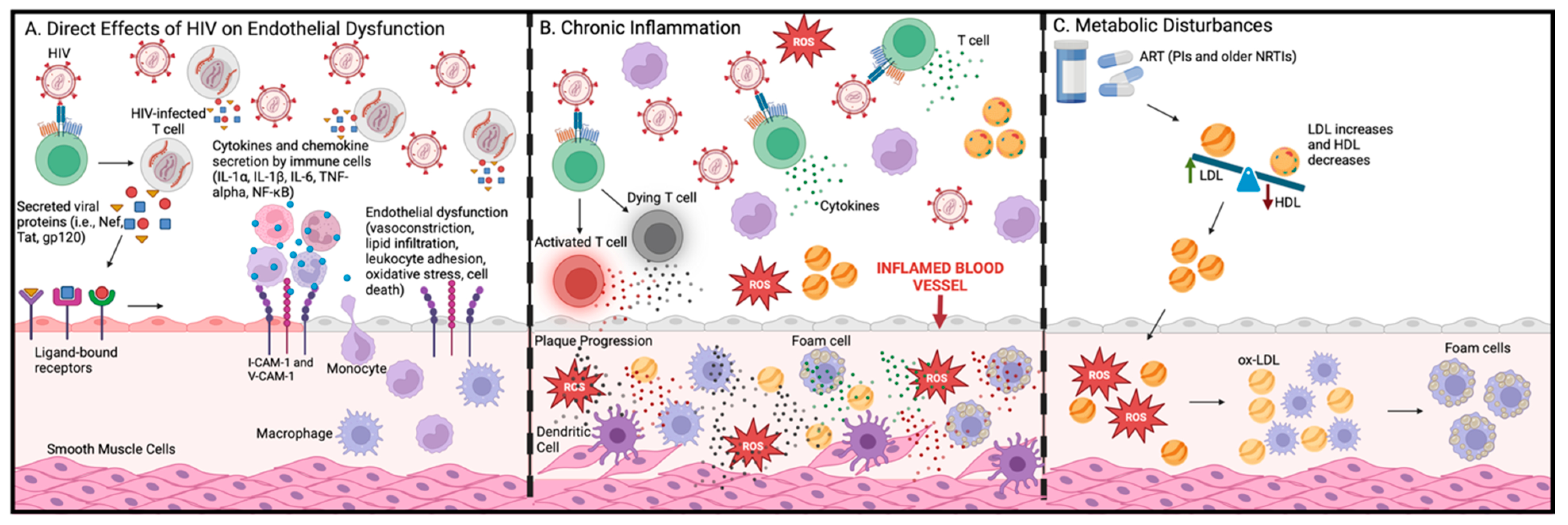
Although HIV cannot undergo active replication in endothelial cells (ECs), HIV-encoded proteins released into the microenvironment or transferred onto ECs during an HIV infection can directly activate ECs and compromise the integrity of the endothelium (Figure 3) [41,48].

6.2. HIV Glycoprotein (gp 120)

Gp120 is expressed on the outer layer of HIV particles and the surface of infected cells, and it facilitates viral entry and binding specificity on target cells. Studies have shown that gp120 can directly upregulate pro-atherogenic cytokines (IL-6 and IL-8) and cell adhesion molecules (CAMs) to facilitate leukocyte recruitment and adherence to the vascular endothelium, respectively [49,50]. Gp120 also increases endothelial cell permeability and induces the expression of matrix metalloproteinase-9, which contributes to endothelial dysfunction by degrading extracellular matrix-associated proteins [51,52,53,54].

6.3. HIV Trans-Activator of Transcription (Tat)

Tat is secreted into the extracellular micromilieu by HIV-infected monocytes, macrophages, and T-cells, enhancing viral transcription. Tat also elicits endothelial dysfunction by activating integrins (Vbeta1, Vbeta3, and alphaVbeta5) to initiate pro-atherogenic signaling cascades in the vascular endothelium [55,56]. Studies have shown that Tat can induce EC apoptosis through cytokine secretion and the activation of Fas-dependent cell signaling pathways [57]. Like gp120, Tat also upregulates the production of pro-inflammatory cytokines and adhesion molecules [58,59]. Specifically, Tat stimulates the secretion of IL-β which can potentially promote apoptosis in macrophages and foam cells, directly contributing to the growth of the lipid-rich necrotic core. MCP-1 secretion is also upregulated in the presence of Tat, facilitating the transendothelial migration of monocytes from the plasma to the intima [60]. Subsequently, these monocytes are differentiated into macrophages, potentially becoming foam cells, a significant component of the necrotic core.

6.4. HIV Negative Regulatory Factor (Nef)

Nef is an accessory protein that aids in increasing the infectivity of HIV. Nef induces endothelial dysfunction by inducing apoptosis via ROS-dependent mechanisms and upregulating MCP-1 via NF-kB [61]. Furthermore, Duffy et al. revealed that Nef decreases nitric oxide production through the inhibition of adequate endothelial nitric oxide synthase (eNOS) expression, which, in turn, reduces endothelium-dependent vasodilation, a hallmark sign of atherosclerosis [62]. Like gp120 and Tat, Nef has also been shown to induce apoptosis in ECs through several mechanisms, including mitochondrial pathways, Fas/FasL-dependent pathways, and the activation of caspases [61,63,64]. Nef also increases the production of MCP-1, ultimately increasing the potential for foam cell formation and the development/progression of atherosclerosis [65,66].

6.5. Inflammation and Immune Activation

While ART can effectively suppress viral replication, aviremic PLWH continue to experience residual and chronic inflammation associated with the virus (Figure 4). Persistent inflammation is associated with an increased risk for multiple comorbidities, including atherosclerosis. Several immune cells affected by HIV also contribute significantly to the development and progression of ASCVD. For example, macrophages are a cellular reservoir for latent HIV. Andrade et al. suggest that macrophages are infected early during the acute phase of the HIV infection due to the identification of rebound HIV particles with macrophage-specific markers on their surface (macrophage-tropic (M-tropic) virus) [67]. Since the M-tropic viruses were identified in viremia from PLWH during treatment interruption, these data suggest that macrophage reservoirs can reignite viremia and contribute to viral persistence. Upon infection, macrophages shift to an M1 phenotype, leading to the expression and secretion of pro-inflammatory cytokines such as IFN-γ. HIV infection also upregulates anti-apoptotic genes in macrophages through various pathways (including NF-kβ/TREM1 and PI3K survival pathway) to promote their survival [68,69,70,71].
Many macrophage-associated inflammatory pathways that contribute to viral persistence are also key drivers of atherogenesis and atherogenic processes. As previously mentioned, HIV promotes macrophage survival by increasing the expression of BCL2, MFN1, MFN2, and BCLXL and decreasing the expression of BAX and BAD [71]. The increase in the number of surviving macrophages potentially fosters the increased recruitment of macrophages into the intima. This, in turn, leads to more cellular uptake of oxidized LDLs (oxLDLs) and the transformation of macrophages into foam cells, which are essential for the initiation, formation, and growth of atherosclerotic lesions. On the other hand, some HIV-infected macrophages undergo apoptosis [72]. This suggests that increased accumulation of apoptotic macrophages and impaired efferocytosis could exacerbate atherosclerosis by enlarging the necrotic core. Furthermore, the increased production of IFN-γ during an HIV infection can promote pro-atherogenic processes by regulating cholesterol accumulation, foam cell formation, plaque formation, and plaque rupture. IFN-γ also stimulates macrophages to produce pro-inflammatory cytokines, contributing to chronic inflammation and increased expression of adhesion molecules, as seen in atherosclerosis [73].
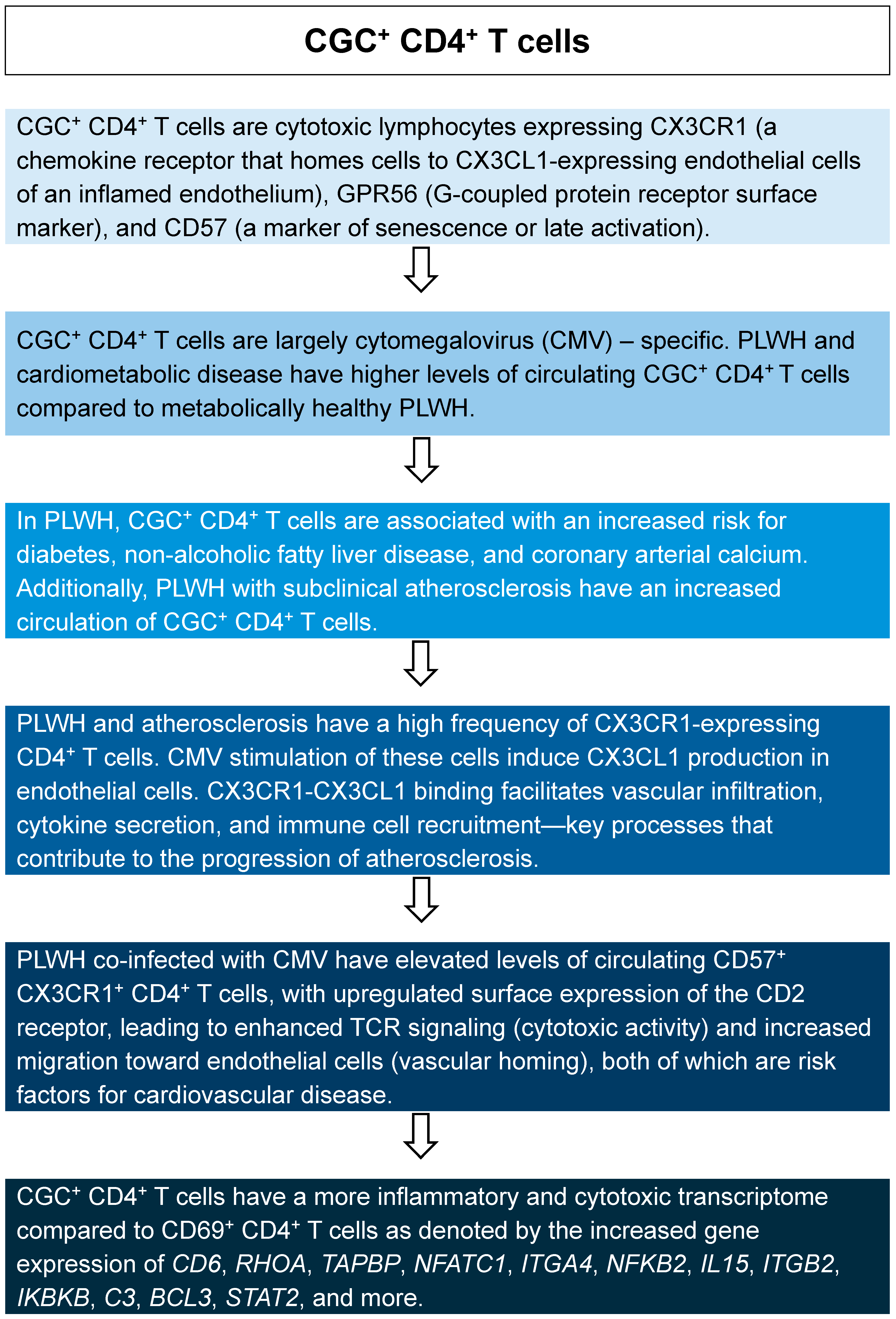
In addition to macrophages, other immune cells and inflammatory components have also been linked to HIV-induced atherosclerosis (Table 3). Among the different subsets, our group has studied a specific CD4+ T cell subset in the context of HIV and cardiometabolic disease. These CD4+ T cells are identified by their co-expression of three surface markers: CX3CR1, GPR56, and CD57, referred to as CGC+ CD4+ T cells (Figure 5). Studies have shown that CGC+ CD4+ T cells are higher in PLWH than PWoH with diabetes and are associated with subclinical atherosclerosis [74,75,76,77].
Table 3. Summary of immune cells and inflammatory components linked to HIV-induced atherosclerosis.
PBMCsRole in HIVPro-atherogenic Features and Mechanism of AtherogenesisRef.
MDMsMonocytes and macrophages are reservoirs for HIV. HIV infection of these cells contributes to persistent inflammation.
  • Chronic inflammation contributes to endothelial dysfunction and adhesion, vascular inflammation, and plaque instability.
  • HIV induces impaired lipid processing and metabolism in these cells.
These mechanisms increase trans-endothelial migration, arterial accumulation, and cholesterol internalization (foam cell formation).
[78,79]
DCsDCs play an important role in viral transmission to CD4+ T cells. HIV also increases the activation of DCs, promoting inflammation.
  • Activated DCs have increased antigen-presenting capabilities, allowing for increased presentation of ox-LDL to T-cells. This leads to heightened inflammation within the arterial wall.
  • DCs also produce pro-inflammatory cytokines that recruit and activate other immune cells, promoting plaque development and rupture.
[80,81,82]
CD4+ T cellsHIV targets CD4+ T cells and uses its machinery to replicate. This causes the cells to die, leading to low CD4+ T cell counts without ART.
  • Low CD4+ T cell counts disrupt the balance between pro- and anti-inflammatory signaling.
  • The increased inflammation promotes endothelial activation and dysfunction, promoting plaque formation and ASCVD progression.
[83,84]
B cellsHIV viral proteins and virions trigger the production of pro-inflammatory cytokines from B cells and increase apoptosis. HIV also alters their class switching capabilities.
  • B cells produce antibodies against ox-LDL and cytokines, likely promoting immune complexes that increase inflammation within the arterial wall.
  • There are no previous studies of B cells in HIV-associated atherosclerosis.
[85]
NK cellsNK cells are crucial for the body’s defense against viruses. However, HIV can impair the antiviral properties of NK cells and inhibit their ability to interact with other immune cells, leading to increased production of inflammatory mediators.
  • NK cells are primarily in the necrotic core of atherosclerotic lesions.
  • They produce IFN-γ, granzyme B, and perforin, which can contribute to enlarging the necrotic core and destabilizing the plaque, leading to plaque rupture.
[86,87]
Abbreviations: peripheral blood mononuclear cells (PBMCs); monocytes and monocyte-derived macrophages (MDMs); dendritic cells (DCs); natural killer (NK) cells.
Figure 5. CGC+ CD4+ T cells. This figure summarizes the main findings on the role of CGC+ CD4+ T cells in cardiometabolic disease among PLWH [74,75,76,88,89].
Ijms 26 03417 g005

6.6. Insights into Pathogenic Mechanisms

HIV-associated atherosclerosis is an active area of investigation. Several studies have highlighted how the interaction between HIV, immune cells, and ECs drives atherogenesis [48]. Oxidative stress, identified by elevated levels of reactive oxygen species (ROS), plays a major role in the development of atherosclerosis [90]. ROS is largely produced by nicotinamide adenine dinucleotide phosphate (NADPH) oxidase (NOX) enzymes, which are abundant in phagocytic leukocytes (i.e., macrophages) and highly expressed in smooth muscle cells and ECs. During hyperlipidemia, NOXs are upregulated and contribute to the increased formation of oxLDL. Cells internalize oxLDL, which undergo lysosomal degradation, and their downstream products mediate inflammatory signaling cascades that induce atherogenic processes (i.e., EC activation, chemokine expression, monocyte/macrophage recruitment, leukocyte recruitment and foam cell formation).
Using models of HIV and patient data, studies have shown that the presence of HIV correlates with elevated levels of ROS and oxLDL, likely due to enhanced production and impaired antioxidant functions [91]. Furthermore, Nef has been shown to increase ROS production in arteries and arterial ECs, contributing to endothelial dysfunction [62]. These data suggest that HIV-induced oxidative stress has the potential to drive atherogenic processes, leading to the development of atherosclerosis.
Increased ROS production following an HIV infection can also trigger endoplasmic reticulum (ER) stress. ER stress subsequently activates the unfolded protein response (UPR) during atherosclerotic lesion formation [92]. ER stress has also been shown to induce macrophage apoptosis [93]. When apoptosis is enhanced, the increase in apoptotic cells can overwhelm phagocytes, leading to impaired efferocytosis, which contributes to the development of the necrotic core in atherosclerotic plaques, as previously mentioned [94,95].
The inflammasome is an integral component of the innate immune system, and it functions by responding to inflammatory stimuli, such as HIV. Since HIV cannot be cleared by the immune system, its persistence leads to prolonged and excessive activation of the inflammasome, which has been linked to atherosclerosis [96]. The accumulation of cholesterol crystals and oxLDL also stimulates NLRP3 inflammasome activation [97,98]. Inflammasome activation triggers caspase-1, which cleaves pro IL-1β and pro IL-18 into their mature forms to mediate inflammatory responses. Interestingly, studies have shown that caspase-1 activation increases rapidly during HIV infection, and it promotes the release of mature and biologically active IL-1β and IL-18, two atherogenic cytokines [99].
The transmigration and adhesion of monocytes to the subendothelial space, where they differentiate into macrophages, internalize oxLDL, and become foam cells, are important for the initiation of atherogenesis. This process is primarily regulated by chemokines, chemokine receptors, and adhesion molecules. In fact, inhibiting the activity of several chemokines and chemokine receptors has been shown to reduce the size of atherosclerotic lesions, highlighting the critical role these molecules play in atherogenesis [100,101]. During HIV infection, chemokines and their receptors are produced and expressed at heightened levels, suggesting that HIV may contribute to atherogenesis by upregulating the production of these molecules.
Autophagy is a highly regulated process in which cells degrade dysfunctional or abnormal cellular components and proteins, recycling them for use in essential cellular processes. During an HIV infection, the role of autophagy is cell-dependent and typically supports the survival of the virus. For example, dendritic cells (DCs) act by accumulating HIV particles and then migrating to lymph nodes where they present and transfer HIV to CD4+ T cells. Studies have shown that immature, resting DCs are readily infected with HIV, whereas mature, activated DCs appear to be resistant to infection (depending on cellular origin) [102]. Mature CD4+ T cells can be activated by TNF-α and poly (I:C) which have been shown to activate and upregulate autophagy [103]. However, Blanchet et al. demonstrated that the HIV-associated Env protein rapidly downregulated autophagy in DCs through the mTOR pathways [104]. This mechanism protects HIV from autophagy-mediated degradation and promotes increased DC to CD4+ T cell viral transmission and survival. This mechanism also increases inflammation, which can promote atherosclerosis.

6.7. Optimizing Immune-Deficient Mouse Models for Atherosclerosis and HIV Research Using Human Immune Cells

Atherosclerosis research in the context of human immune cell function and HIV requires advanced humanized mouse models that support robust PBMC and hematopoietic stem cell (HSC) engraftment, enable atherosclerosis progression, and facilitate HIV persistence within macrophage-rich vascular lesions. Standard NSG mice provide an excellent foundation for human immune cell engraftment but lack optimal myeloid lineage support and atherosclerosis susceptibility [105,106]. Recent models, including NSG-SGM3, NSG-MHC-DKO, and NOD Apoe−/ LDLr−/, offer improvements but require additional modifications to fully capitulate human immune responses in vascular disease [105,106,107].

6.8. Key Features of NSG-Based Mouse Models for the Study of Atherosclerosis Progression in HIV

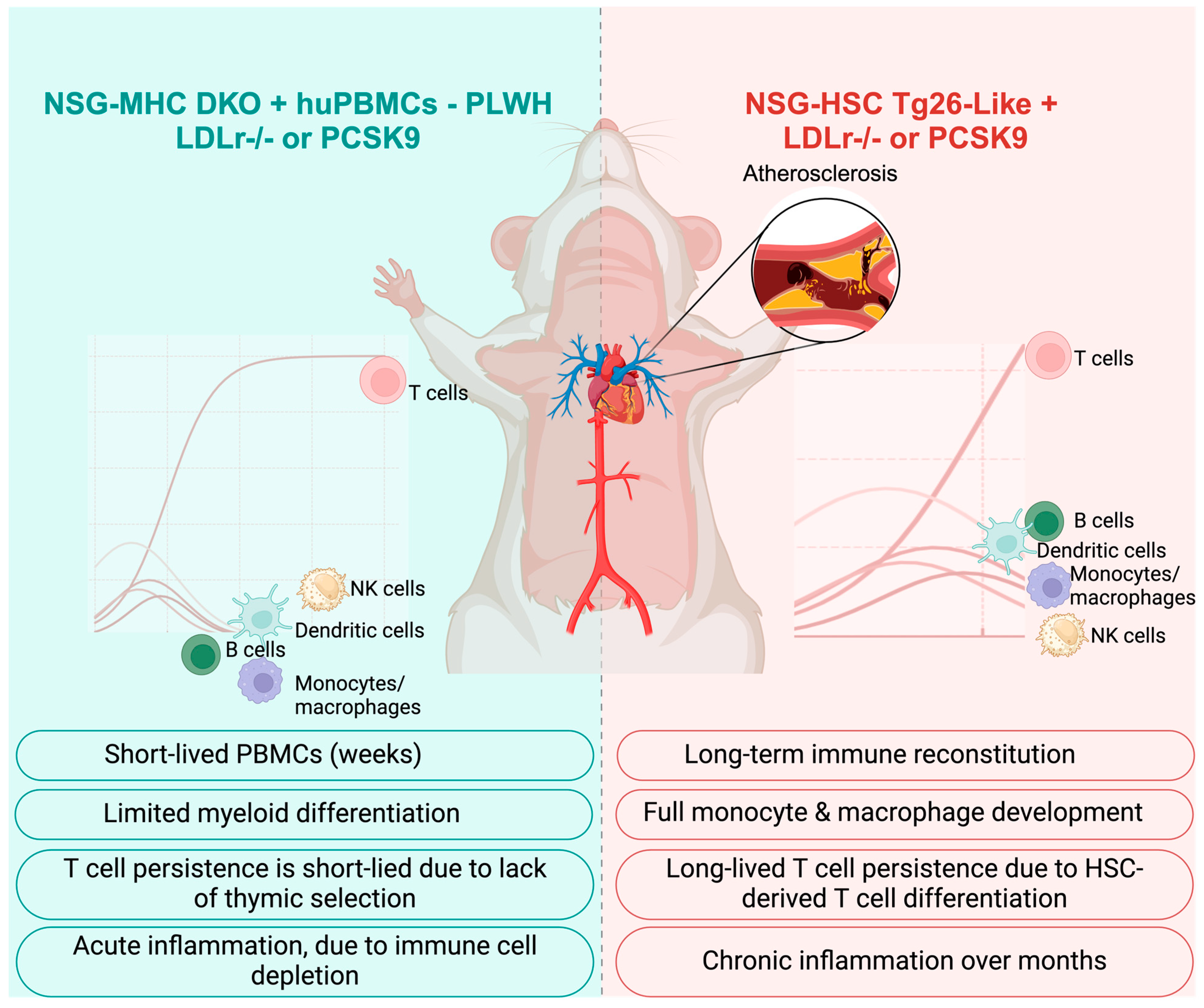
The ideal mouse model would integrate human T cell and monocyte/macrophage function while allowing for severe atherosclerosis progression (Figure 6). For short-term studies, an optimized NSG-based mouse model would incorporate PBMC engraftment for T-cell studies and MHC Class I/II double knockout (MHC-DKO) to prevent xenogeneic GVHD, allowing human PBMC persistence. The mice would also need a genetic predisposition to atherosclerosis with either the PCSK9 gain-of-function mutation to drive severe hypercholesterolemia or LDLr−/ to mimic HFD-induced atherosclerosis comparable to human cardiovascular disease in inflammatory settings. In addition, modifications of IL-7 and IL-15 transgenes to support human T cell and NK cell survival in vivo would support robust adaptive immune responses relevant to atherosclerosis and HIV persistence.
For long-term studies, NSG mice reconstituted with human stem cells will likely yield outcomes similar to those of humans. For HIV-related studies, we would leverage studies from Tg26 (HIV) mice and modify NSG mice to express transgenes with part of the HIV in the NSG mice or the HSC cells engrafted into the mice. Similar to the short-term studies, the genetic predisposition to atherosclerosis would be either the PCSK9 gain-of-function mutation, LDLr−/− and HFD, or a combination. Beyond T cells, enhanced myeloid and macrophage support through the expression of human CSF-1 would support macrophage development and survival following HSC engraftment. This would support HIV-infectible reservoirs in the vascular niche and promote antigen presentation. This idealized NSG-based model would integrate key genetic modifications to support patient-derived immune cells, ensure severe atherosclerosis progression, and provide a relevant platform for studying HIV-infected macrophages in vascular lesions. Further refinements in engraftment optimization, lipid metabolism modifications, and immune system fine-tuning will allow for greater translational relevance in CVD and HIV research.

7. Integration of Omics Approaches to Deepen Understanding

Omics refers to high throughput screening technologies, often providing unbiased molecular measurements within cells and tissues. These methods increase our understanding of biological systems beyond standard, non-computational experimental approaches. Examples of omics technologies include proteomics (global analyses of proteins), metabolomics (global analyses of metabolites), genomics (global analyses of genes), transcriptomics (global analyses of RNA), and more. Using omics data in biomedical research has been paramount in facilitating the exponential growth in medical advancements for more than a decade. These experimental tools provide data on differentially expressed molecules between typical and atypical biological pathways and processes, providing insight into potential disease biomarkers. Although each omics technology is powerful, no single technology can display the full scope of disease processes alone, which is why multi-omics is suggested.
Integrating multiple types of omics data and technologies (multi-omics) increases the potential of uncovering a comprehensive understanding of the pathogenic changes associated with disease. This can include identifying a link between biomolecules and disease or identifying up- and down-regulated signaling pathways relevant to disease phenotypes. A multi-omics approach allows researchers to uncover biological information efficiently and rapidly before verifying those findings using more traditional molecular approaches. As it relates to translational medicine and research, the application of multi-omics data generally has five main objectives: (1) detect disease-associated molecular patterns, (2) subtype identification, (3) diagnosis/prognosis, (4) drug response prediction, and (5) understand regulatory processes [108]. The multi-omics approach also provides an additional tool for validating the use and relevance of animal models, such as mouse models, to study human disease.
Atherosclerosis is an etiopathological factor of cardiovascular diseases, such as coronary artery disease (CAD). Kurt et al. conducted a comprehensive, integrative study using multiomics data resources, which allowed for a systems-level, tissue-specific analysis of CAD in mice and humans. They focused on genome-wide association studies (GWAS) to investigate genetic risk loci, transcriptomics to interrogate gene expression and patterns, and expression quantitative trait loci (eQTLs) to assess gene regulation in a tissue-specific manner [109]. This multi-layered, multi-omics approach revealed that mice and humans shared at least 75% of pathways that lead to CAD. Importantly, multi-omics approaches have been used to interrogate other known HIV-related CVD complications and risk factors, including metabolic syndrome, altered lipid profiles, atrial remodeling, and atrial fibrillation [110,111,112,113].

8. Conclusions

Mouse models have been used extensively to study mechanisms of atherosclerosis and to investigate and test therapeutic targets and drugs. Although more than 90% of protein-encoding genes are homologous between humans and mice, both species have distinct differences that impair direct translational discoveries. Differences in immune cell features between humans and mice are also notable. Furthermore, there are certain risk factors for atherosclerosis development, such as inflammation from HIV, that cannot be studied in most mouse models. Therefore, humanized mouse models are superior tools compared to the previously mentioned mouse models to study HIV-associated atherosclerosis. Using immunocompromised mice, such as NSG mice, allows researchers to reconstitute the immune system of these mice with human immune cells, providing an experimental platform to investigate atherosclerotic mechanisms. Observing how the immune cells drive disease in these animals may better recapitulate what occurs in human atherosclerosis. Furthermore, employing multi-omics approaches to study HIV-associated atherosclerosis in humanized mouse models provides a means to gather a comprehensive understanding of the similarities and differences in molecular mechanisms between the two species, which enhances the value of mouse models in cardiovascular research. Specifically, humanized mouse models provide a means to significantly enhance our capacity to address critical gaps in understanding the immune mechanisms driving HIV-associated atherosclerosis.

Author Contributions

Conceptualization, V.R.S. and C.N.W.; funding acquisition, C.N.W.; investigation, V.R.S., S.A., A.S.M., and C.N.W.; supervision, C.N.W.; visualization, V.R.S., S.A., A.S.M., and C.N.W.; writing—original draft, V.R.S., S.A., A.S.M., and C.N.W.; writing—review and editing, V.R.S., S.A., A.S.M., and C.N.W. All authors have read and agreed to the published version of the manuscript.

Funding

This research was funded by K23 HL156759 (CNW), Burroughs Wellcome Fund G-1021480 (CNW), Veterans Affairs I0BX00533 (A.S.M.), NIAID/NIH R21AI176219 (A.S.M.), and NIAID/NIH R01AI153167 (A.S.M.).

Conflicts of Interest

The authors declare no conflicts of interest.

Abbreviations

The following abbreviations are used in this manuscript:
APOEapolipoprotein E
ARTantiretroviral therapies
ASCVDatherosclerotic cardiovascular disease
CADcoronary artery disease
CAMscell adhesion molecules
cARTcombination antiretroviral therapy
CETPcholesteryl ester transfer protein
CMVcytomegalovirus
CRISPR/Cas9clustered regularly interspaced short palindromic repeats/CRISPR-associated 9
CVDcardiovascular disease
DCsdendritic cells
DKOdouble knockout
DOIDNA of interest
dsRNAdouble-stranded RNA
ECMextracellular matrix
ECsendothelial cells
eNOSendothelial nitric oxide synthase
eQTLsexpression quantitative trait loci
ERendoplasmic reticulum
Fbn1fibrillin 1
gpglycoprotein
GWASgenome-wide association studies
HDLhigh-density lipoprotein
HEshoming endonucleases
HIVhuman immunodeficiency virus
HSChematopoietic stem cell
IFNinterferon
INSTIintegrase strand transfer inhibitors
KOknockout
KTRkynurenine to tryptophan ratio
LDLlow-density lipoproteins
LDLRlow-density lipoproteins receptor
MDMmonocyte-derived macrophage
MHCmajor histocompatibility
M-tropicmacrophage-tropic
mRNAmessenger RNA
NADPHnicotinamide adenine dinucleotide phosphate
NF-kBnuclear factor kappa B
NHEJnon-homologous end joining
NKnatural killer
NOnitric oxide
NOXnicotinamide adenine dinucleotide phosphate oxidase
NNRTInon-nucleoside reverse transcriptase inhibitors
NRTInucleoside reverse transcriptase inhibitors
NSGNOD scid gamma
ox-LDLoxidized-LDL
PBMCperipheral blood mononuclear cells
PIprotease inhibitors
PITTpronuclear injection-based targeted transgenesis
PI3Kphosphoinositide 3-kinase
PLWHpeople living with HIV
PWoHpeople without HIV
RISCRNA-induced silencing complex
RMCErecombination-mediated cassette exchange
RNAribonucleic acid
RNAiRNA interference
ROSreactive oxygen species
siRNAsmall interfering RNAs
TALENstranscription activator-like effector nucleases
Tattrans-activator of transcription
TREMtriggering receptor expressed on myeloid cells
UPRunfolded protein response
VSMCvascular smooth muscle cell
ZFNszinc-finger nucleases

References

  1. Rydell-Törmänen, K.; Johnson, J.R. The Applicability of Mouse Models to the Study of Human Disease. Methods Mol. Biol. 2019, 1940, 3–22. [Google Scholar] [CrossRef] [PubMed]
  2. Bryda, E.C. The Mighty Mouse: The impact of rodents on advances in biomedical research. Mo. Med. 2013, 110, 207–211. [Google Scholar]
  3. Gurumurthy, C.B.; Lloyd, K.C.K. Generating mouse models for biomedical research: Technological advances. Dis. Model. Mech. 2019, 12, dmm029462. [Google Scholar] [CrossRef]
  4. McAlpine, W.; Russell, J.; Murray, A.R.; Beutler, B.; Turer, E. Research Techniques Made Simple: Forward Genetic Screening to Uncover Genes Involved in Skin Biology. J. Invest. Dermatol. 2019, 139, 1848–1853.e1. [Google Scholar] [CrossRef]
  5. Hall, B.; Limaye, A.; Kulkarni, A.B. Overview: Generation of gene knockout mice. Curr. Protoc. Cell Biol. 2009, 44, 19.12.1–19.12.17. [Google Scholar] [CrossRef] [PubMed]
  6. Park, E.C.; Finley, D.; Szostak, J.W. A strategy for the generation of conditional mutations by protein destabilization. Proc. Natl. Acad. Sci. USA 1992, 89, 1249–1252. [Google Scholar] [CrossRef]
  7. Kanasty, R.L.; Whitehead, K.A.; Vegas, A.J.; Anderson, D.G. Action and reaction: The biological response to siRNA and its delivery vehicles. Mol. Ther. 2012, 20, 513–524. [Google Scholar] [CrossRef] [PubMed]
  8. Ohtsuka, M.; Miura, H.; Sato, M.; Kimura, M.; Inoko, H.; Gurumurthy, C.B. PITT: Pronuclear injection-based targeted transgenesis, a reliable transgene expression method in mice. Exp. Anim. 2012, 61, 489–502. [Google Scholar] [CrossRef]
  9. Redman, M.; King, A.; Watson, C.; King, D. What is CRISPR/Cas9? Arch. Dis. Child. Educ. Pract. Ed. 2016, 101, 213–215. [Google Scholar] [CrossRef]
  10. Waterston, R.H.; Lindblad-Toh, K.; Birney, E.; Rogers, J.; Abril, J.F.; Agarwal, P.; Agarwala, R.; Ainscough, R.; Alexandersson, M.; An, P.; et al. Initial sequencing and comparative analysis of the mouse genome. Nature 2002, 420, 520–562. [Google Scholar] [CrossRef]
  11. Thompson, S.; Clarke, A.R.; Pow, A.M.; Hooper, M.L.; Melton, D.W. Germ line transmission and expression of a corrected HPRT gene produced by gene targeting in embryonic stem cells. Cell 1989, 56, 313–321. [Google Scholar] [CrossRef] [PubMed]
  12. Thomas, K.R.; Capecchi, M.R. Site-directed mutagenesis by gene targeting in mouse embryo-derived stem cells. Cell 1987, 51, 503–512. [Google Scholar] [CrossRef]
  13. Pulina, M.V.; Sahr, K.E.; Nowotschin, S.; Baron, M.H.; Hadjantonakis, A.K. A conditional mutant allele for analysis of Mixl1 function in the mouse. Genesis 2014, 52, 417–423. [Google Scholar] [CrossRef]
  14. Gassmann, M.; Casagranda, F.; Orioli, D.; Simon, H.; Lai, C.; Klein, R.; Lemke, G. Aberrant neural and cardiac development in mice lacking the ErbB4 neuregulin receptor. Nature 1995, 378, 390–394. [Google Scholar] [CrossRef] [PubMed]
  15. Gowen, L.C.; Johnson, B.L.; Latour, A.M.; Sulik, K.K.; Koller, B.H. Brca1 deficiency results in early embryonic lethality characterized by neuroepithelial abnormalities. Nat. Genet. 1996, 12, 191–194. [Google Scholar] [CrossRef]
  16. Ludwig, T.; Chapman, D.L.; Papaioannou, V.E.; Efstratiadis, A. Targeted mutations of breast cancer susceptibility gene homologs in mice: Lethal phenotypes of Brca1, Brca2, Brca1/Brca2, Brca1/p53, and Brca2/p53 nullizygous embryos. Genes. Dev. 1997, 11, 1226–1241. [Google Scholar] [CrossRef] [PubMed]
  17. Leung, R.K.; Whittaker, P.A. RNA interference: From gene silencing to gene-specific therapeutics. Pharmacol. Ther. 2005, 107, 222–239. [Google Scholar] [CrossRef]
  18. Tiscornia, G.; Singer, O.; Ikawa, M.; Verma, I.M. A general method for gene knockdown in mice by using lentiviral vectors expressing small interfering RNA. Proc. Natl. Acad. Sci. USA 2003, 100, 1844–1848. [Google Scholar] [CrossRef]
  19. Fire, A.; Xu, S.; Montgomery, M.K.; Kostas, S.A.; Driver, S.E.; Mello, C.C. Potent and specific genetic interference by double-stranded RNA in Caenorhabditis elegans. Nature 1998, 391, 806–811. [Google Scholar] [CrossRef]
  20. Ohtsuka, M.; Ogiwara, S.; Miura, H.; Mizutani, A.; Warita, T.; Sato, M.; Imai, K.; Hozumi, K.; Sato, T.; Tanaka, M.; et al. Pronuclear injection-based mouse targeted transgenesis for reproducible and highly efficient transgene expression. Nucleic Acids Res. 2010, 38, e198. [Google Scholar] [CrossRef]
  21. Hall, B.; Cho, A.; Limaye, A.; Cho, K.; Khillan, J.; Kulkarni, A.B. Genome Editing in Mice Using CRISPR/Cas9 Technology. Curr. Protoc. Cell Biol. 2018, 81, e57. [Google Scholar] [CrossRef]
  22. Jebari-Benslaiman, S.; Galicia-García, U.; Larrea-Sebal, A.; Olaetxea, J.R.; Alloza, I.; Vandenbroeck, K.; Benito-Vicente, A.; Martín, C. Pathophysiology of Atherosclerosis. Int. J. Mol. Sci. 2022, 23, 3346. [Google Scholar] [CrossRef]
  23. Oppi, S.; Lüscher, T.F.; Stein, S. Mouse Models for Atherosclerosis Research-Which Is My Line? Front. Cardiovasc. Med. 2019, 6, 46. [Google Scholar] [CrossRef]
  24. von Scheidt, M.; Zhao, Y.; Kurt, Z.; Pan, C.; Zeng, L.; Yang, X.; Schunkert, H.; Lusis, A.J. Applications and Limitations of Mouse Models for Understanding Human Atherosclerosis. Cell Metab. 2017, 25, 248–261. [Google Scholar] [CrossRef]
  25. Takahashi, S.; Fukami, T.; Masuo, Y.; Brocker, C.N.; Xie, C.; Krausz, K.W.; Wolf, C.R.; Henderson, C.J.; Gonzalez, F.J. Cyp2c70 is responsible for the species difference in bile acid metabolism between mice and humans. J. Lipid Res. 2016, 57, 2130–2137. [Google Scholar] [CrossRef]
  26. Xu, Y.; Li, F.; Zalzala, M.; Xu, J.; Gonzalez, F.J.; Adorini, L.; Lee, Y.K.; Yin, L.; Zhang, Y. Farnesoid X receptor activation increases reverse cholesterol transport by modulating bile acid composition and cholesterol absorption in mice. Hepatology 2016, 64, 1072–1085. [Google Scholar] [CrossRef] [PubMed]
  27. Vinué, Á.; Herrero-Cervera, A.; González-Navarro, H. Understanding the Impact of Dietary Cholesterol on Chronic Metabolic Diseases through Studies in Rodent Models. Nutrients 2018, 10, 939. [Google Scholar] [CrossRef] [PubMed]
  28. Paigen, B.; Holmes, P.A.; Mitchell, D.; Albee, D. Comparison of atherosclerotic lesions and HDL-lipid levels in male, female, and testosterone-treated female mice from strains C57BL/6, BALB/c, and C3H. Atherosclerosis 1987, 64, 215–221. [Google Scholar] [CrossRef]
  29. Paigen, B.; Morrow, A.; Brandon, C.; Mitchell, D.; Holmes, P. Variation in susceptibility to atherosclerosis among inbred strains of mice. Atherosclerosis 1985, 57, 65–73. [Google Scholar] [CrossRef]
  30. Schreyer, S.A.; Wilson, D.L.; LeBoeuf, R.C. C57BL/6 mice fed high fat diets as models for diabetes-accelerated atherosclerosis. Atherosclerosis 1998, 136, 17–24. [Google Scholar] [CrossRef]
  31. Watanabe, H.; Numata, K.; Ito, T.; Takagi, K.; Matsukawa, A. Innate immune response in Th1- and Th2-dominant mouse strains. Shock 2004, 22, 460–466. [Google Scholar] [CrossRef] [PubMed]
  32. Nakashima, Y.; Plump, A.S.; Raines, E.W.; Breslow, J.L.; Ross, R. ApoE-deficient mice develop lesions of all phases of atherosclerosis throughout the arterial tree. Arterioscler. Thromb. 1994, 14, 133–140. [Google Scholar] [CrossRef] [PubMed]
  33. Tian, J.; Pei, H.; James, J.C.; Li, Y.; Matsumoto, A.H.; Helm, G.A.; Shi, W. Circulating adhesion molecules in apoE-deficient mouse strains with different atherosclerosis susceptibility. Biochem. Biophys. Res. Commun. 2005, 329, 1102–1107. [Google Scholar] [CrossRef] [PubMed]
  34. van den Maagdenberg, A.M.; Hofker, M.H.; Krimpenfort, P.J.; de Bruijn, I.; van Vlijmen, B.; van der Boom, H.; Havekes, L.M.; Frants, R.R. Transgenic mice carrying the apolipoprotein E3-Leiden gene exhibit hyperlipoproteinemia. J. Biol. Chem. 1993, 268, 10540–10545. [Google Scholar] [CrossRef]
  35. Westerterp, M.; van der Hoogt, C.C.; de Haan, W.; Offerman, E.H.; Dallinga-Thie, G.M.; Jukema, J.W.; Havekes, L.M.; Rensen, P.C. Cholesteryl ester transfer protein decreases high-density lipoprotein and severely aggravates atherosclerosis in APOE*3-Leiden mice. Arterioscler. Thromb. Vasc. Biol. 2006, 26, 2552–2559. [Google Scholar] [CrossRef]
  36. Ishibashi, S.; Goldstein, J.L.; Brown, M.S.; Herz, J.; Burns, D.K. Massive xanthomatosis and atherosclerosis in cholesterol-fed low density lipoprotein receptor-negative mice. J. Clin. Investig. 1994, 93, 1885–1893. [Google Scholar] [CrossRef]
  37. Goldstein, J.L.; Brown, M.S. Molecular medicine. The cholesterol quartet. Science 2001, 292, 1310–1312. [Google Scholar] [CrossRef]
  38. Van Herck, J.L.; De Meyer, G.R.; Martinet, W.; Van Hove, C.E.; Foubert, K.; Theunis, M.H.; Apers, S.; Bult, H.; Vrints, C.J.; Herman, A.G. Impaired fibrillin-1 function promotes features of plaque instability in apolipoprotein E-deficient mice. Circulation 2009, 120, 2478–2487. [Google Scholar] [CrossRef]
  39. Sato, K.; Nakano, K.; Katsuki, S.; Matoba, T.; Osada, K.; Sawamura, T.; Sunagawa, K.; Egashira, K. Dietary cholesterol oxidation products accelerate plaque destabilization and rupture associated with monocyte infiltration/activation via the MCP-1-CCR2 pathway in mouse brachiocephalic arteries: Therapeutic effects of ezetimibe. J. Atheroscler. Thromb. 2012, 19, 986–998. [Google Scholar] [CrossRef]
  40. Dickie, P.; Felser, J.; Eckhaus, M.; Bryant, J.; Silver, J.; Marinos, N.; Notkins, A.L. HIV-associated nephropathy in transgenic mice expressing HIV-1 genes. Virology 1991, 185, 109–119. [Google Scholar] [CrossRef]
  41. Kress, T.C.; Barris, C.T.; Kovacs, L.; Khakina, B.N.; Jordan, C.R.; Bruder-Nascimento, T.; Stepp, D.W.; MacArthur, R.; Patel, V.S.; Chen, J.; et al. CD4+ T Cells Expressing Viral Proteins Induce HIV-Associated Endothelial Dysfunction and Hypertension Through Interleukin 1α–Mediated Increases in Endothelial NADPH Oxidase 1. Circulation 2025. [Google Scholar] [CrossRef]
  42. Alam, M.A.; Caocci, M.; Ren, M.; Chen, Z.; Liu, F.; Khatun, M.S.; Kolls, J.K.; Qin, X.; Burdo, T.H. Deficiency of Caspase-1 Attenuates HIV-1-Associated Atherogenesis in Mice. Int. J. Mol. Sci. 2023, 24, 12871. [Google Scholar] [CrossRef] [PubMed]
  43. Bandera, A.; Masetti, M.; Fabbiani, M.; Biasin, M.; Muscatello, A.; Squillace, N.; Clerici, M.; Gori, A.; Trabattoni, D. The NLRP3 Inflammasome Is Upregulated in HIV-Infected Antiretroviral Therapy-Treated Individuals with Defective Immune Recovery. Front. Immunol. 2018, 9, 214. [Google Scholar] [CrossRef]
  44. Kearns, A.C.; Velasquez, S.; Liu, F.; Dai, S.; Chen, Y.; Lehmicke, G.; Gordon, J.; Rappaport, J.; Qin, X. Elevated indoleamine-2,3-dioxygenase enzyme activity in a novel mouse model of HIV-associated atherosclerosis. AIDS 2019, 33, 1557–1564. [Google Scholar] [CrossRef]
  45. Morimoto, K.; Shimada, N.; Naganawa, H.; Takita, T.; Umezawa, H.; Kambara, H. Minor congeners of antrimycin: Application of secondary ion mass spectrometry (SIMS) to structure determination. J. Antibiot. 1982, 35, 378–380. [Google Scholar] [CrossRef] [PubMed]
  46. Cui, H.L.; Ditiatkovski, M.; Kesani, R.; Bobryshev, Y.V.; Liu, Y.; Geyer, M.; Mukhamedova, N.; Bukrinsky, M.; Sviridov, D. HIV protein Nef causes dyslipidemia and formation of foam cells in mouse models of atherosclerosis. FASEB J. 2014, 28, 2828–2839. [Google Scholar] [CrossRef] [PubMed]
  47. Pushkarsky, T.; Shilov, E.; Kruglova, N.; Naumann, R.; Brichacek, B.; Jennelle, L.; Sviridov, D.; Kruglov, A.; Nedospasov, S.A.; Bukrinsky, M. Short Communication: Accumulation of Neutral Lipids in Liver and Aorta of Nef-Transgenic Mice. AIDS Res. Hum. Retroviruses 2017, 33, 57–60. [Google Scholar] [CrossRef]
  48. Anand, A.R.; Rachel, G.; Parthasarathy, D. HIV Proteins and Endothelial Dysfunction: Implications in Cardiovascular Disease. Front. Cardiovasc. Med. 2018, 5, 185. [Google Scholar] [CrossRef]
  49. Yang, B.; Akhter, S.; Chaudhuri, A.; Kanmogne, G.D. HIV-1 gp120 induces cytokine expression, leukocyte adhesion, and transmigration across the blood-brain barrier: Modulatory effects of STAT1 signaling. Microvasc. Res. 2009, 77, 212–219. [Google Scholar] [CrossRef]
  50. Ren, Z.; Yao, Q.; Chen, C. HIV-1 envelope glycoprotein 120 increases intercellular adhesion molecule-1 expression by human endothelial cells. Lab. Invest. 2002, 82, 245–255. [Google Scholar] [CrossRef]
  51. Kanmogne, G.D.; Primeaux, C.; Grammas, P. HIV-1 gp120 proteins alter tight junction protein expression and brain endothelial cell permeability: Implications for the pathogenesis of HIV-associated dementia. J. Neuropathol. Exp. Neurol. 2005, 64, 498–505. [Google Scholar] [CrossRef] [PubMed]
  52. Sufiawati, I.; Tugizov, S.M. HIV-induced matrix metalloproteinase-9 activation through mitogen-activated protein kinase signalling promotes HSV-1 cell-to-cell spread in oral epithelial cells. J. Gen. Virol. 2018, 99, 937–947. [Google Scholar] [CrossRef] [PubMed]
  53. Louboutin, J.P.; Reyes, B.A.; Agrawal, L.; Van Bockstaele, E.J.; Strayer, D.S. HIV-1 gp120 upregulates matrix metalloproteinases and their inhibitors in a rat model of HIV encephalopathy. Eur. J. Neurosci. 2011, 34, 2015–2023. [Google Scholar] [CrossRef]
  54. Annunziata, P.; Cioni, C.; Toneatto, S.; Paccagnini, E. HIV-1 gp120 increases the permeability of rat brain endothelium cultures by a mechanism involving substance P. AIDS 1998, 12, 2377–2385. [Google Scholar] [CrossRef]
  55. Urbinati, C.; Mitola, S.; Tanghetti, E.; Kumar, C.; Waltenberger, J.; Ribatti, D.; Presta, M.; Rusnati, M. Integrin alphavbeta3 as a target for blocking HIV-1 Tat-induced endothelial cell activation in vitro and angiogenesis in vivo. Arterioscler. Thromb. Vasc. Biol. 2005, 25, 2315–2320. [Google Scholar] [CrossRef] [PubMed]
  56. Fanales-Belasio, E.; Moretti, S.; Nappi, F.; Barillari, G.; Micheletti, F.; Cafaro, A.; Ensoli, B. Native HIV-1 Tat protein targets monocyte-derived dendritic cells and enhances their maturation, function, and antigen-specific T cell responses. J. Immunol. 2002, 168, 197–206. [Google Scholar] [CrossRef]
  57. Park, I.W.; Ullrich, C.K.; Schoenberger, E.; Ganju, R.K.; Groopman, J.E. HIV-1 Tat induces microvascular endothelial apoptosis through caspase activation. J. Immunol. 2001, 167, 2766–2771. [Google Scholar] [CrossRef]
  58. Ben Haij, N.; Planès, R.; Leghmari, K.; Serrero, M.; Delobel, P.; Izopet, J.; BenMohamed, L.; Bahraoui, E. HIV-1 Tat Protein Induces Production of Proinflammatory Cytokines by Human Dendritic Cells and Monocytes/Macrophages through Engagement of TLR4-MD2-CD14 Complex and Activation of NF-κB Pathway. PLoS ONE 2015, 10, e0129425. [Google Scholar] [CrossRef]
  59. Pu, H.; Tian, J.; Flora, G.; Lee, Y.W.; Nath, A.; Hennig, B.; Toborek, M. HIV-1 Tat protein upregulates inflammatory mediators and induces monocyte invasion into the brain. Mol. Cell Neurosci. 2003, 24, 224–237. [Google Scholar] [CrossRef]
  60. Park, I.W.; Wang, J.F.; Groopman, J.E. HIV-1 Tat promotes monocyte chemoattractant protein-1 secretion followed by transmigration of monocytes. Blood 2001, 97, 352–358. [Google Scholar] [CrossRef]
  61. Wang, T.; Green, L.A.; Gupta, S.K.; Kim, C.; Wang, L.; Almodovar, S.; Flores, S.C.; Prudovsky, I.A.; Jolicoeur, P.; Liu, Z.; et al. Transfer of intracellular HIV Nef to endothelium causes endothelial dysfunction. PLoS ONE 2014, 9, e91063. [Google Scholar] [CrossRef]
  62. Duffy, P.; Wang, X.; Lin, P.H.; Yao, Q.; Chen, C. HIV Nef protein causes endothelial dysfunction in porcine pulmonary arteries and human pulmonary artery endothelial cells. J. Surg. Res. 2009, 156, 257–264. [Google Scholar] [CrossRef] [PubMed]
  63. Acheampong, E.A.; Parveen, Z.; Muthoga, L.W.; Kalayeh, M.; Mukhtar, M.; Pomerantz, R.J. Human Immunodeficiency virus type 1 Nef potently induces apoptosis in primary human brain microvascular endothelial cells via the activation of caspases. J. Virol. 2005, 79, 4257–4269. [Google Scholar] [CrossRef]
  64. Lenassi, M.; Cagney, G.; Liao, M.; Vaupotic, T.; Bartholomeeusen, K.; Cheng, Y.; Krogan, N.J.; Plemenitas, A.; Peterlin, B.M. HIV Nef is secreted in exosomes and triggers apoptosis in bystander CD4+ T cells. Traffic 2010, 11, 110–122. [Google Scholar] [CrossRef] [PubMed]
  65. Lehmann, M.H.; Masanetz, S.; Kramer, S.; Erfle, V. HIV-1 Nef upregulates CCL2/MCP-1 expression in astrocytes in a myristoylation- and calmodulin-dependent manner. J. Cell Sci. 2006, 119, 4520–4530. [Google Scholar] [CrossRef] [PubMed]
  66. Aiello, A.; Giannessi, F.; Percario, Z.A.; Fecchi, K.; Arenaccio, C.; Leone, S.; Carollo, M.; D’Aversa, E.; Chaperot, L.; Gambari, R.; et al. HIV-1 Nef Protein Affects Cytokine and Extracellular Vesicles Production in the GEN2.2 Plasmacytoid Dendritic Cell Line. Viruses 2021, 14, 74. [Google Scholar] [CrossRef]
  67. Andrade, V.M.; Mavian, C.; Babic, D.; Cordeiro, T.; Sharkey, M.; Barrios, L.; Brander, C.; Martinez-Picado, J.; Dalmau, J.; Llano, A.; et al. A minor population of macrophage-tropic HIV-1 variants is identified in recrudescing viremia following analytic treatment interruption. Proc. Natl. Acad. Sci. USA 2020, 117, 9981–9990. [Google Scholar] [CrossRef]
  68. Giri, M.S.; Nebozyhn, M.; Raymond, A.; Gekonge, B.; Hancock, A.; Creer, S.; Nicols, C.; Yousef, M.; Foulkes, A.S.; Mounzer, K.; et al. Circulating monocytes in HIV-1-infected viremic subjects exhibit an antiapoptosis gene signature and virus- and host-mediated apoptosis resistance. J. Immunol. 2009, 182, 4459–4470. [Google Scholar] [CrossRef]
  69. Tyner, J.W.; Uchida, O.; Kajiwara, N.; Kim, E.Y.; Patel, A.C.; O’Sullivan, M.P.; Walter, M.J.; Schwendener, R.A.; Cook, D.N.; Danoff, T.M.; et al. CCL5-CCR5 interaction provides antiapoptotic signals for macrophage survival during viral infection. Nat. Med. 2005, 11, 1180–1187. [Google Scholar] [CrossRef]
  70. Kaul, M.; Ma, Q.; Medders, K.E.; Desai, M.K.; Lipton, S.A. HIV-1 coreceptors CCR5 and CXCR4 both mediate neuronal cell death but CCR5 paradoxically can also contribute to protection. Cell Death Differ. 2007, 14, 296–305. [Google Scholar] [CrossRef]
  71. Campbell, G.R.; To, R.K.; Spector, S.A. TREM-1 Protects HIV-1-Infected Macrophages from Apoptosis through Maintenance of Mitochondrial Function. mBio 2019, 10, 10–1128. [Google Scholar] [CrossRef]
  72. Castellano, P.; Prevedel, L.; Eugenin, E.A. HIV-infected macrophages and microglia that survive acute infection become viral reservoirs by a mechanism involving Bim. Sci. Rep. 2017, 7, 12866. [Google Scholar] [CrossRef]
  73. Voloshyna, I.; Littlefield, M.J.; Reiss, A.B. Atherosclerosis and interferon-γ: New insights and therapeutic targets. Trends Cardiovasc. Med. 2014, 24, 45–51. [Google Scholar] [CrossRef] [PubMed]
  74. Wanjalla, C.N.; Gabriel, C.L.; Fuseini, H.; Bailin, S.S.; Mashayekhi, M.; Simmons, J.; Warren, C.M.; Glass, D.R.; Oakes, J.; Gangula, R.; et al. CD4. Front. Immunol. 2023, 14, 1099356. [Google Scholar] [CrossRef]
  75. Wanjalla, C.N.; McDonnell, W.J.; Ram, R.; Chopra, A.; Gangula, R.; Leary, S.; Mashayekhi, M.; Simmons, J.D.; Warren, C.M.; Bailin, S.; et al. Single-cell analysis shows that adipose tissue of persons with both HIV and diabetes is enriched for clonal, cytotoxic, and CMV-specific CD4+ T cells. Cell Rep. Med. 2021, 2, 100205. [Google Scholar] [CrossRef] [PubMed]
  76. Wanjalla, C.N.; Mashayekhi, M.; Bailin, S.; Gabriel, C.L.; Meenderink, L.M.; Temu, T.; Fuller, D.T.; Guo, L.; Kawai, K.; Virmani, R.; et al. Anticytomegalovirus CD4. Arterioscler. Thromb. Vasc. Biol. 2021, 41, 1459–1473. [Google Scholar] [CrossRef]
  77. Wanjalla, C.N.; McDonnell, W.J.; Barnett, L.; Simmons, J.D.; Furch, B.D.; Lima, M.C.; Woodward, B.O.; Fan, R.; Fei, Y.; Baker, P.G.; et al. Adipose Tissue in Persons With HIV Is Enriched for CD4. Front. Immunol. 2019, 10, 408. [Google Scholar] [CrossRef]
  78. Cui, H.L.; Grant, A.; Mukhamedova, N.; Pushkarsky, T.; Jennelle, L.; Dubrovsky, L.; Gaus, K.; Fitzgerald, M.L.; Sviridov, D.; Bukrinsky, M. HIV-1 Nef mobilizes lipid rafts in macrophages through a pathway that competes with ABCA1-dependent cholesterol efflux. J. Lipid Res. 2012, 53, 696–708. [Google Scholar] [CrossRef]
  79. Veenhuis, R.T.; Abreu, C.M.; Costa, P.A.G.; Ferreira, E.A.; Ratliff, J.; Pohlenz, L.; Shirk, E.N.; Rubin, L.H.; Blankson, J.N.; Gama, L.; et al. Monocyte-derived macrophages contain persistent latent HIV reservoirs. Nat. Microbiol. 2023, 8, 833–844. [Google Scholar] [CrossRef]
  80. St Gelais, C.; Coleman, C.M.; Wang, J.H.; Wu, L. HIV-1 Nef enhances dendritic cell-mediated viral transmission to CD4+ T cells and promotes T-cell activation. PLoS ONE 2012, 7, e34521. [Google Scholar] [CrossRef]
  81. Frostegård, J.; Zhang, Y.; Sun, J.; Yan, K.; Liu, A. Oxidized Low-Density Lipoprotein (OxLDL)-Treated Dendritic Cells Promote Activation of T Cells in Human Atherosclerotic Plaque and Blood, Which Is Repressed by Statins: microRNA let-7c Is Integral to the Effect. J. Am. Heart Assoc. 2016, 5, e003976. [Google Scholar] [CrossRef]
  82. Zhao, Y.; Zhang, J.; Zhang, W.; Xu, Y. A myriad of roles of dendritic cells in atherosclerosis. Clin. Exp. Immunol. 2021, 206, 12–27. [Google Scholar] [CrossRef]
  83. Vidya Vijayan, K.K.; Karthigeyan, K.P.; Tripathi, S.P.; Hanna, L.E. Pathophysiology of CD4+ T-Cell Depletion in HIV-1 and HIV-2 Infections. Front. Immunol. 2017, 8, 580. [Google Scholar] [CrossRef] [PubMed]
  84. Kaplan, R.C.; Kingsley, L.A.; Gange, S.J.; Benning, L.; Jacobson, L.P.; Lazar, J.; Anastos, K.; Tien, P.C.; Sharrett, A.R.; Hodis, H.N. Low CD4+ T-cell count as a major atherosclerosis risk factor in HIV-infected women and men. AIDS 2008, 22, 1615–1624. [Google Scholar] [CrossRef] [PubMed]
  85. Obare, L.M.; Bonami, R.H.; Doran, A.C.; Wanjalla, C.N. B cells and atherosclerosis: A HIV perspective. J. Cell Physiol. 2024, 239, e31270. [Google Scholar] [CrossRef]
  86. Selathurai, A.; Deswaerte, V.; Kanellakis, P.; Tipping, P.; Toh, B.H.; Bobik, A.; Kyaw, T. Natural killer (NK) cells augment atherosclerosis by cytotoxic-dependent mechanisms. Cardiovasc. Res. 2014, 102, 128–137. [Google Scholar] [CrossRef] [PubMed]
  87. Alles, M.; Gunasena, M.; Kettelhut, A.; Ailstock, K.; Musiime, V.; Kityo, C.; Richardson, B.; Mulhern, W.; Tamilselvan, B.; Rubsamen, M.; et al. Activated NK Cells with Pro-inflammatory Features are Associated with Atherogenesis in Perinatally HIV-Acquired Adolescents. medRxiv 2023. [Google Scholar] [CrossRef]
  88. Sacre, K.; Hunt, P.W.; Hsue, P.Y.; Maidji, E.; Martin, J.N.; Deeks, S.G.; Autran, B.; McCune, J.M. A role for cytomegalovirus-specific CD4+CX3CR1+ T cells and cytomegalovirus-induced T-cell immunopathology in HIV-associated atherosclerosis. AIDS 2012, 26, 805–814. [Google Scholar] [CrossRef]
  89. Chen, B.; Morris, S.R.; Panigrahi, S.; Michaelson, G.M.; Wyrick, J.M.; Komissarov, A.A.; Potashnikova, D.; Lebedeva, A.; Younes, S.A.; Harth, K.; et al. Cytomegalovirus Coinfection Is Associated with Increased Vascular-Homing CD57. J. Immunol. 2020, 204, 2722–2733. [Google Scholar] [CrossRef]
  90. Batty, M.; Bennett, M.R.; Yu, E. The Role of Oxidative Stress in Atherosclerosis. Cells 2022, 11, 3843. [Google Scholar] [CrossRef]
  91. Zidar, D.A.; Juchnowski, S.; Ferrari, B.; Clagett, B.; Pilch-Cooper, H.A.; Rose, S.; Rodriguez, B.; McComsey, G.A.; Sieg, S.F.; Mehta, N.N.; et al. Oxidized LDL Levels Are Increased in HIV Infection and May Drive Monocyte Activation. J. Acquir. Immune Defic. Syndr. 2015, 69, 154–160. [Google Scholar] [CrossRef] [PubMed]
  92. Zhou, J.; Lhoták, S.; Hilditch, B.A.; Austin, R.C. Activation of the unfolded protein response occurs at all stages of atherosclerotic lesion development in apolipoprotein E-deficient mice. Circulation 2005, 111, 1814–1821. [Google Scholar] [CrossRef]
  93. Feng, B.; Yao, P.M.; Li, Y.; Devlin, C.M.; Zhang, D.; Harding, H.P.; Sweeney, M.; Rong, J.X.; Kuriakose, G.; Fisher, E.A.; et al. The endoplasmic reticulum is the site of cholesterol-induced cytotoxicity in macrophages. Nat. Cell Biol. 2003, 5, 781–792. [Google Scholar] [CrossRef]
  94. Schrijvers, D.M.; De Meyer, G.R.; Kockx, M.M.; Herman, A.G.; Martinet, W. Phagocytosis of apoptotic cells by macrophages is impaired in atherosclerosis. Arterioscler. Thromb. Vasc. Biol. 2005, 25, 1256–1261. [Google Scholar] [CrossRef] [PubMed]
  95. Clarke, M.C.; Talib, S.; Figg, N.L.; Bennett, M.R. Vascular smooth muscle cell apoptosis induces interleukin-1-directed inflammation: Effects of hyperlipidemia-mediated inhibition of phagocytosis. Circ. Res. 2010, 106, 363–372. [Google Scholar] [CrossRef] [PubMed]
  96. Duewell, P.; Kono, H.; Rayner, K.J.; Sirois, C.M.; Vladimer, G.; Bauernfeind, F.G.; Abela, G.S.; Franchi, L.; Nuñez, G.; Schnurr, M.; et al. NLRP3 inflammasomes are required for atherogenesis and activated by cholesterol crystals. Nature 2010, 464, 1357–1361. [Google Scholar] [CrossRef]
  97. Sheedy, F.J.; Grebe, A.; Rayner, K.J.; Kalantari, P.; Ramkhelawon, B.; Carpenter, S.B.; Becker, C.E.; Ediriweera, H.N.; Mullick, A.E.; Golenbock, D.T.; et al. CD36 coordinates NLRP3 inflammasome activation by facilitating intracellular nucleation of soluble ligands into particulate ligands in sterile inflammation. Nat. Immunol. 2013, 14, 812–820. [Google Scholar] [CrossRef]
  98. Jiang, Y.; Wang, M.; Huang, K.; Zhang, Z.; Shao, N.; Zhang, Y.; Wang, W.; Wang, S. Oxidized low-density lipoprotein induces secretion of interleukin-1β by macrophages via reactive oxygen species-dependent NLRP3 inflammasome activation. Biochem. Biophys. Res. Commun. 2012, 425, 121–126. [Google Scholar] [CrossRef]
  99. Amand, M.; Adams, P.; Schober, R.; Iserentant, G.; Servais, J.Y.; Moutschen, M.; Seguin-Devaux, C. The anti-caspase 1 inhibitor VX-765 reduces immune activation, CD4. eLife 2023, 12, e83207. [Google Scholar] [CrossRef]
  100. Rowinska, Z.; Koeppel, T.A.; Sanati, M.; Schelzig, H.; Jankowski, J.; Weber, C.; Zernecke, A.; Liehn, E.A. Role of the CX3C chemokine receptor CX3CR1 in the pathogenesis of atherosclerosis after aortic transplantation. PLoS ONE 2017, 12, e0170644. [Google Scholar] [CrossRef]
  101. Wan, W.; Murphy, P.M. Regulation of atherogenesis by chemokines and chemokine receptors. Arch. Immunol. Ther. Exp. 2013, 61, 1–14. [Google Scholar] [CrossRef]
  102. Dong, C.; Janas, A.M.; Wang, J.H.; Olson, W.J.; Wu, L. Characterization of human immunodeficiency virus type 1 replication in immature and mature dendritic cells reveals dissociable cis- and trans-infection. J. Virol. 2007, 81, 11352–11362. [Google Scholar] [CrossRef]
  103. Dinkins, C.; Pilli, M.; Kehrl, J.H. Roles of autophagy in HIV infection. Immunol. Cell Biol. 2015, 93, 11–17. [Google Scholar] [CrossRef]
  104. Blanchet, F.P.; Moris, A.; Nikolic, D.S.; Lehmann, M.; Cardinaud, S.; Stalder, R.; Garcia, E.; Dinkins, C.; Leuba, F.; Wu, L.; et al. Human immunodeficiency virus-1 inhibition of immunoamphisomes in dendritic cells impairs early innate and adaptive immune responses. Immunity 2010, 32, 654–669. [Google Scholar] [CrossRef]
  105. Proto, J.D.; Doran, A.C.; Subramanian, M.; Wang, H.; Zhang, M.; Sozen, E.; Rymond, C.C.; Kuriakose, G.; D’Agati, V.; Winchester, R.; et al. Hypercholesterolemia induces T cell expansion in humanized immune mice. J. Clin. Invest. 2018, 128, 2370–2375. [Google Scholar] [CrossRef] [PubMed]
  106. Wang, X.; Huang, R.; Zhang, L.; Li, S.; Luo, J.; Gu, Y.; Chen, Z.; Zheng, Q.; Chao, T.; Zheng, W.; et al. A severe atherosclerosis mouse model on the resistant NOD background. Dis. Model. Mech. 2018, 11, dmm033852. [Google Scholar] [CrossRef]
  107. Chuprin, J.; Buettner, H.; Seedhom, M.O.; Greiner, D.L.; Keck, J.G.; Ishikawa, F.; Shultz, L.D.; Brehm, M.A. Humanized mouse models for immuno-oncology research. Nat. Rev. Clin. Oncol. 2023, 20, 192–206. [Google Scholar] [CrossRef]
  108. Athieniti, E.; Spyrou, G.M. A guide to multi-omics data collection and integration for translational medicine. Comput. Struct. Biotechnol. J. 2023, 21, 134–149. [Google Scholar] [CrossRef] [PubMed]
  109. Kurt, Z.; Cheng, J.; McQuillen, C.N.; Saleem, Z.; Hsu, N.; Jiang, N.; Barrere-Cain, R.; Pan, C.; Franzen, O.; Koplev, S.; et al. Shared and distinct pathways and networks genetically linked to coronary artery disease between human and mouse. bioRxiv 2023. [Google Scholar] [CrossRef]
  110. Mikaeloff, F.; Gelpi, M.; Benfeitas, R.; Knudsen, A.D.; Vestad, B.; Høgh, J.; Hov, J.R.; Benfield, T.; Murray, D.; Giske, C.G.; et al. Network-based multi-omics integration reveals metabolic at-risk profile within treated HIV-infection. eLife 2023, 12, e82785. [Google Scholar] [CrossRef]
  111. Wong, G.; Trevillyan, J.M.; Fatou, B.; Cinel, M.; Weir, J.M.; Hoy, J.F.; Meikle, P.J. Plasma lipidomic profiling of treated HIV-positive individuals and the implications for cardiovascular risk prediction. PLoS ONE 2014, 9, e94810. [Google Scholar] [CrossRef] [PubMed]
  112. Peterson, T.E.; Hahn, V.S.; Moaddel, R.; Zhu, M.; Haberlen, S.A.; Palella, F.J.; Plankey, M.; Bader, J.S.; Lima, J.A.C.; Gerszten, R.E.; et al. Proteomic signature of HIV-associated subclinical left atrial remodeling and incident heart failure. Nat. Commun. 2025, 16, 610. [Google Scholar] [CrossRef] [PubMed]
  113. La Fazia, V.M.; Pierucci, N.; Mohanty, S.; Gianni, C.; Della Rocca, D.G.; Compagnucci, P.; MacDonald, B.; Mayedo, A.; Torlapati, P.G.; Bassiouny, M.; et al. Catheter ablation approach and outcome in HIV+ patients with recurrent atrial fibrillation. J. Cardiovasc. Electrophysiol. 2023, 34, 2527–2534. [Google Scholar] [CrossRef] [PubMed]
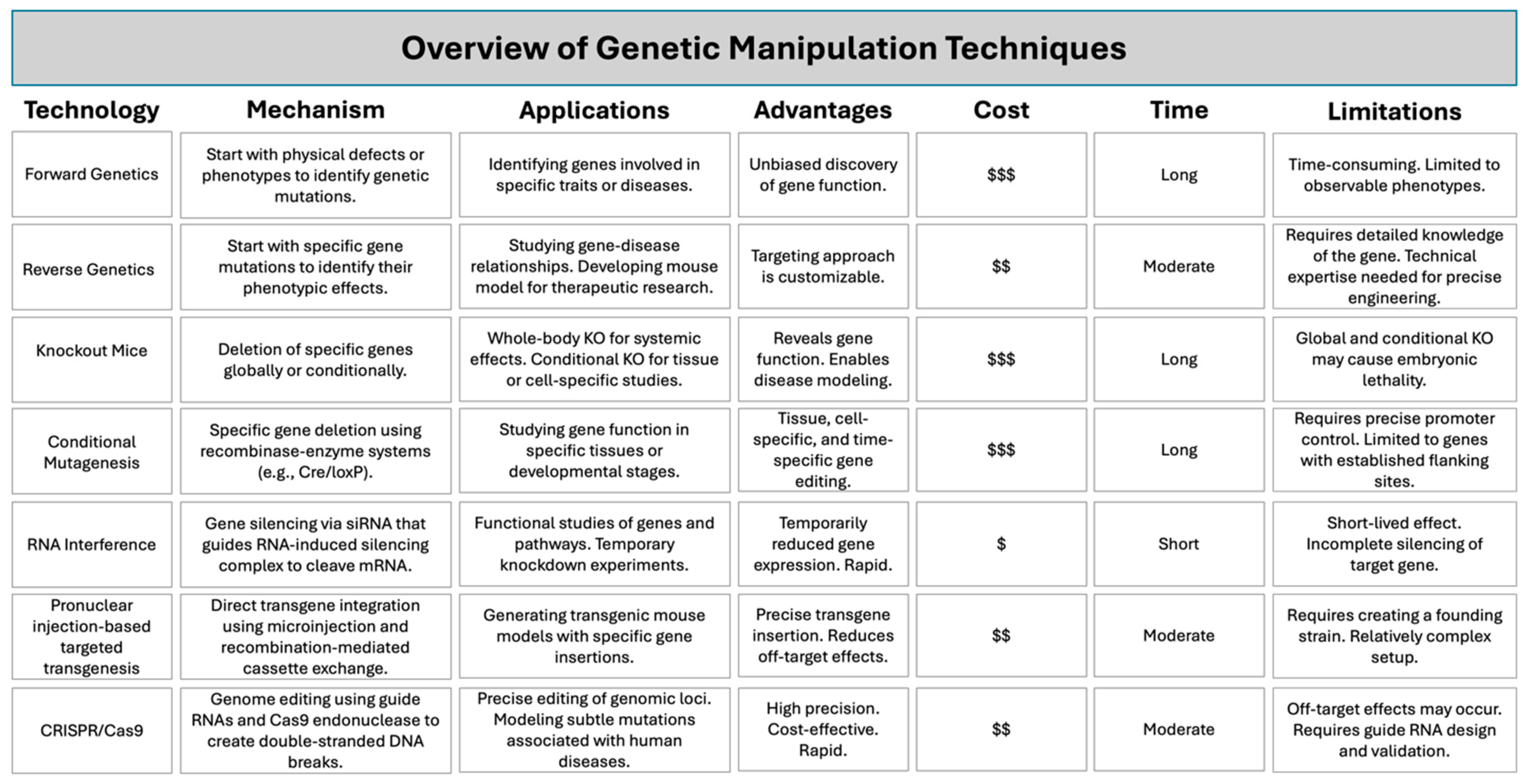
Figure 1. Overview of technologies used to modify mice for research. This table summarizes various genetic manipulation techniques, including their mechanisms, applications, advantages, cost, time, and limitations. Techniques covered include forward and reverse genetics [4], gene knockouts [5], conditional mutagenesis [6], RNA interference (RNAi) [7], pronuclear injection-based targeted transgenesis (PITT) [8], and CRISPR-Cas9 [9]. $ = low cost, $$ = moderate cost, $$$ = high cost based on technical complexity and resource needs.
Ijms 26 03417 g001
Figure 2. Stages of Atheroma Formation. Atherosclerosis develops in sequential stages, starting from endothelial dysfunction to plaque rupture. Healthy Artery: The endothelium functions to maintain minimal inflammation, balance lipid levels, and a healthy vascular environment. Lesion Initiation: Endothelial cells (ECs), which line the blood vessel walls, lose their integrity, leading to increased vasoconstriction, lipid infiltration, leukocyte adhesion, and oxidative stress. The reduced bioavailability of nitric oxide (NO), a key vasoprotective molecule, is a hallmark of endothelial dysfunction. This stage sets the foundation for plaque development. Fatty Streak: Pro-atherogenic mediators, such as cytokines and adhesion molecules, recruit monocytes to the intima. These monocytes differentiate into macrophages, internalizing low-density lipoproteins (LDL) and forming foam cells, initiating fatty streak formation. Fibrous Plaque: A fibrous cap forms over the necrotic core, composed of collagen and other extracellular matrix (ECM) components, stabilizing the plaque. However, the fibrous cap can weaken due to vascular smooth muscle cell (VSMC) apoptosis and the release of matrix metalloproteinases. Plaque Rupture: When the fibrous cap ruptures due to hemodynamic forces, platelets aggregate, and a coagulation cascade leads to thrombus formation. This exacerbates the plaque, potentially obstructing blood flow and contributing to cardiovascular disease. Servier Medical Art was used to create the figure, which is licensed under CC BY 4.0 (https://creativecommons.org/licenses/by/4.0/), assessed on January 30 2023.
Ijms 26 03417 g002
Figure 3. Direct effect of HIV on endothelial dysfunction. (A) HIV binds to the CD4 receptor on the surface of a T cell, fuses with the T cell, and replicates inside the T cell, HIV-secreted viral proteins, such as Nef and Tat, leading to the upregulation of adhesion molecules like ICAM-1 and VCAM-1. Additionally, immune cells secrete pro-inflammatory cytokines and chemokines, including IL-1α, IL-1β, IL-6, TNF-α, and NF-κB, further contributing to endothelial dysfunction. This dysfunction results in pathological outcomes such as vasoconstriction, lipid infiltration, leukocyte adhesion, oxidative stress, and cell death. (B) Chronic inflammation begins when a virus recognized by the T-cell receptor (TCR) causes T-cell death. These dead cells produce more pro-inflammatory cytokines, increasing reactive oxygen species (ROS) and oxidative stress. In endothelial cells, the early stages of atherosclerosis are marked by LDL entering the blood vessel wall, which becomes oxidized and induces monocyte recruitment. Recruited monocytes differentiate into macrophages, taking up oxLDL via scavenger receptors. This scavenger-receptor-mediated uptake of lipoproteins by macrophages leads to the formation of foam cells. (C) ART is a class of medications that suppress viral replication in HIV infections. As a side effect, some inhibitors can cause an imbalance between LDL and HDL levels, increase ROS production, and disrupt the body’s ability to neutralize this production damaging molecules. This results in more oxidative stress, increased oxLDL, the formation of more foam cells, and promotion of plaque formation. Created with BioRender.com, accessed 19 February 2025.
Ijms 26 03417 g003
Figure 4. HIV life cycle, viral inhibitors, and ASCVD-associated mechanisms. (Left Panel) This figure illustrates the stages of the HIV life cycle, including binding and post-attachment, viral entry, reverse transcription, integration, transcription, translation, and budding. It also highlights the points at which various classes of antiretroviral drugs act to disrupt the viral replication process, including binding inhibitors (CCR4 antagonist), entry inhibitors, nucleoside reverse transcriptase inhibitors (NRTIs), non-nucleoside reverse transcriptase inhibitors (NNRTIs), integrase strand transfer inhibitors (INSTIs), protease inhibitors (PIs), and capsid inhibitors. (Right Panel) Among these viral inhibitors, CCR4 antagonists, post-attachment, fusion, and capsid inhibitors are atheroprotective. In contrast, nucleoside reverse transcriptase inhibitors (NRTIs), non-nucleoside reverse transcriptase inhibitors (NNRTIs), integrase strand transfer inhibitors (INSTIs), and protease inhibitors (PIs) may be atherogenic. Created with BioRender.com, accessed on 28 January 2025.
Ijms 26 03417 g004
Figure 6. NSG-mouse models in CVD research related to HIV. On the (left), using NSG-MHC I/II double knock-out (DKO) mice reconstituted with PBMCs, the role of immune cells may be explored in the development stages of atherosclerosis. Due to the short-lived period, T cell functions will likely be better explored in this model. On the (right), we propose that NSG mice reconstituted with human stem cells modified to express HIV proteins, like Tg26 mice, would be used. We propose knocking out the LDL receptor for both models because the cholesterol profile is like humans versus a gain of function PCSK9 mouse, which can be performed using an adenoviral vector [105]. Created with BioRender.com, accessed on 16 February 2025.
Ijms 26 03417 g006
Table 1. Selecting the ideal mouse model for atherosclerosis research.
Mouse ModelResearch Focus
Apoe−/− mice Spontaneous atherosclerosis development
Role of ApoE in atherosclerosis
Inflammation and atherosclerosis
CETP.E3L mice Role of age-related changes in cholesterol transport and lipid profiles in atherogenic processes
Ldlr−/− mice Diet-induced atherosclerosis
Role of LDL receptor in cholesterol handling and atherosclerosis
Interactions between diet and genetic differences in human LDL receptor
Human-like lipid profile and atherosclerosis (for example to study the role of statins)
Interaction between diet and inflammation
Apoe−/− Fbn1C1039G+/− mice Arterial stiffening and plaque development
Tg26+/−/Apoe−/− mice Role of HIV transcripts and proteins in atherosclerosis development and progression
Table 2. Limitations of mouse models: summary of differences in atherosclerosis between mice and humans.
AspectMiceHumans
Cholesterol Transport Transport most cholesterol in HDL particles (atheroprotective). Transport most cholesterol in LDL particles (atherogenic).
Bile Acid Metabolism Produce more hydrophilic bile acids, leading to reduced intestinal cholesterol uptake and increased fecal cholesterol excretion. Produce less hydrophilic bile acids, leading to higher intestinal cholesterol absorption.
Atherosclerotic Plaque Location Plaques develop in the brachiocephalic trunk, aortic arch, aortic sinus, and proximal aorta. Plaques typically develop in the carotid and coronary arteries.
Plaque Development Plaques develop but do not progress to advanced fibrous atheroma. Plaques progress to advanced-stage fibrous atheroma.
Genetic Models Common models include Apoe/ and Ldlr/ mice, manipulated for hypercholesterolemia. Genetic mutations in Ldlr and Apoe are associated with familial hypercholesterolemia and ASCVD.
Plaque Rupture Plaque rupture is rare except in specific models (e.g., Apoe−/− Fbn1C1039G+/− mice, angiotensin II-infused Apoe/ mice). Plaque rupture is common, leading to thrombosis and cardiovascular events.
Inflammatory Response Th1-skewed immune response; IFN-γ plays a key role in lesion development. Mixed Th1/Th2 immune response; inflammation plays a significant role in plaque rupture.
Disclaimer/Publisher’s Note: The statements, opinions and data contained in all publications are solely those of the individual author(s) and contributor(s) and not of MDPI and/or the editor(s). MDPI and/or the editor(s) disclaim responsibility for any injury to people or property resulting from any ideas, methods, instructions or products referred to in the content.