Review

What Can Fluorescence Tell Us About Wine?

by
Izabela Sadowska-Bartosz
* and
Grzegorz Bartosz
Laboratory of Analytical Biochemistry, Institute of Food Technology and Nutrition, Faculty of Technology and Life Sciences, University of Rzeszow, 4 Zelwerowicza Street, 35-601 Rzeszow, Poland
*
Author to whom correspondence should be addressed.
Int. J. Mol. Sci. 2025, 26(7), 3384; https://doi.org/10.3390/ijms26073384
Submission received: 8 March 2025 / Revised: 30 March 2025 / Accepted: 3 April 2025 / Published: 4 April 2025
(This article belongs to the Section Biochemistry)

Abstract

Rapid and cost-effective measurements of the autofluorescence of wine can provide valuable information on the brand, origin, age, and composition of wine and may be helpful for the authentication of wine and detection of forgery. The list of fluorescent components of wines includes flavonoids, phenolic acids, stilbenes, some vitamins, aromatic amino acids, NADH, and Maillard reaction products. Distinguishing between various fluorophores is not simple, and chemometrics are usually employed to analyze the fluorescence spectra of wines. Front-face fluorescence is especially useful in the analysis of wine, obviating the need for sample dilution. Front-face measurements are possible using most plate readers, so they are commonly available. Additionally, the use of fluorescent probes allows for the detection and quantification of specific wine components, such as resveratrol, oxygen, total iron, copper, hydrogen sulfite, and haze-forming proteins. Fluorescence measurements can thus be useful for at least a preliminary rapid evaluation of wine properties.

1. Introduction

Wine is one of the oldest and most commonly consumed beverages, consumed since Neolithic times for social, personal, and religious reasons [1,2,3]. Wine is the product of the alcoholic fermentation of grapes, although it is also produced from other fruits. Natural wines contain 9–14% ethanol, while dessert and appetizer wines may contain 15–21% ethanol [4]. The cardioprotective effects of wine have been postulated and debated. The low prevalence of ischemic heart disease, in spite of the high intake of saturated fat, observed in France, was ascribed to the high consumption of red wine and referred to as the French paradox [5,6,7]. Wine is a complex mixture of various compounds including alcohols (mainly ethanol but also small amounts of other alcohols), sugars, esters, aldehydes, ketones, organic acids, amino acids, biogenic amines, furanic compounds, lactones, acetals, phenols, polyphenols, metal ions, terpenes, norisoprenoids, methoxypyrazines, and thiols. All these compounds may affect the taste, aroma, and ither properties of wine and thus consumer preferences [8].
Among the various techniques used to analyze wine, fluorescence spectroscopy is one of less frequently employed. The term “fluorescence” has been introduced to describe the unusual properties of the mineral fluorite, also called fluorspar (calcium fluoride) [9]. Fluorescence denotes a phenomenon occurring in some molecules (fluorophores), consisting of an immediate (in the nanosecond range) emission of light by a substance irradiated with a light of a shorter wavelength or UV radiation. The phenomenon occurs in three stages. In the first step, the fluorophore is excited to an electronic singlet state by absorption of an external photon (ex). In the second step, the excited-state molecule interacts with the molecular environment in several different ways, including vibrational relaxation, quenching, and energy transfer. In the third and final step, a photon (em) of a longer wavelength than that of the exciting light is emitted, while the fluorophore returns to its ground state. The difference between the positions of the band maxima of the absorption and emission spectra is called the Stokes shift [10,11].
Fluorescence spectroscopy offers several advantages over other analytical methods. It is more sensitive than other spectroscopic techniques by 2–3 orders of magnitude [12,13,14,15,16]. In many cases, this technique allows for the rapid assessment of samples without the need for complex preparation processes. Such a measurement is non-destructive and cost-effective. Another approach is based on the use of specific fluorescent probes that can be introduced for the selective measurement of specific compounds or processes, for example, Ca2+ [17] or Mn2+ concentration [18], the generation of reactive oxygen species [19], or changes in membrane potential [20].
In conventional fluorescence spectroscopy, two types of spectra are usually measured. Recording the emission intensity as a function of the emission wavelength λem at a fixed excitation wavelength λex yields an emission spectrum. When λex is scanned at a fixed λem, an excitation spectrum is recorded [10,11,21]. Synchronous spectra are obtained when excitation and emission are scanned simultaneously, with a fixed interval between the excitation and emission wavelengths [22,23].
Like any technique, fluorescence spectroscopy has its limitations. Usually, right-angle measurements of fluorescence are employed. In this arrangement, fluorescence emitted from the whole sample in a plane perpendicular to that of the incident light is measured (Figure 1). However, if the absorbance of the sample at the excitation wavelength is higher than 0.05, the incident light does not reach the whole volume of the sample. In this situation, the intensity of the emitted light may not be accurately measured due to potential scattering and absorption effects. In such cases, it is crucial to dilute the sample or adjust the experimental setup to ensure that the measurements reflect the true fluorescence characteristics of the sample, proportional to fluorophore concentration. This is a common problem when measuring food products, including wine, which are often turbid or opaque or have high concentrations of the fluorophores. However, dilution can lead to the loss of organization of the food matrix. To avoid these problems, the method of front-face fluorescence spectroscopy is used. In this method, fluorescence is emitted from the surface layer of the probe. Thus, excitation of the sample and measurement of its emitted radiation are carried out in the same cell-face. The passage of radiation through the bulk solution is avoided and the scattered light and depolarization phenomena are minimized. The incidence angle of the excitation radiation is between 30° and 60° [10,23,24,25]. Most microplate readers have the option of front-face fluorescence measurement, which may be an incentive for the broader use of this approach. Front-face fluorescence proved to be especially useful in the analysis of wine [26,27,28,29,30].
In principle, the fluorescence signal of a given sample is the sum of the fluorescence contributions from each of the inherent fluorophores. However, fluorescence emission depends on the surroundings of the fluorophore, and in complex systems and concentrated solutions, the fluorescence may not be additive due to the phenomena of quenching, interactions with the molecular environment of the fluorophores, and fluorescence reabsorption [10,11].

2. Chemometric Approach to Wine Analysis

In fluorometric studies of complex systems, including wine, a simple emission spectrum for one excitation wavelength is not sufficient to characterize the fluorescent properties of the sample. Instead, a set of emission spectra for a range of different excitation wavelengths λex is often recorded to obtain a three-dimensional plot, the so-called fluorescence excitation–emission matrix (EEM) (Figure 2).
The excitation–emission matrix can be obtained for each fluorophore. The overall fluorescence EEM for a sample can be described according to Equation (1):
E E M = i = 1 n a i   ×   b i λ e x × c i λ e m
where i is the number of a fluorophore, n is the total number of fluorescent species present in the sample, ai is a concentration-dependent factor characteristic for each fluorophore, bi(λex) describes the excitation characteristics, and ci (λem) describes the emission characteristics of the ith fluorophore.
The simultaneous presence of multiple fluorophores of interlapping excitation and emission characteristics makes the identification of contributions of individual fluorophores difficult and requires the use of chemometrics, a chemical discipline using mathematics, statistics, and formal logic to analyze the data. Chemometrics uses such tools as parallel factor analysis (PARAFAC), principal component analysis (PCA), or partial least squares (PLS) regression [31,32,33].
The parallel factor analysis (PARAFAC) model is based on a decomposition of a complex set of fluorescence data into several PARAFAC components corresponding to individual fluorophores (or fluorophore groups) present in the samples. In the analysis of data, the relative concentration of components in the mixture can be determined, and the excitation and emission loadings can be used for the identification of fluorophores. The three-way data array is thus decomposed into a set of sample scores, aif, loadings for the emission mode, bjf, and loadings for the excitation mode, ckf. The principle of this approach is to minimize the sum of squares of the residual, eijk in Equation (2), using the least-squares algorithm
x i j k = f = 1 F a i f b j f c k f + e i j k
where xijk represents the data for sample i in variables j and k of the two different variable dimensions [12,31].
Principal component analysis (PCA) is another mathematical procedure that decomposes the data matrix with n samples and p columns (variables, e.g., wavelengths) into the product of a scores matrix, with n rows and d < p columns (principal components, PCs). The scores are the positions of the samples in the space of the principal components, and the loadings are the contributions of the original variables to the PCs. All PCs are mutually orthogonal, and each successive PC contains less of the total variability of the initial dataset. This procedure reduces the dimensionality of the data, which enables the effective visualization, classification, and regression of multivariate data [9]. The PCA components do not necessarily have a clear physical meaning, but they can be efficiently used to understand and classify the wine data. After PCA, data modeling can be further progressed using, e.g., Soft Independent Modeling of Class Analogy (SIMCA) or machine learning as a data modeling alternative [34,35].
Multivariate classification methods or pattern-recognition methods are used for grouping samples with similar characteristics. They include supervised and non-supervised methods.
Non-supervised or exploratory methods can group data into clusters. They are often useful at an early stage of a study to compare subpopulations, such as different batches of a product. Cluster analysis can be performed with simple means, such as hierarchical cluster analysis (HCA) or PCA. HCA compares the similarity between the samples based on their measured variables. The samples are grouped into clusters according to their closeness in a multidimensional space and are usually presented in the form of dendrograms [12]. PCA can also be used to find relationships between different parameters and the detection of possible clusters within the samples [32,36,37].
Sometimes, non-negative matrix factorization (NMF) may be more suitable than PCA. In this method, only positive solution values can be obtained, and thus, this method provides a more realistic approximation to the original data than PCA, which allows for both positive and negative values [38].
In the supervised or discriminant analysis methods, each fluorescence spectrum is preliminarily assigned to a definite class, with comprehensive libraries of spectra representing various versions of each product being constructed in a calibration process. Principal component or partial least squares (PLS) analyses are often applied to spectral datasets to reduce the size of a dataset and co-linearity. Spectral data are analyzed using various methods such as linear discriminant analysis (LDA) [39], factorial discriminate analysis (FDA) [39,40], or k-nearest neighbors (kNN) [41]. The analysis aims at the formulation of weighted linear combinations of the data to minimize the within-class variance and to maximize the between-class variance. If the samples studied are numerous enough, they can be separated into two sets: a training set to elaborate the method (calibration) and a test set to validate it. The elaborated classification rules are later used for allocating new or unknown samples to the most probable subclass [25].
The second stage of analysis is often the factorial discriminate analysis (FDA). This method is useful when the data are preliminarily transformed into their PCs. In the first stage, a stepwise discriminant analysis is performed to select the most relevant PCs for the discrimination of variables when the qualitative classes are initially defined. FDA allows for the construction of new synthetic variables (discriminant factors) from the linear combinations of the selected PCs to achieve a better separation of the centers of gravity of the classes considered. Individual samples are assigned to classes where the distance from the centers of gravity is the shortest. Similarity maps and patterns can be drawn, as in PCA [25,39].
The most frequently used multivariate regression methods for quantitative fluorescence analysis are partial least-squares regression (PLSR) and principal component regression (PCR). Both methods can be used for whole spectra and selected spectral regions, allowing for the inclusion of more information in the calibration model. PCR uses the principal components provided by PCA to perform regression on the sample parameter to be predicted. PLSR points the directions of greatest variability by comparison of the information on both spectral and target properties with the new axes (PLSR components or PLSR factors). The first principal component or factor in PCR represents the widest variations in the spectrum, while in PLSR, it represents the most relevant variations, showing the best correlation with property values of a target [42,43,44].

3. Fluorescent Components of Wines

The main fluorescent components of wines are polyphenols. Phenolic compounds are secondary metabolites found in grapes and wine that can be classified into two groups: flavonoids and non-flavonoids (phenolic acids and stilbenes) [45,46]. The phenolic composition of wine is dependent on many factors, including conditions of grape berry development and ripening, the grape cultivar and ripeness at harvest, and the technology of fermentation and aging [47,48].
Within wine flavonoids, three subgroups are important: flavonols, flavan-3-ols, and anthocyanins. Flavonols are found in grape skins as glycosides of myricetin, quercetin, kaempferol, isorhamnetin, syringetin, rutin, and laricitrin [49,50]. Flavan-3-ols (monomeric catechins and polymeric proanthocyanidins) are another large family of polyphenolic compounds comprising mainly catechin, epicatechin, gallocatechin, epigallocatechin, and their corresponding polymers, which are found in the skin and seed of the grape [51,52]. Proanthocyanidins (condensed tannins) are phenolic compounds of a polyflavan-3-ol structure [53,54]. Anthocyanins are extracted from the red grape skins during maceration and fermentation [55,56]. They are highly reactive and easily enter into chemical reactions with other red wine components, such as aldehydes or polyphenols (e.g., tannins), producing new anthocyanin derivatives. Both anthocyanins and anthocyanin-derived pigments contribute to the color of young red wines and play a crucial role in the evolution of wine color during aging [57,58,59,60,61]. Anthocyanin−pyruvic acid adducts appear to be the major anthocyanin derivatives detected by HPLC after only 1 or 2 years of aging in Port wine [58].
Flavonoids are excited in the region of 260–268 nm and emit in the range of 370–422 nm, except for flavan-3-ols which are excited at 278–290 nm and emit in the wavelength range of 310–360 nm [62], although fluorescence at λexem of 280/310 nm was also reported for catechin [63].
Anthocyanins are weakly fluorescent in solution. A fluorescence quantum yield of 4.1 × 10⁻³ for malvidin 3,5-diglucoside was reported [64], probably due to the efficient excited state proton transfer to water [65]. It may be the main reason why the fluorescence of red wines has been poorly studied. Cyanidin-3-glucoside and malvidin 3,5-diglucoside were reported to have absorption maxima at 220 nm and about 280 nm and fluorescence maxima at 308 and 293 nm, respectively. However, aggregation or complexation to other molecules can induce a significant fluorescence of the resulting anthocyanin-derived compound [66].
Phenolic acids, comprising mainly of caftaric, coutaric, fertaric, and tartaric, are normally found as esters. Hydroxycinnamic esters are one of the most abundant groups of phenolic compounds found in grapes. Stilbene-like compounds include resveratrol, its glucoside piceid, astringin, and viniferins [67,68]. The excitation wavelengths of phenolic acids (both derivatives of cinnamic acid and derivatives of benzoic acid), phenolic aldehydes, and stilbene-like compounds extend between 260 nm and 330 nm, while the emission wave range is 320–440 nm [62].
Other fluorescent molecules present in wine, apart from polyphenols, are vitamins and amino acids. Vitamin A (retinol) is present in wine in very small amounts. The excitation maximum corresponds to about 335 nm, and the emission maximum to about 470 nm [69]. On the other hand, B-complex vitamins are the most abundant. Riboflavin is present mainly as a component of flavin-mononucleotide (FMN) and flavin-adenine-dinucleotide (FAD). Free riboflavin is also present in raw and processed fruits and is present in significant amounts in wine. Flavin absorption is centered at about 450 nm and emission at about 525 nm [70]. The fluorescent amino acid tryptophan and its ethyl ester have been reported to be present in wines [71,72,73]. Tryptophan is excited at wavelengths around 280 nm and emits fluorescence in the range of 300–400 nm [27,71]. NADH is formed in the fermentation processes that take place during the production of wines [74]. NADH is excited at 340–350 nm and emits fluorescence centered at 460–470 nm [75]. Fluorescent oxidation products and Maillard products may be produced in the browning processes during the aging and storage of wines [76]. Due to their heterogeneity, the absorption maxima of Millard reaction products may range from 320 to 450 nm, while emission maxima are in a broad range of 380–530 nm [77].

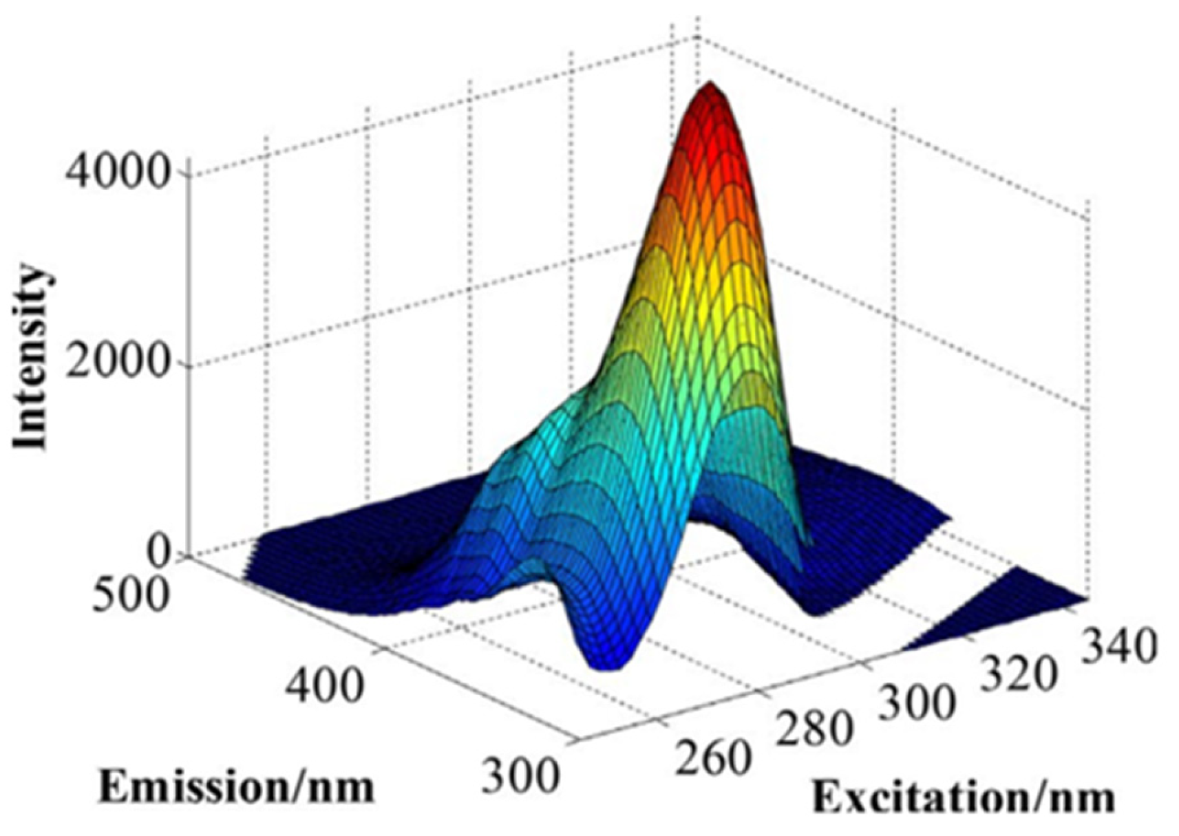
4. Analysis of Wine Autofluorescence

An example of an EEM of wine constructed by the measurement of a series of fluorescence spectra for a range of excitation wavelengths is shown in Figure 2. The list of components tentatively assigned to four PARAFAC components used to interpret EEM of this type is shown in Table 1.
Analysis of these EEMs by PARAFAC makes it possible to determine the relative contributions of each component to the spectra matrices. Other studies found that red wines have four main fluorescence components, with the excitation and emission maxima at the wavelength pairs of 260/380 nm, 275/323 nm, 330/410 nm, and 280/364 nm, respectively [27,78]. A tentative identification of fluorophores performed by matching PARAFAC score values with the HPLC analysis of wine revealed that the third component correlated with concentrations of catechin and epicatechin [30,36]. Such measurements allowed for distinguishing between Rioja and Ribera del Guadiana wines, discrimination between Rioja and non-Rioja samples, discrimination between Crianza or Reserva wines compared to young wines [27], and discrimination of wines according to the country of origin [78].
Components tentatively assigned to four PARAFAC components used for the analysis of Cava wines are shown in Table 2. The fluorescence analysis of sparkling cava wines was found to be a fast alternative method for the quality control of sparkling wines. Specifically, monitoring the fluorophores centered at excitation/emission of 465/530 nm and 280/380 nm can provide useful information about the chemical changes occurring during browning [76].
Pre-barreled New Zealand Pinot Noir wines showed an EEM with a component characterized by excitation and emission maxima at around 277 nm and 330 nm, respectively. The maximum signal intensity of this component was increased about two times in comparison with the grape juice. This increase is contributed by multiple fluorophores in the wine, such as the phenolic acids (syringic, vanillic, gallic, caftaric, p-hydroxybenzoic and caffeic acid, catechin, epicatechin and tryptophan) and other components that were not present or present at much lower levels in grape juice [30].
In the study of red wines by dos Santos et al., the total phenolics region corresponded to 260–360 nm excitation and 370–400 nm emission, the total condensed tannins were the main contributor to the fluorescence in the region of excitation between 285 and 340 nm and emission in the range of 290–350 nm, and the total anthocyanins region contributed to the signals with excitation between 280 and 300 nm and emission between 330 and 380 nm [79]. The analysis of EEMs of 200-times diluted Cabernet Sauvignon wines from three regions of Australia and Bordeaux using discriminant analysis and support vector machine discriminant analysis (SVMDA) made it possible to differentiate wines according to the location of origin [80]. The analysis of EEMs of 150-times diluted Shiraz, Cabernet Sauvignon, and Merlot wines from 10 locations in Australia by PLS and extreme gradient boosting (XGB) discriminant analysis (a machine learning protocol) allowed for the differentiation of samples by their variety and geographical origin [81].
The analysis of wine phenolic content by front-face fluorescence spectroscopy combined with chemometrics was suggested to be a potentially useful tool for authentication and quality control by regulatory bodies [33]. The use of principal component analysis (PCA) and classification by factorial discriminant analysis (FDA) allowed for distinguishing between German and French wines, demonstrating the possibility of identification of wines according to variety and typicality [26]. Discrimination between Shiraz, Cabernet Sauvignon, and Pinot Noir based on fluorescence can be improved by measurements at different pH levels [28].
Front-face fluorescence spectroscopy in combination with PARAFAC was also shown to be a promising tool for the discrimination of grape-derived products from different clonal and vineyard site origins within a small geographical region in New Zealand. The discrimination between grape clones was found to be due to higher concentrations of the component at an excitation maximum of 260 nm and emission maximum of 390 nm, with a shoulder at 370 nm, possibly contributed by caffeic acid-related fluorophores. The effect of discrimination based on the vineyard site was indicated to be due to the component at an excitation maximum of 278 nm and emission maximum of 360 nm, probably contributed mainly by tryptophan and hydroxylated benzoic acid derivatives [30].
The fluorescence of bulk Slovak Tokaj wines was characterized by an excitation range of 390 to 500 nm, with a maximum at 460 nm and emission in the range of 450 to 590 nm, with a maximum of about 530 nm. These wines, when diluted 500 times, had fluorescence characterized by excitation in the range of 250–350 nm and emission in the range of 320–450 nm. An intense band was observed with excitation in the range of 270–280 nm and emission centered at 350 nm, as well as a weak band with excitation at 300–310 and emission at about 430–440 nm. This fluorescence was similar to those of phenolic acids characterized by excitation/emission wavelengths: (gallic acid, 280/360 nm; protocatechuic acid, 270/350 nm; caffeic acid, 262 and 325/426 nm; caftaric acid, 290 and 325/440 nm, p-coumaric acid 290 and 309/404 nm) and catechin, 280/310 nm. The PLS regression allowed for the estimation of the content of gallic, protocatechuic, caffeic, and p-coumaric acids, and (+)-catechin from the fluorescence spectra of wines [63].
Synchronous spectra in the range of 260–290 nm, especially with the wavelength difference between excitation and emission (Δλ) of 60 to 100 nm, allow for prediction of the antioxidant capacity of wines based on the estimation of the concentrations of phenolic compounds in sweet Slovak Tokaj wines [82]. Emission spectra corresponding to excitation at 320 nm or synchronous spectra in the range of 300–400 nm, especially with Δλ of 80 nm, allow for the determination of the sum of concentrations of coumarins in Tokaj wines [83].
A Port red wine aged 3 years showed a fluorescence maximum at about 595 nm when excited at 500 nm. The fluorescence maximum was shifted by about 30 nm towards shorter wavelengths concerning the young wine (fluorescence maximum at about 625 nm). Aged wines usually contain larger amounts of polymerized anthocyanin (pol-Anth) pigments and lower amounts of monomeric anthocyanin pigments than young wines. The fluorescence band of pol-Anth isolated from wine and dissolved in 12% ethanol brought to pH 3.3 was similar to that of aged wines (peak at 597 nm), while the spectra of mon-Anth showed a bathochromic shift of about 40 nm in comparison with pol-Anth. The intensity of fluorescence was higher for pol-Anth than mon-Anth [84].
The fluorescence intensity ratio at 700 nm to that of 560 nm of wines excited at 500 nm was found to decrease when plotted against the relative share of pol-Anth and to decrease in old wines, and it was proposed to be a measure of the pol-Anth/min-Anth ratio, and thus of red wine age.
The treatment of young red wine with sulfur dioxide caused a hypsochromic shift in the fluorescence spectrum to match the spectrum of old Port wine. This effect is due to the formation of colorless and nonfluorescent compounds in the reaction of monomeric anthocyanins with sulfur dioxide, which binds to carbon 4 of the C ring. Polymerized anthocyanins remain unbleached because the site of sulfite binding is the same as that engaged in the anthocyanin polymerization [84,85].
Wine anthocyanins react with pyruvic acid, forming pyranocyanins such as vitisin A [86,87]. It should be noted that the term “vitisin A” is ambiguous since the same name is used for another compound, one of resveratrol tetramers [88,89]. Malvidin 3-O-glucoside (Mv 3-O-glc) is the major anthocyanin detected in young red wine and vitisin A (a pyruvic adduct of Mv 3-O-glc) [58]. The fluorescence spectrum of Mv 3-O-glc at pH 1.0 showed a peak at 610 nm, which increased in intensity and shifted to 638 nm at pH 3.3. Vitisin A showed a broad emission band with a maximum around 720 nm at pH 1.0, while at pH 3.3, a significant increase in fluorescence was observed around 630 nm. The fluorescence quantum yield of vitisin A relative to Mv 3-O-glc was 0.81 and 0.86 at pH 1 and 3.3, respectively. The increase in the fluorescence intensity, for both Mv 3-O-glc and vitisin A, from more to less acidic conditions can be explained by proton quenching, the extent of which is pH-dependent. Based on differences in the excitation spectra, the fluorescence excitation ratio (FER) between wavelengths at the maximal difference and the isosbestic point (FER550nm/425nm and FER350nm/425nm) was proposed to estimate the relative amounts of Mv 3-O-glc and vitisin A, although in whole wines, the strong contribution of pol-Anth to fluorescence makes it difficult to distinguish the two classes of pigments [84].
One technique of measuring the fluorescence of dye molecules concentrated on the surface of wine as a difference between fluorescence was proposed based on the estimation of the difference between the fluorescence from the surface and from inside the wine [90].
Simple measurements of wine fluorescence may not require a spectrofluorimeter or plate reader. The fluorescence spectrum of a red wine from the Lazio region obtained by a computer screen photo-assisted technique (a combination of computer monitors and webcams) was characterized by an excitation maximum at 450 nm, a shoulder at 520 nm, and an emission maximum at about 610 nm [91].

5. Application of Fluorescent Probes for the Analysis of Wine

The use of fluorescent probes allows for the selective estimation of the content of chosen parameters of components of wine using a spectrofluorimeter, plate reader, or fluorescence microscope. The applications of flow cytometry for the analysis of wine are not included in this review, as they require specialized equipment.
A fluorescence-based sensor based on the measurement of the luminescent lifetime of a reference metal/porphyrin complex was proposed for the real-time monitoring of oxygen concentration during wine fermentation. The sensor allows for the determination of the concentrations of O2 in wine below 10–40 µg/L at 20 °C, i.e., between 0.1 and 0.5% O2 [92]. The precise determination of oxygen content during wine production is important, as oxygen induces changes in the chemical and sensory profile of wines, including the final alcohol content. Moderate red wine exposure to oxygen has a positive impact on the color, aroma, and taste of red wine’s properties. However, oxygen negatively affects the quality and sensory properties of white wines [93,94].
A fluorescence assay for resveratrol determination based on Förster Resonance Energy Transfer (FRET), via competitive supramolecular recognition, between p-sulfonated calix [6] arene-(CX6)-modified reduced graphene oxide (CX6@RGO) and Rhodamine B- or rhodamine 123–resveratrol complex was explored. Resveratrol present in wine competes with the complex, releasing rhodamine and causing an increase in the fluorescence intensity. This assay, not requiring any wine pretreatment, allows for the determination of resveratrol with a detection limit of 0.5 µM [95].
The off-odors of wine are of considerable concern in the wine industry. They can develop after the wine has been bottled, and no corrective action is possible. Hydrogen disulfide is the main component of wine off-odors, which are also contributed by mercaptans and volatile sulfur compounds [96,97]. A fluorescent probe, 4-methyl-2-oxo-2H-chromen-7-yl-thiophene-2-carboxylate, was used to estimate the level of hydrogen sulfide in wine. Three red wines bought in a Beijing supermarket were found to contain 0.48, 0.45, and 0.55 μM H2S when estimated using this probe [98].
A fluorescent probe (E)-2-((4-(benzo[d]thiazol-2-yl)benzylidene)amino)phenol (BTPAP) was applied for the determination of total iron (Fe2+/Fe3+) in wine. The proposed assay based on the probe allows for the estimation of iron with a limit of detection of 1.16 µM and a linearity of response up to 200 µM. In three samples of Chinese red wines, the iron concentration was found to be 24–37 µM with this probe [99].
At least three fluorescent probes have been applied for estimation of the copper level in wines. The application of a fluorescent coumarin-based probe for the detection of copper(II) in wine revealed Cu2+ concentrations of 0.22–0.46 in three red wines. The detection limit of the method was 62 nm, and the range of linearity was 0–16 µM [100]. A pH-sensitive chemosensor based on Rhodamine B coupled to a tetraazamacrocyclic ring, 3 [N-(9-(2-((1,4,7,10-tetraazacyclotridecan-5-yl)methyl)-3-oxoisoindolin-1-yl)-6-(diethylamino)-3H-xanthen-3-ylidene)-N-ethylethanamine], was used to determine the Cu2+ concentration in white wine. The detection limit of the sensor is 43.8 nM. The Cu2+ concentration in six samples of a 2016 Burgundy Chardonnay was found to be 0.03–0.76 mg/L (0.47–11.96 µM) [101]. A fluorometric assay based on the simultaneous use of two fluorescent probes and the measurement of the ratio of fluorescence intensities at two wavelengths allows for the determination of the copper concentration in wine, with a limit of detection of 46.5 nM and a linear range up to 4 µM. The Cu2+ concentrations in three samples of red wine were found to be 22–36 µg/L (0.35–0.57 µM) [102].
Ochratoxin A is a hepatotoxic, genotoxic, cytotoxic, and teratogenic mycotoxin produced by several fungal species, mainly of the genera Aspergillus and Penicillium, that can contaminate wine. A fluorescence polarization immunoassay for ochratoxin A possesses a detection limit of 0.11±0.05 ng/mL [103].
Protein haze is an esthetic problem in white wines caused by the persistence of grape pathogenesis-related proteins that are highly stable during winemaking. Some of these proteins precipitate over time, especially at elevated temperatures, forming a turbid haze [104,105]. A rapid fluorescence-based technology to detect haze-forming proteins in white wines was developed based on the use of a new fluorescent probe binding selectively to haze-forming proteins. The method has a detection limit of 2 mg/L and a linear range of 4 to 400 mg/L. It is sensitive enough, since the minimal concentration of proteins to form haze is 12 mg/L [106].
Staining lactic acid bacteria with the Live/Dead BacLightTM staining kit and fluorescent microscopy assessment allows for control of the viability of lactic acid bacteria and thus the process of malolactic fermentation used for the biological deacidification of wines [107].
Examples of the application of fluorescence measurements to wine are listed in Table 3.
Wine has been one of the most popular drinks worldwide for thousands of years. Monitoring wine quality and controlling forgeries concerning the false reporting of wine origin and quality is important. Fluorescence spectroscopy, allowing for a rapid estimation of a range of wine properties, requiring only a fluorimeter or plate reader, and available in most laboratories, may be very useful in this respect, especially for preliminary wine screening.

Author Contributions

Conceptualization, I.S.-B. and G.B.; methodology, I.S.-B. and G.B.; investigation, I.S.-B. and G.B.; writing—original draft preparation, I.S.-B. and G.B.; writing—review and editing, I.S.-B. and G.B.; supervision, I.S.-B.; project administration, I.S.-B.; funding acquisition, I.S.-B. All authors have read and agreed to the published version of the manuscript.

Funding

This study was performed within the project “Modification of anthocyanins/anthocyanidins as new markers of food oxidation” (number of the application 2023/51/B/NZ9/02490), financed by the National Science Centre (NCN), Poland, in the program “Opus 26”.

Institutional Review Board Statement

Not applicable.

Informed Consent Statement

Not applicable.

Data Availability Statement

Data will be available from the corresponding author upon reasonable request.

Conflicts of Interest

The authors declare no conflicts of interest.

References

  1. Harutyunyan, M.; Malfeito-Ferreira, M. The rise of wine among ancient civilizations across the Mediterranean basin. Heritage 2022, 5, 788–812. [Google Scholar] [CrossRef]
  2. Estreicher, S.K. Wine and France: A brief history. Eur. Rev. 2023, 31, 91–179. [Google Scholar]
  3. Mouret, M.; Monaco, G.L.; Urdapilleta, I.; Parr, W.V. Social representations of wine and culture: A comparison between France and New Zealand. Food Qual. Prefer. 2013, 30, 102–107. [Google Scholar]
  4. Swami, S.B.; Thakor, N.J.; Divate, A.D. Fruit wine production: A review. J. Food Res. Technol. 2014, 2, 93–100. [Google Scholar]
  5. Lanzmann-Petithory, D.; Professor Serge, C. Renaud (1927–2012): French Paradox and wine active compounds. Wine Stud. 2022, 1, 4477. [Google Scholar]
  6. de Gaetano, G.; Costanzo, S.; Di Castelnuovo, A. Wine consumption and cardiovascular health: The unresolved French paradox and the promise of objective biomarkers. Eur. Heart J. 2024, 46, 173–175. [Google Scholar]
  7. Buja, L.M. The history, science, and art of wine and the case for health benefits: Perspectives of an oenophilic cardiovascular pathologist. Cardiovasc. Pathol. 2022, 60, 107446. [Google Scholar]
  8. Perestrelo, R.; Silva, C.; Gonçalves, C.; Castillo, M.; Câmara, J.S. An approach of the madeira wine chemistry. Beverages 2020, 6, 12. [Google Scholar] [CrossRef]
  9. Valeur, B.; Berberan-Santos, M.N. A brief history of fluorescence and phosphorescence before the emergence of quantum theory. J. Chem. Educ. 2011, 88, 731–738. [Google Scholar] [CrossRef]
  10. Lakowicz, J.R. Principles of Fluorescence Spectroscopy, 3rd ed.; Springer: New York, NY, USA, 2006. [Google Scholar]
  11. Jameson, D.M. Introduction to Fluorescence, 2nd ed.; CRC Press: Boca Raton, FL, USA, 2025. [Google Scholar]
  12. Sádecká, J.; Tóthová, J. Fluorescence spectroscopy and chemometrics in the food classification—A Review. Czech J. Food Sci. 2007, 25, 159–173. [Google Scholar]
  13. Luykx, D.M.; Van Ruth, S.M. An overview of analytical methods for determining the geographical origin of food products. Food Chem. 2008, 107, 897–911. [Google Scholar] [CrossRef]
  14. Williams, R.T.; Bridges, J.W. Fluorescence of solutions: A review. J. Clin. Pathol. 1964, 17, 371. [Google Scholar] [CrossRef] [PubMed]
  15. Weiß, N.; Armbrecht, M.; Eppendorf, A.G. Absorbance or fluorescence: Which is the best way to quantify nucleic acids? Proteins 2018, 260, 280. [Google Scholar]
  16. Zacharioudaki, D.E.; Fitilis, I.; Kotti, M. Review of fluorescence spectroscopy in environmental quality applications. Molecules 2022, 27, 4801. [Google Scholar] [CrossRef]
  17. Contini, C.; Kuntz, J.; Massing, U.; Merfort, I.; Winkler, K.; Pütz, G. On the validity of fluorimetric intracellular calcium detection: Impact of lipid components. Biochem. Biophys. Commun. 2023, 643, 186–191. [Google Scholar] [CrossRef]
  18. Alosaimi, E.H. Recent developments in colorimetric and fluorimetric chemosensors for the detection of Mn2+ ions: A review (2010–2024). Crit. Rev. Anal. Chem. 2025, 19, 1–21. [Google Scholar] [CrossRef]
  19. Andina, D.; Leroux, J.C.; Luciani, P. Ratiometric fluorescent probes for the detection of reactive oxygen species. Chemistry 2017, 23, 13549–13573. [Google Scholar] [CrossRef]
  20. Fairless, R.; Beck, A.; Kravchenko, M.; Williams, S.K.; Wissenbach, U.; Diem, R.; Cavalié, A. Membrane potential measurements of isolated neurons using a voltage-sensitive dye. PLoS ONE 2013, 8, e58260. [Google Scholar] [CrossRef]
  21. Sauer, M.; Hofkens, J.; Enderlein, J. Handbook of Fluorescence Spectroscopy and Imaging: From Single Molecules to Ensembles; Wiley-VCH Verlag GmbH & Co. KGaA: Weinheim, Germany, 2011. [Google Scholar]
  22. Radotić, K.; Stanković, M.; Bartolić, D.; Natić, M. Intrinsic fluorescence markers for food characteristics, shelf life, and safety estimation: Advanced analytical approach. Foods 2023, 12, 3023. [Google Scholar] [CrossRef]
  23. Freire, P.; Zamora, A.; Castillo, M. Synchronous front-face fluorescence spectra: A review of milk fluorophores. Foods 2024, 13, 812. [Google Scholar] [CrossRef]
  24. Karoui, R.; Dufour, E.; Bosset, J.O.; De Baerdemaeker, J. The use of front face fluorescence spectroscopy to classify the botanical origin of honey samples produced in Switzerland. Food Chem. 2007, 101, 314–323. [Google Scholar]
  25. Karoui, R.; Blecker, C. Fluorescence spectroscopy measurement for quality assessment of food systems—A review. Food Bioprocess Technol. 2011, 4, 364–386. [Google Scholar]
  26. Dufour, É.; Letort, A.; Laguet, A.; Lebecque, A.; Serra, J.N. Investigation of variety, typicality and vintage of French and German wines using front-face fluorescence spectroscopy. Anal. Chim. Acta 2006, 563, 292–299. [Google Scholar]
  27. Airado-Rodríguez, D.; Durán-Merás, I.; Galeano-Díaz, T.; Wold, J.P. Front-face fluorescence spectroscopy: A new tool for control in the wine industry. J. Food Compos. Anal. 2011, 24, 257–264. [Google Scholar] [CrossRef]
  28. Saad, R.; Bouveresse, D.J.R.; Locquet, N.; Rutledge, D.N. Using pH variations to improve the discrimination of wines by 3D front face fluorescence spectroscopy associated to Independent Components Analysis. Talanta 2016, 153, 278–284. [Google Scholar]
  29. Cabrera-Bañegil, M.; del Carmen Hurtado-Sánchez, M.; Galeano-Díaz, T.; Durán-Merás, I. Front-face fluorescence spectroscopy combined with second-order multivariate algorithms for the quantification of polyphenols in red wine samples. Food Chem. 2017, 220, 168–176. [Google Scholar]
  30. Schueuermann, C.; Silcock, P.; Bremer, P. Front-face fluorescence spectroscopy in combination with parallel factor analysis for profiling of clonal and vineyard site differences in commercially produced Pinot Noir grape juices and wines. J. Food Compos. Anal. 2018, 66, 30–38. [Google Scholar] [CrossRef]
  31. Geladi, P. Chemometrics in spectroscopy. Part I. Classical chemometrics. Spectrochim. Acta B 2003, 58, 767–782. [Google Scholar]
  32. Lenhardt Acković, L.; Zeković, I.; Dramićanin, T.; Bro, R.; Dramićanin, M.D. Modeling food fluorescence with PARAFAC. In Reviews in Fluorescence 2017; Geddes, C.D., Ed.; Springer: Cham, Switzerland, 2018; pp. 161–197. [Google Scholar]
  33. Dos Santos, I.; Bosman, G.; Aleixandre-Tudo, J.L.; du Toit, W. Direct quantification of red wine phenolics using fluorescence spectroscopy with chemometrics. Talanta 2022, 236, 122857. [Google Scholar]
  34. Christensen, J.; Nørgaard, L.; Bro, R.; Engelsen, S.B. Multivariate autofluorescence of intact food systems. Chem. Rev. 2006, 106, 1979–1994. [Google Scholar]
  35. Meena, D.; Chakraborty, S.; Mitra, J. Geographical origin identification of red chili powder using NIR spectroscopy combined with SIMCA and machine learning algorithms. Food Anal. Meth. 2024, 17, 1005–1023. [Google Scholar]
  36. Tufariello, M.; Pati, S.; Palombi, L.; Grieco, F.; Losito, I. Use of multivariate statistics in the processing of data on wine volatile compounds obtained by HS-SPME-GC-MS. Foods 2022, 11, 910. [Google Scholar] [CrossRef] [PubMed]
  37. Tsibulskaya, E.; Maslov, N. Decomposition of multi-component fluorescence spectra by narrow peak method based on principal component analysis. J. Chemom. 2021, 35, e3343. [Google Scholar]
  38. Kumar, K.; Giehl, A.; Patz, C.D. Chemometric assisted Fourier Transform Infrared (FTIR) Spectroscopic analysis of fruit wine samples: Optimizing the initialization and convergence criteria in the non-negative factor analysis algorithm for developing a robust classification model. Spectrochim. Acta A Mol. Biomol. Spectrosc. 2019, 209, 22–31. [Google Scholar]
  39. Monakhova, Y.B.; Godelmann, R.; Kuballa, T.; Mushtakova, S.P.; Rutledge, D.N. Independent components analysis to increase efficiency of discriminant analysis methods (FDA and LDA): Application to NMR fingerprinting of wine. Talanta 2015, 141, 60–65. [Google Scholar]
  40. Karoui, R.; Dufour, É. Dynamic testing rheology and fluorescence spectroscopy investigations of surface to centre differences in ripened soft cheeses. Int. Dairy J. 2003, 13, 973–985. [Google Scholar]
  41. dos Santos Freitas, M.M.; Barbosa, J.R.; dos Santos Martins, E.M.; da Silva Martins, L.H.; de Souza Farias, F.; Lourenço, L.D.F.H.; e Silva, N.D.S. KNN algorithm and multivariate analysis to select and classify starch films. Food Packag. Shelf Life 2022, 34, 100976. [Google Scholar]
  42. Han, F.L.; Zhang, W.N.; Pan, Q.H.; Zheng, C.R.; Chen, H.Y.; Duan, C.Q. Principal component regression analysis of the relation between CIELAB color and monomeric anthocyanins in young Cabernet Sauvignon wines. Molecules 2008, 13, 2859–2870. [Google Scholar] [CrossRef]
  43. Barth, J.; Katumullage, D.; Yang, C.; Cao, J. Classification of wines using principal component analysis. J. Wine Econ. 2021, 16, 56–67. [Google Scholar]
  44. Wang, N.; Zhou, Z.; Chen, S. Aging status characterization of Chinese rice wine based on key aging-marker profiles combined with principal components analysis and partial least-squares regression. Eur. Food Res. Technol. 2020, 246, 1283–1296. [Google Scholar]
  45. Garrido, J.; Borges, F. Wine and grape polyphenols—A chemical perspective. Food Res. Int. 2013, 54, 1844–1858. [Google Scholar] [CrossRef]
  46. Pereira, D.M.; Valentão, P.; Pereira, J.A.; Andrade, P.B. Phenolics: From chemistry to biology. Molecules 2009, 14, 2202–2211. [Google Scholar] [CrossRef]
  47. Gutiérrez-Escobar, R.; Aliaño-González, M.J.; Cantos-Villar, E. Wine polyphenol content and its influence on wine quality and properties: A review. Molecules 2021, 26, 718. [Google Scholar] [CrossRef] [PubMed]
  48. Muñoz-Bernal, Ó.A.; Vazquez-Flores, A.A.; de la Rosa, L.A.; Rodrigo-García, J.; Martínez-Ruiz, N.R.; Alvarez-Parrilla, E. Enriched red wine: Phenolic profile, sensory evaluation and in vitro bioaccessibility of phenolic compounds. Foods 2023, 12, 1194. [Google Scholar] [CrossRef]
  49. Makris, D.P.; Kallithraka, S.; Kefalas, P. Flavonols in grapes, grape products and wines: Burden, profile and influential parameters. J. Food Compos. Anal. 2006, 19, 396–404. [Google Scholar]
  50. Castillo-Muñoz, N.; Gómez-Alonso, S.; García-Romero, E.; Hermosín-Gutiérrez, I. Flavonol profiles of Vitis vinifera red grapes and their single-cultivar wines. J Agric. Food Chem. 2007, 55, 992–1002. [Google Scholar]
  51. Favre, G.; Gómez-Alonso, S.; Pérez-Navarro, J.; García-Romero, E.; Mena-Morales, A.; Piccardo, D.; González-Neves, G. Seed and skin-derived flavanols in red wine: A study of Syrah, Marselan, and Tannat cultivars. Eur. Food Res. Technol. 2024, 250, 845–857. [Google Scholar] [CrossRef]
  52. Kuhlman, B.A.; Aleixandre-Tudo, J.L.; Moore, J.P.; du Toit, W. Astringency perception in a red wine context—A review. OENO One 2024, 58, 1–17. [Google Scholar]
  53. de Pascual-Teresa, S.; Rivas-Gonzalo, J.C.; Santos-Buelga, C. Prodelphinidins and related flavanols in wine. Int. J. Food Sci. Technol. 2000, 35, 33–40. [Google Scholar]
  54. Darnal, A.; Poggesi, S.; Ceci, A.T.; Mimmo, T.; Boselli, E.; Longo, E. Effects of pre-and post-fermentative practices on oligomeric cyclic and non-cyclic condensed tannins in wine from Schiava grapes. Curr. Res. Food Sci. 2023, 6, 100513. [Google Scholar] [CrossRef]
  55. Fernandes, I.; Pérez-Gregorio, R.; Soares, S.; Mateus, N.; De Freitas, V. Wine flavonoids in health and disease prevention. Molecules 2017, 22, 292. [Google Scholar] [CrossRef]
  56. Garrido-Bañuelos, G.; Buica, A.; du Toit, W. Relationship between anthocyanins, proanthocyanidins, and cell wall polysaccharides in grapes and red wines. A current state-of-art review. Crit. Rev. Food Sci. Nutr. 2022, 62, 7743–7759. [Google Scholar] [CrossRef] [PubMed]
  57. Pérez-Magariño, S.; González-San José, M.L. Evolution of flavanols, anthocyanins, and their derivatives during the aging of red wines elaborated from grapes harvested at different stages of ripening. J. Agric. Food Chem. 2004, 52, 1181–1189. [Google Scholar] [CrossRef]
  58. Mateus, N.; de Freitas, V. Evolution and stability of anthocyanin-derived pigments during port wine aging. J. Agric. Food Chem. 2001, 49, 5217–5222. [Google Scholar] [CrossRef]
  59. Fang, F.; Li, J.M.; Pan, Q.H.; Huang, W.D. Determination of red wine flavonoids by HPLC and effect of aging. Food Chem. 2007, 101, 428–433. [Google Scholar] [CrossRef]
  60. Li, X.; Teng, Z.; Luo, Z.; Yuan, Y.; Zeng, Y.; Hu, J.; Sun, J.; Bai, W. Pyruvic acid stress caused color attenuation by interfering with anthocyanins metabolism during alcoholic fermentation. Food Chem. 2022, 372, 131251. [Google Scholar] [CrossRef] [PubMed]
  61. Cheng, S.; Wu, T.; Gao, J.; Han, X.; Huang, W.; You, Y.; Zhan, J. Color myth: Anthocyanins reactions and enological approaches achieving their stabilization in the aging process of red wine. Food Innov. Adv. 2023, 2, 255–271. [Google Scholar] [CrossRef]
  62. Poulli, K.I.; Mousdis, G.A.; Georgiou, C.A. Classification of edible and lampante virgin olive oil based on synchronous fluorescence and total luminescence spectroscopy. Anal. Chim. Acta 2005, 542, 151–156. [Google Scholar] [CrossRef]
  63. Jakubíková, M.; Sádecká, J.; Hroboňová, K. Determination of total phenolic content and selected phenolic compounds in sweet wines by fluorescence spectroscopy and multivariate calibration. Microchem. J. 2022, 181, 107834. [Google Scholar] [CrossRef]
  64. Figueiredo, P.; Pina, F. Fluorescence spectra and decays of malvidin 3, 5-diglucoside in aqueous solutions. J. Photochem. Photobiol. A Chem. 1990, 52, 411–424. [Google Scholar] [CrossRef]
  65. Pina, F.; Melo, M.J.; Santos, H.; Lima, J.C.; Abreu, I.; Ballardini, R.; Maestri, M. Excited state proton transfer in synthetic flavylium salts: 4-methyl-7-hydroxyflavylium and 4′, 7-dihydroxyflavylium. Example of a four-level molecular device to invert the population of the excited state. New J. Chem. 1998, 22, 1093–1098. [Google Scholar] [CrossRef]
  66. Drabent, R.; Pliszka, B.; Huszcza-Ciołkowska, G.Y.; Smyk, B. Ultraviolet fluorescence of cyanidin and malvidin glycosides in aqueous environment. Spectrosc. Lett. 2007, 40, 165–182. [Google Scholar]
  67. Kennedy, J.A.; Saucier, C.; Glories, Y. Grape and wine phenolics: History and perspective. Am. J. Enol. Viticult. 2006, 57, 239–248. [Google Scholar]
  68. Clarke, S.; Bosman, G.; du Toit, W.; Aleixandre-Tudo, J.L. White wine phenolics: Current methods of analysis. J. Sci. Food Agric. 2023, 103, 7–25. [Google Scholar]
  69. Futterman, S.; Swanson, D.; Kalina, R.E. A new, rapid fluorometric determination of retinol in serum. Investig. Ophthalmol. 1975, 14, 125–130. [Google Scholar]
  70. Galbán, J.; Sanz-Vicente, I.; Navarro, J.; De Marcos, S. The intrinsic fluorescence of FAD and its application in analytical chemistry: A review. Methods Appl. Fluoresc. 2016, 4, 042005. [Google Scholar]
  71. Fernández-Cruz, E.; Álvarez-Fernández, M.A.; Valero, E.; Troncoso, A.M.; García-Parrilla, M.C. Melatonin and derived L-tryptophan metabolites produced during alcoholic fermentation by different wine yeast strains. Food Chem. 2017, 217, 431–437. [Google Scholar]
  72. Scutarașu, E.C.; Luchian, C.E.; Cioroiu, I.B.; Trincă, L.C.; Cotea, V.V. Increasing amino acids content of white wines with enzymes treatments. Agronomy 2022, 12, 1406. [Google Scholar] [CrossRef]
  73. Abarca-Rivas, C.; Martín-García, A.; Riu-Aumatell, M.; López-Tamames, E. Indole content profiling during biological ageing of cava sparkling wine. Foods 2025, 14, 722. [Google Scholar] [CrossRef]
  74. Zamora, F. Biochemistry of alcoholic fermentation. In Wine Chemistry and Biochemistry; Moreno-Arribas, M.V., Polo, M.C., Eds.; Springer: New York, NY, USA, 2009; pp. 3–26. [Google Scholar]
  75. Cannon, T.M.; Lagarto, J.L.; Dyer, B.T.; Garcia, E.; Kelly, D.J.; Peter, N.S.; Lyon, A.R.; French, P.M.W.; Dunsby, C. Characterization of NADH fluorescence properties under one-photon excitation with respect to temperature, pH, and binding to lactate dehydrogenase. OSA Contin. 2021, 4, 1610–1625. [Google Scholar]
  76. Elcoroaristizabal, S.; Callejón, R.M.; Amigo, J.M.; Ocaña-González, J.A.; Morales, M.L.; Ubeda, C. Fluorescence excitation-emission matrix spectroscopy as a tool for determining quality of sparkling wines. Food Chem. 2016, 206, 284–290. [Google Scholar] [CrossRef] [PubMed]
  77. Risum, A.B.; Bevilacqua, M.; Li, C.; Engholm-Keller, K.; Poojary, M.M.; Rinnan, Å.; Lund, M.N. Resolving fluorescence spectra of Maillard reaction products formed on bovine serum albumin using parallel factor analysis. Food Res. Int. 2024, 178, 113950. [Google Scholar] [CrossRef] [PubMed]
  78. Airado-Rodŕiguez, D.; Durán-Merás, I.; Galeano-Díaz, T.; Wold, J. Usefulness of fluorescence excitation-emission matrices in combination with parafac, as fingerprints of red wines. J. Agric. Food Chem. 2009, 57, 1711–1720. [Google Scholar] [CrossRef] [PubMed]
  79. dos Santos, I.; Bosman, G.; du Toit, W.; Aleixandre-Tudo, J.L. The use of non-invasive fluorescence spectroscopy to quantify phenolic content under red wine real-time fermentation conditions. Food Control 2023, 147, 109616. [Google Scholar] [CrossRef]
  80. Ranaweera, R.K.; Gilmore, A.M.; Capone, D.L.; Bastian, S.E.; Jeffery, D.W. Authentication of the geographical origin of Australian Cabernet Sauvignon wines using spectrofluorometric and multi-element analyses with multivariate statistical modelling. Food Chem. 2021, 335, 127592. [Google Scholar] [CrossRef]
  81. Ranaweera, R.K.; Gilmore, A.M.; Capone, D.L.; Bastian, S.E.; Jeffery, D.W. Spectrofluorometric analysis combined with machine learning for geographical and varietal authentication, and prediction of phenolic compound concentrations in red wine. Food Chem. 2021, 361, 130149. [Google Scholar] [CrossRef]
  82. Sádecká, J.; Hroboňová, K.; Jakubíková, M. synchronous fluorescence spectroscopy as a green method for the prediction of antioxidant activity in wine brandy and sweet wine. Food Bioprocess Technol. 2025, 18, 2070–2082. [Google Scholar] [CrossRef]
  83. Hroboňová, K.; Sádecká, J. Coumarins content in wine: Application of HPLC, fluorescence spectrometry, and chemometric approach. J. Food Sci. Technol. 2020, 57, 200–209. [Google Scholar] [CrossRef]
  84. Agati, G.; Matteini, P.; Oliveira, J.; de Freitas, V.; Mateus, N. Fluorescence approach for measuring anthocyanins and derived pigments in red wine. J. Agric. Food Chem. 2013, 61, 10156–10162. [Google Scholar] [CrossRef]
  85. Berke, B.; Cheze, C.; Deffieux, G.; Vercauteren, J. Sulfur dioxide decolorization or resistance of anthocyanins: NMR structural elucidation of bisulfite-adducts. In Plant Polyphenols 2: Chemistry, Biology, Pharmacology, Ecology; Gross, G.G., Hemingway, R.W., Yoshida, T., Eds.; Kluwer Academic/Plenum Publishers: Dordrecht, The Netherlands, 2012; Volume 66. [Google Scholar]
  86. Nixdorf, S.L.; Hermosín-Gutiérrez, I. Brazilian red wines made from the hybrid grape cultivar Isabel: Phenolic composition and antioxidant capacity. Anal. Chim. Acta 2010, 659, 208–215. [Google Scholar] [CrossRef]
  87. Huang, K.; Hu, J.; Li, X.; Sun, J.; Bai, W. Advancements in the promotion of pyranoanthocyanins formation in wine: A review of current research. Food Chem. 2024, 438, 137990. [Google Scholar] [PubMed]
  88. Seya, K.; Kanemaru, K.; Sugimoto, C.; Suzuki, M.; Takeo, T.; Motomura, S.; Kitahara, H.; Niwa, M.; Oshima, Y.; Furukawa, K. Opposite effects of two resveratrol (trans-3,5,4′-trihydroxystilbene) tetramers, vitisin A and hopeaphenol, on apoptosis of myocytes isolated from adult rat heart. J. Pharmacol. Exp. Ther. 2009, 328, 90–98. [Google Scholar] [PubMed]
  89. Mi, J.S.; Davaatseren, M.; Kim, W.; Park, S.K.; Kim, S.H.; Hur, H.J.; Kim, M.S.; Kim, Y.S.; Kwon, D.Y. Vitisin A suppresses LPS-induced NO production by inhibiting ERK, p38, and NF-kappaB activation in RAW 264.7 cells. Int. Immunopharmacol. 2009, 9, 319–323. [Google Scholar]
  90. Gebala, S.; Przybylowski, P. Possibilities of using fluorescence in wine research. Polish J. Food Nutr. Sci. 2007, 57, 363–366. [Google Scholar]
  91. Alimelli, A.; Filippini, D.; Paolesse, R.; Moretti, S.; Ciolfi, G.; D’Amico, A.; Lundström, I.; Di Natale, C. Direct quantitative evaluation of complex substances using computer screen photo-assisted technology: The case of red wine. Anal. Chim. Acta 2007, 597, 103–112. [Google Scholar] [CrossRef]
  92. Trivellin, N.; Barbisan, D.; Badocco, D.; Pastore, P.; Meneghesso, G.; Meneghini, M.; Zanoni, E.; Belgioioso, G.; Cenedese, A. Study and development of a fluorescence based sensor system for monitoring oxygen in wine production: The WOW project. Sensors 2018, 18, 1130. [Google Scholar] [CrossRef]
  93. Catarino, A.; Alves, S.; Mira, H. Influence of technological operations in the dissolved oxygen content of wines. J. Chem. Chem. Eng. 2014, 8, 390–394. [Google Scholar]
  94. Morales, P.; Rojas, V.; Quirós, M.; Gonzalez, R. The impact of oxygen on the final alcohol content of wine fermented by a mixed starter culture. Appl. Microbiol. Biotechnol. 2015, 99, 3993–4003. [Google Scholar]
  95. Li, C.P.; Tan, S.; Ye, H.; Cao, J.; Zhao, H. A novel fluorescence assay for resveratrol determination in red wine based on competitive host-guest recognition. Food Chem. 2019, 283, 191–198. [Google Scholar]
  96. Ferreira, V.; Franco-Luesma, E.; Vela, E.; López, R.; Hernández-Orte, P. Elusive chemistry of hydrogen sulfide and mercaptans in wine. J. Agric. Food Chem. 2017, 66, 2237–2246. [Google Scholar]
  97. Bekker, M.Z.; Kreitman, G.Y.; Jeffery, D.W.; Danilewicz, J.C. Liberation of hydrogen sulfide from dicysteinyl polysulfanes in model wine. J. Agric. Food Chem. 2018, 66, 13483–13491. [Google Scholar] [CrossRef] [PubMed]
  98. Wang, J.; Wang, H.; Yang, S.; Tian, H.; Liu, Y.; Sun, B. A novel fluorescent probe for detecting hydrogen sulfide in wine. Food Anal. Meth. 2018, 11, 1398–1404. [Google Scholar] [CrossRef]
  99. Duan, N.; Feng, J.; Deng, B.; Yang, S.; Tian, H.; Sun, B. A colourimetric fluorescent probe for the sensitive detection of total iron in wine. Food Chem. 2022, 383, 132594. [Google Scholar] [CrossRef]
  100. Wu, X.; Wang, H.; Yang, S.; Tian, H.; Liu, Y.; Sun, B. A novel coumarin-based fluorescent probe for sensitive detection of copper (II) in wine. Food Chem. 2019, 284, 23–27. [Google Scholar] [CrossRef] [PubMed]
  101. Doumani, N.; Bou-Maroun, E.; Maalouly, J.; Tueni, M.; Dubois, A.; Bernhard, C.; Denat, F.; Cayot, P.; Sok, N.A. New pH-dependent macrocyclic rhodamine b-based fluorescent probe for copper detection in white wine. Sensors 2019, 19, 4514. [Google Scholar] [CrossRef]
  102. Dai, X.; Liang, Z.; Li, Y.; Tao, J.; Qu, L.; Zhao, L.; Yang, R. Rapid self-calibrating fluorescent detection of copper (II) ions in wine with high accuracy. Food Chem. 2023, 405, 134984. [Google Scholar] [CrossRef]
  103. Zezza, F.; Longobardi, F.; Pascale, M.; Eremin, S.A.; Visconti, A. Fluorescence polarization immunoassay for rapid screening of ochratoxin A in red wine. Anal. Bioanalyt. Chem. 2009, 395, 1317–1323. [Google Scholar] [CrossRef]
  104. Van Sluyter, S.C.; McRae, J.M.; Falconer, R.J.; Smith, P.A.; Bacic, A.; Waters, E.J.; Marangon, M. Wine protein haze: Mechanisms of formation and advances in prevention. J. Agric. Food Chem. 2015, 63, 4020–4030. [Google Scholar] [CrossRef]
  105. Liu, Z.; Xu, L.; Wang, J.; Duan, C.; Sun, Y.; Kong, Q.; He, F. Research progress of protein haze in white wines. Food Sci. Hum. Wellness 2023, 12, 1427–1438. [Google Scholar] [CrossRef]
  106. Mierczynska-Vasilev, A.; Vasilev, A.; Reilly, T.; Bindon, K.; Vasilev, K. Fluorescence sensing technology for the rapid detection of haze-forming proteins in white wine. Food Chem. 2022, 374, 131770. [Google Scholar] [CrossRef]
  107. Fernández-Pérez, R.; Rodríguez, C.T.; Ruiz-Larrea, F. Fluorescence microscopy to monitor wine malolactic fermentation. Food Chem. 2019, 274, 228–233. [Google Scholar] [CrossRef] [PubMed]
  108. Azcarate, S.M.; de Araújo Gomes, A.; Alcaraz, M.R.; de Araújo, M.C.U.; Camiña, J.M.; Goicoechea, H.C. Modeling excitation-emission fluorescence matrices with pattern recognition algorithms for classification of Argentine white wines according grape variety. Food Chem. 2015, 184, 214–219. [Google Scholar] [CrossRef] [PubMed]
  109. Sádecká, J.; Jakubíková, M. Varietal classification of white wines by fluorescence spectroscopy. J. Food Sci. Technol. 2020, 57, 2545–2553. [Google Scholar] [CrossRef] [PubMed]
  110. Suciu, R.C.; Zarbo, L.; Guyon, F.; Magdas, D.A. Application of fluorescence spectroscopy using classical right angle technique in white wines classification. Sci. Rep. 2019, 9, 18250. [Google Scholar] [CrossRef]
  111. Sádecká, J.; Jakubíková, M.; Májek, P. Fluorescence spectroscopy for discrimination of botrytized wines. Food Control 2018, 88, 75–84. [Google Scholar] [CrossRef]
  112. Cabrera-Bañegil, M.; Valdés-Sánchez, E.; Moreno, D.; Airado-Rodríguez, D.; Durán-Merás, I. Front-face fluorescence excitation-emission matrices in combination with three-way chemometrics for the discrimination and prediction of phenolic response to vineyard agronomic practices. Food Chem. 2019, 270, 162–172. [Google Scholar] [CrossRef]
  113. Coelho, C.; Aron, A.; Roullier-Gall, C.; Gonsior, M.; Schmitt-Kopplin, P.; Gougeon, R.G.D. Fluorescence fingerprinting of bottled white wines can reveal memories related to sulfur dioxide treatments of the must. Anal. Chem. 2015, 87, 8132–8137. [Google Scholar] [CrossRef]
Figure 1. Right-angle and front-face fluorescence.
Ijms 26 03384 g001
Figure 2. Emission–excitation matrix of a pre-barreled New Zealand wine. From [30], with the kind permission of authors and Elsevier.
Ijms 26 03384 g002
Table 1. Tentative assignment of fluorophores to fluorescence components identified by PARAFAC in New Zealand Pinot Noir wines, following [30].
Componentλmax (exc)λmax (em)Tentatively Assigned Fluorophores
1278315Monomeric catechins
2278360Tryptophan, vanillic acid, syringic acid, gallic acid
3260(370)/
390
Caffeic acid
4(278)/
320
415Caffeic acid, p-coumaric acid, tyrosol
Table 2. Fluorescence components tentatively identified by PARAFAC in cava wines, and tentative assignment of fluorophores. After [76].
Componentλmax (exc)λmax (em)Tentatively Assigned Fluorophores
1395485Unknown
2365440Oxidation products, Maillard products, NADH
3465530Vitamin B2 or riboflavin
4280, shoulder
at 350
415Stilbenes such as trans-piceid and trans-resveratrol
Table 3. Results of fluorescence analysis of wine.
WineAnalysisResultsReference
French and German winesFront phase, PCADifferentiation between Gamay and Dornfelder wines, discrimination between typical and non-typical Beaujolais wines[26]
Red winesPARAFACDiscrimination according to the country of origin and grape variety[78]
Red winesFront phase, PARAFACSeparation between Rioja and Ribera del Guadiana wines, discrimination between Rioja and non-Rioja samples for Crianza and Reserva wines compared to young wines [27]
White Argentinian winesPCA, PARAFAC, other algorithms; best results with U-PLS-DADiscrimination between the type of grape used for wine production[108]
New Zealand Pinot NoirFront phase, PARAFACDetection of differences in vineyard site, grape clone, winemaking process, and barrel properties[30]
South African red winesFront phase, PARAFAC, PCA, Bayesian optimizationClassification of South African red wine cultivars based on unique fluorescent fingerprints[33]
White winesPCA-LDADiscrimination between Furmint, Lipovina, and Muscat Blanc wines[109]
Pinot Gris and Riesling wines (Romania), Riesling (Romania) and Sauvignon (France)Classical right-angle fluorimetry, PARAFAC, SIMCAClassification based on the site of origin[110]
Cabernet Sauvignon wines from 3 regions of Australia and BordeauxEEM of 200 times diluted wines analyzed by DA and SVMDADiscrimination of wines according to location[80]
Shiraz, Cabernet Sauvignon, and Merlot wines from 10 locations in AustraliaEEM of 150-times diluted wines analyzed by XGB discriminant analysis and PLS Discrimination of wine brand and geographical location[81]
Four- to six-butt Tokaj winesPCA followed by LDADistinguishing between botrytized wines of different quality (4-, 5- and 6- butt wines) and between unadulterated and adulterated wines [111]
Cava sparkling winesPARAFACMonitoring of browning in sparkling wines[76]
Ribera del Guadiana and Rioja winesFront phase, U-PLS/RBLGood results for the quantification of caffeic and vanillic acids and resveratrol; acceptable results for epicatechin[112]
Red wines (Cabernet Sauvignon)Front-phase fluorescence; PCA, RMSE and MAEEstimation of the content of total phenolics, total condensed tannins, and total anthocyanins following the course of fermentation[79]
White Chardonnay winesPARAFACDetection of the effect of SO2 treatment and/or vintage, even after several years of bottle aging[113]
Porto wines and table red wines, PortugalDiluted wines, standard fluorescence spectraFluorescence F700nm/F560nm ratio as a measure of monomeric/polymeric anthocyanins; excitation ratio Fex350nm/Fex 550 ratio as a measure of vitisin A/malvidin-3-O-glucoside ratio[84]
Sweet Tokay winesSynchronous emission spectra (260–290 nm), Δλ of 60 to 100 nmPrediction of antioxidant capacity of wines based on estimation of the concentrations of phenolic compounds[82]
Tokaj winesSpectra at λex = 320 nm or synchronous fluorescence spectraDetermination of sum of concentrations of coumarins[83]
Tokaj winesBulk and diluted (500 times), PLSEstimation of concentrations of gallic, protocatechuic, caffeic, and p-coumaric acids and (+) catechin[63]
White and red winesFluorescence sensorEstimation of oxygen level in wine[92]
Red winesFRET-based fluorescence assayEstimation of resveratrol concentration in wine[95]
Red winesFluorescent probe 4-methyl-2-oxo-2H-chromen-7-yl-thiophene-2-carboxylateEstimation of the level of H2S in wine[98]
Red wineBTPAP fluorescent probeEstimation of Fe2+/Fe3+ concentration in wine[99]
Red wineCoumarin-based fluorescent probeEstimation of Cu2+ concentration in wine[100]
White wine Macrocyclic Rhodamine B-based fluorescent probe Estimation of Cu2+ concentration in wine[101]
Red winesSimultaneous use of two fluorescent probes, fluorescence intensity ratio at two wavelengthsEstimation of Cu2+ concentration in wine[102]
White winesNew fluorescent probeEstimation of concentration of haze-forming proteins [106]
Red winesFluorescence polarizationImmunoassay for ochratoxin [103]
DA, discriminant analysis; MAE, mean absolute; PLS, partial least squares regression; RMSE, root mean square error; SIMCA, Soft Independent Modeling Classification Analogy; SVMDA, support vector machine discriminant analysis; U-PLS/RBL, unfolded-partial least squares coupled to residual bilinearization; XGB, extreme gradient boosting.
Disclaimer/Publisher’s Note: The statements, opinions and data contained in all publications are solely those of the individual author(s) and contributor(s) and not of MDPI and/or the editor(s). MDPI and/or the editor(s) disclaim responsibility for any injury to people or property resulting from any ideas, methods, instructions or products referred to in the content.

