Review

Single-Nucleotide Polymorphisms Related to Multiple Myeloma Risk: A Systematic Review and Meta-Analysis

by
Giovanna Gilioli da Costa Nunes
1,2,
Francisco Cezar Aquino de Moraes
1,2,
Aline Beatriz Carvalho de Almeida
1,2,
Felipe Goes Costa
1,2,
Luiz Fernando Duarte de Andrade Junior
1,2,
Maria Vitória Sabino Hupp
1,2,
Ruan Rotondano Assunção
2,
Marianne Rodrigues Fernandes
1,
Sidney Emanuel Batista dos Santos
3 and
Ney Pereira Carneiro dos Santos
1,*
1
Research Center of Oncology, Federal University of Pará Belém, Belém 66073-000, PA, Brazil
2
Faculty of Medicine, Federal University of Pará Belém, Belém 66073-000, PA, Brazil
3
Laboratory of Human and Medical Genetics, Institute of Biological Science, Federal University of Pará Belém, Belém 66077-830, PA, Brazil
*
Author to whom correspondence should be addressed.
Int. J. Mol. Sci. 2025, 26(7), 3369; https://doi.org/10.3390/ijms26073369
Submission received: 22 December 2024 / Revised: 28 January 2025 / Accepted: 29 January 2025 / Published: 4 April 2025

Abstract

Multiple myeloma ranks as the second most common hematopoietic malignancy in terms of both incidence and mortality. Prognostic stratification is critical for optimizing therapeutic strategies, as certain genetic alterations can significantly influence disease progression and treatment response. The meta-analysis analyzed data from 3421 multiple myeloma patients and 14,720 controls. PubMed, Web of Science, and Scopus were used as databases. Associations between the SNPs and multiple myeloma were calculated as a measure of pooled odds ratios (ORs) and 95% confidence intervals. Statistical analysis was performed using Review Manager (RevMan). DNAH11 rs4487645 A/C genotype (OR = 1.35; 95% CI: 1.24–1.46; p < 0.00001; I2 = 0%), ULK4 rs1052501 G/G genotype (OR = 1.21; 95% CI: 0.98–1.50; p = 0.08; I2 = 64%), ULK4 rs1052501 A/G genotype (OR = 1.23; 95% CI: 1.13–1.34; p < 0.00001; I2 = 0%), DTNB rs6746082 A/A genotype (OR = 1.10; 95% CI: 1.01–1.20; p = 0.03; I2 = 45%), and VDR rs1544410 A/G genotype (OR = 1.87; 95% CI: 1.04–3.36; p = 0.04; I2 = 0%) increased multiple myeloma risk. Our study concludes that DNAH11, ULK4, DTNB, and VDR may serve as predictive biomarkers for MM risk.

1. Introduction

Multiple myeloma (MM) is a hematological neoplasm characterized by the clonal proliferation of malignant plasma cells within the bone marrow, leading to the over-production of monoclonal immunoglobulins that contribute to end-organ damage, particularly affecting the bones and kidneys [1]. B cells, as essential components of humoral immunity, are responsible for antibody production during infectious processes, with plasma cells representing their terminal differentiation stage.
MM is a malignancy associated with high incidence and mortality rates, accounting for over 180,000 cases and 120,000 deaths worldwide annually, with the highest burdens reported in Asia, Europe, and North America [2]. The disease predominantly affects individuals over the age of 65 in industrialized nations, with notable risk factors including male sex, occupational exposure (e.g., firefighting), obesity, and exposure to dioxins or Agent Orange [3]. MM ranks as the second most common hematopoietic malignancy in terms of both incidence and mortality, constituting approximately 10% of all hematological cancers [4].
The clinical presentation of MM often encompasses anemia, leukopenia, thrombocytopenia, lytic bone lesions, hypercalcemia, and elevated serum creatinine. These features result from the pathological suppression of normal hematopoiesis, increased osteolysis, and renal impairment caused by the expansion of malignant plasma cells [3]. Diagnostic criteria include the presence of CRAB features—hypercalcemia, renal insufficiency, anemia, or bone lesions—or at least one myeloma-defining event (MDE), which may include clonal plasma cell infiltration ≥60%, a serum free light chain (FLC) ratio ≥100, or more than one focal lesion ≥5 mm detected on magnetic resonance imaging [5].
Research on the genetic underpinnings of MM has highlighted significant contributions of chromosomal abnormalities, particularly translocations involving chromosome 14, as well as the activation of oncogenes such as NRAS, KRAS, and BRAF [6]. However, despite these advancements, the etiology of MM remains incompletely understood. Prognostic stratification is critical for optimizing therapeutic strategies, as certain genetic alterations can significantly influence disease progression and treatment response [7].
The need for further investigation into the genetic determinants of MM is evident. Studies aimed at elucidating the genetic architecture of this malignancy could enhance prognostic accuracy, inform treatment protocols, and guide preventative measures.
This study aims to synthesize data from global literature to investigate genetic variants and candidate genes associated with increased susceptibility to MM. Specifically, this study focuses on evaluating the potential roles of DNAH11, VDR, DTNB, and ULK4 in the pathogenesis of MM, seeking to identify associations between these genetic elements and the epidemiological patterns observed globally.

2. Materials and Methods

2.1. Protocol and Registration

This systematic review followed the Preferred Reporting Items for Systematic Reviews and Meta-Analysis (PRISMA) guidelines [8]. The protocol was registered in the International Prospective Register of Systematic Reviews (PROSPERO) with registration number CRD42024620125.

2.2. Search Strategy

A comprehensive literature search was conducted to identify relevant studies published until 1 November 2024, in the PubMed, Scopus and Web of Science databases. The search strategy with the MeSH terms is detailed in Table S1, Supplementary Materials. Aiming at the inclusion of additional studies, the references of the included articles and systematic reviews of the literature were evaluated, and an alert was established for notifications in each database, in case a study corresponding to the consultation carried out was eventually published. Those found in the databases and in the references of the articles were incorporated into the reference management software (EndNote®, version X7, Thomson Reuters, Philadelphia, PA, USA). Duplicate articles were automatically and manually excluded. Titles and abstracts of articles found in the databases were analyzed independently by four reviewers (A.B.C.d.A., L.F.D.d.A., F.G.C. and M.V.S.H.). Disagreements were resolved by consensus between the two authors and the senior author (G.G.d.C.N., F.C.A.d.M. and N.P.C.S.).

2.3. Study Selection

The population, exposure, comparator, outcomes, and study design (PECOS) model was used to select potential studies: P (population), patients with MM; E (exposure) and C (comparator), SNPs related to MM risk and different genotypes; O (outcomes), susceptibility or greater risk of developing MM; and S (study design), observational (cohort, case-control, or cross-sectional).
Conference abstracts, preprints, theses, dissertations, and studies using biobanks were excluded. The inclusion criteria prioritized studies with diverse ancestral populations to identify genetic variants relevant across different groups. To ensure robust comparisons, only studies explicitly reporting the sample sizes of both patients and controls were included. Additionally, only significant variants were considered, and genotypes had to be present in all analyzed studies for these variants. Exclusion criteria involved studies that lacked information on sample sizes, reported non-significant results, or investigated SNPs unrelated to MM risk. This stringent selection process ensured the reliability and relevance of the findings, considering that the majority of available studies involve populations of European Caucasian ancestry.
Two reviewers (G.G.d.C.N. and F.C.A.d.M.) independently screened the titles and abstracts of citations to identify potentially relevant studies. Full-text articles were obtained, and the same two reviewers independently reviewed the articles according to the inclusion criteria. The third reviewer (R.R.A.) resolved any disagreements or doubts. This process was performed using Rayyan QCRI, a free web application designed to assist researchers in conducting systematic reviews.

2.4. Data Extraction

The following data were extracted by four independent reviewers (A.B.C.d.A., L.F.D.d.A., F.G.C. and M.V.S.H.) using standardized sheets in Microsoft Excel: authors; publication year; country where the study was conducted; total number; gender; ethnicity; mean age and mean age at diagnosis of participants with MM; genotyping method; histologically confirmed MM; and author’s main findings. Disagreements were resolved through discussion with the senior author (N.P.C.S.).

2.5. Quality Assessment

The Newcastle–Ottawa Scale (NOS) [9] was used to evaluate the methodological quality of the studies (risk of bias) by two groups of independent reviewers (G.G.d.C.N. and F.C.A.d.M.), and disagreements were resolved by discussion with another reviewer (R.R.A.). Three primary domains were evaluated in each study, namely, selection, comparability, and exposure. The maximum NOS scores for each domain were 4, 2, and 3 stars, respectively. Therefore, every study could attain a total score of 9. The Strengthening the Reporting of Genetic Association (STREGA) guidelines [10] were also used to assess the quality of reporting in the included studies. These guidelines comprise five main categories (genotyping methods and errors, population stratification, haplotype variation, Hardy–Weinberg equilibrium, and replication). The first category includes five items (genotyping platform, error and call rates, genotyping in batches, centers/laboratories of genotyping, and the number of individuals for successful genotyping). Thus, nine items were evaluated. To compare study quality, a total score was calculated by assigning one point to each item, with a higher score indicating better genetic study quality (range: 0–9). This instrument was applied by three independent reviewers (G.G.d.C.N., F.C.A.d.M. and R.R.A.), and disagreements were resolved by the senior author (N.P.C.S.).

2.6. Data Analysis

Detailed information about the SNPs was obtained from the genetic databases 1000 Genomes Project (1000 Genomes Project Consortium, 2015) [11] and ClinVar (Landrum et al., 2018) [12]. Statistical analyses were conducted using Review Manager (Rev Man), version 5.4.1 (The Cochrane Collaboration, Oxford, England). Associations between the SNPs and MM risk were calculated as a measure of pooled odds ratios (ORs) and 95% confidence intervals (CIs). We consider OR > 1 favoring increased risk of GBM and OR < 1 favoring decreased risk of GBM. Pooled OR was analyzed by the Mantel–Haenszel method (fixed-effect). The I2 range of 60–75% I2 was considered significant, indicating substantial heterogeneity. An I2 > 75% was considered to represent considerable heterogeneity.

3. Results

3.1. Search Results

The electronic search identified 396 potentially relevant records. After removing duplicates, 373 records remain. Finally, after reviewing titles and abstracts, 21 articles were selected for full-text examination. Of these, only four met the inclusion criteria for the review [13,14,15,16,17]. No relevant studies were identified from the reference lists of the included studies. Figure 1 presents a flowchart of the literature search process.

3.2. Characteristics of Studies, Genes/SNPs, and Participants

The characteristics of the 21 studies are summarized in Table 1. All the studies were published between 2008 and 2024, with data collection occurring from November to December 2024. The articles featured populations from diverse countries, with participants originating from Europe (ten studies), Asia (seven studies), North America (two studies), and Brazil (two studies). The number of MM patients in the studies ranged from 40 to 1675. The primary genotyping method employed across the studies was real-time reverse transcriptase–polymerase chain reaction. Table 1 provides details on the total number of participants, number of MM patients, gender distribution, ethnicity, mean age, mean age at diagnosis, genotyping methods, histological confirmation of MM, and key findings from each study. Additionally, Table 2 highlights fou SNPs associated with four genes, with all four SNPs (DNAH11 rs4487645, ULK4 rs1052501, DTNB rs6746082, and VDR rs1544410) being investigated in more than one study.

3.3. Quality Assessment

The methodological quality of the five studies based on the NOS is shown in Table 3. The total score ranged from four to nine stars, with all studies scoring 7. All studies received a star in items 1, 2, 3, and 4 of the “selection” domain, as well as in items 1 and 2 of the “exposure” domain. The “comparability” domain aims to assess whether confounding factors between the case and control groups were identified and adjusted in the analysis so that a maximum of two stars can be alloted in this category. Therefore, none of the analyzed studies received two stars for item 1b; however, all scored for item 1a. The quality of reporting of the included studies based on STREGA is shown in Table 4. The total score ranged from four to eight points, only Martino et al., 2012 [16] obtained the highest score, the other four studies scored 7.

3.4. Meta-Analysis

The studies by Broderick et al. (2011) [13], Hongbing Rui et al. (2019) [14], Kumar et al. (2020) [15], Martino et al. (2012) [16], and Weinhold et al. (2014) [17] were selected for meta-analysis.
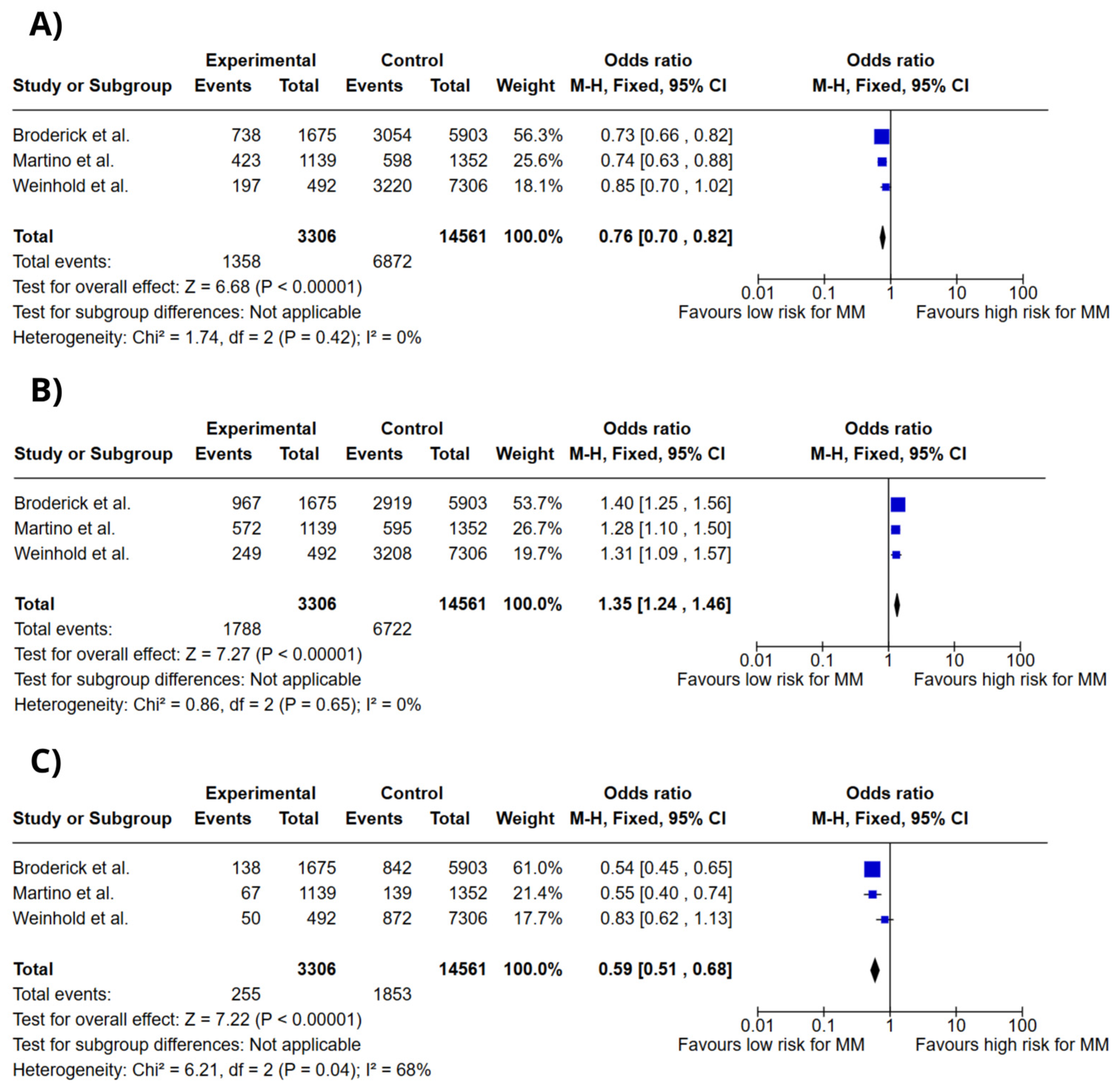
Figure 2 illustrates forest plots for the association between DNAH11 rs4487645 genotypes and multiple myeloma (MM) risk, analyzed under a codominant model. The genotypes assessed included C/C, A/C, and A/A. The meta-analysis revealed a significant association between the DNAH11 rs4487645 A/C genotype and increased risk of MM (OR = 1.35; 95% CI: 1.24–1.46; p < 0.00001; I2 = 0%). Conversely, the C/C genotype (OR = 0.76; 95% CI: 0.70–0.82; p < 0.00001; I2 = 0%) and the A/A genotype (OR = 0.59; 95% CI: 0.51–0.68; p < 0.00001; I2 = 68%) were associated with reduced risk of MM.
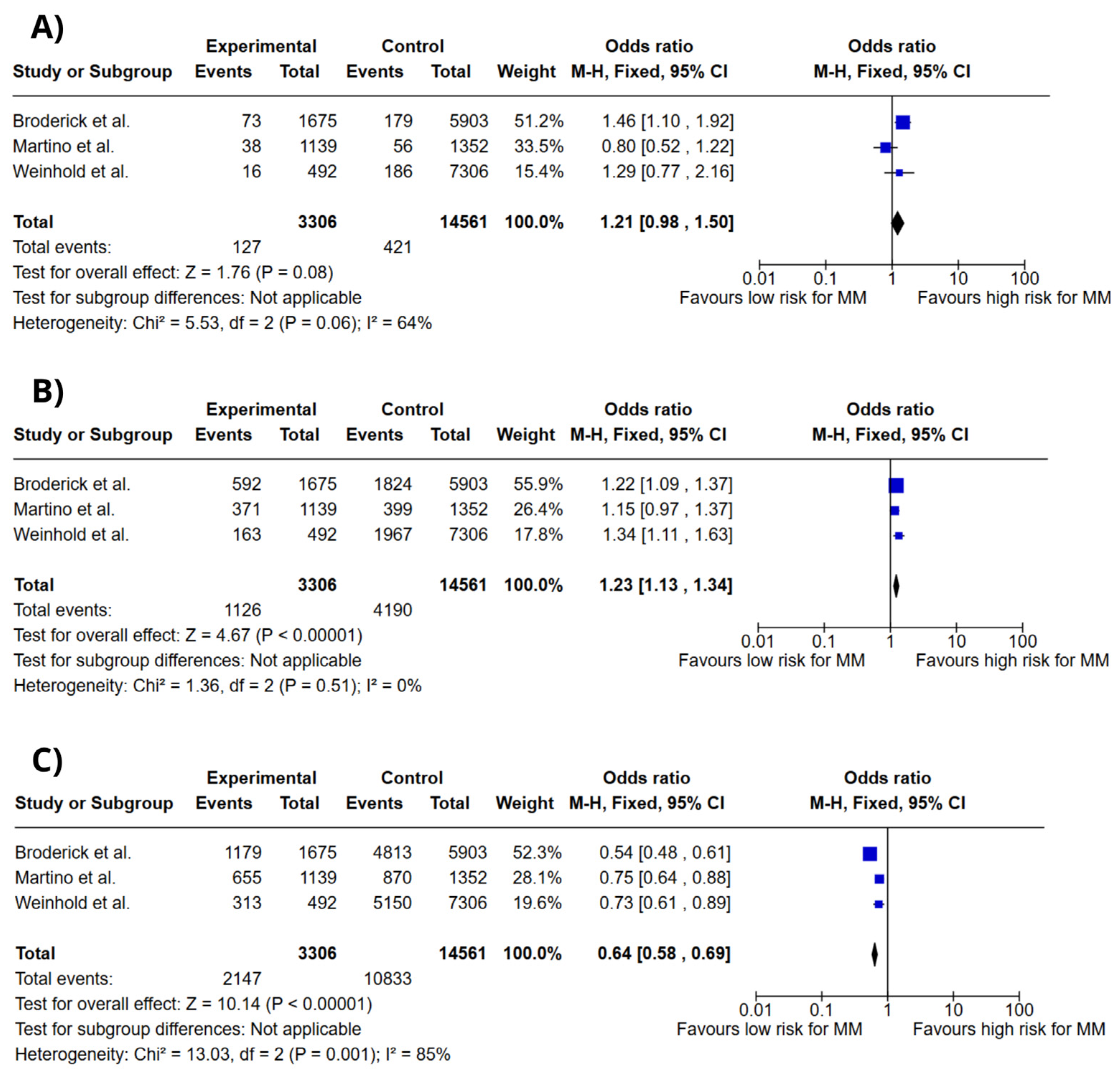
Figure 3 depicts forest plots for the association between ULK4 rs1052501 genotypes and MM risk, also analyzed in a codominant model. The genotypes evaluated were G/G, A/G, and A/A. The meta-analysis indicated that the G/G genotype (OR = 1.21; 95% CI: 0.98–1.50; p = 0.08; I2 = 64%) and the A/G genotype (OR = 1.23; 95% CI: 1.13–1.34; p < 0.00001; I2 = 0%) were associated with increased risk of MM. However, the A/A genotype was linked to a lower risk (OR = 0.64; 95% CI: 0.58–0.69; p < 0.00001; I2 = 85%).
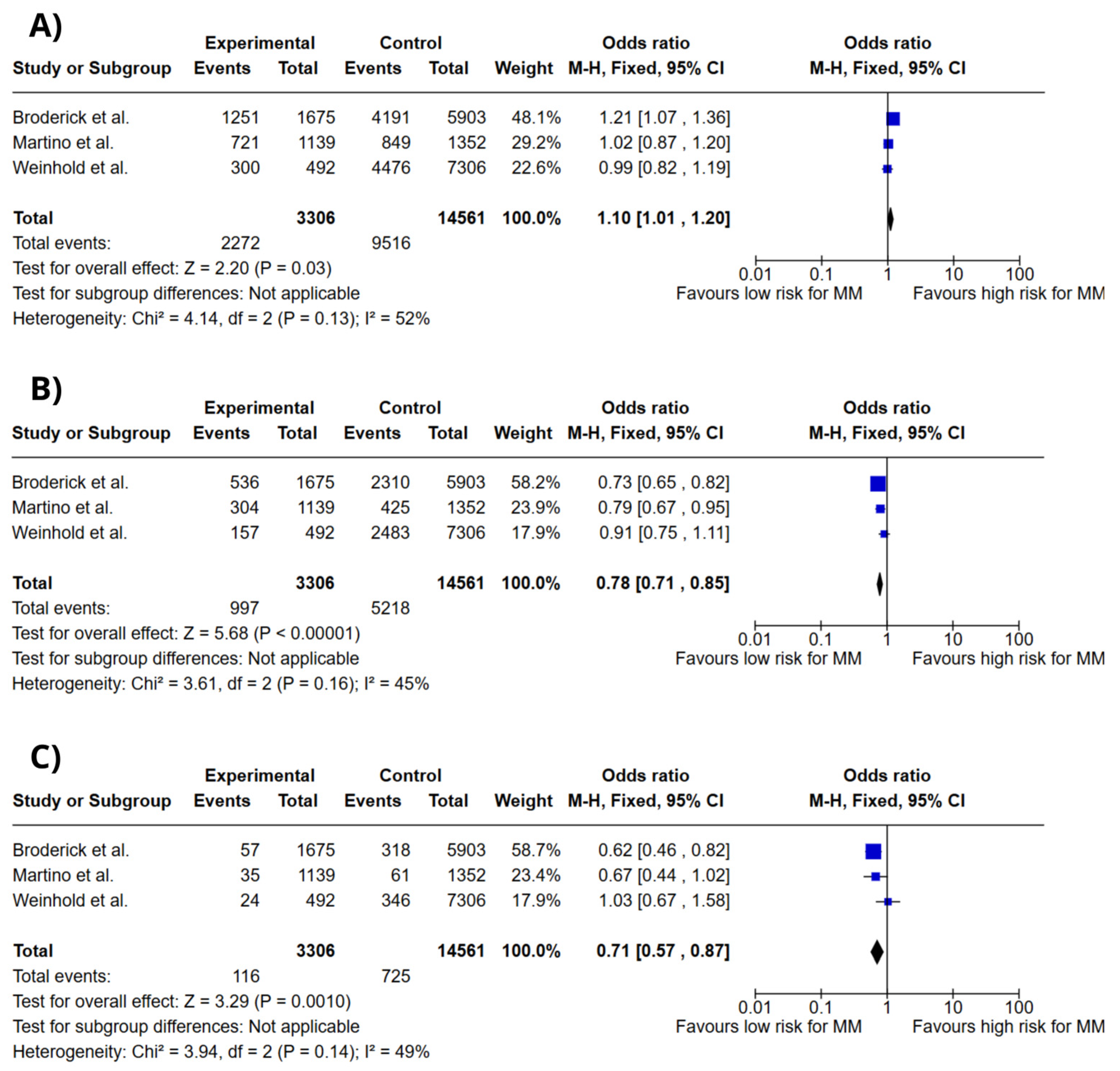
Figure 4 presents forest plots for the association between DTNB rs6746082 genotypes and MM risk under the codominant model. The genotypes analyzed were A/A, A/C, and C/C. The meta-analysis showed a modest association between the A/A genotype and increased MM risk (OR = 1.10; 95% CI: 1.01–1.20; p = 0.03; I2 = 45%). In contrast, the A/C genotype (OR = 0.78; 95% CI: 0.71–0.85; p < 0.00001; I2 = 45%) and the C/C genotype (OR = 0.71; 95% CI: 0.57–0.87; p = 0.001; I2 = 49%) were associated with reduced risk of MM.
Figure 5 illustrates forest plots for the association between VDR rs1544410 genotypes and MM risk under the codominant model. The genotypes assessed included A/A, A/G, and G/G. The A/G genotype was significantly associated with increased MM risk (OR = 1.87; 95% CI: 1.04–3.36; p = 0.04; I2 = 0%). On the other hand, the A/A genotype was associated with reduced risk (OR = 0.44; 95% CI: 0.21–0.92; p = 0.03; I2 = 66%), while the G/G genotype showed no significant association (OR = 0.99; 95% CI: 0.53–1.83; p = 0.97; I2 = 58%).

4. Discussion

This study represents the first systematic review and meta-analysis to correlate data from the literature with clinically significant genetic variants and genes associated with increased risk of MM. Building upon prior research, we identified genetic variants and candidate genes potentially linked to MM development. Moreover, we established correlations between these allelic variants and the global epidemiological patterns of the disease.
Our analyses revealed significant associations between five genetic variants and susceptibility to MM. Specifically, the DNAH11 gene variant rs4487645 (A/C genotype), the VDR gene variant rs1544410 (A/G genotype), and the DTNB gene variant rs6746082 (A/A genotype) were associated with a heightened risk of MM. Additionally, the ULK4 gene variant rs1052501 demonstrated a significant association, with its G/G and A/G genotypes linked to an increased risk of MM.
DNAH11 (dynein axonemal heavy chain 11) encodes a motor ATPase of the dynein heavy chain, a microtubule-dependent protein critical for cilia function [17,34]. This gene has been implicated in primary ciliary dyskinesia [35] and is associated with various malignancies, including esophageal carcinoma [36] and breast cancer [37], underscoring its role in cancer predisposition. The rs4487645 variant is located in intron 80 of the DNAH11 gene [13] within an 88 kb region of linkage disequilibrium (LD) that also encompasses the 3′ portion of the CDCA7L gene. CDCA7L interacts with the oncogene MYC, functioning as a binding partner of p75 and enhancing MYC’s transformational activity [38]. Given that MYC dysregulation is a hallmark of plasma cell neoplasms, CDCA7L emerges as a strong candidate for the functional basis of the rs4487645 association. Our meta-analysis confirmed that the DNAH11 rs4487645 A/C genotype is associated with increased MM risk.
ULK4 (Unc-51-like kinase 4) is located on chromosome 3p22.1 and plays a crucial role in regulating cellular processes such as neurogenesis and intracellular signaling [39]. While it lacks direct kinase activity, ULK4 belongs to the ULK family, which is involved in autophagy-related pathways [39]. It has been identified as a genetic modifier of holoprosencephaly, disrupting Shh signaling and Gli-luc reporter gene expression when knocked down [40]. Recent studies have linked the ULK4 rs1052501 variant with MM, suggesting a role in cell cycle regulation [13,41,42]. Our findings corroborate this association, identifying the rs1052501 G/G and A/G genotypes as risk factors for MM.
DTNB (dystrobrevin beta), located on chromosome 2, encodes a protein within the dystrophin-associated protein complex. This complex is critical for maintaining cellular structure and intracellular signaling, particularly in muscle tissues and the brain. DTNB is predominantly expressed in neurons of the cortex and hippocampus, where it contributes to early neuronal differentiation [43,44]. Dysfunctional DTNB expression has been linked to neurodegenerative conditions such as Alzheimer’s disease and schizophrenia [45]. Additionally, its association with immune cell infiltration highlights its potential role in cancer treatment and prognosis [46]. In our analysis, the DTNB rs6746082 A/A genotype was associated with increased MM risk, while the A/C and C/C genotypes appeared protective.
VDR (vitamin D receptor) plays a pivotal role in mediating the effects of vitamin D. VDR is a nuclear transcription factor that forms heterodimers with RXR (retinoid X receptor) isoforms to regulate gene expression [47]. Polymorphisms in VDR, such as rs1544410, may influence vitamin D metabolism and carcinogenesis. Low vitamin D levels, often observed in MM patients, have been linked to cancer progression and associated symptoms such as fatigue and musculoskeletal pain [48]. Our study identified a strong correlation between the VDR rs1544410 A/G genotype and increased MM risk, highlighting its relevance in disease susceptibility.
DNAH11 rs4487645 A/C genotype demonstrated a significant association with increased MM risk, potentially linked to its role in modifying MYC oncogene activity through its interaction with CDCA7L, a well-established MYC-binding partner. Similarly, ULK4 rs1052501 G/G and A/G genotypes were implicated in MM risk due to their involvement in cell cycle regulation and autophagy-related pathways. DTNB rs6746082 A/A genotype’s association with MM risk may stem from its role in maintaining cellular structure and modulating immune responses. Additionally, VDR rs1544410 A/G genotype underscores the impact of vitamin D metabolism on MM susceptibility.
Although the identified SNPs demonstrate biological plausibility across populations, their specific impact may differ due to variations in allele frequencies, linkage disequilibrium patterns, and interactions with environmental factors. Additional validation in underrepresented populations is essential to ensure the generalizability and applicability of these findings across diverse ethnicities. Collectively, these polymorphic variants hold promise as predictive biomarkers, offering valuable insights for risk stratification and paving the way for personalized therapeutic strategies in MM management.

5. Conclusions

The present study suggests that DNAH11 rs4487645 A/C genotype, ULK4 rs1052501 G/G genotype, ULK4 rs1052501 A/G genotype, DTNB rs6746082 A/A genotype, and VDR rs1544410 A/G genotype may serve as predictive biomarkers for MM risk. While this study provides valuable insights, some aspects warrant consideration. The focus on previously reported SNPs could overlook unexplored genetic variants, population heterogeneity might influence the generalizability of the findings, and the variants cannot be used to predict the clinical outcome of MM. Future research could address these aspects by conducting larger longitudinal studies, exploring additional genetic variants and SNP-environment interactions, and investigating the molecular mechanisms underlying the role of these SNPs in MM pathogenesis.

Supplementary Materials

The following supporting information can be downloaded at: https://www.mdpi.com/article/10.3390/ijms26073369/s1.

Author Contributions

Conceptualization, G.G.d.C.N. and F.C.A.d.M.; methodology, G.G.d.C.N. and F.C.A.d.M.; software, F.C.A.d.M.; validation, G.G.d.C.N., F.C.A.d.M. and R.R.A.; formal analysis, G.G.d.C.N.; investigation, G.G.d.C.N., F.C.A.d.M. and R.R.A.; resources, G.G.d.C.N. and N.P.C.d.S.; data curation, G.G.d.C.N. and F.C.A.d.M.; writing—original draft preparation, G.G.d.C.N., A.B.C.d.A., L.F.D.d.A.J., F.G.C. and M.V.S.H.; writing—review and editing, A.B.C.d.A., L.F.D.d.A.J., F.G.C. and M.V.S.H.; visualization, G.G.d.C.N.; supervision, N.P.C.d.S., M.R.F. and S.E.B.d.S.; project administration, N.P.C.d.S., M.R.F. and S.E.B.d.S. All authors have read and agreed to the published version of the manuscript.

Funding

This research was funded by the Conselho Nacional de Desenvolvimento Científico e Tecnológico (CNPq), the Coordenação de Aperfeiçoamento de Pessoal de Nível Superior (CAPES) and the Pró-Reitoria de Pesquisa e Pós-Graduação da UFPA (PROPESP).

Institutional Review Board Statement

Not applicable.

Informed Consent Statement

Not applicable.

Data Availability Statement

The original contributions presented in this study are included in the article/Supplementary Materials. Further inquiries can be directed to the corresponding author.

Acknowledgments

We thank the Federal University of Pará (UFPA); the Research Center in Oncology (NPO/UFPA). The design of the study, sample collection, data analysis, interpretation, and manuscript writing were conducted independently of any influence or involvement from the funding agencies.

Conflicts of Interest

The authors declare no conflicts of interest.

Abbreviations

The following abbreviations are used in this manuscript:
MMMultiple myeloma
SNPsSingle-nucleotide polymorphisms
NRNot reported

References

  1. Jurczyszyn, A.; Hutch, R.; Waszczuk-Gajda, A.; Suska, A.; Krzanowska, K.; Vesole, D.H. Monoclonal gammopathies of undetermined significance and smoldering myeloma. Acta Haematol. Pol. 2020, 51, 193–202. [Google Scholar] [CrossRef]
  2. International Agency for Research on Cancer. GLOBOCAN 2022: Estimated Cancer Incidence, Mortality and Prevalence Worldwide; IARCP: Lyon, France, 2022. [Google Scholar]
  3. Cowan, A.J.; Green, D.J.; Kwok, M.; Lee, S.; Coffey, D.G.; Holmberg, L.A.; Tuazon, S.; Gopal, A.K.; Libby, E.N. Diagnosis and management of multiple myeloma: A review. JAMA 2022, 327, 464–477. [Google Scholar] [CrossRef]
  4. Zhang, X.; Zhang, H.; Lan, H.; Wu, J.; Xiao, Y. CAR-T cell therapy in multiple myeloma: Current limitations and potential strategies. Front. Immunol. 2023, 14, 1101495. [Google Scholar] [CrossRef]
  5. Rajkumar, S.V. Multiple myeloma: 2024 update on diagnosis, risk-stratification, and management. Am. J. Hematol. 2024, 99, 1802–1824. [Google Scholar] [CrossRef] [PubMed]
  6. Karunarathna, I.; Gunawardana, K.; Aluthge, P.; Gunasena, P.; Gunathilake, S. Advances in Understanding and Managing Multiple Myeloma: A Comprehensive Review. Uva Clin. Med. 2024, 97, 1–15. [Google Scholar]
  7. Li, Z.; Zhao, H.; Li, Z.; He, Y. Correlation analysis of laboratory indicators, genetic abnormalities and staging in patients with newly diagnosed multiple myeloma. Medicine 2024, 103, e40710. [Google Scholar] [CrossRef] [PubMed]
  8. Page, M.J.; McKenzie, J.E.; Bossuyt, P.M.; Boutron, I.; Hoffmann, T.C.; Mulrow, C.D.; Shamseer, L.; Tetzlaff, J.M.; Akl, E.A.; Brennan, S.E.; et al. The PRISMA 2020 statement: An updated guideline for reporting systematic reviews. BMJ 2021, 372, n71. [Google Scholar] [CrossRef] [PubMed] [PubMed Central]
  9. Wells, G.A.; Shea, B.; O’Connell, D.; Peterson, J.; Welch, V.; Losos, M.; Tugwell, P. The Newcastle-Ottawa Scale (NOS) for Assessing the Quality of Nonrandomised Studies in Meta-Analyses; Ottawa Health Research Institute: Ottawa, ON, Canada, 2014. [Google Scholar]
  10. Little, J.; Higgins, J.; Ioannidis, J.; Moher, D.; Gagnon, F.; Vandenbroucke, J.; Zeggini, E. Strengthening the reporting of genetic association studies (STREGA)—An extension of the STROBE statement. Genet. Epidemiol. 2009, 33, 1061–1074. [Google Scholar] [CrossRef]
  11. 1000 Genomes Project Consortium. A Global Reference for Human Genetic Variation. Nature 2015, 526, 68–74. [Google Scholar] [CrossRef] [PubMed] [PubMed Central]
  12. Landrum, M.J.; Lee, J.M.; Benson, M.; Brown, G.R.; Chao, C.; Chitipiralla, S.; Gu, B.; Hart, J.; Hoffman, D.; Jang, W.; et al. ClinVar: Improving Access to Variant Interpretations and Supporting Evidence. Nucleic Acids Res. 2018, 46, D1062–D1067. [Google Scholar] [CrossRef] [PubMed] [PubMed Central]
  13. Broderick, P.; Chubb, D.; Johnson, D.C.; Weinhold, N.; Försti, A.; Lloyd, A.; Olver, B.; Ma, Y.; Dobbins, S.E.; Walker, B.A.; et al. Common variation at 3p22.1 and 7p15.3 influences multiple myeloma risk. Nat. Genet. 2011, 44, 58–61. [Google Scholar] [CrossRef] [PubMed] [PubMed Central]
  14. Rui, H.; Liu, Y.; Lin, M.; Zheng, X. Vitamin D receptor gene polymorphism is associated with multiple myeloma. J. Cell Biochem. 2019, 120, 10147–10153. [Google Scholar] [CrossRef]
  15. Kumar, R.; Himani Gupta, N.; Singh, V.; Kumar, V.; Haq, A.; Mirza, A.A.; Sharma, A. Unveiling molecular associations of polymorphic variants of VDR gene (FokI, BsmI and ApaI) in multiple myeloma patients of Indian population. J. Steroid Biochem. Mol. Biol. 2020, 199, 105588. [Google Scholar] [CrossRef] [PubMed]
  16. Martino, A.; Campa, D.; Jamroziak, K.; Reis, R.M.; Sainz, J.; Buda, G.; García-Sanz, R.; Lesueur, F.; Marques, H.; Moreno, V.; et al. Impact of polymorphic variation at 7p15.3, 3p22.1 and 2p23.3 loci on risk of multiple myeloma. Br. J. Haematol. 2012, 158, 805–809. [Google Scholar] [CrossRef] [PubMed]
  17. Weinhold, N.; Johnson, D.C.; Rawstron, A.C.; Försti, A.; Doughty, C.; Vijayakrishnan, J.; Broderick, P.; Dahir, N.B.; Begum, D.B.; Hosking, F.J.; et al. Inherited genetic susceptibility to monoclonal gammopathy of unknown significance. Blood 2014, 123, 2513–2517, quiz 2593. [Google Scholar] [CrossRef] [PubMed]
  18. Erickson, S.W.; Raj, V.R.; Stephens, O.W.; Dhakal, I.; Chavan, S.S.; Sanathkumar, N.; Coleman, E.A.; Lee, J.Y.; Goodwin, J.A.; Apewokin, S.; et al. Genome-wide scan identifies variant in 2q12.3 associated with risk for multiple myeloma. Blood 2014, 124, 2001–2003. [Google Scholar] [CrossRef] [PubMed]
  19. Hosgood III, H.D.; Baris, D.; Zhang, Y.; Zhu, Y.; Zheng, T.; Yeager, M.; Welch, R.; Zahm, S.; Chanock, S.; Rothman, N.; et al. Caspase polymorphisms and genetic susceptibility to multiple myeloma. Hematol. Oncol. 2008, 26, 148–151. [Google Scholar] [CrossRef] [PubMed]
  20. Karabon, L.; Pawlak-Adamska, E.; Tomkiewicz, A.; Jedynak, A.; Kielbinski, M.; Woszczyk, D.; Potoczek, S.; Jonkisz, A.; Kuliczkowski, K.; Frydecka, I. Variations in suppressor molecule CTLA-4 gene are related to susceptibility to multiple myeloma in a Polish population. Pathol. Oncol. Res. 2012, 18, 219–226. [Google Scholar] [CrossRef] [PubMed]
  21. Du, J.; Huo, J.; Shi, J.; Yuan, Z.; Zhang, C.; Fu, W.; Jiang, H.; Yi, Q.; Hou, J. Polymorphisms of nuclear factor-κB family genes are associated with development of multiple myeloma and treatment outcome in patients receiving bortezomib-based regimens. Haematologica 2011, 96, 729–737. [Google Scholar] [CrossRef] [PubMed]
  22. Campa, D.; Martino, A.; Sainz, J.; Buda, G.; Jamroziak, K.; Weinhold, N.; Reis, R.M.V.; García-Sanz, R.; Jurado, M.; Ríos, R.; et al. Comprehensive investigation of genetic variation in the 8q24 region and multiple myeloma risk in the IMMEnSE consortium. Br. J. Haematol. 2012, 156, 507–514. [Google Scholar] [CrossRef] [PubMed]
  23. Chubb, D.; Weinhold, N.; Broderick, P.; Chen, B.; Johnson, D.C.; Försti, A.; Vijayakrishnan, J.; Migliorini, G.; Dobbins, S.E.; Holroyd, A.; et al. Common variation at 3q26.2, 6p21.33, 17p11.2, and 22q13.1 influences multiple myeloma risk. Nat. Genet. 2013, 45, 1221–1225. [Google Scholar] [CrossRef] [PubMed]
  24. Martino, A.; Campa, D.; Jurczyszyn, A.; Martínez-López, J.; Moreno, M.J.; Varkonyi, J.; Dumontet, C.; García-Sanz, R.; Gemignani, F.; Jamroziak, K.; et al. Genetic variants and multiple myeloma risk: IMMEnSE validation of the best reported associations—An extensive replication of the associations from the candidate gene era. Cancer Epidemiol. Biomarkers Prev. 2014, 23, 670–674. [Google Scholar] [CrossRef] [PubMed]
  25. Faber, E.W.; Lourenço, G.J.; Ortega, M.M.; Lorand-Metze, I.; De Souza, C.A.; Lima, C.S.P. Polymorphisms of VEGF, GSTM1 and GSTT1 genes in multiple myeloma risk. Hematol. Oncol. 2011, 29, 217–223. [Google Scholar] [CrossRef] [PubMed]
  26. Vangsted, A.J.; Nielsen, K.R.; Klausen, T.W.; Haukaas, E.; Tjønneland, A.; Vogel, U. A functional polymorphism in the promoter region of the IL1B gene is associated with risk of multiple myeloma. Hematol. Oncol. 2012, 30, 179–183. [Google Scholar] [CrossRef] [PubMed]
  27. Scionti, F.; Agapito, G.; Caracciolo, D.; Riillo, C.; Grillone, K.; Cannataro, M.; Di Martino, M.T.; Tagliaferri, P.; Tassone, P.; Arbitrio, M. Risk Alleles for Multiple Myeloma Susceptibility in ADME Genes. Cells 2022, 11, 189. [Google Scholar] [CrossRef] [PubMed] [PubMed Central]
  28. Wang, Q.; Wang, Y.; Wu, J.; Xie, X.; Qin, H.; Huang, C.; Li, Z.; Ling, Z.; Li, R. Association between BCL2 interacting protein 3 like (BNIP3L) genetic polymorphisms and the risk of multiple myeloma in China. Hematology 2024, 29, 2367918. [Google Scholar] [CrossRef] [PubMed]
  29. Niebudek, K.; Balcerczak, E.; Mirowski, M.; Żebrowska, M. Association of ABCB1 T-129C polymorphism and multiple myeloma risk in Polish population. Pol. J. Pathol. 2018, 69, 405–409. [Google Scholar] [CrossRef] [PubMed]
  30. Peng, M.; Zhao, G.; Yang, F.; Cheng, G.; Huang, J.; Qin, X.; Liu, Y.; Wang, Q.; Li, Y.; Qin, D. NCOA1 is a novel susceptibility gene for multiple myeloma in the Chinese population: A case-control study. PLoS ONE 2017, 12, e0173298. [Google Scholar] [CrossRef]
  31. Li, B.; Liu, C.; Cheng, G.; Peng, M.; Qin, X.; Liu, Y.; Li, Y.; Qin, D. LRP1B Polymorphisms Are Associated with Multiple Myeloma Risk in a Chinese Han Population. J. Cancer 2019, 10, 577–582. [Google Scholar] [CrossRef]
  32. Szemraj-Rogucka, Z.; Szemraj, J.; Grzybowska-Izydorczyk, O.; Robak, T.; Jamroziak, K. CD38 Gene Polymorphisms and Genetic Predisposition to Multiple Myeloma. Acta Haematol. Pol. 2013, 44, 58–62. [Google Scholar] [CrossRef]
  33. Brito, A.B.C.; Lourenço, G.J.; Oliveira, G.B.; De Souza, C.A.; Vassallo, J.; Lima, C.S.P. Associations of VEGF and VEGFR2 Polymorphisms with Increased Risk and Aggressiveness of Multiple Myeloma. Ann. Hematol. 2014, 93, 1363–1369. [Google Scholar] [CrossRef] [PubMed]
  34. Sodeifian, F.; Samieefar, N.; Shahkarami, S.; Rayzan, E.; Seyedpour, S.; Rohlfs, M.; Klein, C.; Babaie, D.; Rezaei, N. DNAH11 and a Novel Genetic Variant Associated with Situs Inversus: A Case Report and Review of the Literature. Case Rep. Med. 2023, 2023, 8436715. [Google Scholar] [CrossRef] [PubMed] [PubMed Central]
  35. Schultz, R.; Elenius, V.; Lukkarinen, H.; Saarela, T. Two novel mutations in the DNAH11 gene in primary ciliary dyskinesia (CILD7) with considerable variety in the clinical and beating cilia phenotype. BMC Med. Genet. 2020, 2, 237. [Google Scholar] [CrossRef] [PubMed]
  36. Gao, J.; Wu, Y.; Yu, J.; Qiu, Y.; Yi, T.; Luo, C.; Zhang, J.; Lu, G.; Li, X.; Xiong, F.; et al. Impact of genomic and epigenomic alterations of multigene on a multicancer pedigree. Cancer Med. 2024, 13, e7394. [Google Scholar] [CrossRef] [PubMed] [PubMed Central]
  37. Verma, S.; Bakshi, D.; Sharma, V.; Sharma, I.; Shah, R.; Bhat, A.; Bhat, G.R.; Sharma, B.; Wakhloo, A.; Kaul, S.; et al. Genetic variants of DNAH11 and LRFN2 genes and their association with ovarian and breast cancer. Int. J. Gynaecol. Obstet. 2020, 148, 118–122. [Google Scholar] [CrossRef] [PubMed]
  38. Taddesse-Heath, L.; Meloni-Ehrig, A.; Scheerle, J.; Kelly, J.C.; Jaffe, E.S. Plasmablastic lymphoma with MYC translocation: Evidence for a common pathway in the generation of plasmablastic features. Mod. Pathol. 2010, 23, 991–999. [Google Scholar] [CrossRef]
  39. Luo, S.; Zheng, N.; Lang, B. ULK4 in Neurodevelopmental and Neuropsychiatric Disorders. Front. Cell Dev. Biol. 2022, 10, 873706. [Google Scholar] [CrossRef] [PubMed] [PubMed Central]
  40. Mecklenburg, N.; Kowalczyk, I.; Witte, F.; Görne, J.; Laier, A.; Mamo, T.M.; Gonschior, H.; Lehmann, M.; Richter, M.; Sporbert, A.; et al. Identification of disease-relevant modulators of the SHH pathway in the developing brain. Development 2021, 148, dev199307. [Google Scholar] [CrossRef] [PubMed]
  41. Greenberg, A.J.; Lee, A.M.; McDonnell, S.K.; Cerhan, J.R.; Liebow, M.; Larson, D.R.; Colby, C.L.; Norman, A.D.; Kyle, R.A.; Kumar, S.; et al. Single-nucleotide polymorphism rs1052501 associated with monoclonal gammopathy of undetermined significance and multiple myeloma. Leukemia 2013, 27, 515–516. [Google Scholar] [CrossRef]
  42. Went, M.; Kinnersley, B.; Sud, A.; Johnson, D.C.; Weinhold, N.; Försti, A.; van Duin, M.; Orlando, G.; Mitchell, J.S.; Kuiper, R.; et al. Transcriptome-wide association study of multiple myeloma identifies candidate susceptibility genes. Hum. Genom. 2019, 13, 37. [Google Scholar] [CrossRef] [PubMed] [PubMed Central]
  43. Blake, D.J.; Nawrotzki, R.; Loh, N.Y.; Górecki, D.C.; Davies, K.E. beta-dystrobrevin, a member of the dystrophin-related protein family. Proc. Natl. Acad. Sci. USA 1998, 95, 241–246. [Google Scholar] [CrossRef] [PubMed] [PubMed Central]
  44. Quaranta, M.T.; Spinello, I.; Paolillo, R.; Macchia, G.; Boe, A.; Ceccarini, M.; Labbaye, C.; Macioce, P. Identification of β-Dystrobrevin as a Direct Target of miR-143: Involvement in Early Stages of Neural Differentiation. PLoS ONE 2016, 11, e0156325. [Google Scholar] [CrossRef] [PubMed] [PubMed Central]
  45. Sun, J.; Li, Y.; Bie, B.; Tian, H.; Li, J.; Yang, L.; Zhou, Z.; Mu, Y.; Li, Z. Dystrobrevin beta is a promising prognostic biomarker and therapeutic target for hepatocellular carcinoma. Am. J. Transl. Res. 2024, 16, 6072–6096. [Google Scholar] [CrossRef] [PubMed] [PubMed Central]
  46. Lyu, C.; Yin, X.; Li, Z.; Wang, T.; Xu, R. Vitamin D receptor gene polymorphisms and multiple myeloma: A meta-analysis. Clin. Exp. Med. 2024, 24, 118. [Google Scholar] [CrossRef] [PubMed] [PubMed Central]
  47. Bouillon, R.; Carmeliet, G.; Verlinden, L.; van Etten, E.; Verstuyf, A.; Luderer, H.F.; Lieben, L.; Mathieu, C.; Demay, M. Vitamin D and human health: Lessons from vitamin D receptor null mice. Endocr. Rev. 2008, 29, 726–776. [Google Scholar] [CrossRef] [PubMed] [PubMed Central]
  48. Ismail, N.H.; Mussa, A.; Al-Khreisat, M.J.; Mohamed Yusoff, S.; Husin, A.; Johan, M.F.; Islam, M.A. The Global Prevalence of Vitamin D Deficiency and Insufficiency in Patients with Multiple Myeloma: A Systematic Review and Meta-Analysis. Nutrients 2023, 15, 3227. [Google Scholar] [CrossRef] [PubMed] [PubMed Central]
Figure 1. Study selection flowchart through literature search.
Ijms 26 03369 g001
Figure 2. Forest plots of association between DNAH11 gene polymorphisms and MM risk. (A) DNAH11 rs4487645 C/C genotype. (B) DNAH11 rs4487645 A/C genotype. (C) DNAH11 rs4487645 A/A genotype. Colors do not convey additional information and do not influence data interpretation. Broderick et al. [13], Martino et al. [16], Weinhold et al. [17].
Ijms 26 03369 g002
Figure 3. Forest plots of association between ULK4 gene polymorphisms and MM risk. (A) ULK4 rs1052501 G/G genotype. (B) ULK4 rs1052501 A/G genotype. (C) ULK4 rs1052501 A/A genotype. Colors do not convey additional information and do not influence data interpretation. Broderick et al. [13], Martino et al. [16], Weinhold et al. [17].
Ijms 26 03369 g003
Figure 4. Forest plots of association between DTNB gene polymorphisms and MM risk. (A) DTNB rs6746082 A/A genotype. (B) DTNB rs6746082 A/C genotype. (C) DTNB rs6746082 C/C genotype. Colors do not convey additional information and do not influence data interpretation. Broderick et al. [13], Martino et al. [16], Weinhold et al. [17].
Ijms 26 03369 g004
Figure 5. Forest plots of association between VDR gene polymorphisms and MM risk. (A) VDR rs1544410 A/A genotype. (B) VDR rs1544410 A/G genotype. (C) VDR rs1544410 G/G genotype. Colors do not convey additional information and do not influence data interpretation. Hongbing Rui et al. [14], Kumar et al. [15].
Ijms 26 03369 g005
Table 1. Characteristics of included studies and participants.
Authors, YearCountryParticipants (Male/Female)Mean AgeMean Age at Diagnosis (Years)Genotyping MethodMM Histologically ConfirmedEthnicityMain Results
Broderick et al., 2011 [13]United Kingdom
Germany
TOTAL
1371 (819/552)
384 (229/155)
1675 (1048/627)
NR
NR
NR
64.1 ± 10.3
54.5 ± 8.0
NR
MicroarrayYesEuropeanSNP rs1052501within gene ULK4 and SNP rs4487645 were associated with higher risk of MM
Martino et al., 2012 [16]Germany1139NRNRPCRYesEuropeanSNPs rs4487645 and rs6746082 were linked to higher risk of MM
Erickson et al., 2014 [18]United States of America120257.761.0PCRNREuropean/North AmericanThe SNPs rs12614346 and rs73486634 were linked to an increased risk of MM.
Hosgood III et al., 2008 [19]United States of America128 (0/128)NRNRqPCRYesNorth AmericanCASP3 and CASP9 polymorphisms were associated with decreased risk of MM.
Karabon et al., 2012 [20]Poland200 (94/106)67 ± 10.963.5 ± 11.2PCR-RFLPYesDanishThree CTLA variations were identified more frequently in MM patients compared to the control group. Five CTLA polymorphisms were associated with higher risk of MM.
Juan Du et al., 2011 [21]China252 (161/91)58NRRT-PCRNRChineseTRAF3 rs12147254 variant is linked to a reduced risk of developing multiple myeloma, while the rs11160707 genotype has been correlated with improved progression-free survival.
Campa et al., 2011 [22]German1188 (643/545)58.62 ± 10.15NRcastPCRYesEuropeanRs2456449 was correlated with the risk of multiple myeloma.
Chubb et al., 2013 [23]German/United KingdomUK-replication-1—812 (412/400)
UK-replication-2—396 (181/215)
German-replication—1149 (676/473)
NR
NR
NR
NR
66.0
57.6
PCR KASParYesEuropeanThe variants rs10936599, rs2285803, rs4273077, and rs877529 were correlated with the risk of multiple myeloma.
Martino et al., 2014 [24]Italy/Poland/Spain/France/Portugal/Hungary/Denmark1498 (756/742)60.9 ± 10.6NRcastPCRYesEuropeanA possible association between the SNP rs2227667 (SERPINE1) and the risk of multiple myeloma in women has been identified.
Faber et al., 2011 [25]Brazil150 (81/69)54NRmPCR/ RT-PCRNRWhite people/African-BraziliansAn increased risk of MM was observed in individuals with the VEGF CC genotype combined with GSTM1 undeleted and GSTT1 null genotypes.
Vangsted et al., 2012 [26] Denmark348NRNRABI 7500 or HT7900 systemsNRDanishAssociation between IL1B expression and risk of MM.
Scionti et al., 2022 [27]Italy64NRNRDMET Console software version 1.1NRItalianPolymorphism in ADME genes were associated with susceptibility for MM.
Wang et al., 2024 [28]China94 (49/45)59.25 ± 9.36NRUE Blood genomic DNA preparation kitNRChinesePolymorphisms in BNIP3L were associated with susceptibility and prognosis of MM in chinese population.
Hongbing Rui et al., 2019 [14]China40 (23/17)59.5NRPCRNRChineseAssociation between polymorphisms in VDR gene with increased risk of MM.
Niebudek et al., 2018 [29]Poland91 (41/50)63NRPCR-RFLPNRPolishPolymorphism T-129C in ABCB1 gene was not associated with the increased risk of MM development in the polish population.
Kumar et al., 2020 [15]India75 (54/21)57NRlocus-specific PCRNRIndianFokI, ApaI, and BsmI genotypes were associated with risk of
MM in the Indian population.
Peng et al., 2017 [30]China827 (473/354)59.35 ± 9.95over 60 yearslocus-specific PCRyesChinesers79480871 were associated with susceptibility for MM.
Li et al., 2019 [31]China739 (415/324)59.27 ± 10.11older than 65 yearslocus-specific PCRYesChineseAssociation for susceptibility in MM by rs61070260 in LRP1B.
Szemraj-Rogucka, 2013 [32]Poland174 (93/81)6168PCR-RFLPNRCaucasianPredisposition of MM were associated for rs6449182 of CD38.
Brito et al., 2014 [33]Brazil192 (99/93)62over 60 yearsRT-PCRNRCaucasian and Non-CaucasianAggressiveness and risk of MM. were associated with VEGF and VEGFR2.
Weinhold et al., 2014 [17]United Kingdom/Germany492 (234/258)67.5NRKompetitive Allele Specific Polymerase (KASP) chain reactionYesEuropeanrs1052501, rs2285803, rs4487645, and rs4273077 increased MM risk.
Table 2. Investigated single-nucleotide polymorphisms.
SNPsGeneLocationAllelesAncestralFunctional ConsequenceClinical Significance
rs1544410VDRChromosome 12:47846052C/A/G/TCIntron variantBenign, likely risk allele
rs6746082DTNBChromosome 2:25436375A/C/G/TAIntron variantNR
rs1052501ULK4Chromosome 3:41883906C/G/TTMissense variantBenign
rs4487645DNAH11Chromosome 7:21898622C/A/TCIntron variantNR
Table 3. Methodological quality of the studies based on the Newcastle–Ottawa Scale (NOS).
StudiesSelectionItem 2Item 3Item 4ComparabilityItem 1bExposureItem 2Item 3Total Score
Item 1Item 1aItem 1
Broderick et al., 2011 [13]***** ** 7
Hongbing Rui et al., 2019 [14]***** ** 7
Kumar et al., 2020 [15]***** ** 7
Martino et al., 2012 [16]***** ** 7
Weinhold et al., 2014 [17]***** ** 7
* Selection—Item 1: Is the case definition adequate?; Item 2: Representativeness of the cases; Item 3: Selection of controls; Item 4: Definition of controls. Comparability—Item 1a and 1b: Comparability of cases and controls based on the design or analysis. Exposure—Item 1: Ascertainment of exposure; Item 2: Same method of ascertainment for cases and controls; Item 3: Non-response rate.
Table 4. The quality of reporting using the Strengthening the Reporting of Genetic Association (STREGA) guideline.
StudiesDescription of Genotyping Methods and ErrorsDescription of Modeling Population StratificationDescription of Modeling Haplotype VariationHardy–Weinberg Equilibrium Was ConsideredStatement of Whether the Study Is the First Report of a Genetic Association, a Replication Effort, or BothScore
Genotyping Methods and PlatformsError Rates and Call RatesGenotyping in BatchesLaboratory/Center Where Genotyping Was PerformedThe Number of Individuals Successful in Genotyping
Broderick et al., 2011 [13]YesYesYesYesYesYesNoYesNo7
Hongbing Rui et al., 2019 [14]YesYesNoYesYesYesYesYesNo7
Kumar et al., 2020 [15]YesYesYesYesYesYesNoYesNo7
Martino et al., 2012 [16]YesYesYesYesYesYesNoYesYes8
Weinhold et al., 2014 [17]YesYesYesYesYesYesNoYesNo7
Disclaimer/Publisher’s Note: The statements, opinions and data contained in all publications are solely those of the individual author(s) and contributor(s) and not of MDPI and/or the editor(s). MDPI and/or the editor(s) disclaim responsibility for any injury to people or property resulting from any ideas, methods, instructions or products referred to in the content.