
MiR-21-5p and miR-223-3p as Treatment Response Biomarkers in Pediatric Eosinophilic Esophagitis

 , , , ,
Abstract
1. Introduction
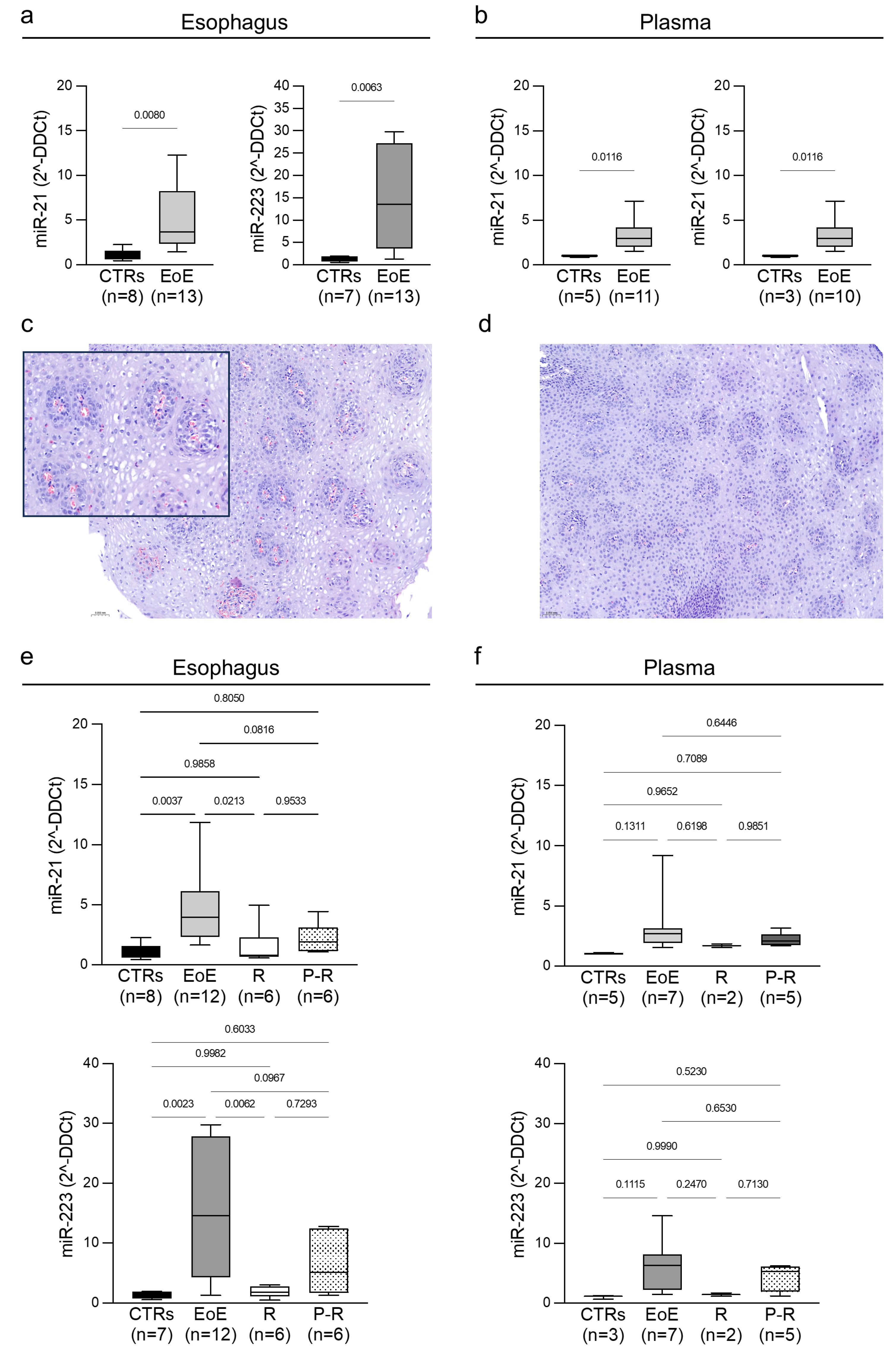
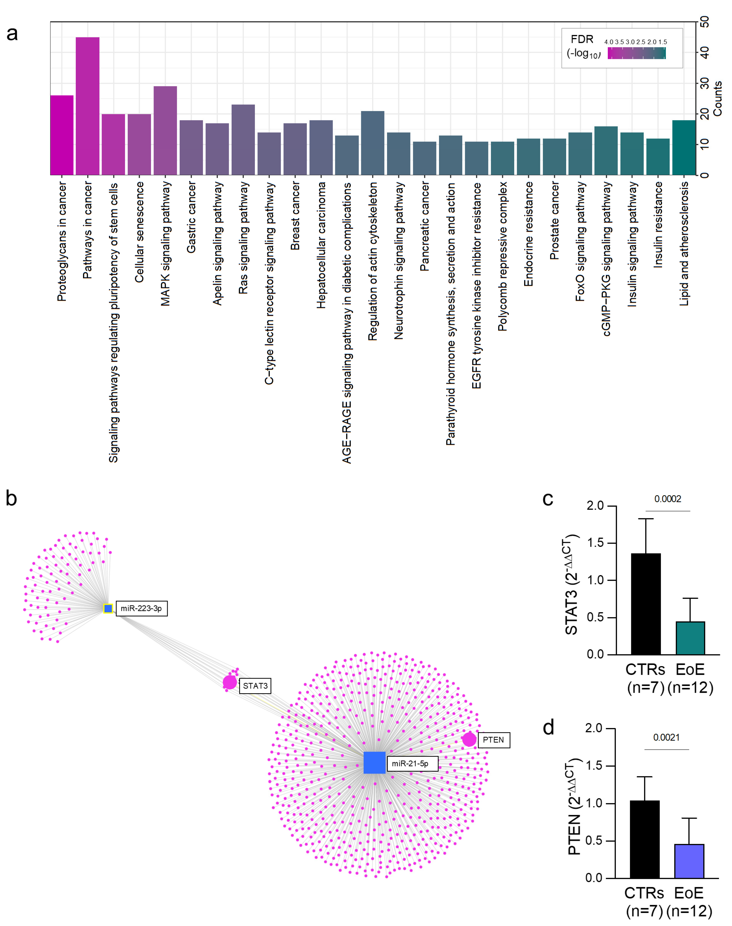
2. Results
3. Discussion
4. Materials and Methods
4.1. Study Design, Patients, and Data Collection
4.2. RNA Isolation and Quantitative Real-Time Polymerase Chain Reaction (qRT-PCR)
4.3. Bioinformatic Analysis
4.4. Histological Analysis
4.5. Statistical Analysis
Author Contributions
Funding
Institutional Review Board Statement
Informed Consent Statement
Data Availability Statement
Acknowledgments
Conflicts of Interest
Abbreviations
| CCS | corticosteroids |
| EoE | eosinophilic esophagitis |
| FDR | false discovery rate |
| H/E | hematoxylin/eosin |
| HPF | high-power field |
| miRNA | MicroRNA |
| PPI | proton pump inhibitors |
| qRT-PCR | quantitative real-time polymerase chain reaction |
| KEGG | Kyoto Encyclopedia of Genes and Genomes |
| David | Database for Annotation, Visualization, and Integrated Discovery |
References
- Furuta, G.T.; Katzka, D.A. Eosinophilic Esophagitis. N. Engl. J. Med. 2015, 373, 1640–1648. [Google Scholar] [PubMed]
- Spergel, J.M.; Brown-Whitehorn, T.A.; Muir, A.; Liacouras, C.A. Medical algorithm: Diagnosis and treatment of eosinophilic esophagitis in children. Allergy 2020, 75, 1522–1524. [Google Scholar] [PubMed]
- Dellon, E.S.; Hirano, I. Epidemiology and Natural History of Eosinophilic Esophagitis. Gastroenterology 2018, 154, 319–332.e3. [Google Scholar]
- Gupta, S.K. Noninvasive markers of eosinophilic esophagitis. Gastrointest. Endosc. Clin. N. Am. 2008, 18, 157–167. [Google Scholar] [PubMed]
- Ho, P.T.B.; Clark, I.M.; Le, L.T.T. MicroRNA-Based Diagnosis and Therapy. Int. J. Mol. Sci. 2022, 23, 7167. [Google Scholar] [CrossRef]
- Tarallo, A.; Carissimo, A.; Gatto, F.; Nusco, E.; Toscano, A.; Musumeci, O.; Coletta, M.; Karali, M.; Acampora, E.; Damiano, C.; et al. microRNAs as biomarkers in Pompe disease. Genet. Med. 2019, 21, 591–600. [Google Scholar] [CrossRef]
- Bartel, D.P. MicroRNAs: Genomics, biogenesis, mechanism, and function. Cell 2004, 116, 281–297. [Google Scholar]
- Allegra, A.; Alonci, A.; Campo, S.; Penna, G.; Petrungaro, A.; Gerace, D.; Musolino, C. Circulating microRNAs: New biomarkers in diagnosis, prognosis and treatment of cancer (review). Int. J. Oncol. 2012, 41, 1897–1912. [Google Scholar]
- Markey, G.E.; Donohoe, C.L.; McNamee, E.N.; Masterson, J.C. MicroRNA dysregulation and therapeutic opportunities in esophageal diseases. Am. J. Physiol. Gastrointest. Liver Physiol. 2023, 325, G1–G13. [Google Scholar]
- Jhaveri, P.B.; Lambert, K.A.; Bogale, K.; Lehman, E.; Alexander, C.; Ishmael, F.; Jhaveri, P.N.; Hicks, S.D. Salivary microRNAs in pediatric eosinophilic esophagitis. Allergy Asthma Proc. 2023, 44, 145–152. [Google Scholar]
- Votto, M.; Strisciuglio, C.; Indolfi, C.; Marseglia, G.L.; Miraglia Del Giudice, M.; Licari, A. Correspondence to “Salivary Immunoinflammatory Proteins Identify Children with Eosinophilic Esophagitis”. Allergy 2024, 79, 3176–3177. [Google Scholar] [PubMed]
- Zahm, A.M.; Menard-Katcher, C.; Benitez, A.J.; Tsoucas, D.M.; Le Guen, C.L.; Hand, N.J.; Friedman, J.R. Pediatric eosinophilic esophagitis is associated with changes in esophageal microRNAs. Am. J. Physiol. Gastrointest. Liver Physiol. 2014, 307, G803–G812. [Google Scholar]
- Cañas, J.A.; Tabares, A.; Barbero, C.; García-Sánchez, D.; Sastre, B.; Rodrigo-Muñoz, J.M.; Mahíllo-Fernández, I.; Rayo, A.; Borrell, B.; Cilleruelo, M.L.; et al. Proton-pump Inhibitor Response Prediction Using Esophageal microRNAs in Children With Eosinophilic Esophagitis. J. Pediatr. Gastroenterol. Nutr. 2020, 71, 755–763. [Google Scholar]
- Lu, T.X.; Sherrill, J.D.; Wen, T.; Plassard, A.J.; Besse, J.A.; Abonia, J.P.; Franciosi, J.P.; Putnam, P.E.; Eby, M.; Martin, L.J.; et al. MicroRNA signature in patients with eosinophilic esophagitis, reversibility with glucocorticoids, and assessment as disease biomarkers. J. Allergy Clin. Immunol. 2012, 129, 1064–1075.e9. [Google Scholar]
- Zou, K.; Dong, H.; Li, M.; Zhang, Y.; Zhang, K.; Song, D.; Chu, C. Comprehensive analysis of transcriptome-wide N6-methyladenosine methylomes in the Barrett’s esophagus in rats. Genomics 2023, 115, 110687. [Google Scholar] [PubMed]
- Dellon, E.S.; Liacouras, C.A.; Molina-Infante, J.; Furuta, G.T.; Spergel, J.M.; Zevit, N.; Spechler, S.J.; Attwood, S.E.; Straumann, A.; Aceves, S.S.; et al. Updated International Consensus Diagnostic Criteria for Eosinophilic Esophagitis: Proceedings of the AGREE Conference. Gastroenterology 2018, 155, 1022–1033.e10. [Google Scholar] [PubMed]
- Wu, Y.; Song, Y.; Xiong, Y.; Wang, X.; Xu, K.; Han, B.; Bai, Y.; Li, L.; Zhang, Y.; Zhou, L. MicroRNA-21 (Mir-21) Promotes Cell Growth and Invasion by Repressing Tumor Suppressor PTEN in Colorectal Cancer. Cell. Physiol. Biochem. 2017, 43, 945–958. [Google Scholar]
- Li, Z.; Dong, X.; Wang, Z.; Liu, W.; Deng, N.; Ding, Y.; Tang, L.; Hla, T.; Zeng, R.; Li, L.; et al. Regulation of PTEN by Rho small GTPases. Nat. Cell Biol. 2005, 7, 399–404. [Google Scholar]
- Yang, X.O.; Panopoulos, A.D.; Nurieva, R.; Chang, S.H.; Wang, D.; Watowich, S.S.; Dong, C. STAT3 regulates cytokine-mediated generation of inflammatory helper T cells. J. Biol. Chem. 2007, 282, 9358–9363. [Google Scholar] [CrossRef]
- Wang, J.; Shen, Y.; Li, C.; Liu, C.; Wang, Z.H.; Li, Y.S.; Ke, X.; Hu, G.H. IL-37 attenuates allergic process via STAT6/STAT3 pathways in murine allergic rhinitis. Int. Immunopharmacol. 2019, 69, 27–33. [Google Scholar] [CrossRef]
- Meng, F.; Henson, R.; Wehbe-Janek, H.; Ghoshal, K.; Jacob, S.T.; Patel, T. MicroRNA-21 regulates expression of the PTEN tumor suppressor gene in human hepatocellular cancer. Gastroenterology 2007, 133, 647–658. [Google Scholar] [PubMed]
- Chang, L.; Zhou, G.; Soufan, O.; Xia, J. miRNet 2.0: Network-based visual analytics for miRNA functional analysis and systems biology. Nucleic Acids Res. 2020, 48, W244–W251. [Google Scholar] [PubMed]
- Hasan, S.H.; Taylor, S.; Garg, S.; Buras, M.R.; Doyle, A.D.; Bauer, C.S.; Wright, B.L.; Schroeder, S. Diagnosis of Pediatric Non-Esophageal Eosinophilic Gastrointestinal Disorders by Eosinophil Peroxidase Immunohistochemistry. Pediatr. Dev. Pathol. 2021, 24, 513–522. [Google Scholar] [PubMed]

| EOE Patients n = 13 | |
|---|---|
| Sex males, n (%) | 8 (61) |
| Age years, median ± SD | 13 ± 4.9 |
| History of atopy, n (%) | 9 (69) |
| 3 (27.1) |
| 5 (38.4) |
| 3 (27.1) |
| Comorbidities (%) | 5 (38.5) |
| 3 (23) |
| 4 (30.8) |
| EoE treatment, n (%) | |
| 4 (30.7) |
| 7 (53.8) |
| 2 (15.3) |
| Eos/HPF (mean) | |
| T0 biopsy | 33.75 |
| T1 biopsy | 13.23 |
| EREFS (mean) | |
| T0 | 2.07 |
| T1 | 1.53 |
| Esophageal miR-223-3p | Estimate | p Value |
|---|---|---|
| (Intercept) | −24.595264 | 0.2176 |
| EC-T0_Blood | 0.011237 | 0.2752 |
| EC-T0_Esophagus | 1.015288 | 0.0318 * |
| Allergy | 6.610019 | 0.4009 |
| EREFS-T0 | −8.176028 | 0.07 |
| CD | 11.834543 | 0.272 |
| DM1 | 7.98814 | 0.3675 |
Disclaimer/Publisher’s Note: The statements, opinions and data contained in all publications are solely those of the individual author(s) and contributor(s) and not of MDPI and/or the editor(s). MDPI and/or the editor(s) disclaim responsibility for any injury to people or property resulting from any ideas, methods, instructions or products referred to in the content. |
© 2025 by the authors. Licensee MDPI, Basel, Switzerland. This article is an open access article distributed under the terms and conditions of the Creative Commons Attribution (CC BY) license (https://creativecommons.org/licenses/by/4.0/).
Share and Cite
Tarallo, A.; Casertano, M.; Valanzano, A.; Cenni, S.; Creoli, M.; Russo, G.; Damiano, C.; Carissimo, A.; Cioce, A.; Martinelli, M.; et al. MiR-21-5p and miR-223-3p as Treatment Response Biomarkers in Pediatric Eosinophilic Esophagitis. Int. J. Mol. Sci. 2025, 26, 3111. https://doi.org/10.3390/ijms26073111
Tarallo A, Casertano M, Valanzano A, Cenni S, Creoli M, Russo G, Damiano C, Carissimo A, Cioce A, Martinelli M, et al. MiR-21-5p and miR-223-3p as Treatment Response Biomarkers in Pediatric Eosinophilic Esophagitis. International Journal of Molecular Sciences. 2025; 26(7):3111. https://doi.org/10.3390/ijms26073111
Chicago/Turabian StyleTarallo, Antonietta, Marianna Casertano, Anna Valanzano, Sabrina Cenni, Mara Creoli, Giuseppina Russo, Carla Damiano, Annamaria Carissimo, Alessandro Cioce, Massimo Martinelli, and et al. 2025. "MiR-21-5p and miR-223-3p as Treatment Response Biomarkers in Pediatric Eosinophilic Esophagitis" International Journal of Molecular Sciences 26, no. 7: 3111. https://doi.org/10.3390/ijms26073111
APA StyleTarallo, A., Casertano, M., Valanzano, A., Cenni, S., Creoli, M., Russo, G., Damiano, C., Carissimo, A., Cioce, A., Martinelli, M., Miele, E., Staiano, A., Iafusco, D., Parenti, G., & Strisciuglio, C. (2025). MiR-21-5p and miR-223-3p as Treatment Response Biomarkers in Pediatric Eosinophilic Esophagitis. International Journal of Molecular Sciences, 26(7), 3111. https://doi.org/10.3390/ijms26073111