Article

Development and Evaluation of a New Measles Detection Assay Using Real-Time RT-PCR

by
Vera A. Chayeb
1,
Anna S. Dolgova
1,*,
Margarita R. Popova
1,
Nina V. Zheleznova
1,
Svetlana A. Shirobokova
1,
Anna V. Shabalina
1,
Alena A. Sharova
1,
Anna S. Gladkikh
1,
Anastasia Yu. Antipova
1,
Anastasiia D. Kirichenko
1,
Edward S. Ramsay
1 and
Vladimir G. Dedkov
1,2
1
Saint-Petersburg Pasteur Institute, Federal Service on Consumers’ Rights Protection and Human Well-Being Surveillance, 197101 Saint-Petersburg, Russia
2
Martsinovsky Institute of Medical Parasitology, Tropical and Vector Borne Diseases, First Moscow State Medical University (Sechenov University), 119048 Moscow, Russia
*
Author to whom correspondence should be addressed.
Int. J. Mol. Sci. 2025, 26(5), 1801; https://doi.org/10.3390/ijms26051801
Submission received: 11 January 2025 / Revised: 9 February 2025 / Accepted: 15 February 2025 / Published: 20 February 2025
(This article belongs to the Section Molecular Pathology, Diagnostics, and Therapeutics)

Abstract

:
The severity of MeV infection has been greatly reduced by the development of a live attenuated vaccine, which has been incorporated into vaccination programs in many countries. However, poor access to health facilities, and above all, the increase in anti-vaccination movements, has prevented the achievement of sufficient vaccination coverage. In outbreak scenarios, a rapid and transportable method can improve differential diagnosis, including removing ambiguity in suspected measles cases, contacts, or a cohort. In response to the need, we have developed a new RT-qPCR-based MeV detection assay. The LOD of the developed assay was determined on different PCR machines and the higher threshold was 1–1.2 103 copies/mL. The joint diagnostic sensitivity of ELISA and RT-PCR (used together) was 100%, and used combinedly, these two methods enable detection of all measles-infected persons, which is extremely important for controlling contagion and spread of infection. During the clinical validation of the assay on 200 clinical samples from measles-suspected cases using ELISA, 157 samples showed a positive result, while 163 positive cases were confirmed by the RT-qPCR assay. The concordance between the two techniques was 93%. According to our results, the real-time RT-PCR approach used in our study is more sensitive and appears to be a more promising method for measles diagnosis during early stages of the disease, likely before the rise of specific IgM antibodies detected by ELISA.

1. Introduction

Measles is an infectious and contagious disease caused by the measles virus, an airborne pathogen spread by inhalation of respiratory droplets. Clinical presentation of the disease is characterized by a prodrome of fever, conjunctivitis, rhinorrhea, and cough, followed by a morbilliform rash. From the middle of 20th century, vast vaccination campaigns were initiated globally, leading to a false hope of definitive measles eradication. Over the last two decades, incoming reports are indicating a progressive reemergence of measles, with multiple outbreaks registered across the globe [1] in countries with a high population index [2], with the omission bias playing a crucial a role in the increase of disease resurgence. In Western countries with a high Human Development Index, regional measles spread is related to vaccine hesitancy and under-immunized groups, including migrants from countries with poor access to health facilities [3,4,5], a fact highlighted by the WHO [6] and the European Center for Disease Prevention and Control [7]. In 2021, the WHO reported an estimated global mortality of 128,000 cases, essentially among unvaccinated or under-vaccinated children [8]. Despite the notion of measles as an already eradicated pathology, recent reports are indicating a progressive reemergence of the disease globally.
Measles virus (MeV) belongs to the Morbillivirus genus in the Paramyxoviridae family. A single-stranded, negative-sense RNA virus, MeV uses a multitude of penetration mechanisms to ensure infection of the cell using different antigenic recognition mechanisms and immune escape strategies [9].
The main risk factors, reported by a significant number of studies, are: a lack or an incompleteness of vaccination; underage for routine vaccination; migration of unvaccinated individuals; and recent contact with a measles case [10,11,12,13]. Malnutrition was also established as an essential contributor to measles infectivity in low-income countries [14]. Among comorbid factors, HIV infection was reported to increase measles susceptibility among children, but not adults [15,16].
Measles can lead to complications such as pneumonia, otitis media, thrombocytopenia, and encephalitis. In cases of malnourishment, the disease may lead to blindness, deafness, or encephalitis-associated intellectual disabilities (acute disseminated encephalomyelitis, subacute sclerosing panencephalitis) [17,18].
To overcome these epidemiological challenges, multiple laboratory detection methods were implemented during recent measles outbreaks across the globe. Currently, the mainstream WHO-recommended measles diagnostic method is ELISA-based immunoglobulin M (IgM) detection [19]. However, the IgM test detects MeV only in symptomatic individuals with late-onset symptoms, such as rash. A study investigating a measles outbreak highlighted that serum specimens collected within 4 days of initial symptoms onset were IgM-negative. However, they were simultaneously MeV RNA-positive in 82.1% of cases when analyzed by RT-qPCR [20]. The RT-PCR method allows a more accurate MeV viral load assessment in a given specimen during initiation of the infectious process and appearance of initial symptoms (fever, coryza, conjunctivitis) [21]. Earlier-diagnosed cases afford timely anti-epidemic measures and restriction of local transmission.
RNA-dependent RNA polymerase (RdRp) is an enzyme involved in replication of the viral genome, wherein RNA is synthesized from an RNA template. The enzyme and its products are key determinants of viral function and survival. Besides being a proprietary target in anti-measles therapeutics, the RdRp gene is also a prime target in measles molecular detection strategies. RT-qPCR targeting the RdRp gene can be useful during outbreak scenarios because it allows rapid detection of MeV-infected persons, increasing the number of secondary transmission cases. This method could be implemented in both stationary and mobile laboratories, particularly in low-resource settings. Such diagnostics can improve differential diagnosis, including removing ambiguity in suspected measles cases, contacts, or a cohort.
This paper describes: the development of a new MeV detection assay; clinical validation of the findings in a sample of MeV-infected patients; and evaluation of the clinical and diagnostic efficiency of several approaches. These are offered in an effort to overcome outbreak management challenges, including improvement of surveillance measures at national and international levels.

2. Results

2.1. MV AmpPS Assay Design

The multiple alignment of the MeV genotype B3 and D8 sequences available in GenBank NCBI (n = 1265) performed at the beginning of the study (Figure 1) allowed the identification of fairly conserved regions required for the designing of the MeV-specific primers and respective probes (Table 3). Based on these data, oligonucleotide primers and fluorescent probes were designed and synthesized, and the MV AmpPS assay was developed. The developed assay includes all components required for RT-PCR. The advantage of this assay is that it allows the verification of all steps of the analysis, including extraction, reverse transcription, and PCR. In addition, by using NEC− and C− controls, the risk of false-positive results because of cross-contamination was minimized.
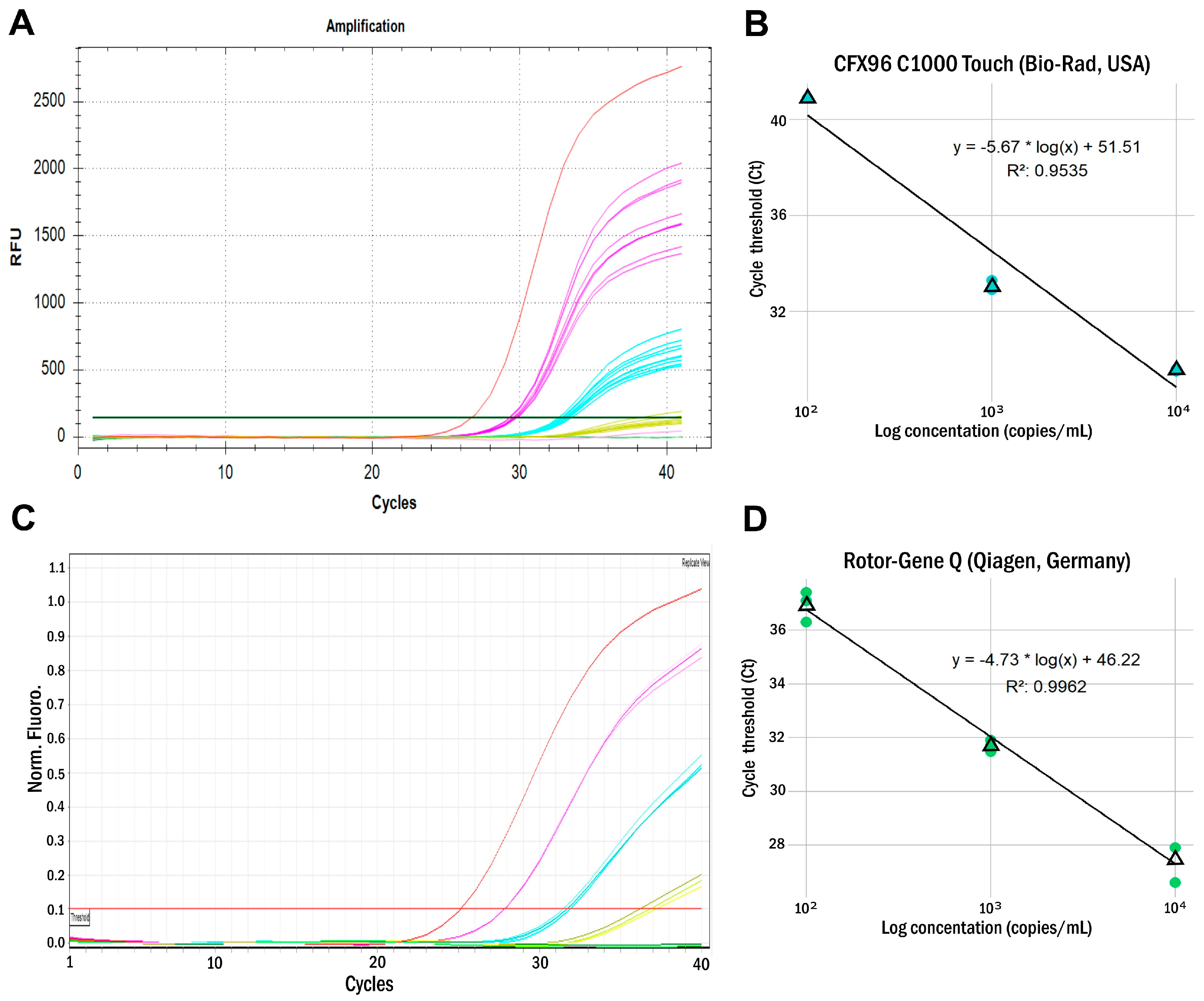
The limit of detection (LOD) was assessed using ARC dilutions on two PCR machines featuring flatbed (Bio-Rad CFX96 C1000 Touch, USA) or rotary (Rotor-Gene Q, Qiagen, Germany) designs (Figure 2). The LOD of the assay on the CFX96 PCR plate machine, measured as the minimum dilution found in all replicates, was 103 RNA copies per mL, while the LOD* measured using Probit analysis was 1.2 × 10³ RNA copies/mL. For the Rotor-Gene Q PCR rotary machine, the measured minimum dilution found in all replicates was above 102 RNA copies/mL, while the LOD* measured using probit analysis was 2.7 × 10² RNA copies/mL (Table 1).
The potential for cross-reactivity was assessed using heterologous RNA/DNA from 18 viral species belonging to 10 viral families. None of the 18 different viral species showed a positive reaction on the MeV real-time RT-PCR assay (Table 5). Consequently, the evaluated analytical specificity was 100%.

2.2. Clinical Evaluation of the MV AmpPS Assay

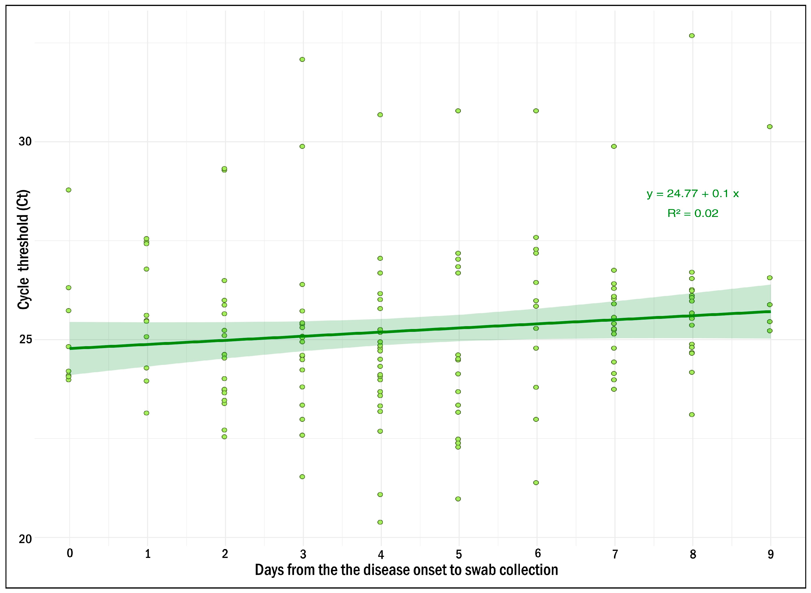
The clinical samples (nasopharyngeal swabs, plasma) from suspected measles cases were simultaneously tested using the MV AmpPS RT-PCR assay (nasopharyngeal swabs, n = 200) and the VectoMeasles-IgM ELISA kit (Vector-Best®, Russia) (plasma, n = 200). The findings were: 163/200 (81.5%) matched positive (Ct values ranging from 20.4 to 32.7) (Figure 3); and 33/200 (16.5%) matched negative using both RT-PCR and ELISA methods. In addition, 13/200 (6.5%) discordant samples were observed: four ELISA-positive cases with negative RT-PCR; nine ELISA-negative cases with positive RT-PCR (Ct values ranging from 22.4 to 29.9); and one ELISA-uncertain case with positive RT-PCR (Ct = 26.8) (Table S1).
The discordant samples were retested using the single-target RT-PCR assay design [22] used at the Centers for Disease Control and Prevention (CDC, Atlanta, GA, USA) and elsewhere for MeV surveillance and case confirmation (CDC assay) with modifications related to recently observed priming mismatch variants [23]. Complete coincidence of the testing results by both real-time RT-PCR assays was observed.
In silico analysis of primers and probe sequences showed an absence of significant substitutions in the target regions of the MeV genotype B3 and D8 sequences available in NCBI GenBank (Figure 1). Thus, the MV AmpPS RT-PCR assay was found to be suitable for measles diagnostics.

3. Discussion

According to WHO reports, 40 million children globally did not receive routine measles vaccinations in 2021 due to the COVID-19 pandemic: 25 million missed the first dose, and 14.7 million missed the second [24,25]. Historically, the National Program for Measles Elimination in Russia (established in 2003) has greatly contributed to the eradication of the infection. However, the importation of cases originating from neighboring countries remains a problem to be solved [26].
The lifting of restrictions related to the COVID-19 pandemic and the resumption of migratory flows towards Russia have resulted in an increase in the number of imported measles cases from Central Asia. This has led to a major measles outbreak since the end of 2022, forcing a prompt reaction from health authorities to contain it. Abramov et al. reported that significant migratory flows and insufficient vaccination against measles (both in Russia and abroad) create a threat that measles virus can be imported and spread across Russia, leading to persistence nationally [27].
Traditionally, the WHO-recommended gold standard for measles diagnostics has been detection of measles-specific serum IgM antibody, mainly using the ELISA approach. With the advent of the RT-PCR methodology, and due to its cost-effectiveness, real-time RT-PCR is now a WHO newly recommended method for importation tracking and rapid diagnostics, especially in terms of commercial kits [19].
In the context of the current epidemiological situation, the Saint Petersburg Pasteur Institute has developed a new real-time RT-PCR assay for MeV detection: MV AmpPS. A summary of the developed assay is shown in Table 2.
Conditions for the real-time RT-PCR assay were initially optimized using purified RNA from modified MS2 phage particles and RNA/DNA from 18 viral species belonging to 10 viral families. For the selected primers and probe, suitable conditions for PCR with real-time detection were determined. Studies have shown that this approach can effectively differentiate MeV from closely related viruses.
The LOD of the developed assay varied on different devices. Regarding the device with a higher threshold, it was 1–1.2 × 10³ copies/mL (15–20 copies/reaction), which is a good indicator for diagnostic kits in the RT-PCR format with real-time detection [28,29].
Evaluation of the assay using clinical samples from measles-suspected cases showed 93% concordance between ELISA and the developed real-time RT-PCR assay. Specifically, 186 of 200 clinical samples showed the same result when tested by RT-PCR and ELISA. The obtained diagnostic sensitivity for ELISA (average) was 94.0% (95% CI: 89.26% to 97.09%). For the real-time RT-PCR assay, it was 97.6% (95% CI: 93.98% to 99.34%). These data are comparable with those described earlier [30]. Four swabs from patients with ELISA-confirmed measles were tested as negative by the assay developed, as well as by the CDC assay. This suggests that a negative test result is not a consequence of mismatches in the target region of MeV genome (the assays target different fragments of the genome). The collection date for these swabs ranged from day 1 to day 9 from disease onset. As shown in Figure 3 and Table S1, the viral load, depending on the day of the disease onset, is quite variable from sample to sample and could reach Ct values up to 24 from day 1 to the day 9 from disease onset. Therefore, the negative test result is probably not related to the virus mutations or the absence of the virus in the patient’s nasopharynx during swab collection, but to the bias during material collection or RNA extraction. Nevertheless, dependence of the viral load on the day of the disease is important for understanding in what time frame the use of real time RT-PCR is the optimal one. This issue has yet to be resolved.
Thus, the real-time RT-PCR approach is more sensitive and appears to be a more promising method for measles diagnosis. This may be the case particularly in the early stages of the disease, likely before the rise of specific IgM antibodies. Meanwhile, the joint diagnostic sensitivity of ELISA and RT-PCR (used together) was 100%. Therefore, usage of both techniques (ELISA and real-time RT-PCR) can enable detection of all measles-infected persons, which is extremely important for controlling contagion and the spread of infection. At the initial stages of the disease and in the prodromal period, the use of the RT-PCR method is more appropriate. Moreover, the method can be used not only in suspected measles cases but also in persons who have been in contact with them.
Ct values of positive swab samples allow us to conclude that the viral load in measles can vary significantly. Also, we can talk about a trend towards a decrease in viral load of swab samples over time (Figure 3). However, the viral load in the samples collected on days 7–10 from disease onset remains at a level that allows confident detection of MeV RNA by the assay developed. Thus, the optimal time frame for swabs collection could be defined in the range from 0 up to 10 days from the disease onset.

4. Methods and Materials

4.1. Development of the MV AmpPS Assay

4.1.1. Identification of Conserved Sites

Complete sequences of MeV genotype B3 and D8 (as of December, 2023) in GenBank (NCBI, n = 1265) were downloaded. Among these, 544 complete RdRp sequences (without gaps or ambiguities of reading) were aligned to identify conserved sites. The only measles genotypes that had been reported since 2020 were B3 and D8, indicating that they were the prevalent genotypes worldwide [23].
Alignment was performed using the BioEdit 7.2.5 software package (Ibis Biosciences, Carlsbad, CA, USA). A 106-nucleotide fragment of the RdRp gene was selected as a target for amplification using PLOTCON (http://emboss.bioinformatics.nl/cgi-bin/emboss/plotcon, accessed on the 3 December 2023). The target corresponded to nucleotide positions 15404–15299 in the MeV reference sequence: strain MVs/Moscow.RUS/20.23/2[B3] (GenBank accession number OR290098).
The primers and probe were designed in accordance with guidelines regarding TaqMan primers and probes for RT-PCR techniques [31,32]. Primer melting temperatures were calculated using an oligonucleotide property calculator (https://bsu.bio/molbiol/oligocalc.html) [33]. In addition, an oligonucleotide property calculator and MFOLD were used to assess the thermodynamic characteristics of the probes and the probability of the appearance of secondary structures (https://www.unafold.org/mfold/applications/dna-folding-form.php, accessed on the 3 December 2023) [34]. Two nucleotides «CA» were added to the 3′ end of the MV-Prb probe sequence to form a hairpin. The rhodamine 6G (R6G) fluorescent reporter dye was covalently attached to the MeV-specific probe at the 5′ end. The black hole quencher (BHQ1) was attached at the 3′ end. The primers and probe were synthesized by Genterra PLC (Moscow, Russia). The primer and probe sequences are shown in Table 3.

4.1.2. Positive and Internal Control Preparation

The assay uses several types of control samples. The control sample set is similar to that already described [35,36], and includes internal extraction control (IC), armored RNA control (ARC), negative extraction control (NEC), and PCR controls (C+, C−). Briefly, the internal extraction control and armored RNA control are modified MS2 phage particles that include either an artificial sequence (internal extraction control, IC) or a sequence of the virus to be detected (armored RNA control, ARC). Extraction control samples are added to each sample during the nucleic acid extraction step. The positive PCR control (C+) contains a mixture of plasmids containing the same regions. The negative PCR control (C−) and negative extraction control (NEC) contain ultrapure water (milli Q, Merck KGaA, Darmstadt, Germany). A 177-nucleotide MeV target fragment was synthesized de novo by a previously described two-step PCR method [37]. Phusion high-fidelity DNA polymerase (NEB, Ipswich, MA, USA) was used with the primers listed (Table 4) to create cDNA and RNA controls.

4.1.3. Reaction Mixture and Amplification Conditions

Reactions were carried out in a volume of 25 μL. Optimized PCR reaction conditions were as follows: 1 µL of BioMaster Mix (Biolabmix, Novosibirsk, Russia); 12.5 µL of 2× reaction buffer (Biolabmix, Novosibirsk, Russia); 10.5 pmol of each MeV specific primer (MV-Fw, MV-Rev); 7.5 pmol of MV-probe; 10.5 pmol of internal control primers; and 7.5 pmol of internal control probe. The sample quantity used was 10 μL, and reactions were brought to 25 µL with ultrapure water (milli Q, Merck Millipore, Merck KGaA, Darmstadt, Germany). The optimized amplification regimen was the following: 50 °C 15 min; 95 °C 5 min; followed by 40 cycles (95 °C 10 s and 60 °C 30 s). Fluorescence was recorded during the 60 °C step in the HEX/yellow channel (for MeV) and FAM/green channel (for IC). The sequences of primers and probes targeting IC have been described previously [35]. The reaction was performed using the CFX96 C1000 Touch (Bio-Rad, Hercules, CA, USA) and Rotor-Gene Q (Qiagen, Hilden, Germany) PCR machines.

4.2. Limit of Detection

The limit of detection (LOD) was determined using a series of 10-fold dilutions of armored RNA particles (ARC, same as positive control for reverse transcription) at known concentrations. Concentrations were measured by droplet digital PCR (ddPCR). The concentrations used to determine the detection limit were 104, 103, 102, and 10 copies of shielded RNA particles per milliliter (200, 20, 2, and 0.2 copies/reaction, respectively). Samples from each dilution (100 µL) were extracted in triplicate using the RIBO-prep kit (AmpliSens®, Moscow, Russia) according to the manufacturer’s instructions (elution volume 60 µL), followed by testing in the developed MV AmpPS RT-PCR assay. The LOD was set as the minimum dilution found in all replicates. Alternatively, we estimated LOD* using probit analysis.

4.3. Analytical Specificity

Analytical specificity was tested on a panel of viral RNA/DNA samples (solutions) representing heterologous viral strains obtained from the collection of the Saint Petersburg Pasteur Institute. Viral loads of the samples were assessed using specific real-time RT-PCR (Table 5, Figure S1).

4.4. Clinical Evaluation of the MV AmpPS Assay

4.4.1. Sample Collection

Nasopharyngeal swabs and blood samples were collected from those suspected to have measles (N = 200), specifically those admitted to hospitals in Saint Petersburg or the Leningrad Region from 3 May to 20 September 2024. Patient ages ranged from four months to 63 years. Women accounted for 46.5% and men for 53.5%.
Blood samples were collected 3–5 days after rash manifestation, while nasopharyngeal swabs were collected 1–3 days after rash manifestation (Table S1, Supplementary Materials).
Epidemiological and clinical information for each patient was collected using a standardized form. To ensure the confidentiality of personal patient data, each clinical record was assigned an individual number, which was subsequently used to label the tubes containing samples for molecular–biological and serological studies. Samples were delivered to the Saint Petersburg Pasteur Institute and kept for further studies.
Nasopharyngeal swabs were collected in 500 µL of special transport medium or phosphate-buffered saline (pH 7.0). Blood samples were taken into vacutainers with EDTA and processed by centrifugation (3000× g for 5 min). Plasma was separated from cellular elements and transferred into plastic tubes. All samples were stored at −80 °C until further analysis.

4.4.2. Enzyme-Linked Immunosorbent Assay

The presence of specific IgM antibodies against the MeV antigen was determined using the VectoMeasles-IgM ELISA kit (Vector-Best®, Novosibirsk, Russia) according to the manufacturer’s instructions.

4.4.3. RNA Extraction

Total nucleic acids from nasopharyngeal swabs were extracted and purified using the RIBO-prep kit (AmpliSens®, Moscow, Russia) according to the manufacturer’s instructions (elution volume 100 µL) and stored at −80 °C until molecular analysis.

5. Conclusions

This manuscript reports the development and evaluation of the MV AmpPS real-time RT-PCR assay for measles virus detection targeting an RdRp gene fragment. The assay contains all of the necessary components to perform the analysis, including internal extraction control (IC), positive reverse-transcription control (ARC), negative control of extraction (NCE), positive PCR control (C+), and negative PCR control (C−). The advantage of this assay is that it allows the verification of all steps of the analysis, including extraction, reverse transcription, and PCR.
Our results revealed that the LOD of the assay is in the range of 1–1.2 × 10³ RNA copies/mL on the CFX96 PCR plate machine. On the Rotor-Gene Q PCR rotary machine, the LOD is in the range of 1—2.7 × 10² RNA copies/mL (10–27 RNA copies/reaction). The LOD of the assay described is comparable with previously described MeV RT-PCR assays (22 copies/reaction and 10 copies/reaction, respectively) [22,23] as well as with RT-RPA CRISPR/Cas12a (120 copies/reaction) and RT-RPA-CRISPR-LFD assays (6.25 copies/reaction) [30]. Thus, this assay can be effectively used for detection of the reemergent B3 and D8 genotype strains [22,38,39] in the course of diagnostics and epidemiological surveillance of measles.

Supplementary Materials

The following supporting information can be downloaded at https://www.mdpi.com/article/10.3390/ijms26051801/s1.

Author Contributions

Conceptualization, A.S.D. and V.G.D.; data curation, A.S.G. and A.Y.A.; formal analysis, V.A.C., A.A.S. and A.D.K.; investigation, V.A.C., M.R.P., N.V.Z., S.A.S. and A.V.S.; methodology, A.S.D. and A.S.G.; project administration, V.G.D.; resources, A.Y.A.; supervision, V.G.D.; validation, A.V.S.; visualization, A.S.D. and A.D.K.; writing—original draft, V.A.C.; writing—review and editing, A.S.D., E.S.R. and V.G.D. All authors have read and agreed to the published version of the manuscript.

Funding

This research was funded by the Russian federal governmental program Sanitary Shield.

Institutional Review Board Statement

The study was conducted according to the guidelines of the Declaration of Helsinki and approved by the Institutional Review Board of the Saint Petersburg Pasteur Institute (St. Petersburg, Russia, No 063-04).

Informed Consent Statement

Not applicable.

Data Availability Statement

The original contributions presented in the study are included in the article. Further inquiries can be directed to the corresponding author.

Conflicts of Interest

The authors declare no conflicts of interest.

References

  1. Kuba, Y.; Kyan, H.; Iha, Y.; Kato, T.; Oyama, M.; Miyahira, M.; Kakita, T.; Takara, T.; Yamauchi, M.; Kamiya, H.; et al. Emergent Measles-Containing Vaccination Recommendation for Aged 6–11 Months and Detection of Vaccine-Associated Measles during a Large Measles Outbreak in Okinawa, Japan, in 2018. Vaccine 2020, 38, 2361–2367. [Google Scholar] [CrossRef]
  2. Qin, S.; Ding, Y.; Yan, R.; He, H. Measles in Zhejiang, China, 2004–2017: Population Density and Proportion of Floating Populations Effects on Measles Epidemic. Health Secur. 2019, 17, 193–199. [Google Scholar] [CrossRef] [PubMed]
  3. Kremer, J.R.; Brown, K.E.; Jin, L.; Santibanez, S.; Shulga, S.V.; Aboudy, Y.; Demchyshyna, I.V.; Djemileva, S.; Echevarría, J.E.; Featherstone, D.; et al. High Genetic Diversity of Measles Virus, World Health Organization European Region, 2005–2006. Emerg. Infect. Dis. 2008, 14, 107–114. [Google Scholar] [CrossRef]
  4. Gogoi, M.; Martin, C.A.; Bird, P.W.; Wiselka, M.J.; Gardener, J.; Ellis, K.; Renals, V.; Lewszuk, A.J.; Hargreaves, S.; Pareek, M. Risk of vaccine preventable diseases in UK migrants: A serosurvey and concordance analysis. J. Migr. Health 2024, 9, 100217. [Google Scholar] [CrossRef]
  5. Knights, F.; Carter, J.; Deal, A.; Crawshaw, A.; Bouaddi, O.; Sanchez-Clemente, N.; Seedat, F.; Vanderslott, S.; Eagan, R.; Holt, D.E.; et al. Strengthening life-course immunisation in migrant populations: Access, equity, and inclusion. Lancet Reg. Health Eur. 2024, 41, 100806. [Google Scholar] [CrossRef] [PubMed]
  6. World Health Organization. Available online: https://www.who.int/publications/i/item/measles-and-rubella-strategic-framework-2021-2030 (accessed on 1 February 2025).
  7. European Center for Disease Prevention and Control. Available online: https://www.ecdc.europa.eu/sites/default/files/documents/measles-eu-threat-assessment-brief-february-2024.pdf (accessed on 1 February 2025).
  8. World Health Organization. Available online: https://www.who.int/news/item/23-11-2022-nearly-40-million-children-are-dangerously-susceptible-to-growing-measles-threat (accessed on 30 January 2025).
  9. Benecke, O.; DeYoung, S.E. Anti-Vaccine Decision-Making and Measles Resurgence in the United States. Glob. Pediatr. Health 2019, 6, 2333794X1986294. [Google Scholar] [CrossRef]
  10. Yaméogo, K.R.; Perry, R.T.; Yaméogo, A.; Kambiré, C.; Kondé, M.K.; Nshimirimana, D.; Kezaala, R.; Hersh, B.S.; Cairns, K.L.; Strebel, P. Migration as a Risk Factor for Measles after a Mass Vaccination Campaign, Burkina Faso, 2002†. Int. J. Epidemiol. 2005, 34, 556–564. [Google Scholar] [CrossRef] [PubMed]
  11. Mat Daud, M.R.H.; Yaacob, N.A.; Ibrahim, M.I.; Wan Muhammad, W.A.R. Five-Year Trend of Measles and Its Associated Factors in Pahang, Malaysia: A Population-Based Study. Int. J. Environ. Res. Public Health 2022, 19, 8017. [Google Scholar] [CrossRef] [PubMed]
  12. W/Kidan, F.; Getachew, D.; Mekonnen, B.; Woldeselassie Hammeso, W. Risk Factors of Measles Outbreak among Students of Mizan-Tepi University, Tepi Campus, Southwest Ethiopia. Infect. Drug Resist. 2021, 14, 963–970. [Google Scholar] [CrossRef]
  13. Hungerford, D.; Cleary, P.; Ghebrehewet, S.; Keenan, A.; Vivancos, R. Risk Factors for Transmission of Measles during an Outbreak: Matched Case–Control Study. J. Hosp. Infect. 2014, 86, 138–143. [Google Scholar] [CrossRef] [PubMed]
  14. Nassar, A.A.H.; Al Amad, M.A.; Qasim, M.; Dureab, F. Risk Factors for Measles Outbreak in Ataq and Habban Districts, Shabwah Governorate, Yemen, February to May 2018. BMC Infect Dis. 2021, 21, 551. [Google Scholar] [CrossRef]
  15. Polonsky, J.A.; Singh, B.; Masiku, C.; Langendorf, C.; Kagoli, M.; Hurtado, N.; Berthelot, M.; Heinzelmann, A.; Puren, A.; Grais, R.F. Exploring HIV Infection and Susceptibility to Measles among Older Children and Adults in Malawi: A Facility-Based Study. Int. J. Infect. Dis. 2015, 31, 61–67. [Google Scholar] [CrossRef]
  16. Wirth, K.E.; Wolf, E.R.; Goldfarb, D.M.; Ho-Foster, A.; Tolle, M.; Jacovides, C.; Kirk, B.; Chise, M.; Steenhoff, A.P. Risk Factors for Measles in HIV-Infected Children and Adolescents in Botswana. Pediatr. Infect. Dis. J. 2015, 34, 1093–1095. [Google Scholar] [CrossRef] [PubMed]
  17. Moss, W.J. Measles. Lancet 2017, 390, 2490–2502. [Google Scholar] [CrossRef] [PubMed]
  18. Donadel, M.; Stanescu, A.; Pistol, A.; Stewart, B.; Butu, C.; Jankovic, D.; Paunescu, B.; Zimmerman, L. Risk Factors for Measles Deaths among Children during a Nationwide Measles Outbreak—Romania, 2016–2018. BMC Infect Dis. 2021, 21, 279. [Google Scholar] [CrossRef] [PubMed]
  19. World Health Organization. Available online: https://www.who.int/publications/m/item/chapter-1-manual-for-the-laboratory-based-surveillance-of-measles-rubella-and-congenital-rubella-syndrome (accessed on 23 October 2023).
  20. Komabayashi, K.; Seto, J.; Tanaka, S.; Suzuki, Y.; Ikeda, T.; Onuki, N.; Yamada, K.; Ahiko, T.; Ishikawa, H.; Mizuta, K. The Largest Measles Outbreak, Including 38 Modified Measles and 22 Typical Measles Cases in Its Elimination Era in Yamagata, Japan, 2017. Jpn. J. Infect. Dis. 2018, 71, 413–418. [Google Scholar] [CrossRef]
  21. Chua, K.Y.L.; Thapa, K.; Yapa, C.M.; Somerville, L.K.; Chen, S.C.-A.; Dwyer, D.E.; Sheppeard, V.; Kok, J. What Assay Is Optimal for the Diagnosis of Measles Virus Infection? An Evaluation of the Performance of a Measles Virus Real-Time Reverse Transcriptase PCR Using the Cepheid SmartCycler ® and Antigen Detection by Immunofluorescence. J. Clin. Virol. 2015, 70, 46–52. [Google Scholar] [CrossRef] [PubMed]
  22. Hummel, K.B.; Lowe, L.; Bellini, W.J.; Rota, P.A. Development of Quantitative Gene-Specific Real-Time RT-PCR Assays for the Detection of Measles Virus in Clinical Specimens. J. Virol. Methods 2006, 132, 166–173. [Google Scholar] [CrossRef]
  23. Beck, A.S.; Lopareva, E.N.; Hwang, H.; Hart, D.; de Almeida, M.; Anderson, R.; Rota, P.A.; Bankamp, B. Evaluation of the Sensitivity of a Measles Diagnostic Real-Time RT-PCR Assay Incorporating Recently Observed Priming Mismatch Variants, 2024. Eurosurveillance 2024, 29, 2400410. [Google Scholar] [CrossRef] [PubMed]
  24. World Health Organization. Available online: https://www.who.int/europe/news/item/14-12-2023-a-30-fold-rise-of-measles-cases-in-2023-in-the-who-european-region-warrants-urgent-action (accessed on 25 October 2023).
  25. World Health Organization. Available online: https://www.who.int/news-room/fact-sheets/detail/measles (accessed on 25 October 2023).
  26. Onishchenko, G.; Ezhlova, E.; Gerasimova, A.; Tsvirkun, O.; Shulga, S.; Lipskaya, G.; Mamayeva, T.; Aleshkin, V.; Tikhonova, N. Progress toward Measles Elimination in the Russian Federation, 2003–2009. J. Infect. Dis. 2011, 204, S366–S372. [Google Scholar] [CrossRef]
  27. Abramov, I.A.; Chernyavskaya, O.P.; Abramov, A.A. Procedure for Assessing Risks of an Infectious Disease Being Imported and Spread in the RF Regions Exemplified with Measles in 2018. Health Risk Anal. 2020, 108–117. [Google Scholar] [CrossRef]
  28. Arnaout, R.; Lee, R.A.; Lee, G.R.; Callahan, C.; Yen, C.F.; Smith, K.P.; Arora, R.; Kirby, J.E. SARS-CoV2 Testing: The Limit of Detection Matters. bioRxiv 2020. [Google Scholar] [CrossRef]
  29. Fung, B.; Gopez, A.; Servellita, V.; Arevalo, S.; Ho, C.; Deucher, A.; Thornborrow, E.; Chiu, C.; Miller, S. Direct Comparison of SARS-CoV-2 Analytical Limits of Detection across Seven Molecular Assays. J. Clin. Microbiol. 2020, 58, e01535-20. [Google Scholar] [CrossRef]
  30. Pinchon, E.; Henry, S.; Leon, F.; Fournier-Wirth, C.; Foulongne, V.; Cantaloube, J.-F. Rapid Detection of Measles Virus Using Reverse Transcriptase/Recombinase Polymerase Amplification Coupled with CRISPR/Cas12a and a Lateral Flow Detection: A Proof-of-Concept Study. Diagnostics 2024, 14, 517. [Google Scholar] [CrossRef] [PubMed]
  31. Taylor, S.C.; Nadeau, K.; Abbasi, M.; Lachance, C.; Nguyen, M.; Fenrich, J. The Ultimate QPCR Experiment: Producing Publication Quality, Reproducible Data the First Time. Trends Biotechnol. 2019, 37, 761–774. [Google Scholar] [CrossRef]
  32. van Pelt-Verkuil, E.; Van Belkum, A.; Hays, J.P. Principles and Technical Aspects of PCR Amplification; Springer, Cop: Heidelberg, Germany, 2010; pp. 63–87. [Google Scholar]
  33. Kibbe, W.A. OligoCalc: An Online Oligonucleotide Properties Calculator. Nucleic Acids Res. 2007, 35, W43–W46. [Google Scholar] [CrossRef] [PubMed]
  34. Zuker, M. Mfold Web Server for Nucleic Acid Folding and Hybridization Prediction. Nucleic Acids Res. 2003, 31, 3406–3415. [Google Scholar] [CrossRef] [PubMed]
  35. Goncharova, E.A.; Dedkov, V.G.; Dolgova, A.S.; Kassirov, I.S.; Safonova, M.V.; Voytsekhovskaya, Y.; Totolian, A.A. One-Step Quantitative RT-PCR Assay with Armored RNA Controls for Detection of SARS-CoV-2. J. Med. Virol. 2021, 93, 1694–1701. [Google Scholar] [CrossRef]
  36. Dolgova, A.; Kanaeva, O.; Antonov, S.; Shabalina, A.; Klyuchnikova, E.; Sbarzaglia, V.; Gladkikh, A.; Ivanova, O.; Kozlovskaya, L.; Dedkov, V. Qualitative Real-Time RT-PCR Assay for NOPV2 Poliovirus Detection. J. Virol. Methods. 2024, 329, 114984. [Google Scholar] [CrossRef] [PubMed]
  37. Dolgova, A.S.; Stukolova, O.A. High-Fidelity PCR Enzyme with DNA-Binding Domain Facilitates de Novo Gene Synthesis. 3 Biotech. 2017, 7, 128. [Google Scholar] [CrossRef] [PubMed]
  38. Kaida, A.; Kubo, H.; Sekiguchi, J.-I.; Ohyama, M.; Goto, K.; Hase, A.; Iritani, N. Detection of Five Rash-Associated Viruses Using Multiplex Real-Time PCR during 2006–2011. Jpn. J. Infect. Dis. 2012, 65, 430–432. [Google Scholar] [CrossRef] [PubMed]
  39. Zaidi, S.S.Z.; Hameed, A.; Suleman Rana, M.; Alam, M.M.; Umair, M.; Aamir, U.B.; Hussain, M.; Sharif, S.; Shaukat, S.; Angez, M.; et al. Identification of Measles Virus Genotype B3 Associated with Outbreaks in Islamabad, Pakistan, 2013–2015. J. Infect. Public Health 2018, 11, 540–545. [Google Scholar] [CrossRef] [PubMed]
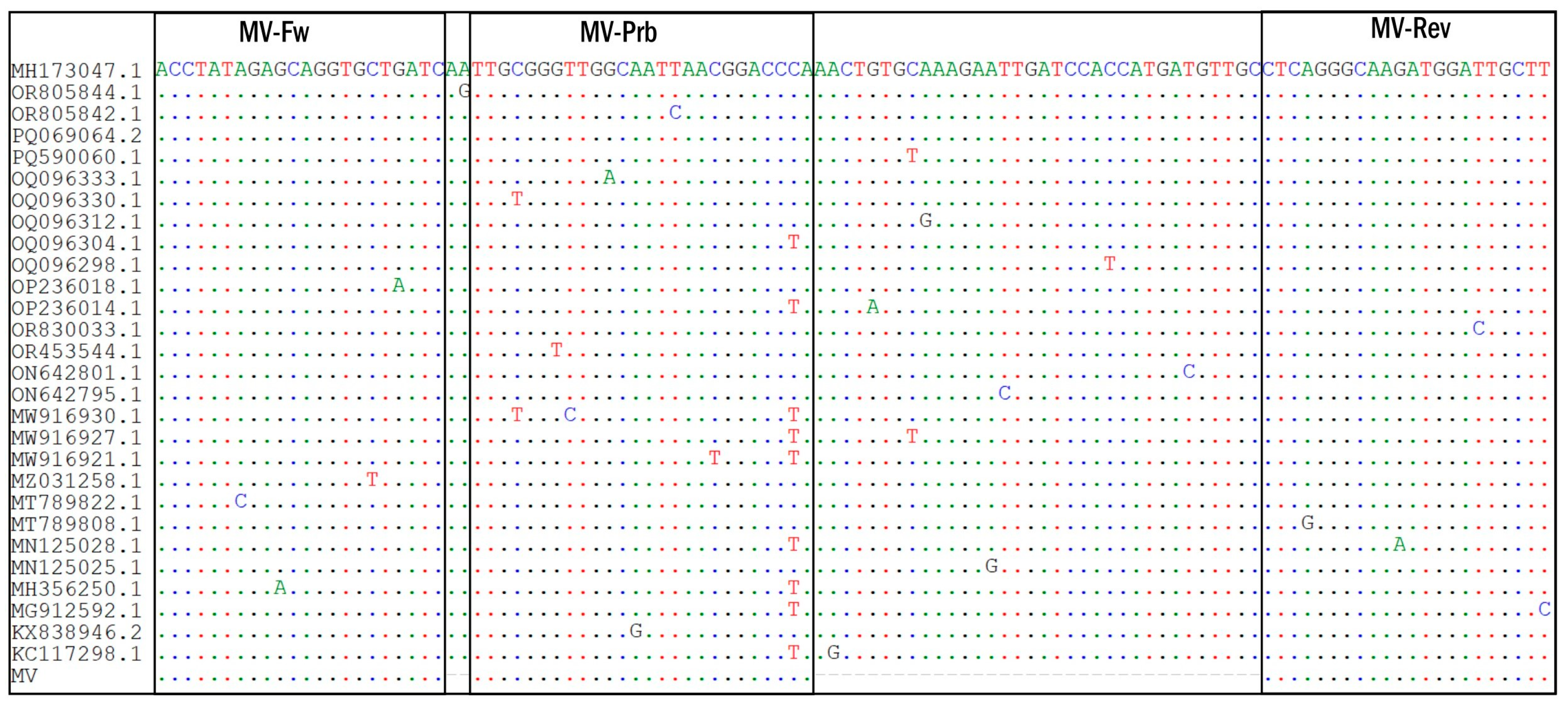
Figure 1. Partial nucleotide sequence alignment of MeV (RdRp gene). Complete sequences of MeV genotype B3 and D8 (as of December 2023) in Gen-Bank (NCBI, n = 1265) were downloaded. Among these, 544 complete RdRp sequences (without gaps and ambiguities of reading) were aligned to identify conserved sites. In sum, 28 unique MeV RdRp gene sequences with the following NCBI GenBank accession numbers are shown: MH173047.1—strain MVs/Ancona.ITA/45.17/1/[D8], OR805844.1—strain MVs/Ahmednagar.IND/48.22/233783[D8]MIBE, OR805842.1—strain MVs/Mumbai.IND/05.23/2/233874[D8]MIBE, PQ069064.2—strain MVi/Rabat.MAR/29.24/[B3], PQ590060.1—strain MVs/SaoPaulo.BRA/41.24[D8], OQ096333.1—strain MVs/Alberta.CAN/27.18[D8], OQ096330.1—strain MVs/Britih_Columbia.CAN/44.18[B3], OQ096312.1—strain MVs/British_Columbia.CAN/6.19[D8], OQ096304.1—strain MVs/British_Columbia/25.18/2[D8], OQ096298.1—strain MVs/Ontario.CAN/10.18[D8], OP236018.1—strain MVi/Pune.IND/38.13[D8], OP236014.1—strain MVi/Tumkur.Ind/06.08[D8], OR830033.1—strain MVs/Sao Paulo.BRA/02.20[D8], OR453544.1—strain MVs/Sao Paulo.BRA/239.19[D8], ON642801.1—strain MVi/Hoima.UGA/01.13 [B3], ON642795.1—strain MVi/Otuke.UGA/13.19[B3], MW916930.1—strain MVi/Krishnagiri.Ind/12.06[D8], MW916927.1—strain MVi/Anand.IND/33.15/2[D8], MW916921.1—strain MVi/Sundargarh.IND/10.14[D8], MZ031258.1—strain MVi/Marseille.FRA/27.19/6[B3], MT789822.1—strain MVs/California.USA/15.19/2[D8], MT789808.1—strain MVs/California.USA/43.19/2[B3], MN125028.1—strain MVi/Udupi.IND/7.15/7, MN125025.1—strain MVi/Kanpur.IND/16.14[D8], MH356250.1—strain MVi/Kozhikode.IND/14.10[D8], KT732240.1—strain MVs/Hull.GBR/47.12/[D8], KX838946.2—strain MVi/Calais.FRA/01.16[B3], KC117298.1—strain MVi/Vicenza.ITA/12.10/2 [D8]. Sequences of the forward (MV_Fw) and reverse (MV_Rev) primers and probes (MV_Prb) targeting this region are indicated in the frames.
Ijms 26 01801 g001
Figure 2. The assay LODs and assay standard curves assessed using ARCs on plate- and rotary-type instruments. Samples: red—C+, purple—ARC 104 copies/mL, blue—ARC 103 copies/mL, yellow—ARC 102 copies/mL. The ARC 101 copies/mL (pink), internal control (green), and C− (green) samples are negative. (A) CFX96 C1000 Touch (Bio-Rad, USA) standard curve; (B) LOD assay, (CFX96 C1000 Touch); (C) Rotor-Gene Q (Qiagen, Germany) standard curve; (D) LOD assay, Rotor-Gene Q.
Ijms 26 01801 g002
Figure 3. Ct value distribution of positive swab samples by collection day after disease onset.
Ijms 26 01801 g003
Table 1. Threshold cycles (Ct) of the HEX/yellow channel (ARC dilution) at various concentrations.
ARC Concentration (Copies/mL)CFX96 C1000 Touch (Bio-Rad, USA)Rotor-Gene Q (Qiagen, Germany)
Replicate, Ct ValuesReplicate, Ct Values
123Median123Median
10429.629.529.629.627.927.927.927.9
10333.332.933.033.131.531.931.731.7
102neg.neg.40.9neg.37.137.436.336.9
101neg.neg.neg.neg.neg.neg.neg.neg.
LOD (copies/mL)103102
LOD* (copies/mL)1.2 × 1032.7 × 102
Note: LOD*—limit of detection measured using probit analysis; neg.—negative.
Table 2. Summary of the MV AmpPS assay.
ParameterReal-Time RT-PCR Assay
Reagent components MeV super mix, RT-PCR enzyme mix, 2× RT-PCR buffer, armored internal control (IC), armored positive RNA control (ARC+), positive PCR control (C+), negative PCR control (C−), and negative extraction control (NEC)
Virus detectedMorbillivirus hominis (MeV)
Genetic targetRdRp gene
Real-time PCR platformCFX96 C1000 Touch (Bio-Rad, USA), Rotor-Gene Q (Qiagen, Germany)
Nucleic acid extraction required yes
Suitable specimens for testing nasopharyngeal swabs
Sensitivity1–1.2 × 10³ copies/mL (CFX96 C1000 Touch), 1–2.7 × 10² copies/mL (Rotor-Gene Q)
Duration of the analysis (excluding extraction time)90 min
Table 3. List of primers and probes used.
Primer or
Probe
Sequence (5′→3′)Reference Sequence Nucleotide Position (GenBank OR290098)5′–3′
Modification
Amplicon Length (bp)
MV-FwACCTATAgAGCAggTgCTgATC15299–15320none106
MV-RevAAgCAATCCATCTTGCCCTgAg15383–15404none
MV-PrbTTgCgggTTggCAATTAACggACCCA *15323–15348R6G—BHQ1-
IC-FwCCggATTgCgTATCTCCggACTnonenone122
IC-RevCACggCggCATCTCTATCACgAnonenone
IC-Prb1CTAgCTgggCgTCAggAATCCCAgg noneFAM-BHQ1-
* The two nucleotides added to the 3′ end of the MV-Prb sequence are marked in bold italic font.
Table 4. Oligonucleotides used for de novo MeV cDNA synthesis by two-step PCR.
NameSequence (5′-3′)
MEV1AGTTATTAGAGGTGATATCAACCCTACTCTGAAAAAACTTACACCTATAGAGCAGGTG
MEV2GTTTGGGTCCGTTAATTGCCAACCCGCAATTGATCAGCACCTGCTCTATAGGTGTAAGTT
MEV3GGCAATTAACGGACCCAAACTGTGCAAAGAATTGATCCACCATGATGTTGCCTCAGGGCA
MEV4AACTCCCTGTAGAGGATGAGTATAGAATTAAGCAATCCATCTTGCCCTGAGGCAACATCA
Table 5. List of viral species used in this study.
SpeciesAcronymFamilyGenusType of Nucleic AcidRT-PCR KitCt ValueMV AmpS Assay
Influenza A/H1N3FLUAV (H1N3)OrthomyxoviridaeAlphainfluenzavirusRNAAmpliSens® Influenza virus A-type-FRT PCR 18.4negative
Influenza A/H3N2FLUAV (H3N2)OrthomyxoviridaeAlphainfluenzavirusRNA17.9negative
Influenza BFLUBVOrthomyxoviridaeBetainfluenzavirusRNAAmpliSens® Influenza virus A/B-FRT PCR23.6negative
Human parainfluenza virus type 1HPIV-1ParamyxoviridaeRubulavirusRNAAmpliSens® ARVI-screen-FRT20.4negative
Human parainfluenza virus type 4b HPIV-4bParamyxoviridaeRubulavirusRNAAmpliSens® ARVI-screen-FRT23.8negative
Human rhinovirus B (Type 17)HRV-BPicornaviridaeEnterovirusRNAAmpliSens® ARVI-screen-FRT24.4negative
Human adenovirus type 6HAdV-6AdenoviridaeMastadenovirusDNAAmpliSens® All screen-FRT19.8negative
Severe acute respiratory syndrome coronavirus 2SARS-CoV-2CoronaviridaeBetacoronavirusRNACOVID-19Amp (St. Petersburg Pasteur Institute) [35] 17.3negative
Rubella virusRUBVMatonaviridaeRubivirusRNANA22.7negative
Respiratory syncytial virus type B1RSV B1PneumoviridaeOrthopneumovirusRNAAmpliSens® ARVI-screen-FRT21.9negative
Human parechovirus type 1HPeV-1PicornaviridaeParechovirusRNANA-negative
Human alphaherpesvirus 1HSV-1HerpesviridaeAlphaherpesvirusDNAAmpliSens® HSV I, II-FRT21.8negative
Human Rotavirus ARVAReoviridaeRotavirusRNAAmpliSens® Rotavirus/Norovirus/Astrovirus-FRT PCR19.9negative
Echovirus 4ECHOV-4PicornaviridaeEnterovirusRNANA-negative
Human Cytomegalovirus 5HCMV-5HerpesviridaeCytomegalovirusDNAAmpliSens® CMV-FRT PCR19.9negative
Human parvovirus B19B19ParvoviridaeErythroparvovirusDNAAmpliSens® Parvovirus B19-FRT PCR22.1negative
Human Coxsackievirus B1CV-B1PicornaviridaeEnterovirusRNANA-negative
Mumps orthorubulavirusMuVParamyxoviridaeOrthorubulavirusRNANA-negative
Disclaimer/Publisher’s Note: The statements, opinions and data contained in all publications are solely those of the individual author(s) and contributor(s) and not of MDPI and/or the editor(s). MDPI and/or the editor(s) disclaim responsibility for any injury to people or property resulting from any ideas, methods, instructions or products referred to in the content.