Review

Role of Kynurenine and Its Derivatives in Liver Diseases: Recent Advances and Future Clinical Perspectives

1
Department of Infectious Disease, Union Hospital, Tongji Medical College, Huazhong University of Science and Technology, Wuhan 430022, China
2
Center for Liver Transplantation, Union Hospital, Tongji Medical College, Huazhong University of Science and Technology, Wuhan 430022, China
3
Department of Nosocomial Infection Management, Union Hospital, Tongji Medical College, Huazhong University of Science and Technology, Wuhan 430022, China
*
Authors to whom correspondence should be addressed.
Int. J. Mol. Sci. 2025, 26(3), 968; https://doi.org/10.3390/ijms26030968
Submission received: 27 December 2024 / Revised: 12 January 2025 / Accepted: 22 January 2025 / Published: 24 January 2025
(This article belongs to the Section Biochemistry)

Abstract

Liver health is integral to overall human well-being and the pathogenesis of various diseases. In recent years, kynurenine and its derivatives have gradually been recognized for their involvement in various pathophysiological processes, especially in the regulation of liver diseases, such as acute liver injury, non-alcoholic fatty liver disease, cirrhosis, and liver cancer. Kynurenine and its derivatives are derived from tryptophan, which is broken down by the enzymes indoleamine 2,3-dioxygenase (IDO) and tryptophan 2,3-dioxygenase (TDO), converting the essential amino acid tryptophan into kynurenine (KYN) and other downstream metabolites, such as kynurenic acid (KYNA), 3-hydroxykynurenine (3-HK), xanthurenic acid (XA), and quinolinic acid (QA). In liver diseases, kynurenine and its derivatives can promote the activity of the transcription factor aryl hydrocarbon receptor (AhR), suppress T cell activity for immune modulation, inhibit the activation of inflammatory signaling pathways, such as NF-κB for anti-inflammatory effects, and inhibit the activation of hepatic stellate cells to slow down fibrosis progression. Additionally, kynurenine and other downstream metabolites can influence the progression of liver diseases by modulating the gut microbiota. Therefore, in this review, we summarize and explore the mechanisms by which kynurenine and its derivatives regulate liver diseases to help develop new diagnostic or prognostic biomarkers and effective therapies targeting the kynurenine pathway for liver disease treatment.

1. Introduction

Over 2 million people die from liver diseases worldwide each year, accounting for about 4% of global deaths, with alcoholic liver disease, cirrhosis, and liver cancer being the most common causes, severely threatening human health [1]. Chronic liver disease affects an estimated 1.5 billion individuals globally, with its prevalence having risen by 13% since 2000, thereby imposing a significant burden on populations worldwide [2]. Chronic liver disease develops from liver injury, with common pathological features of liver injury including hepatocyte degeneration, necrosis, and apoptosis [3]. Hepatocyte death triggers subsequent inflammation, leading to excessive deposition of extracellular matrix proteins, resulting in hepatitis and liver fibrosis, and eventually progressing to cirrhosis and hepatocellular carcinoma [4]. Currently, therapeutic interventions for liver diseases are rapidly advancing, aiming to delay or even reverse the progression of liver diseases.
In recent years, kynurenine and its derivatives have exhibited anti-inflammatory, immunoregulatory, and pro-apoptotic properties, contributing to the regulation of liver lipid metabolism, inflammation, and fibrosis. The kynurenine pathway is the main pathway of tryptophan metabolism. In the body, 2,3-dioxygenase (TDO) and two indoleamine 2,3-dioxygenase isoforms (IDO-1 and IDO-2) oxidize tryptophan into kynurenine and its series of derivatives [5]. Kynurenine (Kyn) has immunoregulatory effects, promoting the stagnation or apoptosis of effector T cells [6] and inducing the conversion of naive CD4+ T cells into immunosuppressive regulatory T cells (Treg) [7]. Ogiso et al. discovered that the lack of the Ido1 gene exacerbated CCl4-induced liver fibrosis in a CCl4-induced liver injury model [8]. In primary liver cancer, the kynurenine pathway (KP) enables tumor cells to escape immune surveillance [9], promoting proliferation and metastasis, as well as suppressing local immune cell populations [10], thereby facilitating tumor growth. Recent studies have highlighted the potential link between the kynurenine pathway and various liver diseases, including acute and chronic liver injury [11,12], non-alcoholic fatty liver disease [13], and liver cancer [14].
To further clarify the regulatory effects of kynurenine and its derivatives on liver diseases, this article comprehensively introduces the synthesis, metabolism, bioactivity, and mechanisms of action in liver disease regulation, and the clinical challenges faced, as well as discusses how to expedite the clinical translation and application of kynurenine and its derivatives. The aim of this review is to bridge the gap between clinical treatment of liver diseases and basic experimental to clinical translation, providing theoretical reference for the development of kynurenine and its derivatives in the field of liver disease treatment.

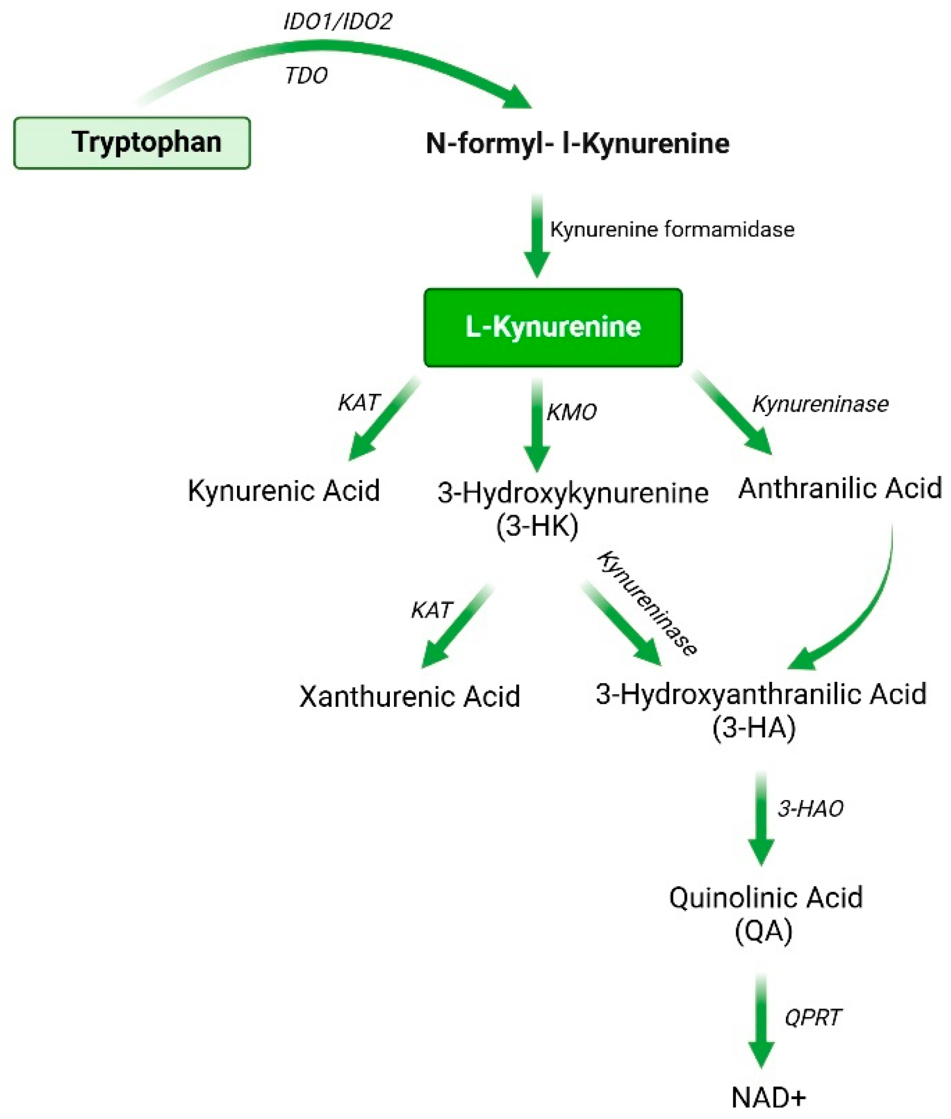
2. Synthesis, Metabolism, and Biological Functions of Kynurenine

2.1. Synthesis and Metabolism of Kynurenine

Kynurenic acid (KYNA) was first isolated by Justus von Liebig from the urine of dogs fed with tryptophan. Subsequently, Kotake and Iwao isolated kynurenine as the main metabolic product of tryptophan (TRP) breakdown through the kynurenine pathway [15].Tryptophan can produce various metabolites through direct or indirect action by the gut microbiota and then be transported by transport proteins to intestinal epithelial cells, reaching the liver via the enterohepatic circulation, influencing liver function and metabolism [16]. In the gut, tryptophan mainly follows three metabolic pathways, including the kynurenine pathway [17], serotonin metabolism pathway [18], and indole and its derivative metabolism pathway [19].
More than 95% of tryptophan is metabolized via the kynurenine pathway, primarily by the rate-limiting enzymes indoleamine 2,3-dioxygenases (IDO1 and IDO2), kynurenine formamidase, kynurenine aminotransferase (KAT), kynurenine 3-monooxygenase (KMO), 3-hydroxyanthranilic acid oxidase (3-HAO), quinolinic acid phosphoribosyl transferase (QPRT), and tryptophan-2,3-dioxygenase (TDO) [20]. Studies have shown that tryptophan 2,3-dioxygenase (TDO) is predominantly expressed in the liver, especially in hepatocytes [21]. Furthermore, the expression level of Tryptophan 2,3-dioxygenase (TDO) is regulated by factors such as L-tryptophan (Trp), corticosteroids, and hypoxic conditions [22,23]. IDO1 is widely expressed in various stem cells, such as bone marrow mesenchymal stem cells (BMSCs) and umbilical cord tissue-derived mesenchymal stromal cells (UC-MSC), and multiple immune cells, including dendritic cells, macrophages, and neutrophils [24,25,26]. It plays a critical role in immune regulation and liver repair and regeneration [27]. The expression of IDO1 is regulated by various factors, such as interferon-γ (IFN-γ), tumor necrosis factor-alpha (TNF-α), and stimulation by other cytokines [28]. Therefore, the activity of IDO1 is associated with immune responses, chronic inflammation, tumor immune escape, and various other pathological processes. Additionally, the kynurenine pathway is tightly regulated under physiological conditions, generating various metabolites through the action of a series of enzymes (Figure 1), such as kynurenine, kynurenic acid (KYNA), 3-hydroxykynurenine (3-HK), anthranilic acid (QA), quinolinic acid (QA), and the NAD(P)/NAD(P)H [29].
Furthermore, kynurenine, as a critical intermediate in tryptophan metabolism, is closely linked to the metabolism and function of the gut microbiota. The kynurenine pathway (KP) plays an essential role in regulating the immune system, neuro-signaling, and oxidative stress. The interaction between the gut and liver is referred to as the “gut–liver axis”, which is a complex bidirectional connection based on the portal vein and involves various signaling pathways. Kynurenine metabolism in the liver is also regulated by the gut (Figure 2). Kynurenine is a key intermediate in tryptophan metabolism, and its metabolic pathway is closely associated with the metabolism and function of the gut microbiota. Tryptophan absorption primarily occurs at the apical membrane of intestinal epithelial cells, where it is transported into the cells via angiotensin-converting enzyme 2 (ACE2) and broad-spectrum neutral amino acid transporter 1 (B0AT1) [30]. Once absorbed by transport proteins in the intestinal epithelial cells, tryptophan (TRP) passes through the portal venous system and is converted to NAD+ in the liver via the kynurenine pathway [31]. The gut, as an important site for tryptophan metabolism, widely expresses IDO in intestinal epithelial cells and immune cells. Gut microbiota significantly modulates the activity of the host’s tryptophan metabolic pathway by secreting indole metabolites, short-chain fatty acids (SCFAs), and other signaling molecules [32].
The gut microbiota regulates the host’s kynurenine metabolism through multiple mechanisms, including direct regulation of IDO1 activity and indirect influence on tryptophan metabolic pathways. The gut microbiota can directly regulate the kynurenine metabolism. Zhao et al. found that in a colitis mouse model, the gut microbiota was found to regulate IDO-1 and KAT, leading to a significant increase in Kyn and kynurenic acid levels [33]. Additionally, gut microbiota can indirectly regulate the kynurenine pathway through metabolites derived from the indole pathway. Furthermore, recent studies have shown that in mice with dysbiosis, gut barrier dysfunction and tryptophan metabolic disorders lead to a significant increase in indole derivatives, which activate the colon aryl hydrocarbon receptor (AhR), promote the kynurenine pathway, and inhibit the serotonin pathway [34]. Similarly, Paydaş Hataysal et al. found that in patients with inflammatory bowel disease, serum levels of tryptophan, kynurenic acid, 3-hydroxykynurenine, and other kynurenine metabolites are lower [35]. Indole metabolites, such as indole-3-propionic acid and indole-lactic acid, are products of gut microbiota metabolism of tryptophan and can regulate IDO1 expression through the aryl hydrocarbon receptor (AhR) [36].
Kynurenine and its metabolites can also feedback to affect the composition and function of the gut microbiota. Kynurenine can also be metabolized in the gut to substances, such as 3-HK and KA, and kynurenine metabolites can indirectly alter the gut microbiota composition by affecting the local immune microenvironment. Kynurenic acid is an antimicrobial substance in the intestinal fluid, first isolated from the Tachypleus tridentatus [37], and it has direct antimicrobial effects at high concentrations [38]. In a high-fat disease mouse model, kynurenic acid can inhibit the increase in Firmicutes and Bacteroidetes, which are associated with the disease. However, kynurenic acid promotes the growth of probiotics, such as Lactobacillus acidophilus, Bifidobacterium and Actobacillus rhamnosus [39]. Additionally, Robert et al. found that kynurenine easily enters probiotics, preferentially converts into kynurenic acid, and is then swiftly released into the extracellular environment to act [40]. Furthermore, the intestinal antimicrobial protein REG3A influences gut microbiota composition by increasing the proportion of lactobacilli, which in turn increases kynurenine levels [41]. Therefore, kynurenine and its metabolites are closely linked with the gut microbiota, working together to regulate the occurrence and development of liver diseases.

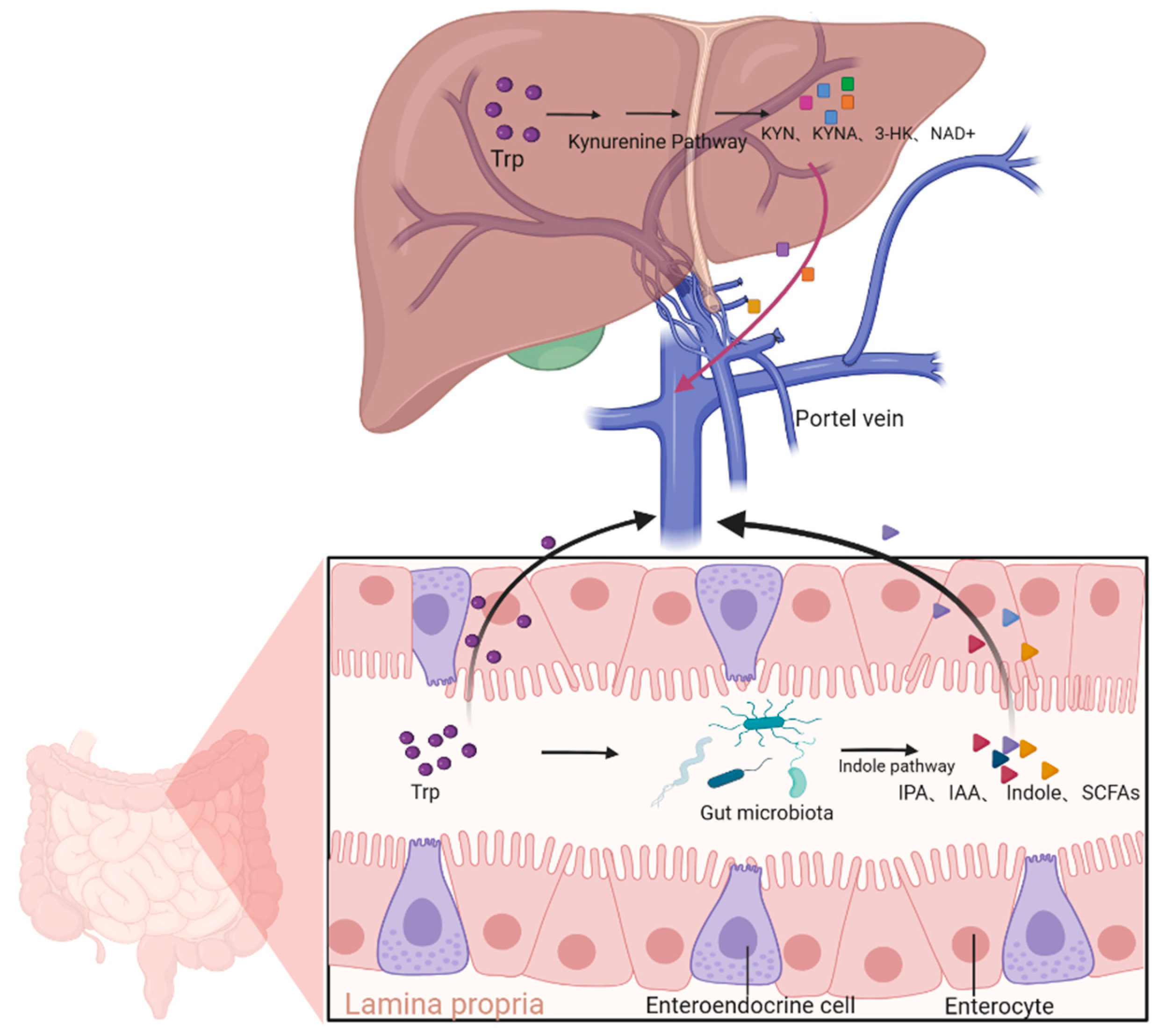
2.2. Biological Activity of the Kynurenine Pathway and Its Derivatives

In the metabolism of tryptophan, kynurenine synthesis is catalyzed by tryptophan-2,3-dioxygenase (TDO) and indoleamine 2,3-dioxygenase (IDO). The role of kynurenine in the immune system has been widely studied. Kynurenine and its derivatives function as agonists of the aryl hydrocarbon receptor (AhR) and serve as critical modulators of inflammatory processes [42]. AhRs are a family of proteins found in various species, including humans, and are ligand-activated transcription factors [43].
AhR signaling is considered a critical component of immune responses at barrier sites. It can influence intestinal epithelial renewal, barrier integrity, and various immune cells such as intraepithelial lymphocytes, Th17 cells, innate lymphoid cells, macrophages, dendritic cells, and neutrophils, which are essential for gut homeostasis [44]. Activated AhR signaling is closely associated with inflammatory diseases, such as ulcerative colitis [45], non-alcoholic steatohepatitis [46], and systemic lupus erythematosus [47]. This process can regulate immune cell functions through multiple pathways, including AhR-induced Cyp1a1 gene expression [48], AhR interaction with NF-κB [49], and AhR regulation of the AhR/Nrf2/NQO1 pathway [50]. The AhR signaling pathway in cells can also be activated by ligands such as Kyn and its derivatives. The kynurenine pathway represents a potential target for modulating gut microbiota in liver inflammatory diseases and plays a crucial role in the inflammatory signaling of the AhR–liver axis [51].
Kynurenine and its derivatives are potential immunosuppressive metabolites produced through the tryptophan metabolic pathway. It has been reported that kynurenine can induce immune tolerance, acting as an endogenous ligand for AhR. Elevated kynurenine levels activate the aryl hydrocarbon receptor, thereby inhibiting effector T cell proliferation [7] and inducing the FoxP3 transcription factor to promote the differentiation of immunosuppressive Treg cells. In addition, kynurenine also suppresses the surface expression of the NKp46 and NKG2D activating receptors, thereby inhibiting T cell receptor expression and NK cell function—key mechanisms implicated in immune evasion across various cancers [52]. For example, Patra et al. [46] found a positive correlation between indoleamine 2,3-dioxygenase-1 (IDO1) and the tumor-promoting reprogramming transcription factor Lin28B in HCC patient samples [53]. Lin28B can increase the production of the enhancer Sox2/Oct4 to upregulate the expression of IDO1. The effect of IDO1 inhibitors is minimal when used alone, but when combined with immune checkpoint inhibitors, the anti-tumor effect is significantly enhanced. In liver cancer cell models, inhibiting IDO1 expression can significantly enhance the immune response against the tumor and slow tumor growth. This result suggests that the role of IDO1 in tumor immune evasion should not be underestimated, and its inhibition may become a new immunotherapeutic strategy.
The role of kynurenine and its metabolites in the central nervous system has also received increasing attention. Kynurenine, through its downstream metabolites, such as kynurenic acid, 3-hydroxykynurenine, 3-hydroxyanthranilic acid, and quinolinic acid, plays an important role in neurotoxicity, neuroinflammation, and the development of neurodegenerative diseases. Kynurenine aminotransferase (KAT) can convert kynurenine into kynurenic acid (KYNA) [54], while KMO converts kynurenine into 3-hydroxykynurenine (3-HK) [20]. Kynurenic acid (KYNA) inhibits the N-methyl-D-aspartate receptor (NMDAR) [55,56], binds to the G protein-coupled receptor 35 (GPR35), [57] and activates the aryl hydrocarbon receptor (AhR) [58], exerting anti-inflammatory, neuroprotective, and immune-regulatory effects. Conversely, the downstream metabolites of kynurenic acid, 3-hydroxykynurenine, and quinolinic acid exhibit opposite effects. 3-Hydroxykynurenine is a toxic metabolite that induces oxidative damage and cell death [59]. Quinolinic acid acts as an NMDAR agonist, leading to excitotoxicity [60]. Alterations in the kynurenine metabolic pathway are considered crucial in the pathological processes of neurodegenerative diseases such as Alzheimer’s disease (AD), Parkinson’s disease (PD), and Huntington’s disease (HD). A clinical study found that in Parkinson’s disease (PD) patients, the levels of kynurenine metabolite 3-hydroxykynurenine (3-HK) were significantly elevated in both peripheral and central systems, and this elevation correlated with the severity of the disease [61]. The kynurenine metabolic pathway not only plays a significant role in neurological diseases but is also closely related to the occurrence of psychiatric disorders [62]. The potential mechanism of elevated KYNA in psychiatric disorders may be related to increased activation of the pro-inflammatory cytokine IL-1β. Kynurenic acid, as an NMDA receptor antagonist, is elevated in the cerebrospinal fluid of patients with schizophrenia and mania [63]. Moreover, central nervous inflammation may have an important relationship with the activation of the kynurenine pathway in mental illnesses [64,65]. In conclusion, inhibiting the kynurenine metabolic pathway is considered a potential therapeutic strategy, with inhibition of IDO1 activity being an important target for neuroprotective treatments to alleviate symptoms of neurodegenerative diseases and neuroinflammation.
Kynurenine can affect liver cell survival and death by regulating apoptosis pathways. During liver injury or disease progression, kynurenine may affect liver health by promoting T cell apoptosis in the liver [66] or inhibiting cell proliferation. The immune tolerance of the liver, the metabolic status of liver cells, and the local inflammatory response are closely related to the metabolism of kynurenine. Liver injury is often accompanied by immune system activation and inflammatory responses. Therefore, kynurenine prevents liver injury by alleviating oxidative stress, inflammation, and cell death. In acute liver injury, kynurenine can suppress T cell proliferation via apoptosis pathways, promoting immunosuppressive responses and thus slowing liver recovery [67]. Moreover, the kynurenine metabolite 3-hydroxyanthranilic acid accumulates in monocytes/macrophages, thereby inducing immune cell apoptosis [68]. Bishnupuri et al. found that in IDO1 knockout mouse colon tumor model, kynurenine pathway metabolites rapidly activated PI3K-Akt signaling in tumor epithelial cells and promoted β-catenin nuclear translocation, cell proliferation, and anti-apoptotic effects [69].

3. The Role of Kynurenine and Its Derivatives in Liver Diseases

The roles of kynurenine and its derivatives in liver diseases are complex and multifaceted. They may play an important role in the occurrence and development of various liver diseases, such as liver injury, liver inflammation, liver fibrosis, and liver cancer, by regulating immune responses, oxidative stress, apoptosis, fibrosis, and other processes. Kynurenine and its derivatives cannot only regulate diseases through various mechanisms, but their metabolite levels are also closely related to the occurrence and development of diseases (Table 1). Therefore, kynurenine and its metabolites not only provide potential biomarkers for early diagnosis of liver diseases [70], but also offer possible new therapeutic targets. This section discusses reprogramming of the kynurenine metabolism, the abnormal expression of key metabolic enzymes and their products in various liver diseases, and their potential clinical significance and prognostic value.

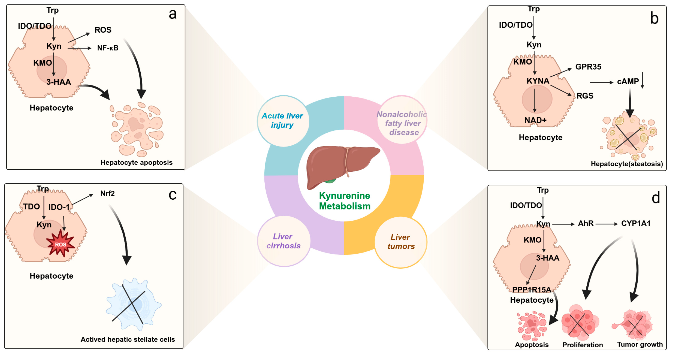
3.1. Acute Liver Injury

Acute liver injury (ALI) is an acute inflammatory response of the liver caused by factors such as drugs, viral infections, alcohol intoxication, and ischemia–reperfusion, characterized by hepatocellular degeneration, necrosis, and apoptosis, and is a multi-etiologic clinical syndrome [87]. Hepatocellular death triggers subsequent inflammation, leading to excessive deposition of extracellular matrix proteins. Therefore, liver injury often leads to hepatitis and liver fibrosis, which are important initiating factors for cirrhosis and hepatocellular carcinoma (HCC).
Recently, studies have shown that in acute liver injury caused by liver ischemia–reperfusion, the kynurenine metabolic pathway undergoes reprogramming, with a decrease in liver 3-hydroxyanthranilic acid (3-HAA) and quinolinic acid (QA), while kynurenine and kynurenic acid (KYNA) levels increase. This is due to the significant upregulation of kynurenine aminotransferase 2 in hepatocytes, shifting the kynurenine pathway from 3-HAA and QA to KYNA and NAD synthesis, alleviating oxidative stress, inflammation, and cell death in hepatocytes [73]. Another study also showed that in a mouse liver injury model induced by hexafluoropropylene oxide dimer acid (HFPO-DA), the correlation between kynurenic acid (KYNA) levels in mouse serum and liver injury was the highest. Exposure to HFPO-DA-induced liver injury in mice, which caused a shift in the kynurenine pathway from kynurenic acid (KYNA) to NAD synthesis, led to endoplasmic reticulum stress and activation of the NF-κB signaling pathway (Figure 3a). Pre-treatment with kynurenic acid (KYNA) significantly alleviated liver injury induced by HFPO-DA exposure in mice, showing protective effects [88]. Hoshi et al. found that in a CCl4-induced acute liver injury mouse model with Ido2 knockout (Ido2(−/−)) and treatment with the Ido2 inhibitor 1-methyltryptophan (D-1MT), hepatocellular damage was alleviated [71]. Additionally, endothelial cells are closely related to liver function, and under the stimulation of inflammatory factors, endothelial cells highly express indoleamine 2,3-dioxygenase (IDO), which metabolizes tryptophan into kynurenine, resulting in relaxation of arterial blood vessels [89]. Kynurenine metabolism is one of the major contributing mechanisms for systemic circulatory dysfunction in acute liver failure. The increase in IDO activity, kynurenine pathway activity, and oxidative stress in endothelial cells, along with kynurenine-mediated small arterial vasodilation, represents a malignant pathological cycle, which may lead to progressive endothelial cell apoptosis [90], microcirculatory dysfunction, and organ failure. Inhibiting the kynurenine pathway can reduce hepatocellular damage and liver fibrosis. This suggests that changes in the kynurenine metabolic pathway play a key role in liver injury. However, a study by Ma et al. found that another key enzyme in the kynurenine pathway, indoleamine 2,3-dioxygenase 1 (IDO-1), increases hepatocyte ferroptosis and M1 polarization, while also enhancing M2 polarization and promoting macrophage phagocytosis [74]. Therefore, the activation of IDO-1 in macrophages plays a critical role in triggering hepatocyte death during liver ischemia–reperfusion injury.
In conclusion, kynurenine plays a role in acute liver injury through metabolic reprogramming, regulating microcirculation, and affecting NAD homeostasis, while also demonstrating some protective effects. Therefore, elucidating the role of kynurenine and its metabolites in the molecular pathology of acute liver injury is essential for understanding the connection between kynurenine metabolism and liver diseases. These findings provide new directions for future research and may help develop new therapeutic strategies to combat liver injury.

3.2. Metabolic Dysfunction-Associated Steatotic Liver Disease

Currently, metabolic dysfunction-associated steatotic liver disease (MASLD) is one of the leading causes of chronic liver disease worldwide [91], and the progression from non-alcoholic fatty liver to non-alcoholic steatohepatitis has become a major threat leading to cirrhosis, acute-on-chronic liver failure, and liver cancer. Among these, the kynurenine metabolic pathway plays a crucial role in the progression of non-alcoholic fatty liver disease. Dorochow et al. analyzed the liver lipidome, metabolome, and immune cells in non-alcoholic hepatitis and found that kynurenine levels were elevated in the liver. This elevation promoted M2-like macrophage polarization and inhibited the activity of natural killer cells, dendritic cells, monocytes, and macrophages, exerting an anti-inflammatory effect and slowing disease progression [76]. Kynurenic acid increases energy expenditure, improves energy metabolism, and reduces inflammation in mice on a high-fat diet by activating Gpr35 and RGS (Figure 3b) [77]. Berge et al. constructed a mouse model of non-alcoholic fatty liver and found that although kynurenine levels decreased in the plasma, the end product of kynurenine metabolism, niacinamide, increased. This change is likely unrelated to increased inflammation and oxidative stress. The increase in NAD+ and niacinamide is likely generated through the salvage pathway, rather than through the de novo kynurenine pathway [75]. Carine et al. found that treatment with 1-triple TTA in male Wistar rats resulted in increased fatty acid oxidation in liver mitochondria and promoted the conversion of tryptophan to NAD+. Therefore, kynurenine and its derivatives are positively correlated with mitochondrial fatty acid oxidation [92]. Agudelo et al. found that treatment with KYNA reduced the levels of quinolinic acid (QA) and 3-hydroxykynurenine (3-HK) in mice while increasing the levels of nicotinic acid [93]. NAD+, as the end product of tryptophan–kynurenine axis de novo synthesis, has therapeutic effects in fatty liver disease [94]. Fatty liver disease is closely associated with metabolic dysfunction, particularly with the increase in obesity. Carmen Arto et al. quantified the in vivo levels of 15 tryptophan-related metabolites in the kynurenine, indole, and serotonin pathways using ultra-high-performance liquid chromatography, and found that only kynurenine-related metabolites were associated with increased gene expression of the tryptophan degradation pathway enzymes, IDO-1 and kynurenine monooxygenase (KMO), as well as elevated concentrations of various kynurenine-related metabolites [95]. Targeting these pathways may have potential therapeutic applications.
In conclusion, kynurenine metabolism presents a pathway with potential for controlling energy homeostasis in the treatment of metabolic dysfunction–associated steatotic liver disease (MASLD). Further understanding of kynurenine and its metabolites in metabolic dysfunction-associated steatotic liver disease (MASLD) is crucial for elucidating the relationship between liver diseases.

3.3. Chronic Liver Injury and Liver Cirrhosis

Chronic liver injury is caused by long-term conditions, such as chronic hepatitis, fatty liver, or alcoholic liver disease, and can eventually develop into liver fibrosis or cirrhosis. Kynurenine and its metabolites play a crucial role in the formation of liver fibrosis, particularly in inhibiting the liver’s repair process through immune escape mechanisms. The pathogenesis of liver fibrosis is closely related to the immune microenvironment, with macrophages playing a key role as important immune cells in the liver, capable of shifting between two polarized phenotypes, classical (M1) and alternative (M2), in response to external stimuli [96]. Indoleamine 2,3-dioxygenase 1 (IDO1) can be detected in macrophages and it regulates various immune responses and plays a regulatory role in dendritic cells. IDO1 impairs the maturation of dendritic cells in wild-type (WT) mice and downregulates key regulators of cellular oxidative stress and inflammation, including nuclear factor E2-related factor 2 (Figure 3c), thus alleviating liver fibrosis [79]. Studies have shown that kynurenine participates in regulating immune tolerance in chronic liver injury via the IDO1 pathway. Wang et al. discovered that activation of IDO1 in a mouse model of liver fibrosis caused an increase in kynurenine levels and influenced macrophage differentiation, thereby delaying immune response recovery. Further analysis indicated that IDO1-mediated kynurenine increased the activation of hepatic stellate cells, thus promoting the progression of liver fibrosis [81]. The role of IDO-1 in liver diseases is not only related to the regulation of immune responses and fibrosis but also involves various physiological processes such as liver regeneration, oxidative stress, and inflammation. Due to the liver’s excellent regenerative capacity, especially after liver injury, hepatocytes can rapidly recover and repair. However, chronic liver diseases, such as cirrhosis, impair liver regeneration due to persistent inflammation and fibrosis. In this process, MSCs can migrate to the damaged tissue, undergo hepatocyte differentiation [97], inhibit the release of inflammatory factors, and enhance hepatocyte proliferation in the body [98]. Additionally, MSCs can secrete indoleamine 2,3-dioxygenase (IDO) to exert immune-regulatory effects [99]. Treatment with IDO1 inhibitors significantly alleviated liver fibrosis, suggesting that kynurenine metabolism plays a critical role in chronic liver injury and fibrosis.
In addition, tryptophan 2,3-dioxygenase (TDO) is another systemic tryptophan-metabolizing enzyme, and its high expression in liver is also associated with the progression of liver fibrosis [82]. Zhong et al. found in a mouse liver fibrosis model that liver lesions correlated positively with serum IDO1 levels, and IDO1 in vivo deficiency led to compensatory increases [100]. TDO exacerbates the liver’s inflammatory response and immune suppression by enhancing tryptophan metabolism to kynurenine [80]. Inhibition of TDO expression improves the degree of liver injury, indicating that TDO-mediated kynurenine metabolism is closely related to the occurrence of chronic liver injury and liver fibrosis. In chronic liver injury, the reprogramming of the kynurenine metabolic pathway is tightly regulated. Therefore, these findings highlight the role of IDO1 and TDO activity in the pathology of liver fibrosis.

3.4. Hepatocellular Carcinoma

Hepatocellular carcinoma (HCC) is one of the most lethal malignant tumors worldwide and can arise from various chronic liver diseases and cirrhosis. Hepatocellular carcinoma (HCC) is a highly invasive liver tumor with a pronounced capacity for metastasis to distant organs, including the lungs, bone, brain, lymph nodes, and adrenal glands. As a malignant tumor, early detection with high precision and the ability to assess the patient’s response to treatment are crucial for the prognosis. The kynurenine pathway’s catabolism plays an immunosuppressive role in HCC and enhances tumor survival and invasiveness [101]. The key enzyme of the kynurenine pathway, TDO, suppresses the tumor immune response by inducing immune tolerance, thus promoting immune escape of the tumor. TDO expression is typically elevated in the HCC microenvironment, accompanied by kynurenine accumulation, which may promote tumor growth and metastasis by affecting immune cell function in the tumor microenvironment [102]. Inhibition of TDO expression diminishes kynurenine production and inactivates the aryl hydrocarbon receptor (AhR), thereby promoting the activation and proliferation of CD3(+) T cells, which contribute to anti-tumor immune responses [84]. TDO is not only involved in immune tolerance but is also closely associated with the metastasis of HCC cells. The zinc-finger transcription factor, ZNF165, which is highly expressed in liver tissue and the immune microenvironment, can activate the kynurenine/AhR/CYP1A1 axis and promote CYP1A1 expression, thus enhancing HCC cell proliferation and migration (Figure 3d). Miyazaki et al. conducted a systematic analysis of metabolites in colon cancer patient samples and found that high levels of kynurenine and TDO2 were positively correlated with liver metastasis [103]. In a mouse colon cancer model, TDO expression significantly enhanced liver metastasis, primarily by inducing AHR-mediated PD-L1 transactivation, which suppressed the immune response and promoted liver metastasis of colon cancer. Additionally, in terms of treatment, a new immunosuppressive agent can improve the effectiveness of tumor chemotherapy. The conjugate obtained by combining it with irinotecan can improve the tumor immune microenvironment by inhibiting TDO enzyme expression to block kynurenine production and induce HCC cell apoptosis by releasing TDO inhibitors and irinotecan to cause DNA damage [85]. Kynurenine metabolic derivatives also play a role in HCC treatment. In a liver cancer nude mouse model, 3-HAA was found to increase apoptosis of HCC in cultured cells and mouse xenografts by upregulating phosphatases PPP1R15A/DUSP6 and reducing AKT phosphorylation, as well as increasing sensitivity to the first-line targeted drug sorafenib in HCC patients [83]. It has been reported that high serum kynurenine levels are associated with poor prognosis in HCC. Bekki et al. demonstrated that elevated kynurenine levels in the serum of HCC patients may serve as a biomarker for predicting survival and prognosis in individuals with early-stage HCC [104]. In conclusion, the kynurenine pathway plays an important role in immune escape and tumor progression in HCC. Elucidating these pathways will open new avenues for therapeutic strategies targeting kynurenine metabolites in liver cancer.

4. Clinical Applications of the Kynurenine Pathway

The kynurenine pathway plays an important role in human health and disease, and the regulation of kynurenine metabolism has clinical and therapeutic significance. Currently, the primary clinical treatments include inhibitors targeting different enzymes in the kynurenine pathway, such as IDO1 inhibitors, TDO inhibitors, and KMO inhibitors, which have shown efficacy in cancer, immune diseases, neurological diseases, and neurodegenerative diseases (Table 2).
Currently, significant exploration of IDO1 inhibitors has shown promising progress in clinical settings, including drugs such as Epacadostat, BMS-986205, 1-methyl-tryptophan (1-MT), and EOS200271 (formerly PF-06840003). Epacadostat is a tryptophan competitive inhibitor with high selectivity for IDO1. Animal studies have shown that Epacadostat can reduce tumor growth and promote the proliferation of T cells and NK cells [114]. In clinical applications, a phase 1/2 ECHO-203 (NCT02318277) study evaluated the combination of IDO1 inhibitor Epacadostat with Durvalumab in treating adult patients with advanced solid tumors, including melanoma (n = 5), non-small cell lung cancer (n = 20), squamous cell carcinoma of the head and neck (n = 27), and bladder cancer (n = 19). The objective response rate (ORR) was greater than 10%, with melanoma patients showing the best objective response rate, reaching 80% [105]. BMS-986205 is an oral IDO1 inhibitor. The combination of BMS-986205 with Nivolumab can improve patient response rates. For example, in bladder cancer patients (n = 25), the objective response rate (ORR) was 32%, and in cervical cancer patients (n = 22), the ORR was 14% [106]. Indoximod, as a competitive IDO1 inhibitor, can modulate the function of T cells, CD4 T cells, and dendritic cells (DCs) [113,115], while reverse IDO activity exerts immune suppression. A single-arm phase II clinical trial demonstrated promising safety and efficacy with the combination of Indoximod and Pembrolizumab. Among 89 non-ocular melanoma patients, the objective response rate (ORR) was 51%, and the confirmed complete response rate was 20% [107]. PF-06840003, due to its ability to cross the blood–brain barrier (BBB), was studied in a phase I trial by David et al. in patients with recurrent malignant glioma (NCT02764151). Eight patients (47%) showed controlled disease, indicating its pharmacodynamic effects and sustained clinical efficacy in recurrent malignant glioma patients [108]. TDO, as another key enzyme in the kynurenine pathway, also holds immense potential in clinical treatment. TDO inhibitors include 680C91, LM10, and others. In a preclinical study, Tsai-Der Chuang et al. found that treatment with 680C91 in immunodeficient mice bearing human fibrosarcoma xenografts reduced the weight of uterine fibroid xenografts by 30% [109] S. Hu et al. discovered that in a squamous cell carcinoma mouse model, the TDO2 inhibitor LM10 alleviated T cell suppression, restored T cell anti-tumor responses, and blocked the malignant progression of squamous cell carcinoma [110].
Due to the tremendous progress in small-molecule inhibitors targeting tryptophan metabolism, especially IDO and TDO inhibitors in cancer therapy, dual-target inhibitors targeting both IDO1 and TDO are being developed for broader and more effective clinical treatments of various diseases, including M4112 and RY103. M4112, as an effective and selective dual inhibitor of IDO1 and TDO2, was shown in a tumor mouse model to possess dual inhibitory effects. It was also preliminarily validated for anti-tumor activity in solid tumors, with the best overall response being disease stabilization in 9 out of 15 patients (60.0%) [111]. Subsequently, Zhang et al. developed a new IDO1/TDO dual-target inhibitor compound 17 (RY103). In an in situ pancreatic cancer mouse model, RY103 was able to inhibit tumor growth and metastasis, promote tumor cell apoptosis, significantly improve the immune suppression state, and show no significant toxicity to the mice [112]. Additionally, KMO is overexpressed in several diseases, especially in cancers like melanoma, colorectal cancer, and breast cancer [116], as well as in neurodegenerative diseases such as Alzheimer’s disease and Huntington’s disease [117,118], contributing to disease progression. Therefore, research on KMO inhibitors shows potential as a promising therapeutic strategy for a variety of diseases. Zwilling et al. discovered that JM6 is a small-molecule prodrug inhibitor of kynurenine 3-monooxygenase (KMO) that can inhibit KMO in the blood. In a transgenic mouse model of Alzheimer’s disease, it prevented spatial memory deficits, anxiety-related behaviors, synapse loss, and prolonged survival [113].
Inhibitors of the kynurenine pathway can exert anti-tumor effects when combined with other immunotherapies, such as immune checkpoint inhibitors, chemotherapy, and radiotherapy; however, their application in liver diseases is not yet widespread, possibly due to the complex role of IDO in the liver and the lack of sufficient clinical evidence to support their broad use in liver-related diseases. Therefore, although basic research has made some progress, indicating that kynurenine and its derivatives have antioxidant, anti-inflammatory, and pro-apoptotic effects on liver cells and are closely related to the occurrence and development of liver diseases, most studies focus on mouse models, and clinical application is still distant. However, related drugs targeting KP enzyme inhibitors have been applied in other disease treatments. A deeper understanding of kynurenine and its derivatives, as well as the rational design of IDO1 inhibitors in liver diseases, could provide new avenues for therapeutic intervention in liver diseases.

5. Conclusions

In summary, the kynurenine pathway and its various metabolites, such as kynurenic acid (KYNA), 3-hydroxykynurenine (3-HK), and quinolinic acid (QA), play an important role in liver diseases. Under pathological conditions, the dysregulation of metabolic enzyme activity in the kynurenine pathway, such as increased IDO expression and imbalance in metabolite levels, leads to immune activation and activation of inflammatory pathways, which play a critical role in the progression of liver diseases. With the development of multi-omics and metagenomic technologies, research on the mechanisms of the kynurenine metabolic pathway in liver diseases has deepened. The kynurenine metabolic pathway, especially the inhibitors of kynurenine pathway enzymes, has become an important target for clinical diagnosis and new drug development in liver diseases [119]. For example, 5-fluorouracil combined with indoleamine 2,3-dioxygenase (IDO) inhibitors, assembled into nanoparticles, effectively reverses drug resistance and enhances immunotherapy for liver cancer [120]. However, the selectivity, efficacy, and safety of enzyme inhibitors pose a challenge in clinical applications. Therefore, targeting the kynurenine metabolism pathway may be an important approach for treating liver diseases in the future, although most research is still in the basic research phase. In the future, conducting more research to improve the molecular mechanisms linking kynurenine metabolites to liver pathological states and achieving a comprehensive understanding of kynurenine and its derivatives will help expedite clinical translation.

Author Contributions

Conceptualization, L.X. and S.D.; writing, editing, and visualization, Q.T.; supervision, S.D. All authors have read and agreed to the published version of the manuscript.

Funding

This research was funded by the Hubei Province Public Health Leading Talent Program, grant number 011043372.

Acknowledgments

We thank Falong Zou and Xueling Wang for their help in figure design. We also thank the editor and reviewers for their insightful comments and suggestions.

Conflicts of Interest

The authors declare no conflicts of interest.

Abbreviations

The following abbreviations are used in this manuscript:
NAD+Nicotinamide adenine dinucleotide
IDOIndoleamine 2,3-dioxygenase
TDOTryptophan 2,3-dioxygenase
KMOKynurenine 3-monooxygenase
KATKynurenine aminotransferases
KMOKynurenine 3-monooxygenase
3-HAO3-Hydroxyanthranilate 3,4-dioxygenase
KYNKynurenine
KYNAKynurenic acid
3-HK3-Hydrokynurenine
TrpTryptophan
IPAIndole-3-propionic acid
IAAIndole-3-acetic acid
SCFAsShort chain fatty acids
3-HA3-Hydroxyanthranilic acid
QAQuinolinic acid
3-HAA3-Hydroxyanthranilic acid
QPRTQuinolinic acid phosphoribosyl transferase
BBBBlood–brain barrier
KPKynurenine pathway
AhRAryl hydrocarbon receptor
ROSReactive oxygen species
IFN-γ Interferon-γ
TNF-αTumor necrosis factor-alpha
ALI Acute liver injury
MASLDMetabolic dysfunction-associated steatotic liver disease
HCCHepatocellular carcinoma
RGSRegulator of G protein
GPR35G protein-coupled receptor 35
CYP1A1Cytochrome P450, family 1, subfamily A, polypeptide 1
NMDARN-methyl-D-aspartate receptor
ORRObjective response rate
CRRComplete response rate

References

  1. He, Y.; Hwang, S.; Ahmed, Y.A.; Feng, D.; Li, N.; Ribeiro, M.; Lafdil, F.; Kisseleva, T.; Szabo, G.; Gao, B. Immunopathobiology and Therapeutic Targets Related to Cytokines in Liver Diseases. Cell Mol. Immunol. 2021, 18, 18–37. [Google Scholar] [CrossRef] [PubMed]
  2. Moon, A.M.; Singal, A.G.; Tapper, E.B. Contemporary Epidemiology of Chronic Liver Disease and Cirrhosis. Clin. Gastroenterol. Hepatol. 2020, 18, 2650–2666. [Google Scholar] [CrossRef] [PubMed]
  3. Gimson, A.E. Fulminant and Late Onset Hepatic Failure. Br. J. Anaesth. 1996, 77, 90–98. [Google Scholar] [CrossRef] [PubMed]
  4. Luedde, T.; Schwabe, R.F. Nf-Κb in the Liver--Linking Injury, Fibrosis and Hepatocellular Carcinoma. Nat. Rev. Gastroenterol. Hepatol. 2011, 8, 108–118. [Google Scholar] [CrossRef] [PubMed]
  5. Terai, M.; Londin, E.; Rochani, A.; Link, E.; Lam, B.; Kaushal, G.; Bhushan, A.; Orloff, M.; Sato, T. Expression of Tryptophan 2,3-Dioxygenase in Metastatic Uveal Melanoma. Cancers 2020, 12, 405. [Google Scholar] [CrossRef] [PubMed]
  6. Grohmann, U.; Fallarino, F.; Puccetti, P. Tolerance, Dcs and Tryptophan: Much Ado About Ido. Trends Immunol. 2003, 24, 242–248. [Google Scholar] [CrossRef]
  7. Wainwright, D.A.; Dey, M.; Chang, A.; Lesniak, M.S. Targeting Tregs in Malignant Brain Cancer: Overcoming Ido. Front. Immunol. 2013, 4, 116. [Google Scholar] [CrossRef]
  8. Ogiso, H.; Ito, H.; Ando, T.; Arioka, Y.; Kanbe, A.; Ando, K.; Ishikawa, T.; Saito, K.; Hara, A.; Moriwaki, H.; et al. The Deficiency of Indoleamine 2,3-Dioxygenase Aggravates the Ccl4-Induced Liver Fibrosis in Mice. PLoS ONE 2016, 11, e0162183. [Google Scholar] [CrossRef]
  9. van Baren, N.; Van den Eynde, B.J. Tumoral Immune Resistance Mediated by Enzymes That Degrade Tryptophan. Cancer Immunol. Res. 2015, 3, 978–985. [Google Scholar] [CrossRef]
  10. Xue, P.; Fu, J.; Zhou, Y. The Aryl Hydrocarbon Receptor and Tumor Immunity. Front. Immunol. 2018, 9, 286. [Google Scholar] [CrossRef]
  11. Ohtaki, H.; Ito, H.; Ando, K.; Ishikawa, T.; Hoshi, M.; Ando, T.; Takamatsu, M.; Hara, A.; Moriwaki, H.; Saito, K.; et al. Kynurenine Production Mediated by Indoleamine 2,3-Dioxygenase Aggravates Liver Injury in Hbv-Specific Ctl-Induced Fulminant Hepatitis. Biochim. Biophys. Acta 2014, 1842, 1464–1471. [Google Scholar] [CrossRef] [PubMed]
  12. Shayesteh, S.; Guillemin, G.J.; Rashidian, A.; Faghir-Ghanesefat, H.; Mani, A.R.; Tavangar, S.M.; Dehpour, A.R. 1-Methyl Tryptophan, an Indoleamine 2,3-Dioxygenase Inhibitor, Attenuates Cardiac and Hepatic Dysfunction in Rats with Biliary Cirrhosis. Eur. J. Pharmacol. 2021, 908, 174309. [Google Scholar] [CrossRef] [PubMed]
  13. Luo, Z.; Liu, Y.; Wang, X.; Fan, F.; Yang, Z.; Luo, D. Exploring Tryptophan Metabolism: The Transition from Disturbed Balance to Diagnostic and Therapeutic Potential in Metabolic Diseases. Biochem. Pharmacol. 2024, 230 Pt 1, 116554. [Google Scholar] [CrossRef]
  14. Xue, C.; Gu, X.; Zheng, Q.; Shi, Q.; Yuan, X.; Chu, Q.; Jia, J.; Su, Y.; Bao, Z.; Lu, J.; et al. Effects of 3-Haa on Hcc by Regulating the Heterogeneous Macrophages-a Scrna-Seq Analysis. Adv. Sci. 2023, 10, e2207074. [Google Scholar] [CrossRef]
  15. Lepkovsky, S.; Roboz, E.; Haagen-Smit, A.J. Xanthurenic Acid and Its Role Ix the Tryptophane Metabolism Ok Pyridoxine-Deficient Rats. Nutr. Rev. 1974, 32, 338–339. [Google Scholar] [CrossRef]
  16. Agus, A.; Planchais, J.; Sokol, H. Gut Microbiota Regulation of Tryptophan Metabolism in Health and Disease. Cell Host Microbe 2018, 23, 716–724. [Google Scholar] [CrossRef]
  17. Clarke, G.; Grenham, S.; Scully, P.; Fitzgerald, P.; Moloney, R.D.; Shanahan, F.; Dinan, T.G.; Cryan, J.F. The Microbiome-Gut-Brain Axis During Early Life Regulates the Hippocampal Serotonergic System in a Sex-Dependent Manner. Mol. Psychiatry 2013, 18, 666–673. [Google Scholar] [CrossRef]
  18. Yano, J.M.; Yu, K.; Donaldson, G.P.; Shastri, G.G.; Ann, P.; Ma, L.; Nagler, C.R.; Ismagilov, R.F.; Mazmanian, S.K.; Hsiao, E.Y. Indigenous Bacteria from the Gut Microbiota Regulate Host Serotonin Biosynthesis. Cell 2015, 161, 264–276. [Google Scholar] [CrossRef]
  19. Alexeev, E.E.; Lanis, J.M.; Kao, D.J.; Campbell, E.L.; Kelly, C.J.; Battista, K.D.; Gerich, M.E.; Jenkins, B.R.; Walk, S.T.; Kominsky, D.J.; et al. Microbiota-Derived Indole Metabolites Promote Human and Murine Intestinal Homeostasis through Regulation of Interleukin-10 Receptor. Am. J. Pathol. 2018, 188, 1183–1194. [Google Scholar] [CrossRef]
  20. Platten, M.; Nollen, E.A.A.; Röhrig, U.F.; Fallarino, F.; Opitz, C.A. Tryptophan Metabolism as a Common Therapeutic Target in Cancer, Neurodegeneration and Beyond. Nat. Rev. Drug Discov. 2019, 18, 379–401. [Google Scholar] [CrossRef]
  21. Mitaka, T.; Sato, F.; Mizuguchi, T.; Yokono, T.; Mochizuki, Y. Reconstruction of Hepatic Organoid by Rat Small Hepatocytes and Hepatic Nonparenchymal Cells. Hepatology 1999, 29, 111–125. [Google Scholar] [CrossRef] [PubMed]
  22. Mohapatra, S.R.; Sadik, A.; Sharma, S.; Poschet, G.; Gegner, H.M.; Lanz, T.V.; Lucarelli, P.; Klingmüller, U.; Platten, M.; Heiland, I.; et al. Hypoxia Routes Tryptophan Homeostasis Towards Increased Tryptamine Production. Front. Immunol. 2021, 12, 590532. [Google Scholar] [CrossRef] [PubMed]
  23. Ott, M.; Litzenburger, U.M.; Rauschenbach, K.J.; Bunse, L.; Ochs, K.; Sahm, F.; Pusch, S.; Opitz, C.A.; Blaes, J.; von Deimling, A.; et al. Suppression of Tdo-Mediated Tryptophan Catabolism in Glioblastoma Cells by a Steroid-Responsive Fkbp52-Dependent Pathway. Glia 2015, 63, 78–90. [Google Scholar] [CrossRef]
  24. Zheng, R.; Wu, X.; Li, S.; Chen, X.; Yan, D.; He, J. Mechanism Exploration on the Immunoregulation of Allogeneic Heart Transplantation Rejection in Rats with Exosome Mirna and Proteins from Overexpressed Ido1 Bmscs. Cell Transplant. 2024, 33, 9636897241245796. [Google Scholar] [CrossRef]
  25. Nagamura-Inoue, T.; Nagamura, F. Umbilical Cord Blood and Cord Tissue Banking as Somatic Stem Cell Resources to Support Medical Cell Modalities. Inflamm. Regen. 2023, 43, 59. [Google Scholar] [CrossRef]
  26. Van der Leek, A.P.; Yanishevsky, Y.; Kozyrskyj, A.L. The Kynurenine Pathway as a Novel Link between Allergy and the Gut Microbiome. Front. Immunol. 2017, 8, 1374. [Google Scholar] [CrossRef]
  27. Barcena, A.J.R.; Owens, T.C.; Melancon, S.; Workeneh, I.; Cao, H.S.T.; Vauthey, J.N.; Huang, S.Y. Current Perspectives and Progress in Preoperative Portal Vein Embolization with Stem Cell Augmentation (Pvesa). Stem Cell Rev. Rep. 2024, 20, 1236–1251. [Google Scholar] [CrossRef]
  28. Prendergast, G.C.; Smith, C.; Thomas, S.; Mandik-Nayak, L.; Laury-Kleintop, L.; Metz, R.; Muller, A.J. Indoleamine 2,3-Dioxygenase Pathways of Pathogenic Inflammation and Immune Escape in Cancer. Cancer Immunol. Immunother. 2014, 63, 721–735. [Google Scholar] [CrossRef]
  29. Gao, J.; Xu, K.; Liu, H.; Liu, G.; Bai, M.; Peng, C.; Li, T.; Yin, Y. Impact of the Gut Microbiota on Intestinal Immunity Mediated by Tryptophan Metabolism. Front. Cell. Infect. Microbiol. 2018, 8, 13. [Google Scholar]
  30. Li, J.; Yan, Y.; Fu, Y.; Chen, Z.; Yang, Y.; Li, Y.; Pan, J.; Li, F.; Zha, C.; Miao, K.; et al. Ace2 Mediates Tryptophan Alleviation on Diarrhea by Repairing Intestine Barrier Involved Mtor Pathway. Cell. Mol. Biol. Lett. 2024, 29, 90. [Google Scholar]
  31. Launay, J.M.; Delorme, R.; Pagan, C.; Callebert, J.; Leboyer, M.; Vodovar, N. Impact of Ido Activation and Alterations in the Kynurenine Pathway on Hyperserotonemia, Nad(+) Production, and Ahr Activation in Autism Spectrum Disorder. Transl. Psychiatry 2023, 13, 380. [Google Scholar] [CrossRef] [PubMed]
  32. Fung, T.C.; Vuong, H.E.; Luna, C.D.G.; Pronovost, G.N.; Aleksandrova, A.A.; Riley, N.G.; Vavilina, A.; McGinn, J.; Rendon, T.; Forrest, L.R.; et al. Intestinal Serotonin and Fluoxetine Exposure Modulate Bacterial Colonization in the Gut. Nat. Microbiol. 2019, 4, 2064–2073. [Google Scholar] [CrossRef] [PubMed]
  33. Zhao, L.P.; Wu, J.; Quan, W.; Zhou, Y.; Hong, H.; Niu, G.Y.; Li, T.; Huang, S.B.; Qiao, C.M.; Zhao, W.J.; et al. Dss-Induced Colitis Activates the Kynurenine Pathway in Serum and Brain by Affecting Ido-1 and Gut Microbiota. Front. Immunol. 2022, 13, 1089200. [Google Scholar] [CrossRef] [PubMed]
  34. Cui, J.; Xiao, S.; Cao, Y.; Zhang, Y.; Yang, J.; Zheng, L.; Zhao, F.; Liu, X.; Liu, D.; Zhou, Z.; et al. Organophosphate Insecticide Malathion Induces Alzheimer’s Disease-Like Cognitive Impairment in Mice: Evidence of the Microbiota-Gut-Brain Axis. Environ. Sci. Technol. 2024, 58, 21966–21977. [Google Scholar] [CrossRef]
  35. Paydaş Hataysal, E.; Körez, M.K.; Guler, E.M.; Vatansev, H.; Bozalı, K.; Basaranoglu, M.; Vatansev, H. Impaired Kynurenine Pathway in Inflammatory Bowel Disease. J. Clin. Med. 2024, 13, 6147. [Google Scholar] [CrossRef]
  36. Salminen, A. Activation of Aryl Hydrocarbon Receptor (Ahr) in Alzheimer’s Disease: Role of Tryptophan Metabolites Generated by Gut Host-Microbiota. J. Mol. Med. 2023, 101, 201–222. [Google Scholar] [CrossRef]
  37. Schwarcz, R.; Du, F.; Schmidt, W.; Turski, W.A.; Gramsbergen, J.B.; Okuno, E.; Roberts, R.C. Kynurenic Acid: A Potential Pathogen in Brain Disorders. Ann. N. Y Acad. Sci. 1992, 648, 140–153. [Google Scholar] [CrossRef]
  38. Kuc, D.; Rahnama, M.; Tomaszewski, T.; Rzeski, W.; Wejksza, K.; Urbanik-Sypniewska, T.; Parada-Turska, J.; Wielosz, M.; Turski, W.A. Kynurenic Acid in Human Saliva—Does It Influence Oral Microflora? Pharmacol. Rep. 2006, 58, 393–398. [Google Scholar]
  39. Dolecka, J.; Urbanik-Sypniewska, T.; Skrzydło-Radomańska, B.; Parada-Turska, J. Effect of Kynurenic Acid on the Viability of Probiotics in Vitro. Pharmacol. Rep. 2011, 63, 548–551. [Google Scholar] [CrossRef]
  40. Schwarcz, R.; Foo, A.; Sathyasaikumar, K.V.; Notarangelo, F.M. The Probiotic Lactobacillus reuteri Preferentially Synthesizes Kynurenic Acid from Kynurenine. Int. J. Mol. Sci. 2024, 25, 3679. [Google Scholar] [CrossRef]
  41. Qi, H.; Li, Y.; Yun, H.; Zhang, T.; Huang, Y.; Zhou, J.; Yan, H.; Wei, J.; Liu, Y.; Zhang, Z.; et al. Lactobacillus Maintains Healthy Gut Mucosa by Producing L-Ornithine. Commun. Biol. 2019, 2, 171. [Google Scholar] [CrossRef] [PubMed]
  42. Haghshenas, R.; Aftabi, Y.; Doaei, S.; Gholamalizadeh, M. Synergistic Effect of Endurance Training and Nettle Leaf Extract on the Ido1-Kyn-Ahr Pathway Homeostasis and Inhibiting of Liver Toxicity in Rats with Stz-Induced Diabetes. Front. Endocrinol. 2023, 14, 1071424. [Google Scholar] [CrossRef] [PubMed]
  43. Mendes, B.G.; Schnabl, B. From Intestinal Dysbiosis to Alcohol-Associated Liver Disease. Clin. Mol. Hepatol. 2020, 26, 595–605. [Google Scholar] [CrossRef] [PubMed]
  44. Lamas, B.; Natividad, J.M.; Sokol, H. Aryl Hydrocarbon Receptor and Intestinal Immunity. Mucosal Immunol. 2018, 11, 1024–1038. [Google Scholar] [CrossRef]
  45. Qiu, J.; Guo, X.; Chen, Z.M.; He, L.; Sonnenberg, G.F.; Artis, D.; Fu, Y.X.; Zhou, L. Group 3 Innate Lymphoid Cells Inhibit T-Cell-Mediated Intestinal Inflammation through Aryl Hydrocarbon Receptor Signaling and Regulation of Microflora. Immunity 2013, 39, 386–399. [Google Scholar] [CrossRef]
  46. Bock, K.W. Modulation of Aryl Hydrocarbon Receptor (Ahr) and the Nad(+)-Consuming Enzyme Cd38: Searches of Therapeutic Options for Nonalcoholic Fatty Liver Disease (Nafld). Biochem. Pharmacol. 2020, 175, 113905. [Google Scholar] [CrossRef] [PubMed]
  47. Hanlon, N.; Gillan, N.; Neil, J.; Seidler, K. The Role of the Aryl Hydrocarbon Receptor (Ahr) in Modulating Intestinal Ilc3s to Optimise Gut Pathogen Resistance in Lupus and Benefits of Nutritional Ahr Ligands. Clin. Nutr. 2024, 43, 1199–1215. [Google Scholar] [CrossRef]
  48. Bolatimi, O.E.; Hua, Y.; Ekuban, F.A.; Gripshover, T.C.; Ekuban, A.; Luulay, B.; Watson, W.H.; Hardesty, J.E.; Wahlang, B. Low Dose Exposure to Dioxins Alters Hepatic Energy Metabolism and Steatotic Liver Disease Development in a Sex-Specific Manner. Environ. Int. 2024, 194, 109152. [Google Scholar] [CrossRef]
  49. Vogel, C.F.; Sciullo, E.; Matsumura, F. Involvement of Relb in Aryl Hydrocarbon Receptor-Mediated Induction of Chemokines. Biochem. Biophys. Res. Commun. 2007, 363, 722–726. [Google Scholar] [CrossRef]
  50. Wang, K.; Lv, Q.; Miao, Y.M.; Qiao, S.M.; Dai, Y.; Wei, Z.F. Cardamonin, a Natural Flavone, Alleviates Inflammatory Bowel Disease by the Inhibition of Nlrp3 Inflammasome Activation Via an Ahr/Nrf2/Nqo1 Pathway. Biochem. Pharmacol. 2018, 155, 494–509. [Google Scholar] [CrossRef]
  51. Savitz, J. The Kynurenine Pathway: A Finger in Every Pie. Mol. Psychiatry 2020, 25, 131–147. [Google Scholar] [CrossRef] [PubMed]
  52. Jennings, M.R.; Munn, D.; Blazeck, J. Immunosuppressive Metabolites in Tumoral Immune Evasion: Redundancies, Clinical Efforts, and Pathways Forward. J. Immunother. Cancer 2021, 9, e003013. [Google Scholar] [CrossRef] [PubMed]
  53. Patra, T.; Cunningham, D.M.; Meyer, K.; Toth, K.; Ray, R.B.; Heczey, A.; Ray, R. Targeting Lin28 Axis Enhances Glypican-3-Car T cell Efficacy against Hepatic Tumor Initiating Cell Population. Mol. Ther. 2023, 31, 715–728. [Google Scholar] [CrossRef] [PubMed]
  54. Cervenka, I.; Agudelo, L.Z.; Ruas, J.L. Kynurenines: Tryptophan’s Metabolites in Exercise, Inflammation, and Mental Health. Science 2017, 357, eaaf9794. [Google Scholar] [CrossRef] [PubMed]
  55. Ovalle Rodríguez, P.; Ortega, D.R.; Ayala, T.B.; Roldán, G.R.; de la Cruz, G.P.; Esquivel, D.F.G.; Gómez-Manzo, S.; Chapul, L.S.; Salazar, A.; Pineda, B.; et al. Modulation of Kynurenic Acid Production by N-Acetylcysteine Prevents Cognitive Impairment in Adulthood Induced by Lead Exposure During Lactation in Mice. Antioxidants 2023, 12, 2035. [Google Scholar] [CrossRef]
  56. Kessler, M.; Terramani, T.; Lynch, G.; Baudry, M. A Glycine Site Associated with N-Methyl-D-Aspartic Acid Receptors: Characterization and Identification of a New Class of Antagonists. J. Neurochem. 1989, 52, 1319–1328. [Google Scholar] [CrossRef]
  57. Wang, J.; Simonavicius, N.; Wu, X.; Swaminath, G.; Reagan, J.; Tian, H.; Ling, L. Kynurenic Acid as a Ligand for Orphan G Protein-Coupled Receptor Gpr35. J. Biol. Chem. 2006, 281, 22021–22028. [Google Scholar] [CrossRef]
  58. DiNatale, B.C.; Murray, I.A.; Schroeder, J.C.; Flaveny, C.A.; Lahoti, T.S.; Laurenzana, E.M.; Omiecinski, C.J.; Perdew, G.H. Kynurenic Acid Is a Potent Endogenous Aryl Hydrocarbon Receptor Ligand That Synergistically Induces Interleukin-6 in the Presence of Inflammatory Signaling. Toxicol. Sci. 2010, 115, 89–97. [Google Scholar] [CrossRef]
  59. Sathyasaikumar, K.V.; Stachowski, E.K.; Amori, L.; Guidetti, P.; Muchowski, P.J.; Schwarcz, R. Dysfunctional Kynurenine Pathway Metabolism in the R6/2 Mouse Model of Huntington’s Disease. J. Neurochem. 2010, 113, 1416–1425. [Google Scholar] [CrossRef]
  60. Hogan-Cann, A.D.; Anderson, C.M. Physiological Roles of Non-Neuronal Nmda Receptors. Trends Pharmacol. Sci. 2016, 37, 750–767. [Google Scholar] [CrossRef]
  61. Heilman, P.L.; Wang, E.W.; Lewis, M.M.; Krzyzanowski, S.; Capan, C.D.; Burmeister, A.R.; Du, G.; Galvis, M.L.E.; Brundin, P.; Huang, X.; et al. Tryptophan Metabolites Are Associated with Symptoms and Nigral Pathology in Parkinson’s Disease. Mov. Disord. 2020, 35, 2028–2037. [Google Scholar] [CrossRef]
  62. Pawlowski, T.; Malyszczak, K.; Inglot, M.; Zalewska, M.; Radkowski, M.; Laskus, T.; Pawlak, D. Alterations in the Metabolism of Tryptophan in Patients with Chronic Hepatitis C Six Months after Pegylated Interferon-A 2a Treatment. Psychoneuroendocrinology 2018, 97, 1–7. [Google Scholar] [CrossRef] [PubMed]
  63. Sellgren, C.M.; Kegel, M.E.; Bergen, S.E.; Ekman, C.J.; Olsson, S.; Larsson, M.; Vawter, M.P.; Backlund, L.; Sullivan, P.F.; Sklar, P.; et al. A Genome-Wide Association Study of Kynurenic Acid in Cerebrospinal Fluid: Implications for Psychosis and Cognitive Impairment in Bipolar Disorder. Mol. Psychiatry 2016, 21, 1342–1350. [Google Scholar] [CrossRef]
  64. Mandal, G.; Kirkpatrick, M.; Alboni, S.; Mariani, N.; Pariante, C.M.; Borsini, A. Ketamine Prevents Inflammation-Induced Reduction of Human Hippocampal Neurogenesis Via Inhibiting the Production of Neurotoxic Metabolites of the Kynurenine Pathway. Int. J. Neuropsychopharmacol. 2024, 27, pyae041. [Google Scholar] [CrossRef] [PubMed]
  65. Antenucci, N.; D’Errico, G.; Fazio, F.; Nicoletti, F.; Bruno, V.; Battaglia, G. Changes in Kynurenine Metabolites in the Gray and White Matter of the Dorsolateral Prefrontal Cortex of Individuals Affected by Schizophrenia. Schizophrenia 2024, 10, 27. [Google Scholar] [CrossRef] [PubMed]
  66. Fallarino, F.; Grohmann, U.; Vacca, C.; Bianchi, R.; Orabona, C.; Spreca, A.; Fioretti, M.C.; Puccetti, P. T Cell Apoptosis by Tryptophan Catabolism. Cell Death Differ. 2002, 9, 1069–1077. [Google Scholar] [CrossRef]
  67. Siska, P.J.; Jiao, J.; Matos, C.; Singer, K.; Berger, R.S.; Dettmer, K.; Oefner, P.J.; Cully, M.D.; Wang, Z.; Quinn, I.W.; et al. Kynurenine Induces T Cell Fat Catabolism and Has Limited Suppressive Effects in Vivo. EBioMedicine 2021, 74, 103734. [Google Scholar] [CrossRef]
  68. Morita, T.; Saito, K.; Takemura, M.; Maekawa, N.; Fujigaki, S.; Fujii, H.; Wada, H.; Takeuchi, S.; Noma, A.; Seishima, M. 3-Hydroxyanthranilic Acid, an L-Tryptophan Metabolite, Induces Apoptosis in Monocyte-Derived Cells Stimulated by Interferon-Gamma. Ann. Clin. Biochem. 2001, 38 Pt. 3, 242–251. [Google Scholar] [CrossRef]
  69. Bishnupuri, K.S.; Alvarado, D.M.; Khouri, A.N.; Shabsovich, M.; Chen, B.; Dieckgraefe, B.K.; Ciorba, M.A. Ido1 and Kynurenine Pathway Metabolites Activate Pi3k-Akt Signaling in the Neoplastic Colon Epithelium to Promote Cancer Cell Proliferation and Inhibit Apoptosis. Cancer Res. 2019, 79, 1138–1150. [Google Scholar] [CrossRef]
  70. Clària, J.; Moreau, R.; Fenaille, F.; Amorós, A.; Junot, C.; Gronbaek, H.; Coenraad, M.J.; Pruvost, A.; Ghettas, A.; Chu-Van, E.; et al. Orchestration of Tryptophan-Kynurenine Pathway, Acute Decompensation, and Acute-on-Chronic Liver Failure in Cirrhosis. Hepatology 2019, 69, 1686–1701. [Google Scholar] [CrossRef]
  71. Hoshi, M.; Osawa, Y.; Nakamoto, K.; Morita, N.; Yamamoto, Y.; Ando, T.; Tashita, C.; Nabeshima, T.; Saito, K. Kynurenine Produced by Indoleamine 2,3-Dioxygenase 2 Exacerbates Acute Liver Injury by Carbon Tetrachloride in Mice. Toxicology 2020, 438, 152458. [Google Scholar] [CrossRef] [PubMed]
  72. Wang, Y.; Guan, Y.; Xie, Q.; Gong, W.; Li, J.; Chen, T.; Xu, Y.; Xu, N.; Chen, S.; Chen, M.; et al. The Metabolites of De Novo Nad(+) Synthesis Are a Valuable Predictor of Acute Kidney Injury. Clin. Kidney J. 2023, 16, 711–721. [Google Scholar] [CrossRef]
  73. Xu, B.; Zhang, P.; Tang, X.; Wang, S.; Shen, J.; Zheng, Y.; Gao, C.; Mi, P.; Zhang, C.; Qu, H.; et al. Metabolic Rewiring of Kynurenine Pathway During Hepatic Ischemia-Reperfusion Injury Exacerbates Liver Damage by Impairing Nad Homeostasis. Adv. Sci. 2022, 9, e2204697. [Google Scholar] [CrossRef]
  74. Ma, S.; Li, J.; Ye, H.; Wu, C.; Zhang, J.; Xu, S.; Song, Y.; Gu, Y.; Gao, L. Indoleamine 2, 3-Dioxygenase 1 Activation in Macrophage Exacerbates Hepatic Ischemia-Reperfusion Injury by Triggering Hepatocyte Ferroptosis. Int. Immunopharmacol. 2024, 130, 111692. [Google Scholar] [CrossRef]
  75. Berge, R.K.; Cacabelos, D.; Señarís, R.; Nordrehaug, J.E.; Nygård, O.; Skorve, J.; Bjørndal, B. Hepatic Steatosis Induced in C57bl/6 Mice by a Non-ß Oxidizable Fatty Acid Analogue Is Associated with Reduced Plasma Kynurenine Metabolites and a Modified Hepatic Nad(+)/Nadh Ratio. Lipids Health Dis. 2020, 19, 94. [Google Scholar] [CrossRef] [PubMed]
  76. Dorochow, E.; Kraus, N.; Chenaux-Repond, N.; Pierre, S.; Kolbinger, A.; Geisslinger, G.; Ortiz, C.; Welsch, C.; Trebicka, J.; Gurke, R.; et al. Differential Lipidomics, Metabolomics and Immunological Analysis of Alcoholic and Non-Alcoholic Steatohepatitis in Mice. Int. J. Mol. Sci. 2023, 24, 10351. [Google Scholar] [CrossRef]
  77. Agudelo, L.Z.; Ferreira, D.M.S.; Cervenka, I.; Bryzgalova, G.; Dadvar, S.; Jannig, P.R.; Pettersson-Klein, A.T.; Lakshmikanth, T.; Sustarsic, E.G.; Porsmyr-Palmertz, M.; et al. Kynurenic Acid and Gpr35 Regulate Adipose Tissue Energy Homeostasis and Inflammation. Cell Metab. 2018, 27, 378–392.e5. [Google Scholar] [CrossRef] [PubMed]
  78. Pyun, D.H.; Kim, T.J.; Kim, M.J.; Hong, S.A.; El-Aty, A.M.A.; Jeong, J.H.; Jung, T.W. Endogenous Metabolite, Kynurenic Acid, Attenuates Nonalcoholic Fatty Liver Disease Via Ampk/Autophagy- and Ampk/Orp150-Mediated Signaling. J. Cell. Physiol. 2021, 236, 4902–4912. [Google Scholar] [CrossRef]
  79. Mo, C.; Xie, S.; Zhong, W.; Zeng, T.; Huang, S.; Lai, Y.; Deng, G.; Zhou, C.; Yan, W.; Chen, Y.; et al. Mutual Antagonism between Indoleamine 2,3-Dioxygenase 1 and Nuclear Factor E2-Related Factor 2 Regulates the Maturation Status of Dcs in Liver Fibrosis. Free Radic. Biol. Med. 2020, 160, 178–190. [Google Scholar] [CrossRef]
  80. Lercher, A.; Popa, A.M.; Viczenczova, C.; Kosack, L.; Klavins, K.; Agerer, B.; Opitz, C.A.; Lanz, T.V.; Platten, M.; Bergthaler, A. Hepatocyte-Intrinsic Type I Interferon Signaling Reprograms Metabolism and Reveals a Novel Compensatory Mechanism of the Tryptophan-Kynurenine Pathway in Viral Hepatitis. PLoS Pathog. 2020, 16, e1008973. [Google Scholar] [CrossRef]
  81. Wang, Y.; Huang, S.; Kong, W.; Wu, C.; Zeng, T.; Xie, S.; Chen, Q.; Kuang, S.; Zheng, R.; Wang, F.; et al. Corilagin Alleviates Liver Fibrosis in Zebrafish and Mice by Repressing Ido1-Mediated M2 Macrophage Repolarization. Phytomedicine 2023, 119, 155016. [Google Scholar] [CrossRef] [PubMed]
  82. Ohta, Y.; Uemura, M.; Saito, K.; Sasaki, E.; Ishiguro, I. Relationship between the Level of Serum L-Tryptophan and Its Hepatic Uptake and Metabolism in Rats with Carbon Tetrachloride-Induced Liver Cirrhosis. Amino Acids 1996, 10, 369–378. [Google Scholar] [CrossRef]
  83. Gan, G.; Shi, Z.; Shangguan, C.; Zhang, J.; Yuan, Y.; Chen, L.; Liu, W.; Li, B.; Meng, S.; Xiong, W.; et al. The Kynurenine Derivative 3-Haa Sensitizes Hepatocellular Carcinoma to Sorafenib by Upregulating Phosphatases. Theranostics 2021, 11, 6006–6018. [Google Scholar] [CrossRef] [PubMed]
  84. Hua, S.; Wang, X.; Chen, F.; Gou, S. Novel Conjugates with Dual Suppression of Glutathione S-Transferases and Tryptophan-2,3-Dioxygenase Activities for Improving Hepatocellular Carcinoma Therapy. Bioorganic Chem. 2019, 92, 103191. [Google Scholar] [CrossRef] [PubMed]
  85. Liu, Q.; Hua, S.; Wang, X.; Chen, F.; Gou, S. The Introduction of Immunosuppressor (Tdo Inhibitor) Significantly Improved the Efficacy of Irinotecan in Treating Hepatocellular Carcinoma. Cancer Immunol. Immunother. 2021, 70, 497–508. [Google Scholar] [CrossRef]
  86. Cecchi, M.; Anceschi, C.; Silvano, A.; Coniglio, M.L.; Chinnici, A.; Magnelli, L.; Lapucci, A.; Laurenzana, A.; Parenti, A. Unveiling the Role of Tryptophan 2,3-Dioxygenase in the Angiogenic Process. Pharmaceuticals 2024, 17, 558. [Google Scholar] [CrossRef]
  87. Xu, J.B.; Gao, G.C.; Yuan, M.J.; Huang, X.; Zhou, H.Y.; Zhang, Y.; Zheng, Y.X.; Wu, Z.; Feng, J.M.; Wu, J.M. Lignans from Schisandra Chinensis Ameliorate Alcohol and Ccl(4)-Induced Long-Term Liver Injury and Reduce Hepatocellular Degeneration Via Blocking Etbr. J. Ethnopharmacol. 2020, 258, 112813. [Google Scholar] [CrossRef]
  88. Hu, J.; Dai, J.; Sheng, N. Kynurenic Acid Plays a Protective Role in Hepatotoxicity Induced by Hfpo-Da in Male Mice. Environ. Sci. Technol. 2024, 58, 1842–1853. [Google Scholar] [CrossRef]
  89. Wang, Y.; Liu, H.; McKenzie, G.; Witting, P.K.; Stasch, J.P.; Hahn, M.; Changsirivathanathamrong, D.; Wu, B.J.; Ball, H.J.; Thomas, S.R.; et al. Kynurenine Is an Endothelium-Derived Relaxing Factor Produced During Inflammation. Nat. Med. 2010, 16, 279–285. [Google Scholar] [CrossRef]
  90. Jung, I.D.; Lee, M.G.; Chang, J.H.; Lee, J.S.; Jeong, Y.I.; Lee, C.M.; Park, W.S.; Han, J.; Seo, S.K.; Lee, S.Y.; et al. Blockade of Indoleamine 2,3-Dioxygenase Protects Mice against Lipopolysaccharide-Induced Endotoxin Shock. J. Immunol. 2009, 182, 3146–3154. [Google Scholar] [CrossRef]
  91. Rinella, M.E.; Lazarus, J.V.; Ratziu, V.; Francque, S.M.; Sanyal, A.J.; Kanwal, F.; Romero, D.; Abdelmalek, M.F.; Anstee, Q.M.; Arab, J.P.; et al. A Multisociety Delphi Consensus Statement on New Fatty Liver Disease Nomenclature. J. Hepatol. 2023, 79, 1542–1556. [Google Scholar] [CrossRef] [PubMed]
  92. Lindquist, C.; Bjørndal, B.; Lund, A.; Slettom, G.; Skorve, J.; Nygård, O.; Svardal, A.; Berge, R.K. Increased Fatty Acid Oxidation and Mitochondrial Proliferation in Liver Are Associated with Increased Plasma Kynurenine Metabolites and Nicotinamide Levels in Normolipidemic and Carnitine-Depleted Rats. Biochim. Biophys. Acta Mol. Cell Biol. Lipids 2020, 1865, 158543. [Google Scholar] [CrossRef] [PubMed]
  93. Agudelo, L.Z.; Femenía, T.; Orhan, F.; Porsmyr-Palmertz, M.; Goiny, M.; Martinez-Redondo, V.; Correia, J.C.; Izadi, M.; Bhat, M.; Schuppe-Koistinen, I.; et al. Skeletal Muscle Pgc-1α1 Modulates Kynurenine Metabolism and Mediates Resilience to Stress-Induced Depression. Cell 2014, 159, 33–45. [Google Scholar] [CrossRef]
  94. Aggarwal, S.; Yadav, V.; Maiwall, R.; Rastogi, A.; Pamecha, V.; Bedi, O.; Maras, J.S.; Trehanpati, N.; Ramakrishna, G. Metabolomic Analysis Shows Dysregulation in Amino Acid and Nad+ Metabolism in Palmitate Treated Hepatocytes and Plasma of Non-Alcoholic Fatty Liver Disease Spectrum. Biochem. Biophys. Res. Commun. 2023, 643, 129–138. [Google Scholar] [CrossRef]
  95. Arto, C.; Rusu, E.C.; Clavero-Mestres, H.; Barrientos-Riosalido, A.; Bertran, L.; Mahmoudian, R.; Aguilar, C.; Riesco, D.; Chicote, J.U.; Parada, D.; et al. Metabolic Profiling of Tryptophan Pathways: Implications for Obesity and Metabolic Dysfunction-Associated Steatotic Liver Disease. Eur. J. Clin. Investig. 2024, 54, e14279. [Google Scholar] [CrossRef]
  96. Locati, M.; Mantovani, A.; Sica, A. Macrophage Activation and Polarization as an Adaptive Component of Innate Immunity. Adv. Immunol. 2013, 120, 163–184. [Google Scholar]
  97. Lee, K.D.; Kuo, T.K.; Whang-Peng, J.; Chung, Y.F.; Lin, C.T.; Chou, S.H.; Chen, J.R.; Chen, Y.P.; Lee, O.K. In Vitro Hepatic Differentiation of Human Mesenchymal Stem Cells. Hepatology 2004, 40, 1275–1284. [Google Scholar] [CrossRef]
  98. Han, Y.; Yang, J.; Fang, J.; Zhou, Y.; Candi, E.; Wang, J.; Hua, D.; Shao, C.; Shi, Y. The Secretion Profile of Mesenchymal Stem Cells and Potential Applications in Treating Human Diseases. Signal Transduct. Target. Ther. 2022, 7, 92. [Google Scholar] [CrossRef]
  99. Su, J.; Chen, X.; Huang, Y.; Li, W.; Li, J.; Cao, K.; Cao, G.; Zhang, L.; Li, F.; Roberts, A.I.; et al. Phylogenetic Distinction of Inos and Ido Function in Mesenchymal Stem Cell-Mediated Immunosuppression in Mammalian Species. Cell Death Differ. 2014, 21, 388–396. [Google Scholar] [CrossRef]
  100. Zhong, W.; Gao, L.; Zhou, Z.; Lin, H.; Chen, C.; Huang, P.; Huang, W.; Zhou, C.; Huang, S.; Nie, L.; et al. Indoleamine 2,3-Dioxygenase 1 Deficiency Attenuates Ccl4-Induced Fibrosis through Th17 Cells Down-Regulation and Tryptophan 2,3-Dioxygenase Compensation. Oncotarget 2017, 8, 40486–40500. [Google Scholar] [CrossRef]
  101. Trézéguet, V.; Fatrouni, H.; Merched, A.J. Immuno-Metabolic Modulation of Liver Oncogenesis by the Tryptophan Metabolism. Cells 2021, 10, 3469. [Google Scholar] [CrossRef] [PubMed]
  102. Yu, C.; Rao, D.; Zhu, H.; Liu, Q.; Huang, W.; Zhang, L.; Liang, H.; Song, J.; Ding, Z. Tdo2 Was Downregulated in Hepatocellular Carcinoma and Inhibited Cell Proliferation by Upregulating the Expression of P21 and P27. BioMed Res. Int. 2021, 2021, 4708439. [Google Scholar] [CrossRef] [PubMed]
  103. Miyazaki, T.; Chung, S.; Sakai, H.; Ohata, H.; Obata, Y.; Shiokawa, D.; Mizoguchi, Y.; Kubo, T.; Ichikawa, H.; Taniguchi, H.; et al. Stemness and Immune Evasion Conferred by the Tdo2-Ahr Pathway Are Associated with Liver Metastasis of Colon Cancer. Cancer Sci. 2022, 113, 170–181. [Google Scholar] [CrossRef] [PubMed]
  104. Bekki, S.; Hashimoto, S.; Yamasaki, K.; Komori, A.; Abiru, S.; Nagaoka, S.; Saeki, A.; Suehiro, T.; Kugiyama, Y.; Beppu, A.; et al. Serum Kynurenine Levels Are a Novel Biomarker to Predict the Prognosis of Patients with Hepatocellular Carcinoma. PLoS ONE 2020, 15, e0241002. [Google Scholar] [CrossRef]
  105. Naing, A.; Algazi, A.P.; Falchook, G.S.; Creelan, B.C.; Powderly, J.; Rosen, S.; Barve, M.; Mettu, N.B.; Triozzi, P.L.; Hamm, J.; et al. Phase 1/2 Study of Epacadostat in Combination with Durvalumab in Patients with Metastatic Solid Tumors. Cancer 2023, 129, 71–81. [Google Scholar] [CrossRef]
  106. Blocking Ido1 Helps Shrink Bladder, Cervical Tumors. Cancer Discov. 2018, 8, Of3. [CrossRef]
  107. Zakharia; McWilliams, R.R.; Rixe, O.; Drabick, J.; Shaheen, M.F.; Grossmann, K.F.; Kolhe, R.; Pacholczyk, R.; Sadek, R.; Tennant, L.L.; et al. Phase Ii Trial of the Ido Pathway Inhibitor Indoximod Plus Pembrolizumab for the Treatment of Patients with Advanced Melanoma. J. Immunother. Cancer 2021, 9, e002057. [Google Scholar]
  108. Reardon, D.A.; Desjardins, A.; Rixe, O.; Cloughesy, T.; Alekar, S.; Williams, J.H.; Li, R.; Taylor, C.T.; Lassman, A.B. A Phase 1 Study of Pf-06840003, an Oral Indoleamine 2,3-Dioxygenase 1 (Ido1) Inhibitor in Patients with Recurrent Malignant Glioma. Investig. New Drugs 2020, 38, 1784–1795. [Google Scholar] [CrossRef]
  109. Chuang, T.D.; Ton, N.; Rysling, S.; Quintanilla, D.; Boos, D.; Khorram, O. Therapeutic Effects of In vivo Administration of an Inhibitor of Tryptophan 2,3-Dioxygenase (680c91) for the Treatment of Fibroids: A Preclinical Study. Fertil. Steril. 2024, 121, 669–678. [Google Scholar] [CrossRef]
  110. Hu, S.; Lu, H.; Xie, W.; Wang, D.; Shan, Z.; Xing, X.; Wang, X.M.; Fang, J.; Dong, W.; Dai, W.; et al. Tdo2+ Myofibroblasts Mediate Immune Suppression in Malignant Transformation of Squamous Cell Carcinoma. J. Clin. Investig. 2022, 132, e157649. [Google Scholar] [CrossRef]
  111. Naing, A.; Eder, J.P.; Piha-Paul, S.A.; Gimmi, C.; Hussey, E.; Zhang, S.; Hildebrand, V.; Hosagrahara, V.; Habermehl, C.; Moisan, J.; et al. Preclinical Investigations and a First-in-Human Phase I Trial of M4112, the First Dual Inhibitor of Indoleamine 2,3-Dioxygenase 1 and Tryptophan 2,3-Dioxygenase 2, in Patients with Advanced Solid Tumors. J. Immunother. Cancer 2020, 8, e000870. [Google Scholar] [CrossRef] [PubMed]
  112. Zhang, Y.; Hu, Z.; Zhang, J.; Ren, C.; Wang, Y. Dual-Target Inhibitors of Indoleamine 2, 3 Dioxygenase 1 (Ido1): A Promising Direction in Cancer Immunotherapy. Eur. J. Med. Chem. 2022, 238, 114524. [Google Scholar] [CrossRef] [PubMed]
  113. Zwilling, D.; Huang, S.Y.; Sathyasaikumar, K.V.; Notarangelo, F.M.; Guidetti, P.; Wu, H.Q.; Lee, J.; Truong, J.; Andrews-Zwilling, Y.; Hsieh, E.W.; et al. Kynurenine 3-Monooxygenase Inhibition in Blood Ameliorates Neurodegeneration. Cell 2011, 145, 863–874. [Google Scholar] [CrossRef] [PubMed]
  114. Jochems, C.; Fantini, M.; Fernando, R.I.; Kwilas, A.R.; Donahue, R.N.; Lepone, L.M.; Grenga, I.; Kim, Y.S.; Brechbiel, M.W.; Gulley, J.L.; et al. The Ido1 Selective Inhibitor Epacadostat Enhances Dendritic Cell Immunogenicity and Lytic Ability of Tumor Antigen-Specific T Cells. Oncotarget 2016, 7, 37762–37772. [Google Scholar] [CrossRef]
  115. Sharma, M.D.; Hou, D.Y.; Baban, B.; Koni, P.A.; He, Y.; Chandler, P.R.; Blazar, B.R.; Mellor, A.L.; Munn, D.H. Reprogrammed Foxp3(+) Regulatory T Cells Provide Essential Help to Support Cross-Presentation and Cd8(+) T Cell Priming in Naive Mice. Immunity 2010, 33, 942–954. [Google Scholar] [CrossRef]
  116. Yan, J.; Chen, D.; Ye, Z.; Zhu, X.; Li, X.; Jiao, H.; Duan, M.; Zhang, C.; Cheng, J.; Xu, L.; et al. Molecular Mechanisms and Therapeutic Significance of Tryptophan Metabolism and Signaling in Cancer. Mol. Cancer 2024, 23, 241. [Google Scholar] [CrossRef]
  117. Erhardt, S.; Pocivavsek, A.; Repici, M.; Liu, X.C.; Imbeault, S.; Maddison, D.C.; Thomas, M.A.R.; Smalley, J.L.; Larsson, M.K.; Muchowski, P.J.; et al. Adaptive and Behavioral Changes in Kynurenine 3-Monooxygenase Knockout Mice: Relevance to Psychotic Disorders. Biol. Psychiatry 2017, 82, 756–765. [Google Scholar] [CrossRef]
  118. Beaumont, V.; Mrzljak, L.; Dijkman, U.; Freije, R.; Heins, M.; Rassoulpour, A.; Tombaugh, G.; Gelman, S.; Bradaia, A.; Steidl, E.; et al. The Novel Kmo Inhibitor Chdi-340246 Leads to a Restoration of Electrophysiological Alterations in Mouse Models of Huntington’s Disease. Exp. Neurol. 2016, 282, 99–118. [Google Scholar] [CrossRef]
  119. Zhou, Q.; Shi, Y.; Chen, C.; Wu, F.; Chen, Z. A Narrative Review of the Roles of Indoleamine 2,3-Dioxygenase and Tryptophan-2,3-Dioxygenase in Liver Diseases. Ann. Transl. Med. 2021, 9, 174. [Google Scholar] [CrossRef]
  120. Zou, T.; Huang, Y.; Zhou, Z.; He, S.; Liu, J.; Chen, Y.; Liu, H.; Luo, Z.; Liu, M.; Wei, H.; et al. A Minimalist Multifunctional Nano-Prodrug for Drug Resistance Reverse and Integration with Pd-L1 Mab for Enhanced Immunotherapy of Hepatocellular Carcinoma. J. Nanobiotechnol. 2024, 22, 750. [Google Scholar] [CrossRef]
Figure 1. Overview of the kynurenine pathway (KP). This figure shows the main metabolites and enzymes of the KP. Abbreviations: NAD+, nicotinamide adenine dinucleotide; IDO, indoleamine 2,3-dioxygenase; TDO, Tryptophan 2,3-dioxygenase; KMO, kynurenine 3-monooxygenase; KAT, kynurenine aminotransferases; KMO, kynurenine 3-monooxygenase; 3-HAO, 3-hydroxyanthranilate 3,4-dioxygenase; QPRT, quinolinic acid phosphoribosyl transferase.
Ijms 26 00968 g001
Figure 2. The interaction between the gut and liver. Tryptophan is partially absorbed in the gut, while the rest is metabolized by gut microbiota through the indole pathway into various beneficial or harmful metabolites like IPA, IAA, indole, SCFAs, etc., which then enter the portal vein system and reach the liver, modulating the kynurenine pathway. On the other hand, kynurenine metabolites, such as KYN, KYNA, 3-HKK, IDO, etc., are also capable of returning to the gut to exert regulatory effects. Abbreviations: KYNA, kynurenic acid; NAD+, nicotinamide adenine dinucleotide; 3-HK, 3-hydrokynurenine; Trp, tryptophan; IPA, indole-3-propionic acid; IAA, indole-3-acetic acid; SCFAs, short-chain fatty acids.
Ijms 26 00968 g002
Figure 3. Molecular mechanisms of kynurenine pathway in liver diseases. (a) In acute liver injury, the metabolic migration of kynurenine pathway from kynurenine (Kyn) to nicotinamide adenine dinucleotide leads to endoplasmic reticulum stress and activation of NF-κB signaling pathway to induce hepatocyte apoptosis. (b) In metabolic dysfunction-associated steatotic liver disease, the kynurenine pathway activates Gpr35 and RGS to increase energy expenditure, regulate fatty acid metabolism, and improve inflammation. (c) In liver cirrhosis, IDO-1 can inhibit hepatic stellate cell activation and scavenge free radicals to reduce oxidative damage by decreasing nuclear factor E2-related factor 2 (Nrf2). (d) The kynurenine pathway activates AHR, thereby inhibiting tumor initiation and progression and promoting tumor cell apoptosis. Abbreviations: Trp, tryptophan; KYN, kynurenine; KYNA, kynurenic acid; 3-HAA, 3-hydroxyanthranilic acid; ROS, reactive oxygen species; NF-κB, nuclear factor-κB; NAD+, nicotinamide adenine dinucleotide; IDO, indoleamine 2,3-dioxygenase; TDO, tryptophan 2,3-dioxygenase; KMO, kynurenine 3-monooxygenase; RGS, regulator of G protein; GPR35, G protein-coupled receptor 35; AhR, aryl hydrocarbon receptor; CYP1A1, cytochrome P450, family 1, subfamily A, polypeptide 1.
Ijms 26 00968 g003
Table 1. Changes and roles of kynurenine and its metabolites in liver diseases.
Liver DiseasesLevels of Kynureine
Metabolites
Roles of Kynureine
Metabolites
Acute liver injuryIncreased KYN
and KYNA [71]
Decreased 3-HAA
and QA [72]
1. Modulate endothelial cells to reduce liver injury [70]
2. Reduce oxidative stress in liver cells [73]
3. Regulate immune cells, such as monocytes/macrophages, T cells, dendritic cells, NK cells, etc. [74]
Metabolic dysfunction-associated steatotic liver diseaseIncreased NAD+ [75]
Decreased KYN [75]
1. Inhibit immune cell activity [76]
2. Promoting the activity and proliferation of regulatory T cells [76]
3. Alleviating oxidative damage in the liver [76]
4. Regulating fatty acid metabolism and lipid transport [77]
5. Promoting hepatocyte repair and regeneration [78]
Liver fibrosis and cirrhosisIncreased KYN [79]1. Reduce cellular oxidative damage and inflammation [79,80]
2. Reducing immune–inflammatory responses [79]
3. Eliminating free radicals to reduce oxidative injury [79]
4. Suppressing the activation of hepatic stellate cell [81]
5. Correcting metabolic disturbances in the liver [82]
Liver tumorsIncreased KYN and
3-HA [83]
1. Inhibit immune cell function [84]
2. Inhibit proliferation and metastasis of liver cancer cells [84]
3. Alleviating tumor-associated inflammation [84]
4. Inhibiting the tumor microenvironment [85]
5. Suppressing tumor angiogenesis [86]
Abbreviations: KYN, kynurenine; KYNA, kynurenic acid; 3-HAA, 3-hydroxyanthranilic acid; 3-HA,3-hydroxyanthranilic acid; QA, quinolinic acid; NAD+, nicotinamide adenine dinucleotide.
Table 2. Clinical applications of kynurenine pathway inhibitors.
Drug NameFormulation and DoseMechanism of ActionDiseaseClinical Results
EpacadostatOral, 100 mg twice dailyIDO1 inhibitorMelanomaIn a phase 1/2 study, ORR was 80% in melanoma patients (n = 5), with 40% of the patients achieving complete response [105].
Bladder cancerIn a phase 1/2 study, ORR was 15.8% in bladder cancer patients (n = 19), and CRR was 5.3% [105].
BMS-986205Oral, 100 mg once dailyIDO1 inhibitorBladder cancerIn a clinical study, ORR in bladder cancer patients (n = 25) was 32% [106].
Cervical cancerIn a clinical study, ORR in cervical cancer patients (n = 22) was 14% [106].
IndoximodOral, 600 mg twice dailyIDO1 inhibitorMelanomaIn a phase II trial, ORR was 51% in melanoma patients (n = 89), with 20% of the patients achieving complete response [107].
PF-06840003Oral, 500 mg twice dailyIDO1 inhibitorRecurrent
malignant glioma
A phase I study showed 47% of patients (n = 8) had disease control, indicating sustained clinical efficacy [108].
680C91Intraperitoneal injection,
100 mL per day
TDO inhibitorUterine fibroidsA preclinical study showed a 30% reduction in the weight of uterine fibroid xenografts in immunodeficient mice [109].
LM10Oral gavage,
160 mg/kg per day
TDO inhibitorSquamous cell carcinomaPreclinical studies in mouse models showed T cell anti-tumor responses and prevented the malignant progression of squamous cell carcinoma [110].
M4112Oral, 600 mg twice dailyDual IDO1–TDO inhibitorSolid tumorsA phase I study showed 60% of patients (n = 15) had disease control [111].
RY103Intraperitoneal injection,
12 mg/kg every 36 h
Dual IDO1–TDO inhibitorPancreatic cancerIn pancreatic cancer mouse model, RY103 inhibited tumor growth, metastasis, and improved immune suppression [112].
JM6Oral gavage,
100 mg/kg per day
KMO inhibitorAlzheimer’s diseaseIn transgenic Alzheimer’s mouse models, JM6 prevented spatial memory deficits, anxiety behaviors, synapse loss, and extended survival [113].
Abbreviations: ORR, objective response rate; CRR, complete response rate.
Disclaimer/Publisher’s Note: The statements, opinions and data contained in all publications are solely those of the individual author(s) and contributor(s) and not of MDPI and/or the editor(s). MDPI and/or the editor(s) disclaim responsibility for any injury to people or property resulting from any ideas, methods, instructions or products referred to in the content.

