Review

Deciphering the Functions of Raphe–Hippocampal Serotonergic and Glutamatergic Circuits and Their Deficits in Alzheimer’s Disease

Hubei Clinical Research Center for Alzheimer’s Disease, Brain Science and Advanced Technology Institute, School of Medicine, Wuhan University of Science and Technology, Wuhan 430065, China
*
Author to whom correspondence should be addressed.
These authors contributed equally to this work.
Int. J. Mol. Sci. 2025, 26(3), 1234; https://doi.org/10.3390/ijms26031234
Submission received: 10 January 2025 / Revised: 28 January 2025 / Accepted: 29 January 2025 / Published: 30 January 2025
(This article belongs to the Special Issue Dysfunctional Neural Circuits and Impairments in Brain Function)

Abstract

Subcortical innervation of the hippocampus by the raphe nucleus is essential for emotional and cognitive control. The two major afferents from raphe to hippocampus originate from serotonergic and glutamatergic neurons, of which the serotonergic control of hippocampal inhibitory network, theta activity, and synaptic plasticity have been extensively explored in the growing body of literature, whereas those of glutamatergic circuits have received little attention. Notably, both serotonergic and glutamatergic circuits between raphe and hippocampus are disrupted in Alzheimer’s disease (AD), which may contribute to initiation and progression of behavioral and psychological symptoms of dementia. Thus, deciphering the mechanism underlying abnormal raphe–hippocampal circuits in AD is crucial to prevent dementia-associated emotional and cognitive symptoms. In this review, we summarize the anatomical, neurochemical, and electrophysiological diversity of raphe nuclei as well as the architecture of raphe–hippocampal circuitry. We then elucidate subcortical control of hippocampal activity by raphe nuclei and their role in regulation of emotion and cognition. Additionally, we present an overview of disrupted raphe–hippocampal circuits in AD pathogenesis and analyze the available therapies that can potentially be used clinically to alleviate the neuropsychiatric symptoms and cognitive decline in AD course.

1. Introduction

Alzheimer’s disease (AD) is a progressive neurodegenerative disorder that is characterized by the pathological accumulation of extracellular β-amyloid (Aβ) plaques and intracellular neurofibrillary tangles, leading to severe brain atrophy and irreversible cognitive decline [1]. Currently, restoring the cholinergic system with cholinesterase inhibitors is the primary therapeutic option for improving cognition in mild and moderate AD [2]. However, in addition to cognitive impairment, an estimated 12.8–66.0% of mild cognitive impairment (MCI) patients have various types of behavioral and psychological symptoms of dementia (BPSD) [3], and approximately 90% of AD patients suffer from BPSD throughout the disease course, including emotional (e.g., depression, anxiety, euphoria, and apathy), verbal, motor, and vegetative symptoms [4]. Unfortunately, immunotherapy against Aβ or cholinergic therapy has shown limited effects in alleviating neuropsychiatric symptoms (NPS) in several clinical trials for AD patients [5,6]. Thus, it is urgent to understand the initiation and progression of NPS in AD courses and to develop effective novel treatments for AD psychosis.
The involvement of serotonin (5-hydroxytryptamine, 5-HT) in the regulation of emotion and cognition has been well documented in the growing body of literature [7,8,9]. 5-HT is originally synthesized in midbrain raphe neurons expressing tryptophan 5-hydroxylase 2 (TPH2) and, subsequently, degraded to 5-hydroxy-3-indoleacetic acid (5-HIAA) by monoamine oxidase A [10]. Anatomically, the midbrain serotonergic neurons are organized into nine distinct clusters, with the dorsal raphe nucleus (DRN) and median raphe nucleus (MRN) representing the two largest neuronal populations [10]. 5-HT neurons within the DRN and MRN exhibit significant heterogeneity in multiple aspects, including cellular morphology, projection patterns, and electrophysiological and neurochemical properties [10,11,12]. In addition, 5-HT can bind to different postsynaptic 5-HT receptors (5-HTRs), including 14 subclasses from 7 families [13]. Overall, the intricate structural organization of the serotonergic system confers both functional diversity and selective vulnerability to pathogenic stimuli upon 5-HT neurons. Notably, serotonin metabolism and neurotransmission are profoundly altered in the hippocampus and cortex of AD patients and mouse models, which may contribute to the emotional and cognitive deficits in AD [14]. Selective serotonin reuptake inhibitors (SSRIs) not only prevent the accumulation of Aβ and Tau [15,16], but also reverse the emotional and cognitive deficits in AD patients and mouse models [17,18,19], indicating that targeting the serotonergic system could be an effective approach to treat cognitive and psychiatric symptoms of AD.
In addition, 5-HT neurons make up only a small proportion of the neuronal architecture in the DRN and MRN [10], and the ascending projections from raphe nuclei to the forebrain are predominantly glutamatergic neurons, the majority of which express vesicular glutamate transporter 3 (vGluT3) [20]. Accumulating evidence suggests that vGluT3-positive raphe neurons are involved in the regulation of serotonergic transmission and hippocampal synaptic plasticity [21,22]. Particularly, strengthening the raphe–hippocampal glutamatergic circuit enhances memory retrieval in a mouse model of AD [23]. Thus, apart from the serotonergic circuit, glutamatergic projections from raphe also play a pivotal role in regulating hippocampal activity, but current knowledge on the role of disrupted raphe–hippocampal glutamatergic circuits in AD is lacking. In this review, we focus on the role of the raphe–hippocampal serotonergic and glutamatergic circuits and their deficits in AD. We first summarize the diversity of the raphe nucleus and the architecture of raphe–hippocampal circuits. We then elucidate the role of raphe–hippocampal serotonergic and glutamatergic circuits in regulating hippocampal activity, as well as emotional and cognitive behavior. Additionally, we explore the role of aberrant raphe–hippocampal circuitry in AD-associated emotional and cognitive deficits, highlighting that stimulation of the raphe–hippocampal circuit could be a valuable strategy to prevent the development of AD.

2. Anatomical, Neurochemical, and Electrophysiological Diversity of Raphe Nuclei

Raphe nuclei, characterized by 5-HT neurons, are evolutionarily conserved brainstem nuclei that exist in both invertebrates and vertebrates. Historically, the first description of raphe nucleus was made by Santiago Ramón y Cajal, with a drawing showing the cell morphology in the subaqueductal nucleus of the raphe [14]. However, the cellular profile in the raphe nucleus remained unclear until Dahlstöm and Fuxe demonstrated the existence of monoamine-containing neurons in the central nervous system, and they subsequently classified the 5-HT-immunopositive neurons into nine clusters (B1–B9) [24]. These clusters were further subdivided into two groups by Jacobs and Azmita [25]. Namely, the superior group, including the DRN (B6–7), the caudal MRN (B5), the caudal liner nucleus, the rostral MRN, the prepontine raphe nucleus (B8), and the supragenual raphe nucleus (B4), which send projection fibers to the brainstem and the forebrain, while neurons in the inferior group, including nucleus raphe pallidus (RPa, B1), nucleus raphe obscurus (ROb, B2), ventrolateral medulla, and nucleus raphe magnus (RMg, B3), project to the cerebellum and spinal cord (Figure 1) [14,25]. The anatomical classification of the raphe nucleus reflects the topographical organization of raphe neurons. It is important to note that these distinct clusters imply cellular or functional differences between the raphe neurons.
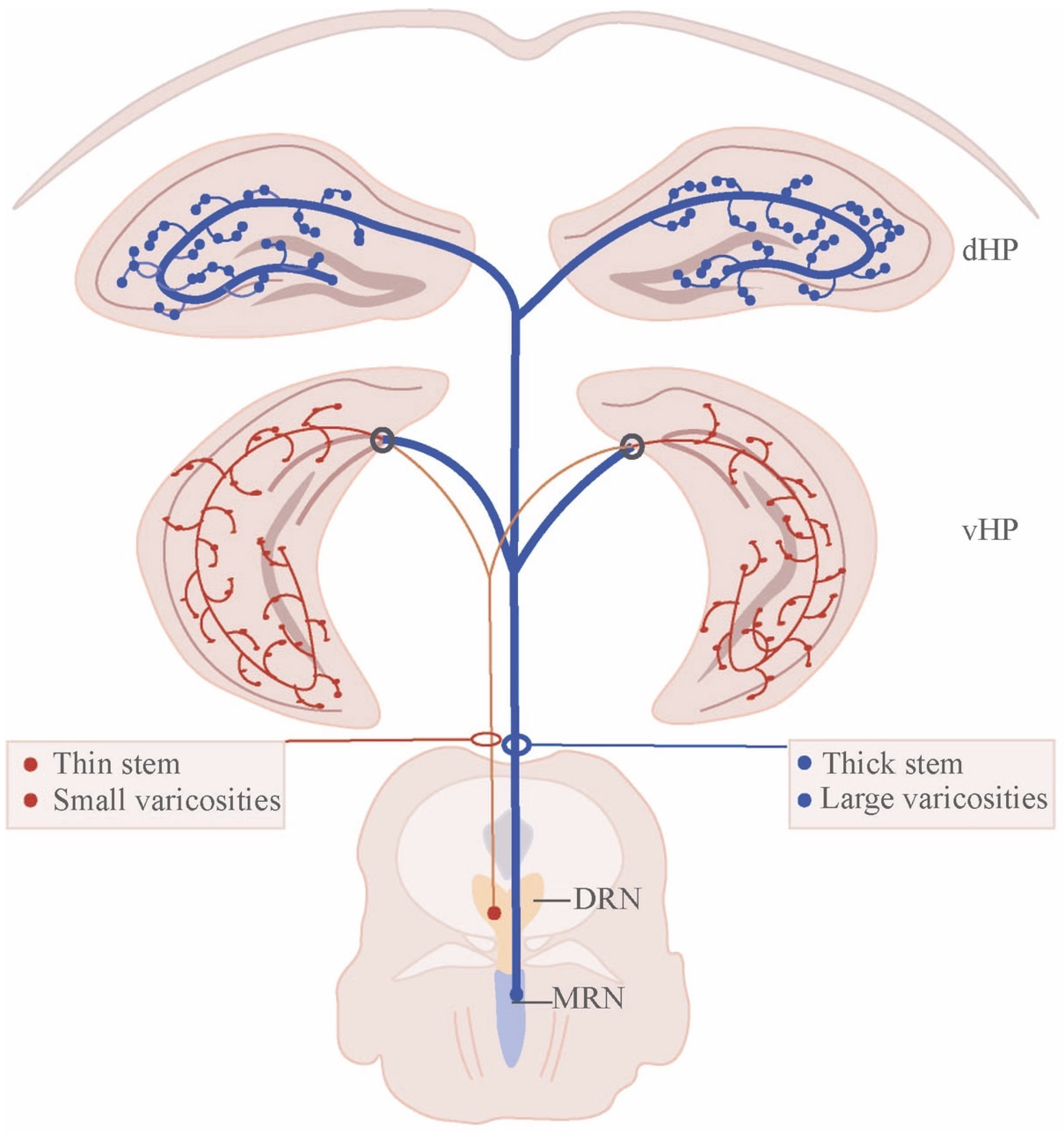
Investigations into the serotonergic system have disclosed the neurochemical, hodological, and electrophysiological diversity of raphe neurons [10]. The majority of 5-HT neurons are located in the DRN and MRN. Anterograde tracing using P. vulgaris leucoagglutinin (PHA-L) has shown that 5-HT neurons in the DRN and MRN differ significantly in their axonal morphology and projection pattern [11,26]. MRN-derived serotonergic neurons send irregularly spaced thick fibers with large and spherical varicosities (beaded fibers, BFs) [11,14,27], which mainly innervate midline brain regions, including dorsal and ventral hippocampus (vHP) [28,29], medial septum (MS) [30], and lateral hypothalamus [31]. In contrast, DRN-derived serotonergic axons are regularly spaced fine fibers (FFs) with typically small pleomorphic varicosities that are granular or fusiform in shape (Figure 2) [11,14]. FFs from DRN are uniquely connected to lateral brain regions associated with emotion and motivation (e.g., amygdala, nucleus accumbens, ventral tegmental area, ventral pallidum, and vHP) [31,32,33,34,35]. Interestingly, the caudal third of the DRN (hereafter referred to as the B6 region) shares similar connectivity with the MRN (B8); both of which dominate over the septum and hippocampus [31]. Thus, DRN and MRN output fibers innervate forebrain structures in a non-overlapping manner.
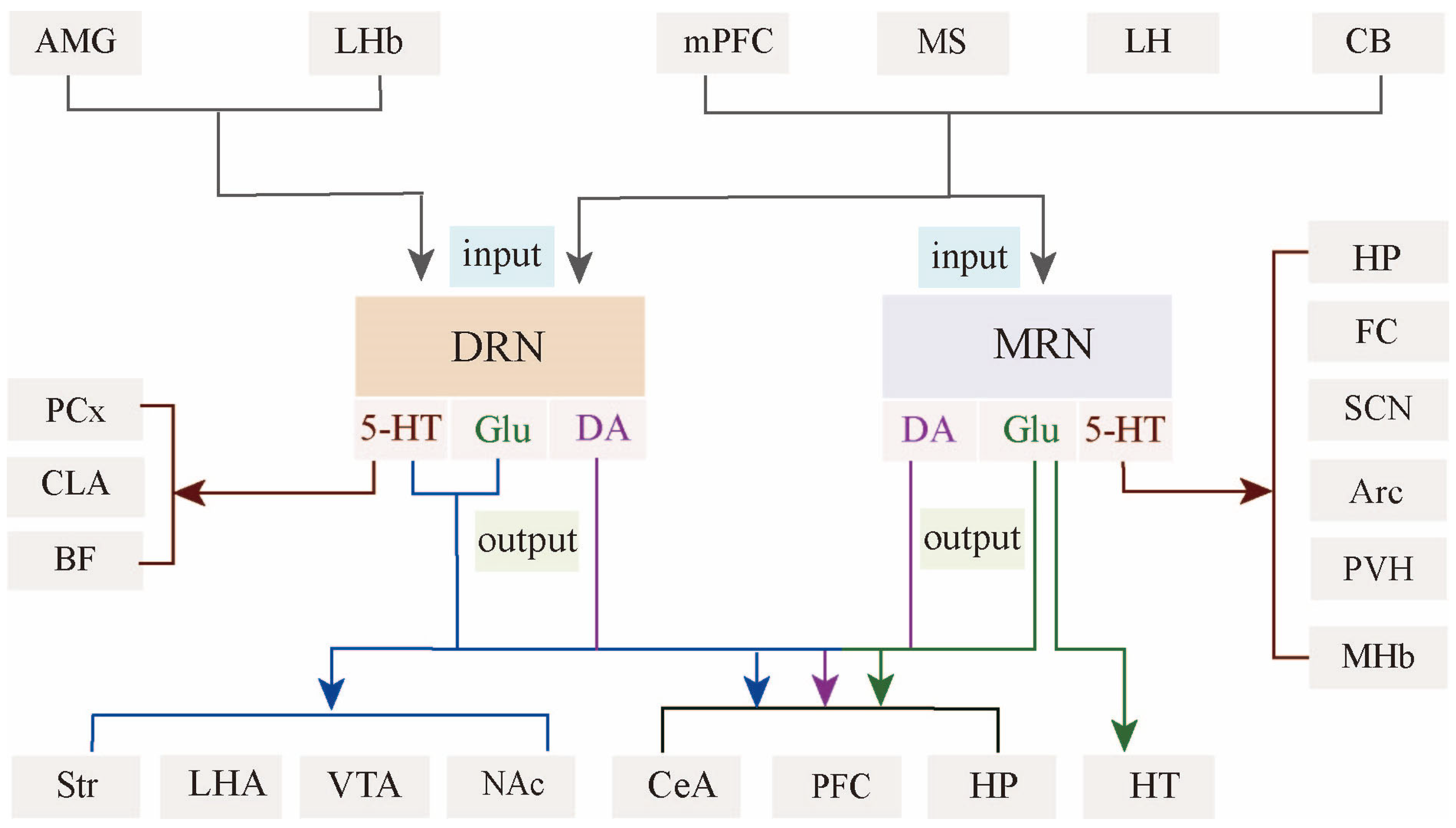
Meanwhile, DRN and MRN have a similar input pattern but display subtle differences in connection strength. For example, MRN5-HT neurons receive fewer afferents from the prefrontal cortex, with a preference for inputs from midline areas such as MS, hypothalamus, and brainstem [37,38,39,40]. Taken together, the efferent and afferent fibers of MRN correspond to midline structures, whereas those of DRN correspond to lateral structures (Figure 3). The different topographic organization of DRN and MRN circuits probably underlies the different neuronal properties and behavioral features.
In fact, 5-HT neurons represent only a small fraction of raphe nuclei [41,42]. Immunohistochemical studies have discerned that 5-HT neurons make up 25–30% of the total neuronal population in the DRN [37,43]. High-throughput single-cell transcriptome profiling of the DRN has uncovered five neuronal clusters in decreasing order of abundance: serotonergic, dopaminergic, GABAergic, glutamatergic, and peptidergic neurons [44]. Several molecularly defined subtypes of 5-HT neurons are found to co-express glutamic acid decarboxylase (GAD) or vGluT3, indicating that these 5-HT subpopulations co-release serotonin, gamma-aminobutyric acid (GABA), or glutamate [45]. Notably, 5-HT neurons only account for a minority of the total neuronal population (8.5%) in the MRN, while 26% of neurons are vGluT3, 12.8% are 5-HT and vGluT3 co-expressing neurons, 37.2% of neurons are GABAergic, and 14.4% are triple-negative [41]. Anterograde and retrograde tracing showed that ascending projections from MRN to the forebrain were predominantly vGluT3 neurons [20,46], reinforcing the notion that vGluT3 neurons make up the majority of projection neurons in the MRN [47].
Spatial single-cell RNA sequencing has identified gene expression in the subdomains of the DRN, such as lateral DRN neurons projecting to the colliculus and auditory brainstem nuclei, which express Pet1, Tph2, Sert, and En1. Dorsolateral DRN neurons express Pet1, Tph2, Sert, Drd2, Gad2, Pax5, and En1. Dorsal DRN neurons projecting to the central amygdala and the paraventricular nucleus of the hypothalamus express Pet1, Tph2, Sert, and En1. Ventromedial DRN neurons projecting to the frontal cortex express Pet1, Tph2, Sert, vGluT3, and En1. Cadual DRN neurons projecting to the septum and vHP express Pet1, Tph2, Sert, Met, and En1 [10]. Apart from the same gene expression of Pet1, Tph2, and Sert, MRN neurons projecting to the hippocampus, septum, and suprachiasmatic nucleus express Hoxa2, Met, Oxtr, Nr2f2, vGluT3, and Tacr3, while MRN neurons projecting to the dorsal tegmental nucleus of Gudden express Nmbr and Chrna4 [10], suggesting that 5-HT neurons exhibit molecular diversity. It is, therefore, convenient for researchers to label and modulate serotonergic neurons with genetically modified mouse models, such as Pet1-Cre [48,49], Tph2-Cre [50], Sert-Cre [51], and 5-HT1A-iCre mice [52].
The intrinsic electrical properties of 5-HT neurons depend on factors such as developmental stage, anatomical location, gene expression, and projection pattern. First, the electrical properties of 5-HT neurons change significantly during the first 3 weeks of life [53]. Due to a lack of 5-HT1A, autoreceptors, and GABA input, 5-HT neurons are hyperexcitable at postnatal day 4 (P4), 5-HT1AR-mediated auto-inhibition starts to develop by P21 when 5-HT neurons give rise to an increase in spontaneous spikes [53]. Second, 5-HT neuron activity is associated with anatomical location, such as the DRN versus the MRN [12], and the lateral DRN versus the medial DRN [54]. In vitro patch-clamp electrophysiological studies have demonstrated that 5-HT neurons in the lateral wing of the DRN are more excitable than ventromedial DRN5-HT neurons [54]. It is important to note that the difference in 5-HT neuron activity between the DRN subregions may be relevant to the input strength of presynaptic partners [39]. Moreover, the electrical properties of DRN5-HT neurons are largely different from those of the MRN, with MRN5-HT neurons having a larger afterhyperpolarization amplitude and a shorter time constant than those of the DRN [12]. Mechanistically, the inhibitory effects elicited by the activation of 5-HT1A autoreceptors are stronger in DRN5-HT neurons than in the MRN; furthermore, non-serotonergic neurons are responsive to 5-HT1AR agonists in the DRN but not in MRN non-serotonergic neurons [12].
DRN and MRN neurons are genetically divided into several clusters, and the electrical properties of raphe neurons vary among these clusters. In the MRN, Hoxa2-Pet1 neurons are more excitable than En1-Pet1 and Egr2-Pet1 neurons [55], and vGluT3 neurons fire faster and more variably than 5-HT neurons [56]. Moreover, 5-HT neurons projecting to the basolateral amygdala fired more frequently than those projecting to the medial prefrontal cortex (mPFC) and the dorsal hippocampus, suggesting that the intrinsic electrical properties of 5-HT neurons are linked to efferent projection target [26]. The electrophysiological diversity of 5-HT neurons indicates that the 5-HT neurons respond differently to external stimuli.

3. Architecture of Raphe Nucleus–Hippocampal Circuits

Ascending fibers from the DRN and MRN rarely innervate the same brain structure, with the exception of MS and hippocampus. Previous studies have shown that both DRN and MRN send dense serotonergic and non-serotonergic afferents to the hippocampus [57,58], but these ascending fibers to the hippocampus differ between subdivisions of DRN and MRN. Retrograde tracing combined with 5-HT immunohistochemistry has shown that a small number of retrogradely labeled 5-HT neurons are found in the B6 region, and a large number of 5-HT neurons are found in the B8 region [57]. Anterograde tracing of individual groups (B5–B9) of raphe nuclei confirmed the similar projection pattern between the B6 region of DRN and MRN [59], but the most notable differences were observed in the projections from the caudal DRN and the rostral DRN, with moderately dense afferents from the caudal DRN to hippocampus and none from the rostral DRN [60]. Interestingly, the forebrain afferents to the raphe nuclei showed disparity between rostral 2/3 of the DRN and the caudal third of the DRN, but similar afferent innervation patterns between B6 region of DRN and MRN [61]. Thus, the anterograde and retrograde tracing together suggest that the B6 region shares the similar afferent and efferent patterns with the MRN. This argument is supported by the same developmental origin that the B6 region and MRN are defined by the common expression of Engrailed-1 (En1, also called rhombomere 1) [62,63], as well as the same genetic lineage that Met is expressed in both the B6 region and MRN [55]. On the other hand, anatomically, the B6 region and MRN are not connected in the coronal view; however, they are closely connected in the sagittal section (Figure 1) [31].
The B6 region, in striking contrast to the rostral portion of DRN, sends projections to the dorsal and vHP [59,60]. It is assumed that the B6 region preferentially innervates the vHP. In support of this assumption, microdialysis experiments showed that electrical stimulations of the DRN (5 Hz, 300 μA, 20 min) evoked a short-lasting 5-HT release (an increase by 70–100%) in the vHP, but the dorsal hippocampus did not respond to the DRN stimulation, whereas stimulation of the MRN induced 70–100% of the 5-HT release in the dorsal hippocampus, vHP, and MS [29]. The assumption is further reinforced by the experiment that optogenetic activation of DRN5-HT neurons caused brain-wide activation, including the mPFC, striatum, and ventral tegmental area, but excluding the dorsal hippocampus [64]. In fact, DRN5-HT neurons are also involved in the regulation of hippocampal activity. For example, selective activation of serotonergic neurons in the DRN by infusion of substance P increased extracellular levels of 5-HT by 30% for 20 min in the vHP compared to the vehicle control [65], whereas perfusion of tetrodotoxin into the DRN simultaneously decreased hippocampal 5-HT release [66]. Chemogenetic activation of DRN neurons projecting to the vHP or axonal terminals in the vHP reduced c-Fos (a marker of neuronal activity) expression in the vHP via activation of postsynaptic 5-HT1BR [35]. In addition, DRN5-HT neurons preferentially reactivate neuronal ensembles in the dorsal dentate gyrus (DG) by activating dopaminergic signaling in the ventral tegmental area [67]. DRN-derived serotonergic fibers also project to the dorsal hippocampal CA1 and modulate excitability of CaMKⅡ-positive neuron via 5-HT1BR or 5-HT4R [68]. DRN5-HT neurons make synaptic contact with 5-HT3AR-positive interneurons, and dysfunction of DRN5-HT-HPHTR3A transmission results in an anxiety phenotype [69].
Among the raphe nuclei, MRN neurons establish strong synaptic contact with the hippocampal region. Retrograde tracing combined with 5-HT immunohistochemistry has shown that only half of the retrogradely labeled MRN neurons projecting to the hippocampus belong to 5-HT neurons, whereas the majority of retrogradely labeled neurons are vGluT3+5-HT neurons [70]. We and others have unveiled that serotonergic fibers from the MRN are largely distributed across the border between the stratum lacunosum moleculare (SLM) and stratum radiatum (SR) of the CA1, as well as in the subgranular zone in the hilus [49,58]. Both serotonergic and non-serotonergic afferents from the MRN have been shown to climb along proximal dendrites and soma of calbindin-positive interneurons [58,71,72], suggesting that GABAergic interneurons are the major postsynaptic target of MRN neurons [71]. For example, MRNvGluT3 neurons make direct synaptic contact with parvalbumin (PV) interneurons, thereby controlling the efficacy of spatial memory retrieval [23]. Both MRNvGluT3 and MRN5-HT neurons can activate 5-HT3AR-positive interneurons, thereby inducing a long-lasting increase in GABAergic transmission in CA1 pyramidal neurons [22,49]. MRNvGluT2 neurons selectively innervate septal PV interneurons that project to hippocampus; therefore, in vivo stimulation of MRNvGluT2 neurons instantly promotes memory acquisition in mice [7]. Another cluster of GABAergic interneurons, neuropeptide Y (NPY)-expressing interneurons, receives a serotonergic input from the MRN [73] and indirectly controls the firing of the hippocampal output neurons. Additionally, PV interneurons were selectively innervated by septal GABAergic neurons but avoided by MRN5-HT neurons [74], whereas a small number of cholecystokinin (CCK) interneurons receive input from both MRN and septal afferents [72]. In the raphe–hippocampal circuits, single or double synaptic contacts were more common on vasoactive intestinal polypeptide (VIP)-positive interneurons, which may ensure the rhythmic synchronization of inhibitory tones in the hippocampus [28].

4. Synaptic Control of Hippocampal Activity by the Raphe Nucleus

4.1. Fast Modulation of Hippocampal Inhibitory Network by Raphe Nucleus

The aforementioned architecture of raphe–hippocampal circuits can be summarized by the synaptic connection between MRN and hippocampal GABAergic interneurons, including calbindin-containing, PV, CCK, VIP, and NPY, as well as 5-HT3AR-expressing interneurons, which underly the fast synaptic control of the hippocampal inhibitory network. Therefore, it is the case that hippocampal inhibitory network is under control of the raphe nucleus. First, serotonin can attenuate feedback inhibition onto interneurons [75]. Increased GABA levels were found in the hippocampus of TPH2 knockout mice, indicating that 5-HT inhibits hippocampal GABA release [76]. Subsequently, Kao et al. found that electrical stimulation of MRN inhibited the majority (60%) of the GABAergic interneuron activity in the CA1 region [77]. These GABAergic interneurons located in the oriens lacunosum-moleculare (hereafter referred to as O-LM interneurons) of the hippocampal CA1 region can be classified into a caudal ganglionic eminence-derived subpopulation expressing 5-HT3AR and a medial ganglionic eminence-derived subpopulation lacking 5-HT3AR [78]. 5-HT released from serotonergic terminals profoundly and reversibly reduces the excitatory input from local CA1 collaterals to 5-HT3AR-expressing O-LM interneurons, and this effect can be mimicked by fenfluramine [75]. Mechanistically, 5-HT, via activation of presynaptic 5-HT1BR, substantially inhibits the excitation of CCK interneurons in the CA1 area, thereby selectively reducing feedback inhibition onto pyramidal neurons and allowing for an increase in the integration of time windows for spike generation by CA1 pyramidal neurons [79]. Overall, subcortical serotonergic neurons can control the hippocampal excitatory output by modulating inhibitory strength.
The innervation of hippocampus by serotonergic neurons depends on multiple postsynaptic 5-HTRs. 5-HT excites GABAergic interneurons through activation of ionotropic 5-HT3Rs [80]. Varga et al. have shown that selective stimulation of MRN5-HT neurons produced rapid activation of hippocampal interneurons, and MDL72222 (a 5-HT3AR antagonist) only reduced the amplitude of excitatory input into interneurons, whereas a combination of both 5-HT3AR antagonists and glutamate receptor blockers can fully abolish the excitation of hippocampal interneurons, suggesting that serotonergic neurons co-release 5-HT and glutamate to excite GABAergic interneurons [81]. In actuality, MRNvGluT3 neurons send primary afferents to the hippocampus. Likewise, MRNvGluT3 neurons can activate 5-HT3AR-expressing GABAergic neurons at the SR/SLM border, and this process is negatively regulated by 5-HT/5-HT1BR signaling [22]. In addition, 5-HT2ARs are predominantly, but not solely, present in the interneurons located at the border of SR/SLM of the CA1 region [82], and 5-HT can stimulate GABA release via activation of 5-HT2AR signaling [83]. Serotonergic neurons have also been found to establish multiple synaptic contacts with NPY interneurons that innervate the distal dendrites of principal cells, thereby controlling the excitability of hippocampal output neurons [73]. Meanwhile, serotonergic neurons also innervate VIP interneurons that mediate feedforward inhibition onto GABAergic interneurons and control rhythmic synchronization of inhibitory networks [28]. Altogether, whether the profound effect of raphe–hippocampal pathway is mediated by GABAergic interneurons or via a direct action on excitatory neurons remains to be determined [74], but it is clear that the serotonergic and glutamatergic input from MRN governs rapid modulation of the hippocampal inhibitory network.

4.2. Subcortical Control of Hippocampal Theta-Rhythm by Raphe Nucleus

Theta rhythm is an oscillatory neural activity that fires at a frequency of 3–10 Hz in the hippocampus and is deeply implicated in mnemonic, motor, and sensory functions [84,85,86]. A growing body of evidence suggests that activation of the MRN suppresses theta rhythms, whereas inhibition of the MRN induces persistent theta [87,88]. Interestingly, there is a dissociable pathway to facilitate theta and non-theta states in the raphe–hippocampal circuit. Selective inactivation of the MRN serotonergic output with 5-HT1AR agonist (8-OH-DAPT) induces hippocampal theta rhythm, whereas activation of non-serotonergic (possibly glutamatergic) MRN-hippocampal pathway promotes hippocampal theta generation [87]. During hippocampal theta oscillations, MRN5-HT neurons exhibit little or no theta-related activity, whereas most GABAergic neurons and vGluT3 neurons in the MRN show increased and decreased activity, respectively [89,90]. GABAergic neurons establish a monosynaptic connection with local serotonergic and vGluT3 neurons; therefore, even if MRN GABAergic neurons do not directly innervate the hippocampus, they could still modulate hippocampal theta oscillations by regulating the activity of local vGluT3 and serotonergic neurons [89,91]. Pharmacological evidence has shown that infusion of an N-methyl-D-aspartate (NMDA) receptor antagonist (MK-801) into the MRN or a GABAA receptor agonist (muscimol) facilitates the hippocampal theta oscillation in urethane-anesthetized rats [88,92,93]. Collectively, these results suggest that the local inhibitory circuit in the MRN may synchronize ascending serotonergic/glutamatergic modulation with hippocampal theta rhythm on a sub-second timescale [94].
Hippocampal theta rhythm is modulated by synchronizing afferents from the MS and desynchronizing afferents from the MRN. Inhibition of the MS suppresses hippocampal theta rhythm, whereas inhibition of the MRN produces sustained theta, and this effect can be reversed by inhibiting the MS, suggesting that MRN–MS–hippocampal circuit promotes different brain states (theta or non-theta) [87]. 5-HT depletion in the hippocampus resulted in an increased expression of high-frequency theta activity, which facilitates spatial learning in the Morris water maze [95]. Concomitantly, 5-HT depletion in the MS induced both low-frequency (4.5–6.5 Hz) and high-frequency (6.5–9.5 Hz) theta activity [95]. Additionally, 5-HT depletion in the supramammillary or posterior hypothalamic nucleus results in theta rhythm alterations in the CA1 region [96]; infusion of 8-OH-DAPT into the pedunculopontine tegmental nucleus induces prolonged spontaneous theta rhythm [97]. These results suggest that ascending serotonergic fibers indirectly modulate hippocampal theta rhythm through synaptic contacts with other nuclei, such as MS, supramammillary nucleus, and posterior hypothalamic nucleus.

4.3. Subcortical Control of Synaptic Plasticity in the Hippocampus

Theta rhythm has been shown to regulate long-term potentiation (LTP) in the hippocampus [98], strongly suggesting that ascending projections from the raphe nucleus may also control hippocampal synaptic transmission and plasticity. Likewise, the effects on hippocampal LTP induction vary across different neuronal types of the MRN. Optogenetic activation of serotonergic terminals in the CA1 region enhances excitatory transmission in both Schaffer collateral pathway and LPP-DG pathway [99,100]. In contrast, activation of MRNvGluT3 afferents suppresses LTP induction in the CA3–CA1 pathways in male mice [22]. The underlying molecular mechanism is also different, with the serotonergic enhancement of LTP acting through activation of 5-HT4R, whereas glutamatergic suppression of LTP occurs through activation of GABAergic interneuron-mediated feedforward inhibition [22,99].

5. Raphe–Hippocampal Serotonergic and Glutamatergic Circuits Regulate Emotional Behavior

5.1. Anxiety

It is well established that DRN5-HT neurons play a pivotal role in regulating anxiety behavior through their projections to emotion-related nuclei, such as amygdala [32,101], bed nucleus of the stria terminals [102], and vHP [69]. Although the role of the MRN in the regulation of anxiety has received less attention than that of the DRN, there is increasing evidence that MRN–hippocampal serotonergic and glutamatergic circuits control anxiety behavior. Previous studies have shown that electrolytic or pharmacological lesions of MRN exert an anxiolytic effect in both restraint and nonrestraint rats [103]. Conversely, Netto et al. showed that lesions of MRN5-HT neurons produced anxiogenic behavior in chronically restrained rats [104]. Although the role of MRN in the regulation of anxiety remains controversial, emerging evidence supports the assumption that activation of MRN5-HT neurons promotes anxiety. For example, selective optogenetic activation of MRN5-HT neurons enhances anxiety-like behavior [105]; inhibition of MRN5-HT neurons promotes anxiolysis [106], whereas activation of DRN5-HT neurons has antidepressant-like effects [105,107]. Activation or inhibition of MRN5-HT neurons via infusion of 5-HT1AR agonists/antagonists yielded similar results, with 8-OH-DAPT inducing anxiolytic effects, whereas WAY-100635 induced the opposite effects [108,109,110]. Another critical question is: How does the MRN modulate anxiety behavior? Electrophysiological recordings combined with in vivo microdialysis confirmed that pharmacological or optogenetic stimulation of MRN5-HT neurons produced rapid 5-HT release in the dorsal hippocampus, which may enhance anxiety [105,110,111]. In support of this view, simultaneous injections of 5-HT-acting drugs into the MRN and dorsal hippocampus demonstrated that the MRN regulates anxiety via serotonergic innervation of the dorsal hippocampus [106].
Ascending glutamatergic afferents from raphe to the hippocampus are also involved in the modulation of anxiety. DRN and MRN contain a mass of vGluT3 neurons projecting to the hippocampus [46]. vGluT3 knockout mice showed increased anxiety behavior [21,112], whereas activation of MRNvGluT3 neurons caused anxiolytic effects [112]. Notably, most of vGluT3 immunopositivity is found on nerve terminals in the hippocampus [113], and the vGluT3 positively modulates 5-HT transmission by decreasing 5-HT1AR-mediated autoinhibition [21]. Therefore, deletion of vGluT3 is likely to excite 5-HT neurons and thereby promote anxious behavior. In contrast, DRN vGluT3 and 5-HT double-positive neurons control separate behavioral features of anxiety via their projection to the amygdala but not the hippocampus [32]. Thus, DRNvGluT3 and MRNvGluT3 neurons modulate anxiety via different pathways.

5.2. Depression

Hypoactive 5-HT neurons have been implicated in the pathophysiology of depressive-like behaviors [114]. However, recent studies have suggested that MRN and DRN neurons respond differently to depression symptoms. A hyperactivated DRN with overactive projections to the amygdala, striatum, and periaqueductal gray, and a hypoactive MRN with underactive projections to the hippocampus have been found in patients with major depressive disorder (MDD) [115]. Normally, MRN input to the hippocampus inhibits aversive memory consolidation, and increased hippocampal hyperactivity in MDD may result from the disrupted MRN–hippocampal pathway [115]. It has been suggested that an activated MRN5-HT-DGCamKII pathway underlies behaviors of despair in mice subjected to a regimen of chronic behavioral despair [116]. MRN contains a large population of vGluT2-expressing neurons that receive input from the negative experience-related nucleus and project to the aversion centers, and these neurons were selectively activated by aversive stimuli, thereby inducing depression-related anhedonia [7]. Overall, MRN serotonergic and glutamatergic afferents to the hippocampus may promote depression. Therefore, it is the case that simulation of the MRN decreased locomotion and increased depressive-like behaviors [117].
In contrast to the MRN, activation of DRN5-HT neurons has antidepressant-like effects [107]. DRN5-HT can retrieve positive memory ensembles in the DG mediated by 5-HT-activated dopaminergic signaling from the ventral tegmental area, providing insight into how DRN5-HT neurons elicit antidepressant effects [67]. In addition, reduced serotonergic transmission from the DRN to LHb neurons would increase LHb activity and contribute to depressive symptoms; optogenetic activation of the DRN–LHb circuit mitigates the depressive symptoms in a rat model of chronic unpredictable mild stress [118].

5.3. Aggression

In the literature, 5-HT is thought to play an inhibitory role in controlling aggression. Inhibition of 5-HT synthesis by genetic ablation of TPH2 reduces anxiety but exaggerates aggression in male rats [119,120]. Similarly, selective lesions of DRN5-HT or MRN5-HT neurons promote submissive cats to engage in dominance fights, and biochemical analysis revealed a significant reduction in 5-HT and 5-HIAA in the hypothalamus, the amygdala, and the hippocampus in either MRN- or DRN-lesioned rats [121]. Furthermore, 5-HT levels were decreased in the prefrontal cortex, the striatum, the amygdala, the hippocampus, and the DRN in stressed animals compared to non-stressed animals [120]. This evidence was sufficient to link the reduced cerebral 5-HT levels and serotonergic activity to exaggerated aggression. Given that chronic social defeat paradigm simultaneously induced a marked increase in serotonin transporter (SERT) expression in the DRN and CA1/CA3 of the hippocampus [122], raising the possibility that the raphe–hippocampal pathway engages in the regulation of aggression. Indeed, while the hyperactivated raphe–hippocampal pathway is involved in the stress-evoked escalated aggression, chemogenetic activation of the DRN–vHP pathway reduces reactive aggression via suppression of vHP activity by activating 5-HT1BR, then the vHP suppresses the downstream ventromedial hypothalamus (VMH), which is the core aggression center [35]. Infusion of 5-HT1BR agonists (anpirtoline) into the vHP reduces neuronal activity projecting to VMH and reactive aggression [35]. These results suggest that DRN–vHP inhibits aggression via innervation of VMH.

5.4. Addiction and Reward

The serotonergic system dynamically interacts with the hippocampus to regulate reward-related information processing. DRN5-HT neurons are responsible for encoding the reward signal by co-releasing 5-HT and glutamate [9,123,124,125]; these neurons are selectively recruited by emotional stimuli rather than neutral stimuli, and subsequently, they activate the downstream ventral tegmental area but not the hippocampus [126]. Meanwhile, in vivo two-photon imaging has screened two distinct neuronal populations in the MRN that project to the hippocampus: one is associated with reward delivery and the other with locomotion, and stimulation of these hippocampal projection fibers modulates reward-induced behavior [127]. During successful goal-directed behavior, there is an increase in serotonergic transmission from MRN to the vHP, which suppresses vHP activity by activating 5-HT3AR. It can be concluded that suppression of the vHP via MRN5-HT-vHP5-HT3AR is required for successful goal-directed behavior [128]. Overall, these findings suggest that DRN and MRN target different nuclei to modulate reward signaling.
Alcohol addiction is associated with an alteration in 5-HT neuronal function, with a transition from the DRN-dependent regulation of short-term alcohol intake to the MRN-dependent regulation of long-term alcohol intake [129]. In mice with long-term voluntary alcohol consumption, the serotonergic fibers projecting from MRN to the DG were severely affected by the altered expression of 5-HT1A autoreceptors in MRN5-HT neurons [129]. Chemogenetic inhibition of hippocampal serotonergic fibers from MRN5-HT neurons alleviates alcohol addiction after long-term alcohol consumption [129].

6. Subcortical Modulation of Memory by Raphe–Hippocampal Circuits

6.1. Aversive Memory

Repeated aversive stimuli, such as enemy odor, foot electric shock, and pain, help to form and consolidate aversive (fear) memories. During this process, serotonergic and glutamatergic afferents from the MRN–hippocampus are motivated to calibrate the memory storage. MRN is believed to control the fear memory via innervation of amygdala, hippocampus, and prefrontal cortex. Either acute or chronic lesions of the MRN clearly suppressed freezing behavior in rats [130]. In contrast, electrical or optical stimulation of the MRN alone consolidates remote but not recent fear memory traces, and optogenetic silencing of the MRN during foot shock, which attenuates conditioned fear memories [131]. Next, it must be asked whether the serotonergic projections from MRN–HP influence fear memories. The results show that infusion of the 5-HT1AR agonist or the corticotropin-releasing factor receptor antagonist into either MRN or hippocampus abolished contextual freezing, suggesting that ascending 5-HT fibers from MRN to the hippocampus are implicated in the acquisition of fear memories [132,133]. It has been proposed that stimulation of the MRN interferes with fear memory consolidation by suppressing hippocampal sharp-wave ripple oscillations [134]. Meanwhile, suppression of MRNvGluT2 neurons impaired both contextual and cued fear memory formation via activation of septal–hippocampal inhibitory circuits [134]. Furthermore, activation of MRNvGluT3 neurons enhances the GABAergic input to pyramidal neurons in the mPFC and selectively attenuates fear memories in female but not male mice [135]. Therefore, it can be concluded that innervation of the hippocampus by MRN provides bottom-up control of fear circuits, complementing the top-down control by mPFC [131,136,137].

6.2. Spatial Memory

The serotonergic system plays a crucial role in the regulation of spatial memory through its projections to the hippocampus. Acute depletion of tryptophan, the primary amino acid used to synthesize 5-HT, impairs memory consolidation but improves focused attention in healthy individuals [138,139]. Postnatal depletion of 5-HT via administration of parachlorophenylalanine (PCPA, an inhibitor of 5-HT synthesis) not only causes neuronal loss in the hippocampus but also impairs spatial memory during adulthood [140]. In line with this finding, global depletion of cerebral 5-HT (Pet1 knockout mice) leads to significant deficits in novel object recognition memory [141]. Taken together, these findings underscore that 5-HT is essential for memory consolidation.
The literature indicates that 5-HT can modulate hippocampal activity in a bidirectional manner, either through direct synaptic contact with hippocampal neurons or indirectly via MS cholinergic/GABAergic inputs to the hippocampus. Selective depletion of 5-HT in MS disrupts the septal-hippocampal information relay [95,142]. Infusion of 5, 7-dihydroxytrptamine (5, 7-DHT) into the MS potentiates the acquisition of spatial information and working memory [95,142]. Additionally, hippocampal 5-HT depletion facilitates place learning in the Morris water maze [95]. However, simultaneous lesions of septal cholinergic innervation of the hippocampus and the serotonergic pathway result in a reduction in hippocampal inhibition and behavioral deficits, and intrahippocampal raphe grafts can ameliorate the spatial memory deficits in rats combined with serotonergic and cholinergic lesions [143,144], suggesting a complementary role for the serotonergic innervation of the hippocampus after loss of septal cholinergic innervation of the hippocampus.
Stimulation of MRN5-HT or DRN5-HT neurons produces conflicting results in spatial memory impairment. It has been reported that inhibition of serotonergic neurons from the MRN, but not DRN, is sufficient to impair object recognition in adult mice [141]. Electrolytic lesions of the MRN have a profound effect on both acquisition and retention in the eight-arm maze task [145]. Moreover, optogenetic activation of serotonergic terminals in the CA1 region enhances excitatory transmission at the CA3–CA1 pathways and spatial memory in mice, whereas optogenetic silencing of serotonergic terminals impairs spatial memory [99]. Blockade of hippocampal activity via activation of 5-HT1A heteroreceptors impairs the acquisition and performance of a spatial task in the water maze [146]. In contrast, several studies have found that inactivation of either the MRN or the hippocampus has no effect on spatial memory retrieval and consolidation [147,148]. Whether serotonergic innervation of the hippocampus is necessary for regulating hippocampal synaptic plasticity and spatial memory remains to be further determined [141]. The different results may depend on MRN cell types, animal models, stimulation methods, etc. For example, it has been found that stimulation of MRNvGluT3 neurons improves spatial memory retrieval by strengthening MRNvGluT3-HPPV synaptic transmission in AD mice [23].
Interestingly, inactivation of serotonergic neurons from the DRN attenuates the spatial learning deficits in mice with the loss of cholinergic innervation of hippocampus [149,150], whereas blocking orexin 1 receptors in the DRN impairs spatial memory consolidation in rats [151]. These results suggest that DRN5-HT neurons also participate in regulating spatial memory but in a manner opposite to that of MRN neurons.

6.3. Social Memory

In the hippocampus, CA2 pyramidal neurons act as a hub receiving a variety of inputs from subcortical areas, including MS, supramammillary nucleus, and MRN, thereby modulating social aggression and memory [152]. However, the serotonergic circuit between raphe and hippocampus is remodeled under social stress. Chronic social stress markedly reduced 5-HT neuronal activity and hippocampal 5-HT reuptake [153,154]. In contrast, stimulation of the MRN increased friendly social interactions in the resident intruder test [117]. 5-HT released from the MRN not only promotes social interaction but also consolidates social memory. Stimulation of MRN5-HT neurons bidirectionally regulates social memory by activating dorsal CA2-projecting MS glutamatergic neurons [155]. With the exception of serotonergic neurons, both GABAergic and dopaminergic neurons in the MRN contribute to social interactions in mice but have no effect on social memory [156,157].

7. Abnormal Raphe–Hippocampal Circuits in AD Patients and Mouse Models

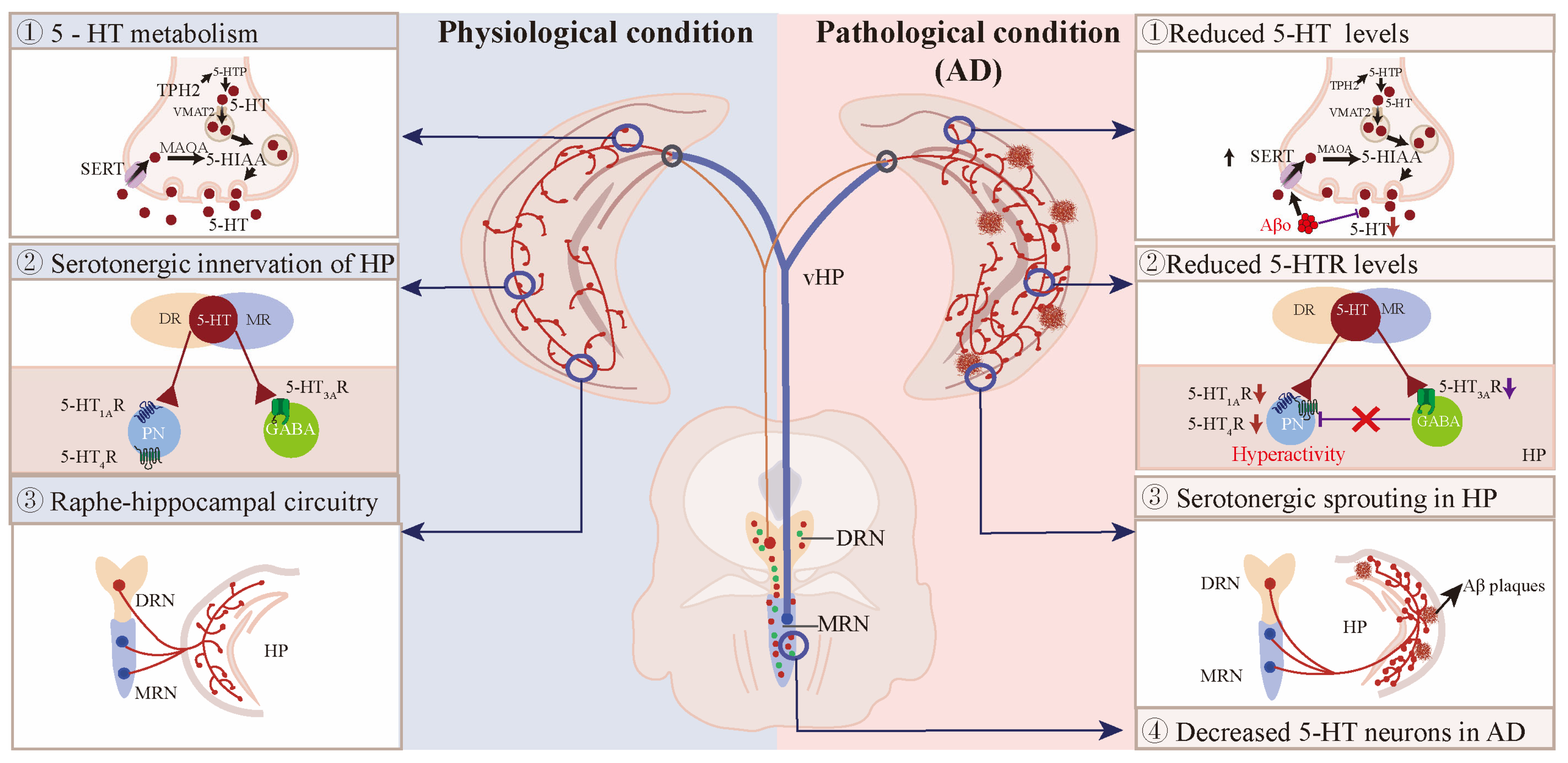
AD is the most common type of neurodegenerative diseases in the elderly, and it has been well established that the clinical symptoms of AD are characterized by β-amyloid (Aβ) plaques, phosphorylated Tau, and gliosis in the cerebral parenchyma, resulting in progressive cognition decline [158]. In addition to the obvious cognitive deficits, AD patients also suffer from anxiety [159], depression [160] and sleep disturbances [161]. These psychiatric disorders have linked the disrupted raphe–hippocampal circuitry to cognitive decline in AD (Figure 4).

7.1. The 5-HT Neurons in AD Patients and Mouse Models

Approximately 90% of AD patients suffer from NPS years prior to cognitive decline, and recent evidence has linked the early behavioral symptoms to pathological changes in the raphe nucleus [3]. The loss of 5-HT neurons in the raphe nuclei, including raphe obscurus and pallidus, MRN, and DRN, has been reported in the early stages of AD (Table 1) [162,163]. Immunostaining for TPH2 showed that although no significant difference was found between young and aged dogs without cortical Aβ deposits, aged dogs with cortical Aβ pathology had 33% fewer 5-HT neurons in the DRN and MRN than those without cortical Aβ pathology [164]. However, in AD mouse models of overexpressing human amyloid precursor protein (hAPP), the results from our group and others have shown that there was no significant difference in the number of 5-HT neurons in either DRN or MRN between non-transgenic and hAPP mice [49,165]. It can be concluded that aging is not responsible for the degeneration of 5-HT neurons, but that the degeneration of serotonergic system correlates with the development of cortical Aβ deposits.
Pathohistological studies have shown that the early onset of tauopathy results in a reduced number of 5-HT neurons in the raphe nucleus in both AD patients and mouse models [163,174,175]. Specifically, AD patients had significantly more neurofibrillary tangles in both MRN and DRN than in controls, leading to a 41% reduction in DRN neuron density and a 29% reduction in MRN neuron density [163]. Moreover, 2.6% of DRN neurons contain hyperphosphorylated Tau in AD patients at Braak stages 0 and 2, and hyperphosphorylated Tau in raphe nuclei deteriorate with AD progression [174]. Another study showed that neurofibrillary tangles were detected in the supratrochlear subnucleus of DRN in 1/5 of AD patients at Braak stage 0, and in all patients at Braak stages above 1 [175]. Consistent with AD patients, mice overexpressing human P301L Tau in the DRN recapitulate the degeneration of serotonergic system in AD patients, including profound 5-HT neuron loss, 5-HT neuron hyperexcitability, inflammation, axonal degeneration, as well as cognitive impairment and behavioral symptoms, including depressive- and anxiety-like behaviors, hyperlocomotion, and social deficits [172,176,177]. Upregulated inflammation in the DRN can exacerbate Tau phosphorylation, aggregation, and propagation alongside serotonergic projection targets [172]. It is also noteworthy that neurofibrillary tangles are thought to initiate from the DRN and then spread to its connected brain regions [176]. Taken together, these findings suggest that tauopathy, rather than that of Aβ pathology, is responsible for the degeneration of serotonergic system in AD.

7.2. Reduced 5-HT and Its Metabolites in the Hippocampus of AD Patients and Mouse Models

Accordingly, 5-HT and its metabolites were markedly altered in various brain regions of AD patients and mouse models (Table 2), including the hippocampus, cortex, amygdala, thalamus, substantia innominata, substantia nigra, putamen, and locus coeruleus [178,179]. Serotonergic deficiency seems to correlate with dementia type. For example, 5-HT levels were significantly increased in the brains of frontotemporal dementia (FTD) patients compared with AD patients, whereas the 5-HIAA/5-HT ratio was decreased in FTD patients compared with healthy controls [170]. In contrast, 5-HT and 5-HIAA were significantly reduced in 15 brain regions of AD patients comorbid with Down syndrome (DS) as compared to early-onset AD patients [180]. DS patients have monoaminergic deficits similar to DS + AD patients, suggesting that 5-HT deficiency is possibly due to early Aβ deposition [180]. Second, cerebral 5-HT levels were closely related to NPS in AD patients. Hippocampal 5-HIAA, 5-HIAA/5-HT, and 5-HIAA levels were significantly decreased in AD patients with aggression compared to those without aggression [171], and hippocampal 5-HIAA levels were negatively correlated with agitation scores in AD patients [181], suggesting that the brain-region specific alterations of 5-HT and its metabolites contribute to the occurrence of NPS in AD [171]. Lastly, the mini-mental state examination (MMSE) score, used as a measure of cognition, correlated positively with both hippocampal 5-HIAA levels and cortical 5-HT levels, indicating that hippocampal 5-HT/5-HIAA levels may predict cognitive decline in AD [181].
Consistent with AD patients, both 5-HT levels and 5-HIAA/5-HT ratio were significantly reduced in AD mouse models compared to age-matched controls. In the hippocampus of 18-month-old APP/PS1 mice, 5-HT was reduced by approximately 25% [182]. Similarly, hippocampal 5-HIAA/5-HT turnover was markedly lower in tgDimer mice than in non-transgenic mice [183]. Therefore, early Aβ deposition is associated with the progressive loss of hippocampal 5-HT with age [184]. Meanwhile, in a mouse model of co-expressing APP and Tau, 3×Tg-AD mice showed a significant decrease in 5-HT levels but a significant increase in 5-HIAA levels in the hippocampus [165]. It has been suggested that reduced hippocampal 5-HT and 5-HIAA levels are associated with increased depressive and aggressive behavior in 12-month-old THY-Tau22 mice [184].
Table 2. 5-HT and 5-HIAA levels in the hippocampus of AD patients and mouse models.
Research
Model
Age
(Year or Month)
Hippocampal
5-HT/5HIAA
References
MCI patient73.2 ± 10.8 y5-HT↓[185]
MCI patient83.0 ± 0 y5-HIAA↓[167]
AD patient75.0 ± 11.9 y5-HT↓, 5-HIAA↓[186]
AD patient70.0 ± 8.7 y5-HT↓, 5-HIAA↓[187,188]
AD patient82.0 ± 8.0 y5-HT↓, 5-HIAA↓[181,189]
hTau mice4 m5-HT↓[172]
hAPP-J20 mice4–5 m5-HT↓, 5-HIAA↓[49]
5×FAD mice9–10 m5-HT↓[190]
APPswe/PS1dE9 mice2, 3m
4 m
5-HIAA (NC),
5-HT↓, 5-HIAA (NC)
[191]
3×Tg-AD mice3 m5-HT↓, 5-HIAA↑[165]
THY-Tau22 12 m5-HT↓, 5-HIAA↑[184]
APP/PS1 mice18 m5-HT↓[182]
Abbreviations: MCI: mild cognitive impairment; AD: Alzheimer’s disease; 5-HT: 5-hydroxytryptamine; 5-HIAA: 5-hydroxyindoleacetic acid; NC: no change; year or month: year or month; APP: amyloid precursor protein; PS1: presenilin-1; FAD: familial Alzheimer’s disease; Tg: transgenic; ↓ indicates a decrease in 5-HT or 5-HTIAA levels; ↑ indicates an increase in 5-HT or 5-HTIAA levels.
The 5-HT deficiency in AD patients can be attributed to decreased upstream production and increased downstream degradation. TPH2 activity was increased by 4.7-fold, and 5-HT and 5-HIAA levels increased by 4- and 2-fold, respectively, in the soma of raphe neurons in AD brains compared to healthy individuals; in contrast, the level of 5-HT and 5-HIAA decreased by 41% and 50%, respectively, in the synaptic terminals projecting to the amygdala [189]. However, the overt TPH protein or TPH-positive neurons were found to be significantly reduced in the DRN of hTau mice [192]. Another explanation for cerebral 5-HT deficiency is the reduced tryptophan levels in the cerebrospinal fluid (CSF) of AD patients [193]. Therefore, the hippocampal 5-HT deficiency may result from the diminished transport of TPH to axon terminals, the overt reduction in TPH protein, or metabolic alterations [192].

7.3. The Expression of 5-HTRs and SERT in the Hippocampus of AD Patients and Mouse Models

The 5-HT-dependent regulation of emotion and cognition relies on its binding to different 5-HTRs. According to their topographic structure, 5-HTRs are divided into two families with 14 subclasses: the first type belongs to G-protein coupled receptors (GPCRs), including 5-HT1A/BR and 5-HT7R (Gi-coupled), 5-HT2A/B/CR (Gq/11-coupled), 5-HT4R, 5-HT6R, and 5-HT7R (Gs-coupled) [13]. Another type is the ligand-gated cation channel, which comprises only 5-HT3AR. Previous functional tomographic imaging combined with immunohistochemical studies have shown that the expression of 5-HTRs is disrupted in the cerebral cortex, hippocampus, amygdala, and hypothalamus area of AD patients and mouse models; some receptors, such as 5-HT6Rs, have been summarized in other excellent reviews [14,194,195]. The main focus here is on the three highly expressed 5-HTRs (5-HT1AR, 5-HT2AR, 5-HT3AR, and 5-HT4R) in the hippocampus. Table 3 summarizes the expression of 5-HTRs in the hippocampus of AD patients and mouse models.
The 5-HT1ARs are located either on the soma and dendrites of serotonergic neurons acting as autoreceptors, where their activation results in the inhibition of 5-HT release, or on the postsynaptic neurons acting as heteroreceptors. Positron emission tomography (PET) combined with isotopically labeled 5-HT1AR antagonists has shown that 5-HT1ARs are significantly reduced in the hippocampi and raphe nuclei of AD patients and mouse models [49,196], and the reduction in hippocampal 5-HT1ARs was positively correlated with cognitive decline while inversely correlated with increased amyloid plaque burden [197,198,199]. Interestingly, the hippocampal 5-HT1ARs in MCI patients are much higher than in the healthy controls but lower than in mild AD patients, suggesting that hippocampal 5-HT1ARs may distinguish MCI patients from mild AD patients [185]. The decreased 5-HT1ARs can be interpreted as a consequence of CA1 neuron loss in the late stages of AD, which is supported by the evidence that hippocampal 5-HT1ARs are preserved until the progression of neurofibrillary tangles in the late stages of AD [199,200]. Moreover, high levels of Aβ40 fibrils, but not Aβ42, can induce the transient astrocytic overexpression of 5-HT1ARs to compensate for hippocampal neuronal loss in AD [201]. Thus, whether the increased expression of 5-HT1ARs in early AD is beneficial or detrimental is yet to be determined [194].
Additionally, there is a 20–30% reduction in cortical 5-HT2ARs in MCI patients compared to healthy controls [202]. Intrahippocampal injection of aggregated Aβ42 in rats reduced hippocampal 5-HT2ARs, and age-related amyloid burden was accompanied by a significant decrease in 5-HT2AR binding, suggesting a causal relationship between Aβ accumulation and the downregulation of hippocampal 5-HT2ARs [203,204]. In contrast, 5-HT2BR mRNA levels were 2-fold higher in Aβ plaque-containing neocortex and hippocampus of old AD patients and mouse models than in age-matched controls [205].
PET imaging results showed that the density of 5-HT3R remains stable in the amygdala and hippocampus of AD patients compared to healthy individuals [206]. However, our previous results showed that the 5-HT3R mRNA started to decline in 4-month-old hAPP-J20 mice, while the 5-HT3R levels were significantly reduced in 8-month-old hAPP-J20 mice. Given that 5-HT3R mediates the tonic inhibition of pyramidal neurons, the reduction in hippocampal 5-HT3R contributes to the hyperexcitability of CA1 pyramidal neurons in hAPP-J20 mice [49]. Meanwhile, PET imaging results also showed a marked loss of 5-HT4R in the hippocampus and frontal cortex of AD patients [207], but there was no significant change in 5-HT4R in the DG of hAPP-J20 mouse models [49]. The discrepant results between AD patients and mouse models may be attributed to factors, such as experimental methods, as well as APP- or Tau-expressing genes’ age.
Table 3. The expression of 5-HTRs in the hippocampus of AD patients and mouse models.
Research
Model
Age
(Year or Month)
Hippocampal
5-HTRs
Reference
MCI patient76.8 ± 11.6 y5-HT1AR↓[197]
MCI patient83.0 ± 0 y5-HT1AR↓[208]
MCI patient73.2 ± 10.8 y5-HT1AR↑[185]
MCI patient73.2 ± 10.8 y5-HT1AR↑[187]
AD patient81.7 ± 2.0 y5-HT1AR↓[208]
AD patient75.0 ± 11.9 y5-HT1AR↓[197,199]
AD patient70.0 ± 8.7 y5-HT1AR↓[185,187]
AD patient82.0 ± 8.0 y5-HT4R↓[209]
AD patient83.1 ± 5.8 y5-HT4R↓[207]
AD patient77.0 ± 4.0 y5-HT3R (NC)[206]
HTau mice4 m5-HT1BR↓, 5-HT4R↑,
5-HT1AR↑, 5-HT7R↑
[172]
hAPP-J20 mice4, 8 m5-HT1AR↓, 5-HT3AR↓,
5-HT4R (NC)
[49]
5×FAD mice3, 6 m5-HT1BR↓, 5-HT2BR↓,
5-HT3BR↓, 5-HT4R↓,
5-HT6R↓, 5-HT7R↓,
5-HT1FR↓
[68]
APPswe/PS1dE9 mice4, 8, 11 m
18 m
5-HT2AR (NC),
5-HT2BR↑
[203,205]
3×Tg-AD mice24 m5-HT1AR↓[196]
Tg2576 mice24 m5-HT1BR↓[210]
APP/PS1 mice9 m5-HT1BR↓[211]
Abbreviations: MCI: mild cognitive impairment; AD: Alzheimer’s disease; 5-HT: 5- hydroxytryptamine; 5-HIAA: 5-hydroxyindoleacetic acid; NC: no change; y or m: year or month; APP: amyloid precursor protein; PS1: presenilin-1; FAD: familial Alzheimer’s disease; Tg: transgenic; 5-HTRs: 5-HT receptors; ↓ indicates a decrease in the 5-HTRs levels; ↑ indicates an increase in the 5-HTRs levels.
SERT is commonly used as a marker of serotonergic fibers. Reduced SERT availability has been observed in the corticolimbic regions of MCI patients compared to the controls [212]. Similarly, dementia patients with Lewy bodies have lower SERT binding in limbic brain regions than patients without dementia [213]. A reduction in SERT expression has also been found in Tg2576 mice at different ages [210]. These findings suggest that the reduced SERT levels may account for the disruption of 5-HT neurotransmission in AD patients.

7.4. Ascending Projections from Raphe to Hippocampus in AD

The monoaminergic neurons show degenerative changes in the early stages of AD, and it has previously been shown that the progression of Aβ deposition is accompanied by a progressive loss of monoaminergic afferents to the forebrain in APP/PS1 mice, and that axonal degeneration is probably due to the atrophy or loss of monoaminergic neurons [173]. However, our results showed that there was a significant increase in the serotonergic fibers projecting to the hippocampus in both AD patients and hAPP-J20 mice [49]. Anterograde and retrograde tracing showed that the aberrant serotonergic fibers projecting to the hippocampus mainly originated from the MRN5-HT neuron. Importantly, no significant change was found between hAPP-J20 mice and non-transgenic mice in either DRN5-HT or MRN5-HT neurons, suggesting that the increased serotonergic sprouting in the hippocampus is not the result of raphe neuron loss [49]. Similarly, J. J. Rodriguez et al. showed that there was a significant increase in SERT fibers in the hippocampus of 3 and 18 month-old 3×Tg-AD mice, and the increased SERT fibers were specifically located in the SR/SLM border of CA1 region [165,214,215]. Thus, we and others have generally shown that serotonergic sprouting from raphe to the hippocampus is abnormal in AD patients and mouse models. In addition, glutamatergic afferents from MRN to the hippocampus are impaired by ETV4-dependent inhibition of vGluT3 transcription in APP/PS1 mice, and enhancing glutamatergic transmission to hippocampal PV interneurons improves the spatial memory retrieval in AD mice [23].
On the other hand, serotonergic projections from DRN to the hippocampus were also impaired in Aβ- or Tau-overexpressing mouse models. The retrograde tracing results showed that the number of Dil-labeled (retrograde tracer) cells was reduced by 44.6% in the DRN of 5×FAD mice compared to littermate controls, suggesting that the attenuated DRN–hippocampal serotonergic input may contribute to the decreased of 5-HT levels in the hippocampus of AD patients [216]. Another independent group also found that both the activity of DRN5-HT neurons and their terminals projecting to the dorsal hippocampal CA1CaMKII neurons were significantly reduced in the brain of 5×FAD mice [68]. Reduced SERT expression has also been observed in the DRN of MCI patients and is associated with lower resting-state hippocampal connectivity [217]. Notably, accumulation of phosphorylated Tau in the DRN leads to the reduced activity of DRN5-HT neurons and the loss of serotonergic innervation of entorhinal cortex and hippocampal DG area, which may explain the depressive symptoms in late AD [172]. Reasonably, specific serotonergic denervation exacerbates Tau phosphorylation and cognition without affecting Aβ pathology in APP/PS1 mice [218]. Overall, tauopathy exerts inhibitory effects on 5-HT neurotransmission, mainly via inhibition of DRN5-HT neurons.

7.5. Interactions Between Serotonergic and Glutamatergic Circuits in AD

Serotonergic and vGluT3+ neurons in the raphe nucleus differ widely in their projection pattern, electrophysiological properties, and modulation of hippocampal theta rhythm [47,56,58,75,87]; however, there is a small population of raphe neurons that co-express 5-HT and vGluT3 [41,56], and these vGluT3-positive serotonergic fibers show little or no SERT expression [21]. Notably, vGluT3 can regulate serotonergic transmission. The loss of vGluT3 in raphe neurons significantly reduces the 5-HT1AR-mediated auto-inhibition of serotonergic neurons [21]. On the other hand, vGluT3 can accelerate synaptic 5-HT transmission in the hippocampus [21]. However, these interactions are disrupted in the AD background. First, vGluT3 protein and mRNA levels are reduced in MRNvGluT3 neurons in 6-month-old APP/PS1 mice, which may contribute to the impairment of MRNvGluT3-DGPV synaptic transmission and spatial memory retrieval in AD [23]. Restoration of vGluT3 expression in MRNvGluT3 neurons rescues the spatial memory deficits in APP/PS1 mice, whereas inhibition of vGluT3 transcription in MRNvGluT3 neurons exacerbates the spatial memory retrieval in APP/PS1 mice [23]. Taken together, loss of vGluT3 in the MRN impairs hippocampus-associated spatial memory retrieval in AD, possibly by decreasing 5-HT synaptic transmission.
On the other hand, glutamatergic hyperactivity in early AD may induce compensatory serotonergic fibers sprouting in the hippocampus. There is extensive serotonergic sprouting and increased APP expression in response to excitotoxic lesions, and compensatory serotonergic sprouting may counteract excitotoxicity via 5-HT-induced membrane hyperpolarization [219]. Our results have previously shown that abnormal serotonergic sprouting correlates with the hyperexcitability of CA1 pyramidal neurons in hAPP-J20 mice [49]. Specifically, reduced 5-HT/5-HT3AR and 5-HT/5-HT1AR signaling in the downstream synaptic terminals contribute to the hyperexcitability of CA1 pyramidal neurons in hAPP-J20 mice, suggesting that hippocampal serotonergic sprouting may compensate for 5-HT/5-HT3AR and 5-HT/5-HT1AR-induced hyperexcitability of excitatory neurons in early AD [49]. In addition, reduced DRN5-HT-CA1CaMKII serotonergic transmission promotes hyperactivity of CA1CaMKII neurons in 5×FAD mice, and activation of DRN5-HT neurons ameliorates cognitive deficits in AD via 5-HT1BR/5-HT4R -mediated modulation of CA1CaMKII neurons [68,211]. Overall, aberrant 5-HT signaling contributes to the hyperexcitability of hippocampal glutamatergic neurons in early AD.

8. Therapeutic Strategies to Rectify Serotonergic System for the Prevention of AD

8.1. Modulating 5-HTRs in the Treatment of AD

The first-line clinical drugs used to treat AD are cholinesterase inhibitors (ChEIs), such as donepezil, rivastigmine, and galantamine. The use of ChEIs has shown promising results in improving cognition, global status, and activities of daily living in patients with mild, moderate, or severe AD [2]. However, a meta-analysis showed that these drugs had limited effects in the treatment of NPS in AD patients [220]. Based on the above-mentioned causal relationship between aberrant 5-HTRs expression and NPS in AD, 5-HTRs can be seen as the therapeutic targets for the treatment of AD (Table 4).
Altered 5-HT1AR expression has been associated with depressive symptoms in AD. A 5-HT1AR partial agonist, tandospirone, has been shown to improve NPS, including delusions, agitation, depression, anxiety, and irritability, in dementia patients without causing severe adverse effects [221]. Meanwhile, application of a selective 5-HT2CR antagonist, RS-102221, can prevent Tau hyperphosphorylation and improve hippocampal LTP and spatial memory in stressed mice [222], suggesting that blockade of 5-HT2CR may attenuate cognitive deficits in AD. Additionally, 5-HT2CR polymorphism (102 T/C) is associated with an increased prevalence of delusional symptoms and treatment resistance to second-generation antipsychotics in AD patients [223]. Although no treatment has been approved by the United States Food and Drug Administration (FDA) for psychosis in AD, a new drug (pimavanserin) was approved by the FDA for the treatment of Parkinsion’s psychosis [224]. Pimavanserin, which served as a selective 5-HT2AR reverse agonist/antagonist, has no significant affinity for dopaminergic, muscarinic, or adrenergic receptors, offering hope that it may achieve positive results in the treatment of AD psychosis [224].
The 5-HT4Rs are highly expressed in the hippocampus, and their downstream signaling has been shown to modify APP processing [225]. Activation of 5-HT4Rs stimulates α-secretase processing, resulting in increased production of soluble forms of APP (sAPPα) and decreased Aβ levels, and this effect can be inhibited by 5-HT4R antagonists [225]. Moreover, a novel high-affinity 5-HT4R agonist, SSP-002392, can increase sAPPα production rate more than traditional 5-HT4R agonists, prucalopride, chronic administration of SSP-002392 reduced soluble and insoluble Aβ levels in the hippocampus of hAPP/PS1 mice [226]. Other 5-HT4R agonists, as well as 5-HT6R antagonists, have shown similar memory-improving effects in AD mouse models [227,228].
We have previously reported that 5-HT1ARs and 5-HT3ARs are downregulated in the hippocampus of hAPP-J20 mice, and stimulation of 5-HT1AR or 5-HT3R alone impairs contextual memory in wild-type mice without affecting memory performance in hAPP-J20 mice [49]. However, 5-HT1AR and 5-HT3AR agonists can work together to improve the contextual and spatial memory in hAPP-J20 mice [49], raising the possibility that a synergistic action of 5-HTRs may have a better performance in AD treatment.
Table 4. Targeting serotonergic or glutamatergic system for the prevention of AD.
DrugsTargetResearch
Model
ResultsReference
Erythropoietin5-HT4R, 5-HT6R,
5-HT7R, 5-HT1AR
C57BL/6 mice injected with AβoErythropoietin ameliorates cognitive deficits in Aβo-induced AD mouse model by modulating 5-HTRs.[229]
T3SERT, 5HT1AR3×Tg-AD miceT3 supplements improve depression- like behavior in 3×Tg-AD mice via activation of 5HT1AR.[230]
ParoxetineSERTAPP/PS1 miceParoxetine treatment ameliorates motional dysfunction in APP/PS1 mice. [231]
Desloratadine5-HT2ARAPP/PS1 miceDesloratadine represses Aβ level via upregulation of 5HT2AR-mediated Sirt1 expression and stimulation of autophagy.[232]
Amisulpride5-HT7RTauP301L-BiFC
mice
Amisulpride mitigates Tauopathy via blockade of 5-HT7R activity.[233]
Pimavanserin5-HT2ARAPP/PS1 micePimavanserin reduces Aβ levels via suppression of 5HT2A-R activity.[234]
RiluzoleEAAT2/GLT-1AβPP/PS1 miceRiluzole benefits cognition in AβPP/PS1 mice by reducing glutamatergic tone.[235]
Δ9-THC and CBDGLT-1, EAAT3APP/PS1 miceChronic combined treatment with Δ9-THC and CBD reduces hippocampal glutamate levels in APP/PS1 mice.[236]
Decanoic acidAMPAR5×FAD miceDecanoic acid improves cognitive function in 5×FAD mice by normalizing AMPAR-mediated signaling in CA1 hippocampal cells.[237]
PerampanelAMPARC57BL/6 mice injected with Aβ1–42 Perampanel restores Aβ-impaired hippocampal LTP and blocks Aβ-induced network hyperexcitability.[238]
TroriluzolevGlut13×Tg-ADTroriluzole improves memory in 3×Tg mice by reducing amyloid, tau, vGlut1 levels and restoring glutamate, synaptic functions.[239]
Abbreviations: MCI: mild cognitive impairment; AD: Alzheimer’s disease; 5-HT: 5-hydroxytryptamine; Aβ: beta amyloid; Aβo: Aβ oligomers; APP: amyloid precursor protein; PS1: presenilin-1; FAD: familial Alzheimer’s disease; Tg: transgenic; 5-HTRs: 5-HT receptors; Δ9-THC: Δ9-tetrahydrocannabinol; CBD: cannabidiol; NMDAR: N-methyl-D-aspartic acid receptor; EAAT: excitatory amino acid transporter; GLT: glutamate transporter; AMPAR: α-amino-3-hydroxy-5-methyl-4-isoxazolepropionic acid receptor; GlyT: glycine transporter; vGlut1: vesicular glutamate transporter 1.

8.2. SSRIs in the Treatment of AD

The loss of 5-HT release is associated with amyloid plaque deposition [164], while dietary tryptophan supplementation or the direct infusion of 5-HT into the hippocampus significantly reduced Aβ levels in the CA1 pyramidal neurons and brain interstitial fluid (ISF) through the Erk-dependent regulation of APP processing [15,215]. These findings suggest that 5-HT deficiency is implicated in APP processing and cerebral Aβ accumulation; therefore, 5-HT augmentation therapies are widely used in clinical trials for AD treatment. The most commonly used drugs to increase 5-HT are SSRIs, such as fluoxetine, paroxetine, escitalopram, fluvoxamine maleate, and sertraline. In the mouse models of AD, acute treatment with escitalopram reduced ISF Aβ by 25%, while chronic administration of escitalopram (5 mg/day) markedly decreased Aβ plaque load by 38% [16]. Another analog of escitalopram, citalopram, reduced brain plaque load in AD mice by as much as 50% [15]. Additionally, paroxetine treatment not only reduced intraneuronal Aβ levels, but also reduced Tau pathology in male 3×Tg-AD mice [240]. In line with these results, short-term escitalopram treatment induces a 9.4% reduction in CSF Aβ42 in cognitively normal older adults compared to placebo groups [241]. In particular, prodromal AD patients exposed to long-term antidepressants drugs (≥5 years) had a significantly lower amyloid burden than those not exposed [15]. Compared with short-term SSRI treatment in MCI patients with depression symptoms, long-term SSRI treatment (>5 years) delayed the progression from MCI to AD by approximately 3 years [17].
In contrast to the beneficial effects of SSRIs, several independent groups have found that SSRIs have minimal effects on mitigating Aβ pathology in AD mouse models [242,243,244]. For example, three months of 5-HT depletion had no effect on reducing Aβ plaques and Aβ42/Aβ40 ratios in 12-month-old APP/PS1 mice [242], nor did TPH2 inactivation or serotonergic deafferentation, which exacerbate Aβ pathology [244,245]. Concomitantly, increasing cerebral 5-HT levels by chronic paroxetine treatment did not reduce the number and size of Aβ plaques in 18-month-old APP/PS1 mice [242,243]. Even worse, chronic paroxetine treatment not only failed to mitigate Aβ pathology, but also increased mortality in APP/PS1 mice [244]. These results suggest that chronic SSRI treatment does not reverse Aβ pathology.
Overall, in agreement with the view of J.J Rodriguez [14], the discrepant outcomes of SSRI treatment need to take into account the heterogeneity of experimental design, e.g., treatment duration, species and age of experimental models, history of drug use, number of animals or patients enrolled, and drug dosage.

8.3. Stimulation of Raphe Nuclei in the Treatment of AD

Given that MR neurons can regulate hippocampal ripple activity and memory consolidation [99,134], stimulation of raphe neurons is considered a novel strategy to prevent the development of AD. Stimulation of midbrain MRN and DRN has been tested for its ability to regulate recovery from traumatic brain injury (TBI), with results showing that 8 Hz stimulation of the MRN not only restored the reference memory in the Morris water maze, but also markedly reduced the cortical neuronal loss in TBI mice [246]. Mechanistically, repeated electroconvulsive stimulation enhanced serotonergic sprouting and increased 5-HT metabolism in the partially lesioned hippocampus [247]. Electroacupuncture at Shenshu and Baihui sites effectively improved synaptic plasticity and memory in AD via activation of DRN5-HT neurons, whereas chemogenetic inhibition of DRN5-HT neurons abolished the beneficial effects of electroacupuncture in APP/PS1 mice [211]. These results remind us that stimulation of raphe nuclei holds great potential in the treatment of AD.
We have previously shown that chemogenetic activation of MRN5-HT neurons improves memory in hAPP-J20 mice via 5-HT3AR- and 5-HT1AR-mediated inhibition of hyperexcitability of CA1 pyramidal neurons [49]. In alignment with our findings, Chen et al. found that stimulation of DRN5-HT neurons attenuated depressive symptoms and cognitive impairment in 5×FAD mice via 5-HT1BR and 5-HT4R-mediated activation of dorsal CA1 CaMKII neurons [68]. He et al. showed that activation of MRNvGluT3 neurons rescued spatial memory deficits in APP/PS1 mice by activating DGPV interneurons [23]. Taken together, these results provide solid evidence that stimulation of raphe nuclei could be a promising therapeutic target for AD treatment.

9. Concluding Remarks

Previous studies have extensively elaborated the roles of raphe–hippocampal serotonergic circuits in the regulation of emotional and cognitive behaviors [10,14,158,248]. However, compared to 5-HT neurons, the role of raphe glutamatergic neurons in modulating hippocampal activity has received little attention. Notably, glutamatergic neurons within the raphe nuclei markedly outnumber 5-HT neurons in their innervation of the hippocampus, and in some instances, these glutamatergic neurons may exert opposing effects in modulating hippocampal activity. In addition, almost all AD patients suffer from BPSD throughout the disease, and there are currently no FDA-approved drugs for the treatment of psychosis in AD. Given that the disrupted raphe–hippocampal serotonergic and glutamatergic circuits are closely associated with the NPS and cognitive deficits in AD, SSRIs remain the first-line options for the management of NPS, but concerns have been raised regarding the potential adverse effects and abuse. It is encouraging that agomelatine, a new type of antidepressant, has shown promising results in alleviating the BPSD [249], so there is an urgent clinical need for new drugs and techniques without side effects to treat NPS in the early stages of AD.

Author Contributions

Writing—original draft, W.Y.; Visualization and writing—original draft, R.Z.; Visualization, A.Z.; Funding acquisition, project administration, supervision, validation, and writing—review and editing, Y.M. All authors have read and agreed to the published version of the manuscript.

Funding

This work was supported by grants National Natural Science Foundation of China (82201351) to Yufei Mei.

Institutional Review Board Statement

Not applicable.

Informed Consent Statement

Not applicable.

Data Availability Statement

Data will be made available on request.

Conflicts of Interest

The authors declare no conflicts of interest.

References

  1. Frisoni, G.B.; Altomare, D.; Thal, D.R.; Ribaldi, F.; van der Kant, R.; Ossenkoppele, R.; Blennow, K.; Cummings, J.; van Duijn, C.; Nilsson, P.M.; et al. The probabilistic model of Alzheimer disease: The amyloid hypothesis revised. Nat. Rev. Neurosci. 2022, 23, 53–66. [Google Scholar] [CrossRef] [PubMed]
  2. Giacobini, E.; Cuello, A.C.; Fisher, A. Reimagining cholinergic therapy for Alzheimer’s disease. Brain 2022, 145, 2250–2275. [Google Scholar] [CrossRef] [PubMed]
  3. Magierski, R.; Sobow, T. Serotonergic drugs for the treatment of neuropsychiatric symptoms in dementia. Expert. Rev. Neurother. 2016, 16, 375–387. [Google Scholar] [CrossRef] [PubMed]
  4. Amrapala, A.; Sabe, M.; Solmi, M.; Maes, M. Neuropsychiatric disturbances in mild cognitive impairment: A scientometric analysis. Ageing Res. Rev. 2023, 92, 102129. [Google Scholar] [CrossRef]
  5. Sperling, R.; Salloway, S.; Brooks, D.J.; Tampieri, D.; Barakos, J.; Fox, N.C.; Raskind, M.; Sabbagh, M.; Honig, L.S.; Porsteinsson, A.P.; et al. Amyloid-related imaging abnormalities in patients with Alzheimer’s disease treated with bapineuzumab: A retrospective analysis. Lancet Neurol. 2012, 11, 241–249. [Google Scholar] [CrossRef] [PubMed]
  6. Li, J.; Haj Ebrahimi, A.; Ali, A.B. Advances in Therapeutics to Alleviate Cognitive Decline and Neuropsychiatric Symptoms of Alzheimer’s Disease. Int. J. Mol. Sci. 2024, 25, 5169. [Google Scholar] [CrossRef] [PubMed]
  7. Szonyi, A.; Zicho, K.; Barth, A.M.; Gonczi, R.T.; Schlingloff, D.; Torok, B.; Sipos, E.; Major, A.; Bardoczi, Z.; Sos, K.E.; et al. Median raphe controls acquisition of negative experience in the mouse. Science 2019, 366, 6469. [Google Scholar] [CrossRef] [PubMed]
  8. Teissier, A.; Chemiakine, A.; Inbar, B.; Bagchi, S.; Ray, R.S.; Palmiter, R.D.; Dymecki, S.M.; Moore, H.; Ansorge, M.S. Activity of Raphe Serotonergic Neurons Controls Emotional Behaviors. Cell Rep. 2015, 13, 1965–1976. [Google Scholar] [CrossRef] [PubMed]
  9. Liu, Z.; Zhou, J.; Li, Y.; Hu, F.; Lu, Y.; Ma, M.; Feng, Q.; Zhang, J.E.; Wang, D.; Zeng, J.; et al. Dorsal raphe neurons signal reward through 5-HT and glutamate. Neuron 2014, 81, 1360–1374. [Google Scholar] [CrossRef] [PubMed]
  10. Okaty, B.W.; Commons, K.G.; Dymecki, S.M. Embracing diversity in the 5-HT neuronal system. Nat. Rev. Neurosci. 2019, 20, 397–424. [Google Scholar] [CrossRef] [PubMed]
  11. Kosofsky, B.E.; Molliver, M.E. The serotoninergic innervation of cerebral cortex: Different classes of axon terminals arise from dorsal and median raphe nuclei. Synapse 1987, 1, 153–168. [Google Scholar] [CrossRef] [PubMed]
  12. Beck, S.G.; Pan, Y.Z.; Akanwa, A.C.; Kirby, L.G. Median and dorsal raphe neurons are not electrophysiologically identical. J. Neurophysiol. 2004, 91, 994–1005. [Google Scholar] [CrossRef] [PubMed]
  13. Slifirski, G.; Krol, M.; Turlo, J. 5-HT Receptors and the Development of New Antidepressants. Int. J. Mol. Sci. 2021, 22, 9015. [Google Scholar] [CrossRef] [PubMed]
  14. Rodriguez, J.J.; Noristani, H.N.; Verkhratsky, A. The serotonergic system in ageing and Alzheimer’s disease. Prog. Neurobiol. 2012, 99, 15–41. [Google Scholar] [CrossRef] [PubMed]
  15. Cirrito, J.R.; Disabato, B.M.; Restivo, J.L.; Verges, D.K.; Goebel, W.D.; Sathyan, A.; Hayreh, D.; D’Angelo, G.; Benzinger, T.; Yoon, H.; et al. Serotonin signaling is associated with lower amyloid-beta levels and plaques in transgenic mice and humans. Proc. Natl. Acad. Sci. USA 2011, 108, 14968–14973. [Google Scholar] [CrossRef] [PubMed]
  16. Cirrito, J.R.; Wallace, C.E.; Yan, P.; Davis, T.A.; Gardiner, W.D.; Doherty, B.M.; King, D.; Yuede, C.M.; Lee, J.M.; Sheline, Y.I. Effect of escitalopram on Abeta levels and plaque load in an Alzheimer mouse model. Neurology 2020, 95, e2666–e2674. [Google Scholar] [CrossRef] [PubMed]
  17. Bartels, C.; Wagner, M.; Wolfsgruber, S.; Ehrenreich, H.; Schneider, A.; Alzheimer’s Disease Neuroimaging, I. Impact of SSRI Therapy on Risk of Conversion from Mild Cognitive Impairment to Alzheimer’s Dementia in Individuals with Previous Depression. Am. J. Psychiatry 2018, 175, 232–241. [Google Scholar] [CrossRef]
  18. Qiao, J.; Wang, J.; Wang, H.; Zhang, Y.; Zhu, S.; Adilijiang, A.; Guo, H.; Zhang, R.; Guo, W.; Luo, G.; et al. Regulation of astrocyte pathology by fluoxetine prevents the deterioration of Alzheimer phenotypes in an APP/PS1 mouse model. Glia 2016, 64, 240–254. [Google Scholar] [CrossRef] [PubMed]
  19. Bougea, A.; Angelopoulou, E.; Vasilopoulos, E.; Gourzis, P.; Papageorgiou, S. Emerging Therapeutic Potential of Fluoxetine on Cognitive Decline in Alzheimer’s Disease: Systematic Review. Int. J. Mol. Sci. 2024, 25, 6542. [Google Scholar] [CrossRef]
  20. Szonyi, A.; Mayer, M.I.; Cserep, C.; Takacs, V.T.; Watanabe, M.; Freund, T.F.; Nyiri, G. The ascending median raphe projections are mainly glutamatergic in the mouse forebrain. Brain Struct. Funct. 2016, 221, 735–751. [Google Scholar] [CrossRef] [PubMed]
  21. Amilhon, B.; Lepicard, E.; Renoir, T.; Mongeau, R.; Popa, D.; Poirel, O.; Miot, S.; Gras, C.; Gardier, A.M.; Gallego, J.; et al. VGLUT3 (vesicular glutamate transporter type 3) contribution to the regulation of serotonergic transmission and anxiety. J. Neurosci. 2010, 30, 2198–2210. [Google Scholar] [CrossRef] [PubMed]
  22. Stinson, H.E.; Ninan, I. Median raphe glutamatergic neuron-mediated enhancement of GABAergic transmission and suppression of long-term potentiation in the hippocampus. Heliyon 2024, 10, e38192. [Google Scholar] [CrossRef] [PubMed]
  23. He, A.; Zhang, C.; Ke, X.; Yi, Y.; Yu, Q.; Zhang, T.; Yu, H.; Du, H.; Li, H.; Tian, Q.; et al. VGLUT3 neurons in median raphe control the efficacy of spatial memory retrieval via ETV4 regulation of VGLUT3 transcription. Sci. China Life Sci. 2022, 65, 1590–1607. [Google Scholar] [CrossRef] [PubMed]
  24. Dahlstroem, A.; Fuxe, K. Evidence for the Existence of Monoamine-Containing Neurons in the Central Nervous System. I. Demonstration of Monoamines in the Cell Bodies of Brain Stem Neurons. Acta Physiol. Scand Suppl. 1964, 232, 1–55. [Google Scholar]
  25. Jacobs, B.L.; Azmitia, E.C. Structure and function of the brain serotonin system. Physiol. Rev. 1992, 72, 165–229. [Google Scholar] [CrossRef]
  26. Fernandez, S.P.; Cauli, B.; Cabezas, C.; Muzerelle, A.; Poncer, J.C.; Gaspar, P. Multiscale single-cell analysis reveals unique phenotypes of raphe 5-HT neurons projecting to the forebrain. Brain Struct. Funct. 2016, 221, 4007–4025. [Google Scholar] [CrossRef]
  27. Bjarkam, C.R.; Sorensen, J.C.; Geneser, F.A. Distribution and morphology of serotonin-immunoreactive axons in the retrohippocampal areas of the New Zealand white rabbit. Anat. Embryol. 2005, 210, 199–207. [Google Scholar] [CrossRef]
  28. Papp, E.C.; Hajos, N.; Acsady, L.; Freund, T.F. Medial septal and median raphe innervation of vasoactive intestinal polypeptide-containing interneurons in the hippocampus. Neuroscience 1999, 90, 369–382. [Google Scholar] [CrossRef]
  29. McQuade, R.; Sharp, T. Functional mapping of dorsal and median raphe 5-hydroxytryptamine pathways in forebrain of the rat using microdialysis. J. Neurochem. 1997, 69, 791–796. [Google Scholar] [CrossRef] [PubMed]
  30. Crunelli, V.; Segal, M. An electrophysiological study of neurones in the rat median raphe and their projections to septum and hippocampus. Neuroscience 1985, 15, 47–60. [Google Scholar] [CrossRef]
  31. Commons, K.G. Ascending serotonin neuron diversity under two umbrellas. Brain Struct. Funct. 2016, 221, 3347–3360. [Google Scholar] [CrossRef] [PubMed]
  32. Yu, X.D.; Zhu, Y.; Sun, Q.X.; Deng, F.; Wan, J.; Zheng, D.; Gong, W.; Xie, S.Z.; Shen, C.J.; Fu, J.Y.; et al. Distinct serotonergic pathways to the amygdala underlie separate behavioral features of anxiety. Nat. Neurosci. 2022, 25, 1651–1663. [Google Scholar] [CrossRef]
  33. Wang, X.Y.; Jia, W.B.; Xu, X.; Chen, R.; Wang, L.B.; Su, X.J.; Xu, P.F.; Liu, X.Q.; Wen, J.; Song, X.Y.; et al. A glutamatergic DRN-VTA pathway modulates neuropathic pain and comorbid anhedonia-like behavior in mice. Nat. Commun. 2023, 14, 5124. [Google Scholar] [CrossRef]
  34. Walsh, J.J.; Christoffel, D.J.; Heifets, B.D.; Ben-Dor, G.A.; Selimbeyoglu, A.; Hung, L.W.; Deisseroth, K.; Malenka, R.C. 5-HT release in nucleus accumbens rescues social deficits in mouse autism model. Nature 2018, 560, 589–594. [Google Scholar] [CrossRef] [PubMed]
  35. Chien, P.Y.; Su, C.L.; Liu, P.H.; Chang, C.H.; Gean, P.W. The dorsal raphe-to-ventral hippocampal projection modulates reactive aggression through 5-HT(1B) receptors. Eur. J. Pharmacol. 2024, 981, 176918. [Google Scholar] [CrossRef]
  36. Hensler, J.G. Serotonergic modulation of the limbic system. Neurosci. Biobehav. Rev. 2006, 30, 203–214. [Google Scholar] [CrossRef] [PubMed]
  37. Dorocic, I.P.; Furth, D.; Xuan, Y.; Johansson, Y.; Pozzi, L.; Silberberg, G.; Carlen, M.; Meletis, K. A Whole-Brain Atlas of Inputs to Serotonergic Neurons of the Dorsal and Median Raphe Nuclei. Neuron 2014, 83, 663–678. [Google Scholar]
  38. Weissbourd, B.; Ren, J.; DeLoach, K.E.; Guenthner, C.J.; Miyamichi, K.; Luo, L. Presynaptic partners of dorsal raphe serotonergic and GABAergic neurons. Neuron 2014, 83, 645–662. [Google Scholar] [CrossRef]
  39. Zhou, L.; Liu, M.Z.; Li, Q.; Deng, J.; Mu, D.; Sun, Y.G. Organization of Functional Long-Range Circuits Controlling the Activity of Serotonergic Neurons in the Dorsal Raphe Nucleus. Cell Rep. 2017, 18, 3018–3032. [Google Scholar] [CrossRef] [PubMed]
  40. Ogawa, S.K.; Cohen, J.Y.; Hwang, D.; Uchida, N.; Watabe-Uchida, M. Organization of monosynaptic inputs to the serotonin and dopamine neuromodulatory systems. Cell Rep. 2014, 8, 1105–1118. [Google Scholar] [CrossRef]
  41. Sos, K.E.; Mayer, M.I.; Cserep, C.; Takacs, F.S.; Szonyi, A.; Freund, T.F.; Nyiri, G. Cellular architecture and transmitter phenotypes of neurons of the mouse median raphe region. Brain Struct. Funct. 2017, 222, 287–299. [Google Scholar] [CrossRef] [PubMed]
  42. Commons, K.G. Dorsal raphe organization. J. Chem. Neuroanat. 2020, 110, 101868. [Google Scholar] [CrossRef]
  43. Descarries, L.; Watkins, K.C.; Garcia, S.; Beaudet, A. The serotonin neurons in nucleus raphe dorsalis of adult rat: A light and electron microscope radioautographic study. J. Comp. Neurol. 1982, 207, 239–254. [Google Scholar] [CrossRef] [PubMed]
  44. Huang, K.W.; Ochandarena, N.E.; Philson, A.C.; Hyun, M.; Birnbaum, J.E.; Cicconet, M.; Sabatini, B.L. Molecular and anatomical organization of the dorsal raphe nucleus. eLife 2019, 8, e46464. [Google Scholar] [CrossRef] [PubMed]
  45. Fu, W.; Le Maitre, E.; Fabre, V.; Bernard, J.F.; David Xu, Z.Q.; Hokfelt, T. Chemical neuroanatomy of the dorsal raphe nucleus and adjacent structures of the mouse brain. J. Comp. Neurol. 2010, 518, 3464–3494. [Google Scholar] [CrossRef]
  46. Fortin-Houde, J.; Henderson, F.; Dumas, S.; Ducharme, G.; Amilhon, B. Parallel streams of raphe VGLUT3-positive inputs target the dorsal and ventral hippocampus in each hemisphere. J. Comp. Neurol. 2023, 531, 702–719. [Google Scholar] [CrossRef] [PubMed]
  47. Senft, R.A.; Freret, M.E.; Sturrock, N.; Dymecki, S.M. Neurochemically and Hodologically Distinct Ascending VGLUT3 versus Serotonin Subsystems Comprise the r2-Pet1 Median Raphe. J. Neurosci. 2021, 41, 2581–2600. [Google Scholar] [CrossRef] [PubMed]
  48. Liu, C.; Maejima, T.; Wyler, S.C.; Casadesus, G.; Herlitze, S.; Deneris, E.S. Pet-1 is required across different stages of life to regulate serotonergic function. Nat. Neurosci. 2010, 13, 1190–1198. [Google Scholar] [CrossRef] [PubMed]
  49. Wang, J.; Mei, Y.; Zhang, X.; Wei, X.; Zhang, Y.; Wang, D.; Huang, J.; Zhu, K.; Peng, G.; Sun, B. Aberrant serotonergic signaling contributes to the hyperexcitability of CA1 pyramidal neurons in a mouse model of Alzheimer’s disease. Cell Rep. 2023, 42, 112152. [Google Scholar] [CrossRef] [PubMed]
  50. Okaty, B.W.; Sturrock, N.; Escobedo Lozoya, Y.; Chang, Y.; Senft, R.A.; Lyon, K.A.; Alekseyenko, O.V.; Dymecki, S.M. A single-cell transcriptomic and anatomic atlas of mouse dorsal raphe Pet1 neurons. eLife 2020, 9, e55523. [Google Scholar] [CrossRef] [PubMed]
  51. Narboux-Neme, N.; Pavone, L.M.; Avallone, L.; Zhuang, X.; Gaspar, P. Serotonin transporter transgenic (SERTcre) mouse line reveals developmental targets of serotonin specific reuptake inhibitors (SSRIs). Neuropharmacology 2008, 55, 994–1005. [Google Scholar] [CrossRef]
  52. Sahly, I.; Fabre, V.; Vyas, S.; Milet, A.; Rouzeau, J.D.; Hamon, M.; Lazar, M.; Tronche, F. 5-HT1A-iCre, a new transgenic mouse line for genetic analyses of the serotonergic pathway. Mol. Cell Neurosci. 2007, 36, 27–35. [Google Scholar] [CrossRef] [PubMed]
  53. Rood, B.D.; Calizo, L.H.; Piel, D.; Spangler, Z.P.; Campbell, K.; Beck, S.G. Dorsal raphe serotonin neurons in mice: Immature hyperexcitability transitions to adult state during first three postnatal weeks suggesting sensitive period for environmental perturbation. J. Neurosci. 2014, 34, 4809–4821. [Google Scholar] [CrossRef] [PubMed]
  54. Crawford, L.K.; Craige, C.P.; Beck, S.G. Increased intrinsic excitability of lateral wing serotonin neurons of the dorsal raphe: A mechanism for selective activation in stress circuits. J. Neurophysiol. 2010, 103, 2652–2663. [Google Scholar] [CrossRef]
  55. Okaty, B.W.; Freret, M.E.; Rood, B.D.; Brust, R.D.; Hennessy, M.L.; deBairos, D.; Kim, J.C.; Cook, M.N.; Dymecki, S.M. Multi-Scale Molecular Deconstruction of the Serotonin Neuron System. Neuron 2015, 88, 774–791. [Google Scholar] [CrossRef]
  56. Domonkos, A.; Nikitidou Ledri, L.; Laszlovszky, T.; Cserep, C.; Borhegyi, Z.; Papp, E.; Nyiri, G.; Freund, T.F.; Varga, V. Divergent in vivo activity of non-serotonergic and serotonergic VGluT3-neurones in the median raphe region. J. Physiol. 2016, 594, 3775–3790. [Google Scholar] [CrossRef]
  57. Kohler, C.; Steinbusch, H. Identification of serotonin and non-serotonin-containing neurons of the mid-brain raphe projecting to the entorhinal area and the hippocampal formation. A combined immunohistochemical and fluorescent retrograde tracing study in the rat brain. Neuroscience 1982, 7, 951–975. [Google Scholar] [CrossRef] [PubMed]
  58. Aznar, S.; Qian, Z.X.; Knudsen, G.M. Non-serotonergic dorsal and median raphe projection onto parvalbumin- and calbindin-containing neurons in hippocampus and septum. Neuroscience 2004, 124, 573–581. [Google Scholar] [CrossRef]
  59. Muzerelle, A.; Scotto-Lomassese, S.; Bernard, J.F.; Soiza-Reilly, M.; Gaspar, P. Conditional anterograde tracing reveals distinct targeting of individual serotonin cell groups (B5-B9) to the forebrain and brainstem. Brain Struct. Funct. 2016, 221, 535–561. [Google Scholar] [CrossRef] [PubMed]
  60. Vertes, R.P. A PHA-L analysis of ascending projections of the dorsal raphe nucleus in the rat. J. Comp. Neurol. 1991, 313, 643–668. [Google Scholar] [CrossRef] [PubMed]
  61. Commons, K.G. Two major network domains in the dorsal raphe nucleus. J. Comp. Neurol. 2015, 523, 1488–1504. [Google Scholar] [CrossRef] [PubMed]
  62. Jensen, P.; Farago, A.F.; Awatramani, R.B.; Scott, M.M.; Deneris, E.S.; Dymecki, S.M. Redefining the serotonergic system by genetic lineage. Nat. Neurosci. 2008, 11, 417–419. [Google Scholar] [CrossRef]
  63. Alonso, A.; Merchan, P.; Sandoval, J.E.; Sanchez-Arrones, L.; Garcia-Cazorla, A.; Artuch, R.; Ferran, J.L.; Martinez-de-la-Torre, M.; Puelles, L. Development of the serotonergic cells in murine raphe nuclei and their relations with rhombomeric domains. Brain Struct. Funct. 2013, 218, 1229–1277. [Google Scholar] [CrossRef]
  64. Hamada, H.T.; Abe, Y.; Takata, N.; Taira, M.; Tanaka, K.F.; Doya, K. Optogenetic activation of dorsal raphe serotonin neurons induces brain-wide activation. Nat. Commun. 2024, 15, 4152. [Google Scholar] [CrossRef] [PubMed]
  65. Gradin, K.; Qadri, F.; Nomikos, G.G.; Hillegaart, V.; Svensson, T.H. Substance P injection into the dorsal raphe increases blood pressure and serotonin release in hippocampus of conscious rats. Eur. J. Pharmacol. 1992, 218, 363–367. [Google Scholar] [CrossRef]
  66. Matos, F.F.; Urban, C.; Yocca, F.D. Serotonin (5-HT) release in the dorsal raphe and ventral hippocampus: Raphe control of somatodendritic and terminal 5-HT release. J. Neural Transm. 1996, 103, 173–190. [Google Scholar] [CrossRef] [PubMed]
  67. Nagai, Y.; Kisaka, Y.; Nomura, K.; Nishitani, N.; Andoh, C.; Koda, M.; Kawai, H.; Seiriki, K.; Nagayasu, K.; Kasai, A.; et al. Dorsal raphe serotonergic neurons preferentially reactivate dorsal dentate gyrus cell ensembles associated with positive experience. Cell Rep. 2023, 42, 112149. [Google Scholar] [CrossRef] [PubMed]
  68. Chen, M.; Wang, C.; Lin, Y.; Chen, Y.; Xie, W.; Huang, X.; Zhang, F.; Fu, C.; Zhuang, K.; Zou, T.; et al. Dorsal raphe nucleus-hippocampus serotonergic circuit underlies the depressive and cognitive impairments in 5×FAD male mice. Transl. Neurodegener. 2024, 13, 34. [Google Scholar] [CrossRef]
  69. Cheng, J.; Chen, L.; Zheng, Y.N.; Liu, J.; Zhang, L.; Zhang, X.M.; Huang, L.; Yuan, Q.L. Disfunction of dorsal raphe nucleus-hippocampus serotonergic-HTR3 transmission results in anxiety phenotype of Neuroplastin 65-deficient mice. Acta Pharmacol. Sin. 2024, 45, 1393–1405. [Google Scholar] [CrossRef] [PubMed]
  70. Jackson, J.; Bland, B.H.; Antle, M.C. Nonserotonergic projection neurons in the midbrain raphe nuclei contain the vesicular glutamate transporter VGLUT3. Synapse 2009, 63, 31–41. [Google Scholar] [CrossRef]
  71. Halasy, K.; Miettinen, R.; Szabat, E.; Freund, T.F. GABAergic Interneurons are the Major Postsynaptic Targets of Median Raphe Afferents in the Rat Dentate Gyrus. Eur. J. Neurosci. 1992, 4, 144–153. [Google Scholar] [CrossRef] [PubMed]
  72. Miettinen, R.; Freund, T.F. Convergence and segregation of septal and median raphe inputs onto different subsets of hippocampal inhibitory interneurons. Brain Res. 1992, 594, 263–272. [Google Scholar] [CrossRef] [PubMed]
  73. Miettinen, R.; Freund, T.F. Neuropeptide Y-containing interneurons in the hippocampus receive synaptic input from median raphe and GABAergic septal afferents. Neuropeptides 1992, 22, 185–193. [Google Scholar] [CrossRef]
  74. Freund, T.F.; Gulyas, A.I.; Acsady, L.; Gorcs, T.; Toth, K. Serotonergic control of the hippocampus via local inhibitory interneurons. Proc. Natl. Acad. Sci. USA 1990, 87, 8501–8505. [Google Scholar] [CrossRef]
  75. Bohm, C.; Pangalos, M.; Schmitz, D.; Winterer, J. Serotonin Attenuates Feedback Excitation onto O-LM Interneurons. Cereb. Cortex 2015, 25, 4572–4583. [Google Scholar] [CrossRef] [PubMed]
  76. Waider, J.; Proft, F.; Langlhofer, G.; Asan, E.; Lesch, K.P.; Gutknecht, L. GABA concentration and GABAergic neuron populations in limbic areas are differentially altered by brain serotonin deficiency in Tph2 knockout mice. Histochem. Cell Biol. 2013, 139, 267–281. [Google Scholar] [CrossRef]
  77. Kao, K.; Sanders, M.J.; Green, E.J. Physiological evidence for hippocampal disinhibition resulting from activation of the median raphe. Brain Res. 1997, 752, 90–98. [Google Scholar] [CrossRef] [PubMed]
  78. Chittajallu, R.; Craig, M.T.; McFarland, A.; Yuan, X.; Gerfen, S.; Tricoire, L.; Erkkila, B.; Barron, S.C.; Lopez, C.M.; Liang, B.J.; et al. Dual origins of functionally distinct O-LM interneurons revealed by differential 5-HT(3A)R expression. Nat. Neurosci. 2013, 16, 1598–1607. [Google Scholar] [CrossRef] [PubMed]
  79. Winterer, J.; Stempel, A.V.; Dugladze, T.; Foldy, C.; Maziashvili, N.; Zivkovic, A.R.; Priller, J.; Soltesz, I.; Gloveli, T.; Schmitz, D. Cell-type-specific modulation of feedback inhibition by serotonin in the hippocampus. J. Neurosci. 2011, 31, 8464–8475. [Google Scholar] [CrossRef]
  80. McMahon, L.L.; Kauer, J.A. Hippocampal interneurons are excited via serotonin-gated ion channels. J. Neurophysiol. 1997, 78, 2493–2502. [Google Scholar] [CrossRef] [PubMed]
  81. Varga, V.; Losonczy, A.; Zemelman, B.V.; Borhegyi, Z.; Nyiri, G.; Domonkos, A.; Hangya, B.; Holderith, N.; Magee, J.C.; Freund, T.F. Fast synaptic subcortical control of hippocampal circuits. Science 2009, 326, 449–453. [Google Scholar] [CrossRef] [PubMed]
  82. Wyskiel, D.R.; Andrade, R. Serotonin excites hippocampal CA1 GABAergic interneurons at the stratum radiatum-stratum lacunosum moleculare border. Hippocampus 2016, 26, 1107–1114. [Google Scholar] [CrossRef]
  83. Luttgen, M.; Ove Ogren, S.; Meister, B. Chemical identity of 5-HT2A receptor immunoreactive neurons of the rat septal complex and dorsal hippocampus. Brain Res. 2004, 1010, 156–165. [Google Scholar] [CrossRef] [PubMed]
  84. Leung, L.S.; Law, C.S.H. Phasic modulation of hippocampal synaptic plasticity by theta rhythm. Behav. Neurosci. 2020, 134, 595–612. [Google Scholar] [CrossRef] [PubMed]
  85. Vertes, R.P.; Hoover, W.B.; Viana Di Prisco, G. Theta rhythm of the hippocampus: Subcortical control and functional significance. Behav. Cogn. Neurosci. Rev. 2004, 3, 173–200. [Google Scholar] [CrossRef]
  86. Bland, B.H.; Bland, C.E.; MacIver, M.B. Median raphe stimulation-induced motor inhibition concurrent with suppression of type 1 and type 2 hippocampal theta. Hippocampus 2016, 26, 289–300. [Google Scholar] [CrossRef] [PubMed]
  87. Crooks, R.; Jackson, J.; Bland, B.H. Dissociable pathways facilitate theta and non-theta states in the median raphe--septohippocampal circuit. Hippocampus 2012, 22, 1567–1576. [Google Scholar] [CrossRef]
  88. Kinney, G.G.; Kocsis, B.; Vertes, R.P. Injections of excitatory amino acid antagonists into the median raphe nucleus produce hippocampal theta rhythm in the urethane-anesthetized rat. Brain Res. 1994, 654, 96–104. [Google Scholar] [CrossRef] [PubMed]
  89. Huang, W.; Ikemoto, S.; Wang, D.V. Median Raphe Nonserotonergic Neurons Modulate Hippocampal Theta Oscillations. J. Neurosci. 2022, 42, 1987–1998. [Google Scholar] [CrossRef] [PubMed]
  90. Kocsis, B.; Varga, V.; Dahan, L.; Sik, A. Serotonergic neuron diversity: Identification of raphe neurons with discharges time-locked to the hippocampal theta rhythm. Proc. Natl. Acad. Sci. USA 2006, 103, 1059–1064. [Google Scholar] [CrossRef]
  91. Sinnamon, H.M.; Jassen, A.K.; Ilch, C. Hippocampal theta activity and facilitated locomotor stepping produced by GABA injections in the midbrain raphe region. Behav. Brain Res. 2000, 107, 93–103. [Google Scholar] [CrossRef]
  92. Kinney, G.G.; Kocsis, B.; Vertes, R.P. Injections of muscimol into the median raphe nucleus produce hippocampal theta rhythm in the urethane anesthetized rat. Psychopharmacology 1995, 120, 244–248. [Google Scholar] [CrossRef] [PubMed]
  93. Li, S.; Varga, V.; Sik, A.; Kocsis, B. GABAergic control of the ascending input from the median raphe nucleus to the limbic system. J. Neurophysiol. 2005, 94, 2561–2574. [Google Scholar] [CrossRef]
  94. Jelitai, M.; Barth, A.M.; Komlosi, F.; Freund, T.F.; Varga, V. Activity and Coupling to Hippocampal Oscillations of Median Raphe GABAergic Cells in Awake Mice. Front. Neural Circuits 2021, 15, 784034. [Google Scholar] [CrossRef] [PubMed]
  95. Lopez-Vazquez, M.A.; Lopez-Loeza, E.; Lajud Avila, N.; Gutierrez-Guzman, B.E.; Hernandez-Perez, J.J.; Reyes, Y.E.; Olvera-Cortes, M.E. Septal serotonin depletion in rats facilitates working memory in the radial arm maze and increases hippocampal high-frequency theta activity. Eur. J. Pharmacol. 2014, 734, 105–113. [Google Scholar] [CrossRef]
  96. Hernandez-Perez, J.J.; Gutierrez-Guzman, B.E.; Lopez-Vazquez, M.A.; Olvera-Cortes, M.E. Supramammillary serotonin reduction alters place learning and concomitant hippocampal, septal, and supramammillar theta activity in a Morris water maze. Front. Pharmacol. 2015, 6, 250. [Google Scholar] [CrossRef] [PubMed]
  97. Matulewicz, P.; Orzel-Gryglewska, J.; Hunt, M.J.; Trojniar, W.; Jurkowlaniec, E. Hippocampal theta rhythm after serotonergic activation of the pedunculopontine tegmental nucleus in anesthetized rats. Brain Res. Bull. 2010, 83, 257–261. [Google Scholar] [CrossRef] [PubMed]
  98. Law, C.S.H.; Leung, L.S. Long-Term Potentiation and Excitability in the Hippocampus Are Modulated Differently by theta Rhythm. eNeuro 2018, 5, ENEURO.0236-18.2018. [Google Scholar] [CrossRef]
  99. Teixeira, C.M.; Rosen, Z.B.; Suri, D.; Sun, Q.; Hersh, M.; Sargin, D.; Dincheva, I.; Morgan, A.A.; Spivack, S.; Krok, A.C.; et al. Hippocampal 5-HT Input Regulates Memory Formation and Schaffer Collateral Excitation. Neuron 2018, 98, 992–1004 e4. [Google Scholar] [CrossRef]
  100. Nozaki, K.; Kubo, R.; Furukawa, Y. Serotonin modulates the excitatory synaptic transmission in the dentate granule cells. J. Neurophysiol. 2016, 115, 2997–3007. [Google Scholar] [CrossRef] [PubMed]
  101. Ren, J.; Friedmann, D.; Xiong, J.; Liu, C.D.; Ferguson, B.R.; Weerakkody, T.; DeLoach, K.E.; Ran, C.; Pun, A.; Sun, Y.; et al. Anatomically Defined and Functionally Distinct Dorsal Raphe Serotonin Sub-systems. Cell 2018, 175, 472–487.e20. [Google Scholar] [CrossRef] [PubMed]
  102. Marcinkiewcz, C.A.; Mazzone, C.M.; D’Agostino, G.; Halladay, L.R.; Hardaway, J.A.; DiBerto, J.F.; Navarro, M.; Burnham, N.; Cristiano, C.; Dorrier, C.E.; et al. Serotonin engages an anxiety and fear-promoting circuit in the extended amygdala. Nature 2016, 537, 97–101. [Google Scholar] [CrossRef] [PubMed]
  103. Andrade, T.G.; Graeff, F.G. Effect of electrolytic and neurotoxic lesions of the median raphe nucleus on anxiety and stress. Pharmacol. Biochem. Behav. 2001, 70, 1–14. [Google Scholar] [CrossRef] [PubMed]
  104. Netto, S.M.; Silveira, R.; Coimbra, N.C.; Joca, S.R.; Guimaraes, F.S. Anxiogenic effect of median raphe nucleus lesion in stressed rats. Prog. Neuropsychopharmacol. Biol. Psychiatry 2002, 26, 1135–1141. [Google Scholar] [CrossRef]
  105. Ohmura, Y.; Tanaka, K.F.; Tsunematsu, T.; Yamanaka, A.; Yoshioka, M. Optogenetic activation of serotonergic neurons enhances anxiety-like behaviour in mice. Int. J. Neuropsychopharmacol. 2014, 17, 1777–1783. [Google Scholar] [CrossRef] [PubMed]
  106. Andrade, T.G.; Zangrossi, H., Jr.; Graeff, F.G. The median raphe nucleus in anxiety revisited. J. Psychopharmacol. 2013, 27, 1107–1115. [Google Scholar] [CrossRef]
  107. Ohmura, Y.; Tsutsui-Kimura, I.; Sasamori, H.; Nebuka, M.; Nishitani, N.; Tanaka, K.F.; Yamanaka, A.; Yoshioka, M. Different roles of distinct serotonergic pathways in anxiety-like behavior, antidepressant-like, and anti-impulsive effects. Neuropharmacology 2020, 167, 107703. [Google Scholar] [CrossRef] [PubMed]
  108. Vicente, M.A.; Zangrossi, H., Jr.; dos Santos, L.; de Macedo, C.E.; Andrade, T.G. Involvement of median raphe nucleus 5-HT1A receptors in the regulation of generalized anxiety-related defensive behaviours in rats. Neurosci. Lett. 2008, 445, 204–208. [Google Scholar] [CrossRef]
  109. Andrews, N.; Hogg, S.; Gonzalez, L.E.; File, S.E. 5-HT1A receptors in the median raphe nucleus and dorsal hippocampus may mediate anxiolytic and anxiogenic behaviours respectively. Eur. J. Pharmacol. 1994, 264, 259–264. [Google Scholar] [CrossRef] [PubMed]
  110. Andrews, N.; File, S.E.; Fernandes, C.; Gonzalez, L.E.; Barnes, N.M. Evidence that the median raphe nucleus--dorsal hippocampal pathway mediates diazepam withdrawal-induced anxiety. Psychopharmacology 1997, 130, 228–234. [Google Scholar] [CrossRef]
  111. Abela, A.R.; Browne, C.J.; Sargin, D.; Prevot, T.D.; Ji, X.D.; Li, Z.; Lambe, E.K.; Fletcher, P.J. Median raphe serotonin neurons promote anxiety-like behavior via inputs to the dorsal hippocampus. Neuropharmacology 2020, 168, 107985. [Google Scholar] [CrossRef]
  112. Fazekas, C.L.; Torok, B.; Correia, P.; Chaves, T.; Bellardie, M.; Sipos, E.; Horvath, H.R.; Gaszner, B.; Dora, F.; Dobolyi, A.; et al. The Role of Vesicular Glutamate Transporter Type 3 in Social Behavior, with a Focus on the Median Raphe Region. eNeuro 2024, 11, ENEURO.0332-23.2024. [Google Scholar] [CrossRef] [PubMed]
  113. Fazekas, C.L.; Szabo, A.; Torok, B.; Banrevi, K.; Correia, P.; Chaves, T.; Daumas, S.; Zelena, D. A New Player in the Hippocampus: A Review on VGLUT3+ Neurons and Their Role in the Regulation of Hippocampal Activity and Behaviour. Int. J. Mol. Sci. 2022, 23, 790. [Google Scholar] [CrossRef] [PubMed]
  114. Yoo, S.B.; Kim, B.T.; Kim, J.Y.; Ryu, V.; Kang, D.W.; Lee, J.H.; Jahng, J.W. Adolescence fluoxetine increases serotonergic activity in the raphe-hippocampus axis and improves depression-like behaviors in female rats that experienced neonatal maternal separation. Psychoneuroendocrinology 2013, 38, 777–788. [Google Scholar] [CrossRef] [PubMed]
  115. Johnston, B.A.; Tolomeo, S.; Gradin, V.; Christmas, D.; Matthews, K.; Steele, J.D. Failure of hippocampal deactivation during loss events in treatment-resistant depression. Brain 2015, 138 Pt 9, 2766–2776. [Google Scholar] [CrossRef] [PubMed]
  116. Huang, X.; Hu, S.S.; Zhang, Q.L.; Han, X.M.; Chen, Z.G.; Nie, R.Z.; Cao, X.; Yuan, D.H.; Long, Y.; Hong, H.; et al. A circuit from lateral hypothalamic to dorsal hippocampal dentate gyrus modulates behavioral despair in mice. Cereb. Cortex 2024, 34, bhae399. [Google Scholar] [CrossRef]
  117. Fazekas, C.L.; Bellardie, M.; Torok, B.; Sipos, E.; Toth, B.; Baranyi, M.; Sperlagh, B.; Dobos-Kovacs, M.; Chaillou, E.; Zelena, D. Pharmacogenetic excitation of the median raphe region affects social and depressive-like behavior and core body temperature in male mice. Life Sci. 2021, 286, 120037. [Google Scholar] [CrossRef] [PubMed]
  118. Zhang, H.; Li, K.; Chen, H.S.; Gao, S.Q.; Xia, Z.X.; Zhang, J.T.; Wang, F.; Chen, J.G. Dorsal raphe projection inhibits the excitatory inputs on lateral habenula and alleviates depressive behaviors in rats. Brain Struct. Funct. 2018, 223, 2243–2258. [Google Scholar] [CrossRef]
  119. Meng, X.; Grandjean, J.; Sbrini, G.; Schipper, P.; Hofwijks, N.; Stoop, J.; Calabrese, F.; Homberg, J. Tryptophan Hydroxylase 2 Knockout Male Rats Exhibit a Strengthened Oxytocin System, Are Aggressive, and Are Less Anxious. ACS Chem. Neurosci. 2022, 13, 2974–2981. [Google Scholar] [CrossRef]
  120. Gorlova, A.; Ortega, G.; Waider, J.; Bazhenova, N.; Veniaminova, E.; Proshin, A.; Kalueff, A.V.; Anthony, D.C.; Lesch, K.P.; Strekalova, T. Stress-induced aggression in heterozygous TPH2 mutant mice is associated with alterations in serotonin turnover and expression of 5-HT6 and AMPA subunit 2A receptors. J. Affect. Disord. 2020, 272, 440–451. [Google Scholar] [CrossRef]
  121. Koprowska, M.; Romaniuk, A. Behavioral and biochemical alterations in median and dorsal raphe nuclei lesioned cats. Pharmacol. Biochem. Behav. 1997, 56, 529–540. [Google Scholar] [CrossRef] [PubMed]
  122. Zhang, J.; Fan, Y.; Raza, M.U.; Zhan, Y.; Du, X.D.; Patel, P.D.; Zhu, M.Y. The regulation of corticosteroid receptors in response to chronic social defeat. Neurochem. Int. 2017, 108, 397–409. [Google Scholar] [CrossRef]
  123. Li, Y.; Zhong, W.; Wang, D.; Feng, Q.; Liu, Z.; Zhou, J.; Jia, C.; Hu, F.; Zeng, J.; Guo, Q.; et al. Serotonin neurons in the dorsal raphe nucleus encode reward signals. Nat. Commun. 2016, 7, 10503. [Google Scholar] [CrossRef] [PubMed]
  124. Wang, H.L.; Zhang, S.; Qi, J.; Wang, H.; Cachope, R.; Mejias-Aponte, C.A.; Gomez, J.A.; Mateo-Semidey, G.E.; Beaudoin, G.M.J.; Paladini, C.A.; et al. Dorsal Raphe Dual Serotonin-Glutamate Neurons Drive Reward by Establishing Excitatory Synapses on VTA Mesoaccumbens Dopamine Neurons. Cell Rep. 2019, 26, 1128–1142.e7. [Google Scholar] [CrossRef] [PubMed]
  125. Feng, Y.Y.; Bromberg-Martin, E.S.; Monosov, I.E. Dorsal raphe neurons integrate the values of reward amount, delay, and uncertainty in multi-attribute decision-making. Cell Rep. 2024, 43, 114341. [Google Scholar] [CrossRef]
  126. Paquelet, G.E.; Carrion, K.; Lacefield, C.O.; Zhou, P.; Hen, R.; Miller, B.R. Single-cell activity and network properties of dorsal raphe nucleus serotonin neurons during emotionally salient behaviors. Neuron 2022, 110, 2664–2679 e8. [Google Scholar] [CrossRef] [PubMed]
  127. Luchetti, A.; Bota, A.; Weitemier, A.; Mizuta, K.; Sato, M.; Islam, T.; McHugh, T.J.; Tashiro, A.; Hayashi, Y. Two Functionally Distinct Serotonergic Projections into Hippocampus. J. Neurosci. 2020, 40, 4936–4944. [Google Scholar] [CrossRef] [PubMed]
  128. Yoshida, K.; Drew, M.R.; Mimura, M.; Tanaka, K.F. Serotonin-mediated inhibition of ventral hippocampus is required for sustained goal-directed behavior. Nat. Neurosci. 2019, 22, 770–777. [Google Scholar] [CrossRef] [PubMed]
  129. Belmer, A.; Depoortere, R.; Beecher, K.; Newman-Tancredi, A.; Bartlett, S.E. Neural serotonergic circuits for controlling long-term voluntary alcohol consumption in mice. Mol. Psychiatry 2022, 27, 4599–4610. [Google Scholar] [CrossRef] [PubMed]
  130. Avanzi, V.; Castilho, V.M.; de Andrade, T.G.; Brandao, M.L. Regulation of contextual conditioning by the median raphe nucleus. Brain Res. 1998, 790, 178–184. [Google Scholar] [CrossRef]
  131. Balazsfi, D.G.; Zelena, D.; Farkas, L.; Demeter, K.; Barna, I.; Cserep, C.; Takacs, V.T.; Nyiri, G.; Goloncser, F.; Sperlagh, B.; et al. Median raphe region stimulation alone generates remote, but not recent fear memory traces. PLoS ONE 2017, 12, e0181264. [Google Scholar] [CrossRef]
  132. Almada, R.C.; Borelli, K.G.; Albrechet-Souza, L.; Brandao, M.L. Serotonergic mechanisms of the median raphe nucleus-dorsal hippocampus in conditioned fear: Output circuit involves the prefrontal cortex and amygdala. Behav. Brain Res. 2009, 203, 279–287. [Google Scholar] [CrossRef]
  133. Ohmura, Y.; Izumi, T.; Yamaguchi, T.; Tsutsui-Kimura, I.; Yoshida, T.; Yoshioka, M. The serotonergic projection from the median raphe nucleus to the ventral hippocampus is involved in the retrieval of fear memory through the corticotropin-releasing factor type 2 receptor. Neuropsychopharmacology 2010, 35, 1271–1278. [Google Scholar] [CrossRef] [PubMed]
  134. Wang, D.V.; Yau, H.J.; Broker, C.J.; Tsou, J.H.; Bonci, A.; Ikemoto, S. Mesopontine median raphe regulates hippocampal ripple oscillation and memory consolidation. Nat. Neurosci. 2015, 18, 728–735. [Google Scholar] [CrossRef]
  135. Collins, S.A.; Stinson, H.E.; Himes, A.; Nestor-Kalinoski, A.; Ninan, I. Sex-specific modulation of the medial prefrontal cortex by glutamatergic median raphe neurons. Sci. Adv. 2023, 9, eadg4800. [Google Scholar] [CrossRef]
  136. Silva, R.C.; Cruz, A.P.; Avanzi, V.; Landeira-Fernandez, J.; Brandao, M.L. Distinct contributions of median raphe nucleus to contextual fear conditioning and fear-potentiated startle. Neural Plast. 2002, 9, 233–247. [Google Scholar] [CrossRef] [PubMed]
  137. Borelli, K.G.; Gargaro, A.C.; dos Santos, J.M.; Brandao, M.L. Effects of inactivation of serotonergic neurons of the median raphe nucleus on learning and performance of contextual fear conditioning. Neurosci. Lett. 2005, 387, 105–110. [Google Scholar] [CrossRef]
  138. Riedel, W.J.; Klaassen, T.; Schmitt, J.A. Tryptophan, mood, and cognitive function. Brain Behav. Immun. 2002, 16, 581–589. [Google Scholar] [CrossRef] [PubMed]
  139. Schmitt, J.A.; Jorissen, B.L.; Sobczak, S.; van Boxtel, M.P.; Hogervorst, E.; Deutz, N.E.; Riedel, W.J. Tryptophan depletion impairs memory consolidation but improves focussed attention in healthy young volunteers. J. Psychopharmacol. 2000, 14, 21–29. [Google Scholar] [CrossRef] [PubMed]
  140. Ghaheri, S.; Niapour, A.; Sakhaie, N.; Sadegzadeh, F.; Saadati, H. Postnatal depletion of serotonin affects the morphology of neurons and the function of the hippocampus in male rats. Int. J. Dev. Neurosci. 2022, 82, 222–230. [Google Scholar] [CrossRef]
  141. Fernandez, S.P.; Muzerelle, A.; Scotto-Lomassese, S.; Barik, J.; Gruart, A.; Delgado-Garcia, J.M.; Gaspar, P. Constitutive and Acquired Serotonin Deficiency Alters Memory and Hippocampal Synaptic Plasticity. Neuropsychopharmacology 2017, 42, 512–523. [Google Scholar] [CrossRef]
  142. Gutierrez-Guzman, B.E.; Hernandez-Perez, J.J.; Olvera-Cortes, M.E. Serotonergic modulation of septo-hippocampal and septo-mammillary theta activity during spatial learning, in the rat. Behav. Brain Res. 2017, 319, 73–86. [Google Scholar] [CrossRef] [PubMed]
  143. Richter-Levin, G.; Segal, M. The effects of serotonin depletion and raphe grafts on hippocampal electrophysiology and behavior. J. Neurosci. 1991, 11, 1585–1596. [Google Scholar] [CrossRef] [PubMed]
  144. Richter-Levin, G.; Segal, M. Raphe cells grafted into the hippocampus can ameliorate spatial memory deficits in rats with combined serotonergic/cholinergic deficiencies. Brain Res. 1989, 478, 184–186. [Google Scholar] [CrossRef] [PubMed]
  145. Wirtshafter, D.; Asin, K.E. Impaired radial maze performance in rats with electrolytic median raphe lesions. Exp. Neurol. 1983, 79, 412–421. [Google Scholar] [CrossRef] [PubMed]
  146. Carli, M.; Lazarova, M.; Tatarczynska, E.; Samanin, R. Stimulation of 5-HT1A receptors in the dorsal hippocampus impairs acquisition and performance of a spatial task in a water maze. Brain Res. 1992, 595, 50–56. [Google Scholar] [CrossRef]
  147. Sarihi, A.; Motamedi, F.; Naghdi, N.; Rashidy-Pour, A. Lidocaine reversible inactivation of the median raphe nucleus has no effect on reference memory but enhances working memory versions of the Morris water maze task. Behav. Brain Res. 2000, 114, 1–9. [Google Scholar] [CrossRef] [PubMed]
  148. Adams, W.; Kusljic, S.; van den Buuse, M. Serotonin depletion in the dorsal and ventral hippocampus: Effects on locomotor hyperactivity, prepulse inhibition and learning and memory. Neuropharmacology 2008, 55, 1048–1055. [Google Scholar] [CrossRef]
  149. Carli, M.; Bonalumi, P.; Samanin, R. Stimulation of 5-HT1A receptors in the dorsal raphe reverses the impairment of spatial learning caused by intrahippocampal scopolamine in rats. Eur. J. Neurosci. 1998, 10, 221–230. [Google Scholar] [CrossRef]
  150. Carli, M.; Balducci, C.; Samanin, R. Low doses of 8-OH-DPAT prevent the impairment of spatial learning caused by intrahippocampal scopolamine through 5-HT(1A) receptors in the dorsal raphe. Br. J. Pharmacol. 2000, 131, 375–381. [Google Scholar] [CrossRef] [PubMed]
  151. Khodabande, F.; Akbari, E.; Ardeshiri, M.R. The modulation of the spatial reference memory by the orexinergic system of the dorsal raphe nucleus. Life Sci. 2021, 265, 118777. [Google Scholar] [CrossRef] [PubMed]
  152. Diethorn, E.J.; Gould, E. Development of the hippocampal CA2 region and the emergence of social recognition. Dev. Neurobiol. 2023, 83, 143–156. [Google Scholar] [CrossRef]
  153. McKittrick, C.R.; Magarinos, A.M.; Blanchard, D.C.; Blanchard, R.J.; McEwen, B.S.; Sakai, R.R. Chronic social stress reduces dendritic arbors in CA3 of hippocampus and decreases binding to serotonin transporter sites. Synapse 2000, 36, 85–94. [Google Scholar] [CrossRef]
  154. Sargin, D.; Oliver, D.K.; Lambe, E.K. Chronic social isolation reduces 5-HT neuronal activity via upregulated SK3 calcium-activated potassium channels. eLife 2016, 5, e21416. [Google Scholar] [CrossRef]
  155. Wu, X.; Morishita, W.; Beier, K.T.; Heifets, B.D.; Malenka, R.C. 5-HT modulation of a medial septal circuit tunes social memory stability. Nature 2021, 599, 96–101. [Google Scholar] [CrossRef] [PubMed]
  156. Chaves, T.; Torok, B.; Fazekas, C.L.; Correia, P.; Sipos, E.; Varkonyi, D.; Hellinger, A.; Erk, D.; Zelena, D. Median raphe region GABAergic neurons contribute to social interest in mouse. Life Sci. 2022, 289, 120223. [Google Scholar] [CrossRef]
  157. Chaves, T.; Torok, B.; Fazekas, C.L.; Correia, P.; Sipos, E.; Varkonyi, D.; Toth, Z.E.; Dora, F.; Dobolyi, A.; Zelena, D. The Dopaminergic Cells in the Median Raphe Region Regulate Social Behavior in Male Mice. Int. J. Mol. Sci. 2024, 25, 4315. [Google Scholar] [CrossRef]
  158. Li, J.; Liu, Y.; Yin, C.; Zeng, Y.; Mei, Y. Structural and functional remodeling of neural networks in beta-amyloid driven hippocampal hyperactivity. Ageing Res. Rev. 2024, 101, 102468. [Google Scholar] [CrossRef] [PubMed]
  159. Chakraborty, S.; Lennon, J.C.; Malkaram, S.A.; Zeng, Y.; Fisher, D.W.; Dong, H. Serotonergic system, cognition, and BPSD in Alzheimer’s disease. Neurosci. Lett. 2019, 704, 36–44. [Google Scholar] [CrossRef] [PubMed]
  160. Huang, Y.Y.; Gan, Y.H.; Yang, L.; Cheng, W.; Yu, J.T. Depression in Alzheimer’s Disease: Epidemiology, Mechanisms, and Treatment. Biol. Psychiatry 2024, 95, 992–1005. [Google Scholar] [CrossRef] [PubMed]
  161. Slutsky, I. Linking activity dyshomeostasis and sleep disturbances in Alzheimer disease. Nat. Rev. Neurosci. 2024, 25, 272–284. [Google Scholar] [CrossRef]
  162. Kovacs, G.G.; Kloppel, S.; Fischer, I.; Dorner, S.; Lindeck-Pozza, E.; Birner, P.; Botefur, I.C.; Pilz, P.; Volk, B.; Budka, H. Nucleus-specific alteration of raphe neurons in human neurodegenerative disorders. Neuroreport 2003, 14, 73–76. [Google Scholar] [CrossRef] [PubMed]
  163. Chen, C.P.; Eastwood, S.L.; Hope, T.; McDonald, B.; Francis, P.T.; Esiri, M.M. Immunocytochemical study of the dorsal and median raphe nuclei in patients with Alzheimer’s disease prospectively assessed for behavioural changes. Neuropathol. Appl. Neurobiol. 2000, 26, 347–355. [Google Scholar] [CrossRef] [PubMed]
  164. Bernedo, V.; Insua, D.; Suarez, M.L.; Santamarina, G.; Sarasa, M.; Pesini, P. Beta-amyloid cortical deposits are accompanied by the loss of serotonergic neurons in the dog. J. Comp. Neurol. 2009, 513, 417–429. [Google Scholar] [CrossRef] [PubMed]
  165. Noristani, H.N.; Olabarria, M.; Verkhratsky, A.; Rodriguez, J.J. Serotonin fibre sprouting and increase in serotonin transporter immunoreactivity in the CA1 area of hippocampus in a triple transgenic mouse model of Alzheimer’s disease. Eur. J. Neurosci. 2010, 32, 71–79. [Google Scholar] [CrossRef] [PubMed]
  166. Gottfries, C.G. Alzheimer’s disease and senile dementia: Biochemical characteristics and aspects of treatment. Psychopharmacology 1985, 86, 245–252. [Google Scholar] [CrossRef] [PubMed]
  167. Cross, A.J.; Crow, T.J.; Johnson, J.A.; Joseph, M.H.; Perry, E.K.; Perry, R.H.; Blessed, G.; Tomlinson, B.E. Monoamine metabolism in senile dementia of Alzheimer type. J. Neurol. Sci. 1983, 60, 383–392. [Google Scholar] [CrossRef] [PubMed]
  168. Hendricksen, M.; Thomas, A.J.; Ferrier, I.N.; Ince, P.; O’Brien, J.T. Neuropathological study of the dorsal raphe nuclei in late-life depression and Alzheimer’s disease with and without depression. Am. J. Psychiatry 2004, 161, 1096–1102. [Google Scholar] [CrossRef] [PubMed]
  169. Tejani-Butt, S.M.; Yang, J.; Pawlyk, A.C. Altered serotonin transporter sites in Alzheimer’s disease raphe and hippocampus. Neuroreport 1995, 6, 1207–1210. [Google Scholar] [CrossRef]
  170. Vermeiren, Y.; Janssens, J.; Aerts, T.; Martin, J.J.; Sieben, A.; Van Dam, D.; De Deyn, P.P. Brain Serotonergic and Noradrenergic Deficiencies in Behavioral Variant Frontotemporal Dementia Compared to Early-Onset Alzheimer’s Disease. J. Alzheimers Dis. 2016, 53, 1079–1096. [Google Scholar] [CrossRef]
  171. Vermeiren, Y.; Van Dam, D.; Aerts, T.; Engelborghs, S.; De Deyn, P.P. Monoaminergic neurotransmitter alterations in postmortem brain regions of depressed and aggressive patients with Alzheimer’s disease. Neurobiol. Aging 2014, 35, 2691–2700. [Google Scholar] [CrossRef]
  172. Khan, K.M.; Balasubramanian, N.; Gaudencio, G.; Wang, R.; Selvakumar, G.P.; Kolling, L.; Pierson, S.; Tadinada, S.M.; Abel, T.; Hefti, M.; et al. Human tau-overexpressing mice recapitulate brainstem involvement and neuropsychiatric features of early Alzheimer’s disease. Acta Neuropathol. Commun. 2023, 11, 57. [Google Scholar] [CrossRef]
  173. Liu, Y.; Yoo, M.J.; Savonenko, A.; Stirling, W.; Price, D.L.; Borchelt, D.R.; Mamounas, L.; Lyons, W.E.; Blue, M.E.; Lee, M.K. Amyloid pathology is associated with progressive monoaminergic neurodegeneration in a transgenic mouse model of Alzheimer’s disease. J. Neurosci. 2008, 28, 13805–13814. [Google Scholar] [CrossRef] [PubMed]
  174. Ehrenberg, A.J.; Nguy, A.K.; Theofilas, P.; Dunlop, S.; Suemoto, C.K.; Di Lorenzo Alho, A.T.; Leite, R.P.; Diehl Rodriguez, R.; Mejia, M.B.; Rub, U.; et al. Quantifying the accretion of hyperphosphorylated tau in the locus coeruleus and dorsal raphe nucleus: The pathological building blocks of early Alzheimer’s disease. Neuropathol. Appl. Neurobiol. 2017, 43, 393–408. [Google Scholar] [CrossRef] [PubMed]
  175. Grinberg, L.T.; Rub, U.; Ferretti, R.E.; Nitrini, R.; Farfel, J.M.; Polichiso, L.; Gierga, K.; Jacob-Filho, W.; Heinsen, H.; Brazilian Brain Bank Study, G. The dorsal raphe nucleus shows phospho-tau neurofibrillary changes before the transentorhinal region in Alzheimer’s disease. A precocious onset? Neuropathol. Appl. Neurobiol. 2009, 35, 406–416. [Google Scholar] [CrossRef]
  176. Pierson, S.R.; Fiock, K.L.; Wang, R.; Balasubramanian, N.; Reinhardt, J.; Khan, K.M.; James, T.D.; Hunter, M.L.; Cooper, B.J.; Williamsen, H.R.; et al. Tau pathology in the dorsal raphe may be a prodromal indicator of Alzheimer’s disease. Mol. Psychiatry 2024, 30, 532–546. [Google Scholar] [CrossRef] [PubMed]
  177. Khan, N.S.; Uribe Isaza, J.; Rouhi, N.; Jamani, N.F.; Jabeen, S.; Gill, A.K.; Tsutsui, M.; Visser, F.; Sargin, D. Behavioral and Neurophysiological Implications of Pathological Human Tau Expression in Serotonin Neurons. ACS Chem. Neurosci. 2024, 15, 932–943. [Google Scholar] [CrossRef] [PubMed]
  178. Arai, H.; Kosaka, K.; Iizuka, R. Changes of biogenic amines and their metabolites in postmortem brains from patients with Alzheimer-type dementia. J. Neurochem. 1984, 43, 388–393. [Google Scholar]
  179. Reinikainen, K.J.; Paljarvi, L.; Huuskonen, M.; Soininen, H.; Laakso, M.; Riekkinen, P.J. A post-mortem study of noradrenergic, serotonergic and GABAergic neurons in Alzheimer’s disease. J. Neurol. Sci. 1988, 84, 101–116. [Google Scholar] [CrossRef] [PubMed]
  180. Dekker, A.D.; Vermeiren, Y.; Carmona-Iragui, M.; Benejam, B.; Videla, L.; Gelpi, E.; Aerts, T.; Van Dam, D.; Fernandez, S.; Lleo, A.; et al. Monoaminergic impairment in Down syndrome with Alzheimer’s disease compared to early-onset Alzheimer’s disease. Alzheimers Dement. 2018, 10, 99–111. [Google Scholar] [CrossRef]
  181. Vermeiren, Y.; Van Dam, D.; Aerts, T.; Engelborghs, S.; De Deyn, P.P. Brain region-specific monoaminergic correlates of neuropsychiatric symptoms in Alzheimer’s disease. J. Alzheimers Dis. 2014, 41, 819–833. [Google Scholar] [CrossRef] [PubMed]
  182. Szapacs, M.E.; Numis, A.L.; Andrews, A.M. Late onset loss of hippocampal 5-HT and NE is accompanied by increases in BDNF protein expression in mice co-expressing mutant APP and PS1. Neurobiol. Dis. 2004, 16, 572–580. [Google Scholar] [CrossRef] [PubMed]
  183. Abdel-Hafiz, L.; Muller-Schiffmann, A.; Korth, C.; Fazari, B.; Chao, O.Y.; Nikolaus, S.; Schable, S.; Herring, A.; Keyvani, K.; Lamounier-Zepter, V.; et al. Abeta dimers induce behavioral and neurochemical deficits of relevance to early Alzheimer’s disease. Neurobiol. Aging 2018, 69, 1–9. [Google Scholar] [CrossRef] [PubMed]
  184. Van der Jeugd, A.; Blum, D.; Raison, S.; Eddarkaoui, S.; Buee, L.; D’Hooge, R. Observations in THY-Tau22 mice that resemble behavioral and psychological signs and symptoms of dementia. Behav. Brain Res. 2013, 242, 34–39. [Google Scholar] [CrossRef]
  185. Truchot, L.; Costes, S.N.; Zimmer, L.; Laurent, B.; Le Bars, D.; Thomas-Antérion, C.; Croisile, B.; Mercier, B.; Hermier, M.; Vighetto, A.; et al. Up-regulation of hippocampal serotonin metabolism in mild cognitive impairment. Neurology 2007, 69, 1012–1017. [Google Scholar] [CrossRef]
  186. Gottfries, C.G.; Bartfai, T.; Carlsson, A.; Eckernäs, S.; Svennerholm, L. Multiple biochemical deficits in both gray and white matter of Alzheimer brains. Prog. Progress. Neuro-Psychopharmacol. Biol. Psychiatry 1986, 10, 405–413. [Google Scholar] [CrossRef]
  187. Truchot, L.; Costes, N.; Zimmer, L.; Laurent, B.; Le Bars, D.; Thomas-Antérion, C.; Mercier, B.; Hermier, M.; Vighetto, A.; Krolak-Salmon, P. A distinct [18F]MPPF PET profile in amnestic mild cognitive impairment compared to mild Alzheimer’s disease. NeuroImage 2008, 40, 1251–1256. [Google Scholar] [CrossRef]
  188. Gottfries, C.G. Disturbance of the 5-hydroxytryptamine metabolism in brains from patients with Alzheimer’s dementia. J. Neural Transm. Suppl. 1990, 30, 33–43. [Google Scholar] [PubMed]
  189. Burke, W.J.; Park, D.H.; Chung, H.D.; Marshall, G.L.; Haring, J.H.; Joh, T.H. Evidence for decreased transport of tryptophan hydroxylase in Alzheimer’s disease. Brain Res. 1990, 537, 83–87. [Google Scholar] [CrossRef]
  190. Tian, J.; Stucky, C.S.; Wang, T.; Muma, N.A.; Johnson, M.; Du, H. Mitochondrial Dysfunction Links to Impaired Hippocampal Serotonin Release in a Mouse Model of Alzheimer’s Disease. J. Alzheimer’s Dis. JAD 2023, 93, 605–619. [Google Scholar] [CrossRef] [PubMed]
  191. Lin, T.W.; Liu, Y.F.; Shih, Y.H.; Chen, S.J.; Huang, T.Y.; Chang, C.Y.; Lien, C.H.; Yu, L.; Chen, S.H.; Kuo, Y.M. Neurodegeneration in Amygdala Precedes Hippocampus in the APPswe/PS1dE9 Mouse Model of Alzheimer’s Disease. Curr. Alzheimer Res. 2015, 12, 951–963. [Google Scholar] [CrossRef]
  192. Dengler-Crish, C.M.; Smith, M.A.; Wilson, G.N. Early Evidence of Low Bone Density and Decreased Serotonergic Synthesis in the Dorsal Raphe of a Tauopathy Model of Alzheimer’s Disease. J. Alzheimers Dis. 2017, 55, 1605–1619. [Google Scholar] [CrossRef]
  193. Aquilani, R.; Cotta Ramusino, M.; Maestri, R.; Iadarola, P.; Boselli, M.; Perini, G.; Boschi, F.; Dossena, M.; Bellini, A.; Buonocore, D.; et al. Several dementia subtypes and mild cognitive impairment share brain reduction of neurotransmitter precursor amino acids, impaired energy metabolism, and lipid hyperoxidation. Front. Aging Neurosci. 2023, 15, 1237469. [Google Scholar] [CrossRef]
  194. Verdurand, M.; Zimmer, L. Hippocampal 5-HT(1A) receptor expression changes in prodromal stages of Alzheimer’s disease: Beneficial or deleterious? Neuropharmacology 2017, 123, 446–454. [Google Scholar] [CrossRef] [PubMed]
  195. Ferrero, H.; Solas, M.; Francis, P.T.; Ramirez, M.J. Serotonin 5-HT(6) Receptor Antagonists in Alzheimer’s Disease: Therapeutic Rationale and Current Development Status. CNS Drugs 2017, 31, 19–32. [Google Scholar] [CrossRef]
  196. Zhang, M.; Liu, L.Y.; Xu, Y.; Wang, W.Z.; Qiu, N.Z.; Zhang, F.F.; Zhang, F.; Wang, X.D.; Chen, W.; Xu, X.Y.; et al. Imbalance of multiple neurotransmitter pathways leading to depression-like behavior and cognitive dysfunction in the triple transgenic mouse model of Alzheimer disease. Metab. Brain Dis. 2023, 38, 2465–2476. [Google Scholar] [CrossRef]
  197. Kepe, V.; Barrio, J.R.; Huang, S.C.; Ercoli, L.; Siddarth, P.; Shoghi-Jadid, K.; Cole, G.M.; Satyamurthy, N.; Cummings, J.L.; Small, G.W.; et al. Serotonin 1A receptors in the living brain of Alzheimer’s disease patients. Proc. Natl. Acad. Sci. USA 2006, 103, 702–707. [Google Scholar] [CrossRef] [PubMed]
  198. Vidal, B.; Sebti, J.; Verdurand, M.; Fieux, S.; Billard, T.; Streichenberger, N.; Troakes, C.; Newman-Tancredi, A.; Zimmer, L. Agonist and antagonist bind differently to 5-HT1A receptors during Alzheimer’s disease: A post-mortem study with PET radiopharmaceuticals. Neuropharmacology 2016, 109, 88–95. [Google Scholar] [CrossRef] [PubMed]
  199. Mizukami, K.; Ishikawa, M.; Akatsu, H.; Abrahamson, E.E.; Ikonomovic, M.D.; Asada, T. An immunohistochemical study of the serotonin 1A receptor in the hippocampus of subjects with Alzheimer’s disease. Neuropathology 2011, 31, 503–509. [Google Scholar] [CrossRef] [PubMed]
  200. Mattsson, P.; Cselenyi, Z.; Andree, B.; Borg, J.; Nag, S.; Halldin, C.; Farde, L. Decreased 5-HT(1A) binding in mild Alzheimer’s disease-A positron emission tomography study. Synapse 2022, 76, e22235. [Google Scholar] [CrossRef] [PubMed]
  201. Verdurand, M.; Chauveau, F.; Daoust, A.; Morel, A.L.; Bonnefoi, F.; Liger, F.; Berod, A.; Zimmer, L. Differential effects of amyloid-beta 1-40 and 1-42 fibrils on 5-HT1A serotonin receptors in rat brain. Neurobiol. Aging 2016, 40, 11–21. [Google Scholar] [CrossRef]
  202. Marner, L.; Knudsen, G.M.; Madsen, K.; Holm, S.; Baare, W.; Hasselbalch, S.G. The reduction of baseline serotonin 2A receptors in mild cognitive impairment is stable at two-year follow-up. J. Alzheimers Dis. 2011, 23, 453–459. [Google Scholar] [CrossRef] [PubMed]
  203. Holm, P.; Ettrup, A.; Klein, A.B.; Santini, M.A.; El-Sayed, M.; Elvang, A.B.; Stensbol, T.B.; Mikkelsen, J.D.; Knudsen, G.M.; Aznar, S. Plaque deposition dependent decrease in 5-HT2A serotonin receptor in AbetaPPswe/PS1dE9 amyloid overexpressing mice. J. Alzheimers Dis. 2010, 20, 1201–1213. [Google Scholar] [CrossRef]
  204. Christensen, R.; Marcussen, A.B.; Wortwein, G.; Knudsen, G.M.; Aznar, S. Abeta(1-42) injection causes memory impairment, lowered cortical and serum BDNF levels, and decreased hippocampal 5-HT(2A) levels. Exp. Neurol. 2008, 210, 164–171. [Google Scholar] [CrossRef] [PubMed]
  205. Anzalone, M.; Karam, S.A.; Briting, S.R.R.; Petersen, S.; Thomsen, M.B.; Babcock, A.A.; Landau, A.M.; Finsen, B.; Metaxas, A. Serotonin-2B receptor (5-HT(2B)R) expression and binding in the brain of APP(swe)/PS1(dE9) transgenic mice and in Alzheimer’s disease brain tissue. Neurosci. Lett. 2025, 844, 138013. [Google Scholar] [CrossRef]
  206. Barnes, N.M.; Costall, B.; Naylor, R.J.; Williams, T.J.; Wischik, C.M. Normal densities of 5-HT3 receptor recognition sites in Alzheimer’s disease. Neuroreport 1990, 1, 253–254. [Google Scholar] [CrossRef] [PubMed]
  207. Reynolds, G.P.; Mason, S.L.; Meldrum, A.; De Keczer, S.; Parnes, H.; Eglen, R.M.; Wong, E.H. 5-Hydroxytryptamine (5-HT)4 receptors in post mortem human brain tissue: Distribution, pharmacology and effects of neurodegenerative diseases. Br. J. Pharmacol. 1995, 114, 993–998. [Google Scholar] [CrossRef]
  208. Lai, M.K.; Tsang, S.W.; Esiri, M.M.; Francis, P.T.; Wong, P.T.; Chen, C.P. Differential involvement of hippocampal serotonin1A receptors and re-uptake sites in non-cognitive behaviors of Alzheimer’s disease. Psychopharmacology 2011, 213, 431–439. [Google Scholar] [CrossRef]
  209. Rebholz, H.; Friedman, E.; Castello, J. Alterations of Expression of the Serotonin 5-HT4 Receptor in Brain Disorders. Int. J. Mol. Sci. 2018, 19, 3581. [Google Scholar] [CrossRef] [PubMed]
  210. Tajeddinn, W.; Persson, T.; Maioli, S.; Calvo-Garrido, J.; Parrado-Fernandez, C.; Yoshitake, T.; Kehr, J.; Francis, P.; Winblad, B.; Hoglund, K.; et al. 5-HT1B and other related serotonergic proteins are altered in APPswe mutation. Neurosci. Lett. 2015, 594, 137–143. [Google Scholar] [CrossRef] [PubMed]
  211. Yu, C.C.; Wang, X.F.; Wang, J.; Li, C.; Xiao, J.; Wang, X.S.; Han, R.; Wang, S.Q.; Lin, Y.F.; Kong, L.H.; et al. Electroacupuncture Alleviates Memory Deficits in APP/PS1 Mice by Targeting Serotonergic Neurons in Dorsal Raphe Nucleus. Curr. Med. Sci. 2024, 44, 987–1000. [Google Scholar] [CrossRef]
  212. Smith, G.S.; Barrett, F.S.; Joo, J.H.; Nassery, N.; Savonenko, A.; Sodums, D.J.; Marano, C.M.; Munro, C.A.; Brandt, J.; Kraut, M.A.; et al. Molecular imaging of serotonin degeneration in mild cognitive impairment. Neurobiol. Dis. 2017, 105, 33–41. [Google Scholar] [CrossRef] [PubMed]
  213. van der Zande, J.J.; Joling, M.; Happach, I.G.; Vriend, C.; Scheltens, P.; Booij, J.; Lemstra, A.W. Serotonergic deficits in dementia with Lewy bodies with concomitant Alzheimer’s disease pathology: An (123)I-FP-CIT SPECT study. Neuroimage Clin. 2020, 25, 102062. [Google Scholar] [CrossRef] [PubMed]
  214. Noristani, H.N.; Meadows, R.S.; Olabarria, M.; Verkhratsky, A.; Rodriguez, J.J. Increased hippocampal CA1 density of serotonergic terminals in a triple transgenic mouse model of Alzheimer’s disease: An ultrastructural study. Cell Death Dis. 2011, 2, e210. [Google Scholar] [CrossRef]
  215. Noristani, H.N.; Verkhratsky, A.; Rodriguez, J.J. High tryptophan diet reduces CA1 intraneuronal beta-amyloid in the triple transgenic mouse model of Alzheimer’s disease. Aging Cell 2012, 11, 810–822. [Google Scholar] [CrossRef]
  216. Jeon, S.G.; Kim, Y.J.; Kim, K.A.; Mook-Jung, I.; Moon, M. Visualization of Altered Hippocampal Connectivity in an Animal Model of Alzheimer’s Disease. Mol. Neurobiol. 2018, 55, 7886–7899. [Google Scholar] [CrossRef]
  217. Barrett, F.S.; Workman, C.I.; Sair, H.I.; Savonenko, A.V.; Kraut, M.A.; Sodums, D.J.; Joo, J.J.; Nassery, N.; Marano, C.M.; Munro, C.A.; et al. Association between serotonin denervation and resting-state functional connectivity in mild cognitive impairment. Hum. Brain Mapp. 2017, 38, 3391–3401. [Google Scholar] [CrossRef] [PubMed]
  218. Ramos-Rodriguez, J.J.; Molina-Gil, S.; Rey-Brea, R.; Berrocoso, E.; Garcia-Alloza, M. Specific serotonergic denervation affects tau pathology and cognition without altering senile plaques deposition in APP/PS1 mice. PLoS ONE 2013, 8, e79947. [Google Scholar] [CrossRef]
  219. Harkany, T.; Dijkstra, I.M.; Oosterink, B.J.; Horvath, K.M.; Abraham, I.; Keijser, J.; Van der Zee, E.A.; Luiten, P.G. Increased amyloid precursor protein expression and serotonergic sprouting following excitotoxic lesion of the rat magnocellular nucleus basalis: Neuroprotection by Ca(2+) antagonist nimodipine. Neuroscience 2000, 101, 101–114. [Google Scholar] [CrossRef]
  220. d’Angremont, E.; Begemann, M.J.H.; van Laar, T.; Sommer, I.E.C. Cholinesterase Inhibitors for Treatment of Psychotic Symptoms in Alzheimer Disease and Parkinson Disease: A Meta-analysis. JAMA Neurol. 2023, 80, 813–823. [Google Scholar] [CrossRef]
  221. Sato, S.; Mizukami, K.; Asada, T. A preliminary open-label study of 5-HT1A partial agonist tandospirone for behavioural and psychological symptoms associated with dementia. Int. J. Neuropsychopharmacol. 2007, 10, 281–283. [Google Scholar] [CrossRef]
  222. Busceti, C.L.; Di Pietro, P.; Riozzi, B.; Traficante, A.; Biagioni, F.; Nistico, R.; Fornai, F.; Battaglia, G.; Nicoletti, F.; Bruno, V. 5-HT(2C) serotonin receptor blockade prevents tau protein hyperphosphorylation and corrects the defect in hippocampal synaptic plasticity caused by a combination of environmental stressors in mice. Pharmacol. Res. 2015, 99, 258–268. [Google Scholar] [CrossRef] [PubMed]
  223. Angelucci, F.; Bernardini, S.; Gravina, P.; Bellincampi, L.; Trequattrini, A.; Di Iulio, F.; Vanni, D.; Federici, G.; Caltagirone, C.; Bossu, P.; et al. Delusion symptoms and response to antipsychotic treatment are associated with the 5-HT2A receptor polymorphism (102T/C) in Alzheimer’s disease: A 3-year follow-up longitudinal study. J. Alzheimers Dis. 2009, 17, 203–211. [Google Scholar] [CrossRef] [PubMed]
  224. Kurhan, F.; Akin, M. A New Hope in Alzheimer’s Disease Psychosis: Pimavanserin. Curr. Alzheimer Res. 2023, 20, 403–408. [Google Scholar] [CrossRef]
  225. Robert, S.J.; Zugaza, J.L.; Fischmeister, R.; Gardier, A.M.; Lezoualc’h, F. The human serotonin 5-HT4 receptor regulates secretion of non-amyloidogenic precursor protein. J. Biol. Chem. 2001, 276, 44881–44888. [Google Scholar] [CrossRef] [PubMed]
  226. Tesseur, I.; Pimenova, A.A.; Lo, A.C.; Ciesielska, M.; Lichtenthaler, S.F.; De Maeyer, J.H.; Schuurkes, J.A.; D’Hooge, R.; De Strooper, B. Chronic 5-HT4 receptor activation decreases Abeta production and deposition in hAPP/PS1 mice. Neurobiol. Aging 2013, 34, 1779–1789. [Google Scholar] [CrossRef]
  227. Hashemi-Firouzi, N.; Shahidi, S.; Soleimani Asl, S. Chronic stimulation of the serotonergic 5-HT4 receptor modulates amyloid-beta-related impairments in synaptic plasticity and memory deficits in male rats. Brain Res. 2021, 1773, 147701. [Google Scholar] [CrossRef]
  228. Shahidi, S.; Hashemi-Firouzi, N.; Asl, S.S.; Komaki, A. Serotonin type 6 receptor antagonist attenuates the impairment of long-term potentiation and memory induced by Abeta. Behav. Brain Res. 2019, 364, 205–212. [Google Scholar] [CrossRef] [PubMed]
  229. Shim, K.H.; Ha, S.; Choung, J.S.; Choi, J.I.; Kim, D.Y.; Kim, J.M.; Kim, M. Therapeutic Effect of Erythropoietin on Alzheimer’s Disease by Activating the Serotonin Pathway. Int. J. Mol. Sci. 2022, 23, 8144. [Google Scholar] [CrossRef] [PubMed]
  230. Maglione, A.V.; do Nascimento, B.P.P.; Ribeiro, M.O.; de Souza, T.J.L.; da Silva, R.E.C.; Sato, M.A.; Penatti, C.A.A.; Britto, L.R.G.; de Souza, J.S.; Maciel, R.M.B.; et al. Triiodothyronine Treatment reverses Depression-Like Behavior in a triple-transgenic animal model of Alzheimer’s Disease. Metab. Brain Dis. 2022, 37, 2735–2750. [Google Scholar] [CrossRef]
  231. Ai, P.H.; Chen, S.; Liu, X.D.; Zhu, X.N.; Pan, Y.B.; Feng, D.F.; Chen, S.; Xu, N.J.; Sun, S. Paroxetine ameliorates prodromal emotional dysfunction and late-onset memory deficit in Alzheimer’s disease mice. Transl. Neurodegener. 2020, 9, 18. [Google Scholar] [CrossRef]
  232. Lu, J.; Zhang, C.; Lv, J.; Zhu, X.; Jiang, X.; Lu, W.; Lu, Y.; Tang, Z.; Wang, J.; Shen, X. Antiallergic drug desloratadine as a selective antagonist of 5HT(2A) receptor ameliorates pathology of Alzheimer’s disease model mice by improving microglial dysfunction. Aging Cell 2021, 20, e13286. [Google Scholar] [CrossRef]
  233. Jahreis, K.; Brüge, A.; Borsdorf, S.; Müller, F.E.; Sun, W.; Jia, S.; Kang, D.M.; Boesen, N.; Shin, S.; Lim, S.; et al. Amisulpride as a potential disease-modifying drug in the treatment of tauopathies. Alzheimer’s Dement. J. Alzheimer’s Assoc. 2023, 19, 5482–5497. [Google Scholar] [CrossRef]
  234. Yuede, C.M.; Wallace, C.E.; Davis, T.A.; Gardiner, W.D.; Hettinger, J.C.; Edwards, H.M.; Hendrix, R.D.; Doherty, B.M.; Yuede, K.M.; Burstein, E.S.; et al. Pimavanserin, a 5HT(2A) receptor inverse agonist, rapidly suppresses Aβ production and related pathology in a mouse model of Alzheimer’s disease. J. Neurochem. 2021, 156, 658–673. [Google Scholar] [CrossRef] [PubMed]
  235. Hascup, K.N.; Findley, C.A.; Britz, J.; Esperant-Hilaire, N.; Broderick, S.O.; Delfino, K.; Tischkau, S.; Bartke, A.; Hascup, E.R. Riluzole attenuates glutamatergic tone and cognitive decline in AβPP/PS1 mice. J. Neurochem. 2021, 156, 513–523. [Google Scholar] [CrossRef]
  236. Sánchez-Fernández, N.; Gómez-Acero, L.; Castañé, A.; Adell, A.; Campa, L.; Bonaventura, J.; Brito, V.; Ginés, S.; Queiróz, F.; Silva, H.; et al. A combination of Δ(9)-tetrahydrocannabinol and cannabidiol modulates glutamate dynamics in the hippocampus of an animal model of Alzheimer’s disease. Neurother. J. Am. Soc. Exp. NeuroTherapeutics 2024, 21, e00439. [Google Scholar] [CrossRef]
  237. Abghari, M.; Vu, J.; Eckberg, N.; Aldana, B.I.; Kohlmeier, K.A. Decanoic Acid Rescues Differences in AMPA-Mediated Calcium Rises in Hippocampal CA1 Astrocytes and Neurons in the 5×FAD Mouse Model of Alzheimer’s Disease. Biomolecules 2023, 13, 1461. [Google Scholar] [CrossRef] [PubMed]
  238. Bellingacci, L.; Tallarico, M.; Mancini, A.; Megaro, A.; De Caro, C.; Citraro, R.; De Sarro, G.; Tozzi, A.; Di Filippo, M.; Sciaccaluga, M.; et al. Non-competitive AMPA glutamate receptors antagonism by perampanel as a strategy to counteract hippocampal hyper-excitability and cognitive deficits in cerebral amyloidosis. Neuropharmacology 2023, 225, 109373. [Google Scholar] [CrossRef] [PubMed]
  239. Pfitzer, J.; Pinky, P.D.; Perman, S.; Redmon, E.; Cmelak, L.; Suppiramaniam, V.; Coric, V.; Qureshi, I.A.; Gramlich, M.W.; Reed, M.N. Troriluzole rescues glutamatergic deficits, amyloid and tau pathology, and synaptic and memory impairments in 3×Tg-AD mice. J. Neurochem. 2025, 169, e16215. [Google Scholar] [CrossRef]
  240. Nelson, R.L.; Guo, Z.; Halagappa, V.M.; Pearson, M.; Gray, A.J.; Matsuoka, Y.; Brown, M.; Martin, B.; Iyun, T.; Maudsley, S.; et al. Prophylactic treatment with paroxetine ameliorates behavioral deficits and retards the development of amyloid and tau pathologies in 3×TgAD mice. Exp. Neurol. 2007, 205, 166–176. [Google Scholar] [CrossRef] [PubMed]
  241. Sheline, Y.I.; Snider, B.J.; Beer, J.C.; Seok, D.; Fagan, A.M.; Suckow, R.F.; Lee, J.M.; Waligorska, T.; Korecka, M.; Aselcioglu, I.; et al. Effect of escitalopram dose and treatment duration on CSF Abeta levels in healthy older adults: A controlled clinical trial. Neurology 2020, 95, e2658–e2665. [Google Scholar] [CrossRef]
  242. Sivasaravanaparan, M.; Olesen, L.O.; Severino, M.; von Linstow, C.U.; Lambertsen, K.L.; Gramsbergen, J.B.; Hasselstrom, J.; Metaxas, A.; Wiborg, O.; Finsen, B. Efficacy of Chronic Paroxetine Treatment in Mitigating Amyloid Pathology and Microgliosis in APPSWE/PS1DeltaE9 Transgenic Mice. J. Alzheimers Dis. 2022, 87, 685–699. [Google Scholar] [CrossRef]
  243. von Linstow, C.U.; Waider, J.; Grebing, M.; Metaxas, A.; Lesch, K.P.; Finsen, B. Serotonin augmentation therapy by escitalopram has minimal effects on amyloid-beta levels in early-stage Alzheimer’s-like disease in mice. Alzheimers Res. Ther. 2017, 9, 74. [Google Scholar] [CrossRef]
  244. Severino, M.; Sivasaravanaparan, M.; Olesen, L.O.; von Linstow, C.U.; Metaxas, A.; Bouzinova, E.V.; Khan, A.M.; Lambertsen, K.L.; Babcock, A.A.; Gramsbergen, J.B.; et al. Established amyloid-beta pathology is unaffected by chronic treatment with the selective serotonin reuptake inhibitor paroxetine. Alzheimers Dement. 2018, 4, 215–223. [Google Scholar]
  245. von Linstow, C.U.; Waider, J.; Bergh, M.S.; Anzalone, M.; Madsen, C.; Nicolau, A.B.; Wirenfeldt, M.; Lesch, K.P.; Finsen, B. The Combined Effects of Amyloidosis and Serotonin Deficiency by Tryptophan Hydroxylase-2 Knockout Impacts Viability of the APP/PS1 Mouse Model of Alzheimer’s Disease. J. Alzheimers Dis. 2022, 85, 1283–1300. [Google Scholar] [CrossRef] [PubMed]
  246. Carballosa Gonzalez, M.M.; Blaya, M.O.; Alonso, O.F.; Bramlett, H.M.; Hentall, I.D. Midbrain raphe stimulation improves behavioral and anatomical recovery from fluid-percussion brain injury. J. Neurotrauma 2013, 30, 119–130. [Google Scholar] [CrossRef]
  247. Madhav, T.R.; Pei, Q.; Grahame-Smith, D.G.; Zetterstrom, T.S. Repeated electroconvulsive shock promotes the sprouting of serotonergic axons in the lesioned rat hippocampus. Neuroscience 2000, 97, 677–683. [Google Scholar] [CrossRef]
  248. Lesch, K.P.; Waider, J. Serotonin in the modulation of neural plasticity and networks: Implications for neurodevelopmental disorders. Neuron 2012, 76, 175–191. [Google Scholar] [CrossRef] [PubMed]
  249. Li, H.H.; Yao, X.Y.; Tao, S.; Sun, X.; Li, P.P.; Li, X.X.; Liu, Z.L.; Ren, C. Serotonin 2 Receptors, Agomelatine, and Behavioral and Psychological Symptoms of Dementia in Alzheimer’s Disease. Behav. Neurol. 2021, 2021, 5533827. [Google Scholar] [CrossRef]
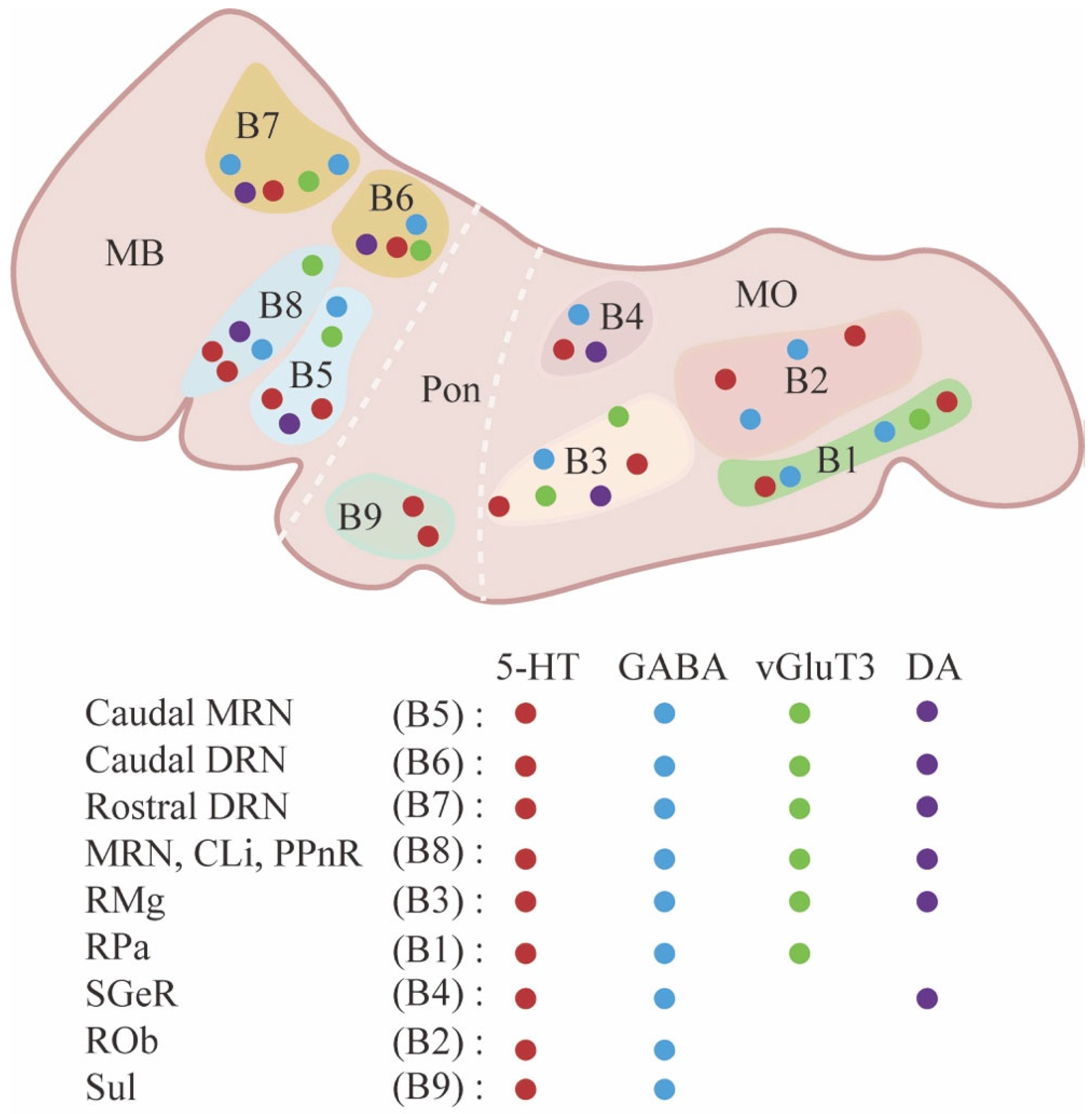
Figure 1. Anatomical and neurochemical diversity of raphe nuclei. Abbreviations: Caudal MRN: caudal median raphe nucleus; Caudal DRN: caudal dorsal raphe nucleus; Rostral DRN: rostral dorsal raphe nucleus; CLi: caudal linear nucleus; PPnR: prepontine raphe nucleus; RMg: raphe magnus nucleus; Rpa: raphe pallidus nucleus; SGeR: supragenual raphe nucleus; ROb: raphe obscurus nucleus; Sul: supralemniscal raphe complex; MB: midbrain; Pon: pons; MO: medulla oblongata.
Ijms 26 01234 g001
Figure 2. Innervation of dorsal and ventral hippocampus by the raphe nucleus. MRN mainly innervates the dorsal and ventral hippocampus. MRN-derived serotonergic neurons send beaded fibers, whereas DRN-derived neurons mainly innervate the vHP, and send regularly spaced fine fibers [36]. Abbreviations: dHP: dorsal hippocampus; vHP: ventral hippocampus; DRN: dorsal raphe nucleus; MRN: medial raphe nucleus.
Ijms 26 01234 g002
Figure 3. The efferent and afferent fibers of MRN correspond to midline structures, whereas those of DRN correspond to lateral structures. Both MRN and DRN innervate CeA, PFC, and HP. Abbreviations: DRN: dorsal raphe nucleus; MRN: medial raphe nucleus; AMG: amygdala; LHb: lateral habenula; mPFC: medial prefrontal cortex; MS: medial septal area; LH: lateral hypothalamus; CB: cerebellum; Pcx: piriform cortex; CLA: claustrum; BF: basal forebrain; Str: striatum; LHA: lateral hypothalamic area; VTA: ventral tegmental area; NAc: nucleus accumbens; CeA: central amygdala; PFC: prefrontal cortex; HP: hippocampus; HT: hypothalamus; FC: frontal cortex; SCN: suprachiasmatic nucleus; Arc: arcuate nucleus; PVH: paraventricular hypothalamic nucleus; MHb: medial habenula.
Ijms 26 01234 g003
Figure 4. A schematic diagram for comparison of raphe–hippocampal circuit under physiological and pathological conditions (AD). Abbreviations: dHP: dorsal hippocampus; vHP: ventral hippocampus; DRN: dorsal raphe nucleus; MRN: medial raphe nucleus; SERT: serotonin transporter; AD: Alzheimer’s disease; 5-HT: 5- hydroxytryptamine; 5-HIAA: 5-hydroxyindoleacetic acid; 5-HTRs: 5-HT receptors; PN: pyramidal neuron; GABA: gamma-aminobutyric acid.
Ijms 26 01234 g004
Table 1. The number of 5-HT neurons in AD patients and mouse models.
Sample
Type
Age
(Year or Month)
Measurement
Method
Raphe NeuronsReference
MCI patient73.2 ± 10.8 yBiochemical DetectionDRN5-HT[166]
MCI patient83.0 ± 0 yHPLCDRN5-HT↓, MRN5-HT[167]
AD patient70.0 ± 8.7 yBiochemical DetectionDRN5-HT[166]
AD patient83.1 ± 5.8 yICC, 2DIAMRN5-HT[168]
AD patient79.3 ± 8.7 yQARDRN5-HT[169]
AD patient82.0 ± 1.0 yICCDRN5-HT[163]
AD patient76.5 ± 10.2 yRP-HPLCDRN5-HT↓, MRN5-HT[170]
AD patient75.9 ± 7.3 yRP-HPLCDRN5-HT[171]
hTau mice4 mIFDRN5-HT[172]
hAPP-J20 mice4 mIF, IHCNC[49]
3×Tg-AD mice3–18 mIHCNC[165]
5×FAD mice3 mIFDRN5-HT[68]
APPswe/PS1dE9 mice24 mIHCDRN5-HT[173]
Abbreviations: MCI: mild cognitive impairment; AD: Alzheimer’s disease; 5-HT: 5-hydroxytryptamine; 5-HIAA: 5-hydroxyindoleacetic acid; DR: dorsal raphe nucleus; MR: median raphe; NC: no change; y or m: year or month; IF: immunofluorescence; IHC: immunohistochemistry; RP-HPLC: reversed-phase high-performance liquid chromatography; ICC: immunocytochemistry; QAR: quantitative autoradiography; 2DIA: two-dimensional image analysis; ↓ indicates a decrease in the number of 5-HT neurons; ↑ indicates an increase in the number 5-HT neurons.
Disclaimer/Publisher’s Note: The statements, opinions and data contained in all publications are solely those of the individual author(s) and contributor(s) and not of MDPI and/or the editor(s). MDPI and/or the editor(s) disclaim responsibility for any injury to people or property resulting from any ideas, methods, instructions or products referred to in the content.