Article

Role of ERβ in Triple-Negative Breast Cancer Associated with p53 and Androgen Receptor

1
Department of Pathology, Toho University Faculty of Medicine, Omori-Nishi 5-21-16, Ota-ku, Tokyo 143-8540, Japan
2
Department of Medical Technology, Faculty of Health Sciences, Tsukuba International University, Manabe 6-20-1, Tsuchiura 300-0051, Japan
3
Department of Surgical Pathology, Toho University Faculty of Medicine, Omori-Nishi 5-21-16, Ota-ku, Tokyo 143-8540, Japan
4
Department of Breast and Endocrine Surgery, Toho University Omori Medical Center, Omori-Nishi 6-11-1, Ota-ku, Tokyo 143-8541, Japan
5
Department of Gastroenterological Surgery, Yokohama City University Graduate School of Medicine, Fukuura 3-9, Kanazawa-ku, Yokohama 236-0004, Japan
6
Palliative Care Nursing, Department of Human Health Sciences, Graduate School of Medicine, Kyoto University, Shogoin-Kawahara-cho 53, Sakyo-ku, Kyoto 606-8507, Japan
7
Department of Pathology, Tokyo Metropolitan Institute for Geriatrics and Gerontology, Sakaecho 35-2, Itabashi-ku, Tokyo 173-0015, Japan
8
Department of Pathology and Molecular Diagnostics, Aichi Cancer Center Hospital, Kanokoden 1-1, Chikusa-ku, Nagoya 464-8681, Japan
9
Department of Breast Oncology, Nagoya Medical Center, National Hospital Organization 4-1-1 Sannomaru, Naka-ku, Nagoya 460-0001, Japan
*
Author to whom correspondence should be addressed.
Int. J. Mol. Sci. 2025, 26(23), 11459; https://doi.org/10.3390/ijms262311459
Submission received: 24 September 2025 / Revised: 6 November 2025 / Accepted: 21 November 2025 / Published: 26 November 2025

Abstract

In triple-negative breast cancer (TNBC), the clinicopathological significance of the expression of a second estrogen receptor, ERβ, remains unclear. Further, although the clinicopathological significance of mutant p53 and androgen receptor (AR) has been investigated in TNBC, they have not been established as therapeutic targets. Experimental studies reported the importance of cross-talk between ERβ and p53 or AR in TNBC. In this study, we immunohistochemically examined ERβ expression in surgical specimens of TNBC obtained from postmenopausal patients who underwent surgery without neoadjuvant therapy and investigated the relationship between ERβ expression and various clinicopathological factors, including clinical outcome, while also considering p53 and AR. No significant difference in clinical outcome was noted according to the ERβ status alone (p = 0.2908). However, the ERβ status did affect the relationship between the clinical outcome and p53 or AR status; p53-positive or AR-positive group exhibited significantly more favorable clinical outcomes than p53-negative or AR-negative group, respectively, in the ERβ-positive group (p53, p = 0.0265; AR, p = 0.0285), but not in the ERβ-negative group (p53, p = 0.7228; AR, p = 0.7734). This may be the result of a functional interaction between ERβ and p53 or AR. The role of ERβ in TNBC will be elucidated in further complex studies considering multiple molecules.

1. Introduction

Since triple-negative breast cancer (TNBC) lacks an effective treatment target, that is, classic estrogen receptor (ERα), progesterone receptor (PgR), or human epidermal growth factor receptor-2 (HER2), chemotherapy is the most established pharmacotherapy for patients with TNBC. Most cases of TNBC involve an undifferentiated tumor and are biologically aggressive, resulting in unfavorable outcomes for patients [1]. TNBC is relatively frequent in younger women or specific ethnicities and is closely related to germline gene mutations, such as BRCA, in these patients. The therapeutic options available for patients with TNBC have been increasing, such as PARP inhibitors for BRCA-mutated patients, immune checkpoint inhibitors for PD-L1-positive tumors [1], or oncolytic peptides in the context of cancer immunotherapy [2]. TNBC, however, is known to be heterogeneous, suggesting the need for appropriate biomarkers to optimize treatment. It may be meaningful to focus on TNBC in postmenopausal women, including older women, who may benefit from pharmacotherapies other than the current options. Although TNBC is characterized as non-responsive to estrogen and its related systems, the expression of receptors for sex steroid hormones other than ERα has now been demonstrated in TNBC, and the molecular mechanisms and clinical relevance of these receptors have been investigated [3,4,5,6,7,8,9,10,11,12,13]. Receptors for estrogen, including a second ER (ERβ) or G protein-coupled estrogen receptor (GPER), and that for androgen (AR), are known, but their roles in TNBC remain unclear. From a clinicopathological point of view, AR has been reported to be expressed in about 40% of TNBC cases, forming the so-called ‘luminal AR’ subtype (LAR), for which AR-targeted therapy has been in a clinical trial [13]. The clinicopathological role of AR has been examined in many studies, but its localization, antibodies, and cut-off values vary, with some reports suggesting a favorable prognostic effect of AR, some suggesting a poor prognostic effect, and others suggesting no prognostic role at all [14,15,16,17,18,19]. We recently showed that AR expression is closely related to patient age [18]. ERβ has also been reported to have various effects in TNBC, which at least partly reflect the differences in study methodologies (in vitro or in vivo experimental studies, real-time RT-PCR or immunohistochemistry on surgical materials, antibody used, intracellular structure estimation, cut-off values, etc.) or settings (eligibility of included patients). Honma et al. reported favorable clinical outcomes in a group of postmenopausal ERβ-positive TNBC patients treated with adjuvant tamoxifen monotherapy [20], whereas others reported an unfavorable role of ERβ [21,22,23]. The role of p53 in TNBC is also controversial [24]. p53 is a representative cancer suppressor protein, inducing cell cycle arrest or apoptosis in genetically damaged cells [25,26,27]. Aberrant p53 promotes tumors [25] and is reportedly frequent in TNBC. Tumors with mutated TP53 have been reported to frequently achieve pathologic complete remission with neoadjuvant chemotherapy [28]. In addition to the respective roles of ERβ, AR, and p53 in TNBC, there have also been reports of molecular cross-talk between ERβ and p53 or AR in basic studies [12,29,30,31,32,33]. In this study, we examined the clinicopathological role of ERβ alone or in combination with p53 and AR, based on immunohistochemical findings.

2. Results

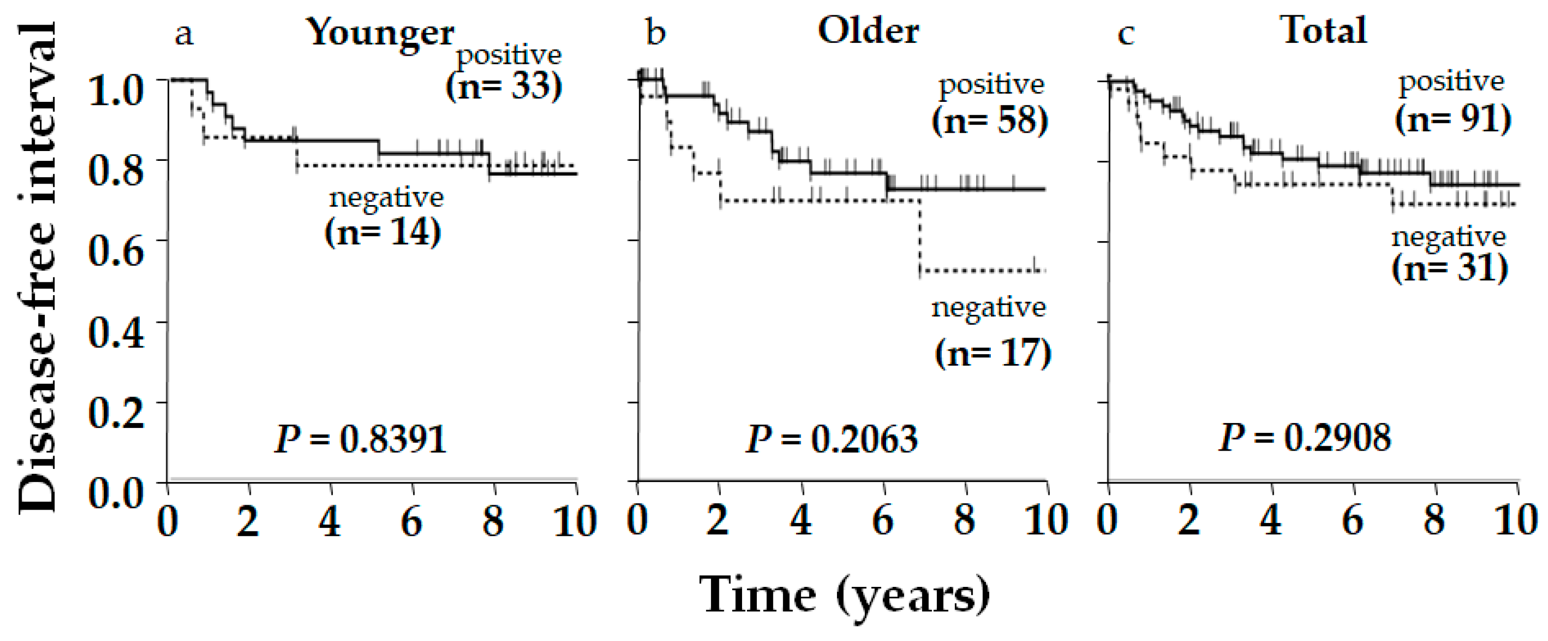
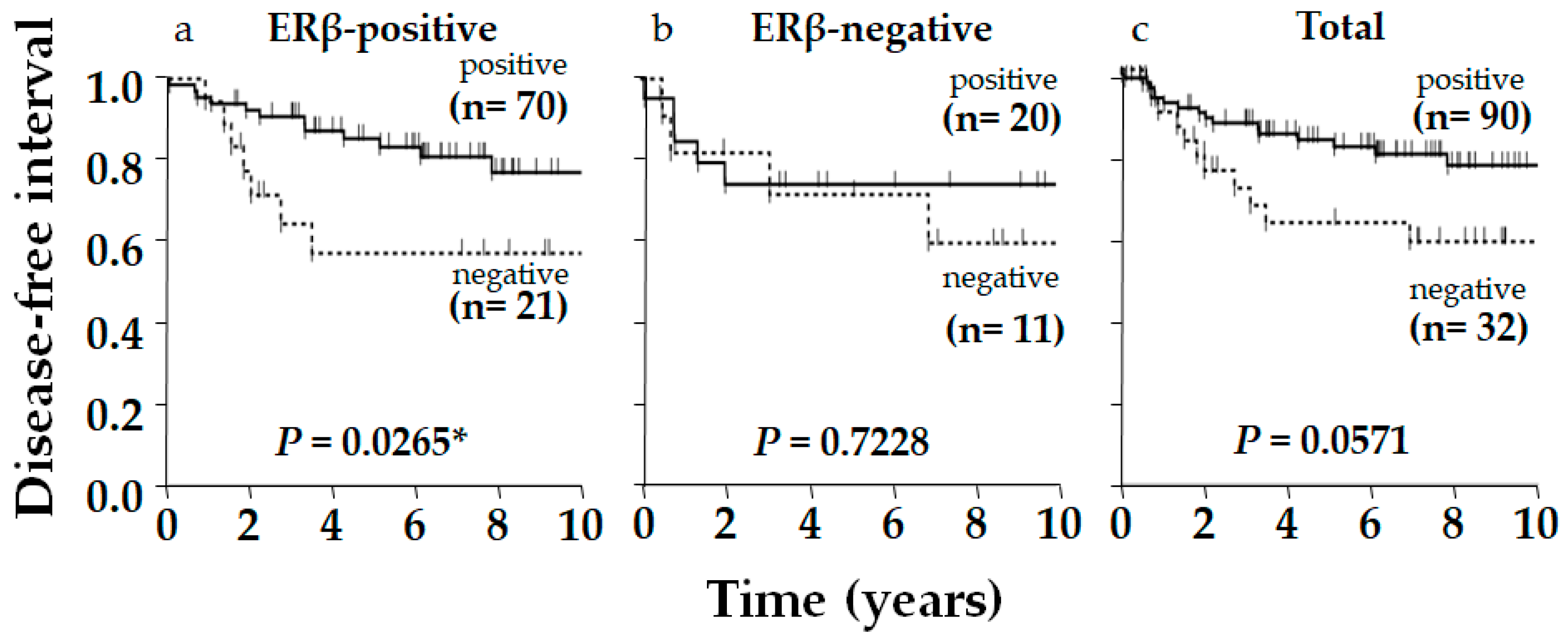
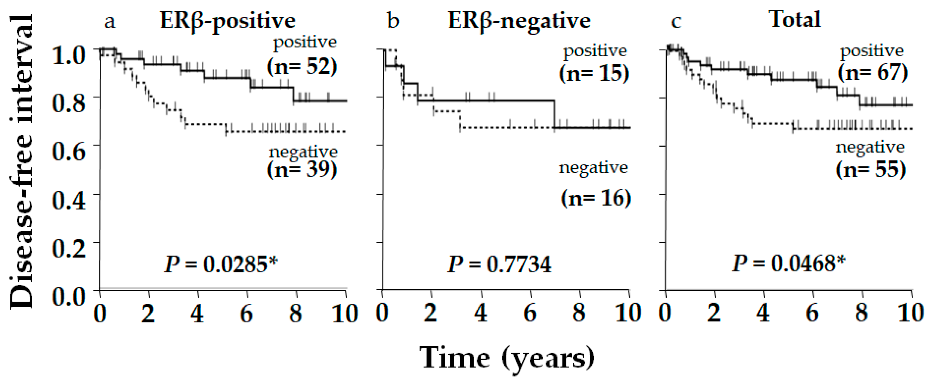
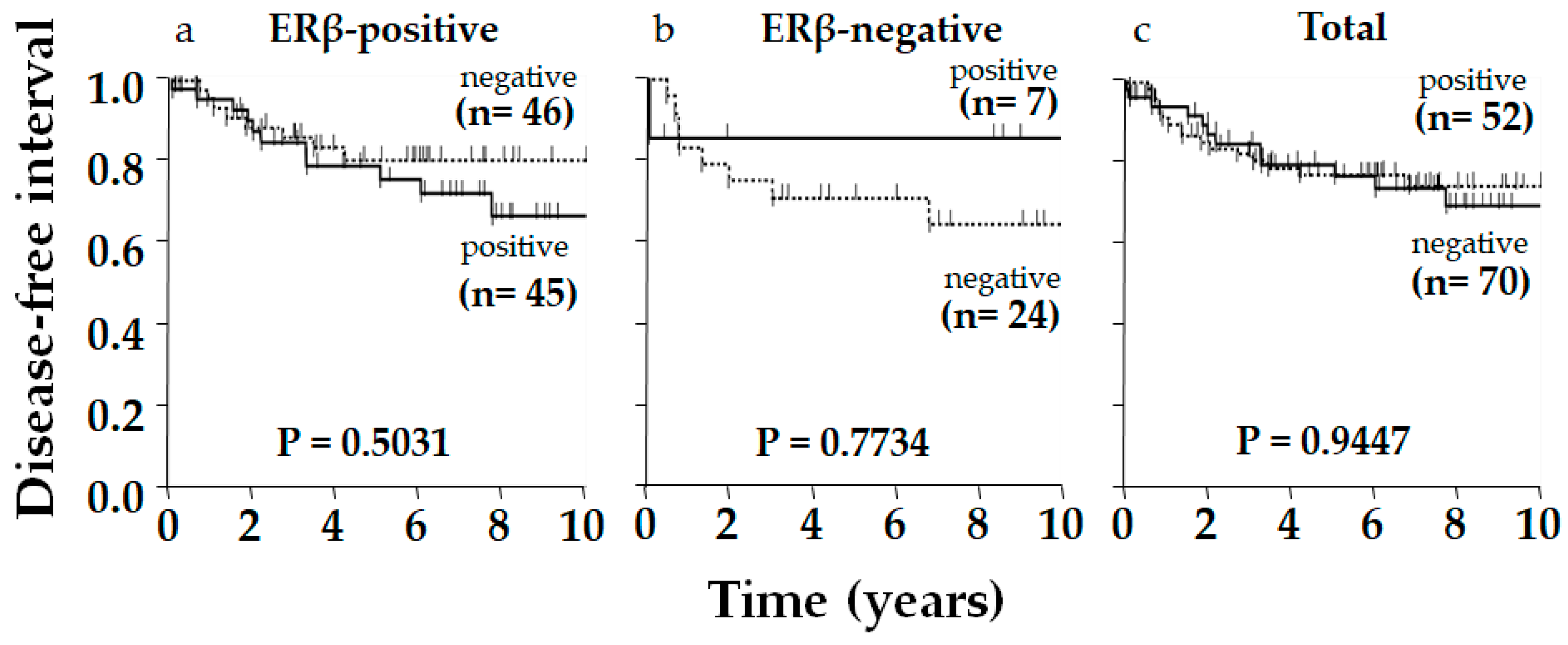
Typical immunostaining images of ERβ, p53, and AR are shown in Figure 1. Of 122 patients, 91 (75%), 90 (74%), and 67 (55%) were positive for ERβ, p53, and AR, respectively. The relationship between ERβ expression and various clinicopathological factors, including patients’ age, tumor size, nodal status, histological grade, Ki-67 status, AR status, and p53 status, were examined; however, no significant correlation was observed, except for Ki-67 status, which showed a positive correlation with ERβ expression (p = 0.009, Table 1). Although insignificant, ERβ positivity was related to smaller tumor size (p = 0.128), negative nodal metastasis (p = 0.142), and lower pStage (p = 0.060) (Table 1). Aside from ERβ, AR and p53 positively correlated with each other (p = 0.0211, Table 2). Regarding the clinical outcome, we focused on the disease-free interval (DFI), in which recurrence/metastasis was considered as event, to elucidate the outcome of the tumor itself, specifically in older patients, who frequently die from causes other than the tumor. No significant difference in DFI was observed according to ERβ status alone, though positivity was slightly related to a favorable outcome in total (p = 0.2908) and in the older group (p = 0.2063), though this was not observed in the younger group (p = 0.8391, Figure 2). As for p53 in total, DFI in the p53-positive group was almost significantly more favorable than in the p53-negative group (p = 0.0571), and this was clearer in the ERβ-positive group yielding significance (p = 0.0265), contrasting with the ERβ-negative group where the difference in DFI according to p53 status disappeared (p = 0.7228, Figure 3). Similar phenomena were observed for AR. A marginally significant difference in DFI according to AR status was shown in total (p = 0.0468, more favorable DFI for AR-positive group than AR-negative group); however, the difference in DFI between AR-positive and -negative groups was even clearer in the ERβ-positive group (p = 0.0285) but was lost in the ERβ-negative group (p = 0.7734, Figure 4). Since ERβ was positively correlated with Ki-67, we conducted the same analyses, comparing DFI according to Ki-67 status in total and ERβ-positive and -negative groups; however, no significant results were obtained (p = 0.9447, p = 0.5031, and p = 0.7734 in total, ERβ-positive, and ERβ-negative groups, respectively; Figure 5).

3. Discussion

In the present study, ERβ alone had no prognostic impact on TNBC. In ERα-positive tumors, ERβ reportedly suppresses cell growth, forming a heterodimer with ERα and inhibiting its tumor-promoting effect or suppressing the transcription of the c-myc, cyclin D1, or cyclin A genes. In ERα-negative tumors, on the contrary, ERβ slightly promotes tumor growth, forming an ERβ homodimer and sending proliferative signals [13,34,35]. The positive correlation between ERβ and Ki-67 observed in this study might reflect the tumor-promoting role of ERβ in TNBC. Honma et al. [20] reported a favorable clinical outcome in an ERβ-positive TNBC patient group treated with adjuvant tamoxifen monotherapy, specifically comprising postmenopausal women (most patients were younger than 75 y/o); however, it was unclear whether the results reflected a favorable prognostic effect of ERβ independent of endocrine therapy or the predictive role of ERβ for tamoxifen therapy. Since tamoxifen was not administered to the current patients in this study, the previous results may have reflected the predictive value of ERβ for tamoxifen treatment, but not an independent prognostic value of ERβ by itself.
However, as there have been reports of molecular cross-talk between ERβ and p53 or AR [12,29,30,31,32,33], we examined the clinicopathological significance of ERβ in combination with p53 or AR and found a greater prognostic impact of p53 and AR in ERβ-positive compared with ERβ-negative patients.
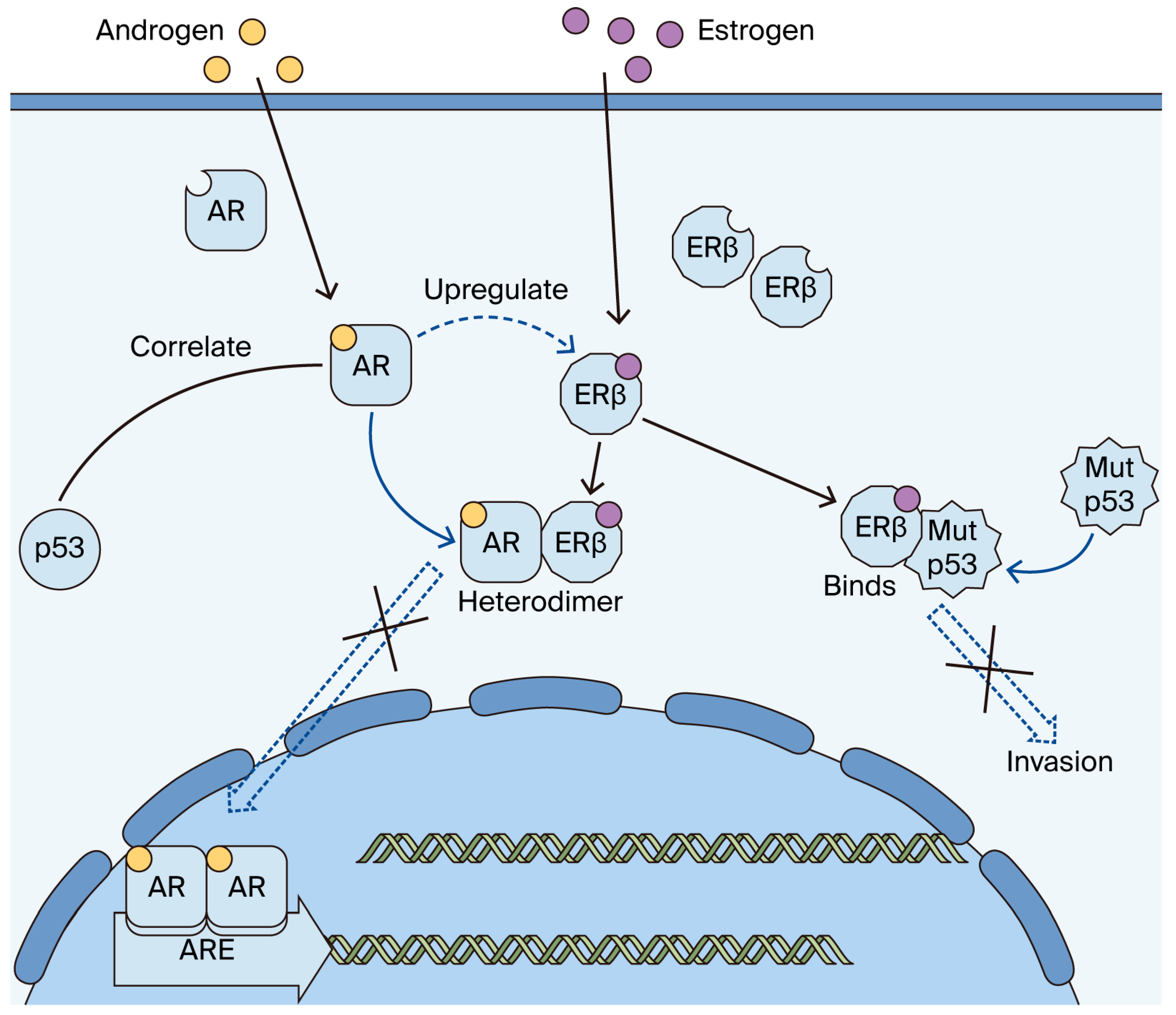
In breast cancer, p53 needs to be considered by subtype due to the influence of ERα and other factors [36]. In ERα-positive cancer, estrogen signaling has been reported to suppress p53 activity, which inhibits apoptosis induced by chemotherapy and causes resistance [28], suggesting the importance of estrogen signaling in the pathobiology of p53. p53-mutated breast cancer reportedly shows a high rate of pathologic complete response with neoadjuvant chemotherapy [28]. In our previous study on p53 in TNBC, there was a trend, though insignificant, toward a better prognosis in the p53-positive group [24]. In the present study, p53 was correlated with a significantly better prognosis when limited to the ERβ-positive group. This is consistent with a basic study reporting that ERβ regulates mutant p53 and reduces invasiveness in TNBC (Figure 6) [29].
The pathobiological role of androgen is also complex in breast tumors. AR is frequently expressed in normal breast epithelial cells and is considered one of the markers of mammary epithelial cell differentiation and a favorable prognostic factor in breast cancer patients. AR in the ERα-positive state exerts an anti-estrogenic, growth-inhibitory effect and antagonizes ERα signaling [37,38], while in TNBC, AR reportedly promotes the proliferation of tumor cells or metastasis [39]; thus, the role of AR in TNBC is similar to that of ERα in ERα-positive tumors, having both favorable prognostic value and a tumor-promoting effect. With such a complicated background, the prognostic role of AR alone may be controversial in TNBC. Androgens may exhibit diverse effects depending on their interactions with other steroid receptors and related signaling pathways and have also been reported to increase ERβ gene expression irrespective of ERα expression [40]. ERβ reportedly forms a heterodimer with AR, resulting in a better prognosis by blocking AR proliferative signals in TNBC [33] (Figure 6). AR alone has a favorable prognostic value in TNBC [18]; however, in the present study, the difference in DFI according to AR status was even more marked in ERβ-positive group but disappeared in ERβ-negative group, suggesting the importance of the interaction between ERβ and AR. Furthermore, the co-expression of ERβ and AR has been reported to increase the efficacy of antihormonal therapy in TNBC [41], so the clinical significance of AR may need to be considered together with ERβ.
In the present study, expressions of AR and p53 were positively correlated. Although the biological mechanism is unknown, some molecules may mediate between them. For example, KLLN has been identified as an AR-induced tumor suppressor, and studies have shown that KLLN directly promotes p53 expression in breast cancer, causing apoptosis and cell cycle arrest [42]. Identifying such mediating molecules is necessary to elucidate the association between AR and p53.
Figure 6. Molecular interactions between ERβ, p53, and AR in triple-negative breast cancer (TNBC) hypothesized in the present study. ERβ regulates mutant p53 and reduces invasiveness in TNBC [29]. Androgens have also been reported to increase ERβ gene expression irrespective of ERα expression [40]. ERβ reportedly forms a heterodimer with AR, resulting in a better prognosis by blocking AR proliferative signals in TNBC [33]. The co-expression of ERβ and AR has been reported to increase the efficacy of antihormonal therapy in TNBC [41]. X means blocked reactions, full arrows mean action of molecules, dashed arrow means influence of a molecule on another molecule.
Ijms 26 11459 g006
Previously, in a combined investigation of Bcl-2 and p53 in TNBC, we demonstrated that a Bcl-2-negative/p53-positive group had a markedly longer DFI than the other groups [24]. These findings provide further evidence supporting the importance of combined investigations of multiple molecules and their interactions. As there has been no consistent research data regarding molecular interactions including ERβ, p53, and AR, it will be necessary to conduct further research based on a combinatory viewpoint, similar to that in this study.
The limitation of this study is its purely observational nature, as it was conducted on surgical samples using immunohistochemistry. Extensive molecular analyses are needed to prove the hypothesis presented in this study.

4. Materials and Methods

4.1. Subjects

A retrospective cross-sectional study was conducted, involving postmenopausal patients with TNBC who were surgically treated without neoadjuvant therapy at Toho University Medical Center Omori Hospital (Tokyo, Japan), Aichi Cancer Center (Nagoya, Japan), Tokyo Metropolitan Geriatric Hospital (Tokyo, Japan), Yokohama City University Hospital (Yokohama, Japan), or Kagawa University Hospital (Kagawa, Japan) in 2004–2013. Patients matched for pathological stage (pStage) and aged 75 years or older (older group, n = 75) and 55–64 years (younger group, n = 47) were included in this study as part of a consecutive series. The inclusion criteria were as follows: (1) primary invasive breast cancer; (2) TNBC, defined as <1% staining for estrogen receptor and progesterone receptor, and 0 or 1+ as per immunohistochemistry or negative as per in situ hybridization for human epidermal growth factor receptor 2 (HER2); (3) unilateral; and (4) female sex. The exclusion criteria were as follows: (1) noninfiltrating carcinoma or carcinoma with microinvasion, (2) no residual carcinoma after biopsy, (3) stage IV tumors, (4) bilateral, (5) male sex, and (6) undergoing neoadjuvant therapy.

4.2. Immunohistochemistry

The expression of each protein was examined immunohistochemically on representative slides of formalin-fixed and paraffin-embedded tissue, according to the established method [18,24,43,44]. Endogenous peroxidase was blocked with 0.5% hydrogen peroxide in methanol for 30 min. For antigen retrieval, the sections were treated with 98 °C Target Retrieval Solution pH 6 (for p53) or pH 9 (for ERβ and AR) (Dako, Carpinteria, CA, USA) for 40 min each. After the application of primary antibodies for ERβ (clone PPG5/10, Bio-Rad, Hercules, CA, USA) and AR (clone AR27, Novocastra, Melbourne, Australia), and p53 (clone DO-7, Dako. X1000) for 60 min at room temperature, the slides were washed with phosphate-buffered saline and incubated with secondary EnVision+System-HRP labeled polymer (Dako). Finally, tissue sections were visualized with Stable DAB (Falma, Tokyo, Japan). Nuclear immunoreactivity was estimated independently by two researchers (KI and NH), who scored both the intensity and percentage of cancer cells (Allred score). The cut-off was set at 10% for ERβ and AR. For p53, a positive result was defined as one in which tumor cells stained more strongly than adjacent noncancerous epithelial cells [18,20,24].

4.3. Statistical Analysis

The expression of ERβ was compared with classical clinicopathological factors and AR or p53 expression. The Chi-square test or Fisher’s exact test were used to compare categorical variables, while the disease-free interval (DFI) was analyzed using the Kaplan–Meier method and log-rank test. Data were analyzed with JMP version 13, and p < 0.05 was considered significant.

Author Contributions

Conceptualization, N.H., H.O., A.Y., M.M. and M.S.; methodology, N.H.; validation, K.I. and N.H.; formal analysis, all authors; investigation, K.I. and N.H.; resources, N.H., H.O., A.Y., T.A., E.S., K.S., T.M. and M.S.; data curation, K.I. and N.H.; writing—original draft preparation, all authors; writing—review and editing, all authors; visualization, K.I. and N.H.; supervision, N.H., T.A., K.S., T.M. and M.S.; project administration, N.H. and M.S.; funding acquisition, N.H. and M.S. All authors have read and agreed to the published version of the manuscript.

Funding

This work was supported by MEXT/JSPS KAKENHI grant number 16K08660, a Grant-in-Aid for Scientific Research from the Japanese Breast Cancer Society (No.24), and The Research Promotion Grant from Toho University Graduate School of Medicine (No. 25-01 to NH). These funding sources had no role in the study.

Institutional Review Board Statement

This study was conducted in accordance with the Declaration of Helsinki and approved by the Ethics Committee of each institution (Toho University A19079_A18116, 25 March 2020; Aichi Cancer Center 2019-1-007, 24 May 2019; Tokyo Metropolitan Geriatric Hospital R20-10, 9 June 2020; Yokohama City University Hospital B191000003, 10 October 2019; and Kagawa University Hospital 2020–023, 15 May 2020). All subjects provided comprehensive consent for the use of clinical data and were given the opportunity to opt out.

Informed Consent Statement

In this kind of study, informed consent is not required in Japan, so the ethics committee waived the requirement for informed consent. Instead, information about the study was posted on the Institution’s website, and potential participants were free to opt out.

Data Availability Statement

The datasets presented in this article are not readily available because the data are part of an ongoing study. Requests to access the datasets should be directed to Naoko Honma.

Acknowledgments

We thank all collaborative researchers involved in this study. We also thank Kayo Tsuburaya and Maho Yokoyama and all the staff of the Department of Pathology, Surgical Pathology, and Breast and Endocrine Surgery, Toho University Faculty of Medicine, Toho University Medical Center Omori Hospital, Aichi Cancer Center, Tokyo Metropolitan Geriatric Hospital, Yokohama City University Hospital, and Kagawa University Hospital for their help.

Conflicts of Interest

The authors declare no conflict of interest.

Abbreviations

The following abbreviations are used in this manuscript:
ARandrogen receptor
ERαestrogen receptor-α
ERβestrogen receptor-β
GPERG protein-coupled estrogen receptor
HER2human epidermal growth factor receptor-2
PgRprogesterone receptor
TNBCtriple-negative breast cancer(s)

References

  1. Denkert, C.; Liedtke, C.; Tutt, A.; von Minckwitz, G. Molecular Alterations in Triple-Negative Breast Cancer-the Road to New Treatment Strategies. Lancet 2017, 389, 2430–2442. [Google Scholar] [CrossRef]
  2. Fu, X.; Yin, H.; Chen, X.; Yao, J.; Ma, Y.; Song, M.; Xu, H.; Yu, Q.; Du, S.; Qi, Y.; et al. Three Rounds of Stability-Guided Optimization and Systematical Evaluation of Oncolytic Peptide LTX-315. J. Med. Chem. 2024, 67, 3885–3908. [Google Scholar] [CrossRef]
  3. Treeck, O.; Schüler-Toprak, S.; Ortmann, O. Estrogen Actions in Triple-Negative Breast Cancer. Cells 2020, 9, 2358. [Google Scholar] [CrossRef] [PubMed]
  4. Shaaban, A.M.; O’neill, P.A.; Davies, M.P.A.; Sibson, R.; West, C.R.; Smith, P.H.; Foster, C.S. Declining Estrogen Receptor-beta Expression Defines Malignant Progression of Human Breast Neoplasia. Am. J. Pathol. 2003, 27, 1502–1512. [Google Scholar] [CrossRef] [PubMed]
  5. Grober, O.M.V.; Mutarelli, M.; Giurato, G.; Ravo, M.; Cicatiello, L.; De Filippo, M.R.; Ferraro, L.; Nassa, G.; Papa, M.F.; Paris, O.; et al. Global Analysis of Estrogen Receptor Beta Binding to Breast Cancer Cell Genome Reveals an Extensive Interplay with Estrogen Receptor Alpha for Target Gene Regulation. BMC Genom. 2011, 12, 36. [Google Scholar] [CrossRef]
  6. Treeck, O.; Juhasz-Boess, I.; Lattrich, C.; Horn, F.; Goerse, R.; Ortmann, O. Effects of Exon-Deleted Estrogen Receptor Β Transcript Variants on Growth, Apoptosis and Gene Expression of Human Breast Cancer Cell Lines. Breast Cancer Res. Treat. 2007, 110, 507–520. [Google Scholar] [CrossRef] [PubMed]
  7. Song, P.; Li, Y.; Dong, Y.; Liang, Y.; Qu, H.; Qi, D.; Lu, Y.; Jin, X.; Guo, Y.; Jia, Y.; et al. Estrogen Receptor Β Inhibits Breast Cancer Cells Migration and Invasion through CLDN6-Mediated Autophagy. J. Exp. Clin. Cancer Res. 2019, 38, 354. [Google Scholar] [CrossRef]
  8. Prossnitz, E.R.; Arterburn, J.B.; Smith, H.O.; Oprea, T.I.; Sklar, L.A.; Hathaway, H.J. Estrogen Signaling through the Transmembrane G Protein–Coupled Receptor GPR30. Annu. Rev. Physiol. 2008, 70, 165–190. [Google Scholar] [CrossRef]
  9. Sartorius, C.A.; Hanna, C.T.; Gril, B.; Cruz, H.; Serkova, N.J.; Huber, K.M.; Kabos, P.; Schedin, T.B.; Borges, V.F.; Steeg, P.S.; et al. Estrogen Promotes the Brain Metastatic Colonization of Triple Negative Breast Cancer Cells Via an Astrocyte-Mediated Paracrine Mechanism. Oncogene 2016, 35, 2881–2892. [Google Scholar] [CrossRef]
  10. Schüler-Toprak, S.; Häring, J.; Inwald, E.C.; Moehle, C.; Ortmann, O.; Treeck, O. Agonists and Knockdown of Estrogen Receptor Β Differentially Affect Invasion of Triple-Negative Breast Cancer Cells in Vitro. BMC Cancer 2016, 16, 951. [Google Scholar] [CrossRef]
  11. Hinsche, O.; Girgert, R.; Emons, G.; Gründker, C. Estrogen Receptor Β Selective Agonists Reduce Invasiveness of Triple-Negative Breast Cancer Cells. Int. J. Oncol. 2014, 46, 878–884. [Google Scholar] [CrossRef]
  12. Mukhopadhyay, U.K.; Oturkar, C.C.; Adams, C.; Wickramasekera, N.; Bansal, S.; Medisetty, R.; Miller, A.; Swetzig, W.M.; Silwal-Pandit, L.; Børresen-Dale, A.; et al. TP53 Status as a Determinant of Pro- vs Anti-Tumorigenic Effects of Estrogen Receptor-Beta in Breast Cancer. J. Natl. Cancer Inst. 2019, 111, 1202–1215. [Google Scholar] [CrossRef]
  13. Honma, N.; Matsuda, Y.; Mikami, T. Carcinogenesis of Triple-Negative Breast Cancer and Sex Steroid Hormones. Cancers 2021, 13, 2588. [Google Scholar] [CrossRef] [PubMed]
  14. Bhattarai, S.; Klimov, S.; Mittal, K.; Krishnamurti, U.; Li, X.; Oprea-Ilies, G.; Wetherilt, C.; Riaz, A.; Aleskandarany, M.A.; Green, A.R.; et al. Prognostic Role of Androgen Receptor in Triple Negative Breast Cancer: A Multi-Institutional Study. Cancers 2019, 11, 995. [Google Scholar] [CrossRef] [PubMed]
  15. Gasparini, P.; Fassan, M.; Cascione, L.; Guler, G.; Balci, S.; Irkkan, C.; Paisie, C.; Lovat, F.; Morrison, C.; Zhang, J.; et al. Androgen Receptor Status is a Prognostic Marker in Non-Basal Triple Negative Breast Cancers and Determines Novel Therapeutic Options. PLoS ONE 2014, 9, e88525. [Google Scholar] [CrossRef]
  16. Choi, J.E.; Kang, S.H.; Lee, S.J.; Bae, Y.K. Androgen Receptor Expression Predicts Decreased Survival in Early Stage Triple-Negative Breast Cancer. Ann. Surg. Oncol. 2014, 22, 82–89. [Google Scholar] [CrossRef]
  17. Mishra, A.; Mishra, S.K.; Sharanappa, V.; Krishnani, N.; Kumari, N.; Agarwal, G. Incidence and Prognostic Significance of Androgen Receptors (AR) in Indian Triple-Negative Breast Cancer (TNBC). Indian J. Surg. Oncol. 2024, 15, 250–257. [Google Scholar] [CrossRef] [PubMed]
  18. Honma, N.; Ogata, H.; Yamada, A.; Matsuda, Y.; Kontani, K.; Miyashita, M.; Arai, T.; Sasaki, E.; Shibuya, K.; Mikami, T.; et al. Clinicopathological Characteristics and Prognostic Marker of Triple-Negative Breast Cancer in Older Women. Hum. Pathol. 2021, 111, 10–20. [Google Scholar] [CrossRef]
  19. Hu, X.; Chen, W.; Ma, H.; Jiang, K. Androgen Receptor Expression Identifies Patient with Favorable Outcome in Operable Triple Negative Breast Cancer. Oncotarget 2017, 8, 56364–56374. [Google Scholar] [CrossRef] [PubMed]
  20. Honma, N.; Horii, R.; Iwase, T.; Saji, S.; Younes, M.; Takubo, K.; Matsuura, M.; Ito, Y.; Akiyama, F.; Sakamoto, G. Clinical Importance of Estrogen Receptor-Β Evaluation in Breast Cancer Patients Treated with Adjuvant Tamoxifen Therapy. J. Clin. Oncol. 2008, 26, 3727–3734. [Google Scholar] [CrossRef]
  21. Wang, J.; Zhang, C.; Chen, K.; Tang, H.; Tang, J.; Song, C.; Xie, X. ERβ1 Inversely Correlates with PTEN/PI3K/AKT Pathway and Predicts a Favorable Prognosis in Triple-Negative Breast Cancer. Breast Cancer Res. Treat. 2015, 152, 255–269. [Google Scholar]
  22. Wimberly, H.; Han, G.; Pinnaduwage, D.; Murphy, L.C.; Yang, X.R.; Andrulis, I.L.; Sherman, M.; Figueroa, J.; Rimm, D.L. ERβ Splice Variant Expression in Four Large Cohorts of Human Breast Cancer Patient Tumors. Breast Cancer Res. Treat. 2020, 146, 657–667. [Google Scholar]
  23. Shanle, E.K.; Onitilo, A.A.; Huang, W.; Kim, K.; Zang, C.; Engel, J.M.; Xu, W.; Wisinski, K.B. Prognostic Significance of Full-Length Estrogen Receptor Beta Expression in Stage I-III Triple Negative Breast Cancer. Am. J. Transl. Res. 2015, 5, 1246–1259. [Google Scholar]
  24. Ito, K.; Honma, N.; Ogata, H.; Yamada, A.; Miyashita, M.; Arai, T.; Sasaki, E.; Shibuya, K.; Mikami, T.; Sawaki, M. Clinicopathological Importance of Bcl-2 and p53 in Postmenopausal Triple-negative Breast Carcinoma and Association with Age. Pathol. Int. 2024, 74, 574–582. [Google Scholar] [CrossRef]
  25. Allred, D.C.; Clark, G.M.; Elledge, R.; Fuqua, S.A.; Brown, R.W.; Chamness, G.C.; Osborne, C.K.; McGuire, W.L. Association of p53 Protein Expression with Tumor Cell Proliferation Rate and Clinical Outcome in Node-Negative Breast Cancer. J. Natl. Cancer Inst. 1993, 85, 200–206. [Google Scholar]
  26. Green, D.R.; Kroemer, G. Cytoplasmic Functions of the Tumour Suppressor p53. Nature 2009, 458, 1127–1130. [Google Scholar] [CrossRef] [PubMed]
  27. Hao, Q.; Chen, J.; Liao, J.; Huang, Y.; Gan, Y.; Larisch, S.; Zeng, S.X.; Lu, H.; Zhou, X. p53 Induces ARTS to Promote Mitochondrial Apoptosis. Cell Death Dis. 2021, 12, 204. [Google Scholar] [CrossRef] [PubMed]
  28. Bertheau, P.; Lehmann-Che, J.; Varna, M.; Dumay, A.; Poirot, B.; Porcher, R.; Turpin, E.; Plassa, L.; de Roquancourt, A.; Bourstyn, E.; et al. P53 in Breast Cancer Subtypes and New Insights into Response to Chemotherapy. Breast 2013, 22 (Suppl. S2), 27. [Google Scholar] [CrossRef]
  29. Bado, I.; Nikolos, F.; Rajapaksa, G.; Gustafsson, J.; Thomas, C. ERβ Decreases the Invasiveness of Triple-Negative Breast Cancer Cells by Regulating Mutant p53 Oncogenic Function. Oncotarget 2016, 7, 13599–13611. [Google Scholar] [CrossRef]
  30. Oturkar, C.C.; Gandhi, N.; Rao, P.; Eng, K.H.; Miller, A.; Singh, P.K.; Zsiros, E.; Odunsi, K.O.; Das, G.M. Estrogen Receptor-Beta2 (ERβ2)–Mutant p53–FOXM1 Axis: A Novel Driver of Proliferation, Chemoresistance, and Disease Progression in High Grade Serous Ovarian Cancer (HGSOC). Cancers 2022, 14, 1120. [Google Scholar] [PubMed]
  31. Scarpetti, L.; Oturkar, C.C.; Juric, D.; Shellock, M.; Malvarosa, G.; Post, K.; Isakoff, S.; Wang, N.; Nahed, B.; Oh, K.; et al. Therapeutic Role of Tamoxifen for Triple-Negative Breast Cancer: Leveraging the Interaction between ERβ and Mutant p53. Oncologist 2023, 28, 358–363. [Google Scholar] [CrossRef]
  32. Song, W.; Tang, L.; Xu, Y.; Sun, Q.; Yang, F.; Guan, X. ERβ1 Inhibits Metastasis of Androgen Receptor-Positive Triple-Negative Breast Cancer by Suppressing ZEB1. J. Exp. Clin. Cancer Res. 2017, 36, 75. [Google Scholar] [CrossRef]
  33. Anestis, A.; Sarantis, P.; Theocharis, S.; Zoi, I.; Tryfonopoulos, D.; Korogiannos, A.; Koumarianou, A.; Xingi, E.; Thomaidou, D.; Kontos, M.; et al. Estrogen Receptor Beta Increases Sensitivity to Enzalutamide in Androgen Receptor-Positive Triple-Negative Breast Cancer. J. Cancer Res. Clin. Oncol. 2019, 145, 1221–1233. [Google Scholar] [CrossRef]
  34. Kuiper, G.G.; Lemmen, J.G.; Carlsson, B.; Corton, J.C.; Safe, S.H.; van der Saag, P.T.; van der Burg, B.; Gustafsson, J.A. Interaction of Estrogenic Chemicals and Phytoestrogens with Estrogen Receptor Beta. Endocrinology 1998, 139, 4252–4263. [Google Scholar] [CrossRef] [PubMed]
  35. Paruthiyil, S.; Parmar, H.; Kerekatte, V.; Cunha, G.R.; Firestone, G.L.; Leitman, D.C. Estrogen Receptor Beta Inhibits Human Breast Cancer Cell Proliferation and Tumor Formation by Causing a G2 Cell Cycle Arrest. Cancer Res. 2004, 64, 423–428. [Google Scholar] [CrossRef]
  36. Silwal-Pandit, L.; Vollan, H.K.M.; Chin, S.; Rueda, O.M.; Mckinney, S.; Osako, T.; Quigley, D.A.; Kristensen, V.N.; Aparicio, S.; Børresen-Dale, A.; et al. TP53 Mutation Spectrum in Breast Cancer is Subtype Specific and has Distinct Prognostic Relevance. Clin. Cancer Res. 2014, 20, 3569–3580, Erratum in Clin. Cancer Res. 2015, 21, 1502. [Google Scholar] [CrossRef]
  37. Hickey, T.E.; Robinson, J.L.L.; Carroll, J.S.; Tilley, W.D. Minireview: The Androgen Receptor in Breast Tissues: Growth Inhibitor, Tumor Suppressor, Oncogene? Mol. Endocrinol. 2012, 26, 1252–1267. [Google Scholar] [CrossRef]
  38. Ravaioli, S.; Maltoni, R.; Pasculli, B.; Parrella, P.; Giudetti, A.M.; Vergara, D.; Tumedei, M.M.; Pirini, F.; Bravaccini, S. Androgen Receptor in Breast Cancer: The “5W” Questions. Front. Endocrinol. 2022, 13, 977331. [Google Scholar] [CrossRef]
  39. Doane, A.S.; Danso, M.; Lal, P.; Donaton, M.; Zhang, L.; Hudis, C.; Gerald, W.L. An Estrogen Receptor-Negative Breast Cancer Subset Characterized by a Hormonally Regulated Transcriptional Program and Response to Androgen. Oncogene 2006, 25, 3994–4008. [Google Scholar] [CrossRef] [PubMed]
  40. Rizza, P.; Barone, I.; Zito, D.; Giordano, F.; Lanzino, M.; De Amicis, F.; Mauro, L.; Sisci, D.; Catalano, S.; Dahlman, K.; et al. Estrogen Receptor Beta as a Novel Target of Androgen Receptor Action in Breast Cancer Cell Lines. Breast Cancer Res. 2014, 16, R21. [Google Scholar] [CrossRef] [PubMed]
  41. Crespo, B.; Illera, J.C.; Silvan, G.; Lopez-Plaza, P.; Herrera De La Muela, M.; De La Puente Yagüe, M.; Diaz Del Arco, C.; Illera, M.J.; Caceres, S. Androgen and Estrogen Β Receptor Expression Enhances Efficacy of Antihormonal Treatments in Triple-Negative Breast Cancer Cell Lines. Int. J. Mol. Sci. 2024, 25, 1471. [Google Scholar] [CrossRef] [PubMed]
  42. Wang, Y.; He, X.; Yu, Q.; Eng, C. Androgen Receptor-Induced Tumor Suppressor, KLLN, Inhibits Breast Cancer Growth and Transcriptionally Activates p53/p73-Mediated Apoptosis in Breast Carcinomas. Human Mol. Genet. 2013, 22, 2263–2272. [Google Scholar] [CrossRef]
  43. Honma, N.; Saji, S.; Mikami, T.; Yoshimura, N.; Mori, S.; Saito, Y.; Murayama, S.; Harada, N. Estrogen-Related Factors in the Frontal Lobe of Alzheimer’s Disease Patients and Importance of Body Mass Index. Sci. Rep. 2017, 7, 726. [Google Scholar] [CrossRef] [PubMed]
  44. Honma, N.; Sakamoto, G.; Akiyama, F.; Esaki, Y.; Sawabe, M.; Arai, T.; Hosoi, T.; Harada, N.; Younes, M.; Takubo, K. Breast Carcinoma in Women over the Age of 85: Distinct Histological Pattern and Androgen, Oestrogen, and Progesterone Receptor Status. Histopathology 2003, 42, 120–127. [Google Scholar] [CrossRef] [PubMed]
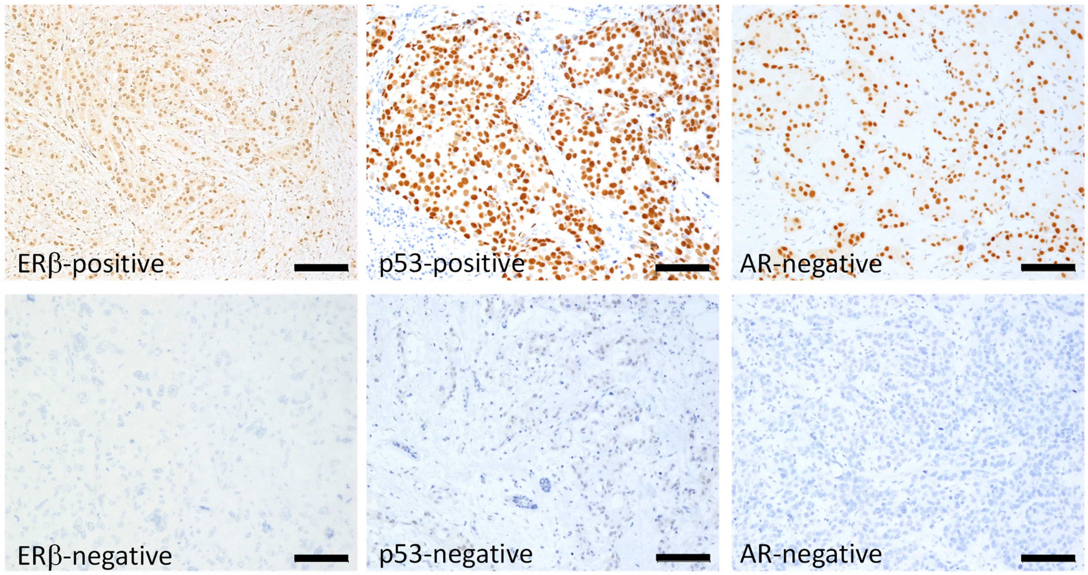
Figure 1. Typical positive and negative nuclear staining of breast cancerous cells for ERβ, p53, and AR: immunoperoxidase staining with hematoxylin counterstaining. Bar: 100 μm. The cut-off was set at 10% for ERβ and AR. For p53, a positive result was defined as one in which tumor cells stained more strongly than adjacent noncancerous epithelial cells.
Ijms 26 11459 g001
Figure 2. Kaplan–Meier disease-free interval (DFI) curve of postmenopausal women with triple-negative breast cancer according to estrogen receptor-β (ERβ) status. Younger: 47 patients aged 55–64 y/o; older: 75 patients aged 75 y/o or older; total: 122 postmenopausal patients. ERβ positivity was slightly related to favorable DFI in total and older patients, but the difference was insignificant. p-value was determined using the log-rank test.
Ijms 26 11459 g002
Figure 3. Kaplan–Meier disease-free interval (DFI) of postmenopausal women with triple-negative breast cancer according to p53 status in ERβ-positive group, ERβ-negative group, and total. In total, DFI of p53-positive group was almost significantly more favorable than p53-negative group (p = 0.0571), which was cleared in ERβ-positive group yielding significance (p = 0.0265), contrasting with ERβ-negative group where difference disappeared (p = 0.7228). p-value was determined using log-rank test. (*) Significance, p < 0.05.
Ijms 26 11459 g003
Figure 4. Kaplan–Meier disease-free interval (DFI) curve of postmenopausal women with triple-negative breast cancer according to androgen receptor (AR) status in ERβ-positive group, ERβ-negative group, and total. AR-positive group exhibited more favorable DFI than AR-negative group in total with marginal significance (p = 0.0468), which was much clearer in ERβ-positive group (p = 0.0285), contrasting with ERβ-negative group where difference in DFI according to AR status was lost (p = 0.7734). p-value was determined using log-rank test. (*) Significance, p < 0.05.
Ijms 26 11459 g004
Figure 5. Kaplan–Meier disease-free interval (DFI) curve of postmenopausal women with triple-negative breast cancer according to Ki-67 status in the ERβ-positive group, ERβ-negative group, and total. Ki-67 status affected DFI in none of the groups. p-value was determined using the log-rank test.
Ijms 26 11459 g005
Table 1. The relationship between ERβ and various clinicopathological factors. No significant correlation was observed except for Ki-67 status, which showed a positive correlation with ERβ expression.
FactorsERβ
PositiveNegativePos %p-Value a
Age (y)Younger3314700.38NS
Older581777
Size (mm)≤20378820.128NS
20<532370
Nodal statsPositive3116660.142NS
Negative541578
pStage (I and II vs. III)III1510600.060NS
I and II762178
Histological gradeIII6921770.377NS
I and II221069
Ki-67Positive457870.009 *Positive
Correlation
Negative462466
ARPositive5215780.396NS
Negative391671
p53Positive7020780.175NS
Negative211166
a Pearson Chi-squared test; Pos %, % of positive cases; NS, not significant; pStage, pathological stage; AR, androgen receptor. (*) Statistically significant.
Table 2. The relationship between the immunohistochemical statuses of AR and p53, which were positively correlated.
p53
PositiveNegativePos %p-Value a
AR-Positive5512820.0211 *Positive
Correlation
AR-Negative352064
a Pearson Chi-squared test; Pos %, % of positive cases; (*) statistically significant.
Disclaimer/Publisher’s Note: The statements, opinions and data contained in all publications are solely those of the individual author(s) and contributor(s) and not of MDPI and/or the editor(s). MDPI and/or the editor(s) disclaim responsibility for any injury to people or property resulting from any ideas, methods, instructions or products referred to in the content.