Article

Arthrospira platensis Preserves Uterine Function by Modulating Electromechanical Coupling and Redox Pathways During Resistance Training in Female Rats

by
Bárbara Cavalcanti Barros
1,
Anderson Fellyp Avelino Diniz
1,2,*,
Francisco Fernandes Lacerda-Júnior
1,
Petruska Pessoa da Silva Souza
3,
Adriano Francisco Alves
1,4,
Paula Benvindo Ferreira
1,
Fabiana de Andrade Cavalcante
1,4 and
Bagnólia Araújo da Silva
1,5
1
Postgraduate Program in Natural and Synthetic Products Bioactive/Health Sciences Center, Federal University of Paraiba, João Pessoa 58051-900, Brazil
2
Postdoctoral National Council for Scientific and Technological Development, Functional Pharmacology Laboratory, Federal University of Paraíba, João Pessoa 58051-900, Brazil
3
Health Sciences Center, Federal University of Paraiba, João Pessoa 58051-900, Brazil
4
Department of Biomedical Sciences, Health Sciences Center, Federal University of Paraíba, João Pessoa 58051-900, Brazil
5
Pharmaceutical Sciences Department, Health Sciences Center, Federal University of Paraíba, João Pessoa 58051-900, Brazil
*
Author to whom correspondence should be addressed.
Int. J. Mol. Sci. 2025, 26(23), 11440; https://doi.org/10.3390/ijms262311440
Submission received: 25 July 2025 / Revised: 10 October 2025 / Accepted: 13 October 2025 / Published: 26 November 2025

Abstract

Algae-derived bioactives have emerged as promising nutraceuticals due to their ability to modulate key molecular pathways under physiological stress. Arthrospira platensis (Spirulina), a cyanobacterium widely recognized for its antioxidant and anti-inflammatory properties, is proposed as a functional supplement to preserve smooth muscle physiology. Progressive strength training (PST) can induce oxidative stress and disrupt electromechanical coupling in the uterus, potentially impairing female reproductive function. This study investigated whether supplementation with A. platensis prevents PST-induced uterine dysfunction and elucidated the molecular mechanisms involved. Virgin Wistar rats were divided into five groups: sedentary with saline (GS), sedentary with A. platensis (GAP100), adapted control (GC), PST-trained (GT), and PST-trained with A. platensis (GTAP100). An eight-week water-jump PST protocol was applied. Uterine contractile responses were recorded in isolated organ baths after cumulative KCl stimulation, in the absence or presence of pathway-specific inhibitors targeting nitric oxide synthase, cyclooxygenase, NADPH oxidase, or superoxide dismutase. Histological evaluations of uterine and ovarian tissues were also performed. PST increased contractile efficacy and myometrial thickness, associated with oxidative stress and activation of NO, COX, and NADPH oxidase pathways. Supplementation with A. platensis attenuated these alterations by enhancing NO signaling, stimulating relaxant prostanoids, and reducing superoxide production. These protective effects were abolished by inhibitors, confirming mechanistic involvement. Overall, our findings provide molecular evidence that A. platensis supplementation preserves uterine smooth muscle physiology under high-intensity resistance training, supporting its potential as a nutraceutical strategy for female reproductive health.

Graphical Abstract

1. Introduction

Progressive strength training (PST) is a widely used exercise modality known to induce muscle hypertrophy, neuromuscular adaptations, and improvements in maximal strength [1,2,3]. Beyond these well-established benefits, PST also promotes metabolic adaptations, including increased glycolytic enzyme activity, elevated intracellular ATP and phosphocreatine levels, and reductions in mitochondrial content and capillarization. These effects vary according to training intensity, frequency, and individual factors such as age, sex, genetic background, and training history [4,5,6,7].
The female reproductive system is particularly sensitive to physiological stress. Intense PST may lead to dysfunctions such as delayed menarche, amenorrhea, and oligomenorrhea, reported in up to 79% of athletes [8,9,10,11]. This indicates that the uterine tissue may respond to metabolic and oxidative stimuli even without direct exposure to exercise. Moreover, PST increases the production of reactive oxygen species (ROS) and reactive nitrogen species (RNS), both of which play dual roles: modulating reproductive processes such as oocyte quality and implantation, but causing damage when produced in excess—depending on exercise intensity, conditioning, and nutritional status [6,7,8,9].
These reactive species also profoundly impact calcium-mediated uterine contraction mechanisms. Ca2+ release through channels such as the inositol trisphosphate receptor (IP3R) and the sensitivity of uterine smooth muscle to calcium are strongly influenced by redox states; under hypoxic conditions, ROS production increases via mitochondrial complexes I and III, compromising myometrial contractility [3,10,11,12,13,14,15,16,17]. Although progressive strength training (PST) does not directly stimulate uterine smooth muscle, systemic responses to intense and repetitive exercise—such as hormonal fluctuations, oxidative stress, and ischemia/reperfusion phenomena—can indirectly modulate uterine contractility. Repeated intense uterine contractions have been shown to cause transient ischemia and hypoxia, triggering adaptive or pathological responses [18]. In addition, oxidative stress is strongly implicated in female reproductive dysfunction [19,20] and intense exercise is associated with altered gonadotropin release and menstrual irregularities [10]. Resistance exercise has also been shown to modify ovarian physiology [21,22], and activation of MAPK/ERK signaling has been linked to increased uterine contractility [23,24]. These mechanisms support the rationale that PST may indirectly affect uterine physiology.
In the uterus, repeated or intense contractions can transiently reduce uteroplacental blood flow, leading to local hypoxia and eliciting adaptive or pathological responses depending on the context. Experimental and clinical studies have shown that excessive uterine activity during labor is associated with transient ischemia of the myometrium and can modulate cellular signaling, cytokine expression, and contractile protein activity, which may be protective or, in some cases, deleterious to both maternal and fetal tissues [25,26,27,28]. These studies provide clear evidence that the intensity and frequency of uterine contractions are key determinants of tissue oxygenation and the subsequent cellular response.
This redox modulation of calcium-signaling pathways becomes particularly relevant in different physiological and pathological contexts within the uterus. For instance, during the menstrual phase, small myometrial contractions, associated with vasoconstriction of the endometrial spiral arteries, induce transient endometrial hypoxia—a physiological event that signals the end of menstruation [29]. Moreover, in pregnant animal models, moderate-to-intense exercise results in reduced uterine blood flow—estimated at 15–25% during moderate exercise and approximately 30% during prolonged efforts or late gestation—accentuating conditions of critical oxygenation [30]. Although this decrease in perfusion does not constitute complete ischemia, it can significantly alter the oxygen microenvironment, contributing to either adaptive or pathological responses depending on the duration and intensity of the redox/kinetic stimulus.
Furthermore, no previous studies have directly evaluated the impact of PST on uterine contractility. However, Ferreira et al. investigated this connection in an animal model (rats), assessing uterine reactivity after strength training with or without dietary supplementation. Their results demonstrated that PST increased contractile efficacy, both in pharmacomechanical coupling (using oxytocin) and electromechanical coupling (using KCl), while also elevating oxidative stress in the uterine tissue. These findings indicate that the uterus is a functional target of exercise and responds to the physiological stress of PST. Notably, when combined with Arthrospira (Spirulina) platensis supplementation, this effect was prevented, while the tissue’s antioxidant capacity was further enhanced [31].
Additionally, it is well established that intense physical training, particularly when combined with inadequate recovery, can disrupt systemic redox balance and cause hormonal dysregulation. Such alterations compromise the homeostasis of the reproductive system, amplifying adverse effects through oxidative stress [1,3,4]. Thus, even though exercise does not act directly on the uterus as a local mechanical stimulus, several systemic mechanisms justify the relevance of the uterus in this experimental context (Figure 1).
Our group has previously demonstrated the antioxidant and pharmacological potential of Arthrospira platensis in smooth muscle tissues, including the uterus and ileum [31,32]. This cyanobacterium protects against excessive contractile activity and also acts in other tissues such as the aorta, trachea, and corpus cavernosum, reinforcing its relevance in the modulation of smooth muscle tone [33,34,35,36,37].
Given the uterus’s unique contractile profile and its high sensitivity to oxidative stress, it is essential to investigate how PST affects electromechanical coupling mechanisms in this tissue. Although previous studies are limited, Ferreira et al. provided valuable evidence in a Wistar rat model subjected to PST with or without dietary supplementation, demonstrating that PST increased contractile efficacy (Emax) and reduced potency (pCE50) in response to KCl-induced stimulation. Notably, supplementation with A. platensis (50 and 100 mg/kg) prevented these alterations, with the 100 mg/kg dose yielding the most pronounced effects. These findings highlight the protective action of A. platensis and underscore the need to elucidate the molecular mechanisms involved in this response.
Most of the available evidence on exercise and female reproductive function derives from studies on high-intensity interval training (HIIT) or sustained excessive exercise. However, to our knowledge, there is no data specifying how progressive resistance training influences ovarian or uterine physiology. This lack of direct evidence highlights the novelty of our study and underscores the need to investigate whether the uterus is a functional target of resistance exercise.
This study should be viewed as exploratory and hypothesis-generating, providing an initial step toward understanding how resistance training affects uterine physiology and how Arthrospira platensis supplementation may mitigate these effects.

2. Results

2.1. Histomorphometric Evaluation of Uterine Tissue

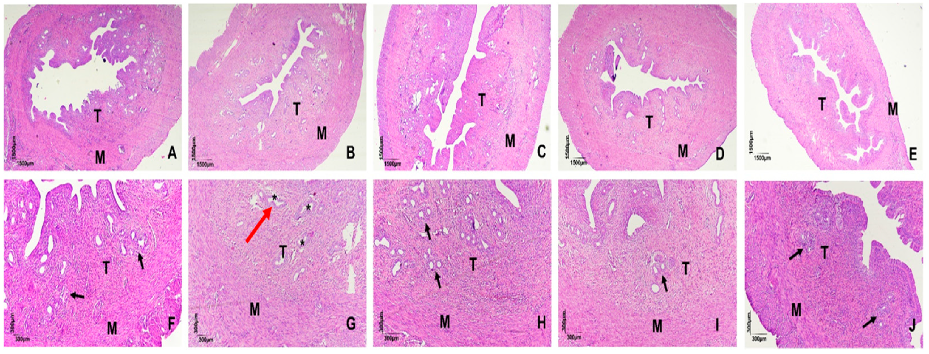
Histological examination of uterine sections stained with hematoxylin and eosin revealed preserved endometrial morphology and circular glandular formations in the GC (control), GTAP100 (trained + AP), GS (saline), and GAP100 (saline + AP) groups. In contrast, GT (trained) animals exhibited disrupted endometrial architecture with individualized and diffusely distributed glands, suggesting morphological alterations induced by PST (Figure 2).
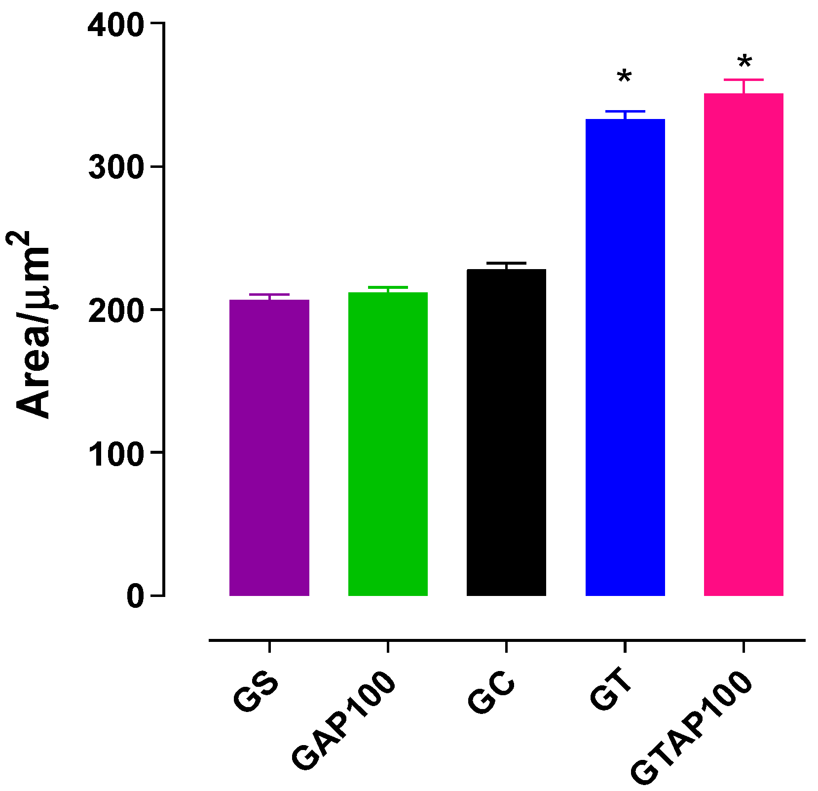
Morphometric analysis of the myometrial layer showed no significant difference in muscle thickness between the GS (206.7 ± 3.8 µm2) and GAP100 (211.7 ± 3.6 µm2) groups. However, GT rats demonstrated a significant increase in myometrial thickness (333.0 ± 5.5 µm2) compared to GC (227.8 ± 4.6 µm2), indicating PST-induced hypertrophy. Supplementation with A. platensis did not reverse this increase in GTAP100 rats (351.2 ± 9.6 µm2) (Figure 3).

2.2. Effect of PST and A. platensis on KCl-Induced Contractility in the Presence of L-NAME

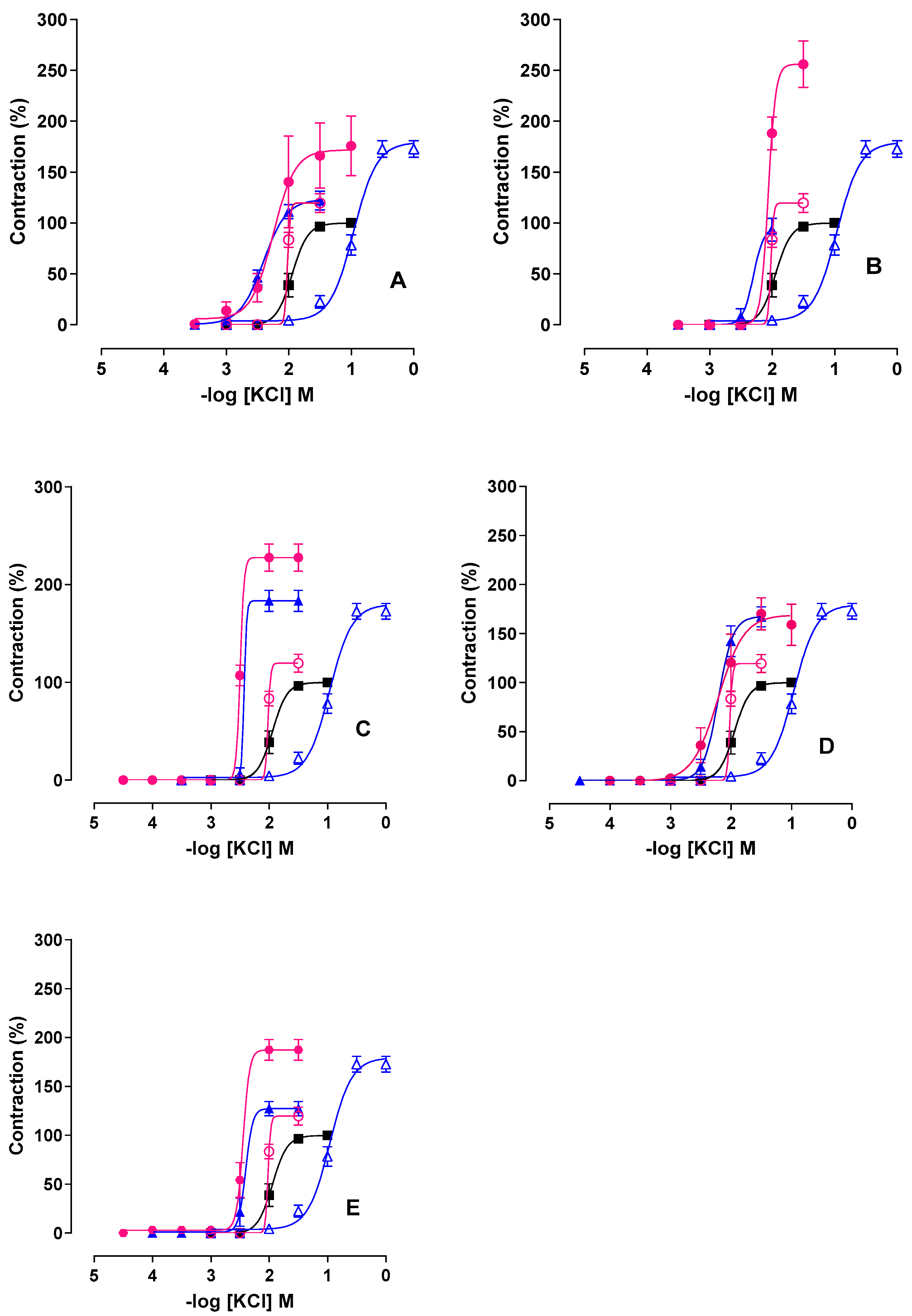
In GT animals, the presence of L-NAME (NOS inhibitor) shifted the KCl concentration–response curve to the left and decreased efficacy by approximately 50% (Emax = 122.1 ± 9.1%; pCE50 = 2.4 ± 0.05) compared to the absence of inhibitor (Emax = 172.7 ± 8.1%; pCE50 = 1.0 ± 0.03) (Figure 4, Table 1). In GTAP100 rats, L-NAME abolished the protective effect of AP, resulting in a marked increase in contractile efficacy (Emax = 203.0 ± 27.8%) without significant change in potency (pCE50 = 2.1 ± 0.2).

2.3. Effect of PST and A. platensis in the Presence of Indomethacin

The COX inhibitor indomethacin significantly reduced KCl efficacy in GT rats (Emax = 93.4 ± 11.3%; pCE50 = 2.2 ± 0.06), compared to the untreated condition (Emax = 172.7 ± 8.1%; pCE50 = 1.0 ± 0.03) (Figure 4, Table 1). In GTAP100 rats, indomethacin markedly increased contractile efficacy (Emax = 256.0 ± 22.9%), suggesting involvement of prostanoid pathways in AP-mediated protection (Figure 4 and Table 1, n = 5).

2.4. Combined Effect of L-NAME and Indomethacin

Simultaneous administration of L-NAME and indomethacin in GT rats maintained high contractile efficacy (Emax = 183.5 ± 10.85%) and significantly increased KCl potency (pCE50 = 2.3 ± 0.05) compared to GT alone (Figure 4, Table 1). In the GTAP100 group, co-incubation led to even higher efficacy (Emax = 227.8 ± 14.0%; pCE50 = 2.5 ± 0.005), indicating synergistic modulation of NO and COX pathways by AP.

2.5. Effect of NADPH Oxidase Inhibition with Apocynin

In GT rats, apocynin shifted the KCl curve to the left (pCE50 = 2.2 ± 0.06) but had minimal effect on efficacy (Emax = 167.5 ± 10.3%), indicating ROS involvement (Figure 4, Table 1). In the GTAP100 group, apocynin increased KCl efficacy to 170.0 ± 16.3% without changing potency, suggesting AP competes with NADPH oxidase-derived ROS signaling.

2.6. Effect of Tempol (SOD Mimetic) on Contractility

Tempol significantly reduced KCl efficacy in GT rats (Emax = 127.2 ± 7.3%) and increased potency (pCE50 = 2.3 ± 0.06), compared to untreated GT (Figure 4, Table 1). In GTAP100 rats, tempol further enhanced both efficacy (Emax = 187.3 ± 10.6%) and potency (pCE50 = 2.4 ± 0.06), suggesting an additive antioxidant effect with AP supplementation.
These results confirm that PST enhances uterine contractility through oxidative and inflammatory mechanisms, and that Arthrospira platensis supplementation modulates these effects via nitric oxide, prostanoid, and ROS pathways.

3. Discussion

This study demonstrated that progressive strength training (PST) induces not only uterine hypertrophy but also histomorphological alterations in female rats. The increase in uterine contractility observed likely involves nitric oxide (NO) production and its interaction with reactive oxygen species (ROS), resulting in peroxynitrite formation. This process appears to activate the PLA2 pathway and stimulate the synthesis of contractile prostanoids, in addition to an inhibitory crosstalk between the COX and NO pathways, increased superoxide anion production via NADPH oxidase, and possibly decreased superoxide dismutase (SOD) activity.
Supplementation with Arthrospira platensis did not inhibit PST-induced myometrial hypertrophy. This finding suggests that myometrial thickening represents a compensatory structural adaptation to repeated contractile and hemodynamic stress during training. Such hypertrophy should not necessarily be interpreted as a pathological dysfunction but rather as part of the uterine remodeling process induced by exercise. In contrast, the protective role of Arthrospira platensis was manifested by preventing oxidative stress, preserving endometrial architecture, and normalizing contractile hyperreactivity. Thus, the alga did not directly block hypertrophic remodeling but mitigated the functional and molecular dysfunctions associated with PST. The underlying electromechanical mechanism seems to involve positive modulation of the NO pathway, release of relaxant prostanoids, and activation of crosstalk between these pathways. The antioxidant properties of AP may act by neutralizing ROS with contractile effects on uterine smooth muscle, as previously demonstrated in other smooth muscle tissues [32,33,34,35,36].
Physical training is known to induce morphological, metabolic, and functional adaptations in various biological systems, thereby enhancing performance and overall health. However, when improperly prescribed or combined with caloric restriction, it may cause muscle damage and elevate ROS levels, which can lead to hypothalamic dysfunction, altered gonadotropin release, menstrual disorders, infertility, and even osteoporosis [10,37,38,39,40,41,42]. Despite the importance of these consequences for female reproductive health, few studies have assessed the impact of physical training on uterine contractility [31].
Animal models are essential for understanding these mechanisms. Prior studies from our group have demonstrated that AP supplementation in rats undergoing PST improves physical performance and reduces muscle damage, oxidative stress, and inflammation [33,40]. Specifically, in the uterus, Ferreira et al. [31] showed that AP at doses of 50 and 100 mg/kg did not alter contractile potency in sedentary rats, but significantly reduced KCl-induced contractile efficacy. Moreover, when combined with PST, AP supplementation prevented the increase in contractility and decrease in potency induced by training, with 100 mg/kg proving more effective.
The uterus, due to its specific contractile patterns and sensitivity to oxidative stress, is particularly affected by PST. In our study, rats in the trained group (GT) presented with individualized and diffusely distributed endometrial glands, in contrast to the circular and preserved morphology observed in other groups. This suggests that PST, even at early stages (within 2 weeks), can induce structural alterations in the uterus that may compromise reproductive function. These changes may result from reduced levels of FSH, LH, and progesterone, which have been reported in female rats following resistance training [41,42].
It is also important to emphasize the physiological relevance of the “water-jump” protocol adopted in this study. Although this model does not directly apply mechanical load to the uterus, it is widely validated to reproduce the systemic stress of resistance training in rodents, including cardiorespiratory, metabolic, and redox adaptations. Repetitive jumping in water induces marked increases in intra-abdominal pressure and sympathetic activation, which promote redistribution of blood flow toward active skeletal muscles and transiently reduce uterine perfusion. Previous studies have demonstrated that increased intraperitoneal pressure reduces cardiac output and visceral blood flow in rodents [43], while forced swimming and diving paradigms trigger reflex vasoconstriction and redistribution of circulation [44]. In pregnant models, exercise has been shown to modulate angiogenic imbalance under reduced uterine perfusion, confirming that systemic hemodynamic stress can affect uterine physiology [45,46]. Together, these findings support the mechanistic rationale that transient ischemia and hypoxia induced by water-jump training can mimic the uterine hemodynamic and redox alterations observed during high-intensity resistance exercise in women.
Oxidative stress-induced proliferation of endometrial cells can also activate the mitogen-activated protein kinase (MAPK) pathway and ERK1/2, contributing to increased uterine contractility [47]. The observed increase in myometrial thickness in GT rats may explain the enhanced KCl contractile efficacy and decreased potency previously reported by Ferreira et al. [31]. This effect may reflect an increase in smooth muscle fiber content [48], coupled with oxidative imbalance and p38-MAPK pathway activation, which promotes smooth muscle growth [49].
Exercise-induced inflammation plays a critical role in tissue remodeling, involving local and systemic release of pro-inflammatory cytokines [50,51]. Under physiological conditions, NO is continuously produced in small amounts by endothelial nitric oxide synthase (eNOS), exerting vasodilatory effects. However, during injury or inflammation, inducible nitric oxide synthase (iNOS) is upregulated and produces large quantities of NO, which can react with ROS to form peroxynitrite, which is a cytotoxic and contractile molecule [38,52].
In our study, the non-selective NOS inhibitor L-NAME abolished the PST-induced increase in contractility in the GT group, suggesting that iNOS-derived NO reacts with superoxide to form peroxynitrite, enhancing uterine contractile responses. In contrast, in AP-supplemented trained rats (GTAP100), this protective effect was lost in the presence of L-NAME, indicating that AP may enhance eNOS phosphorylation or expression via phycocyanin, which is a key antioxidant pigment in Arthrospira [40,53], thereby increasing NO bioavailability.
Exercise also activates PLA2, which in turn stimulates NADPH oxidase activity and ROS generation in muscle mitochondria and cytosol [54,55,56]. Using indomethacin, a non-selective COX inhibitor, we observed that PST-induced increases in uterine contractility were attenuated, suggesting that COX activation and synthesis of contractile prostanoids, such as PGF2α, are involved [57,58].
In trained rats supplemented with AP, indomethacin abolished the preventive effect of the alga, indicating that AP modulates COX activity, possibly shifting the prostanoid balance toward relaxant mediators such as PGE2 [59,60]. This may be attributed to Arthrospira content of γ-linolenic, linoleic, arachidonic, and eicosapentaenoic acids—precursors of relaxant prostanoids [61,62]—as well as to the COX-2 inhibitory properties of phycocyanin [63].
Crosstalk between the NO and prostanoid pathways is well described in the literature. Activation of cPLA2 increases intracellular calcium, enhancing membrane permeability, and stimulating NO production via eNOS and neuronal NOS (nNOS) [64]. Under basal conditions, NO favors the synthesis of relaxant prostanoids, whereas its inhibition favors contractile mediators [65]. S-nitrosylation and peroxynitrite-mediated COX activation may also be involved [66,67,68].
When L-NAME and indomethacin were administered simultaneously, the PST-induced loss of KCl potency was reversed, but contractile efficacy remained high. This supports the hypothesis of an inhibitory crosstalk between the NO and COX pathways. In the GTAP100 group, however, simultaneous inhibition led to a 3.5-fold increase in KCl efficacy, suggesting that AP enhances the activation of this crosstalk in favor of relaxant signaling pathways such as NO and PGE2 [6,65,66,67,68,69]. We further investigated the role of ROS, particularly superoxide anion and hydrogen peroxide, in modulating uterine contractility during PST. Apocynin, an NADPH oxidase inhibitor, restored KCl potency in GT rats but did not affect contractile efficacy, confirming the role of NADPH oxidase-derived ROS in PST-induced uterine hyperreactivity [38,70,71,72].
Thus, it was evaluated whether the alterations in uterine contractile reactivity of rats subjected to PFT or to its combination with algae supplementation were correlated with the modulation of the crosstalk between the NO and prostanoid pathways, through simultaneous incubation with L-NAME and indomethacin, followed by a contraction–response curve induced with KCl.
Interestingly, in AP-supplemented rats, apocynin abolished the protective effects of the alga, suggesting that AP may compete for the same NADPH oxidase inhibition sites. Alternatively, increased cGMP induced by AP might inhibit NADPH oxidase activity and restore NO availability [73].
Finally, using tempol, an SOD mimetic, we observed that it prevented the increase in KCl-induced efficacy in GT rats while enhancing potency, supporting the detrimental role of superoxide in uterine contractility [33]. In the GTAP100 group, tempol enhanced both efficacy and potency, possibly due to the antioxidant constituents of AP (e.g., phycocyanin, β-carotene), which may act downstream of superoxide formation and promote hydrogen peroxide detoxification via SOD and glutathione reductase [63,74,75]. However, high antioxidant doses may blunt exercise-induced upregulation of endogenous enzymes such as SOD and glutathione peroxidase [76,77].
Altogether, these findings suggest that AP supplementation exerts protective effects on uterine physiology during PST by mitigating oxidative damage, restoring NO signaling, promoting relaxant prostanoid synthesis, inhibiting NADPH oxidase activity, and enhancing antioxidant defenses. These results support further investigation of AP as a functional nutraceutical for female reproductive health under physical stress conditions.
A limitation of the present work is that the signaling pathways involved were investigated using a functional pharmacological approach with specific pathway inhibitors (L-NAME, indomethacin, apocynin, and tempol). Although this strategy is classical in smooth muscle pharmacology and provides robust mechanistic evidence, it does not directly demonstrate alterations in protein expression or gene transcription. We therefore acknowledge that this is a limitation of the present study. Future investigations employing molecular biology techniques such as Western blotting, qPCR, or immunohistochemistry are required to confirm changes in NO synthase isoforms, COX enzymes, and NADPH oxidase subunits in uterine tissue. Such complementary approaches will provide more precise mechanistic insight into how Arthrospira platensis modulates redox and signaling pathways.

4. Methods and Materials

4.1. Obtaining and Preparing Arthrospira Platensis

Lyophilized Arthrospira platensis powder (Lot No. 405894) was obtained from Infinity Pharma (Hong Kong, China) and certified and fractionated by Roval Manipulação (João Pessoa, Brazil). The powder was freshly dissolved in 0.9% of NaCl saline solution before administration at a dose of 100 mg/kg. Supplementation was administered via oral gavage once daily, 30 min prior to each training session, for a total duration of 8 weeks [37].

4.2. Animals and Experimental Groups

Virgin female Wistar rats (Rattus norvegicus), aged 8 weeks and weighing between 150 and 250 g, were obtained from the Animal Production Unit (UPA) at the Institute of Drug and Medicine Research (IPeFarM), Federal University of Paraíba (UFPB), Brazil. Prior to the experiments, animals were housed under controlled conditions (21 ± 1 °C, 12 h light/dark cycle, lights on from 6:00 a.m. to 6:00 p.m.) with unrestricted access to a standard pellet diet (Nuvilab®, Quimtia S.A., São Paulo, Brazil) and water. To minimize hormonal variability, only virgin females of the same age range were included, and housing conditions were maintained to reduce natural estrous oscillations. Additionally, all animals were hormonally synchronized by administration of diethylstilbestrol (1 mg/kg, s.c.) 24 h before euthanasia, ensuring that uterine tissues used for functional assays were collected under the same estrous stage.
All procedures complied with institutional ethical standards and were approved by the UFPB Animal Ethics Committee (Protocol No. 5191200320, ID 000946).
Animals were randomly allocated into five groups:
1.
Sedentary control (GS): Did not undergo either adaptation or training and remained completely sedentary, receiving only saline by gavage.
2.
Sedentary supplemented with A. platensis (GAP100, 100 mg/kg): Did not undergo exercise intervention or an adaptation period and received A. platensis at 100 mg/kg by gavage.
3.
Adapted control (GC): Underwent the one-week adaptation protocol only, which consisted of light water-jumping sessions with reduced overload (50% of body weight) and limited repetitions. This adaptation was designed to familiarize animals with the aquatic environment and apparatus, minimize stress, and prevent confounding acute effects of exercise initiation. Importantly, GC animals did not undergo subsequent progressive strength training, allowing the differentiation between physiological effects of the adaptation process itself and those induced by the full training program.
4.
Trained (GT): Underwent the adaptation week followed by 8 weeks of progressive resistance training and received saline by gavage.
5.
Trained supplemented with A. platensis (GTAP100, 100 mg/kg): Underwent the adaptation week followed by 8 weeks of progressive resistance training and received A. platensis at 100 mg/kg by gavage.

4.3. Progressive Strength Training Program (PST)

The animals in the progressive strength training group underwent a specific jumping program in a polyvinyl chloride (PVC) cylinder, which was 30 cm in diameter and 70 cm long, containing water. The depth of the water contained in the tanks was 50 cm, approximately equivalent to twice the length of the rat, aiming to limit the alternative of climbing until grabbing the edge of the cylinder. Before starting the exercise, the water was heated to a temperature of around 32 °C, as it is considered neutral in relation to the rat’s body temperature [37].
Strength training was based on a protocol of jumping in water, consisting of 4 sets of 10 to 12 repetitions, with a 30-second interval between sets, with progressive overload adjusted according to the animal’s weight. The overload was applied to the animals’ thorax through a special vest that allowed them to perform the jumps without the load disconnecting from the body or impeding their movements. An overload corresponding to the weight of the vest when wet (25 g) was considered and deducted from the specific load corresponding to the animal’s body mass for better training precision. Exercise sessions were always carried out from 12 pm to 2 pm. During each exercise series, the time spent by the animal to perform the exercise was analyzed with the aim of evaluating the effectiveness of the exercise on muscle performance.
Strength training was developed as follows: (a) adaptation week—the first week of training was intended for the adaptation of the animals; for this, the animals performed three alternating exercise sessions, with an overload equivalent to 50% of the animal’s body mass, with the number of sets and repetitions adjusted daily and a 30-second rest interval between sets. 1st day: 2 sets × 5 jumps; 2nd day: 4 sets × 5 jumps; 3rd day: 4 sets × 9 jumps. After the adaptation week, training was performed 3 days a week (alternate days) as follows: (a) 1st and 2nd weeks—the animals performed 4 sets of 10 jumps, with 30 s of rest between sets and an overload equivalent to 50% of the animal’s body weight; (b) 3rd and 4th weeks—4 sets of 10 jumps were performed, with 30 s of rest between sets and an overload of 60% of the body weight; (c) 5th and 6th weeks—the 4 sets of 10 jumps remained, with 30 s of rest between sets, but with an overload of 80% of the animal’s body weight; (d) 7th and 8th weeks—the 4 sets, 30 s of rest between sets, and an overload of 80% of the animal’s body weight remained, but with 12 jumps performed (Table 2).

4.4. Reagents and Solutions

All chemicals were of analytical grade. Calcium chloride dihydrate, sodium bicarbonate, potassium chloride, magnesium chloride dihydrate, glucose, sodium chloride, hydrochloric acid, and sodium hydroxide were purchased from Êxodo Científica (João Pessoa, Brazil). Diethylstilbestrol was obtained from Cayman Chemical (Ann Arbor, MI, USA). Tempol, apocynin, L-NAME (Nω-Nitro-L-arginine methyl ester hydrochloride), and indomethacin were purchased from Sigma-Aldrich (São Paulo, Brazil). Ketamine and xylazine were obtained from commercial veterinary suppliers (ketamine hydrochloride and xylazine hydrochloride).

4.5. Nutrient Solutions

The nutrient solution used was Locke–Ringer [78], adjusted to pH 7.4 with 1 N HCl or NaOH, continuously bubbled with carbogen (95% O2 and 5% CO2), and maintained at 32 °C. Locke–Ringer’s physiological solution was freshly prepared on the day of the experiments and had the following composition (mM): NaCl (154.0), KCl (5.6), MgCl2 (2.1), CaCl2 (2.2), glucose (5.6), and NaHCO3 (6.0). Solutions and incubation stocks were stored at −20 °C when appropriate.

4.6. Histological Analysis

Uterine and ovarian samples were fixed in 10% of neutral buffered formalin (10% NBF, with approximately 4% of w/v formaldehyde in phosphate buffer, pH 7.0–7.4) for 24–48 h at room temperature. After fixation, samples were dehydrated through an ascending ethanol series, cleared in xylene and embedded in paraffin. Dehydration and clearing steps were as follows: 70% EtOH, 5 min; 80% EtOH, 5 min; 95% EtOH, 2 × 5 min; absolute ethanol, 2 × 5 min; xylene, 2 × 10 min. Paraffin-embedding was performed using histological paraffin (melting point 56–58 °C). Embedded samples were sectioned at 4 µm thickness on a rotary microtome and mounted on glass slides.
For staining, sections were deparaffinized in xylene (2 × 10 min), rehydrated through a descending ethanol series (absolute EtOH 2 × 5 min, 95% 5 min, 80% 5 min, 70% 5 min), rinsed in distilled water, and stained with Harris hematoxylin for 1 min. Slides were then washed under running water for 5 min and stained with eosin Y (0.5% w/v in 95% ethanol) for 3 min. After staining, slides were dehydrated through the ascending ethanol series, cleared in xylene (2 × 5–10 min), and mounted with the Entellan® mounting medium (Merck; ready-to-use synthetic resin mounting medium—used as supplied). The Harris hematoxylin used was a commercial Harris hematoxylin solution (11;39) [79].

4.7. Investigation of the Preventive Mechanism of A. platensis Supplementation on Contractile Changes Induced by Strength Training

4.7.1. Preparation of Isolated Rat Uterus

The rats were treated 24 hours beforehand with diethylstilbestrol (1 mg/kg) subcutaneously for hormonal synchronization of estrus. After this time, the rats were euthanized by anesthesia with ketamine 100 mg/kg (i.p.) and xylazine 10 mg/kg (i.p.), followed by a complementary method of decapitation with the aid of a guillotine. The abdominal cavity was opened with a longitudinal cut, and the uterus was removed and cleaned of all connective and adipose tissue. Then, the two uterine horns were separated by means of an incision, opened longitudinally, and suspended vertically in bath tanks for isolated organs, containing the Locke–Ringer nutrient solution, which was heated to 32 °C and gassed with carbogen.
These tissues were kept under tension of 1 g and remained at rest for 40 min, which was the time required for stabilization of the preparation. During this period, the nutrient solution was changed every 10 min (adapted from [78]). After the stabilization period, a contraction was induced with 60 mM of KCl to verify the functionality of the organ. After 15 min, a contraction was induced with cumulative concentrations of KCl (10−4–10−1 M). The results were evaluated by comparing the amplitude of the contractile response of the uterus of the rats in the groups that received supplementation with A. platensis and/or were subjected to progressive strength training, with that obtained by the average of the maximum amplitudes of the control curves.

4.7.2. Investigation of the Participation of the Nitric Oxide Pathway and Cyclooxygenases

Obtaining Cumulative Concentration–Response Curves to KCl, in the Absence and Presence of L-NAME or Indomethacin
The uterus was assembled as described in item 2.3.1 After the stabilization period, L NAME (10−4 M), a non-selective NOS inhibitor [80], or indomethacin (10−5 M), a non-selective COX inhibitor [81], was incubated for 20 min in different preparations, and then the cumulative concentration–response curves to KCl (10−4–10−1 M) were induced in the absence and presence of the inhibitors. In addition, cumulative curves to KCl were performed in other preparations in the simultaneous presence of both inhibitors.

4.7.3. Investigation of the Participation of the Enzymes Nicotinamide Adenine Dinucleotide Phosphate Oxidase and Superoxide Dismutase

Obtaining Cumulative Concentration–Response Curves to KCl, in the Absence and Presence of Apocynin and Tempol
The uterus was assembled as described in item 2,3.1 After the stabilization period, it was incubated for 20 min with apocynin (10−4 M), an NADPH oxidase inhibitor [82], or tempol (10−3 M), an SOD mimetic [83], in different preparations and then the cumulative concentration–response curves were induced to KCl (10−4–10−1 M) in the absence and presence of the inhibitors.

4.8. Statistical Analysis

Data are expressed as mean ± standard error of the mean (SEM). The number of animals per experimental group is indicated in each figure legend (n = 5). Data were tested for normality (Shapiro–Wilk test) and homogeneity of variances. Statistical analyses were conducted using unpaired Student’s t-test or one-way ANOVA followed by Tukey’s post hoc test. Differences were considered significant at p < 0.05. Negative logarithm of the concentration of agonist that produces 50% of the maximal effect (pCE50) values were calculated using nonlinear regression. Emax values were defined as the maximum contractile response. Analyses were performed using GraphPad Prism® 7.01 (GraphPad Software, San Diego, CA, USA).

5. Conclusions

The electromechanical mechanism underlying progressive strength training involves myometrial hypertrophy and is likely mediated by increased production of contractile prostanoids, NO, and superoxide anion, along with inhibitory crosstalk between the NO and COX pathways and reduced SOD activity. In female rats supplemented with Arthrospira platensis, PST did not prevent compensatory myometrial hypertrophy but preserved uterine function by reducing oxidative stress, preserving endometrial morphology, enhancing NO signaling, stimulating relaxant prostanoid synthesis, and inhibiting NADPH oxidase-derived ROS production. These protective effects indicate that Arthrospira platensis primarily modulates redox and signaling pathways rather than interfering with structural hypertrophic remodeling, thereby preventing the development of functional uterine dysfunction during resistance training. These findings suggest that Arthrospira platensis supplementation may serve as a promising dietary strategy to prevent oxidative damage and contractile dysregulation in the uterus induced by high-intensity strength training. If confirmed in human studies, Arthrospira platensis could represent a safe and effective functional supplement to support female reproductive health during resistance exercise regimens.

Author Contributions

Conceptualization, B.C.B. and B.A.d.S.; methodology, B.C.B., F.F.L.-J., P.B.F. and A.F.A.; validation, B.C.B., A.F.A.D., P.B.F., F.d.A.C. and B.A.d.S.; formal analysis, B.C.B., A.F.A.D., F.F.L.-J., P.P.d.S.S., A.F.A. and P.B.F.; investigation, B.C.B., P.B.F. and B.A.d.S.; data curation, B.C.B., A.F.A.D., F.F.L.-J., P.P.d.S.S., A.F.A. and P.B.F.; writing—original draft, B.C.B. and A.F.A.D.; writing—review and editing, B.C.B., A.F.A.D. and F.d.A.C.; visualization, B.C.B.; supervision, A.F.A.D., P.B.F., F.d.A.C. and B.A.d.S.; software, B.C.B. and A.F.A.; project administration, B.A.d.S. All authors have read and agreed to the published version of the manuscript.

Funding

This research received no external funding.

Institutional Review Board Statement

The study was conducted in accordance with the Declaration of Helsinki, and approved by the Ethics Committee of Use of Animals of UFPB, Brazil (protocol code 5191200320-ID 000946 and approval date 30 March 2020).

Data Availability Statement

The data supporting the findings of this study are available from the corresponding author upon reasonable request.

Acknowledgments

We thank Coordination for the Improvement of Higher Education Personnel (CAPES) and the National Council for Scientific and Technological Development (CNPq) for their financial support and Federal University of Paraíba for logistical support.

Conflicts of Interest

The authors declare that they have no known competing financial interests or personal relationships that could have appeared to influence the work reported in this paper.

Abbreviations

ANOVAAnalysis of variance
APArthrospira platensis
AQPAquaporins
ATPAdenosine triphosphate
CAPESCoordination for the Improvement of Higher Education Personnel (Brazil)
CATCatalase
CLC3Chloride channel 3
CNPqNational Council for Scientific and Technological Development (Brazil)
CO2Carbon dioxide
COXCyclooxygenase
cPLA2Cytosolic phospholipase A2
CYP450Cytochrome P450 monooxygenases
ERKExtracellular signal-regulated kinase
EtOHEthanol
FSHFollicle-stimulating hormone
GAP100Sedentary group supplemented with A. platensis (100 mg/kg)
GCControl group
GSSaline group
GTTrained group
GTAP100  Trained group supplemented with A. platensis (100 mg/kg)
GTSP100  Trained group supplemented with Spirulina platensis (100 mg/kg)
HClHydrochloric acid
HIITHigh-intensity interval training
IL-1βInterleukin-1 beta
iNOSInducible nitric oxide synthase
IP3RInositol trisphosphate receptor
LHLuteinizing hormone
LOXLipoxygenase
L-NAME  Nω-Nitro-L-arginine methyl ester hydrochloride
MAPKMitogen-activated protein kinase
NADPHNicotinamide adenine dinucleotide phosphate
NBFNeutral buffered formalin
NONitric oxide
NOSNitric oxide synthase
O2Superoxide anion
PGE2Prostaglandin E2
PGF2αProstaglandin F2α
PLA2Phospholipase A2
PVCPolyvinyl chloride
PSTProgressive strength training
qPCRQuantitative polymerase chain reaction
RNSReactive nitrogen species
ROSReactive oxygen species
SEMStandard error of the mean
SODSuperoxide dismutase
UFPBFederal University of Paraíba (Brazil)
UPAAnimal Production Unit (UFPB)
XOXanthine oxidase

References

  1. McGregor, R.A.; Cameron-Smith, D.; Poppitt, S.D. It is not just muscle mass: A review of muscle quality, composition and metabolism during ageing as determinants of muscle function and mobility in later life. Clin. Nutr. 2014, 33, 406–414. [Google Scholar] [CrossRef] [PubMed]
  2. Cartee, G.D.; Hepple, R.T.; Bamman, M.M.; Zierath, J.R. Exercise promotes healthy aging of skeletal muscle. Cell Metab. 2016, 23, 1034–1047. [Google Scholar] [CrossRef] [PubMed]
  3. Hughes, D.C.; Ellefsen, S.; Baar, K. Adaptations to endurance and strength training. Cold Spring Harb. Perspect. Med. 2018, 8, a029769. [Google Scholar] [CrossRef]
  4. Zampieri, S.; Pietrangelo, L.; Loefler, S.; Fruhmann, H.; Vogelauer, M.; Burggraf, S.; Pond, A.; Grim-Stieger, M.; Cvecka, J.; Sedliak, M.; et al. Lifelong physical exercise delays age-associated skeletal muscle decline. J. Gerontol. A Biol. Sci. Med. Sci. 2015, 70, 163–173. [Google Scholar] [CrossRef]
  5. Dudley, G.A.; Tesch, P.A.; Miller, B.J.; Buchanan, P. Importance of eccentric actions in performance adaptations to resistance training. Aviat. Space Environ. Med. 1991, 62, 543–550. [Google Scholar] [PubMed]
  6. Powers, S.K.; Radak, Z.; Ji, L.L.; Jackson, M. Reactive oxygen species promote endurance exercise-induced adaptations in skeletal muscles. J. Sport Health Sci. 2024, 13, 780–792. [Google Scholar] [CrossRef]
  7. Karolkiewicz, J.; Szczêsniak, L.; Deskur-Smielecka, E.; Nowak, A.; Stemplewski, R.; Szeklicki, R. Oxidative stress and antioxidant defense system in healthy, elderly men: Relationship to physical activity. Aging Male. 2003, 6, 100–105. [Google Scholar] [CrossRef] [PubMed]
  8. Pereira, A.S.; Spagnol, A.R.; Luciano, E.; Leme, J.A.C.A. Influence of aerobic exercise training on serum markers of oxidative stress in diabetic rats. J. Phys. Educ. 2016, 27, e2726. [Google Scholar] [CrossRef]
  9. Murrell, G.A.; Francis, M.J.; Bromley, L. Modulation of fibroblast proliferation by oxygen free radicals. Biochem. J. 1990, 265, 659–665. [Google Scholar] [CrossRef]
  10. Warren, M.P.; Perlroth, N.E. The effects of intense exercise on the female reproductive system. J. Endocrinol. 2001, 170, 3–11. [Google Scholar] [CrossRef]
  11. Soltaninejad, F.; Shafiei, G.; Rafie, F.; Nematollahi-Mahani, S.N.; Pourjafari, F.; Haghpanah, T.; Afarinesh, M.R. Ovarian epigenetics modifications following lifestyle interventions by exercise and alternate-day feeding in letrozole-induced PCOS rats. Sci. Rep. 2025, 15, 25557. [Google Scholar] [CrossRef]
  12. Di Meo, S.; Iossa, S.; Venditti, P. Skeletal muscle insulin resistance: Role of mitochondria and other ROS sources. J. Endocrinol. 2017, 233, R15–R42. [Google Scholar] [CrossRef] [PubMed]
  13. De Souza, M.J.; Williams, N.I. Physiological aspects and clinical sequelae of energy deficiency and hypoestrogenism in exercising women. Hum. Reprod. Update 2004, 10, 433–448. [Google Scholar] [CrossRef] [PubMed]
  14. Belza, B.; Warms, C. Physical activity and exercise in women’s health. Nurs. Clin. N. Am. 2004, 39, 181–193. [Google Scholar] [CrossRef] [PubMed]
  15. Hamanaka, R.B.; Chandel, N.S. Mitochondrial reactive oxygen species regulate hypoxic signaling. Curr. Opin. Cell Biol. 2009, 21, 894–899. [Google Scholar] [CrossRef]
  16. Lee, P.; Chandel, N.S.; Simon, M.C. Cellular Adaptation to Hypoxia through Hypoxia Inducible Factors and Beyond. Nat. Rev. Mol. Cell Biol. 2020, 21, 268–283. [Google Scholar] [CrossRef] [PubMed]
  17. Olson, S.L.; Akbar, R.J.; Gorniak, A.; Fuhr, L.I.; Borahay, M.A. Hypoxia in uterine fibroids: Role in pathobiology and therapeutic opportunities. Oxygen 2024, 4, 236–252. [Google Scholar] [CrossRef]
  18. Žitkutė, V.; Kvietkauskas, M.; Maskoliūnaitė, V.; Leber, B.; Ramašauskaitė, D.; Strupas, K.; Stiegler, P.; Schemmer, P. Melatonin and Glycine Reduce Uterus Ischemia/Reperfusion Injury in a Rat Model of Warm Ischemia. Int. J. Mol. Sci. 2021, 22, 8373. [Google Scholar] [CrossRef]
  19. Agarwal, A.; Gupta, S.; Sharma, R.K. Role of oxidative stress in female reproduction. Reprod. Biol. Endocrinol. 2005, 3, 28. [Google Scholar] [CrossRef]
  20. Agarwal, A.; Aponte-Mellado, A.; Premkumar, B.J.; Amani, X.; Gupta, S. The effects of oxidative stress on female reproduction: A review. Reprod. Biol. Endocrinol. 2012, 10, 49. [Google Scholar] [CrossRef]
  21. Lara, L.A.; Ramos, F.K.; Kogure, G.S.; Costa, R.S.; Silva de Sá, M.F.; Ferriani, R.A.; dos Reis, R.M. Impact of Physical Resistance Training on the Sexual Function of Women with Polycystic Ovary Syndrome. J. Sex. Med. 2015, 12, 1584–1590. [Google Scholar] [CrossRef] [PubMed]
  22. Jourkesh, M.; Soori, R.; Earnest, C.P.; Mirheidari, L.; Ravasi, A.A.; Stannard, S.R.; Monsalves-Alvarez, M. Effects of six weeks of resistance-endurance training on microRNA-29 expression in the heart of ovariectomised rats. Prz. Menopauzalny 2018, 17, 155–160. [Google Scholar] [CrossRef] [PubMed]
  23. Streuli, I.; Santulli, P.; Chouzenoux, S.; Chapron, C.; Batteux, F. Activation of the MAPK/ERK cell-signaling pathway in uterine smooth muscle cells of women with adenomyosis. Reprod. Sci. 2015, 22, 1549–1560. [Google Scholar] [CrossRef]
  24. Chen, C.; Zhu, S.; Bai, L.; Sui, M.; Chen, D. The role of formyl peptide receptor 1 in uterine contraction during parturition. Front. Pharmacol. 2021, 12, 696697. [Google Scholar] [CrossRef] [PubMed]
  25. Reynolds, A.J.; Geary, M.P.; Hayes, B.C. Intrapartum uterine activity and neonatal outcomes: A systematic review. BMC Pregnancy Childbirth 2020, 20, 532. [Google Scholar] [CrossRef]
  26. Hellman, K.M.; Yu, P.Y.; Oladosu, F.A.; Segel, C.; Han, A.; Prasad, P.V.; Tu, F.F. The effects of platelet-activating factor on uterine contractility, perfusion, hypoxia, and pain in mice. Reprod. Sci. 2018, 25, 384–394. [Google Scholar] [CrossRef]
  27. Tian, R.; Li, X.; Su, J.; Yu, H.; Fei, J.; Xu, C.; Du, X.; Yu, B.; Cao, Y.; Yin, Z. Regional uterine contractility differences during pregnancy: The role of hypoxia and ferroptosis in vitro. Life Sci. 2025, 371, 123603. [Google Scholar] [CrossRef]
  28. Turner, J.M.; Mitchell, M.D.; Kumar, S.S. The physiology of intrapartum fetal compromise at term. Am. J. Obstet. Gynecol. 2020, 222, 17–26. [Google Scholar] [CrossRef]
  29. Critchley, H.O.; Kelly, R.W.; Baird, D.T.; Brenner, R.M. Regulation of human endometrial function: Mechanisms relevant to uterine bleeding. Reprod. Biol. Endocrinol. 2006, 4 (Suppl. 1), S5. [Google Scholar] [CrossRef]
  30. Lashley, C.J.; Supik, D.A.; Atkinson, J.T.; Murphy, R.J.; O’Hagan, K.P. Effect of pregnancy on the uterine vasoconstrictor response to exercise in rats. Physiol. Rep. 2015, 3, e12337. [Google Scholar] [CrossRef]
  31. Ferreira, P.B.; Diniz, A.F.A.; Lacerda Júnior, F.F.; Silva, M.d.C.C.; Cardoso, G.A.; Silva, A.S.; da Silva, B.A. Supplementation with Spirulina platensis Prevents Uterine Diseases Related to Muscle Reactivity and Oxidative Stress in Rats Undergoing Strength Training. Nutrients 2021, 13, 3763. [Google Scholar] [CrossRef]
  32. Diniz, A.F.A.; Claudino, B.F.O.; Francelino, D.M.C.; Junior, E.B.A.; Ferreira, P.B.; Lacerda-Júnior, F.F.L.; Barros, B.C.; Arruda, R.R.A.; Alves, A.F.; Batista, L.M.; et al. Arthrospira platensis prevents oxidative stress and suppresses IL-1β expression in the ileum of rats fed a hypercaloric diet. J. Funct. Foods 2023, 106, 105586. [Google Scholar] [CrossRef]
  33. Araujo, L.C.C.; Brito, A.F.; Souza, I.L.L.; Ferreira, P.B.; Vasconcelos, L.H.C.; Silva, A.S.; Silva, B.A. Spirulina platensis Supplementation Coupled to Strength Exercise Improves Redox Balance and Reduces Intestinal Contractile Reactivity in Rat Ileum. Mar. Drugs 2020, 18, 89. [Google Scholar] [CrossRef]
  34. Souza, I.L.L.d.; Barros, B.C.; Ferreira, E.d.S.; Queiroga, F.R.; Vasconcelos, L.H.C.; Toscano, L.d.L.T.; Silva, A.S.; Silva, P.M.d.; Cavalcante, F.d.A.; Silva, B.A.d. Supplementation with Spirulina platensis Prevents Damage to Rat Erections in a Model of Erectile Dysfunction Promoted by Hypercaloric Diet-Induced Obesity. Mar. Drugs 2022, 20, 467. [Google Scholar] [CrossRef]
  35. Diniz, A.F.A.; Souza, I.L.L.; Ferreira, E.S.; Carvalho, M.T.L.; Barros, B.C.; Ferreira, P.B.; Silva, M.C.C.; Lacerda-Júnior, F.F.; Toscano, L.L.T.; Silva, A.S.; et al. Potential Therapeutic Role of Dietary Supplementation with Spirulina platensis on the Erectile Function of Obese Rats Fed a Hypercaloric Diet. Oxidative Med. Cell. Longev. 2020, 2020, 3293065. [Google Scholar] [CrossRef] [PubMed]
  36. Brito, A.F.; Silva, A.S.; Souza, A.A.; Ferreira, P.B.; Souza, I.L.L.; Araujo, L.C.C.; Félix, G.S.; Sampaio, R.S.; Silva, M.C.C.; Tavares, R.L.; et al. Aortic Response to Strength Training and Spirulina platensis Dependent on Nitric Oxide and Antioxidants. Front. Physiol. 2018, 9, 1522. [Google Scholar] [CrossRef] [PubMed]
  37. Brito, A.F.; Silva, A.S.; Souza, I.L.L.; Pereira, J.C.; Silva, B.A. Intensity of Swimming Exercise Influences Aortic Reactivity in Rats. Braz. J. Med. Biol. Res. 2015, 48, 996–1003. [Google Scholar] [CrossRef] [PubMed]
  38. Kruk, J.; Aboul-Enein, H.Y.; Kładna, A.; Bowser, J.E. Oxidative stress in biological systems and its relation with pathophysiological functions: The effect of physical activity on cellular redox homeostasis. Free Radic. Res. 2019, 53, 497–521. [Google Scholar] [CrossRef]
  39. Fisher-Wellman, K.H.; Bloomer, R.J. Acute exercise and oxidative stress: A 30 year history. Dyn. Med. 2009, 8, 1. [Google Scholar] [CrossRef]
  40. Brito, A.F.; Silva, A.S.; de Oliveira, C.V.C.; de Souza, A.A.; Ferreira, P.B.; de Souza, I.L.L.; da Cunha Araujo, L.C.; da Silva Félix, G.; de Souza Sampaio, R.; Tavares, R.L.; et al. Spirulina platensis prevents oxidative stress and inflammation promoted by strength training in rats: Dose-response relation study. Sci. Rep. 2020, 10, 6382. [Google Scholar] [CrossRef]
  41. Mosavat, M.; Ooi, F.K.; Mohamed, M. Stress hormone and reproductive system in response to honey supplementation combined with different jumping exercise intensities in female rats. BioMed Res. Int. 2014, 2014, 123640. [Google Scholar] [CrossRef] [PubMed]
  42. Mosavat, M.; Ooi, F.K.; Mohamed, M. Effects of honey supplementation combined with different jumping exercise intensities on bone mass, serum bone metabolism markers and gonadotropins in female rats. BMC Complement. Altern. Med. 2014, 14, 126. [Google Scholar] [CrossRef] [PubMed]
  43. Lindström, P.; Källskog, O.; Wadström, J.; Persson, A.E. Blood flow distribution during elevated intraperitoneal pressure in the rat. Acta Physiol. Scand. 2003, 177, 149–156. [Google Scholar] [CrossRef]
  44. McCulloch, P.F. Animal models for investigating the central control of the Mammalian diving response. Front. Physiol. 2012, 3, 169. [Google Scholar] [CrossRef]
  45. Gilbert, J.S.; Banek, C.T.; Bauer, A.J.; Gingery, A.; Needham, K. Exercise training attenuates placental ischemia-induced hypertension and angiogenic imbalance in the rat. Hypertension 2012, 60, 1545–1551. [Google Scholar] [CrossRef]
  46. Gilbert, J.S.; Banek, C.T.; Bauer, A.J.; Gingery, A.; Dreyer, H.C. Placental and vascular adaptations to exercise training before and during pregnancy in the rat. Am. J. Physiol. Regul. Integr. Comp. Physiol. 2012, 303, R520–R526. [Google Scholar] [CrossRef] [PubMed]
  47. Cho, Y.J.; Bin, P.S.; Han, M. Di-(2-ethylhexyl)-phthalate induces oxidative stress in human endometrial stromal cells in vitro. Mol. Cell. Endocrinol. 2015, 407, 9–17. [Google Scholar] [CrossRef]
  48. Neubig, R.R.; Spedding, M.; Kenakin, T.; Christopoulos, A. International Union of Pharmacology Committee on Receptor Nomenclature and Drug Classification. XXXVIII. Update on terms and symbols in quantitative pharmacology. Pharmacol. Rev. 2003, 55, 597–606. [Google Scholar] [CrossRef]
  49. Merry, T.L.; Ristow, M. Do antioxidant supplements interfere with skeletal muscle adaptation to exercise training? J. Physiol. 2016, 594, 5135–5147. [Google Scholar] [CrossRef]
  50. Petersen, A.M.W.; Pedersen, B.K. The anti-inflammatory effect of exercise. J. Appl. Physiol. 2006, 98, 1154–1162. [Google Scholar] [CrossRef]
  51. Fernandes, T.; Hashimoto, N.Y.; Magalhães, F.C.; Fernandes, F.B.; Casarini, D.E.; Carmona, A.K.; Krieger, J.E.; Phillips, M.I.; Oliveira, E.M. Aerobic exercise training-induced left ventricular hypertrophy involves regulatory microRNAs, decreased angiotensin-converting enzyme-angiotensin II, and activation of the PI3K-Akt-mTOR signaling pathway. Hypertension 2011, 58, 1097–1104. [Google Scholar] [CrossRef] [PubMed]
  52. Caramori, P.R.A.; Zago, A.J. Endothelial Dysfunction and Coronary Artery Disease. Arq. Bras. Cardiol. 2000, 75, 163–172. [Google Scholar] [CrossRef]
  53. Carrizzo, A.; Conte, G.M.; Sommella, E.; Damato, A.; Ambrosio, M.; Sala, M.; Scala, M.C.; Aquino, R.P.; De Lucia, M.; Madonna, M.; et al. Novel Potent Decameric Peptide of Spirulina platensis Reduces Blood Pressure Levels Through a PI3K/AKT/eNOS-Dependent Mechanism. Hypertension 2019, 73, 449–457. [Google Scholar] [CrossRef]
  54. Zhao, W.; Zhang, J.; Lu, Y.; Wang, R. The vasorelaxant effect of hydrogen sulfide as a novel endogenous gaseous KATP channel opener. EMBO J. 2002, 20, 6008–6016. [Google Scholar] [CrossRef]
  55. Nethery, D.; Stofan, D.; Callahan, L.A.; DiMarco, A.F.; Supinski, G.S. Formation of reactive oxygen species by the contracting diaphragm is PLA2 dependent. J. Appl. Physiol. 2000, 89, 216–224. [Google Scholar] [CrossRef]
  56. Bouvière, J.; Fortunato, R.; Dupuy, C.; Werneck-De-Castro, J.; Carvalho, D.; Louzada, R. Exercise-Stimulated ROS Sensitive Signaling Pathways in Skeletal Muscle. Antioxidants 2021, 10, 537. [Google Scholar] [CrossRef]
  57. Palmer, R.M.J.; Ferrige, A.G.; Moncada, S. Nitric oxide release accounts for the biological activity of endothelium-derived relaxing factor. Nature 1987, 327, 524–526. [Google Scholar] [CrossRef]
  58. Trappe, T.A.; Fluckey, J.D.; White, F.; Lambert, C.P.; Evans, W.J. Skeletal muscle PGF2α and PGE2 in response to eccentric resistance exercise: Influence of ibuprofen acetaminophen. J. Clin. Endocrinol. Metab. 2001, 86, 5067–5070. [Google Scholar] [CrossRef]
  59. Richmond, G.S. Prostaglandins in smooth muscle. In Handbook of Experimental Pharmacology; Furchgott, R.F., Vane, J.R., Eds.; Springer: Berlin/Heidelberg, Germany, 2004; pp. 217–232. [Google Scholar]
  60. Abdel-Moneim, A.E.; El-Saadony, M.T.; Shehata, A.M.; Saad, A.M.; Aldhumri, S.A.; Ouda, S.M.; Mesalam, N.M. Antioxidant and antimicrobial activities of Spirulina platensis extracts and biogenic selenium nanoparticles against selected pathogenic bacteria and fungi. Saudi J. Biol. Sci. 2022, 29, 1197–1209. [Google Scholar] [CrossRef] [PubMed]
  61. Cozza, K.L.; Costa, J.A.V. Lipídios em Spirulina.Spirulina. VETOR Rev. Ciências Exatas E Eng. 2007, 10, 69–80. [Google Scholar]
  62. Diniz, A.F.A.; de Oliveira Claudino, B.F.; Francelino, D.M.C.; da Silva, J.M.A.; Barros, B.C.; Arruda, R.R.A.; Melchiades, M.K.D.N.; Ferreira, P.B.; Júnior, F.F.L.; Abreu, L.S.; et al. Arthrospira platensis prevents contractile reactivity damage in obese rats fed a hypercaloric diet by positive modulating the Rho-A/Rho-kinase pathway, inflammation and oxidative stress. J. Funct. Foods 2024, 115, 106116. [Google Scholar] [CrossRef]
  63. Liu, Q.; Huang, Y.; Zhang, R.; Cai, T.; Cai, Y. Medical Application of Spirulina platensis Derived C-Phycocyanin. Evid. Based Complement. Altern. Med. 2016, 2016, 7803846. [Google Scholar] [CrossRef]
  64. Slemmer, J.E.; Shacka, J.J.; Sweeney, M.I.; Weber, J.T. Antioxidants and free radical scavengers for the treatment of stroke, traumatic brain injury and aging. Curr. Med. Chem. 2008, 15, 404–414. [Google Scholar] [CrossRef]
  65. Laemmel, E.; Bonnardel-Phu, E.; Hou, X.; Seror, J.; Vicaut, E. Interaction between nitric oxide and prostanoids in arterioles of rat cremaster muscle in vivo. Am. J. Physiol. Heart Circ. Physiol. 2003, 285, H1254–H1260. [Google Scholar] [CrossRef]
  66. Kennedy, B.P.; Payette, P.; Mudgett, J.; Vadas, P.; Pruzanski, W.; Kwan, M.; Tang, C.; Rancourt, D.E.; Cromlish, W.A. A natural disruption of the secretory group II phospholipase A2 gene in inbred mouse strains. J. Biol. Chem. 1995, 270, 22378–22385. [Google Scholar] [CrossRef]
  67. Hajjar, D.P.; Lander, H.M.; Pearce, S.F.A.; Upmacis, R.; Pomerantz, K.B. Nitric oxide enhances prostaglandin-H synthase-1 activity by a hemeindependent mechanism: Evidence implicating nitrosothiols. J. Am. Chem. Soc. 1995, 117, 3340–3346. [Google Scholar] [CrossRef]
  68. Kim, S.F.; Huri, D.A.; Snyder, S.H. Inducible nitric oxide synthase binds, S-nitrosylates, and activates cyclooxygenase-2. Science 2005, 310, 1966–1970. [Google Scholar] [CrossRef] [PubMed]
  69. Davidge, S.T.; Baker, P.N.; Mclaughlin, M.K.; Roberts, J.M. Nitric oxide produced by endothelial cells increases production of eicosanoids through activation of prostaglandin H synthase. Circ. Res. 1995, 77, 274–283. [Google Scholar] [CrossRef] [PubMed]
  70. Wang, Y.; Branicky, R.; Noë, A.; Hekimi, S. Superoxide dismutases: Dual roles in controlling ROS damage and regulating ROS signaling. J. Cell Biol. 2018, 217, 1915–1928. [Google Scholar] [CrossRef] [PubMed]
  71. Powers, S.K.; Duarte, J.; Kavazis, A.N.; Talbert, E.E. Reactive oxygen species are signalling molecules for skeletal muscle adaptation. Exp. Physiol. 2010, 95, 1–9. [Google Scholar] [CrossRef]
  72. Fridovich, I. Superoxide radical and superoxide dismutases. Annu. Rev. Biochem. 1995, 64, 97–112. [Google Scholar] [CrossRef]
  73. Balarini, C.M.; Leal, M.A.; Gomes, I.B.; Pereira, T.M.; Gava, A.L.; Meyrelles, S.S.; Vasquez, E.C. Sildenafil restores endothelial function in the apolipoprotein E knockout mouse. J. Transl. Med. 2013, 11, 3. [Google Scholar] [CrossRef] [PubMed]
  74. Wu, Q.; Liu, L.; Miron, A.; Klímová, B.; Wan, D.; Kuca, K. The antioxidant, immunomodulatory, and anti-inflammatory activities of Spirulina: An overview. Arch. Toxicol. 2016, 90, 1817–1840. [Google Scholar] [CrossRef]
  75. Khalil, S.R.; Salem, H.F.A.; Metwally, M.M.M.; Emad, R.M.; Elbohi, K.M.; Ali, S.A. Protective effect of Spirulina platensis against physiological, ultrastructural and cell proliferation damage induced by furan in kidney and liver of rat. Ecotoxicol. Environ. Saf. 2020, 192, 110256. [Google Scholar] [CrossRef]
  76. Ristow, M.; Zarse, K.; Oberbach, A.; Klöting, N.; Birringer, M.; Kiehntopf, M.; Stumvoll, M.; Kahn, C.R.; Blüher, M. Antioxidants prevent health-promoting effects of physical exercise in humans. Proc. Natl. Acad. Sci. USA 2009, 106, 8665–8670. [Google Scholar] [CrossRef] [PubMed]
  77. Meitzler, J.L.; Antony, S.; Wu, Y.; Juhasz, A.; Liu, H.; Jiang, G.; Lu, J.; Roy, K.; Doroshow, J.H. NADPH oxidases: A perspective on reactive oxygen species production in tumor biology. Antioxid. Redox Signal. 2014, 20, 2873–2889. [Google Scholar] [CrossRef] [PubMed]
  78. Revuelta, A.V.; Cantabrana, B.; Hidalgo, A. Depolarization-dependent effect of flavonoids in rat uterine smooth muscle contraction elicited by CaCl2. Gen. Pharmacol. Vasc. Syst. 1997, 29, 847–857. [Google Scholar] [CrossRef]
  79. Caliari, M.V. Principles of Digital Morphometry: KS300 for Beginners; UFMG Press: Belo Horizonte, Brazil, 1997; p. 149. [Google Scholar]
  80. Vignozzi, L.; Morelli, A.; Filippi, S.; Vannelli, G.B.; Mungai, S.; Marini, M.; Boddi, V.; Forti, G.; Maggi, M. Effect of sildenafil administration on penile hypoxia induced by cavernous neurotomy in the rat. Int. J. Impot. Res. 2008, 20, 60–67. [Google Scholar] [CrossRef]
  81. Cartledge, J.J.; Eardley, I.; Morrison, J.F. Impairment of corpus cavernosal smooth muscle relaxation by glycosylated human haemoglobin. Br. J. Urol. 2000, 85, 735–741. [Google Scholar] [CrossRef]
  82. Côco, H.; Pernomian, L.; Marchia, K.C.; Gomes, M.S.; Andrade, C.R.; Ramalho, L.N.Z.; Tirapelli, C.R.; Oliveira, A.M. Consequence of hyperhomocysteinaemia on α1-adrenoceptor-mediated contraction in the rat corpus cavernosum: The role of reactive oxygen species. J. Pharm. Pharmacol. 2016, 68, 63–75. [Google Scholar] [CrossRef]
  83. Peixoto, E.B.; Pessoa, B.S.; Biskwas, S.K.; Faria, J.B.L. Antioxidant SOD mimetic prevents NADPH oxidase-induced oxidative stress and renal damage in the early stage of experimental diabetes and hypertension. Am. J. Nephrol. 2009, 29, 309–318. [Google Scholar] [CrossRef] [PubMed]
Figure 1. Schematic representation of redox signaling in mamma-lian cells. In aerobic organisms, diverse sources of superoxide (O2) production include the mitochondrial electron transport chain (ETC), NADPH oxidase, xanthine oxidase (XO), cytochrome P450 monooxygenases (CYP450), cyclooxygenase (COX), and lipoxygenase (LOX); in addition, nitric oxide synthase (NOS—e.g., NOS2) also contributes to ROS production and subsequent signaling. SOD catalyzes the dismutation of O2 to hydrogen peroxide (H2O2), which in turn can cross membranes via aquaporins (AQPs) or react internally, being neutralized by catalase (CAT) or glutathione peroxidase (GPx). Other components illustrated include chloride channels (CLC3), which allow superoxide anion entry, and cellular ROS-scavenging systems. This figure is for illustrative purposes only and does not reflect the original data from this study.
Ijms 26 11440 g001
Figure 2. Photomicrograph of rat uterus. Effect of progressive strength training and supplementation with Arthrospira platensis on the morphology of the uterus of rats, in the groups GC (A,F), GT (B,G), GTAP100 (C,H), GS (D,I) and GAP100 (E,J). Endometrium (T) and myometrium (M); Circular and multiple glands (black arrows). Individualized endometrial glands with single and diffuse morphological distribution (* and red arrow).
Ijms 26 11440 g002
Figure 3. Effects of supplementation with Arthrospira platensis in the GS, GAP100 groups, and progressive strength training and supplementation with A. platensis in the GC, GT, and GTAP100 groups on the muscular area in the rat uterus. Symbols and vertical bars represent the mean and s.e.m, respectively (n = 5). * p < 0.05 (GT and GTAP100 vs. GC). GC = control group; GS = saline group; GT = trained group; GAP100 = group supplemented with A. platensis at a dose of 100 mg/kg; GTAP100 = group trained and supplemented with A. platensis at a dose of 100 mg/kg.
Ijms 26 11440 g003
Figure 4. Effect of progressive strength training and/or supplementation with A. platensis, in the presence and/or absence of inhibitors, on the cumulative KCl dose–response curves in isolated rat uterus. (A) L-NAME; (B) indomethacin; (C) L-NAME + indomethacin; (D) apocynin; (E) tempol. Symbols and vertical bars represent mean ± s.e.m. (n = 5). (A) GC (■), GT (Δ), GTAP100 (), GT + L-NAME (), GTAP100 + L-NAME (); (B) GC (■), GT (Δ), GTAP100 (), GT+ indomethacin (), GTAP100 + indomethacin (); (C) GC (■), GT (Δ), GTAP100 (), GT+ L-NAME + indomethacin (), GTAP100 + L-NAME + indomethacin (); (D) GC (■), GT (Δ), GTAP100 (), GT + apocynin (), GTAP100+ apocynin (); (E) GC (■), GT (Δ), GTAP100 (), GT + tempol (), GTAP100 + tempol (). The x- and y-axis scales were kept identical across all graphs to allow direct comparison of responses between experimental groups. GC = control group; GT = trained group; GT + inhibitor = trained group in the presence of inhibitor; GTAP100 = trained and supplemented group (100 mg/kg A. platensis); GTAP100 + inhibitor = trained and supplemented group (100 mg/kg A. platensis) in the presence of inhibitor.
Ijms 26 11440 g004
Table 1. Emax and pCE50 values of KCl in isolated rat uterus, in GC, GT, GT + L-NAME, GTAP100, and GTAP100 + L-NAME.
InhibitorGCGTGT + InhibitorGTAP100GTAP100 + Inhibitor
Emax (%)
L-NAME

122.1 ± 9.2 #

203.0 ± 27.8 *$
Indomethacin L-NAME +
Indomethacin

100

172.7 ± 8.1 *
93.4 ± 11.3 #
183.5 ± 10.85 *

119.7 ± 9.1
256.0 ± 22.9 *$
227.8 ± 14.0 *$
Apocynin
Tempol
167.0 ± 10.3 *
127.2 ± 7.3 #
170.2 ± 16.3 *$
187.3 ± 10.6 *$
pCE50
L-NAME

2.4 ± 0.05 *#

2.1 ± 0.2
Indomethacin L-NAME +
Indomethacin

2.0 ± 0.07

1.0 ± 0.03 *
2.2 ± 0.06 *#
2.3 ± 0.05 #

2.0 ± 0.05
2.0 ± 0.03
2.5 ± 0.005 *$
Apocynin
Tempol
2.2 ± 0.06 #
2.3 ± 0.06 *#
2.2 ± 0.1
2.4 ± 0.06 *$
One-way ANOVA followed by Tukey’s post-test, * p < 0.05 (GT, GT + Inhibitor, GTAP100, and GTAP100 + Inhibitor vs. GC); # p < 0.05 (GT vs. GT + Inhibitor); $ p < 0.05 (GTAP100 vs. GTAP100 + Inhibitor). GC = control group; GT = trained group; GT + inhibitor = trained group in the presence of inhibitor; GTAP100 = trained and supplemented group (100 mg/kg A. platensis); GTAP100 + inhibitor = trained and supplemented group (100 mg/kg A. platensis) in the presence of inhibitor. Data are expressed as mean ± s.e.m. (n = 5).
Table 2. Strength training protocol outline.
Adaptation (Days)Weeks
1st2nd3rd1st and 2nd3rd and 4th5th and 6th7th and 8th
2 series ×4 series ×4 series ×4 series ×4 series ×4 series ×4 series ×
5 jumps5 jumps9 jumps10 jumps10 jumps10 jumps12 jumps
50%50%50%50%60%80%80%
Disclaimer/Publisher’s Note: The statements, opinions and data contained in all publications are solely those of the individual author(s) and contributor(s) and not of MDPI and/or the editor(s). MDPI and/or the editor(s) disclaim responsibility for any injury to people or property resulting from any ideas, methods, instructions or products referred to in the content.