Article

Accurate RET Fusion Detection in Solid Tumors Using RNA Sequencing Coverage Imbalance Analysis

1
Institute for Personalized Oncology, Biomedical Science & Technology Park, FSAEI HE I.M. Sechenov First Moscow State Medical University of MOH of Russia (Sechenovskiy University), 119991 Moscow, Russia
2
Department of Genetics and Life Sciences, Sirius University of Science and Technology, 354340 Sirius Federal Territory, Russia
3
National Medical Research Center for Endocrinology, 117292 Moscow, Russia
4
Oncobox LLC., 119991 Moscow, Russia
5
PathoBiology Group, European Organization for Research and Treatment of Cancer (EORTC), 1200 Brussels, Belgium
*
Author to whom correspondence should be addressed.
These authors contributed equally to this work.
Int. J. Mol. Sci. 2025, 26(23), 11300; https://doi.org/10.3390/ijms262311300 (registering DOI)
Submission received: 16 October 2025 / Revised: 15 November 2025 / Accepted: 19 November 2025 / Published: 22 November 2025
(This article belongs to the Special Issue Molecular Diagnostics and Genomics of Tumors, 2nd Edition)

Abstract

Accurate detection of oncogenic gene fusions is becoming increasingly important given the availability of highly effective targeted therapies. However, their identification in clinical practice remains challenging due to the rarity of individual events, diversity of partner genes, and variability of breakpoint locations. Conventional approaches such as immunohistochemistry (IHC) and fluorescence in situ hybridization (FISH) lack multiplexing capacity and demonstrate variable sensitivity and specificity, while direct identification of fusion transcripts in whole-transcriptome sequencing (RNA-seq) profiles provides broader applicability but limited sensitivity, as fusion junctions are frequently supported by a minimal number of reads or even no reads at all. In this study, a novel approach was employed to accurately detect clinically actionable RET (REarranged during Transfection) fusions. This approach entailed the measurement of the imbalance in RNA-seq read coverage of potential fusion oncogenes at their 3′ and 5′ exons. A total of 1327 experimental solid tumor RNA-seq profiles were screened, including 154 non-small cell lung cancer and 221 thyroid cancer samples. The RET status was validated in 78 selected cases by targeted NGS and Sanger sequencing. An analysis of the coverage imbalance was conducted, which enabled the accurate discrimination between true and false positive RET fusions. This approach outperformed other methods and yielded 100% sensitivity and specificity with optimized thresholds. The findings were validated using an independent cohort of 79 thyroid cancer cases, confirming the reliability of the results. Among the 18 RET fusion-positive samples, one was identified as an extremely rare case (RUFY3::RET), and two were determined to be novel fusions (FN1::RET, PPP1R21::RET). The findings of this study demonstrate that exon coverage imbalance analysis serves as a robust complement to computational RNA-seq analysis pipelines for the detection of clinically relevant RET fusions.

1. Introduction

The detection of fusion oncogenes is becoming increasingly significant for two primary reasons. Firstly, there has been a notable increase in the availability of new targeted medications. Secondly, molecular diagnostics methods are undergoing constant evolution, thereby facilitating more precise and effective detection of oncogenes [1]. While RET (REarranged during Transfection) fusions are comparatively uncommon across various human cancers, their identification is of paramount clinical significance. According to the findings of phase 1/2 studies [2,3,4], two selective RET inhibitors, selpercatinib and pralsetinib, received accelerated FDA approval in 2020 for patients with RET fusion-positive non-small cell lung cancer (NSCLC) and thyroid cancer only. However, the indication was subsequently expanded to include any locally advanced or metastatic solid tumors harboring a RET gene fusion. The objective response rates to selective RET inhibitors in RET-altered cancers are very high, reaching 64–70% [2,5], thus underscoring the importance of RET testing.
The RET proto-oncogene, which is located on chromosome 10 (10q11.2), encodes a transmembrane receptor tyrosine kinase [6]. RET is activated by ligands of the glial cell line-derived neurotrophic factor (GDNF) family through interaction with GFRα (GDNF family receptor-α) co-receptors. Ligand binding promotes RET dimerization, which subsequently results in the autophosphorylation of its intracellular tyrosine kinase domain. This process, in turn, activates a series of downstream signaling cascades, including the PI3K-AKT, RAS-MAPK, and JAK-STAT pathways [7,8,9]. These pathways regulate essential cellular processes, including proliferation, migration, differentiation, and neuronal guidance. RET is critical for renal and neuronal development and is generally expressed at high levels in neuronal tissues, the enteric nervous system, and the ureteric bud in adults.
Structurally, the extracellular region of RET comprises four cadherin-like domains (CLD1–4) and a cysteine-rich domain (CRD), whereas the intracellular region harbors a catalytically active tyrosine kinase domain (Figure 1). Alternative splicing at the 3′ end of the gene generates two major protein isoforms, RET51 (P07949-1, ENST00000355710.8) and RET9 (P07949-2, ENST00000340058.6), named according to the number of unique C-terminal amino acids. These isoforms also differ in their biological functions [10,11].
Aberrant activation of RET has been observed in various types of human cancer. The oncogenic fusion transcripts maintain the tyrosine kinase (TK) domain of RET at the 3′ end. One underlying mechanism is chromosomal rearrangement, which generates RET fusion transcripts and chimeric proteins. In such fusions, the partner gene frequently encodes a coiled-coil (CCD) or LIS1 homology (LisH) motif that promotes homodimerization and autophosphorylation, thereby enabling ligand-independent activation of the RET kinase [12,13,14,15]. The second mechanism of RET activation through gene fusion involves high constitutive RET expression driven by a ubiquitously expressed upstream partner. Subsequently, enhanced kinase activity engages downstream signaling cascades, thereby sustaining proliferative and survival programs in tumor cells.
The most frequent RET fusions have been observed in thyroid cancer (approximately 3% of all cases and 9–11% of the papillary subtype) and in lung adenocarcinoma (approximately 1%) [16,17]. In pediatric and adolescent patients diagnosed with papillary thyroid carcinoma, the prevalence of RET fusions can reach up to 30% [18].
Among the extensive list of over 100 fusion partners described for the RET gene [16,19,20], the most frequent 5′ partners are genes located on the same chromosome. These genes include CCDC6 (Coiled Coil Domain Containing 6), KIF5B (Kinesin Family Member 5B), and NCOA4 (Nuclear Receptor Coactivator 4). However, the predominant partner varies depending on the specific type of cancer: in the case of lung cancer, KIF5B accounts for 66–68% of all RET fusions, CCDC6 for 18–22%, and NCOA4 for only 2–3% [16,17]. In contrast, in thyroid cancer, the pattern is reversed, with CCDC6 and NCOA4 contributing 41–62% and 24–36% of rearrangements, respectively, and KIF5B contributing only 2–6% [16,17]. In colorectal cancer, NCOA4 has been identified as the most prevalent partner, accounting for 63% of cases, followed by CCDC6, which accounts for 21% of cases, and GPHN (Gephyrin), accounting for 5% of cases [17]. In the vast majority of cases (87%), the RET breakpoint is located in intron 11, in 5% of cases in intron 10, and in other 5% in exon 11 [16,21], resulting in the loss of the extracellular and, in most cases, also the transmembrane domains (Figure 1).
A variety of methods are available for the detection of RET fusions. These methods include immunohistochemistry (IHC), fluorescent in situ hybridization (FISH), reverse transcription-PCR (RT-PCR), high-throughput nucleic acid hybridization technologies (e.g., NanoString), and various DNA- and RNA-based next-generation sequencing (NGS) approaches [22,23,24,25]. According to the 2021 European Society for Medical Oncology (ESMO) guidelines, the use of immunohistochemistry (IHC) is not recommended for RET fusion testing due to its relatively low (~60%) sensitivity and highly variable specificity, ranging from 40% to 85% [26,27]. Furthermore, the sensitivity of IHC is contingent upon the fusion partner, with KIF5B exhibiting 100% sensitivity and NCOA4 demonstrating approximately 50% sensitivity [27].
FISH is the standard method for RET fusion detection, providing single-cell resolution and the capacity to discern rearrangements irrespective of the fusion partner. However, it has notable limitations, including challenges in interpretation (particularly in the context of pericentric fusions, when partners are located close to RET, such as KIF5B and NCOA4, or in the presence of deletions), labor intensity, lack of standardized cutoff criteria, and subjectivity in interpretation [26,27]. FISH is unable to identify the fusion partner, which has the potential to be clinically relevant for the selection of therapy, as demonstrated in the case of KIF5B::RET, which has been associated with poor response to multi-kinase inhibitors [28,29,30]. Furthermore, positive FISH results are not always confirmed at the transcript level [27,31,32].
Alternatively, molecular genetic methods are increasingly being utilized in clinical practice for fusion screening purposes [33]. In light of the considerable diversity inherent in potential fusion partners, methodologies that facilitate partner-agnostic fusion detection assume particular significance. These primarily include high-throughput sequencing approaches, such as whole-genome sequencing (WGS), whole-transcriptome sequencing (WTS), and targeted DNA and/or RNA sequencing [34]. Notably, among NGS-based methods, RNA-based NGS is the only approach capable of assessing the functionality of the rearrangement, encompassing the presence of transcripts, the preservation of reading frames, and the evaluation of kinase domain integrity [1,25,27].
Targeted sequencing is a method that typically involves the use of hybridization probe panels or amplification-based enrichment of target genes. In the latter case, anchored PCR is employed, which uses a combination of a gene-specific primer and an adapter-ligated primer to amplify target regions [35]. This approach facilitates the amplification of the fusion junction, even in cases where the precise fusion partner is not known. For instance, this strategy has been implemented in the commercial Archer FusionPlex platform [36] and in the ATOM-Seq method [37].
An alternative methodology for identifying 3′ fusions of druggable tyrosine kinases with unknown partners or uncommon breakpoints involves the analysis of 5′/3′ coverage imbalance [25]. This approach is based on the premise that, in the presence of a fusion, the 3′ end of the target gene is preserved in the transcript, while the 5′ end may be lost or truncated. This results in increased expression of the 3′ end relative to the 5′ end, which can be detected through various methods. For instance, disparities in expression levels can be evaluated by comparing threshold cycles in RT-qPCR for primer pairs designed at the 5′ and 3′ ends of the gene [38,39]. In amplicon-based next-generation sequencing (NGS) panels, in addition to primers targeting known fusions, primers can also be included for the 3′ and 5′ ends of genes, as in the Oncomine and AmpliSeq panels [40,41]. In the context of NanoString technology, this objective is accomplished through the utilization of fluorescence-labeled probes that are designed to target a 5′ region situated upstream of the kinase domain exons and a 3′ region [41,42]. As is the case with PCR-based methods, the NanoString approach necessitates prior selection and validation of probes for all target genes. Consequently, the analysis of 5′/3′ expression imbalance is frequently employed as a complementary approach to detect predefined fusions when partner-agnostic fusion detection is not viable.
However, the same phenomenon can also be utilized in another group of methods involving RNA-based sequencing [25]. In sum, RNA-seq provides a comprehensive means of characterizing individual tumor-specific expression profiles. The knowledge of tumor-activated drug target genes and molecular pathways can guide personalized prescription of targeted therapeutics in clinical oncology. This approach offers approximately a 10-month benefit per treatment line in progression-free survival for patients with advanced solid cancers, as demonstrated in a recent prospective clinical study [43]. Additionally, bulk tumor RNA-seq profiles can be used to detect chimeric sequencing reads indicative of fusion oncogenes [44]. However, in RNA-seq profiles, fusion events are often supported by only a few junction reads (typically 1–5) [33], which complicates the distinction between true fusions and random chimeric artifacts introduced during library preparation [25].We have demonstrated that incorporating RNA-seq coverage 5′/3′ expression asymmetry analysis in addition to fusion-read detection significantly enhances the sensitivity of ALK fusion identification, while maintaining specificity near 1 [34].
Nevertheless, the efficacy of 5′/3′ imbalance analysis is contingent upon numerous factors, including baseline expression levels of 5′- and 3′-fusion partner genes, splicing and isoform patterns, alternative gene transcripts, and overlapping antisense RNAs [34]. Therefore, for each type of fusion oncogene, specific investigation is required to explore whether the 5′/3′ exon coverage imbalance can be informative in cancer diagnostics.
In this study, the applicability of RNA-seq data 5′/3′ exon coverage imbalance analysis for the detection of RET fusion oncogenes was evaluated. A survey of the extant literature reveals that the majority of studies on RET 5′/3′ expression imbalance have focused on lung cancer [24,40,41,45,46,47] or on isolated cases of rare tumors [42]. To the best of our knowledge, however, no studies have yet investigated thyroid cancer. Conversely, a pan-cancer analysis of 1327 experimental tumor RNA-seq profiles was conducted, encompassing 221 thyroid cancer cases.
We ascertained that a 5′/3′ RNA-seq RET coverage imbalance can effectively predict clinically actionable RET fusions with 100% sensitivity and specificity, even in the absence of sequenced chimeric RNA-seq reads. In our sampling, we also identified one extremely rare (RUFY3::RET) and two novel (FN1::RET, PPP1R21::RET) fusions of the RET gene. The results of the study demonstrate that exon coverage imbalance analysis is a robust complement to computational RNA-seq pipelines for detecting clinically relevant RET fusions.

2. Results

2.1. RET Coverage Asymmetry Screening

In our previous research [34], we demonstrated that RNA-seq coverage imbalance between the 5′ and 3′ ends of the ALK gene could be used to accurately detect its oncogenic rearrangements, thereby extending the utility of whole-transcriptome RNA-seq. In the present study, we sought to ascertain whether the aforementioned strategy could be applied to the detection of oncogenic rearrangements of the RET gene.
Preliminary analysis indicates that, according to the COSMIC database [48] (https://cancer.sanger.ac.uk/cosmic; accessed on 19 March 2025) and the FusionGDB 2.0 database [49] (https://compbio.uth.edu/FusionGDB2/; accessed on 19 March 2025), the breakpoint in RET 3′ fusions is located in intron 11 in approximately 99% of cases. A similar trend is reported in other published studies [16,21], with approximately 90% of breakpoints located in this region. Consequently, in the presence of an oncogenic RET fusion, coverage of exon 12 and downstream exons are expected to be covered to a greater extent than exons located upstream of the breakpoint.
We employed our original approach to assess RET coverage asymmetry in our experimental RNA-seq dataset, which included 1248 whole-transcriptome sequencing (WTS) profiles obtained for samples representing different tumor types (Table 1). In each sample, the normalized expression values were calculated for all individual RET exons. The normalized expression values were defined as the read coverage normalized to exon length and the total number of reads in the sample. The assessment of RET coverage asymmetry was conducted by employing the p-value from a one-sided Mann–Whitney U test, which was used to make a comparison between the coverage of exons 12–16 and that of exons 2–6. The choice of exons was based on the following considerations: (i) to ensure sufficient statistical power of differential coverage to reach the 5% significance threshold, and (ii) to confirm the functional integrity of a putative RET fusion by confirming the preservation of the tyrosine kinase (TK) domain (Figure 1). Accordingly, the first five exons coding TK domain were used for the TK region coverage calculation, and the same number of upstream exons was selected on the 5′ moiety of the gene, excluding exon 1, which was expected to be underrepresented due to the cDNA priming protocol.
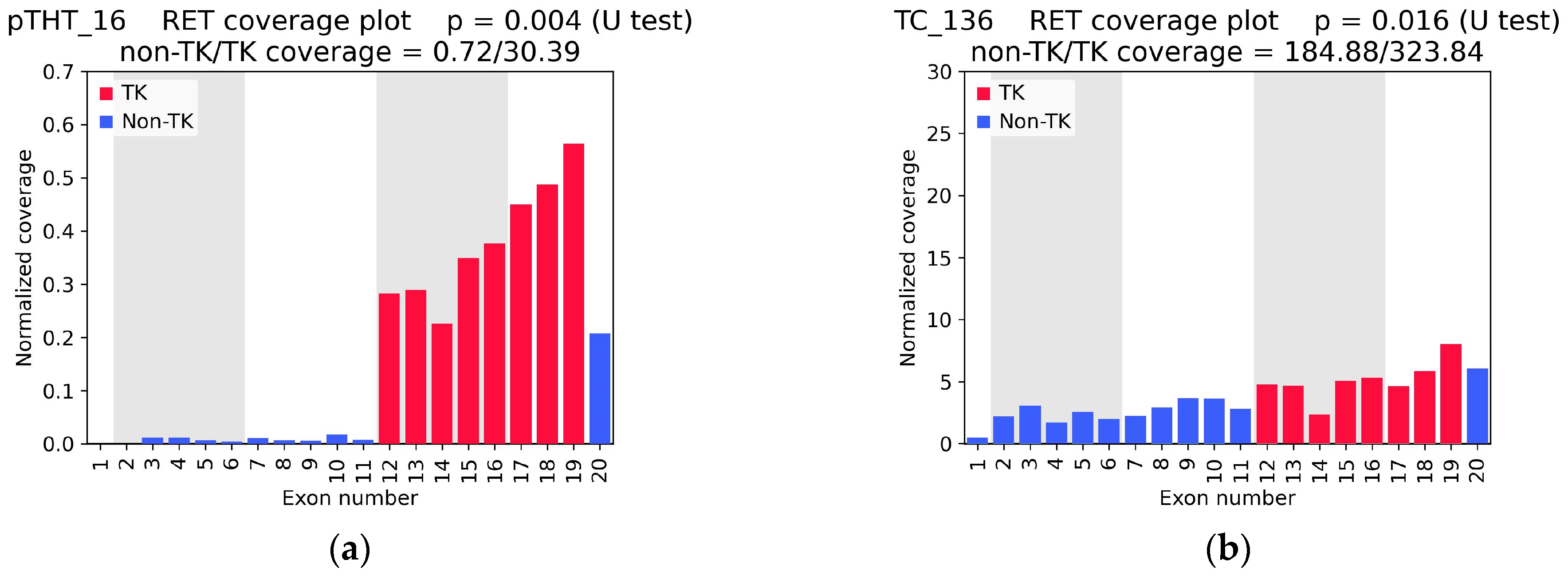
A total of 197 out of 1248 samples showed experimentally cohort demonstrated asymmetric coverage, as indicated by a U-test p-value of less than 0.05. This finding significantly exceeded the anticipated pan-tumor frequency of oncogenic RET fusions. This finding likely reflects a high rate of false-positive predictions when relying solely on the p-value threshold. Concurrently, it was observed that samples in which junction reads were detected by WTS (Figure 2a, Supplementary Table S1) exhibited a more pronounced difference in 5′/3′ coverage compared with samples devoid of fusion-supporting reads (Figure 2b, Supplementary Table S1). This finding suggests the possibility of RNA-seq coverage asymmetry in the context of wild-type RET expression. This observation indicated the necessity of incorporating an additional parameter to account for the extent of variations in coverage between non-TK and TK exons.
In order to further identify coverage characteristics that could help distinguish true positives from false positives, and to test our hypothesis that samples with a statistically significant but modest difference in 5′/3′ coverage reflect expression of wild-type RET rather than RET fusions, we assessed RET status in 78 selected cases using targeted sequencing.

2.2. RET Testing with Targeted NGS Panels and Sanger Sequencing

For the purpose of experimental validation, a cohort of 78 tumor samples was analyzed (Table 2). The samples exhibited diverse RET coverage profiles (Supplementary Figure S1), spanning high and low expression levels, p-values for coverage asymmetry both above and below 0.05, and variable degrees of 5′/3′ coverage imbalance (Supplementary Table S1).
To ascertain RET status, a targeted NGS approach was employed, leveraging two specialized panels designed for the identification of fusion transcripts: the TruSight RNA Fusion Panel (Illumina, San Diego, CA USA) and the OncoFu Elite RNA Panel v1.0 (Nanodigmbio, Nanjing, China). Both panels utilize hybridization-based enrichment process. For the OncoFu panel, the same cDNA libraries generated for RNA-seq were used as input. For the TruSight panel, new libraries were prepared directly from tumor tissue RNA samples according to the manufacturer’s protocol. Subsequently, the STAR-Fusion package was employed to identify fusion transcripts in the resulting sequencing reads [50]. In cases where multiple RET structural variants were detected, the canonical variant and/or the one with the strongest sequencing read support was selected for further analysis. For samples in which reads supporting a RET fusion were directly identified in the WTS RNA-seq data, the putative fusion breakpoint was confirmed by Sanger sequencing.
In 23 tumor samples, a statistically significant RET coverage imbalance in WTS RNA-seq reads was identified (p < 0.05), as shown in Table 3 and Supplementary Table S1. In the remaining 55/78 samples, neither RET coverage asymmetry nor fusion RNA reads were detected by targeted panel NGS.
In the RNA-seq data, junction reads were identified in 11 samples, including 7 cases of pediatric papillary thyroid carcinoma. The confirmation of all detected fusions was achieved through targeted panel sequencing and/or Sanger sequencing (Table 3). In these samples, the p-values for 5′/3′ imbalance were found to be less than 0.01, with the 5′/3′ RET exon coverage ratio measuring below 0.28.
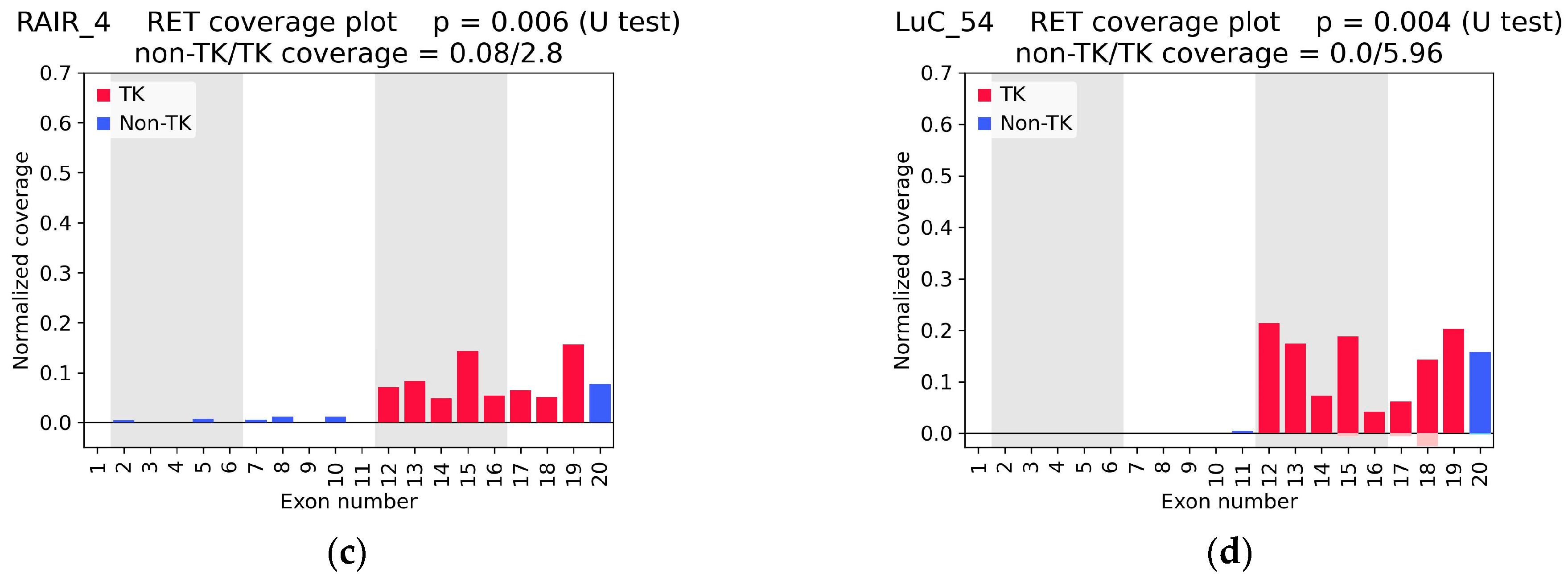
In one additional specimen of papillary thyroid cancer (RAIR_4), a solitary supporting read was identified, corresponding to the standard NCOA4(7)::RET(12) fusion. However, this particular sample exhibited a pronounced RET coverage imbalance (Figure 2c). Sanger sequencing has validated the RET fusion oncogene in this particular case (Supplementary Figure S2).
Additionally, in one lung adenocarcinoma specimen (LuC_54) exhibiting no WTS RNA-seq fusion reads but with significant RET coverage asymmetry, panel sequencing has identified the KIF5B(15)::RET(12) fusion (Figure 2d).
The 10 samples that exhibited statistically significant coverage asymmetry but where no fusions were detected by either WTS or targeted sequencing differed from the cases with true confirmed fusions by virtue of their relatively high 5′/3′ RET exon coverage ratio (Supplementary Figure S1, Table 3, bottom).
Thus, exon coverage of wild-type RET was also somewhat uneven, although to a considerably lesser extent than the 5′/3′ coverage imbalance observed in RET fusion-positive samples. In general, variability in exon coverage in whole-transcriptome sequencing can arise from two categories of factors. The first category is biological, and includes factors such as differences between transcript isoforms. The second category is technical, and includes well-known biases in fragment amplification during library preparation or the exclusion of non-uniquely mapped reads. Further studies are necessary to elucidate the underlying causes of uneven wt RET coverage. For example, long-read sequencing approaches, which are feasible only with fresh samples containing intact RNA, could be used to achieve this objective.
In light of the findings, an effort was made to delineate a threshold p-value and a 5′/3′ RET exon coverage ratio that would facilitate the differentiation between true-positive and false-positive samples.

2.3. RNA-Seq Coverage Threshold Values for Detection of RET Fusions

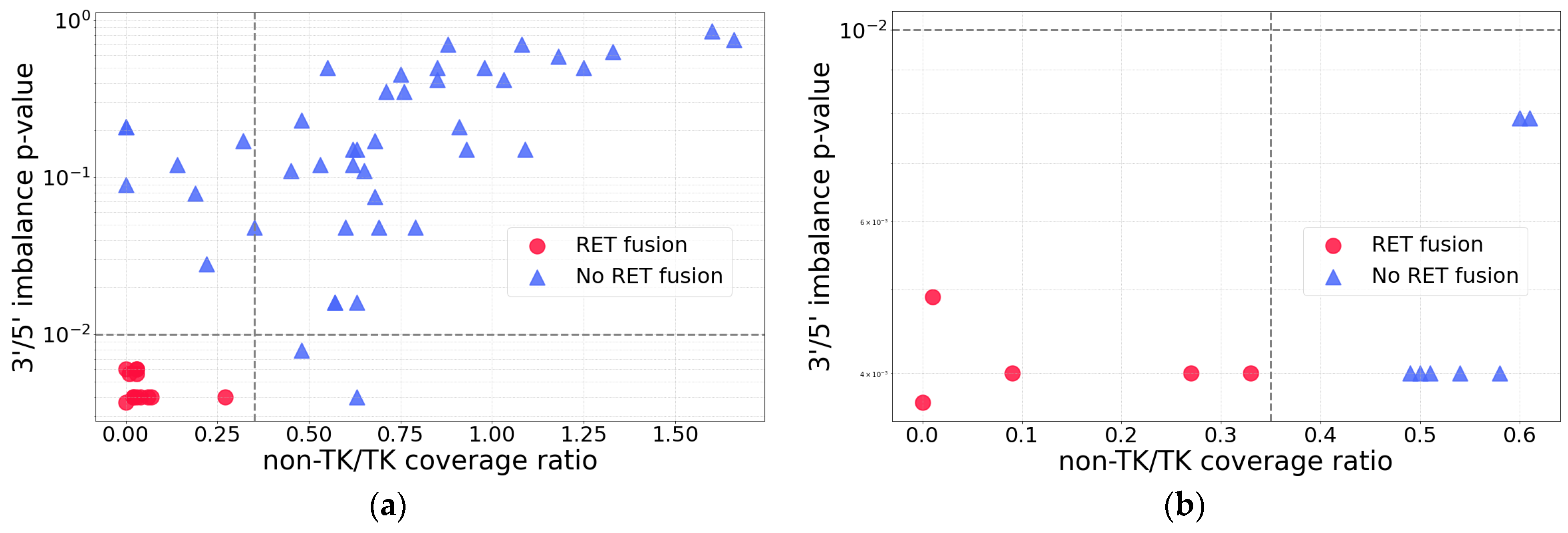
A systematic comparison of all the cases under analysis with known RET status demonstrated lower p-values and 5′/3′ RET exon coverage ratios for the true RET fusion cases (Figure 3a). This approach enabled the establishment of threshold values for p = 0.01 and a 5′/3′ RET exon coverage ratio of 0.35. Cases exhibiting both parameters below these thresholds were designated as true RET fusion cases.
In light of our prior experience with 5′/3′ imbalance analysis of ALK, it has been established that inadequate RNA-seq coverage can result in both false-positive and false-negative outcomes [34]. For ALK, a minimum coverage depth of 0.7 had previously been established as the threshold for reliable detection based on coverage imbalance [34]. To determine an appropriate threshold for RET, RNA-seq data for three fusion-positive samples (pTHT_16, pTHT_21, pTHT_25) were subsampled. From the original FASTQ files, random subsets comprising 2–80% of reads were generated in triplicate for each sample. For each subsample, the RET exon 12–16 coverage and U-test p-values for asymmetry were calculated. As shown in Supplementary Figure S3, simulated 3′ coverage depths below 1.5 yielded U-test p-values that exceeded the selected threshold of 0.01 in true-positive samples, thereby losing statistical significance. These results support the use of 1.5 as the minimum coverage depth necessary for accurate detection of RET fusions.
In order to validate the aforementioned findings, the previously established diagnostic thresholds were applied to an additional cohort of samples. To this end, we additionally performed WTS RNA-seq for 79 new thyroid cancer samples (RET coverage plots given in Supplementary Figure S4).
According to the predetermined criteria (p < 0.01; 5′/3′ RET exon coverage ratio < 0.35, 3′ coverage depth > 1.5), RET fusions were predicted in four samples (THT_51, THT_53, THT_58, and THT_70; see Supplementary Table S2). In order to assess the accuracy of these predictions, a total of 11 thyroid cancer samples with p < 0.01—including the four cases with predicted RET fusions—were subjected to further experimental validation using the targeted NGS panel OncoFu and Sanger sequencing. Furthermore, the analysis encompassed one sample (CC_29) from the primary cohort that satisfied the aforementioned criteria but lacked splice junction reads and had not been previously analyzed by targeted sequencing (Supplementary Table S2).
Targeted panel and Sanger sequencing confirmed the presence of RET fusions in all predicted samples, while no RET fusions were detected in the remaining tested samples (Figure 4b, Supplementary Table S2). Therefore, the selected thresholds are effective in avoiding false-positive predictions arising from overexpression of the wild-type RET gene.
It is noteworthy that all medullary thyroid cancer samples (i.e., THT_1–50) expressed wild-type RET at a high level (see Supplementary Table S2). RET amplification is considered as promising target for anti-RET therapy, along with RET rearrangements and activating point mutations [51]. For all pTHT and THT samples, WES profiles were available in addition to WTS data. Therefore, the potential of wild-type RET overexpression being explained by gene amplification was investigated. However, CNV analysis did not reveal RET amplification in any of the thyroid cancer samples. Consequently, the underlying causes of wild-type RET overexpression in medullary thyroid cancer samples, such as aberrations in RET regulatory factors, necessitate further investigation.

2.4. Rare and Previously Unreported RET Fusions

In the present study, three rare RET fusions with preserved open reading frames were identified. Two of these fusions (FN1(20)::RET(11) and PPP1R21(15)::RET(12)) have not been previously reported in the literature or deposited in public databases, while one (RUFY3(11)::RET(12)) has been reported only once, in a different cancer type (Figure 4a). Coverage plots for RET, derived from RNA-seq reads for the respective samples, are presented in Figure 4b–d.
These fusions are considered clinically relevant because they maintain an intact open reading frame and conserve the full RET tyrosine kinase domain (denoted as TK in Figure 4a), which is essential for kinase activation and therapeutic targeting.
All three 5′ partner genes are located on chromosomes distinct from RET: FN1 (fibronectin 1) on chromosome 2q35, RUFY3 (RUN and FYVE domain-containing 3) on chromosome 4q13.3, and PPP1R21 (protein phosphatase 1 regulatory subunit 21) on chromosome 2p16.3.
The FN1 gene is responsible for encoding fibronectin, a protein that is found in the extracellular matrix and is involved in a variety of essential cellular processes. These processes include wound healing and embryonic morphogenesis. Additionally, fibronectin plays a role in regulating cell behaviors such as adhesion and migration [52]. Fibronectin has also been demonstrated to exhibit self-association properties.
RUFY3 is a gene belonging to a five-member RUFY gene family that is conserved across species. It is expressed in a variety of human tissues and has been implicated in endosomal trafficking, autophagy, and cell migration [53,54].
The PPP1R21 gene is responsible for encoding a cofactor of protein phosphatase 1 (PP1) [55], a key serine/threonine phosphatase that is imperative for several processes within cells, including cell division, glycogen metabolism, protein synthesis, and muscle contractility. PPP1R21 has been identified as a participant in endosomal maturation and exhibits broad expression in multiple tissues, including the thyroid (https://www.proteinatlas.org/ENSG00000162869-PPP1R21/tissue, accessed on 11 September 2025).
In the FGF23-producing adenoma sample (FGF_7), a fusion transcript involving exon 20 of FN1 and exon 11 of RET genes was detected (Figure 4a). In this case, RET activation may ensue as a consequence of its fusion with a gene that is driven by a constitutively active promoter. This process ultimately leads to the overproduction of the RET tyrosine kinase domain. Furthermore, since the fusion also retains FN1 domains critical for intermolecular interactions (type I repeats 1–5 and type III repeat 1) [52], activation is likely mediated through oligomerization of the fusion protein. It is noteworthy that this fusion maintained the integrity of the RET transmembrane domain, a component that frequently becomes lost during rearrangements. This observation suggests that the FN1(20)::RET(11) fusion product may have the potential to localize within the membrane. Although previous studies have reported various fusions involving the FN1 gene and different tyrosine kinase genes [56,57,58], only a single case of a FN1::RET fusion has been described. This case involved a different tumor type, namely pseudosarcomatous myofibroblastic proliferation of the urinary bladder, and different partner exons, namely FN1(28)::RET(6) [59].
In turn, the activation of RET in the rearrangements involving RUFY3 and PPP1R21 is most likely mediated by ligand-independent dimerization of these fusion proteins, since both 5′ partner moieties retain coiled-coil domains (Figure 4a). To the best of our knowledge, the RUFY3(11)::RET(12) fusion has hitherto been reported only once, in a non-small cell lung cancer specimen [60]. In the present study, the presence of the aforementioned element was detected in a sample of pediatric papillary thyroid cancer (pTHT_22). A RET fusion with RUFY3 was also mentioned in [61], described as identified through the analysis of three public datasets (TCGA-THCA, MSK-MetTropism, and MSK-IMPACT), although no further details were provided.
The PPP1R21::RET rearrangement has been previously reported in the context of thyroid cancer, albeit with a distinct breakpoint, namely PPP1R21(9)::RET(12) [60]. In the present study, the breakpoint in PPP1R21 was identified as occurring subsequent to exon 15.

3. Discussion

Despite the growing clinical importance of detecting fusions involving various tyrosine kinases, driven by the increasing availability of highly effective targeted therapies, their identification in routine practice remains challenging. This phenomenon is primarily attributable to the exceedingly low frequency of fusions for each individual gene, in conjunction with the substantial diversity of potential partner genes and possible breakpoint locations. Conventional methods, including IHC and FISH, are inadequate for this purpose due to their reliance on the specific fusion partner involved, which often compromises the accuracy and precision of the results. Furthermore, these approaches are deficient in multiplexing capability and are susceptible to errors resulting from the substantial subjectivity inherent in data interpretation.
In this context, high-throughput methods capable of detecting fusions in a partner-gene-agnostic manner across large panels or even genome-wide have become indispensable. These include targeted sequencing panels and whole-transcriptome sequencing (WTS, RNA-seq). The former is more widely adopted in clinical practice due to its lower cost, higher coverage of target loci, and simpler bioinformatic analysis. Concurrently, while WTS is associated with a higher financial investment and a more intricate analytical framework, it offers a remarkably comprehensive array of information concerning the molecular characteristics of tumors [62].
At the same time, the sensitivity of WTS for fusion detection is relatively limited, as only a few reads typically span the fusion junction. We previously demonstrated that the sensitivity of ALK fusion detection in RNA-seq data increases to 100% when, in addition to chimeric reads, exon expression profiles are analyzed to identify 5′/3′ coverage imbalance [34]. In the present study, the efficacy of this approach in detecting RET fusions was assessed.
An investigation was conducted to identify RET coverage disparities in a substantial collection of experimental RNA-seq data (n = 1327 profiles, comprising 154 non-small cell lung cancer and 221 thyroid cancer samples). Subsequent to this screening process, the validity of RET status was confirmed in 78 selected cases exhibiting diverse RET coverage patterns. To confirm the RET status, targeted panel sequencing and Sanger sequencing were employed. The selection of threshold values for the p-value and the 5′/3′ RET exon coverage ratio was made based on the obtained data. The purpose of this selection was to allow for the distinction between true-positive and false-positive predictions. The selected threshold parameters for RET coverage imbalance in RNA-seq were subsequently validated in an independent cohort of 79 thyroid cancer samples.
While the majority of the RET fusions identified in the experimental cohort (11 out of 14) exhibited junction reads in the RNA-seq data, RET coverage asymmetry analysis facilitated the detection and validation of RET fusions in three additional samples (RAIR_4, LuC_54, and CC_29) among a total of more than 1300 samples examined. Therefore, the analysis of RET coverage asymmetry in RNA-seq data has the potential to facilitate the detection of extremely rare fusion events, even in cases of low coverage depth. The sensitivity and specificity of the 5′/3′ RET exon coverage imbalance analysis of RNA-seq data were both 100%, as determined by the selected 5′/3′ RET exon coverage ratio and p-value thresholds.
It is important to note that the proposed approach can be applied to different tumor types. As RET not typically expressed in lung tissue, RET coverage imbalance has been identified as a reliable marker of fusion presence in lung cancer, as confirmed by previously published studies [24,40,41,45,46,47]. However, in the context of thyroid cancer, a notable exception emerges, as numerous samples manifest an elevated expression of wild-type RET. It was determined that such samples also manifest a statistically significant 5′/3′ coverage imbalance, with pronounced differences between 5′ and 3′ RET exon coverage. This can result in a high rate of false-positive predictions when relying solely on this method. However, the utilization of the established threshold values for the assessment of coverage enables the differentiation of true fusion-positive cases from those exhibiting high wild-type RET expression.
The described approach has been observed to be deficient in its inability to identify the fuse partner or the precise breakpoint within the target gene, although in most cases, the latter can be inferred from the exon coverage profile. It should be noted that IHC and FISH methods are similarly incapable of determining the partner gene and exact breakpoints and this information is not currently required for therapeutic decision-making.
According to the ESMO recommendations, NGS is the preferred method over IHC and FISH, and targeted RNA-based assays are the methods of choice for RET fusion screening [26]. While whole-transcriptome RNA sequencing is not a first-line diagnostic tool due to its higher cost and analytical complexity, it is increasingly applied in research and in diagnostically challenging clinical cases. In such settings, coverage imbalance analysis can serve as an additional interpretive layer that increases the diagnostic yield of existing WTS data. When exon coverage asymmetry is detected, confirmatory testing using targeted NGS panels or anchored PCR-based NGS can be employed to identify the fusion partner and confirm precise breakpoint. The proposed method increases the practical utility and cost-effectiveness of WTS by enabling the identification of clinically actionable RET fusions that might otherwise remain undetected.
Therefore, we propose that the detection of RET exon coverage asymmetry can serve as an independent, highly accurate clinical testing procedure. Nonetheless, given the nascent stage of technology adoption, we suggest a workflow that aligns with current practices. Initially, a 5′/3′ coverage imbalance analysis should be conducted on the RNA-seq data. In instances where the imbalance analysis predicts the presence of RET fusion, the subsequent step involves validating the results using established methods (Supplementary Figure S5).
In the present study, we observed significant overexpression of wild-type RET in all medullary thyroid cancer samples (THT_1–50). Although gene amplification is considered another potential targetable alteration for anti-RET therapy [51], WES analysis did not reveal RET amplification in these cases. According to the extant data, RET overexpression occurs in 40–60% of breast tumors and it is reported to be more common than RET rearrangements or mutations in breast cancer [63]. RET overexpression typically correlates with elevated protein levels [63]. However, extant research on this aspect remains largely limited to preclinical cell-based studies [64,65]. Nonetheless, RET overexpression, irrespective of amplification status, may signify an ancillary opportunity for the implementation of selective RET inhibitors. These should be evaluated in conjunction with RET rearrangements and activating mutations, for which RNA-seq is similarly informative. A further imperative is to undertake a thorough investigation into the mechanisms that underlie RET overexpression in the absence of gene amplification.
Among the 18 RET fusion-positive samples identified in this study, we detected one rare fusion event (RUFY3(11)::RET(12)) as well as two new RET fusions that had not been previously described in the literature (FN1(20)::RET(11) and PPP1R21(15)::RET(12)).
In summary, the present study demonstrated the efficacy of exon coverage imbalance analysis as a substitute for other RET fusion detection methods. The present study offers a valuable complement to RNA sequencing data analysis pipelines, thereby enabling the effective detection of clinically relevant oncogenic RET alterations.

4. Materials and Methods

4.1. Biosamples and RNA Sequencing

In this study, experimental whole-transcriptome data obtained for 1327 tumor samples representing 28 cancer types and 2 benign tumor types were used. The tumor tissue samples were either obtained during the course of the Oncobox clinical trial (Clinicaltrials.gov ID NCT03724097, NCT03521245) (n = 764) or obtained from individual patients who submitted their biosamples for Oncobox molecular testing. For each biospecimen, written informed consent for participation in this study was obtained from the patient or the patient’s legal representative. The consent process and study design were guided and approved by the local ethics committees of the Vitamed Clinic (Moscow, Russia). In addition, 113 thyroid tissue specimens were obtained from the National Medical Research Center for Endocrinology (Moscow, Russia). The study was approved by the local ethics committees of the Vitamed Clinic, Moscow, Russia (protocol #1, approval date: 16 October 2017), and the National Medical Research Center for Endocrinology, Moscow, Russia (protocol #1, approval date: 15 January 2025).
The samples were formalin-fixed paraffin-embedded (FFPE) tissue blocks that were macrodissected for tumor-rich regions identified on hematoxylin and eosin (H&E)-stained slides. RNA extraction, library preparation, sequencing, and RNA-seq data processing were performed as described in [25,66], and the resulting BAM files were then analyzed.
Most NGS libraries were sequenced on either the HiSeq 3000 or the NextSeq 550 platforms (both from Illumina, San Diego, CA, USA) in single-end mode, with read lengths of 50 bp and 75 bp, respectively. More recent samples were sequenced on the FASTASeq 300 (GeneMind Biosciences, Shenzhen, China) with paired read lengths of 75 bp. In total, 1327 RNA-seq tumor profiles had more than the threshold of 2.5 million uniquely mapped reads (Table 1) [25,66]. The gender and age characteristics of all cohorts are shown in Table 1.

4.2. Exon Coverage Calculation

RNA-seq reads were aligned to the human genome (hg38, GCF_000001405.39) using STAR v2.7.4a, and duplicate reads were removed using the Picard MarkDuplicates tool with the default parameters. Only reads with a mapping quality (MQ) > 100 and an alignment length ≥ 10 bp were considered. Exon coordinates were obtained from Ensembl (for ENST00000355710.8), and coverage was normalized to both exon length and to the total number of mapped reads. Strand specificity was taken into account. Coverage imbalance for RET was assessed by comparing the 5′ non-kinase region exons (2–6) with the tyrosine kinase domain exons (12–16) using a one-sided Mann–Whitney U test with the alternative hypothesis that the coverage of non-TK-related exons is lower than that of TK-related exons. Statistical significance was determined based on the p-value. The Mann–Whitney U test was used due to the non-normality of the exon coverage distribution, as assessed by the Shapiro–Wilk test for each sample (with the null hypothesis that the distribution is normal).
The coverage depth of RET exons 12–16 was calculated by dividing the total number of aligned bases by the combined exon length. Samples with undetectable or insufficient RET transcript coverage were excluded from the analysis. The threshold for low coverage is described in the Results section.
The code used for the analysis and the accompanying data are available at https://github.com/akhristichenko/RET_coverage_asymmetry. (accessed on 5 October 2025).

4.3. Experimental Validation of RET Fusion Transcripts by Targeted NGS Panels

Two targeted NGS panels were used for ALK rearrangement verification: the TruSight RNA Fusion Panel (Illumina, San Diego, CA, USA) and the OncoFu Elite (for RNA) Panel v1.0 (Nanodigmbio, Nanjing, China). Both panels utilize hybridization-based enrichment and cover all coding regions of transcripts, as well as selected untranslated regions (UTRs) of genes that are most frequently or clinically implicated in fusions in solid tumors. These panels enable the detection of both known and previously unreported fusions.
NGS libraries were prepared according to the manufacturer’s protocol. For the TruSight panel, libraries were prepared using previously extracted RNA samples. For the OncoFu panel, hybridization enrichment was performed on cDNA libraries that had been prepared previously and were used for whole transcriptome sequencing.
Sequencing was performed on the FASTASeq 300 platform (GeneMind Biosciences, Shenzhen, China) with 2 × 75 bp paired-end reads. The expected yield was approximately 4.5 million reads per sample for the TruSight panel and 2.5 million reads per sample for the OncoFu panel.

4.4. Experimental Validation of RET Fusion Transcripts by RT-PCR and Sanger Sequencing

To generate amplicons for Sanger sequencing, single-stranded cDNA was synthesized from 100 ng of total RNA using the MMLV RT kit (Evrogen, Moscow, Russia), according to the manufacturer’s instructions. The reverse transcriptase was pre-diluted tenfold with enzyme dilution buffer. Reverse transcription was performed using a mix of gene-specific reverse primers for RET exons 11 and 12 (5′-GACAGCAGCACCGAGAC-3′ and 5′-CAAGAACCAAGTTCTTCCGAG-3′), at a final concentration of 500 nM.
The following reagents were used for preparative PCR (all from Evrogen, Moscow, Russia): 10× Turbo Buffer, a dNTP mix (10 mM each), HS-Taq DNA polymerase, and PCR-grade water. PCR was performed in 50 µL volumes using a T100 Thermal Cycler (Bio-Rad, Hercules, CA, USA). The thermal cycling protocol was as follows: (1) 2 min at 95 °C; (2) 40 cycles of 15 s at 95 °C, 20 s at 60 °C, 10 s at 70 °C; (3) 1 min at 70 °C.
The primer design was performed as follows: (1) the region surrounding the fusion breakpoint (150 bp upstream and downstream) was examined for paralogous sequences using BLAT (https://genome-euro.ucsc.edu/cgi-bin/hgBlat?hgsid=324308355_bHclKcKtEyJWfpBYXjHTsAh5QF7h&command=start; accessed on 8 September 2025); (2) primers were designed so that the 3′ end would align to regions without detectable homology; (3) the forward primer was placed on the last exon of the 5′ fusion partner gene immediately upstream of the breakpoint, and the reverse primer was placed on the first exon of RET, immediately downstream of the breakpoint; (4) due to the degraded nature of RNA in FFPE samples, amplicon lengths were kept between 100 and 150 bp; (5) primers were designed with a melting temperature of 61 ± 1 °C and were checked to avoid hairpin formation, homodimer formation, and heterodimer formation using the OligoAnalyzer tool (https://www.idtdna.com/pages/tools/oligoanalyzer; accessed on 8 September 2025). Primer pairs are listed in Table 4.
To verify PCR specificity, negative controls included a non-template control and cDNA from a RET fusion-negative tumor sample, neither of which showed amplification.
Amplicons were purified using the standard NGS Clean and Select Beads protocol (Meridian Bioscience, Cincinnati, OH, USA) with a twofold excess of the volume of the magnetic bead suspension. Sanger sequencing was performed using a Genetic Analyzer 3500xL (Applied Biosystems, Foster City, CA, USA).

4.5. Bioinformatic Detection of RET Fusion Transcripts

The reads quality was assessed with FastQC (https://github.com/s-andrews/FastQC; accessed on 11 May 2025), and reads were filtered using PRINSEQ-lite (https://github.com/uwb-linux/prinseq; accessed on 11 May 2025) (mean Phred quality ≥ 30).
RET fusion transcripts were identified in RNA-seq profiles and data from targeted NGS panels using STAR-Fusion software [50] (version STAR-2.7.2b; https://github.com/STAR-Fusion/STAR-Fusion; accessed on 23 May 2025) with the pre-built CTAT plug-and-play reference GRCh38_gencode_v22_CTAT_lib_Mar012021. Default parameters were used unless otherwise specified.
Fusion candidate files were generated and the relevant RNA sequencing reads associated with RET were extracted. The resulting data were verified through manual inspection using UCSC BLAT and the UCSC Genome Browser (https://genome-euro.ucsc.edu, accessed 3 June 2025). Candidate RET fusions were evaluated based on the following criteria: (i) whether the sequencing read spans an exon junction between two distinct, previously characterized transcripts; (ii) whether the junction site precisely corresponds to the boundaries of exons of known genes, taking established splice sites into account; and (iii) whether both transcripts align in the same orientation, indicating the presence of a putative fusion RNA. Reads that met these criteria were considered evidence of fusion.

4.6. Statistical Analysis

The statistical significance of differences between the groups was estimated using the log-rank test, with the ‘logrank’ function from the Python 3 ‘scipy’ library (https://github.com/scipy/scipy; accessed on 5 October 2025). A p-value less than 0.05 was considered to be statistically significant.

Supplementary Materials

The following supporting information can be downloaded at: https://www.mdpi.com/article/10.3390/ijms262311300/s1.

Author Contributions

Conceptualization, A.B., M.S. (Maksim Sorokin) and M.S. (Maria Suntsova); methodology, M.S. (Maria Suntsova), M.S. (Maksim Sorokin) and G.Z.; software, A.K. and D.L.; investigation, I.G., A.K., E.B., M.R., A.M. and N.K.; validation, D.L., M.S. (Maria Suntsova) and G.Z.; formal analysis, A.K., I.G. and D.L.; resources, E.P., M.S. (Marina Sekacheva) and A.B.; data curation, A.K., N.K., A.M. and G.Z.; writing—original draft preparation, G.Z.; writing—review and editing, A.B.; visualization, A.K. and G.Z.; supervision, A.B. and M.S. (Marina Sekacheva); project administration, M.S. (Maria Suntsova) and G.Z.; funding acquisition, E.P., A.B. and M.S. (Marina Sekacheva). All authors have read and agreed to the published version of the manuscript.

Funding

This study was financed by the Russian Science Foundation, grant number 25-75-20022. The funder had no role in the design of the study; in the collection, analyses, or interpretation of data; in the writing of the manuscript; or in the decision to publish the results.

Institutional Review Board Statement

The study was conducted in accordance with the Declaration of Helsinki and was approved by the local ethics committees of the Vitamed Clinic, Moscow, Russia (approval date: 16 October 2017), and the National Medical Research Center for Endocrinology, Moscow, Russia (approval date: 15 January 2025), for studies involving human cancer tissue biopsy materials.

Informed Consent Statement

Informed consent was obtained from all subjects involved in the study.

Data Availability Statement

The original contributions presented in this study are included in the article/supplementary material. Further inquiries can be directed to the corresponding author(s).

Acknowledgments

The Academic Leadership Program “Priority 2030” supported sample collection and primary processing. We would also like to express our gratitude to Alexandra Emelyanova and Daria Golikova for their invaluable assistance with the clinical annotation of tumor samples.

Conflicts of Interest

Maksim Sorokin was employed in Oncobox LLC. The authors declare that this research was conducted in the absence of any commercial or financial relationships that could be construed as a potential conflict of interest. The funders had no role in the design of the study; in the collection, analyses, or interpretation of data; in the writing of the manuscript, or in the decision to publish the results.

References

  1. Sorokin, M.; Rabushko, E.; Rozenberg, J.M.; Mohammad, T.; Seryakov, A.; Sekacheva, M.; Buzdin, A. Clinically Relevant Fusion Oncogenes: Detection and Practical Implications. Ther. Adv. Med. Oncol. 2022, 14. [Google Scholar] [CrossRef]
  2. Drilon, A.; Oxnard, G.R.; Tan, D.S.W.; Loong, H.H.F.; Johnson, M.; Gainor, J.; McCoach, C.E.; Gautschi, O.; Besse, B.; Cho, B.C.; et al. Efficacy of Selpercatinib in RET Fusion–Positive Non–Small-Cell Lung Cancer. N. Engl. J. Med. 2020, 383, 813–824. [Google Scholar] [CrossRef]
  3. Wirth, L.J.; Sherman, E.; Robinson, B.; Solomon, B.; Kang, H.; Lorch, J.; Worden, F.; Brose, M.; Patel, J.; Leboulleux, S.; et al. Efficacy of Selpercatinib in RET-Altered Thyroid Cancers. N. Engl. J. Med. 2020, 383, 825–835. [Google Scholar] [CrossRef] [PubMed]
  4. Lopes, G.; Gainor, J.F.; Subbiah, V.; Bowles, D.W.; Doebele, R.C.; Mansfield, A.S.; Baik, C.S.; Gadgeel, S.M.; Kalemkerian, G.P.; Ou, S.-H.I.; et al. Efficacy and Safety of Pralsetinib in Patients with Advanced RET-Fusion-Positive NSCLC: Final Data from the Phase 1/2 ARROW Study. J. Clin. Oncol. 2025, 43, 8644. [Google Scholar] [CrossRef]
  5. Ke, J.; Huang, S.; Jing, Z.; Duan, M. The Efficacy and Safety of Selective RET Inhibitors in RET Fusion-Positive Non-Small Cell Lung Cancer: A Meta-Analysis. Investig. New Drugs 2023, 41, 768–776. [Google Scholar] [CrossRef]
  6. Takahashi, M.; Ritz, J.; Cooper, G.M. Activation of a Novel Human Transforming Gene, Ret, by DNA Rearrangement. Cell 1985, 42, 581–588. [Google Scholar] [CrossRef]
  7. Kawai, K.; Takahashi, M. Intracellular RET Signaling Pathways Activated by GDNF. Cell Tissue Res. 2020, 382, 113–123. [Google Scholar] [CrossRef]
  8. Regua, A.T.; Najjar, M.; Lo, H.W. RET Signaling Pathway and RET Inhibitors in Human Cancer. Front. Oncol. 2022, 12, 932353. [Google Scholar] [CrossRef]
  9. Mahato, A.K.; Saarma, M. Biology of RET Receptor and Its Ligands: Focus on the Nervous System. Endocr. Relat. Cancer 2025, 32, e240174. [Google Scholar] [CrossRef] [PubMed]
  10. Ivanchuk, S.M.; Myers, S.M.; Mulligan, L.M. Expression of RET 3′ Splicing Variants during Human Kidney Development. Oncogene 1998, 16, 991–996. [Google Scholar] [CrossRef]
  11. Richardson, D.S.; Rodrigues, D.M.; Hyndman, B.D.; Crupi, M.J.F.; Nicolescu, A.C.; Mulligan, L.M. Alternative Splicing Results in RET Isoforms with Distinct Trafficking Properties. Mol. Biol. Cell 2012, 23, 3838–3850. [Google Scholar] [CrossRef] [PubMed]
  12. Ju, Y.S.; Lee, W.-C.; Shin, J.-Y.; Lee, S.; Bleazard, T.; Won, J.-K.; Kim, Y.T.; Kim, J.-I.; Kang, J.-H.; Seo, J.-S. A Transforming KIF5B and RET Gene Fusion in Lung Adenocarcinoma Revealed from Whole-Genome and Transcriptome Sequencing. Genome Res. 2012, 22, 436–445. [Google Scholar] [CrossRef]
  13. Santoro, M.; Carlomagno, F. Central Role of RET in Thyroid Cancer. Cold Spring Harb. Perspect. Biol. 2013, 5, a009233. [Google Scholar] [CrossRef]
  14. Bossi, D.; Carlomagno, F.; Pallavicini, I.; Pruneri, G.; Trubia, M.; Raviele, P.R.; Marinelli, A.; Anaganti, S.; Cox, M.C.; Viale, G.; et al. Functional Characterization of a Novel FGFR1OP-RET Rearrangement in Hematopoietic Malignancies. Mol. Oncol. 2014, 8, 221–231. [Google Scholar] [CrossRef] [PubMed]
  15. Staubitz, J.I.; Schad, A.; Springer, E.; Rajalingam, K.; Lang, H.; Roth, W.; Hartmann, N.; Musholt, T.J. Novel Rearrangements Involving the RET Gene in Papillary Thyroid Carcinoma. Cancer Genet. 2019, 230, 13–20. [Google Scholar] [CrossRef]
  16. Parimi, V.; Tolba, K.; Danziger, N.; Kuang, Z.; Sun, D.; Lin, D.I.; Hiemenz, M.C.; Schrock, A.B.; Ross, J.S.; Oxnard, G.R.; et al. Genomic Landscape of 891 RET Fusions Detected across Diverse Solid Tumor Types. npj Precis. Oncol. 2023, 7, 10. [Google Scholar] [CrossRef]
  17. Nagasaka, M.; Brazel, D.; Baca, Y.; Xiu, J.; Al-Hallak, M.N.; Kim, C.; Nieva, J.; Swensen, J.J.; Spetzler, D.; Korn, W.M.; et al. Pan-Tumor Survey of RET Fusions as Detected by next-Generation RNA Sequencing Identified RET Fusion Positive Colorectal Carcinoma as a Unique Molecular Subset. Transl. Oncol. 2023, 36, 101744. [Google Scholar] [CrossRef]
  18. Pekova, B.B.; Sykorova, V.; Mastnikova, K.; Vaclavikova, E.; Moravcova, J.; Vlcek, P.; Lancova, L.; Lastuvka, P.; Katra, R.; Bavor, P.; et al. RET Fusion Genes in Pediatric and Adult Thyroid Carcinomas: Cohort Characteristics and Prognosis. Endocr. Relat. Cancer 2023, 30, e230117. [Google Scholar] [CrossRef]
  19. Ou, S.H.I.; Zhu, V.W. Catalog of 5′ Fusion Partners in RET+ NSCLC Circa 2020. JTO Clin. Res. Rep. 2020, 1, 100037. [Google Scholar] [CrossRef] [PubMed]
  20. Ding, Y.; Chen, G.; He, X.; Shi, Y.; He, C.; Yin, J.C.; Zhang, Z. Characterization of RET Fusions via Integrated DNA and RNA Sequencing in Early-Stage Non-Small Cell Lung Cancer: A Retrospective Study. Transl. Lung Cancer Res. 2025, 14, 4384–4397. [Google Scholar] [CrossRef] [PubMed]
  21. Mizukami, T.; Shiraishi, K.; Shimada, Y.; Ogiwara, H.; Tsuta, K.; Ichikawa, H.; Sakamoto, H.; Kato, M.; Shibata, T.; Nakano, T.; et al. Molecular Mechanisms Underlying Oncogenic RET Fusion in Lung Adenocarcinoma. J. Thorac. Oncol. 2014, 9, 622–630. [Google Scholar] [CrossRef]
  22. Goytain, A.; Ng, T. NanoString NCounter Technology: High-Throughput RNA Validation. In Chimeric RNA: Methods and Protocols; Springer: New York, NY, USA, 2020; pp. 125–139. [Google Scholar]
  23. Reguart, N.; Teixidó, C.; Giménez-Capitán, A.; Paré, L.; Galván, P.; Viteri, S.; Rodríguez, S.; Peg, V.; Aldeguer, E.; Viñolas, N.; et al. Identification of ALK, ROS1, and RET Fusions by a Multiplexed MRNA-Based Assay in Formalin-Fixed, Paraffin-Embedded Samples from Advanced Non–Small-Cell Lung Cancer Patients. Clin. Chem. 2017, 63, 751–760. [Google Scholar] [CrossRef]
  24. Novaes, L.A.C.; da Silva, L.S.; de Marchi, P.; de Oliveira Cavagna, R.; de Paula, F.E.; Zanon, M.F.; Evangelista, A.F.; da Silva, E.C.A.; da Silva, V.D.; Leal, L.F.; et al. Simultaneous Analysis of ALK, RET, and ROS1 Gene Fusions by NanoString in Brazilian Lung Adenocarcinoma Patients. Transl. Lung Cancer Res. 2021, 10, 292–303. [Google Scholar] [CrossRef]
  25. Rabushko, E.; Sorokin, M.; Suntsova, M.; Seryakov, A.P.; Kuzmin, D.V.; Poddubskaya, E.; Buzdin, A.A. Experimentally Deduced Criteria for Detection of Clinically Relevant Fusion 3′ Oncogenes from FFPE Bulk RNA Sequencing Data. Biomedicines 2022, 10, 1866. [Google Scholar] [CrossRef]
  26. Belli, C.; Penault-Llorca, F.; Ladanyi, M.; Normanno, N.; Scoazec, J.-Y.; Lacroix, L.; Reis-Filho, J.S.; Subbiah, V.; Gainor, J.F.; Endris, V.; et al. ESMO Recommendations on the Standard Methods to Detect RET Fusions and Mutations in Daily Practice and Clinical Research. Ann. Oncol. 2021, 32, 337–350. [Google Scholar] [CrossRef] [PubMed]
  27. Yang, S.-R.; Aypar, U.; Rosen, E.Y.; Mata, D.A.; Benayed, R.; Mullaney, K.; Jayakumaran, G.; Zhang, Y.; Frosina, D.; Drilon, A.; et al. A Performance Comparison of Commonly Used Assays to Detect RET Fusions. Clin. Cancer Res. 2021, 27, 1316–1328. [Google Scholar] [CrossRef] [PubMed]
  28. Yoh, K.; Seto, T.; Satouchi, M.; Nishio, M.; Yamamoto, N.; Murakami, H.; Nogami, N.; Matsumoto, S.; Kohno, T.; Tsuta, K.; et al. Vandetanib in Patients with Previously Treated RET-Rearranged Advanced Non-Small-Cell Lung Cancer (LURET): An Open-Label, Multicentre Phase 2 Trial. Lancet Respir. Med. 2017, 5, 42–50. [Google Scholar] [CrossRef] [PubMed]
  29. Drilon, A.; Fu, S.; Patel, M.R.; Fakih, M.; Wang, D.; Olszanski, A.J.; Morgensztern, D.; Liu, S.V.; Cho, B.C.; Bazhenova, L.; et al. A Phase I/Ib Trial of the VEGFR-Sparing Multikinase RET Inhibitor RXDX-105. Cancer Discov. 2019, 9, 384–395. [Google Scholar] [CrossRef]
  30. Das, T.K.; Cagan, R.L. KIF5B-RET Oncoprotein Signals through a Multi-Kinase Signaling Hub. Cell Rep. 2017, 20, 2368–2383. [Google Scholar] [CrossRef]
  31. Mc Leer, A.; Mondet, J.; Magnat, N.; Mersch, M.; Giovannini, D.; Emprou, C.; Toffart, A.C.; Sturm, N.; Lantuéjoul, S.; Benito, D. Rearranged During Transfection Rearrangement Detection by Fluorescence In Situ Hybridization Compared With Other Techniques in NSCLC. JTO Clin. Res. Rep. 2024, 5, 100714. [Google Scholar] [CrossRef]
  32. Radonic, T.; Geurts-Giele, W.R.R.; Samsom, K.G.; Roemen, G.M.J.M.; von der Thüsen, J.H.; Thunnissen, E.; Meijssen, I.C.; Sleddens, H.F.B.M.; Dinjens, W.N.M.; Boelens, M.C.; et al. RET Fluorescence In Situ Hybridization Analysis Is a Sensitive but Highly Unspecific Screening Method for RET Fusions in Lung Cancer. J. Thorac. Oncol. 2021, 16, 798–806. [Google Scholar] [CrossRef] [PubMed]
  33. Sorokin, M.; Lyadov, V.; Suntsova, M.; Garipov, M.; Semenova, A.; Popova, N.; Guguchkin, E.; Heydarov, R.; Zolotovskaia, M.; Zhao, X.; et al. Detection of Fusion Events by RNA Sequencing in FFPE versus Freshly Frozen Colorectal Cancer Tissue Samples. Front. Mol. Biosci. 2025, 11, 1448792. [Google Scholar] [CrossRef] [PubMed]
  34. Zakharova, G.; Suntsova, M.; Rabushko, E.; Mohammad, T.; Drobyshev, A.; Seryakov, A.; Poddubskaya, E.; Moisseev, A.; Smirnova, A.; Sorokin, M.; et al. A New Approach of Detecting ALK Fusion Oncogenes by RNA Sequencing Exon Coverage Analysis. Cancers 2024, 16, 3851. [Google Scholar] [CrossRef]
  35. Zheng, Z.; Liebers, M.; Zhelyazkova, B.; Cao, Y.; Panditi, D.; Lynch, K.D.; Chen, J.; Robinson, H.E.; Shim, H.S.; Chmielecki, J.; et al. Anchored Multiplex PCR for Targeted Next-Generation Sequencing. Nat. Med. 2014, 20, 1479–1484. [Google Scholar] [CrossRef]
  36. Beg, S.; Bareja, R.; Ohara, K.; Eng, K.W.; Wilkes, D.C.; Pisapia, D.J.; Al Zoughbi, W.; Kudman, S.; Zhang, W.; Rao, R.; et al. Integration of Whole-Exome and Anchored PCR-Based next Generation Sequencing Significantly Increases Detection of Actionable Alterations in Precision Oncology. Transl. Oncol. 2021, 14, 100944. [Google Scholar] [CrossRef] [PubMed]
  37. Dunwell, T.L.; Dailey, S.C.; Ottestad, A.L.; Yu, J.; Becker, P.W.; Scaife, S.; Richman, S.D.; Wood, H.M.; Slaney, H.; Bottomley, D.; et al. Adaptor Template Oligo-Mediated Sequencing (ATOM-Seq) Is a New Ultra-Sensitive UMI-Based NGS Library Preparation Technology for Use with CfDNA and CfRNA. Sci. Rep. 2021, 11, 3138. [Google Scholar] [CrossRef]
  38. Leone, A.; Muscarella, L.A.; Graziano, P.; Tornese, A.; Grillo, L.R.; Di Lorenzo, A.; Bronzini, M.; Scarpino, S.; Sparaneo, A.; Rossi, G. Robust Performance of the Novel Research-Use-Only Idylla GeneFusion Assay Using a Diverse Set of Pathological Samples with a Proposed 1-Day Workflow for Advanced NSCLC Evaluation. Cancers 2022, 15, 292. [Google Scholar] [CrossRef]
  39. Tong, Y.; Zhao, Z.; Liu, B.; Bao, A.; Zheng, H.; Gu, J.; McGrath, M.; Xia, Y.; Tan, B.; Song, C.; et al. 5′/3′ Imbalance Strategy to Detect ALK Fusion Genes in Circulating Tumor RNA from Patients with Non-Small Cell Lung Cancer. J. Exp. Clin. Cancer Res. 2018, 37, 68. [Google Scholar] [CrossRef]
  40. Vaughn, C.P.; Costa, J.L.; Feilotter, H.E.; Petraroli, R.; Bagai, V.; Rachiglio, A.M.; Marino, F.Z.; Tops, B.; Kurth, H.M.; Sakai, K.; et al. Simultaneous Detection of Lung Fusions Using a Multiplex RT-PCR next Generation Sequencing-Based Approach: A Multi-Institutional Research Study. BMC Cancer 2018, 18, 828. [Google Scholar] [CrossRef]
  41. Rogers, T.M.; Arnau, G.M.; Ryland, G.L.; Huang, S.; Lira, M.E.; Emmanuel, Y.; Perez, O.D.; Irwin, D.; Fellowes, A.P.; Wong, S.Q.; et al. Multiplexed Transcriptome Analysis to Detect ALK, ROS1 and RET Rearrangements in Lung Cancer. Sci. Rep. 2017, 7, 42259. [Google Scholar] [CrossRef]
  42. Goytain, A.; Chang, K.T.E.; Goh, J.Y.; Nielsen, T.O.; Ng, T.L. Diagnosis of Fusion-Associated Sarcomas by Exon Expression Imbalance and Gene Expression. J. Mol. Diagn. 2023, 25, 121–131. [Google Scholar] [CrossRef]
  43. Sorokin, M.; Garazha, A.; Suntsova, M.; Tkachev, V.; Poddubskaya, E.; Gaifullin, N.; Sushinskaya, T.; Lantsov, D.; Borisov, V.; Naskhletashvili, D.; et al. Prospective Trial of the Oncobox Platform RNA Sequencing Bioinformatic Analysis for Personalized Prescription of Targeted Drugs. Comput. Biol. Med. 2025, 187, 109716. [Google Scholar] [CrossRef] [PubMed]
  44. Samii, A.; Sorokin, M.; Kar, S.; Makovskaia, L.; Garazha, A.; Hartmann, C.; Moisseev, A.; Kim, E.; Giese, A.; Buzdin, A. Case of Multifocal Glioblastoma with Four Fusion Transcripts of ALK, FGFR2, NTRK2, and NTRK3 Genes Stresses the Need for Tumor Tissue Multisampling for Transcriptomic Analysis. Mol. Case Stud. 2021, 7, a006100. [Google Scholar] [CrossRef] [PubMed]
  45. Lira, M.E.; Choi, Y.L.; Lim, S.M.; Deng, S.; Huang, D.; Ozeck, M.; Han, J.; Jeong, J.Y.; Shim, H.S.; Cho, B.C.; et al. A Single-Tube Multiplexed Assay for Detecting ALK, ROS1, and RET Fusions in Lung Cancer. J. Mol. Diagn. 2014, 16, 229–243. [Google Scholar] [CrossRef]
  46. Hofman, V.; Heeke, S.; Bontoux, C.; Chalabreysse, L.; Barritault, M.; Bringuier, P.P.; Fenouil, T.; Benzerdjeb, N.; Begueret, H.; Merlio, J.P.; et al. Ultrafast Gene Fusion Assessment for Nonsquamous NSCLC. JTO Clin. Res. Rep. 2023, 4, 100457. [Google Scholar] [CrossRef] [PubMed]
  47. Heydt, C.; Wölwer, C.B.; Velazquez Camacho, O.; Wagener-Ryczek, S.; Pappesch, R.; Siemanowski, J.; Rehker, J.; Haller, F.; Agaimy, A.; Worm, K.; et al. Detection of Gene Fusions Using Targeted Next-Generation Sequencing: A Comparative Evaluation. BMC Med. Genom. 2021, 14, 62. [Google Scholar] [CrossRef]
  48. Sondka, Z.; Dhir, N.B.; Carvalho-Silva, D.; Jupe, S.; Madhumita; McLaren, K.; Starkey, M.; Ward, S.; Wilding, J.; Ahmed, M.; et al. COSMIC: A Curated Database of Somatic Variants and Clinical Data for Cancer. Nucleic Acids Res. 2024, 52, D1210–D1217. [Google Scholar] [CrossRef]
  49. Kim, P.; Zhou, X. FusionGDB: Fusion Gene Annotation DataBase. Nucleic Acids Res. 2019, 47, D994–D1004. [Google Scholar] [CrossRef]
  50. Haas, B.J.; Dobin, A.; Li, B.; Stransky, N.; Pochet, N.; Regev, A. Accuracy Assessment of Fusion Transcript Detection via Read-Mapping and de Novo Fusion Transcript Assembly-Based Methods. Genome Biol. 2019, 20, 213. [Google Scholar] [CrossRef]
  51. Gandhi, M.M.; Ricciuti, B.; Harada, G.; Repetto, M.; Gildenberg, M.S.; Singh, A.; Li, Y.Y.; Gagné, A.; Wang, X.; Aizer, A.; et al. Amplification of Wild-Type RET Represents a Novel Molecular Subtype of Several Cancer Types With Clinical Response to Selpercatinib. JCO Precis. Oncol. 2023, 7, e2300295. [Google Scholar] [CrossRef]
  52. Zollinger, A.J.; Smith, M.L. Fibronectin, the Extracellular Glue. Matrix Biol. 2017, 60–61, 27–37. [Google Scholar] [CrossRef]
  53. Char, R.; Pierre, P. The RUFYs, a Family of Effector Proteins Involved in Intracellular Trafficking and Cytoskeleton Dynamics. Front. Cell Dev. Biol. 2020, 8, 779. [Google Scholar] [CrossRef]
  54. Char, R.; Liu, Z.; Jacqueline, C.; Davieau, M.; Delgado, M.-G.; Soufflet, C.; Fallet, M.; Chasson, L.; Chapuy, R.; Camosseto, V.; et al. RUFY3 Regulates Endolysosomes Perinuclear Positioning, Antigen Presentation and Migration in Activated Phagocytes. Nat. Commun. 2023, 14, 4290. [Google Scholar] [CrossRef] [PubMed]
  55. Hendrickx, A.; Beullens, M.; Ceulemans, H.; Den Abt, T.; Van Eynde, A.; Nicolaescu, E.; Lesage, B.; Bollen, M. Docking Motif-Guided Mapping of the Interactome of Protein Phosphatase-1. Chem. Biol. 2009, 16, 365–371. [Google Scholar] [CrossRef] [PubMed]
  56. Amary, F.; Perez-Casanova, L.; Ye, H.; Cottone, L.; Strobl, A.-C.; Cool, P.; Miranda, E.; Berisha, F.; Aston, W.; Rocha, M.; et al. Synovial Chondromatosis and Soft Tissue Chondroma: Extraosseous Cartilaginous Tumor Defined by FN1 Gene Rearrangement. Mod. Pathol. 2019, 32, 1762–1771. [Google Scholar] [CrossRef] [PubMed]
  57. Kallen, M.E.; Michal, M.; Meyer, A.; Suster, D.I.; Olson, N.J.; Charville, G.W.; Perret, R.; Gross, J.M. Calcified Chondroid Mesenchymal Neoplasm. Am. J. Surg. Pathol. 2023, 47, 725–737. [Google Scholar] [CrossRef]
  58. Liu, Y.J.; Wang, W.; Yeh, J.; Wu, Y.; Mantilla, J.G.; Fletcher, C.D.M.; Ricciotti, R.W.; Chen, E.Y. Calcified Chondroid Mesenchymal Neoplasms with FN1-Receptor Tyrosine Kinase Gene Fusions Including FGFR2, FGFR1, MERTK, NTRK1, and TEK: A Molecular and Clinicopathologic Analysis. Mod. Pathol. 2021, 34, 1373–1383. [Google Scholar] [CrossRef]
  59. Bertz, S.; Stöhr, R.; Gaisa, N.T.; Wullich, B.; Hartmann, A.; Agaimy, A. TERT Promoter Mutation Analysis as a Surrogate to Morphology and Immunohistochemistry in Problematic Spindle Cell Lesions of the Urinary Bladder. Histopathology 2020, 77, 949–962. [Google Scholar] [CrossRef]
  60. Tyurin, V.I.; Preobrazhenskaya, E.V.; Mityushkina, N.V.; Romanko, A.A.; Anuskina, A.A.; Mulkidjan, R.S.; Saitova, E.S.; Shevyakov, M.P.; Aleksakhina, S.N.; Venina, A.R.; et al. Prevalence of RET Translocations in Non-Small Cell Lung Cancer. In Proceedings of the IX St. Petersburg International Oncology Forum “White Nights 2023”, St. Petersburg, Russia, 3–8 July 2023; pp. 343–344. (In Russian). [Google Scholar]
  61. Zhang, W.; Lin, S.; Wang, Z.; Zhang, W.; Xing, M. Coexisting RET/PTC and TERT Promoter Mutation Predict Poor Prognosis but Effective RET and MEK Targeting in Thyroid Cancer. J. Clin. Endocrinol. Metab. 2024, 109, 3166–3175. [Google Scholar] [CrossRef]
  62. Buzdin, A.; Sorokin, M.; Garazha, A.; Glusker, A.; Aleshin, A.; Poddubskaya, E.; Sekacheva, M.; Kim, E.; Gaifullin, N.; Giese, A.; et al. RNA Sequencing for Research and Diagnostics in Clinical Oncology. Semin. Cancer Biol. 2020, 60, 311–323. [Google Scholar] [CrossRef]
  63. Di Grazia, G.; Conti, C.; Nucera, S.; Motta, G.; Martorana, F.; Stella, S.; Massimino, M.; Giuliano, M.; Vigneri, P. REThinking the Role of the RET Oncogene in Breast Cancer. Front. Oncol. 2024, 14, 1427228. [Google Scholar] [CrossRef] [PubMed]
  64. Jagust, P.; Powell, A.M.; Ola, M.; Watson, L.; de Pablos-Aragoneses, A.; García- Gómez, P.; Fallon, R.; Bane, F.; Heiland, M.; Morris, G.; et al. RET Overexpression Leads to Increased Brain Metastatic Competency in Luminal Breast Cancer. JNCI J. Natl. Cancer Inst. 2024, 116, 1632–1644. [Google Scholar] [CrossRef] [PubMed]
  65. Spanheimer, P.M.; Lorenzen, A.W.; De Andrade, J.P.; Kulak, M.V.; Carr, J.C.; Woodfield, G.W.; Sugg, S.L.; Weigel, R.J. Receptor Tyrosine Kinase Expression Predicts Response to Sunitinib in Breast Cancer. Ann. Surg. Oncol. 2015, 22, 4287–4294. [Google Scholar] [CrossRef] [PubMed]
  66. Suntsova, M.; Gaifullin, N.; Allina, D.; Reshetun, A.; Li, X.; Mendeleeva, L.; Surin, V.; Sergeeva, A.; Spirin, P.; Prassolov, V.; et al. Atlas of RNA Sequencing Profiles for Normal Human Tissues. Sci. Data 2019, 6, 36. [Google Scholar] [CrossRef]
Figure 1. Schematic structure of the wild-type RET (REarranged during Transfection) gene (exons are given according to the reference transcript ENST00000355710.8) and its corresponding protein (isoform RET51). SP—signal peptide; CLD1–4—cadherin-like domains 1–4; CRD—cysteine-rich domain; TM—transmembrane domain; TK—tyrosine kinase domain. The most frequent breakpoint in RET rearrangements is indicated by a red arrow. Gray frames indicate groups of five exons located at the 5′ end of RET (“non-TK exons”) and five exons located at the 3′ end of RET (“TK exons”), whose expression levels were compared to assess RET coverage imbalance (see Section 4.2 for details).
Ijms 26 11300 g001
Figure 2. Examples of RET coverage plots based on RNA-seq data, normalized to exon length and the total number of reads in each sample. (a,c,d) Samples pTHT_16, RAIR_4, and LuC_54 showed distinct coverage asymmetry, with overexpression of exons 12–19 encoding the tyrosine kinase domain; (b) Sample TC_136 showed a statistically significant difference in coverage between the 5′ and 3′ ends of the gene, while maintaining high coverage across all exons. TK—tyrosine kinase domain-related exons; non-TK—exons not related to the tyrosine kinase domain; non-TK/TK coverage—ratio of the mean coverage of five non-TK exons (exons 2–6) to that of five TK exons (exons 12–16).
Ijms 26 11300 g002aIjms 26 11300 g002b
Figure 3. Relationship between RET fusion status and RNA-seq coverage asymmetry characteristics in the experimental cohort. Red dots represent cases with RET fusions confirmed by targeted NGS panel(s) and/or Sanger sequencing, and blue dots represent cases where no RET fusions were detected. Non-TK/TK coverage (5′/3′ RET exon coverage ratio)—ratio of the mean coverage of five non-TK exons (exons 2–6) to that of five TK exons (exons 12–16). The selected threshold values are indicated with dashed lines (p = 0.01 and non-TK/TK coverage ratio = 0.35). (a) Data for the main validation cohort. (b) Data for the additional validation cohort.
Ijms 26 11300 g003
Figure 4. (a). Schematic representation of rare RET fusions identified in this study. Domains abbreviations for FN1: fibronectin type-I (1–9), fibronectin type-II (1–2), and fibronectin type-III (1–5) domains. Domains abbreviations for RUFY3: RUN—RUN domain; CC1—coiled-coil domain 1. Domain abbreviation for PPP1R21: CC1—coiled-coil domain 1. Domains abbreviation for RET: TM—transmembrane domain; TK—tyrosine kinase domain. (bd) RET coverage plots based on RNA-seq data, normalized to exon length and the total number of reads in each sample: (b) sample FGF_7 with FN1(20)::RET(11) fusion; (c) sample pTHT_22 with RUFY3(11)::RET(12) fusion; (d) sample THT_70 with PPP1R21(15)::RET(12) fusion. TK—tyrosine kinase domain-related exons; non-TK—exons not related to the tyrosine kinase domain; non-TK/TK coverage—ratio of the mean coverage of five non-TK exons (exons 2–6) to that of five TK exons (exons 12–16).
Ijms 26 11300 g004
Table 1. Clinical characteristics of the included tumor cases.
Tumor Type 1Total Cases Sequenced, n (%)Gender, Male/FemaleAge of Onset
MeanMedianRange
TC221 (16.7%)21.3%/74.7% 242.744.56–81
CRC164 (12.4%)43.9%/56.1%59.05932–87
NSCLC154 (11.6%)68.2%/31.8%60.762.529–81
BC147 (11.1%)0%/100%53.05427–89
CNS82 (6.2%)57.3%/36.6% 239.8403–70
OC80 (6.0%)0%/100%52.35227–80
SC67 (5.0%)55.2%/44.8%57.45929–81
PC61 (4.6%)55.7%/44.3%60.26231–79
MM59 (4.2%)55.9%/42.4% 258.45929–78
AL50 (3.8%)54.0%/46.0%5.851–15
KC37 (2.6%)67.6%/32.4%56.05740–72
Other211 (15.9%)37.9%/60.7% 251.65214–83
1 Tumor type abbreviations: NSCLC—non-small-cell lung cancer; CRC—colorectal cancer; TC—thyroid cancer; BC—breast cancer; CNS—central nervous system cancer; MM—multiple myeloma; AL—acute lymphoblastic/myeloid leukemia; OC—ovarian cancer; SC—stomach cancer; PC—pancreatic cancer; KC—kidney cancer; SCLC—small-cell lung cancer. 2 For the remaining several samples, the annotation was lost.
Table 2. Tumor samples used for validation of RET rearrangement detection.
Tumor TypeNumber of SamplesNumber of Samples Tested with
TruSight NGSOncoFu NGSSanger
Breast cancer4440
Cervical cancer1110
Cholangiocarcinoma2020
FGF23-producing adenoma1001
Fibrosarcoma1001
Glioblastoma, IDH wild type2120
Hemangioendothelioma2020
Leukemia5550
Liposarcoma1110
Lung cancer2422230
Melanoma1110
Mesothelioma1110
Mucoepidermoid carcinoma1010
Ovarian cancer4340
Pancreatic cancer3330
Papillary thyroid cancer192128
Parathyroid cancer1010
Prostate cancer1010
Rectal cancer3230
Salivary gland cancer1110
Overall78476810
Table 3. Results of RET fusion validation using the TruSight and OncoFu targeted NGS panels.
Sample IDRNA-Seq Coverage
Characteristics
Found RET Fusion
(Number of Junction and Spanning Reads) 2
Confirmed by Sanger Sequencing
5′/3′ Asymmetry
p-Value
TK Coverage DepthNon-TK/TK 1 Coverage RatioRNA-SeqTruSightOncoFu
CC_1620.0068.110.03NCOA4(9)::RET(12)
(6 + 11) 2
-NCOA4(9)::RET(12)
(329 + 423)
-
FGF_70.0061 487.740.00FN1(20)::RET(11)
(937 + 267)
--yes
FS_10.0048.900.06CCDC6(1)::RET(12)
(3 + 0)
--yes
LuC_1000.0066.850.03KIF5B(16)::RET(12)
(5 + 0)
KIF5B(16)::RET(12)
(10 + 0)
KIF5B(16)::RET(12)
(93 + 59)
-
pTHT_150.005629.420.01CCDC6(1)::RET(12)
(18 + 4)
--yes
pTHT_160.00430.390.02CCDC6(1)::RET(12)
(31 + 5)
--yes
pTHT_210.00412.440.07NCOA4(7)::RET(12)
(16 + 1)
--yes
pTHT_220.00418.870.02RUFY3(11)::RET(12)
(13 + 2)
--yes
pThT_240.0049.350.27NCOA4(7)::RET(12)
(6 + 0)
-NCOA4(7)::RET(12)
(52 + 45)
yes
pTHT_250.00415.830.04CCDC6(1)::RET(12)
(10 + 1)
--yes
pTHT_260.00420.390.03TRIM27(3)::RET(12)
(15 + 5)
--yes
RAIR_40.00562.800.03NCOA4(7)::RET(12)
(1 + 0)
--yes
LuC_540.00375.960.00no fusionKIF5B(15)::RET(12)
(68 + 10)
KIF5B(15)::RET(12)
(12 + 4)
-
BC_1000.0486.940.79no fusionno fusionno fusion-
OC_490.0481.970.69no fusionno fusionno fusion-
OC_800.0281.240.22no fusion-no fusion-
pTHT_40.0167.260.63no fusion-no fusion-
pTHT_50.0043.380.63no fusion-no fusion-
pTHT_60.0483.190.60no fusion-no fusion-
pTHT_190.00799.820.48no fusion-no fusion-
pTHT_320.0166.770.57no fusion-no fusion-
TC_1360.016323.840.57no fusion-no fusion-
SgC_20.0481.040.35no fusionno fusionno fusion-
1 TK—RET tyrosine kinase-associated exons 12–16; non-TK—RET exons 2–6. 2 The numbers in parentheses denote the counts of junction and spanning reads as determined by STAR-Fusion. Junction reads directly span the fusion breakpoints, whereas spanning pairwise reads map to opposite sides of a chimeric transcript without overlapping the breakpoint, both evidencing the presence of the predicted fusion.
Table 4. List of primers used to amplify the fusion transcript fragment around the junction point.
Target FusionForward Primer, 5′–3′Reverse Primer, 5′–3′
CCDC6(1)::RET(12)TGGAGACCTACAAACTGAAGTGCAAGAACCAAGTTCTTCCGAG
FN1(20)::RET(11)CCCAAAGCCACTGGAGTCGACAGCAGCACCGAGAC
NCOA4(7)::RET(12)ACCTGCCAGTGGTTATCAAGCAAGAACCAAGTTCTTCCGAG
PPP1R21(15)::RET(12)GTGGATTCATTAGTCCTCTTTCAGCAAGAACCAAGTTCTTCCGAG
RUFY3(11)::RET(12)GCAGGATGCCCTGGTATCCAAGAACCAAGTTCTTCCGAG
TRIM27(3)::RET(12)CATCTCCCACCTCAGCAGCAAGAACCAAGTTCTTCCGAG
Disclaimer/Publisher’s Note: The statements, opinions and data contained in all publications are solely those of the individual author(s) and contributor(s) and not of MDPI and/or the editor(s). MDPI and/or the editor(s) disclaim responsibility for any injury to people or property resulting from any ideas, methods, instructions or products referred to in the content.