
Cryopreserved Tissue Biospecimens Offer Superior Quality for Whole-Genome Sequencing of Various Cancers Compared to Paired Formalin-Fixed Paraffin-Embedded Tissues
 , , ,
, , , 
Abstract
1. Introduction
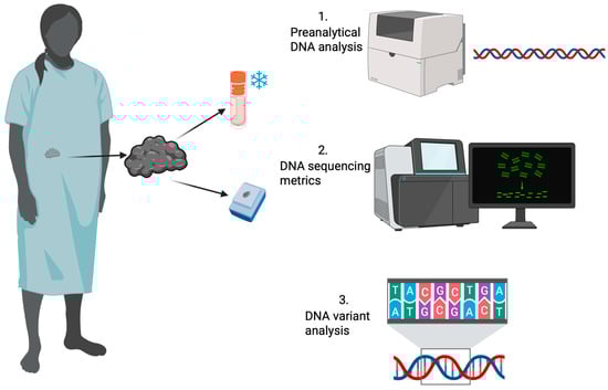
2. Results
2.1. Sample Types
2.2. Presequencing Metrics
2.3. Whole-Genome Sequencing Metrics
2.4. Variant Call Analysis
2.5. Tumor Mutational Burden Analysis
2.6. Cancer Gene Set
3. Discussion
4. Materials and Methods
4.1. Study Design
4.2. WGS Methods
4.2.1. DNA Extraction and Sequencing
4.2.2. Bioinformatic Analysis of WGS Data
4.3. Statistical Analysis
4.4. Bias Mitigation
5. Conclusions
Supplementary Materials
Author Contributions
Funding
Institutional Review Board Statement
Informed Consent Statement
Data Availability Statement
Acknowledgments
Conflicts of Interest
Abbreviations
| CP | Cryopreserved |
| FF | Fresh-frozen |
| FFPE | Formalin-fixed, paraffin-embedded |
| Indel | Insertions and deletions |
| Mb | Megabase |
| MNV | Multinucleotide variant |
| NGS | Next-generation sequencing |
| PCR | Polymerase chain reaction |
| SNV | Single nucleotide variant |
| SV | Structural variant |
| TMB | Tumor mutational burden |
| VAF | Variant allele fraction |
| WGS | Whole-genome sequencing |
References
- Bray, F.; Laversanne, M.; Sung, H.; Ferlay, J.; Siegel, R.L.; Soerjomataram, I.; Jemal, A. Global cancer statistics 2022: GLOBOCAN estimates of incidence and mortality worldwide for 36 cancers in 185 countries. CA Cancer J. Clin. 2024, 74, 229–263. [Google Scholar] [CrossRef]
- Siegel, R.L.; Giaquinto, A.N.; Jemal, A. Cancer statistics, 2024. CA Cancer J. Clin. 2024, 74, 12–49, Erratum in CA Cancer J. Clin. 2024, 74, 203. https://doi.org/10.3322/caac.21830. [Google Scholar] [CrossRef]
- Shin, S.H.; Bode, A.M.; Dong, Z. Precision medicine: The foundation of future cancer therapeutics. npj Precis. Oncol. 2017, 1, 12. [Google Scholar] [CrossRef]
- Normanno, N.; Rachiglio, A.M.; Roma, C.; Fenizia, F.; Esposito, C.; Pasquale, R.; La Porta, M.L.; Iannaccone, A.; Micheli, F.; Santangelo, M.; et al. Molecular diagnostics and personalized medicine in oncology: Challenges and opportunities. J. Cell. Biochem. 2013, 114, 514–524. [Google Scholar] [CrossRef]
- Colomer, R.; Miranda, J.; Romero-Laorden, N.; Hornedo, J.; González-Cortijo, L.; Mouron, S.; Bueno, M.J.; Mondéjar, R.; Quintela-Fandino, M. Usefulness and real-world outcomes of next generation sequencing testing in patients with cancer: An observational study on the impact of selection based on clinical judgement. eClinicalMedicine 2023, 60, 102029. [Google Scholar] [CrossRef] [PubMed]
- Akhoundova, D.; Rubin, M.A. Clinical application of advanced multi-omics tumor profiling: Shaping precision oncology of the future. Cancer Cell 2022, 40, 920–938. [Google Scholar] [CrossRef] [PubMed]
- Do, H.; Dobrovic, A. Sequence artifacts in DNA from formalin-fixed tissues: Causes and strategies for minimization. Clin. Chem. 2015, 61, 64–71. [Google Scholar] [CrossRef] [PubMed]
- Cazzato, G.; Caporusso, C.; Arezzo, F.; Cimmino, A.; Colagrande, A.; Loizzi, V.; Cormio, G.; Lettini, T.; Maiorano, E.; Scarcella, V.S.; et al. Formalin-Fixed and Paraffin-Embedded Samples for Next Generation Sequencing: Problems and Solutions. Genes 2021, 12, 1472. [Google Scholar] [CrossRef]
- Basyuni, S.; Heskin, L.; Degasperi, A.; Black, D.; Koh, G.C.C.; Chmelova, L.; Rinaldi, G.; Bell, S.; Grybowicz, L.; Elgar, G.; et al. Large-scale analysis of whole genome sequencing data from formalin-fixed paraffin-embedded cancer specimens demonstrates preservation of clinical utility. Nat. Commun. 2024, 15, 7731. [Google Scholar] [CrossRef]
- Robbe, P.; Popitsch, N.; Knight, S.J.L.; Antoniou, P.; Becq, J.; He, M.; Kanapin, A.; Samsonova, A.; Vavoulis, D.V.; Ross, M.T.; et al. Clinical whole-genome sequencing from routine formalin-fixed, paraffin-embedded specimens: Pilot study for the 100,000 Genomes Project. Genet. Med. 2018, 20, 1196–1205. [Google Scholar] [CrossRef]
- Gao, X.H.; Li, J.; Gong, H.F.; Yu, G.Y.; Liu, P.; Hao, L.Q.; Liu, L.J.; Bai, C.G.; Zhang, W. Comparison of Fresh Frozen Tissue With Formalin-Fixed Paraffin-Embedded Tissue for Mutation Analysis Using a Multi-Gene Panel in Patients With Colorectal Cancer. Front. Oncol. 2020, 10, 310. [Google Scholar] [CrossRef]
- Okojie, J.; O’Neal, N.; Burr, M.; Worley, P.; Packer, I.; Anderson, D.; Davis, J.; Kearns, B.; Fatema, K.; Dixon, K.; et al. DNA Quantity and Quality Comparisons between Cryopreserved and FFPE Tumors from Matched Pan-Cancer Samples. Curr. Oncol. 2024, 31, 2441–2452. [Google Scholar] [CrossRef] [PubMed]
- Cho, M.; Ahn, S.; Hong, M.; Bang, H.; Van Vrancken, M.; Kim, S.; Lee, J.; Hoon Park, S.; Oh Park, J.; Suk Park, Y.; et al. Tissue recommendations for precision cancer therapy using next generation sequencing: A comprehensive single cancer center’s experiences. Oncotarget 2017, 8, 42478–42486. [Google Scholar] [CrossRef] [PubMed]
- Suciu, B.A.; Pap, Z.; Dénes, L.; Brînzaniuc, K.; Copotoiu, C.; Pávai, Z. Allele-specific PCR method for identification of EGFR mutations in non-small cell lung cancer: Formalin-fixed paraffin-embedded tissue versus fresh tissue. Rom. J. Morphol. Embryol. 2016, 57, 495–500. [Google Scholar]
- Jovanović, B.; Sheng, Q.; Seitz, R.S.; Lawrence, K.D.; Morris, S.W.; Thomas, L.R.; Hout, D.R.; Schweitzer, B.L.; Guo, Y.; Pietenpol, J.A.; et al. Comparison of triple-negative breast cancer molecular subtyping using RNA from matched fresh-frozen versus formalin-fixed paraffin-embedded tissue. BMC Cancer 2017, 17, 241. [Google Scholar] [CrossRef]
- Jacobsen, S.B.; Tfelt-Hansen, J.; Smerup, M.H.; Andersen, J.D.; Morling, N. Comparison of whole transcriptome sequencing of fresh, frozen, and formalin-fixed, paraffin-embedded cardiac tissue. PLoS ONE 2023, 18, e0283159. [Google Scholar] [CrossRef]
- Munchel, S.; Hoang, Y.; Zhao, Y.; Cottrell, J.; Klotzle, B.; Godwin, A.K.; Koestler, D.; Beyerlein, P.; Fan, J.B.; Bibikova, M.; et al. Targeted or whole genome sequencing of formalin fixed tissue samples: Potential applications in cancer genomics. Oncotarget 2015, 6, 25943–25961. [Google Scholar] [CrossRef] [PubMed]
- Greytak, S.R.; Engel, K.B.; Bass, B.P.; Moore, H.M. Accuracy of Molecular Data Generated with FFPE Biospecimens: Lessons from the Literature. Cancer Res. 2015, 75, 1541–1547. [Google Scholar] [CrossRef]
- Steiert, T.A.; Parra, G.; Gut, M.; Arnold, N.; Trotta, J.-R.; Tonda, R.; Moussy, A.; Gerber, Z.; Abuja, P.M.; Zatloukal, K.; et al. A critical spotlight on the paradigms of FFPE-DNA sequencing. Nucleic Acids Res. 2023, 51, 7143–7162. [Google Scholar] [CrossRef]
- Al-Kateb, H.; Nguyen, T.T.; Steger-May, K.; Pfeifer, J.D. Identification of major factors associated with failed clinical molecular oncology testing performed by next generation sequencing (NGS). Mol. Oncol. 2015, 9, 1737–1743. [Google Scholar] [CrossRef]
- Bhagwate, A.V.; Liu, Y.; Winham, S.J.; McDonough, S.J.; Stallings-Mann, M.L.; Heinzen, E.P.; Davila, J.I.; Vierkant, R.A.; Hoskin, T.L.; Frost, M.; et al. Bioinformatics and DNA-extraction strategies to reliably detect genetic variants from FFPE breast tissue samples. BMC Genom. 2019, 20, 689. [Google Scholar] [CrossRef] [PubMed]
- Roepman, P.; de Bruijn, E.; van Lieshout, S.; Schoenmaker, L.; Boelens, M.C.; Dubbink, H.J.; Geurts-Giele, W.R.R.; Groenendijk, F.H.; Huibers, M.M.H.; Kranendonk, M.E.G.; et al. Clinical Validation of Whole Genome Sequencing for Cancer Diagnostics. J. Mol. Diagn. 2021, 23, 816–833. [Google Scholar] [CrossRef]
- Solassol, J.; Ramos, J.; Crapez, E.; Saifi, M.; Mangé, A.; Vianès, E.; Lamy, P.-J.; Costes, V.; Maudelonde, T. KRAS Mutation Detection in Paired Frozen and Formalin-Fixed Paraffin-Embedded (FFPE) Colorectal Cancer Tissues. Int. J. Mol. Sci. 2011, 12, 3191–3204. [Google Scholar] [CrossRef] [PubMed]
- De Paoli-Iseppi, R.; Johansson, P.A.; Menzies, A.M.; Dias, K.R.; Pupo, G.M.; Kakavand, H.; Wilmott, J.S.; Mann, G.J.; Hayward, N.K.; Dinger, M.E.; et al. Comparison of whole-exome sequencing of matched fresh and formalin fixed paraffin embedded melanoma tumours: Implications for clinical decision making. Pathology 2016, 48, 261–266. [Google Scholar] [CrossRef]
- Lu, X.; Wei, Y.; Sun, J.; Xiao, B.; Zhang, X.; Li, W.; Chen, Y.; Lin, F.; Zhang, L.; Wang, Y.; et al. A Comparative Study of Three Nucleic Acid Integrity Assay Systems. Biopreserv. Biobank. 2023, 21, 624–630. [Google Scholar] [CrossRef] [PubMed]
- Mathieson, W.; Thomas, G. Using FFPE Tissue in Genomic Analyses: Advantages, Disadvantages and the Role of Biospecimen Science. Curr. Pathobiol. Rep. 2019, 7, 35–40. [Google Scholar] [CrossRef]
- Gaffney, E.F.; Riegman, P.H.; Grizzle, W.E.; Watson, P.H. Factors that drive the increasing use of FFPE tissue in basic and translational cancer research. Biotech. Histochem. 2018, 93, 373–386. [Google Scholar] [CrossRef]
- Janssen, J.; Chirico, N.; Ainsworth, M.J.; Cedillo-Servin, G.; Viola, M.; Dokter, I.; Vermonden, T.; Doevendans, P.A.; Serra, M.; Voets, I.K.; et al. Hypothermic and cryogenic preservation of cardiac tissue-engineered constructs. Biomater. Sci. 2024, 12, 3866–3881. [Google Scholar] [CrossRef]
- Day, A.G.E.; Bhangra, K.S.; Murray-Dunning, C.; Stevanato, L.; Phillips, J.B. The Effect of Hypothermic and Cryogenic Preservation on Engineered Neural Tissue. Tissue Eng. Part C Methods 2017, 23, 575–582. [Google Scholar] [CrossRef]
- He, A.; Powell, S.; Kyle, M.; Rose, M.; Masmila, E.; Estrada, V.; Sicklick, J.K.; Molinolo, A.; Kaushal, S. Cryopreservation of Viable Human Tissues: Renewable Resource for Viable Tissue, Cell Lines, and Organoid Development. Biopreserv. Biobank. 2020, 18, 222–227. [Google Scholar] [CrossRef]
- Daly, M.B.; Pal, T.; Maxwell, K.N.; Churpek, J.; Kohlmann, W.; AlHilli, Z.; Arun, B.; Buys, S.S.; Cheng, H.; Domchek, S.M.; et al. NCCN Guidelines® Insights: Genetic/Familial High-Risk Assessment: Breast, Ovarian, and Pancreatic, Version 2.2024. J. Natl. Compr. Cancer Netw. 2023, 21, 1000–1010. [Google Scholar] [CrossRef]
- Ferguson, S.; Sriram, S.; Wallace, J.K.; Lee, J.; Kim, J.A.; Lee, Y.; Oh, B.B.; Lee, W.C.; Lee, S.; Connolly-Strong, E. Analytical and Clinical Validation of a Target-Enhanced Whole Genome Sequencing-Based Comprehensive Genomic Profiling Test. Cancer Investig. 2024, 42, 390–399. [Google Scholar] [CrossRef]
- Li, H.; Durbin, R. Fast and accurate short read alignment with Burrows-Wheeler transform. Bioinformatics 2009, 25, 1754–1760. [Google Scholar] [CrossRef]
- Faust, G.G.; Hall, I.M. SAMBLASTER: Fast duplicate marking and structural variant read extraction. Bioinformatics 2014, 30, 2503–2505. [Google Scholar] [CrossRef]
- Chen, X.; Schulz-Trieglaff, O.; Shaw, R.; Barnes, B.; Schlesinger, F.; Källberg, M.; Cox, A.J.; Kruglyak, S.; Saunders, C.T. Manta: Rapid detection of structural variants and indels for germline and cancer sequencing applications. Bioinformatics 2016, 32, 1220–1222. [Google Scholar] [CrossRef]
- McLaren, W.; Gil, L.; Hunt, S.E.; Riat, H.S.; Ritchie, G.R.; Thormann, A.; Flicek, P.; Cunningham, F. The Ensembl Variant Effect Predictor. Genome Biol. 2016, 17, 122. [Google Scholar] [CrossRef] [PubMed]
- Favero, F.; Joshi, T.; Marquard, A.M.; Birkbak, N.J.; Krzystanek, M.; Li, Q.; Szallasi, Z.; Eklund, A.C. Sequenza: Allele-specific copy number and mutation profiles from tumor sequencing data. Ann. Oncol. 2015, 26, 64–70. [Google Scholar] [CrossRef] [PubMed]
- Chang, M.T.; Bhattarai, T.S.; Schram, A.M.; Bielski, C.M.; Donoghue, M.T.A.; Jonsson, P.; Chakravarty, D.; Phillips, S.; Kandoth, C.; Penson, A.; et al. Accelerating Discovery of Functional Mutant Alleles in Cancer. Cancer Discov. 2018, 8, 174–183. [Google Scholar] [CrossRef] [PubMed]
- Ramírez, F.; Ryan, D.P.; Grüning, B.; Bhardwaj, V.; Kilpert, F.; Richter, A.S.; Heyne, S.; Dündar, F.; Manke, T. deepTools2: A next generation web server for deep-sequencing data analysis. Nucleic Acids Res. 2016, 44, W160–W165. [Google Scholar] [CrossRef]
- Davies, H.; Glodzik, D.; Morganella, S.; Yates, L.R.; Staaf, J.; Zou, X.; Ramakrishna, M.; Martin, S.; Boyault, S.; Sieuwerts, A.M.; et al. HRDetect is a predictor of BRCA1 and BRCA2 deficiency based on mutational signatures. Nat. Med. 2017, 23, 517–525. [Google Scholar] [CrossRef]


| Patient (n = 50) | |||
| Age, year | 63.6 ± 11.8 | ||
| Sex assigned at birth | Male Female | 18 (36.0%) 32 (64.0%) | |
| Tumor (n = 50) | |||
| Specimen Source | n | (%) | Histological type |
| Ovary | 14 | 28% | Serous carcinoma 7 Clear cell carcinoma 2 Papillary serous adenocarcinoma 1 Carcinoma 1 Carcinosarcoma 1 Benign stromal tumor 1 Undetermined 1 |
| Uterus | 7 | 14% | Carcinosarcoma 3 Adenocarcinoma 1 Endometrioid carcinoma 1 Leiomyosarcoma 1 * Undetermined 1 |
| Kidney | 6 | 12% | Renal clear cell carcinoma 4 Oncocytoma 1 Urothelial cancer 1 * |
| Liver | 5 | 10% | Hepatocellular carcinoma 2 Adenocarcinoma 2 * Poorly differentiated carcinoma 1 |
| Colon | 5 | 10% | Adenocarcinoma 3 Granulosa cell tumor 1 * Papillary serous carcinoma 1 * |
| Adrenal gland | 2 | 4% | Cortical carcinoma 1 Diffuse large B-cell lymphoma 1 * |
| Lung | 2 | 4% | Epithelioid mesothelioma 1 Leiomyosarcoma 1 * |
| Peritoneum | 2 | 4% | Ovarian adenocarcinoma 1 * Ovarian serous carcinoma 1 * |
| Breast | 2 | 4% | Invasive ductal carcinoma 1 Ovarian adenocarcinoma 1 * |
| Bone | 1 | 2% | Prostate adenocarcinoma 1 * |
| Thyroid | 1 | 2% | Papillary carcinoma 1 |
| Pelvis | 1 | 2% | Liposarcoma 1 |
| Stomach | 1 | 2% | Gastrointestinal stromal tumor 1 |
| Appendix | 1 | 2% | Signet ring carcinoma 1 |
| FFPE | CP | p Value * | |
|---|---|---|---|
| TMB, mut/Mb | 13.7 (12.2–16.0) | 6.4 (5.5–8.5) | 0.02 |
| PM (SNV + MNV + IND) | 39,613 (34,839–45,786) | 20,339 (16,638–24,959) | 0.02 |
| SNV | |||
| Total | 21,998 (16,789–26,046) | 10,660 (8373–15,675) | 0.10 |
| C>A | 2699 (2169–3234) | 4492 (2773–4692) | 0.31 |
| C>G | 1602 (1166–2068) | 568 (262–1189) | 0.11 |
| C>T | 5914 (4563–8282) | 2084 (1328–3429) | 0.14 |
| T>A | 2852 (1501–3310) | 552 (449–893) | 0.054 |
| T>C | 3769 (2459–4967) | 1275 (950–1721) | 0.04 |
| T>G | 2937 (2632–3733) | 1504 (1271–2734) | 0.20 |
| MNV | 1076 (492–1660) | 83 (67–134) | 0.008 |
| Indel | 18,174 (13,704–20,919) | 8871 (8199–11,231) | 0.02 |
| SV | |||
| Total | 21 (13–62) | 38 (12–81) | 0.94 |
| Deletion | 4 (2–8) | 12 (2–24) | 0.11 |
| Duplication | 2 (0–3) | 4 (3–14) | 0.08 |
| Inversion | 11 (2–49) | 10 (2–15) | 0.15 |
| Translocation | 6 (4–7) | 8 (5–27) | 0.46 |
| Variant Types | FFPE | CP | p Value * |
|---|---|---|---|
| Overall | |||
| SNV | 3 (2–4) | 3 (2–5) | 0.02 |
| Indel | 0 (0–1) | 0 (0–1) | 0.82 |
| SV | 1 (1–2) | 1 (1–3) | 0.007 |
| CNV | 0 (0–1.25) | 0 (0–1) | 0.58 |
| Subtotal | 5 (4–8) | 6 (4–10.25) | 0.02 |
| Overlapped | |||
| SNV | 2.5 (1–4) | ||
| Indel | 0 (0–1) | ||
| SV | 1 (0–2) | ||
| CNV | 0 (0–0) | ||
| Subtotal | 4 (3–6) | ||
| Unique | |||
| SNV | 0 (0–0) | 0 (0–0) | 0.02 |
| Indel | 0 (0–0) | 0 (0–0) | 1.00 |
| SV | 0 (0–0) | 0 (0–0) | 0.001 |
| CNV | 0 (0–0) | 0 (0–0) | 0.82 |
| Subtotal | 1 (1–2) | 1.5 (0–2) | 0.009 |
Disclaimer/Publisher’s Note: The statements, opinions and data contained in all publications are solely those of the individual author(s) and contributor(s) and not of MDPI and/or the editor(s). MDPI and/or the editor(s) disclaim responsibility for any injury to people or property resulting from any ideas, methods, instructions or products referred to in the content. |
© 2025 by the authors. Licensee MDPI, Basel, Switzerland. This article is an open access article distributed under the terms and conditions of the Creative Commons Attribution (CC BY) license (https://creativecommons.org/licenses/by/4.0/).
Share and Cite
Dixon, K.; Lee, J.-H.; Miller, R.; Booker, D.; Anderson, D.; Okojie, J.; Kirkham, M.; Lee, E.K.; Bao, C.; Tuncay, I.O.; et al. Cryopreserved Tissue Biospecimens Offer Superior Quality for Whole-Genome Sequencing of Various Cancers Compared to Paired Formalin-Fixed Paraffin-Embedded Tissues. Int. J. Mol. Sci. 2025, 26, 11038. https://doi.org/10.3390/ijms262211038
Dixon K, Lee J-H, Miller R, Booker D, Anderson D, Okojie J, Kirkham M, Lee EK, Bao C, Tuncay IO, et al. Cryopreserved Tissue Biospecimens Offer Superior Quality for Whole-Genome Sequencing of Various Cancers Compared to Paired Formalin-Fixed Paraffin-Embedded Tissues. International Journal of Molecular Sciences. 2025; 26(22):11038. https://doi.org/10.3390/ijms262211038
Chicago/Turabian StyleDixon, Ken, Jeong-Hoon Lee, Ryan Miller, David Booker, DeLaney Anderson, Jeffrey Okojie, Matthew Kirkham, Eun Kyoung Lee, Chunyang Bao, Islam Oguz Tuncay, and et al. 2025. "Cryopreserved Tissue Biospecimens Offer Superior Quality for Whole-Genome Sequencing of Various Cancers Compared to Paired Formalin-Fixed Paraffin-Embedded Tissues" International Journal of Molecular Sciences 26, no. 22: 11038. https://doi.org/10.3390/ijms262211038
APA StyleDixon, K., Lee, J.-H., Miller, R., Booker, D., Anderson, D., Okojie, J., Kirkham, M., Lee, E. K., Bao, C., Tuncay, I. O., Kim, J.-A., Lee, S., & Barrott, J. (2025). Cryopreserved Tissue Biospecimens Offer Superior Quality for Whole-Genome Sequencing of Various Cancers Compared to Paired Formalin-Fixed Paraffin-Embedded Tissues. International Journal of Molecular Sciences, 26(22), 11038. https://doi.org/10.3390/ijms262211038