Article

FGFR2 Might Be a Promising Therapeutic Target for Some Solid Tumors: Analysis of 1312 Cancers with FGFR2 Abnormalities

1
Molecular Oncology and Therapeutics, Osaka Metropolitan University Graduate School of Medicine, 1-4-3 Asahimachi, Abeno-ku, Osaka 545-8585, Japan
2
Department of Clinical Pharmacy, School of Pharmacy and Pharmaceutical Sciences, Mukogawa Women’s University, Nishinomiya 663-8179, Japan
3
Cancer Center for Translational Research, Osaka Metropolitan University Graduate School of Medicine, 1-4-3 Asahimachi, Abeno-ku, Osaka 545-8585, Japan
*
Author to whom correspondence should be addressed.
Int. J. Mol. Sci. 2025, 26(21), 10777; https://doi.org/10.3390/ijms262110777
Submission received: 29 September 2025 / Revised: 30 October 2025 / Accepted: 1 November 2025 / Published: 5 November 2025
(This article belongs to the Special Issue 25th Anniversary of IJMS: Updates and Advances in Molecular Oncology)

Abstract

Genetic abnormalities of the fibroblast growth factor receptor 2 (FGFR2) gene, including amplification, fusions, and mutations, have been reported in various solid tumors. While molecular targeted therapies against FGFR2 fusion have been proved to be useful in cholangiocarcinoma, the therapeutic significance of FGFR2 inhibitors remains unclear in other various solid cancers. Genomic and clinical information from solid tumor cancer gene panel testing cases is consolidated in the Center for Cancer Genomics and Advanced Therapeutics (C-CAT) database in Japan. This study aimed to utilize the C-CAT database to clarify the clinical–pathological significance of FGFR2 abnormalities. A total of 101,231 patients with solid cancer have been registered in the C-CAT database between June 2019 and June 2025. Of the 101,231 cases, 1312 cases with FGFR2 gene abnormalities were analyzed. FGFR2 alterations included amplification in 515 cases, fusion in 280 cases, and mutations in 568 cases. They were detected most frequently in the biliary tract (271 cases), esophagus/stomach (231 cases), and breast (211 cases). Amplification was frequent in the esophagus/stomach (205 cases) and breast (105 cases). Mutations were frequent in the uterus (111 cases), breast (89 cases), and biliary tract (86 cases). Among 515 FGFR2 alteration cases, FGFR2 inhibitors were administered in 85 cases. Of the 85 cases, disease control was achieved in 49 cases, 44 cases of which were biliary tract cancer. FGFR2 might be a promising therapeutic target not only for cholangiocarcinoma with fusion but also for esophagus/stomach cancer and breast cancer with FGFR2 alterations.

1. Introduction

Fibroblast growth factor receptors (FGFRs) are transmembrane receptor tyrosine kinases and consist of four subtypes, i.e., FGFR-1, -2, -3, and -4. FGFRs are involved in a lot of functions such as cell proliferation, differentiation, invasion, and angiogenesis [1,2]. Recently, molecular targeted therapies against the FGFR2 fusion gene have been approved. Actually, three small molecular inhibitors of FGFR2 including futibatinib [3,4], pemigatinib [5], and tasurgratinib [6] have been approved as of 2024 and are clinically used against cholangiocarcinoma with FGFR2 fusion genes in Japan. The inhibitors target the ATP binding site of the FGFR2 kinase domain and inhibit cell proliferation, which suggests that FGFR2 inhibitors might be effective for cancer with not only FGFR2 fusion but also active abnormalities [7,8].
Some solid tumors, such as cholangiocarcinoma, endometrial cancer, breast cancer, gastric cancer, and ovarian cancer, have been reported to show FGFR abnormalities including amplification, fusion, and mutation. FGFR2 amplification has been found in 7–23% of breast cancers, 5–10% of gastric cancers, and 12% of endometrial cancers [9,10]. We previously reported that some diffuse types of gastric cancer showed K-samII amplification, which encodes FGFR2 [11], and FGFR2 inhibitors were therapeutically promising for gastric carcinoma with FGFR2 amplification using several gastric cancer cell lines [12,13,14,15]. Particularly, the extremely malignant type of gastric cancer is associated with FGFR2 amplification; however, few useful drugs have yet been clinically approved for cancers with FGFR2 amplification [16]. Furthermore, the therapeutic significance of FGFR2 abnormalities for these cancers, except cholangiocarcinoma, remains unclear.
Recently, precision medicine has become mainstream for the treatment of tumors based on their genomic abnormalities. Comprehensive genomic panel (CGP) tests have been clinically applied for patients with solid tumors in Japan. Genomic and clinical information of patients who have undergone multi-CGP tests are collected at the Center for Cancer Genomics and Advanced Therapeutics (C-CAT) database in Japan [17,18,19]. A total of 101,231 patients have been registered in the C-CAT database on 30 June 2025. Currently, five types of multi-CGP tests, including FoundationOne CDx (F1), FoundationOne Liquid CDx (F1L), GenMineTOP (GMT), NCC Oncopanel (NCC), and Guardant360 (G360), have been approved in Japan. Analysis using large-scale genomic databases is important to clarify the significance of FGFR2 abnormalities in carcinomas. The aim of this study is to clarify the clinicopathologic significance of FGFR2 abnormalities by using the C-CAT database.

2. Results

2.1. Patient Background of Cases with FGFR2 Gene Abnormalities

Gene abnormalities in the FGFR2 were detected in 1312 cases out of 101,231 cases by the multi-CGP tests. Table 1 summarizes the patient background of cases with FGFR2 genetic abnormalities. Among patients with FGFR2 gene abnormalities, there were 513 males (39.1%) and 799 females (60.9%). More females were detected than males (p < 0.001). Furthermore, concerning the age of patients, abnormalities were detected in significantly higher numbers in those under 60 years of age than in those over 60 years of age. Multi-CGP tests more frequently underwent a DNA assay than RNA assay, but there was no significant difference between the two in the detection of the FGFR2 gene. Submitted specimens were more frequently tissue samples than blood samples, and tissue samples had a significantly higher detection rate of the FGFR2 gene (p < 0.001). Submitted locations of tissue samples were primary and metastasis, and both locations could be detected in cases of FGFR2 abnormalities. FGFR2 gene abnormalities were negatively correlated with smoking and tended to be higher in non-drinkers than drinkers.

2.2. Distribution of FGFR2 Genetic Abnormalities

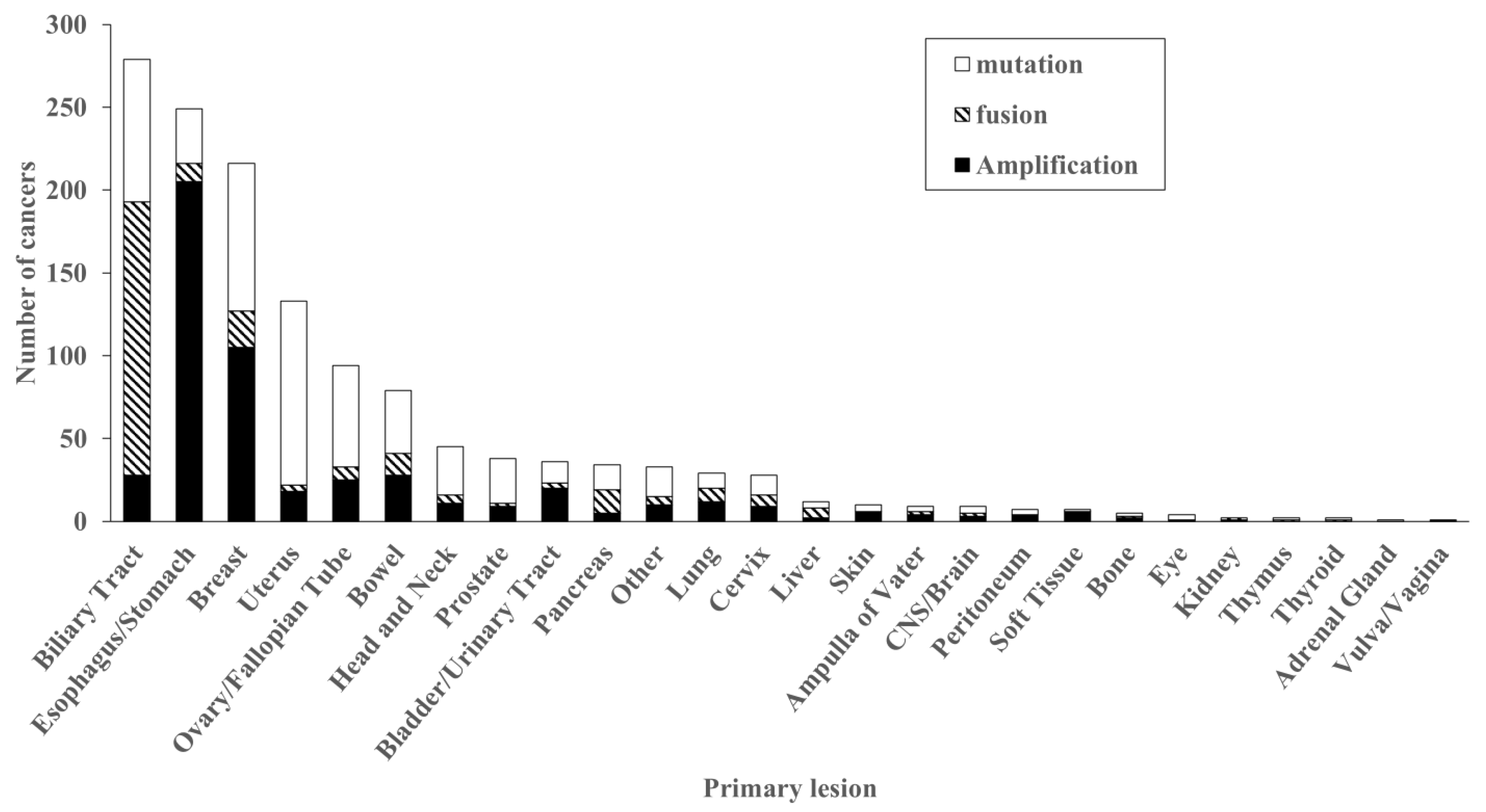
In 1312 cancers with FGFR2 abnormalities, FGFR2 amplification was detected in 515 cases (39.2%), fusion in 280 cases (20.9%), and mutation in 568 cases (43.3%) (Figure 1).
Of these, 29 cases showed both gene amplification and fusions, and 16 showed both gene amplification and mutations. Figure 2 summarizes the cancer types with FGFR2 genetic abnormalities. The most frequent cancer types were found in the biliary tract (271 cases), followed by esophagus/stomach (231 cases), breast (211 cases), uterus (131 cases), and ovary (93 cases).
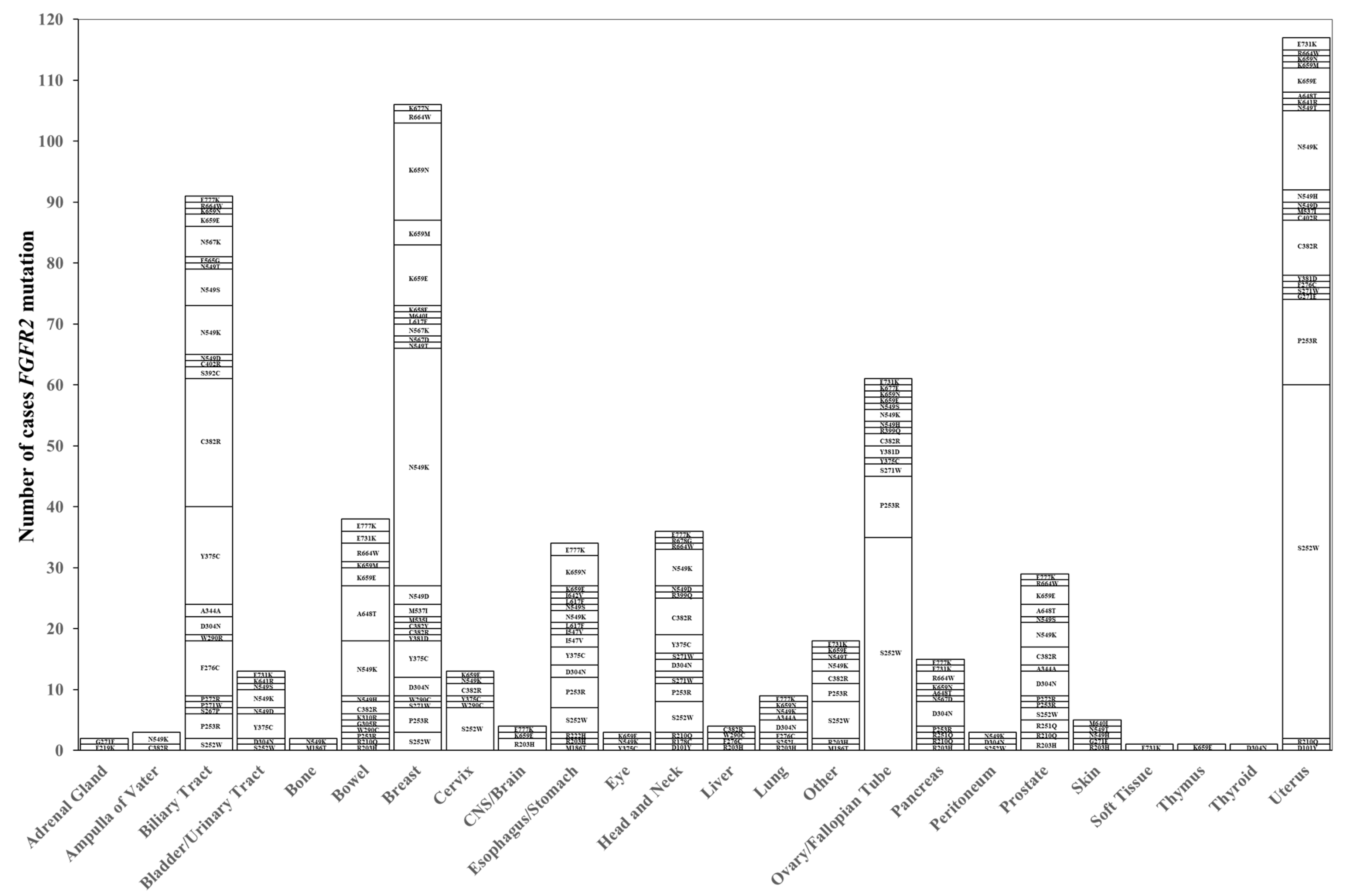
FGFR2 amplification was frequently detected in the esophagus/stomach (205 cases), breast (105 cases), biliary tract (28 cases), bowel (28 cases), and ovary/fallopian tube (25 cases). FGFR2 mutations are frequently detected in the uterus (111 cases), breast (89 cases), biliary tract (86 cases), ovary/fallopian tube (61 cases), and bowel (38 cases). There were frequent hot spot mutations such as S252W (124 cases), N549K (95 cases), C382R (52 cases), P253R (46 cases), and Y375C (35 cases). In contrast, A648T was frequently identified in 23% (9 of 38 cases) of colorectal cancer with FGFR2 mutations (Figure 3).
FGFR2 fusions are frequently detected in the biliary tract (163 cases), followed by breast (21 cases), pancreas (13 cases), bowel (12 cases), and esophagus/stomach (11 cases). Some partner genes were frequently identified, such as BICC1, TACC2, CTNNA3, and KIAA1217 in the biliary tract, while these partner genes were not detected in any other cancers. (Table 2).

2.3. Treatment with FGFR Inhibitors

Out of 1312 cancers with the FGFR2 abnormalities, FGFR2 inhibitors were administered in 85 cases. Of 85 cases, 14 cases received futibatinib, 69 cases received pemigatinib, and 2 cases received both. The cases treated by FGFR2 inhibitors included 77 fusion gene cases, 10 amplification cases, and 2 mutation cases. Of these cases, two cases had both fusion and amplification, and one case had both amplification and mutation. Table 3 shows the correlations between FGFR2 alteration type and therapeutic response by FGFR2 inhibitors. FGFR2 gene abnormalities confirmed to be effectively treated with FGFR2 inhibitors were identified in 44 cases of fusions, 1 case of amplification, 1 case of mutation (N549S), 2 cases of both fusion and amplification, and 1 case of both amplification and mutation.
This summarizes the best response to FGFR2 inhibitor administration and the administered FGFR2 gene abnormalities. There were 0 cases of CR (complete response), 21 cases of PR (partial response), 27 cases of SD (stable disease), 14 cases of PD (progressive disease), and 23 cases of NE (not evaluable). Most patients who achieved PR received treatment directed at the fusion gene. However, some patients also responded to treatment directed at the gene amplification. In addition, in most cases of SD, treatment was directed at the fusion gene, but disease control was also achieved in some cases where treatment was directed at the gene mutation.
Table 4 shows the correlations between cancer type and therapeutic response by FGFR2 inhibitors. Cancer types were biliary tract (73 cases), esophagus/stomach (3 cases), liver (3 cases), pancreas (2 cases), ampulla of vater (2 cases), and others (2 cases). A total of 48 cases achieved disease control; 44 cases were in the biliary tract, 1 case was in the ampulla of vater, 1 case was in the liver, and there were 2 other cases.
FGFR2 inhibitors were administered to 85 patients, of whom 48 patients (56.5%) achieved disease control (DCR). Most of the DCR cases (44 patients) were in the bile duct, but disease control was also achieved in the ampulla of vater and liver. However, there were 37 cases that did not achieve disease control, including 29 in the bile duct, 3 in the esophagus/stomach, 2 in the liver, 2 in the pancreas, and 1 in the ampulla of vater.

3. Discussion

In this study, we analyzed the clinical characteristics of FGFR2 gene abnormalities and treatment outcomes with inhibitors using a large-scale genomic database in Japan, i.e., the C-CAT database. Our analysis revealed that FGFR2 gene abnormalities were significantly frequent in women. This finding might suggest that FGFR2 abnormalities are prevalent in areas such as the uterus, ovaries/fallopian tubes, and breast, as previously reported [20,21].
FGFR2 gene abnormalities were also significantly frequent in younger patients under 60 years of age. One of the reasons might be that FGFR2 abnormalities were frequent in cholangiocarcinoma and gastric cancers, which were relatively frequent in younger age [22,23,24]. In addition, FGFR2 gene abnormalities were negatively correlated with smoking and alcohol. It was indicated that FGFR2 gene abnormalities may be caused by factors within the tumor without external factors and old ages. No significant differences were found between DNA and RNA analysis in the submitted multi-CGP tests, but the submitted specimens showed significantly higher detection rates in tissue than in liquid.
FGFR2 amplification was frequent in the esophagus/stomach, followed by the breast and biliary tract, while FGFR2 fusion was the highest in the biliary tract (for which treatment with FGFR2 inhibitors is approved in Japan), followed by the breast and pancreas. FGFR2 mutations were frequently detected in the uterus, breast, and biliary tract. FGFR2 hotspots such as S252W, P253R, and N549K were frequently detected in endometrial cancer, as previously reported [21,25]. On the other hand, A648T mutation was detected only in colon cancer; this analysis is the first report of this finding.
FGFR2 fusion partners were several common genes such as BICC1 and TACC2, which are frequently detected in the biliary tract, while FGFR2 fusion partners of the other cancers were different from these common partner genes. These findings suggest that the mechanisms of FGFR2 fusions may resemble cholangiocarcinoma, but not in the other cancers.
In a recent study, pemigatinib showed no difference in ORR, OS, and PFS between BICC1 and other partner genes, suggesting that FGFR2 inhibitors may be effective for cancer with FGFR2 fusion regardless of the partner gene [5].
The outcome of FGFR2 inhibitors for FGFR2 abnormalities revealed that disease control was achieved in 49 of 85 patients. Most of the 49 cases were treated for cholangiocarcinoma. This suggests that FGFR2 inhibitors were effective for tumors with FGFR2 fusion. On the other hand, FGFR2 inhibitors also achieved disease control for not only fusion cases but also gene amplification or mutation. It is necessary to examine the effect of FGFR2 inhibitors for cancer with FGFR2 amplification or FGFR2 mutation.
Currently, the multi-CGP test, Hemesight®, has been clinically applied for patients with hematopoietic tumors including leukemia or malignant lymphoma. Hemesight® is also detectable for FGFR abnormalities. FGFR2 inhibitors might be promising for hematopoietic tumors with FGFR abnormalities. It will be necessary to examine the significance of FGFR2 inhibitors for hematopoietic tumors with FGFR abnormalities using C-CAT data in the near future.

4. Materials and Methods

4.1. Extraction of Subject

This study used a total of 101,231 cases registered in the C-CAT database from June 2019 to June 2025. Only facilities authorized to use C-CAT can access this information through the data sharing system of the C-CAT Research Use Portal (https://www.ncc.go.jp/en/c_cat/use/index.html (accessed on 19 September 2025).
Of these data, 3512 cases had FGFR2 gene abnormalities. The gene alteration of a tumor was examined by one of five multi-CGP tests: FoundationOne® CDx (F1; Foundation Medicine Inc., Cambridge, MA, USA), OncoGuide™ NCC Oncopanel System (NCC; Sysmex Co., Ltd., Kobe, Japan), GenMineTOP® Cancer Genome Profiling Systems (GMT; KONICA MI-NOLTA REALM Co., Inc., Tokyo, Japan), FoundationOne® Liquid CDx (F1L; Foundation Medicine Inc.), or Guardant360® CDx sequencing technology (G360; Guardant Health, Palo Alto, CA, USA). F1 was performed using genomic DNA extracted from tumor tissue. The NCC test analyzed both tumor tissue and circulating tumor DNA (ctDNA) in the blood, while the GMT test used DNA and RNA extracted from tumor tissue as well as DNA from whole blood samples. F1L and G360 tests, on the other hand, use peripheral blood to examine tumor-derived ctDNA. Regarding the number of genes analyzed, the NCC test examined 114 gene mutations [26], the F1 test examined 324 gene mutations [27], and the F1L test, which analyzes circulating ctDNA in the blood, also examined 324 gene mutations [28]. The GMT test, on the other hand, examined a total of 737 gene mutations [29]. All five tests were capable of detecting FGFR2 gene abnormalities. Of these, F1 was used in 69,275 cases, F1L was used in 15,445 cases, GMT was used in 4737 cases, NCC was used in 9473 cases, and G360 was used in 2301 cases. This study was approved by the C-CAT review committee (C-CAT management number: CDU2022-044N) and by the medical ethics committee of Osaka Metropolitan University (approval number 2022-111).

4.2. Extraction of Genetic Abnormalities

Multi-CGP tests use next-generation sequencers (NGSs) to detect nucleotide substitutions, insertion/deletion, amplifications/deletions, and fusions. Among the genes extracted from cancer gene panel tests, only those evaluated as pathogenic, likely pathogenic, oncogenic, or likely oncogenic in the clinical annotation of C-CAT findings were extracted. In this study, genes were selected to evaluate the true targets, and variants of uncertain significance (VUS) were not included. Pathogenic, likely pathogenic, oncogenic, likely oncogenic, and VUS were evaluated based on the definitions outlined in the interpretation guidelines for sequence variants established by a joint consensus recommendation from the American College of Medical Genetics and Genomics and the Association for Molecular Pathology. Oncogenic/likely oncogenic classifications follow C-CAT’s proprietary clinical significance criteria. These classifications serve as evidence demonstrating the associations between genetic abnormalities and cancer, based on database analysis and research findings.

4.3. Patients’ Clinicopathologic Analysis

A retrospective study was conducted using the C-CAT database, which includes clinicopathologic information such as age, gender, smoking, alcohol, assay sample, sample type, and sampling site, cancer type, genomic abnormalities such as mutation, amplification, and fusions, and best response. Treatment efficacy was assessed as complete response (CR), partial response (PR), stable disease (SD), progressive disease (PD), or not evaluable (NE) based on the best response assessment for patients treated with FGFR2 inhibitors. The overall response rate (ORR) was defined as CR or PR, and the disease control rate (DCR) was defined as CR, PR, or SD.

4.4. Statistical Analysis

Significance tests were conducted using the chi-square test or Fisher’s exact probability test. A p-value of <0.05 was defined as statistically significant in all tests. Statistical analyses were performed using SPSS® version 28 (IBM Corp., Armonk, NY, USA) and the EZR (Easy R) software package version 1.65 (Saitama Medical Center, Jichi Medical University, Saitama, Japan).

5. Conclusions

This study revealed the distribution of FGFR2 abnormalities in some types of carcinomas and suggested the clinical usefulness of FGFR2 inhibitors for tumors with FGFR2 abnormalities not only fusion but also amplification and mutation.

Author Contributions

Software, D.M., T.S. (Takashi Sakuma) and C.F.; methodology, T.S. (Tomoya Sano) and D.I.; investigation, Y.Y. and M.Y. (Motohiro Yamamori); writing—original draft, H.N.; project administration, M.Y. (Masakazu Yashiro). All authors have read and agreed to the published version of the manuscript.

Funding

This research is partially funded by KAKENHI (24K02525), Daiichi Sankyo Co. (J162631012), Eisai Co. (J172631017), and JST SPRING (Grant Number JPMJSP2139).

Data Availability Statement

The datasets presented in this article are not readily available because the data are part of an ongoing study or due to limitations. Requests to access the datasets should be directed to the Center for Cancer Genomics and Advanced Therapeutics (C-CAT).

Conflicts of Interest

The authors declare no conflicts of interest.

References

  1. Kuroda, K.; Yashiro, M.; Miki, Y.; Sera, T.; Yamamoto, Y.; Sugimoto, A.; Nishimura, S.; Kushiyama, S.; Togano, S.; Okuno, T.; et al. Circulating Tumor Cells with FGFR2 Expression Might Be Useful to Identify Patients with Existing FGFR2-overexpressing Tumor. Cancer Sci. 2020, 111, 4500–4509. [Google Scholar] [CrossRef] [PubMed]
  2. Ito, S.; Otsuki, S.; Ohsawa, H.; Hirano, A.; Kazuno, H.; Yamashita, S.; Egami, K.; Shibata, Y.; Yamamiya, I.; Yamashita, F.; et al. Discovery of Futibatinib: The First Covalent FGFR Kinase Inhibitor in Clinical Use. ACS Med. Chem. Lett. 2023, 14, 396–404. [Google Scholar] [CrossRef] [PubMed]
  3. Sootome, H.; Fujita, H.; Ito, K.; Ochiiwa, H.; Fujioka, Y.; Ito, K.; Miura, A.; Sagara, T.; Ito, S.; Ohsawa, H.; et al. Futibatinib Is a Novel Irreversible FGFR 1–4 Inhibitor That Shows Selective Antitumor Activity against FGFR-Deregulated Tumors. Cancer Res. 2020, 80, 4986–4997. [Google Scholar] [CrossRef]
  4. Goyal, L.; Meric-Bernstam, F.; Hollebecque, A.; Valle, J.W.; Morizane, C.; Karasic, T.B.; Abrams, T.A.; Furuse, J.; Kelley, R.K.; Cassier, P.A.; et al. Futibatinib for FGFR2-Rearranged Intrahepatic Cholangiocarcinoma. N. Engl. J. Med. 2023, 388, 228–239. [Google Scholar] [CrossRef]
  5. Silverman, I.M.; Hollebecque, A.; Friboulet, L.; Owens, S.; Newton, R.C.; Zhen, H.; Féliz, L.; Zecchetto, C.; Melisi, D.; Burn, T.C. Clinicogenomic Analysis of FGFR2-Rearranged Cholangiocarcinoma Identifies Correlates of Response and Mechanisms of Resistance to Pemigatinib. Cancer Discov. 2021, 11, 326–339. [Google Scholar] [CrossRef]
  6. Morizane, C.; Ueno, M.; Ioka, T.; Tajika, M.; Ikeda, M.; Yamaguchi, K.; Hara, H.; Yabusaki, H.; Miyamoto, A.; Iwasa, S.; et al. Tasurgratinib in Patients with Cholangiocarcinoma or Gastric Cancer: Expansion Part of the First-in-human Phase I Study. Cancer Sci. 2025, 116, 192–203. [Google Scholar] [CrossRef]
  7. Kawakami, H. New Therapeutic Target Molecules for Gastric and Gastroesophageal Junction Cancer. Int. J. Clin. Oncol. 2024, 29, 1228–1236. [Google Scholar] [CrossRef]
  8. Hanssens, C.; Mouna, O.; Meyers, M.; Hendlisz, A. State-of-the-Art and Trends in Fibroblast Growth Factor Receptor-Directed Therapies in Gastro-Intestinal Malignancies. Curr. Opin. Oncol. 2024, 36, 320–325. [Google Scholar] [CrossRef]
  9. Helsten, T.; Elkin, S.; Arthur, E.; Tomson, B.N.; Carter, J.; Kurzrock, R. The FGFR Landscape in Cancer: Analysis of 4,853 Tumors by Next-Generation Sequencing. Clin. Cancer Res. 2016, 22, 259–267. [Google Scholar] [CrossRef]
  10. Krook, M.A.; Reeser, J.W.; Ernst, G.; Barker, H.; Wilberding, M.; Li, G.; Chen, H.-Z.; Roychowdhury, S. Fibroblast Growth Factor Receptors in Cancer: Genetic Alterations, Diagnostics, Therapeutic Targets and Mechanisms of Resistance. Br. J. Cancer 2021, 124, 880–892. [Google Scholar] [CrossRef]
  11. Nakazawa, K.; Yashiro, M.; Hirakawa, K. Keratinocyte Growth Factor Produced by Gastric Fibroblasts Specifically Stimulates Proliferation of Cancer Cells from Scirrhous Gastric Carcinoma. Cancer Res. 2003, 63, 8848–8852. [Google Scholar]
  12. Nakamura, K.; Yashiro, M.; Matsuoka, T.; Tendo, M.; Shimizu, T.; Miwa, A.; Hirakawa, K. A Novel Molecular Targeting Compound as K-SamII/FGF-R2 Phosphorylation Inhibitor, Ki23057, for Scirrhous Gastric Cancer. Gastroenterology 2006, 131, 1530–1541. [Google Scholar] [CrossRef]
  13. Yashiro, M.; Shinto, O.; Nakamura, K.; Tendo, M.; Matsuoka, T.; Matsuzaki, T.; Kaizaki, R.; Miwa, A.; Hirakawa, K. Synergistic Antitumor Effects of FGFR2 Inhibitor with 5-Fluorouracil on Scirrhous Gastric Carcinoma. Int. J. Cancer 2010, 126, 1004–1016. [Google Scholar] [CrossRef] [PubMed]
  14. Zhao, W.; Wang, L.; Park, H.; Chhim, S.; Tanphanich, M.; Yashiro, M.; Kim, K.J. Monoclonal Antibodies to Fibroblast Growth Factor Receptor 2 Effectively Inhibit Growth of Gastric Tumor Xenografts. Clin. Cancer Res. 2010, 16, 5750–5758. [Google Scholar] [CrossRef] [PubMed]
  15. Kinoshita, H.; Yashiro, M.; Fukuoka, T.; Hasegawa, T.; Morisaki, T.; Kasashima, H.; Masuda, G.; Noda, S.; Hirakawa, K. Diffuse-Type Gastric Cancer Cells Switch Their Driver Pathways from FGFR2 Signaling to SDF1/CXCR4 Axis in Hypoxic Tumor Microenvironments. Carcinogenesis 2015, 36, 1511–1520. [Google Scholar] [CrossRef] [PubMed]
  16. Yashiro, M.; Kuroda, K.; Masuda, G.; Okuno, T.; Miki, Y.; Yamamoto, Y.; Sera, T.; Sugimoto, A.; Kushiyama, S.; Nishimura, S.; et al. Clinical Difference between Fibroblast Growth Factor Receptor 2 Subclass, Type IIIb and Type IIIc, in Gastric Cancer. Sci. Rep. 2021, 11, 4698. [Google Scholar] [CrossRef]
  17. Kohno, T.; Kato, M.; Kohsaka, S.; Sudo, T.; Tamai, I.; Shiraishi, Y.; Okuma, Y.; Ogasawara, D.; Suzuki, T.; Yoshida, T.; et al. C-CAT: The National Datacenter for Cancer Genomic Medicine in Japan. Cancer Discov. 2022, 12, 2509–2515. [Google Scholar] [CrossRef]
  18. Mukai, Y.; Ueno, H. Establishment and Implementation of Cancer Genomic Medicine in Japan. Cancer Sci. 2021, 112, 970–977. [Google Scholar] [CrossRef]
  19. Nishikubo, H.; Kawabata, K.; Kanei, S.; Aoyama, R.; Ma, D.; Sano, T.; Imanishi, D.; Sakuma, T.; Maruo, K.; Fan, C.; et al. Multi-Cancer Genome Profiling for Neurotrophic Tropomyosin Receptor Kinase (NTRK) Fusion Genes: Analysis of Profiling Database of 88,688 Tumors. Cancers 2025, 17, 2250. [Google Scholar] [CrossRef]
  20. Jeske, Y.W.; Ali, S.; Byron, S.A.; Gao, F.; Mannel, R.S.; Ghebre, R.G.; DiSilvestro, P.A.; Lele, S.B.; Pearl, M.L.; Schmidt, A.P.; et al. FGFR2 Mutations Are Associated with Poor Outcomes in Endometrioid Endometrial Cancer: An NRG Oncology/Gynecologic Oncology Group Study. Gynecol. Oncol. 2017, 145, 366–373. [Google Scholar] [CrossRef]
  21. Pollock, P.M.; Gartside, M.G.; Dejeza, L.C.; Powell, M.A.; Mallon, M.A.; Davies, H.; Mohammadi, M.; Futreal, P.A.; Stratton, M.R.; Trent, J.M.; et al. Frequent Activating FGFR2 Mutations in Endometrial Carcinomas Parallel Germline Mutations Associated with Craniosynostosis and Skeletal Dysplasia Syndromes. Oncogene 2007, 26, 7158–7162. [Google Scholar] [CrossRef]
  22. Hyung, S.; Han, B.; Jung, J.; Kim, S.T.; Hong, J.Y.; Park, S.H.; Zang, D.Y.; Park, J.O.; Park, Y.S.; Kim, K.-M.; et al. Incidence of FGFR2 Amplification and FGFR2 Fusion in Patients with Metastatic Cancer Using Clinical Sequencing. J. Oncol. 2022, 2022, 9714570. [Google Scholar] [CrossRef]
  23. Hu, Z.I.; Ross, J.S.; Pavlick, D.; Hsiehchen, D. Age and Sex Affects the Frequency and Mutation Type of FGFR2 Alterations in Cholangiocarcinoma. JCO Oncol. Adv. 2024, 1, e2400027. [Google Scholar] [CrossRef]
  24. Borad, M.J.; Gores, G.J.; Roberts, L.R. Fibroblast Growth Factor Receptor 2 Fusions as a Target for Treating Cholangiocarcinoma. Curr. Opin. Gastroenterol. 2015, 31, 264–268. [Google Scholar] [CrossRef] [PubMed]
  25. Dutt, A.; Salvesen, H.B.; Chen, T.-H.; Ramos, A.H.; Onofrio, R.C.; Hatton, C.; Nicoletti, R.; Winckler, W.; Grewal, R.; Hanna, M.; et al. Drug-Sensitive FGFR2 Mutations in Endometrial Carcinoma. Proc. Natl. Acad. Sci. USA 2008, 105, 8713–8717. [Google Scholar] [CrossRef] [PubMed]
  26. Sunami, K.; Ichikawa, H.; Kubo, T.; Kato, M.; Fujiwara, Y.; Shimomura, A.; Koyama, T.; Kakishima, H.; Kitami, M.; Matsushita, H.; et al. Feasibility and Utility of a Panel Testing for 114 Cancer-Associated Genes in a Clinical Setting: A Hospital-Based Study. Cancer Sci. 2019, 110, 1480–1490. [Google Scholar] [CrossRef]
  27. Yatabe, Y.; Sunami, K.; Goto, K.; Nishio, K.; Aragane, N.; Ikeda, S.; Inoue, A.; Kinoshita, I.; Kimura, H.; Sakamoto, T.; et al. Multiplex Gene-Panel Testing for Lung Cancer Patients. In Pathology International; Blackwell Publishing: Oxford, UK, 2020; pp. 921–931. [Google Scholar] [CrossRef]
  28. Woodhouse, R.; Li, M.; Hughes, J.; Delfosse, D.; Skoletsky, J.; Ma, P.; Meng, W.; Dewal, N.; Milbury, C.; Clark, T.; et al. Clinical and Analytical Validation of Foundation One Liquid CDx, a Novel 324-Gene CfDNA-Based Comprehensive Genomic Profiling Assay for Cancers of Solid Tumor Origin. PLoS ONE 2020, 15, e0237802. [Google Scholar] [CrossRef]
  29. Watanabe, K.; Ogawa, M.; Shinozaki-Ushiku, A.; Tsutsumi, S.; Tatsuno, K.; Aburatani, H.; Kage, H.; Oda, K. Real-World Data Analysis of Genomic Alterations Detected by a Dual DNA–RNA Comprehensive Genomic Profiling Test. Cancer Sci. 2025, 116, 1984–1995. [Google Scholar] [CrossRef]
Figure 1. Flow diagram of this study. Among 101,231 cases registered in C-CAT, FGFR2 gene abnormalities were detected in 3513 cases. Excluding 2201 cases determined to have non-pathogenic variants, 1312 cases were analyzed. Of these 1312 cases, 515 had gene amplification, 274 had fusion genes, and 568 had gene mutations. Among them, 28 cases had both gene amplification and fusion genes coexisting, and 17 cases had both gene amplification and gene mutations coexisting.
Ijms 26 10777 g001
Figure 2. Distribution of FGFR2 gene abnormalities. Organs in which FGFR2 gene abnormalities were detected are arranged in descending order. They were most frequently detected in the biliary tract, with fusion genes accounting for half of the cases. Next, they were frequently detected in the esophagus and stomach, where gene amplification accounted for the majority. Organs where gene mutations were frequently detected included the breast, uterus, and ovary/fallopian tube.
Ijms 26 10777 g002
Figure 3. Correlation between tumor types and mutation types of FGFR2. A total of 609 cases exhibited FGFR2 mutation spots. FGFR2 S252W was a frequent mutation spot (21.8%, 124/568), followed by N549K (16.7%, 95/568). The organ in which FGFR2 mutation was most frequently detected was the uterus (20.6%, 117/568 cases), followed by breast (18.7%, 106/568 cases).
Ijms 26 10777 g003
Table 1. Correlations between FGFR2 alteration and samples.
Patient and SamplesFGFR2 Alteration
(n = 1312)
FGFR2 Wild
(n = 99,919)
p-Value
GenderMale (n = 50,437)513 (39.1%)49,924 (50.0%)
Female (n = 50,790)799 (60.9%)49,991 (50.0%)<0.001
Age<60 (n = 38,129)539 (41.1%)37,406 (37.4%)
60 ≦ (n = 63,103)773 (58.9%)62,514 (62.6%)<0.01
SmokingPositive (n = 43,945)476 (36.3%)43,469 (43.5%)
Negative (n = 51,599)763 (58.2%)50,836 (50.9%)<0.001
AlcoholPositive (n = 11,627)138 (10.5%)11,489 (11.5%)
Negative (n = 77,339)1031 (78.6%)76,308 (76.4%)<0.001
Assay sampleDNA assay (n = 96,494)1261 (96.1%)95,233 (95.3%)
RNA assay (n = 4737)51 (3.9%)4686 (4.7%)N.S.
Sample typeTissue (n = 83,485)1126 (85.8%)82,359 (82.4%)
Blood (n = 17,746)186 (14.2%)17,560 (17.6%)<0.001
Sampling sitePrimary tumor (n = 57,323)797 (60.7%)56,526 (56.6%)
Metastatic tumor (n = 25,777)326 (24.8%)25,451 (25.5%)<0.001
When comparing patient backgrounds between FGFR2 alteration and FGFR2 wild, significant differences were observed in sex, age, smoking, alcohol consumption, submitted specimens, and the site of specimen collection (p < 0.01).
Table 2. Fusion partners of FGFR2 in each organ.
OrganFusion Partner (Number of Cases)
Ampulla of Vater (n = 2)CCSER2 (1), RBM2 (1)
Biliary Tract (n = 165)BICC1 (29), TACC2 (9), AHCYL1 (6), CCDC6 (5), VCL (5), CTNNA3 (6), KIAA1217 (6), PAWR (5), KIAA1598 (4), SHROOM3 (4), INA (3), TACC1 (3), AFF3 (2), ANKRD28 (2), BEND3 (2), CASP7 (2), CCAR1 (2), CLIP1 (2), COL16A1 (2), DDX21 (2), NOL4 (2), NRBF2 (2), POC1B (2), RBFOX2 (2), SORBS1 (2), SORBS2 (2), SYNPO2 (2), A1CF (1), ALS2CR12 (1), ARHGAP21 (1), ATAD2 (1), BAIAP2L1 (1), CBY1 (1), CCDC147 (1), CCDC171 (1), CCSER2 (1), CCT7 (1), CD2AP (1), CLASP2 (1), CROCC (1), CUX1 (1), DNAH8 (1), DSP (1), EBF3 (1), EEA1 (1), FOXP1 (1), GPHN (1), GPX3 (1), GRB2 (1), HDX (1), HOOK1 (1), KIAA1210 (1), MITF (1), MYLK (1), NELL2 (1), NRAP (1), PAH (1), PDE4DIP (1), PKD2L1 (1), POLDIP3 (1), PPP1R21 (1), PRDM16 (1), RASAL2 (1), RMND1 (1), SEC63 (1), 14-Sep (1), SLMAP (1), TRA2B (1), TRIM54 (1), TTC28 (1), TULP3 (1), TXLNA (1), UBE2K (1), WAC (1), WARS1 (1), WDR65 (1), ZMYM4 (1)
Bladder/Urinary Tract (n = 3)TACC2 (2), CCDC6 (1)
Bone (n = 1)ATE1 (1)
Bowel (n = 12)TACC2 (2), ATE1 (1), CIT (1), FOXP1 (1), GLS (1), KIAA1217 (1), POC1B (1), POF1B (1), PTBP3 (1), STK31 (1), TBC1D1 (1)
Breast (n = 22)ATE1 (3), C10orf88 (1), CCDC6 (1), CCSER2 (1), CLIP1 (1), CPXM2 (1), CTNNA3 (1), FAM160B1 (1), GAB2 (1), KIAA1598 (1), KIF11 (1), MPP7 (1), PRDX3 (1), PTPRE (1), RBM20 (1), RGS10 (1), SLMAP (1), TACC2 (1), VCL (1)
Cervix (n = 7)TACC2 (2), ATE1 (1), BICC1 (1), CASP7 (1), FARS2 (1), PRKN (1)
CNS/Brain (n = 2)SOGA1 (1), UGP2 (1)
Esophagus/Stomach (n = 11)TACC2 (3), TRIM44 (2), ATE1 (2), DMBT1 (1), LAMP1 (1), PPAPDC1A (1), SKIL (1)
Head and Neck (n = 5)CCDC102A (1), FILIP1 (1), FOXP1 (1), KIAA1217 (1), PBLD (1)
Kidney (n = 1)KIFC1 (1)
Liver (n = 6)BICC1 (3), AMTN (1), CCDC6 (1), KIAA1217 (1)
Lung (n = 8)CIT (1), CTNNA3 (1), EEA1 (1), FOXP4 (1), MCC (1), NAV2 (1), NRBF2 (1), TACC2 (1)
Other (n = 5)BICC1 (2), ACPP (1), CCDC6 (1), YPEL5 (1)
Ovary/Fallopian Tube (n = 8)C10orf11 (1), DLG5 (1), KIAA1217 (1), KIF24 (1), RASEF (1), TRIM23 (1), TRIM8 (1), ZMYND11 (1)
Pancreas (n = 14)SYCP1 (2), CAT (1), CGNL1 (1), CIT (1), FOXP1 (1), FRMD4A (1), KIAA1598 (1), POLDIP3 (1), TACC2 (1), TBC1D1 (1), TFCP2 (1), TMEM132B (1), TXLNA (1)
Prostate (n = 2)CTNNA3 (1), TACC2 (1)
Thymus (n = 1)KIAA1217 (1)
Thyroid (n = 1)TACC2 (1)
Uterus (n = 4)ACTN1 (1), ATE1 (1), CCDC6 (1), PPP2R5C (1)
A total of 274 cases exhibited FGFR2 fusion genes. BICC1 was a frequent fusion partner (12.8%, 35/274). The organ in which FGFR2 fusion was most frequently detected was the biliary tract (60.2%, 165/274 cases), followed by breast (8.0%, 22/274 cases).
Table 3. Correlations between FGFR2 alteration type and therapeutic response by FGFR2 inhibitors.
Therapeutic Response of FGFR2 InhibitorsFGFR2 Alteration Type
Amplification
(n = 8)
Fusion
(n = 138)
Mutation
(n = 2)
Amplification and Fusion
(n = 4)
Mutation and Fusion (n = 2)
CR (n = 0)00000
PR (n = 21)120000
SD (n = 27)024111
PD (n = 14)112010
NE (n = 23)418010
ORR; CR + PR
(n = 21)
120000
DCR; CR + PR + SD (n = 48)144111
CR, complete response; PR, partial response; SD, stable disease; PD, progressive disease; NE, not evaluable; ORR, overall response rate; DCR, disease control rate.
Table 4. Correlations between cancer type and therapeutic response by FGFR2 inhibitors.
Cancer TypeDCR (n = 48)PD + NE (n = 37)
Ampulla of vater (n = 2)11
Biliary tract (n = 73)4429
Esophagus/Stomach (n = 3)03
Liver (n = 3)12
Other (n = 2)20
Pancreas (n = 2)02
CR, complete response; PR, partial response; SD, stable disease; PD, progressive disease; NE, not evaluable; DCR, disease control rate: CR + PR + SD.
Disclaimer/Publisher’s Note: The statements, opinions and data contained in all publications are solely those of the individual author(s) and contributor(s) and not of MDPI and/or the editor(s). MDPI and/or the editor(s) disclaim responsibility for any injury to people or property resulting from any ideas, methods, instructions or products referred to in the content.