Article

Assessment of AOPP, TBARS, and Inflammatory Status in Diabetic Nephropathy and Hemodialyzed Patients

by
Daniel Cosmin Caragea
1,†,
Lidia Boldeanu
2,†,
Mohamed-Zakaria Assani
3,4,*,
Mariana-Emilia Caragea
3,*,
Alexandra-Ștefania Stroe-Ionescu
3,
Romeo Popa
5,
Daniela-Teodora Maria
1,
Vlad Pădureanu
6,
Cristin Constantin Vere
7 and
Mihail Virgil Boldeanu
4
1
Department of Nephrology, Faculty of Medicine, University of Medicine and Pharmacy of Craiova, 200349 Craiova, Romania
2
Department of Microbiology, Faculty of Medicine, University of Medicine and Pharmacy of Craiova, 200349 Craiova, Romania
3
Doctoral School, University of Medicine and Pharmacy of Craiova, 200349 Craiova, Romania
4
Department of Immunology, Faculty of Medicine, University of Medicine and Pharmacy of Craiova, 200349 Craiova, Romania
5
Department of Pharmacology, University of Medicine and Pharmacy of Craiova, 200349 Craiova, Romania
6
Department of Internal Medicine, University of Medicine and Pharmacy of Craiova, 200349 Craiova, Romania
7
Department of Gastroenterology, University of Medicine and Pharmacy of Craiova, 200349 Craiova, Romania
*
Authors to whom correspondence should be addressed.
These authors contributed equally to this work.
Int. J. Mol. Sci. 2025, 26(21), 10670; https://doi.org/10.3390/ijms262110670
Submission received: 6 October 2025 / Revised: 20 October 2025 / Accepted: 27 October 2025 / Published: 1 November 2025
(This article belongs to the Special Issue Chronic Kidney Disease: The State of the Art and Future Perspectives)

Abstract

We compared oxidative markers and their links to inflammation in diabetic nephropathy and hemodialysis to identify independent determinants. We studied 180 adults, 90 patients with type 2 diabetes and diabetic nephropathy and 90 patients on hemodialysis. We measured serum advanced oxidation protein products (AOPP) and thiobarbituric acid reactive substances (TBARS) by enzyme-linked immunosorbent assay (ELISA). Partial correlations were adjusted for age, sex, and albumin with false discovery rate (FDR) control. Principal component analysis (PCA) summarized inflammatory indices and linear models tested predictors of AOPP and TBARS. Oxidative damage was higher in hemodialysis, with AOPP median 25.80 versus 5.06 and TBARS 8.49 versus 1.89, p less than 0.0001. C reactive protein (CRP) and mean corpuscular volume-to-lymphocyte ratio (MCVL) were higher in patients ongoing hemodialysis; systemic immune-inflammation index (SII) was higher in diabetic nephropathy. PCA yielded a dominant inflammation axis in both cohorts, 74.73 percent in hemodialysis and 85.20 percent in diabetic nephropathy. In regression, creatinine (β = 2.47, p = 0.026) predicted AOPP in hemodialysis. Dialysis vintage inversely predicted TBARS, beta minus 0.2305, p = 0.0209. In diabetic nephropathy, the PCA inflammation score predicted AOPP, β = 1.134, p = 0.0003. Protein oxidation tracked systemic inflammation in diabetic nephropathy, but not in hemodialysis. AOPP outperformed TBARS as an inflammatory partner and a practical monitoring candidate in diabetic kidney disease. Prospective studies should test for prognostic value and therapy sensitivity.

1. Introduction

Kidney pathology represents a critical public health concern worldwide, with diagnosis frequently occurring at advanced stages characterized by elevated serum creatinine and urea levels. Prior to the manifestation of overt clinical symptoms, molecular perturbations within renal tissue, particularly within proximal tubular cells densely populated with mitochondria, may be detectable. Mitochondria, as primary sources of reactive oxygen species (ROS), render renal parenchyma especially vulnerable to oxidative insult. During episodes of both acute and chronic renal injury, an overproduction of ROS and other pro-oxidants can exacerbate tissue damage and precipitate secondary pathologies [1].
Diabetic nephropathy (DN) remains one of the most serious microvascular complications of diabetes mellitus and a leading cause of end-stage renal disease worldwide. Chronic hyperglycemia causes multiple metabolic and hemodynamic disturbances, which lead to oxidative stress, endothelial dysfunction, inflammation, and glomerular damage in DN patients. Recent reviews highlight that oxidative stress and the related inflammation create a vicious cycle that causes renal cell injury, glomerulosclerosis, tubular atrophy, and fibrosis in diabetic kidneys [2].
Oxidative stress refers to an imbalance between the production of ROS and the body’s antioxidant defenses. In DN, ongoing high blood sugar levels increase ROS generation via mechanisms such as mitochondrial overload, activation of nicotinamide adenine dinucleotide phosphate (NADPH) oxidase, formation of advanced glycation end products (AGEs), and stimulation of protein kinase C (PKC) [3]. Elevated ROS leads to lipid peroxidation, protein oxidation, deoxyribonucleic acid (DNA) damage, and activates proinflammatory pathways like nuclear factor kappa-light-chain-enhancer of activated B-cells (NF-κB). Some researchers suggest that endoplasmic reticulum (ER) stress and mitochondrial dysfunction also play roles in linking oxidative stress to diabetic kidney damage [3,4]. Patients with chronic kidney disease (CKD) face ongoing oxidative stress and systemic inflammation, which not only hasten the decline of renal function but also elevate the risk of cardiovascular morbidity and mortality [5].
Patients with advanced diabetic nephropathy may need dialysis, such as hemodialysis or peritoneal dialysis. During these treatments, oxidative stress typically increases due to factors like bioincompatibility of the dialysis membrane, endotoxins in dialysate, uremic toxin buildup, and ongoing inflammation. Those on hemodialysis often face more oxidative damage to proteins and higher levels of lipid peroxidation products. These oxidative stresses contribute to cardiovascular problems, endothelial dysfunction, atherosclerosis, and further damage to the kidneys [6,7].
Advanced oxidation protein products (AOPP) indicate oxidative changes in plasma proteins, mainly albumin, caused by chlorinated oxidants. Elevated AOPP levels have been linked to inflammation and vascular damage in cases of renal injury. AOPP have been demonstrated to promote inflammation by stimulating ROS production and activating macrophages, potentially contributing to tissue damage in CKD. Elevated levels of AOPP are consistently found in patients with CKD and those on dialysis, with levels tending to rise as kidney function declines. In fact, dialysis patients typically exhibit higher AOPP levels than CKD patients who are not on dialysis [8,9,10,11,12].
Thiobarbituric acid reactive substances (TBARS) are commonly used as an indicator of lipid peroxidation in biological systems, signifying damage to membrane lipids during oxidative stress. In population research, TBARS correlate positively with blood glucose levels and negatively with antioxidant defenses such as glutathione, confirming its role as a broad marker of redox imbalance [13,14].
Our study aimed to assess the levels of AOPP and TBARS in patients with diabetic nephropathy and those on dialysis. The specific objectives included:
I.
To compare AOPP and TBARS values between the two patient groups.
II.
To analyze correlations between AOPP, TBARS, and selected clinical and biochemical indices.
III.
To assess the potential role of these oxidative stress markers as indicators of disease severity and progression.

2. Results

2.1. Demographic and Clinical Characteristics

Table 1 presents the demographic, anthropometric, hematological, renal, and biochemical parameters of the study population. These data were analyzed to compare baseline characteristics between the hemodialysis (HD) and type 2 diabetes mellitus with diabetic nephropathy (T2DM-DN) groups and to identify distinct metabolic and clinical patterns associated with each condition.
The median age was significantly higher in the T2DM-DN group compared with the HD group (67 vs. 61 years, p = 0.03). The sex distribution did not differ significantly between groups (p = 0.454). A higher proportion of T2DM-DN patients lived in urban areas compared with HD (p < 0.0001). The T2DM-DN group had significantly higher body weight and BMI than the HD group (p < 0.0001 for both). Height was comparable between groups (p = 0.281).
White blood cell (WBC), neutrophil (NEU), lymphocyte (LYM), and platelet (PLT) counts were significantly higher in the T2DM-DN group (p < 0.0001 for all but LYM, with a p of 0.0005). Basophil (BAS) and eosinophil (EOS) counts were lower in HD patients (p = 0.0006 and p = 0.012, respectively). Red blood cell (RBC) count, hematocrit (HCT), and hemoglobin (HGB) levels were significantly higher in T2DM-DN (p < 0.0001 for RBC and HGB, and a p of 0.0003 for HCT). Mean corpuscular volume (MCV) was higher in HD patients (p < 0.0001).
Serum creatinine and urea levels were markedly higher in HD patients (p < 0.0001 for both). The estimated glomerular filtration rate (eGFR) was significantly lower in HD (median 5.51 vs. 57.39 mL/min/1.73 m2, p < 0.0001), consistent with severe renal impairment in this group. Serum sodium levels did not differ significantly between groups (p = 0.198). Potassium levels were slightly higher in HD (p = 0.006), while chloride was also elevated in HD (p < 0.0001). Alanine transaminase (ALT) and aspartate aminotransferase (AST) activities were significantly higher in the T2DM-DN group (p = 0.001 and p < 0.0001, respectively). There were no significant differences in triglyceride (TG) or total cholesterol (TC) levels between HD and T2DM-DN groups (p = 0.668 and p = 0.777, respectively).
The T2DM-DN group exhibited higher BMI, inflammatory cell counts, and liver enzyme activity, reflecting a metabolic and inflammatory burden. The HD group showed severe renal dysfunction characterized by elevated creatinine and urea levels, low eGFR, and electrolyte imbalances.

2.2. Levels of AOPP and TBARS and Inflammatory Status

Table 2 summarizes systemic inflammation and oxidative stress markers in HD and T2DM-DN patients. These indicators were assessed to determine the balance between inflammatory activity, oxidative damage, and immune cell response, with emphasis on AOPP, TBARS, and leukocyte parameters, such as the ratio between mean corpuscular volume and lymphocytes (MCVL).
C-reactive protein (CRP) was significantly higher in the HD group compared with T2DM-DN (median 4.50 vs. 1.08 mg/dL, p < 0.0001), indicating a stronger systemic inflammatory response in hemodialysis patients.
Albumin (ALB) levels were lower in HD patients (p < 0.0001), consistent with chronic inflammation and protein-energy wasting commonly observed in end-stage renal disease. The inflammatory indices AISI, SIRI, NPR, NLR, and PLR showed no significant differences between groups (p > 0.05). However, SII was significantly elevated in T2DM-DN (p = 0.002), suggesting a higher inflammatory burden related to metabolic dysregulation.
Among leukocyte-derived ratios, the monocyte-to-lymphocyte ratio (MLR) and MCVL differed markedly between groups. MCVL was significantly higher in HD patients (median 61.42 vs. 46.28, p < 0.0001). This finding indicates altered leukocyte activation and volume regulation in chronic uremic conditions, likely linked to persistent oxidative and osmotic stress. MLR was lower in T2DM-DN (p < 0.0001), suggesting different immune cell dynamics compared with HD.
AOPP and TBARS, both markers of oxidative damage, were markedly higher in HD patients (AOPP: 25.80 vs. 5.06, TBARS: 8.49 vs. 1.89, both p < 0.0001). These results demonstrate a strong oxidative stress state in hemodialysis, reflecting protein and lipid oxidation caused by repeated exposure to bioincompatible dialysis membranes and uremic toxins.
Overall, HD patients exhibited pronounced oxidative stress, reflected by elevated AOPP and TBARS, along with higher CRP and MCVL levels, which point to sustained systemic inflammation. In contrast, T2DM-DN patients showed higher SII values, suggesting inflammation driven by metabolic and vascular factors rather than uremic toxicity.

2.3. Partial Correlations of AOPP and TBARS and Inflammatory Status

To explore the relationship between oxidative stress and systemic inflammation while minimizing the influence of demographic and nutritional factors, partial correlation analyses were performed. The analyses assessed associations between oxidative stress markers (AOPP and TBARS) and inflammatory indices (AISI, IIC, SII, SIRI, MCVL, NLR, MLR, PLR, dNLR, NPR) in two patient groups: individuals undergoing hemodialysis and those with diabetic nephropathy.
All correlations were adjusted for age, sex, and serum ALB to account for the confounding effects of these variables on both oxidative and inflammatory parameters. The results were further corrected for multiple comparisons using the Benjamini–Hochberg false discovery rate (FDR) procedure to reduce the likelihood of type I error. The corrected partial correlation coefficients (r), unadjusted p-values, and FDR-adjusted q-values are presented in Table 3, below.
After controlling for age, sex, and serum albumin, distinct differences were observed between the HD and T2DM-DN groups regarding the association between oxidative stress and inflammatory indices.
In the hemodialysis group, no significant correlations remained after FDR adjustment. Although AOPP showed a weak positive association with MCVL (r = 0.238, p = 0.024), the relationship lost significance after correction (q = 0.489). All other correlations between oxidative markers (AOPP, TBARS) and inflammation-related indices (AISI, IIC, SII, SIRI, NLR, MLR, PLR, dNLR, NPR) were weak and nonsignificant. These results might indicate that in end-stage renal disease, once demographic and nutritional effects are accounted for, oxidative stress and inflammatory activity appear largely independent.
In contrast, the T2DM-DN group demonstrated robust and consistent positive correlations between AOPP and multiple inflammatory markers even after FDR adjustment. Moderate correlations were observed with IIC (r = 0.428, q = 0.0002), NLR (r = 0.429, q = 0.0002), MCVL (r = 0.401, q = 0.0004), and dNLR (r = 0.404, q = 0.0004), among others. The association between AOPP and composite indices such as SII and SIRI also remained statistically significant after correction (q < 0.001). However, TBARS showed no significant associations with inflammatory markers after adjustment, suggesting that protein oxidation rather than lipid peroxidation is more closely linked to systemic inflammation in diabetic nephropathy.
These findings highlight a clear divergence between disease stages: oxidative and inflammatory coupling are strong and statistically robust in diabetic nephropathy but largely absent in hemodialysis. The persistence of AOPP-related associations after FDR correction reinforces the role of protein oxidation as a central driver of inflammatory activation in early renal injury. In contrast, in advanced disease, external factors related to dialysis may disrupt this relationship.
Partial correlation matrices were computed for both study groups to visualize the strength and direction of associations between oxidative stress and inflammatory markers. The analyses controlled for potential confounders, including age, sex, and serum ALB. Heatmaps were generated to illustrate the adjusted correlation coefficients between oxidative stress markers (AOPP, TBARS) and a panel of inflammatory indices (AISI, IIC, SII, SIRI, MCVL, NLR, MLR, PLR, dNLR, NPR). The color gradient represents the strength of correlation (r), ranging from −1 to +1, with warmer colors indicating stronger positive associations (Figure 1).
The heatmap for the diabetic nephropathy group shows a consistent pattern of positive correlations between AOPP and multiple inflammatory indices after controlling for age, sex, and albumin. The higher intensity of the green–yellow hues across several indices (such as IIC, NLR, and SIRI) indicates that oxidative protein damage remains closely associated with inflammatory activation in diabetic nephropathy. TBARS, however, exhibit weaker or negligible correlations, suggesting that lipid peroxidation contributes less significantly to the systemic inflammatory response in this group.
In contrast, the hemodialysis group displays a less coherent pattern, with generally weaker and more variable associations across markers. The colors remain predominantly within the low-to-mid range, reflecting modest correlations often approaching zero after adjustment. This attenuation of relationships suggests that the oxidative and inflammatory link is less stable in end-stage renal disease, likely due to the multifactorial effects of dialysis treatment, fluctuations in metabolic parameters, and broader interindividual variability.
Taken together, these visual findings align with the quantitative FDR-adjusted results, reinforcing that the coupling between oxidative stress and inflammation is more pronounced and statistically reliable in diabetic nephropathy than in hemodialysis.

2.4. Principal Component Analysis of Inflammatory Indices in Hemodialysis and Diabetic Nephropathy Cohorts

We performed principal component analysis (PCA) on ten inflammatory indices (AISI, IIC, SII, SIRI, MCVL, NLR, MLR, PLR, dNLR, NPR) in the two independent cohorts: patients on HD and patients with DN. The component retention was decided by parallel analysis (1000 simulations). Each cohort contributed 90 analyzable cases; no data were missing (Table 4 and Table 5).
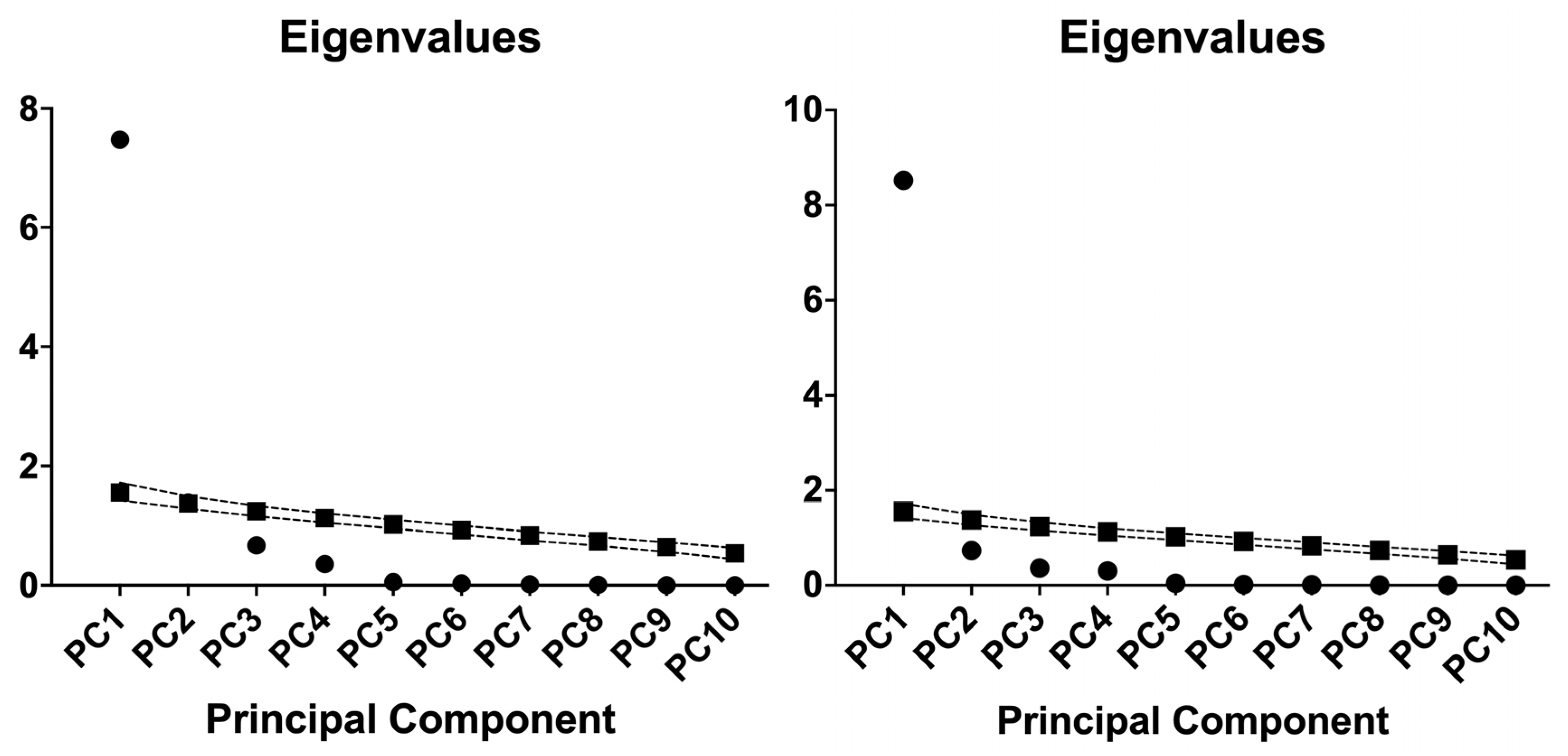
The principal component analysis revealed a dominant first component in both groups. In HD, PC1 had an eigenvalue of 7.47, accounting for 74.73% of the variance. PC2 had an eigenvalue of 1.39, explaining 13.86%, bringing the total explained variance to 88.60% when combined. Each additional component contributed less than 7% to the total variance.
Figure 2 shows the eigenvalue scree plots with the parallel-analysis reference for each cohort, documenting the one-component solution.
In DN, PC1 had an eigenvalue of 8.52, accounting for 85.20% of the variance. No later component surpassed 7.5%. The cumulative variance was 92.52% by PC2 and 96.16% by PC3. PC1 exhibited consistent loadings across various inflammatory indices. In HD, the most significant absolute loadings were for NLR (−0.982), SII (−0.977), PLR (−0.973), IIC (−0.973), SIRI (−0.967), and MLR (−0.956). AISI (−0.942) and MCVL (−0.882) also showed strong loadings. dNLR had a modest loading of −0.376, while NPR’s loading was close to zero at almost 0.080.
In DN, all indices loaded highly on PC1. IIC and NLR had the largest absolute loadings, −0.979 and −0.978. SIRI and SII followed, −0.965 and −0.965, with dNLR at −0.941 and MLR at −0.924. AISI and MCVL loaded at −0.915 and −0.910. NPR and PLR also contributed, −0.875 and −0.757.
These patterns may indicate a common latent dimension shared by the indices in both cohorts, stronger and more uniform in DN. The negligible PC1 loading of NPR in HD and the modest loading of dNLR in HD suggest cohort specific differences in how these markers co-vary with the broader inflammatory signal.

2.5. Multiple Linear Regressions

2.5.1. Multiple Linear Regressions of HD Cohort

Multiple Linear Regressions of HD Cohort for AOPP
Two predefined models were examined. Model 1 incorporated demographic and dialysis-related covariates: sex, dialysis vintage, membrane type, age, and BMI. Model 2 included laboratory and inflammation-related covariates: creatinine, albumin, the PCA inflammation score (PC-inflammation), CRP, and eGFR. All analyses employed ordinary least squares. The alternative, Model 2, was slightly favored based on the small-sample corrected Akaike Information Criterion (AICc) (probability 60.6% versus 39.4% for Model 1; ΔAICc = 0.86; likelihood ratio = 1.54), suggesting only modest evidence supporting Model 2 over the simpler model (Table 6 and Table 7).
Model 1 (clinical/dialysis covariates): Overall fit was not significant, with low explained variance (R2 = 0.072; F (5, 84) = 1.298, p = 0.273). No individual covariate reached significance: sex (β = 4.26, 95% CI −2.83 to 11.34, p = 0.236), dialysis vintage (β = 0.229 per month, −0.244 to 0.701, p = 0.339), membrane type (β = −6.31, −14.68 to 2.07, p = 0.138), age (β = −0.361 per year, −0.860 to 0.138, p = 0.154), and BMI (β = 0.032 per kg/m2, −0.641 to 0.704, p = 0.926). Multicollinearity was acceptable (all VIF ≤ 4.3). Residuals were non-normal by several tests (e.g., D’Agostino–Pearson K2 = 26.95, p < 0.0001).
Model 2 (laboratory/inflammation covariates): Overall fit remained modest (R2 = 0.081; F (5, 84) = 1.472, p = 0.208). Serum creatinine was the only independent predictor of AOPP (β = 2.47 per mg/dL, 95% CI 0.30 to 4.63, p = 0.026). eGFR showed a borderline positive association (β = 1.71, −0.23 to 3.65, p = 0.084). Albumin (β = −3.06, p = 0.515), PC-inflammation (β = 0.747, p = 0.306), and CRP (β = 0.031, p = 0.865) were not significant. Multicollinearity was low (VIFs ≤ 2.87). Residuals were non-normal (K2 = 60.38, p < 0.0001).
Interpretation: Models across different covariate sets accounted for approximately 7–8% of the variability in AOPP. After adjustment, higher serum creatinine levels were independently linked to increased AOPP. Other clinical and inflammatory markers, such as the composite PC-inflammation score and CRP, showed no association in this cohort. Due to residual non-normality and a small R2, effect sizes should be interpreted with caution; the use of robust or transformed models may be advisable in sensitivity analyses.
Multiple Linear Regressions of HD Cohort for TBARS
Two prespecified models were compared. Model 1 used clinical/dialysis covariates (sex, dialysis vintage, membrane type, age, BMI), while Model 2 used laboratory/inflammation covariates (creatinine, albumin, PCA-derived inflammation score, CRP, eGFR). Model 1 was preferred by AICc with an 80.9% probability versus 19.1% for Model 2 (probability ratio 4.231; ΔAICc = −2.885), although overall fit was modest (R2 = 0.086, F (5, 84) = 1.587, p = 0.173). Within Model 1, longer dialysis vintage was independently associated with lower TBARS (β = −0.2305 per month, 95% CI −0.4253 to −0.0357, p = 0.0209); sex, membrane type, age, and BMI were not significant. Model 2 performed worse (R2 = 0.056, F (5, 84) = 1.007, p = 0.419), and none of its covariates were significant (all p ≥ 0.137). Residuals in both models failed normality tests, so effect sizes should be interpreted with caution (Table 8 and Table 9).

2.5.2. Multiple Linear Regressions of T2DM-DN Cohort

Multiple Linear Regressions of T2DM-DN Cohort for AOPP
Two prespecified models were compared. Model 1 included demographic and disease–history covariates (sex, years since diabetes diagnosis, age, BMI). Model 2 included laboratory and inflammation covariates (albumin, HbA1c, PCA-derived inflammation score, CRP, eGFR). Model 2 was decisively preferred by AICc (probability 99.9% vs. 0.08% for Model 1; probability ratio 1220; ΔAICc = 14.21), and it explained more variance (R2 = 0.214) with a significant overall F test (F(5, 84) = 4.584, p = 0.0010). Within Model 2, the PC-inflammation score was the only independent predictor of AOPP (β = 1.134 per unit, 95% CI 0.533–1.734, p = 0.0003), while albumin, HbA1c, CRP, and eGFR were not significant. Model 1 fit was weak and non-significant (R2 = 0.0556; F(4, 85) = 1.250, p = 0.296) and none of its covariates reached significance. Residuals were non-normal in both models, so estimates should be interpreted with appropriate caution or confirmed in sensitivity analyses (Table 10 and Table 11).
Multiple Linear Regressions of T2DM-DN Cohort for TBARS
We compared two models. Model 1 used sex, years since diabetes diagnosis, age, and BMI; Model 2 used albumin, HbA1c, PCA-derived inflammation score, CRP, and eGFR. By AICc, Model 1 was preferred (probability 68.98% vs. 31.02% for Model 2; probability ratio 2.224; ΔAICc = −1.598), but both models fit poorly (Table 12 and Table 13).
Model 1: R2 = 0.021, overall F (4, 85) = 0.4639, p = 0.762. None of the covariates was significant (β [95% CI]; p): sex −0.231 [−1.627, 1.165]; p = 0.743; years since diagnosis 0.026 [−0.059, 0.111]; p = 0.544; age 0.013 [−0.067, 0.094]; p = 0.743; BMI −0.056 [−0.190, 0.079]; p = 0.413. Residuals failed normality (K2 = 56.23, A 2* = 7.423, W = 0.739, all p < 0.0001).
Model 2: R2 = 0.0295, overall F (5, 84) = 0.5114, p = 0.767. No covariate reached significance: albumin 0.119 (p = 0.881), HbA1c −0.102 (p = 0.515), PC-inflammation −0.128 (p = 0.446), CRP 0.025 (p = 0.641), eGFR 0.020 (p = 0.239). Residuals were again non-normal (K2 = 52.06, A 2* = 7.507, W = 0.745, all p < 0.0001).
In T2DM-DN, TBARS were not explained by the tested clinical or laboratory covariates; effect sizes are small and imprecise, and the non-normal residuals argue for cautious interpretation or robust/transform checks.

3. Discussion

This study revealed clear differences in how oxidative stress markers (AOPP, TBARS) relate to systemic inflammation in patients with DN versus those on HD. After controlling for age, sex, and serum albumin, the DN group displayed more significant and stable partial correlations, many of which stayed significant after FDR correction. Conversely, the HD group showed fewer and weaker associations after adjustments. These results provide valuable insight into how oxidative stress and inflammation interact across different stages of renal disease.
The stronger oxidative and inflammatory coupling in the DN group suggests that in early or moderate renal impairment, protein oxidation (reflected by AOPP) may act as a key mediator connecting metabolic dysfunction and immune activation. This aligns with mechanistic models of diabetic kidney disease, where hyperglycemia increases ROS formation, activates NADPH oxidases, and upregulates inflammatory cascades (e.g., NF-κB) that mutually amplify ROS production and renal damage [2,3,15]. The continued presence of associations after adjusting for albumin and demographic factors supports the idea that these links are biologically significant rather than influenced by nutritional or demographic differences.
In HD patients, the attenuation of correlations may reflect a more complex milieu. Patients on dialysis are subject to frequent fluctuations in biochemical parameters, interventions such as dialysis vintage, vascular access inflammation, and exposure to bioincompatible membranes. These external perturbations can introduce noise and weaken the detectability of underlying biological relationships. Indeed, oxidative stress in end-stage renal disease is multifactorial, influenced by uremic toxins, the dialysis procedure itself, comorbidities, and antioxidant deficits [1,7,16]. In such settings, a pure oxidative and inflammatory relationship may be overshadowed by episodic events or external stressors, which could explain why fewer associations survived FDR correction in HD.
Moreover, the relative absence of significant TBARS correlations in both groups suggests that lipid peroxidation might play a less consistent role in systemic inflammation, compared to protein oxidation. This differential pattern underscores the possibility that not all oxidative pathways contribute equally to immune activation in renal disease. Markers such as AOPP may better reflect the pathophysiologically relevant oxidative stress in renal patients, especially in DN, where oxidative protein damage has been implicated in glomerular and tubular injury [2,17,18].
In diabetic kidney disease, hyperglycemia induces excessive ROS through activation of NADPH oxidases, mitochondrial dysfunction, and the advanced glycation end products/receptor for AGEs (AGE–RAGE) pathway. This oxidative stress activates NF-κB signaling and cytokine release, including interleukin-6 (IL-6) and tumor necrosis factor-α (TNF-α), promoting chronic inflammation and renal injury. The strong partial correlations between AOPP and inflammation-related indices in our DN group support this mechanistic link, consistent with evidence that oxidative stress and inflammation form a self-amplifying cycle in diabetic nephropathy [2,19].
AOPP are considered more stable indicators of oxidative damage than lipid peroxidation markers like TBARS. AOPP reflect cumulative oxidative modification of plasma proteins and correlate closely with renal function decline and inflammatory cytokine activity [18,20]. The limited associations observed for TBARS in our study are consistent with evidence that the thiobarbituric acid assay lacks specificity and is influenced by multiple reactive carbonyl compounds, making it a less reliable indicator of lipid peroxidation compared to more stable markers such as F2-isoprostanes. According to Ito et al., TBARS can overestimate malondialdehyde levels due to interference from amino acids, bilirubin, and albumin, which may explain the variability and weaker correlations observed in clinical populations [21].
Sources of oxidative stress during HD include exposure to bioincompatible membranes, dialysate contamination, and intermittent ischemia–reperfusion phenomena. Furthermore, accumulation of uremic toxins and repetitive inflammatory stimuli contribute to a persistently activated yet dysregulated immune state [6]. These processes may weaken specific oxidative and inflammatory correlations and produce greater data variability.
In hemodialysis patients, elevated AOPP levels have been linked to chronic microinflammation and endothelial dysfunction, reflecting continuous oxidative stress induced by the dialysis procedure itself rather than metabolic imbalance [7,22,23,24].
Across both cohorts (patients on HD and DN), principal component analysis (PCA) revealed that ten hemogram-derived inflammatory indices grouped into a single latent axis of systemic inflammation (PC-inflammation) with a greater proportion of variance in DN than in HD, reflecting the central role of inflammation and oxidative stress in chronic kidney disease [25]. This pattern aligns with evidence that complete blood count (CBC)-based indices such as the NLR, PLR, SII, and SIRI effectively capture clinically relevant inflammatory burden across CKD severities. SII, in particular, has emerged as a promising biomarker of immune–inflammatory status in end-stage renal disease and hemodialysis populations [26,27,28,29,30].
In HD, AOPP (advanced oxidation protein products) showed an independent association with serum creatinine, a finding biologically plausible given uremic oxidative stress that both produces AOPP and may be further amplified by AOPP in a positive feedback loop. In DN, AOPP were independently linked to the composite PC-inflammation score, strengthening the connection between systemic inflammatory activation and protein oxidation in diabetic kidney disease. Beyond association, recent work suggests AOPP measurement has diagnostic utility for CKD among individuals with T2DM, supporting its translational relevance [9,31,32].
TBARS are widely used as a readout of lipid peroxidation but are method-sensitive and context-dependent, which can dilute associations in multivariable settings [33].
Methodologically, summarizing correlated biomarkers with PCA reduces multicollinearity and distills shared inflammatory variance, a strategy increasingly used in renal omics and cytokine studies, including HD cohorts [34,35].
Clinically, the replication of a one-factor inflammatory structure across HD and DN indicates that a single inflammatory pathway underpins various renal disease contexts; linking this pathway to AOPP in DN and to creatinine in HD suggests shared but cohort-specific oxidative mechanisms that could be targeted with anti-inflammatory and antioxidant therapies [25,32]. Meanwhile, the weaker and context-dependent signal for TBARS demonstrates that lipid peroxidation markers may require prolonged sampling and consideration of dialysis modality, membrane type, and adjunct therapies to establish consistent associations [36,37].
Our pattern shows that AOPP are linked to creatinine in HD and to the PC-inflammation score in DN, with weak or context-sensitive TBARS. This aligns with the review of AN69 membranes, which indicates that more biocompatible, adsorptive dialyzers reduce complement and cytokine activation, thereby decreasing carbonyl and oxidative stress in HD. This diminishes the inflammation’s additional effect on protein oxidation. In contrast, DN without extracorporeal modulation maintains a direct inflammation–protein oxidation pathway [38].

3.1. Strengths and Limitations

This study possesses several significant strengths that improve both the reliability and clarity of its results.
First, it offers a comparative analysis between two distinct clinical groups: patients with DN and those undergoing chronic HD. This dual-cohort approach encompasses various stages of renal dysfunction, enabling a direct study of the progression of oxidative and inflammatory interactions from metabolic injury to end-stage renal failure.
Second, the use of partial correlations adjusted for age, sex, and serum ALB reduces bias from key demographic and nutritional confounders known to influence both oxidative and inflammatory biomarkers. Including ALB as a covariate is especially important in nephrology, as hypoalbuminemia indicates both malnutrition and systemic inflammation.
Third, the application of FDR correction strengthens the statistical robustness of the reported associations, ensuring that the observed relationships are less likely to represent chance findings due to multiple testing.
Fourth, evaluating a broad panel of inflammatory indices (AISI, IIC, SII, SIRI, MCVL, NLR, MLR, PLR, dNLR, NPR) together with oxidative stress markers (AOPP, TBARS) provides a multidimensional insight into the redox–immune axis. This comprehensive approach, seldom used in renal populations, offers a wider perspective than studies focusing on a single marker and emphasizes inflammatory pathways linked with oxidative stress, even after adjustments.
Finally, by controlling for major clinical covariates and using quantitative visualization, the study offers a clear, reproducible framework for exploring redox–inflammation relationships in clinical nephrology.
Despite these strengths, some limitations are important to recognize. The cross-sectional design restricts the ability to determine causality; additional longitudinal research is needed to understand whether oxidative stress causes inflammation or is a result of it. While major confounders were accounted for, potential residual confounding from unmeasured factors—such as dialysis membrane composition, dialysate quality, medication use (including antioxidants and erythropoietin), or intercurrent infections—cannot be ruled out.
The sample size, especially in the HD cohort, may limit the power to detect smaller but biologically significant associations and restricts subgroup analyses by sex, dialysis duration, or comorbidity profile. Regarding biomarkers, AOPP and TBARS offer valuable but incomplete insights into oxidative stress pathways, whereas hematologic indices of inflammation are indirect and might not fully capture cytokine-mediated activity.
Finally, the study was conducted in a single-center population, which may limit its applicability to other clinical settings with different patient management strategies or dialysis technologies.
Overall, these strengths and limitations suggest that although the current findings offer a strong basis for understanding the stage-specific oxidative–inflammatory dynamics in kidney disease, they need to be validated with multicenter, long-term studies that include biochemical, cytokine, and metabolomic profiling.

3.2. Future Directions

Future research should focus on clarifying the sequence of events and mechanisms connecting oxidative stress and inflammation in the progression of chronic kidney disease. Long-term cohort studies tracking patients from early diabetic nephropathy to dialysis initiation can help determine if changes in oxidative protein damage lead to inflammatory activation or are simply downstream effects of kidney injury. Incorporating multi-omics strategies, such as redox proteomics, lipidomics, and targeted cytokine analysis, can help specify the biochemical nature of these links and uncover causal molecular pathways.
Since oxidative and inflammatory coupling seems strongest in diabetic nephropathy, interventional studies should investigate whether targeted antioxidant or anti-inflammatory treatments (such as NADPH oxidase inhibitors, Nrf2 activators, or advanced glycation end-product (AGE) pathway modulators) can break this feedback loop before permanent renal fibrosis occurs. In hemodialysis, where procedural and metabolic stressors are prevalent, research should focus on dialysis biocompatibility and oxidative load, testing strategies like ultrapure dialysate, vitamin E-coated membranes, and antioxidant supplements to lessen external oxidative stress.
Finally, future clinical translation will rely on developing standardized, reproducible biomarker panels that combine oxidative and inflammatory markers for disease monitoring. Such tools could assist in risk stratification, guiding therapy, and early detection of patients most likely to benefit from targeted redox treatments. Through integrated mechanistic, clinical, and interventional research, the oxidative and inflammatory axis may be transformed from a passive injury marker into an active therapeutic target in kidney disease.

4. Materials and Methods

4.1. Study Design and Setting

This cross-sectional, non-interventional, observational study was conducted over 6 months in the Nephrology Department of the University Hospital of Craiova, Romania, and the Outpatient Diabetes, Nutrition, and Metabolic Diseases Departments of the Filantropia Municipal Clinical Hospital, Craiova, Romania. The research included two patient cohorts: individuals with T2DM-DN and patients undergoing chronic HD. The study aimed to assess the relationship between oxidative stress and systemic inflammation in different stages of renal impairment (Figure 3).

4.2. Ethical Considerations

The protocol was approved by the Ethics Committee of the University Hospital of Craiova, Dolj, Romania (approval no. 6/10 January 2025) and by the Ethics Committee of the Filantropia Municipal Clinical Hospital Craiova, Dolj, Romania (approval no. 5024/4 March 2025). All participants provided written informed consent prior to enrollment, in accordance with the ethical principles outlined in the Declaration of Helsinki.

4.3. Participants

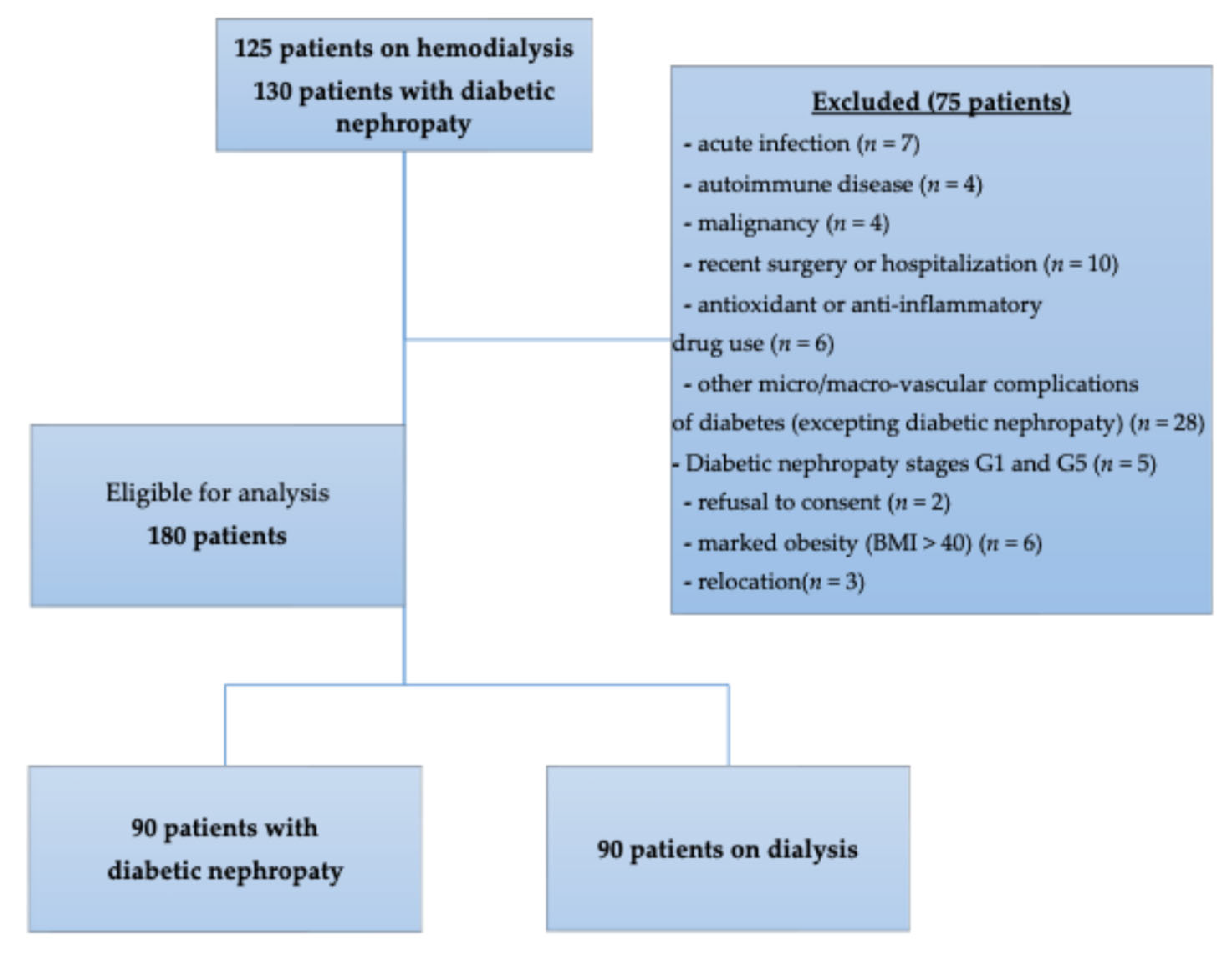
Eligible participants were adults with confirmed diagnoses of T2DM-DN or end-stage renal disease requiring maintenance hemodialysis for at least 6 months. We enrolled 130 patients with T2DM-DN and 125 patients on HD. Exclusion criteria included acute infection, autoimmune disease, malignancy, recent surgery or hospitalization (<3 months), antioxidant or anti-inflammatory drug use, other micro-/macrovascular complications of diabetes, marked obesity (BMI > 40), relocation, and refusal to consent. After applying the exclusion criteria, both cohorts had a final count of 90 patients. Demographic and clinical data, including age, sex, and BMI were recorded from medical files. The diagnosis of T2DM-DN or end-stage renal disease requiring maintenance hemodialysis were in accordance with American Diabetes Association (ADA), Kidney Disease: Improving Global Outcomes (KDIGO) [39,40]. Patients with T2DM-DN that were enrolled and classified into stages between G2 and G4 conform to KDIGO staging.

4.4. Evaluation of Inflammatory Status

In order to evaluate the inflammatory status, we utilized the formulas from Table 14.
Furthermore, eGFR was determined utilizing the serum creatinine-based CKD-EPI equation, as operationalized within the validated MDCalc calculator [45].

4.5. Sampling Procedure

During biological sampling, venous blood was drawn from each participant using two types of vacutainer tubes. For patients on ongoing HD, samples were collected before the procedure. Two additive-free tubes (Becton Dickinson Vacutainer, Franklin Lakes, NJ, USA), each holding roughly 5 mL of blood, were used for serum analyses. The samples were allowed to clot at room temperature and processed within four hours of collection. Centrifugation was carried out at 3000× g for 10 min using a Hermle centrifuge (Hermle AG, Gosheim, Germany).
The serum was meticulously separated to reduce hemolysis. A portion was divided into pre-labeled cryovials, hermetically sealed, and stored at temperatures from −20 °C to −80 °C to preserve protein integrity. Freeze–thaw cycles were strictly avoided. Before analysis, aliquots were brought to room temperature under controlled conditions. One aliquot was set aside for immunological assays, and the serum from the second vial was reserved for biochemical analyses.
For hematological analysis, a third blood sample was collected in an EDTA vacutainer tube (Becton Dickinson, Franklin Lakes, NJ, USA).

4.6. Laboratory Assays

Serum biochemical parameters were measured using the ARCHITECT C4000 automated chemistry analyzer (Abbott Diagnostics, Abbott Park, IL, USA), following standardized clinical laboratory protocols to ensure accuracy and reproducibility.
A comprehensive leukocyte differential count was performed using the MINDRAY BC-6800 hematology analyzer (Mindray, Shenzhen, China). This system combines flow cytometry and impedance technology (Coulter principle) to accurately identify and count leukocyte subsets. The method provides precise measurement of neutrophils, lymphocytes, monocytes, eosinophils, and basophils, offering a detailed leukocyte profile and supporting the calculation of inflammatory indices for further analysis.

4.7. Enzyme-Linked Immunosorbent Assay of AOPP and TBARS

At the Immunology Laboratory of the University of Medicine and Pharmacy of Craiova, serum concentrations of AOPP and TBARS were measured using the enzyme-linked immunosorbent assay (ELISA) method. We followed the protocol instructions to determine serum levels of AOPP and TBARS, employing ELISA kits from MyBioSource (San Diego, CA, USA) and using a standard optical analyzer set at a wavelength of 450 nm for the process (Table 15).

4.8. Statistical Assessment

The patient data extracted from medical records were systematically organized and curated using Microsoft Excel. Statistical analyses were conducted with GraphPad Prism 10.6.1 (GraphPad Software, San Diego, CA, USA) and R v4.3.2. The data distribution’s normality was assessed using the Shapiro–Wilk and D’Agostino’s tests. Parametric data were analyzed using Welch’s t-test, whereas non-parametric data were evaluated with the Mann–Whitney U test. Categorical variables are presented as percentages to depict frequency distributions. Partial correlation analyses were conducted to explore the relationships between oxidative stress markers (AOPP and TBARS) and inflammatory indices (AISI, IIC, SII, SIRI, MCVL, NLR, MLR, PLR, dNLR, NPR) while adjusting for age, sex, and serum ALB. The Benjamini–Hochberg FDR method was used to control for multiple testing. A two-tailed p-value < 0.05 was considered statistically significant before correction, and a q-value < 0.05 was considered significant after FDR adjustment. Values with 0.05 ≤ q < 0.10 were interpreted as trends toward significance. For MLR, normality of Residuals was assessed with: D’Agostino-Pearson omnibus (K2), Anderson-Darling (A 2*), Shapiro-Wilk (W).

5. Conclusions

AOPP and TBARS were markedly higher in hemodialysis than in diabetic nephropathy, supporting a stronger oxidative burden in end-stage disease. Both differences were large and statistically significant.
In diabetic nephropathy, AOPP showed consistent positive correlations with multiple inflammatory indices after adjustment and false discovery rate correction. This pattern indicates tight coupling between protein oxidation and systemic inflammation in this group. TBARS showed no adjusted associations. In hemodialysis, correlations between oxidative markers and inflammatory indices did not remain significant after correction. This suggests that dialysis-related factors and uremic toxicity weaken the oxidative inflammation link.
Principal-component analysis supported a single latent inflammatory axis in both cohorts, stronger in diabetic nephropathy. In regression models, AOPP associated independently with creatinine in hemodialysis and with the composite inflammation score in diabetic nephropathy. AOPP are a practical marker to track the inflammation related oxidative signal in diabetic nephropathy. Their association with creatinine in hemodialysis points to uremic load and dialysis milieu rather than inflammatory activation. These insights can guide monitoring, for example, pairing AOPP with CBC-derived indices in diabetic nephropathy and targeting dialysis biocompatibility and uremic control in hemodialysis.

Author Contributions

Conceptualization, M.-Z.A. and L.B.; methodology, M.V.B. and D.C.C.; software, D.C.C. and M.-E.C.; validation, M.-Z.A. and L.B.; formal analysis, A.-Ș.S.-I., V.P. and M.-E.C.; investigation, C.C.V., R.P. and D.-T.M.; resources, V.P. and R.P.; data curation, A.-Ș.S.-I., R.P. and D.-T.M.; writing—original draft preparation, D.C.C., M.-Z.A. and M.-E.C.; writing—review and editing, C.C.V., L.B. and M.V.B.; visualization, M.V.B. and M.-E.C.; supervision, M.-Z.A. and M.-E.C.; project administration, M.V.B. All authors have read and agreed to the published version of the manuscript.

Funding

The Article Processing Charges were funded by the University of Medicine and Pharmacy of Craiova, Romania.

Institutional Review Board Statement

The study was conducted following the Declaration of Helsinki and approved by the Ethics Committee of the University Hospital of Craiova, Romania (approval no. 6/10 January 2025) and by the Ethics Committee of the Clinical Municipal Hospital Filantropia Craiova (no. approval no. 5024/4 March 2025), Dolj, Romania.

Informed Consent Statement

Informed consent was obtained from all subjects involved in the study. Written informed consent was obtained from the patient(s) to publish this paper.

Data Availability Statement

The data used to support the findings of this study are available from the corresponding author upon reasonable request.

Acknowledgments

Lidia Boldeanu and Daniel Cosmin Caragea share equal contributions and status as main/first authors.

Conflicts of Interest

The authors declare no conflicts of interest.

References

  1. Gyurászová, M.; Gurecká, R.; Bábíčková, J.; Tóthová, Ľ. Oxidative Stress in the Pathophysiology of Kidney Disease: Implications for Noninvasive Monitoring and Identification of Biomarkers. Oxid. Med. Cell. Longev. 2020, 2020, 5478708. [Google Scholar] [CrossRef]
  2. Jin, Q.; Liu, T.; Qiao, Y.; Liu, D.; Yang, L.; Mao, H.; Ma, F.; Wang, Y.; Peng, L.; Zhan, Y. Oxidative stress and inflammation in diabetic nephropathy: Role of polyphenols. Front. Immunol. 2023, 14, 1185317. [Google Scholar] [CrossRef]
  3. Wang, N.; Zhang, C. Oxidative Stress: A Culprit in the Progression of Diabetic Kidney Disease. Antioxidants 2024, 13, 455. [Google Scholar] [CrossRef]
  4. Sagoo, M.K.; Gnudi, L. Diabetic nephropathy: Is there a role for oxidative stress? Free Radic. Biol. Med. 2018, 116, 50–63. [Google Scholar] [CrossRef]
  5. Joumaa, J.P.; Raffoul, A.; Sarkis, C.; Chatrieh, E.; Zaidan, S.; Attieh, P.; Harb, F.; Azar, S.; Ghadieh, H.E. Mechanisms, Biomarkers, and Treatment Approaches for Diabetic Kidney Disease: Current Insights and Future Perspectives. J. Clin. Med. 2025, 14, 727. [Google Scholar] [CrossRef] [PubMed]
  6. Liakopoulos, V.; Roumeliotis, S.; Gorny, X.; Dounousi, E.; Mertens, P.R. Oxidative Stress in Hemodialysis Patients: A Review of the Literature. Oxid. Med. Cell. Longev. 2017, 2017, 3081856. [Google Scholar] [CrossRef]
  7. Ling, X.C.; Kuo, K.-L. Oxidative stress in chronic kidney disease. Ren. Replace. Ther. 2018, 4, 53. [Google Scholar] [CrossRef]
  8. Arimura, N.; Watanabe, H.; Kato, H.; Imafuku, T.; Nakano, T.; Sueyoshi, M.; Chikamatsu, M.; Tokumaru, K.; Nagasaki, T.; Maeda, H.; et al. Advanced Oxidation Protein Products Contribute to Chronic-Kidney-Disease-Induced Adipose Inflammation through Macrophage Activation. Toxins 2023, 15, 179. [Google Scholar] [CrossRef]
  9. Vodošek Hojs, N.; Bevc, S.; Ekart, R.; Hojs, R. Oxidative Stress Markers in Chronic Kidney Disease with Emphasis on Diabetic Nephropathy. Antioxidants 2020, 9, 925. [Google Scholar] [CrossRef]
  10. Xue, K.; Wang, Y.; Wang, Y.; Fang, H. Advanced Oxidation Protein Product Promotes Oxidative Accentuation in Renal Epithelial Cells via the Soluble (Pro)renin Receptor-Mediated Intrarenal Renin-Angiotensin System and Nox4-H2O2 Signaling. Oxid. Med. Cell. Longev. 2021, 2021, 5710440. [Google Scholar] [CrossRef] [PubMed]
  11. Vinereanu, I.V.; Peride, I.; Niculae, A.; Tiron, A.T.; Caragheorgheopol, A.; Manda, D.; Checherita, I.A. The Relationship between Advanced Oxidation Protein Products, Vascular Calcifications and Arterial Stiffness in Predialysis Chronic Kidney Disease Patients. Medicina 2021, 57, 452. [Google Scholar] [CrossRef]
  12. Pasterk, L.; Lemesch, S.; Leber, B.; Trieb, M.; Curcic, S.; Stadlbauer, V.; Schuligoi, R.; Schicho, R.; Heinemann, A.; Marsche, G. Oxidized plasma albumin promotes platelet-endothelial crosstalk and endothelial tissue factor expression. Sci. Rep. 2016, 6, 22104. [Google Scholar] [CrossRef]
  13. Aguilar Diaz De Leon, J.; Borges, C.R. Evaluation of Oxidative Stress in Biological Samples Using the Thiobarbituric Acid Reactive Substances Assay. J. Vis. Exp. 2020, 159. [Google Scholar] [CrossRef]
  14. Trevisan, M.; Browne, R.; Ram, M.; Muti, P.; Freudenheim, J.; Carosella, A.M.; Armstrong, D. Correlates of Markers of Oxidative Status in the General Population. Am. J. Epidemiol. 2001, 154, 348–356. [Google Scholar] [CrossRef] [PubMed]
  15. Ma, X.; Ma, J.; Leng, T.; Yuan, Z.; Hu, T.; Liu, Q.; Shen, T. Advances in oxidative stress in pathogenesis of diabetic kidney disease and efficacy of TCM intervention. Ren. Fail. 2023, 45, 2146512. [Google Scholar] [CrossRef]
  16. Rapa, S.F.; Di Iorio, B.R.; Campiglia, P.; Heidland, A.; Marzocco, S. Inflammation and Oxidative Stress in Chronic Kidney Disease-Potential Therapeutic Role of Minerals, Vitamins and Plant-Derived Metabolites. Int. J. Mol. Sci. 2019, 21, 263. [Google Scholar] [CrossRef] [PubMed]
  17. Tanase, D.M.; Gosav, E.M.; Anton, M.I.; Floria, M.; Seritean Isac, P.N.; Hurjui, L.L.; Tarniceriu, C.C.; Costea, C.F.; Ciocoiu, M.; Rezus, C. Oxidative Stress and NRF2/KEAP1/ARE Pathway in Diabetic Kidney Disease (DKD): New Perspectives. Biomolecules 2022, 12, 1227. [Google Scholar] [CrossRef]
  18. Darenskaya, M.; Kolesnikov, S.; Semenova, N.; Kolesnikova, L. Diabetic Nephropathy: Significance of Determining Oxidative Stress and Opportunities for Antioxidant Therapies. Int. J. Mol. Sci. 2023, 24, 12378. [Google Scholar] [CrossRef]
  19. Winiarska, A.; Knysak, M.; Nabrdalik, K.; Gumprecht, J.; Stompór, T. Inflammation and Oxidative Stress in Diabetic Kidney Disease: The Targets for SGLT2 Inhibitors and GLP-1 Receptor Agonists. Int. J. Mol. Sci. 2021, 22, 10822. [Google Scholar] [CrossRef]
  20. Conti, G.; Caccamo, D.; Siligato, R.; Gembillo, G.; Satta, E.; Pazzano, D.; Carucci, N.; Carella, A.; Del Campo, G.; Salvo, A.; et al. Association of Higher Advanced Oxidation Protein Products (AOPPs) Levels in Patients with Diabetic and Hypertensive Nephropathy. Medicina 2019, 55, 675. [Google Scholar] [CrossRef]
  21. Ito, F.; Sono, Y.; Ito, T. Measurement and Clinical Significance of Lipid Peroxidation as a Biomarker of Oxidative Stress: Oxidative Stress in Diabetes, Atherosclerosis, and Chronic Inflammation. Antioxidants 2019, 8, 72. [Google Scholar] [CrossRef]
  22. Sung, C.-C.; Hsu, Y.-C.; Chen, C.-C.; Lin, Y.-F.; Wu, C.-C. Oxidative Stress and Nucleic Acid Oxidation in Patients with Chronic Kidney Disease. Oxidative Med. Cell. Longev. 2013, 2013, 301982. [Google Scholar] [CrossRef]
  23. Varan, H.I.; Dursun, B.; Dursun, E.; Ozben, T.; Suleymanlar, G. Acute effects of hemodialysis on oxidative stress parameters in chronic uremic patients: Comparison of two dialysis membranes. Int. J. Nephrol. Renov. Dis. 2010, 3, 39–45. [Google Scholar] [CrossRef] [PubMed]
  24. Dounousi, E.; Papavasiliou, E.; Makedou, A.; Ioannou, K.; Katopodis, K.P.; Tselepis, A.; Siamopoulos, K.C.; Tsakiris, D. Oxidative Stress Is Progressively Enhanced with Advancing Stages of CKD. Am. J. Kidney Dis. 2006, 48, 752–760. [Google Scholar] [CrossRef]
  25. Podkowińska, A.; Formanowicz, D. Chronic Kidney Disease as Oxidative Stress- and Inflammatory-Mediated Cardiovascular Disease. Antioxidants 2020, 9, 752. [Google Scholar] [CrossRef] [PubMed]
  26. Mancinelli, M.; Moscucci, F.; Cofini, V.; De Nino, A.L.; Bocale, R.; Savoia, C.; Baratta, F.; Desideri, G. Metabolic Determinants of Systemic Inflammation Dynamics During Hemodialysis: Insights from the Systemic Immune–Inflammation Index in a Single-Center Observational Study. Metabolites 2025, 15, 651. [Google Scholar] [CrossRef]
  27. Burlacu, A.; Namolovan, C.A.; Brinza, C.; Covic, A.; Floria, M.; Voroneanu, L.; Covic, A. Neutrophil-to-Lymphocyte Ratio (NLR)—Independent Prognostic Marker of Renal Function Decline in Chronic Kidney Disease: A Systematic Review and Meta-Analysis. J. Clin. Med. 2025, 14, 6822. [Google Scholar] [CrossRef]
  28. Mureșan, A.V.; Russu, E.; Arbănași, E.M.; Kaller, R.; Hosu, I.; Arbănași, E.M.; Voidăzan, S.T. The Predictive Value of NLR, MLR, and PLR in the Outcome of End-Stage Kidney Disease Patients. Biomedicines 2022, 10, 1272. [Google Scholar] [CrossRef]
  29. Kawalec, A.; Stojanowski, J.; Mazurkiewicz, P.; Choma, A.; Gaik, M.; Pluta, M.; Szymański, M.; Bruciak, A.; Gołębiowski, T.; Musiał, K. Systemic Immune Inflammation Index as a Key Predictor of Dialysis in Pediatric Chronic Kidney Disease with the Use of Random Forest Classifier. J. Clin. Med. 2023, 12, 6911. [Google Scholar] [CrossRef] [PubMed]
  30. Dopierała, M.; Nitz, N.; Król, O.; Wasicka-Przewoźna, K.; Schwermer, K.; Pawlaczyk, K. New and Emerging Biomarkers in Chronic Kidney Disease. Biomedicines 2025, 13, 1423. [Google Scholar] [CrossRef]
  31. Rysz, J.; Franczyk, B.; Ławiński, J.; Gluba-Brzózka, A. Oxidative Stress in ESRD Patients on Dialysis and the Risk of Cardiovascular Diseases. Antioxidants 2020, 9, 1079. [Google Scholar] [CrossRef]
  32. Villalpando-Sánchez, D.C.; Barajas-Medina, C.A.; Alvarez-Aguilar, C.; López-Ortiz, G.; Romero-Henríquez, L.F.; Gómez-García, A. Advanced Oxidative Protein Products Had a Diagnostic Accuracy for Identifying Chronic Kidney Disease in Adult Population. Metabolites 2024, 14, 37. [Google Scholar] [CrossRef]
  33. Deus, L.A.; Corrêa, H.d.L.; Neves, R.V.P.; Reis, A.L.; Honorato, F.S.; Silva, V.L.; Souza, M.K.; de Araújo, T.B.; de Gusmão Alves, L.S.; Sousa, C.V.; et al. Are Resistance Training-Induced BDNF in Hemodialysis Patients Associated with Depressive Symptoms, Quality of Life, Antioxidant Capacity, and Muscle Strength? An Insight for the Muscle–Brain–Renal Axis. Int. J. Environ. Res. Public Health 2021, 18, 11299. [Google Scholar] [CrossRef]
  34. Kozlowska, L.; Jagiello, K.; Ciura, K.; Sosnowska, A.; Zwiech, R.; Zbrog, Z.; Wasowicz, W.; Gromadzinska, J. The Effects of Two Kinds of Dietary Interventions on Serum Metabolic Profiles in Haemodialysis Patients. Biomolecules 2023, 13, 854. [Google Scholar] [CrossRef] [PubMed]
  35. Santos, I.S.R.; Martin-Pastor, M.; Tavares Júnior, A.G.; Queiroz, K.A.; da Silva Sólon, L.G.; Sousa, F.F.O.d. Metabolomic Profile and Its Correlation with the Plasmatic Levels of Losartan, EXP3174 and Blood Pressure Control in Hypertensive and Chronic Kidney Disease Patients. Int. J. Mol. Sci. 2023, 24, 9832. [Google Scholar] [CrossRef]
  36. Chaghouri, P.; Maalouf, N.; Peters, S.L.; Nowak, P.J.; Peczek, K.; Zasowska-Nowak, A.; Nowicki, M. Two Faces of Vitamin C in Hemodialysis Patients: Relation to Oxidative Stress and Inflammation. Nutrients 2021, 13, 791. [Google Scholar] [CrossRef] [PubMed]
  37. Navarro-García, J.A.; Rodríguez-Sánchez, E.; Aceves-Ripoll, J.; Abarca-Zabalía, J.; Susmozas-Sánchez, A.; González Lafuente, L.; Bada-Bosch, T.; Hernández, E.; Mérida-Herrero, E.; Praga, M.; et al. Oxidative Status before and after Renal Replacement Therapy: Differences Between Conventional High Flux Hemodialysis and on-Line Hemodiafiltration. Nutrients 2019, 11, 2809. [Google Scholar] [CrossRef] [PubMed]
  38. Kobayashi, S.; Ohtake, T. The Characteristics of Dialysis Membranes: Benefits of the AN69 Membrane in Hemodialysis Patients. J. Clin. Med. 2023, 12, 1123. [Google Scholar] [CrossRef]
  39. American Diabetes Association Professional Practice Committee. 2. Diagnosis and Classification of Diabetes: Standards of Care in Diabetes—2024. Diabetes Care 2023, 47, S20–S42. [Google Scholar] [CrossRef]
  40. Stevens, P.E.; Ahmed, S.B.; Carrero, J.J.; Foster, B.; Francis, A.; Hall, R.K.; Herrington, W.G.; Hill, G.; Inker, L.A.; Kazancıoğlu, R.; et al. KDIGO 2024 Clinical Practice Guideline for the Evaluation and Management of Chronic Kidney Disease. Kidney Int. 2024, 105, S117–S314. [Google Scholar] [CrossRef]
  41. Xie, H.; Ruan, G.; Wei, L.; Deng, L.; Zhang, Q.; Ge, Y.; Song, M.; Zhang, X.; Lin, S.; Liu, X.; et al. The inflammatory burden index is a superior systemic inflammation biomarker for the prognosis of non-small cell lung cancer. J. Cachexia Sarcopenia Muscle 2023, 14, 869–878. [Google Scholar] [CrossRef] [PubMed]
  42. Șerban, R.E.; Popescu, D.M.; Boldeanu, M.V.; Florescu, D.N.; Șerbănescu, M.S.; Șandru, V.; Panaitescu-Damian, A.; Forțofoiu, D.; Șerban, R.C.; Gherghina, F.L.; et al. The Diagnostic and Prognostic Role of Inflammatory Markers, Including the New Cumulative Inflammatory Index (IIC) and Mean Corpuscular Volume/Lymphocyte (MCVL), in Colorectal Adenocarcinoma. Cancers 2025, 17, 990. [Google Scholar] [CrossRef] [PubMed]
  43. Poenariu, I.S.; Boldeanu, L.; Ungureanu, B.S.; Caragea, D.C.; Cristea, O.M.; Pădureanu, V.; Siloși, I.; Ungureanu, A.M.; Statie, R.-C.; Ciobanu, A.E.; et al. Interrelation of Hypoxia-Inducible Factor-1 Alpha (HIF-1 α) and the Ratio between the Mean Corpuscular Volume/Lymphocytes (MCVL) and the Cumulative Inflammatory Index (IIC) in Ulcerative Colitis. Biomedicines 2023, 11, 3137. [Google Scholar] [CrossRef] [PubMed]
  44. Lin, H.; Bai, Z.; Wu, Q.; Chu, G.; Zhang, Y.; Guo, X.; Qi, X. Inflammatory Indexes for Assessing the Severity and Disease Progression of Ulcerative Colitis: A Single-Center Retrospective Study. Front. Public Health 2022, 10, 851295. [Google Scholar] [CrossRef]
  45. CKD-EPI Equations for Glomerular Filtration Rate (GFR). Available online: https://www.mdcalc.com/calc/3939/ckd-epi-equations-glomerular-filtration-rate-gfr#evidence (accessed on 25 September 2025).
  46. AOPP Elisa Kit :: Human Advanced Oxidation Protein Products ELISA Kit. Available online: https://www.mybiosource.com/aopp-human-elisa-kits/advanced-oxidation-protein-products/722252 (accessed on 25 September 2025).
  47. TBARS Elisa Kit :: Human Thiobarbituric Acid Reactive Substance (TBARS) ELISA Kit. Available online: https://www.mybiosource.com/human-elisa-kits/thiobarbituric-acid-reactive-substance-tbars/166987 (accessed on 25 September 2025).
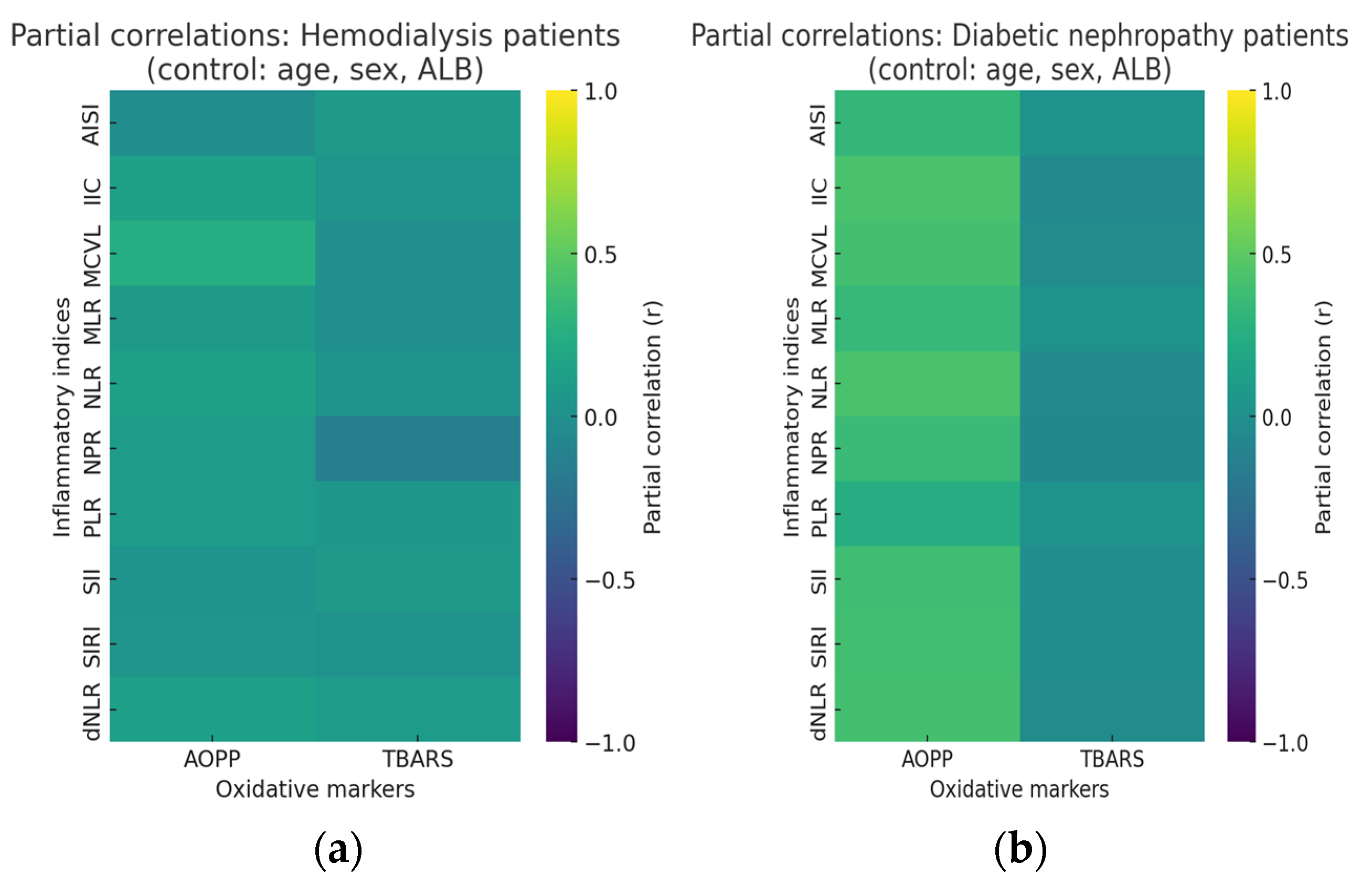
Figure 1. Partial correlation heatmaps between oxidative stress and inflammatory markers in diabetic nephropathy and hemodialysis patients after adjustment for age, sex, and serum ALB. The color scale represents the strength and direction of partial correlations (r), ranging from −1 to +1. In the diabetic nephropathy group, AOPP shows consistent positive correlations with several inflammatory indices, suggesting a close link between protein oxidation and systemic inflammation. In the hemodialysis group, correlations appear weaker and more variable, indicating a less stable oxidative and inflammatory relationship likely influenced by treatment-related and metabolic factors. (a) Partial correlations of HD patients, (b) partial correlations of T2DM-DN patients.
Ijms 26 10670 g001
Figure 2. Eigenvalue scree plots for HD (left), T2DM-DN (right). Scree plots with parallel analysis for PCA of ten inflammatory indices in two cohorts. Circles: Eigenvalue (from data). Squares: Eigenvalue (from Parallel Analysis). The figure shows a clear elbow at the first component in both cohorts. Eigenvalues drop sharply after PC1. In DN, only PC1 exceeds 1.0, with 85.20% variance explained. The curve flattens from PC2 onward. In HD, PC1 and PC2 exceed 1.0. PC1 explains 74.73% and PC2 adds 13.86%. From PC3 onward, each component contributes under 7%. These scree profiles indicate a dominant general component, stronger in DN. The loading patterns support this. In DN, all indices load highly on PC1, for example, IIC −0.979, NLR −0.978, SIRI −0.965, dNLR −0.941. In HD, PC1 is also coherent, but dNLR shows a weaker loading, −0.376, and NPR is near zero, approximately 0.080. This heterogeneity in HD aligns with the presence of a small secondary component. Overall, the figure supports a primarily unidimensional structure for these indices, most clearly in DN. The HD scree curve and loadings indicate a strong first component with limited residual structure. These observations are consistent with the variance profiles reported in Table 4.
Ijms 26 10670 g002
Figure 3. Flowchart of patient inclusion.
Ijms 26 10670 g003
Table 1. Demographic, clinical, and paraclinical characteristics.
ParameterHD
90 Patients
T2DM-DN
90 Patients
p-Value
Age (years)
[median (range)]
61
(23–89)
67
(40–83)
0.03 *
Sex (n)
Male/Female
43/4748/420.454
Residence (n)
Urban/Rural
24/6660/30<0.0001 *
Weight (kg)
[median (range)]
74.25
(39–103)
84.50
(45–137)
<0.0001 *
Height (m)
[median (range)]
1.66
(1.19–1.98)
1.68
(1.47–1.89)
0.281
BMI (kg/m2)
[median (range)]
26.40
(17.33–35.43)
31.11
(20.82–45.55)
<0.0001 *
TG (mg/dL)
[median (range)]
133
(49–401)
145
(57–397)
0.668
TC (mg/dL)
[median (range)]
171.50
(92–291)
162
(84.00–364)
0.777
WBC (×103/μL)
[median (range)]
6.77
(2.72–10.30)
8.42
(4.90–25.36)
<0.0001 *
NEU (×103/μL)
[median (range)]
4.20
(1.12–7.20)
5.23
(2.68–23.67)
<0.0001 *
LYM (×103/μL)
[median (range)]
1.59
(0.20–3.22)
1.92
(0.28–6.05)
0.0005 *
BAS (×103/μL)
[median (range)]
0.05
(0.00–0.17)
0.04
(0.01–0.40)
0.0006 *
EOS (×103/μL)
[median (range)]
0.20
(0.00–0.82)
0.16
(0.01–0.76)
0.012 *
MON (×103/μL)
[median (range)]
0.60
(0.31–1.77)
0.58
(0.20–1.19)
0.211
RBC (×103/μL)
(Mean ± SD)
3.60 ± 0.544.21 ± 0.67<0.0001 *
HCT (%)
(Mean ± SD)
34.21 ± 4.5737.42 ± 6.710.0003 *
HGB (g/dL)
[median (range)]
10.90
(7.90–14.20)
12.65
(8.10–17.30)
<0.0001 *
MCV (fL)
[median (range)]
96.60
(64.60–125.00)
89.35
(69.00–106.30)
<0.0001 *
PLT (×103/μL)
[median (range)]
192.00
(57.20–438.00)
240.50
(116.00–573.00)
<0.0001 *
Creatinine (mg/dL)
[median (range)]
8.81
(3.25–16.00)
1.09
(0.59–2.83)
<0.0001 *
Urea (mg/dL)
[median (range)]
135.90
(36.38–387.30)
52.00
(24.00–159.00)
<0.0001 *
eGFR (mL/min/1.73 m2)
CKD-EPI
[median (range)]
5.51
(2.14–20.81)
57.39
(16.55–89.96)
<0.0001 *
Na
(Mean ± SD)
138.6 ± 2.90137.90 ± 4.040.198
K
[median (range)]
4.80
(2.50–7.80)
4.55
(3.10–7)
0.006 *
Cl
[median (range)]
105.50
(99.00–122.00)
102
(90.00–110)
<0.0001 *
ALT (mg/dL)
[median (range)]
16
(6.00–150)
22
(7.00–136)
0.001 *
AST (mg/dL)
[median (range)]
16
(4.00–186)
21.39
(9.85–65.43)
<0.0001 *
Hemodialysis Vintage
(months)
48 ± 22--
Membrane type
(Synthetic high-flux/Synthetic low-flux) (n/%)
-77/13
85.56%/14.44%
-
Years of diagnosis
(years)
-12.06 ± 8.23-
HbA1c (%)-9.57 ± 2.19-
Demographic, anthropometric, hematological, renal, and biochemical characteristics of patients with hemodialysis (HD) and type 2 diabetes mellitus with diabetic nephropathy (T2DM-DN). Data are presented as median (range) or mean ± standard deviation, as appropriate. Significant differences (p < 0.05) indicate distinct metabolic and renal profiles between groups, reflecting advanced renal dysfunction in HD and higher metabolic burden in T2DM-DN. BMI: body mass index; TG: triglycerides; TC: total cholesterol; WBC: white blood cells; NEU: neutrophils; MON: monocytes; LYM: lymphocytes; EOS: eosinophils; BAS: basophils; RBC: red blood cells; HCT: hematocrit; HGB: hemoglobin; MCV: mean corpuscular volume; PLT: plaquettes; eGFR: estimated glomerular filtration rate; CKD-EPI: Chronic Kidney Disease Epidemiology Collaboration; ALT: alanine transaminase; AST: aspartate aminotransferase; Na: Sodium; K: Potassium; Cl: Chloride; *: reached the significant differences (p < 0.05).
Table 2. Levels of AOPP, TBARS, and inflammatory status.
ParameterHDT2DM-DNp-ValueParameterHDT2DM-DNp-Value
CRP (mg/dL)
[median (range)]
4.50
(0.30–49.40)
1.08
(0.02–30.42)
<0.0001 *AISI
[median (range)]
317.30
(24.98–6087)
355.80
(69.36–4707)
0.09
ALB (g/dL)
[median (range)]
4.10
(3.20–4.80)
4.40
(3.50–5.30)
<0.0001 *SIRI
[median (range)]
1.67
(0.30–21.97)
1.43
(0.32–39.47)
0.339
IIC
[median (range)]
3.72
(1.44–34.66)
3.00
(1.19–59.68)
0.02 *SII
[median (range)]
505.20
(77.83–6745)
603.30
(192.70–6272)
0.002 *
NPR
[median (range)]
22.25
(9.59–72.99)
21.75
(8.67–130.50)
0.775NLR
[median (range)]
2.57
(0.95–24.35)
2.56
(1.00–54.07)
0.06
MCVL
[median (range)]
61.42
(23.99–434)
46.28
(13.60–315.40)
<0.0001 *MLR
[median (range)]
0.39
(0.11–4.51)
0.28
(0.10–2.61)
<0.0001 *
AOPP
[median (range)]
25.80
(2.48–50)
5.06
(2.29–25.79)
<0.0001 *PLR
[median (range)]
128.40
(45.32–1385)
124.70
(44.96–521.90)
0.485
TBARS
[median (range)]
8.49
(0.53–29.94)
1.89
(0.31–15.78)
<0.0001 *dNLR
[median (range)]
1.66
(0.70–4.25)
1.81
(0.86–14.56)
0.03 *
Inflammatory and oxidative stress parameters in HD and T2DM-DN patients. Variables include systemic inflammation indices (AISI, IIC, SII, SIRI, NLR, MLR, PLR, dNLR, NPR) and oxidative stress markers (AOPP, TBARS). Data are expressed as median (range). HD patients exhibited higher CRP, AOPP, TBARS, and MCVL levels, indicating a more pronounced oxidative and inflammatory state, while T2DM-DN patients showed higher SII values, reflecting metabolic inflammation. *: reached the significant differences (p < 0.05); AOPP: advanced oxidation protein products; TBARS: thiobarbituric acid reactive substances; ALB: albumin; CRP: C-reactive protein; AISI: aggregate index of systemic inflammation; IIC: immune inflammation coefficient; SII: systemic immune–inflammation index; SIRI: systemic inflammation response index; MCVL: ratio between mean corpuscular volume and lymphocytes; NLR: neutrophil-to-lymphocyte ratio; MLR: monocyte-to-lymphocyte ratio; PLR: platelet-to-lymphocyte ratio; dNLR: derived neutrophil-to-lymphocyte ratio; NPR: neutrophil-to-platelet ratio.
Table 3. Partial correlations of AOPP and TBARS and inflammatory status.
ParameterNumberr-Valuep-Valueq-ValueParameterNumberr-Valuep-Valueq-Value
HD
AOPPAISI90−0.0180.8640.911TBARSAISI900.0830.4390.798
IIC900.1480.1670.798IIC900.0410.7040.911
SII900.0360.7410.911SII900.0850.4310.798
SIRI900.0430.6910.911SIRI900.0190.8630.911
MCVL900.2380.024 *0.489MCVL90−0.0150.8860.911
NLR900.1470.1700.798NLR900.0190.8620.911
MLR900.0850.4280.798MLR90−0.0120.9120.911
PLR900.0890.4070.798PLR900.0670.5300.882
dNLR900.1310.2190.798dNLR900.0920.3890.798
NPR900.0860.4230.798NPR90−0.1410.1870.798
T2DM-DN
AOPPAISI900.3250.001 *0.004TBARSAISI900.0180.8640.882
IIC900.428<0.0001 *0.0002IIC90−0.0600.5740.882
SII900.3840.0002 *0.0006SII90−0.0210.8470.882
SIRI900.3910.0001 *0.0005SIRI90−0.0280.7970.882
MCVL900.4010.0001 *0.0004MCVL90−0.0330.7600.882
NLR900.429<0.0001 *0.0002NLR90−0.0550.6070.882
MLR900.3420.001 *0.002MLR900.0160.8820.882
PLR900.2480.018 *0.03PLR900.0180.8680.882
dNLR900.4040.0001 *0.0004dNLR90−0.0390.7190.882
NPR900.3530.0007 *0.001NPR90−0.0750.4820.876
Partial correlations between oxidative stress markers (AOPP and TBARS) and inflammatory indices in HD and T2DM-DN patients after adjustment for age, sex, and serum ALB. Correlation coefficients (r), unadjusted p-values, and false discovery rate-adjusted q-values (Benjamini–Hochberg correction) are presented. In the T2DM-DN group, AOPP shows moderate positive correlations with multiple inflammatory markers (IIC, SII, SIRI, MCVL, NLR, MLR, PLR, dNLR, and NPR), indicating a robust link between protein oxidation and systemic inflammation. No correlations remained significant after adjustment in the HD group, suggesting that oxidative and inflammatory coupling weakens in advanced renal failure. *: reached the significant differences (p < 0.05)
Table 4. Eigenvalues and variance explained for each component, PC1 to PC10, with cumulative variance.
ComponentEigenvalueVariance
Explained (%)
Cumulative Proportion
of Variance (%)
ComponentEigenvalueVariance
Explained (%)
Cumulative Proportion
of Variance (%)
HDT2DM-DN
PC17.47374.73%74.73%PC18.5285.20%85.20%
PC21.38613.86%88.60%PC20.73277.33%92.52%
PC30.67266.73%95.32%PC30.36363.64%96.16%
PC40.35743.57%98.90%PC40.30533.05%99.21%
PC50.0530.53%99.43%PC50.04280.43%99.64%
PC60.02970.30%99.72%PC60.01610.16%99.80%
PC70.01690.17%99.89%PC70.01260.13%99.93%
PC80.00760.08%99.97%PC80.00560.06%99.98%
PC90.00190.02%99.99%PC90.00110.01%99.99%
PC100.00120.01%100.00%PC100.00070.01%100.00%
Eigenvalues and variance explained by principal components in the HD and DN cohorts. The table lists eigenvalues for PC1 to PC10, percent variance, and cumulative percent variance, separately for each cohort. Variables were z standardized, and PCA used the correlation matrix. Parallel analysis with 1000 simulations guided component retention. Each cohort contributed 90 complete cases.
Table 5. Loadings of the ten indices on PC1.
VariableAISIIICSIISIRIMCVLNLRMLRPLRdNLRNPR
PC1 loading-HD−0.9415−0.9728−0.9769−0.9674−0.8821−0.9819−0.9558−0.9731−0.37620.0798
PC1 loading-T2DM-DN−0.9149−0.9791−0.9647−0.9653−0.9095−0.9783−0.924−0.7565−0.9409−0.8751
PC1 loadings of the ten inflammatory indices in the HD and DN cohorts. The table reports loadings for AISI, IIC, SII, SIRI, MCVL, NLR, MLR, PLR, dNLR, and NPR, estimated from PCA of the correlation matrix with z standardized inputs. Loadings quantify the strength and direction of association with PC1, higher absolute values indicate stronger contributions. The sign of loadings is arbitrary and does not affect interpretation. Each cohort included 90 complete cases.
Table 6. Model comparison and analysis of variance for multiple linear regression of AOPP in hemodialysis patients.
Analysis of Variance SS DF MS F (DFn, DFd) p Value
Model 1Regression17435348.7F (5, 84) = 1.2980.2728
Sex-HD383.41383.4F (1, 84) = 1.4260.2357
HD vintage248.31248.3F (1, 84) = 0.92400.3392
Membrane-HD602.81602.8F (1, 84) = 2.2430.1380
Age-HD555.31555.3F (1, 84) = 2.0660.1543
BMI-HD2.3412.34F (1, 84) = 0.0087060.9259
Model 2Regression19585391.7F (5, 84) = 1.4720.2078
Creatinine-HD136611366F (1, 84) = 5.1310.0261
ALB-HD114.11114.1F (1, 84) = 0.42860.5145
PC-inflammation282.31282.3F (1, 84) = 1.0610.3060
CRP-HD7.80317.803F (1, 84) = 0.029320.8645
eGFR-HD816.11816.1F (1, 84) = 3.0660.0836
Two prespecified models were evaluated. Model 1 covariates: sex, dialysis vintage, membrane type, age, BMI. Model 2 covariates: creatinine, albumin, PCA-derived inflammation score (PC-inflammation), CRP, eGFR. The table reports SS, DF, MS, F, and two-sided p for the overall model and for each term; AICc model comparison favored Model 2 (probability 0.606 vs. 0.394; ΔAICc = 0.861). Residual df = 84. Abbreviations: AOPP, advanced oxidation protein products; HD, hemodialysis; BMI, body mass index; CRP, C-reactive protein; eGFR, estimated glomerular filtration rate. Overall fits were modest and not significant (Model 1 F (5, 84) = 1.298, p = 0.2728; Model 2 F (5, 84) = 1.472, p = 0.2078); AICc provided only weak preference for Model 2 over Model 1. SS: Sum of Squares; DF: Degrees of Freedom; MS: Mean Square; DFn: numerator DF; DFd: denominator DF.
Table 7. Parameter estimates for multiple linear regression of AOPP in hemodialysis patients.
VariableEstimate95% CI (Profile Likelihood)|t|p Value
Model 1β0Intercept40.5518.11 to 62.983.5940.0005
β1Sex-HD4.256−2.830 to 11.341.1940.2357
β2HD vintage0.2286−0.2443 to 0.70140.96130.3392
β3Membrane-HD−6.305−14.68 to 2.0661.4980.138
β4Age-HD−0.3607−0.8598 to 0.13831.4380.1543
β5BMI-HD0.03156−0.6411 to 0.70420.093310.9259
Model 2β0Intercept10.15−40.08 to 60.380.40170.6889
β1Creatinine-HD2.4670.3011 to 4.6322.2650.0261
β2ALB-HD−3.055−12.34 to 6.2260.65470.5145
β3PC-inflammation0.7471−0.6956 to 2.1901.030.306
β4CRP-HD0.03098−0.3288 to 0.39080.17120.8645
β5eGFR-HD1.708−0.2318 to 3.6481.7510.0836
Unstandardized coefficients (β), profile-likelihood 95% CIs, |t|, and two-sided p values are shown for both models. In Model 1, none of the clinical/dialysis covariates was significant. In Model 2, creatinine was the only independent predictor of AOPP (β = 2.467 per mg/dL, 95% CI 0.301–4.632, p = 0.026), while albumin, PC-inflammation, CRP, and eGFR were not significant (eGFR borderline, p = 0.084). |t|, absolute Student’s t statistic (|β/SE|; degrees of freedom = residual df); PC-inflammation is the first principal-component score summarizing the 10 inflammatory indices.
Table 8. Model comparison and analysis of variance for multiple linear regression of TBARS in hemodialysis patients.
Analysis of VarianceSSDFMSF (DFn, DFd)p Value
Model 1Regression361.8572.37F (5, 84) = 1.5870.1727
Sex-HD25.99125.99F (1, 84) = 0.56990.4524
HD vintage252.61252.6F (1, 84) = 5.5380.0209
Membrane-HD0.0915510.09155F (1, 84) = 0.0020070.9644
Age-HD118.81118.8F (1, 84) = 2.6040.1103
BMI-HD1.29711.297F (1, 84) = 0.028440.8665
Model 2Regression237547.41F (5, 84) = 1.0070.4190
Creatinine-HD0.0634110.06341F (1, 84) = 0.0013460.9708
ALB-HD106.21106.2F (1, 84) = 2.2560.1369
PC-inflammation5.30615.306F (1, 84) = 0.11270.7380
CRP-HD75.69175.69F (1, 84) = 1.6070.2084
eGFR-HD21.49121.49F (1, 84) = 0.45640.5012
Two models were evaluated. Model 1 covariates: sex, dialysis vintage, membrane type, age, BMI. Model 2 covariates: creatinine, albumin, PCA-derived inflammation score (PC-inflammation), CRP, eGFR. The table reports SS, DF, MS, F, and two-sided p for the overall model and each term, plus AICc model comparison statistics (Model 1 probability 80.88%, Model 2 probability 19.12%, probability ratio 4.231, ΔAICc −2.885; residual df 84). TBARS, thiobarbituric acid reactive substances; HD, hemodialysis; BMI, body mass index; CRP, C-reactive protein; eGFR, estimated glomerular filtration rate.
Table 9. Parameter estimates for multiple linear regression of TBARS in hemodialysis patients.
VariableEstimate95% CI (Profile Likelihood)|t|p Value
Model 1β0Intercept10.871.627 to 20.112.3390.0217
β1Sex-HD1.108−1.811 to 4.0280.75490.4524
β2HD vintage−0.2305−0.4253 to −0.035722.3530.0209
β3Membrane-HD0.0777−3.371 to 3.5260.04480.9644
β4Age-HD0.1668−0.03876 to 0.37241.6140.1103
β5BMI-HD0.0235−0.2536 to 0.30060.16860.8665
Model 2β0Intercept−2.857−23.99 to 18.270.26890.7887
β1Creatinine-HD0.01681−0.8940 to 0.92760.036690.9708
β2ALB-HD2.948−0.9556 to 6.8521.5020.1369
β3PC-inflammation0.1024−0.5044 to 0.70930.33570.738
β4CRP-HD−0.0965−0.2478 to 0.054861.2680.2084
β5eGFR-HD0.2772−0.5388 to 1.0930.67560.5012
Unstandardized coefficients (β), profile-likelihood 95% CIs, |t|, and two-sided p values are shown for each model. In Model 1, only dialysis vintage was significant (β = −0.2305 per month, 95% CI −0.4253 to −0.0357, p = 0.0209). In Model 2, creatinine, albumin, PC-inflammation, CRP, and eGFR were not significant. PC-inflammation is the first principal-component score summarizing the 10 inflammatory indices.
Table 10. Model comparison and analysis of variance for multiple linear regression of AOPP in diabetic nephropathy.
Analysis of VarianceSSDFMSF (DFn, DFd)p Value
Model 1Regression198.3449.57F (4, 85) = 1.2500.2960
Sex-DN2.67212.672F (1, 85) = 0.067390.7958
Years of diagnosis-DN20.49120.49F (1, 85) = 0.51670.4742
Age-DN87.12187.12F (1, 85) = 2.1970.1419
BMI-DN92.49192.49F (1, 85) = 2.3330.1304
Model 2Regression764.95153F (5, 84) = 4.5840.0010
ALB-DN29.13129.13F (1, 84) = 0.87270.3529
HbA1c-DN59.15159.15F (1, 84) = 1.7720.1867
PC-inflammation470.51470.5F (1, 84) = 14.100.0003
CRP-DN0.0451310.04513F (1, 84) = 0.0013520.9708
eGFR-DN0.672910.6729F (1, 84) = 0.020160.8874
Two models were evaluated. Model 1 covariates: sex, years since diabetes diagnosis, age, BMI. Model 2 covariates: albumin, HbA1c, PCA-derived inflammation score (PC-inflammation), CRP, eGFR. Shown are SS, DF, MS, F, and two-sided p for the overall model and each term, plus AICc model comparison (Model 2 probability 99.92%, Model 1 probability 0.0819%, probability ratio 1220, ΔAICc 14.21; residual df 84–85). Abbreviations: AOPP, advanced oxidation protein products; DN, diabetic nephropathy; BMI, body mass index; CRP, C-reactive protein; eGFR, estimated glomerular filtration rate. Model 2 fit the data significantly better; Model 1 showed a poor fit and no significant terms.
Table 11. Parameter estimates for multiple linear regression of AOPP in diabetic nephropathy.
VariableEstimate95% CI (Profile Likelihood)|t|p Value
Model 1β0Intercept8.702−4.840 to 22.241.2780.2049
β1Sex-DN−0.3596−3.114 to 2.3940.25960.7958
β2Years of diagnosis-DN0.06061−0.1070 to 0.22820.71880.4742
β3Age-DN−0.118−0.2764 to 0.040291.4820.1419
β4BMI-DN0.2035−0.06140 to 0.46831.5270.1304
Model 2β0Intercept3.975−10.43 to 18.380.54870.5847
β1ALB-DN1.342−1.514 to 4.1970.93420.3529
β2HbA1c-DN−0.3768−0.9396 to 0.18601.3310.1867
β3PC-inflammation1.1340.5333 to 1.7343.7550.0003
β4CRP-DN0.0035−0.1856 to 0.19260.036770.9708
β5eGFR-DN−0.0042−0.06343 to 0.054980.1420.8874
Unstandardized coefficients (β), profile-likelihood 95% CIs, |t|, and two-sided p values are reported for both models. In Model 2, PC-inflammation independently predicted higher AOPP (β = 1.134, 95% CI 0.533–1.734, p = 0.0003); albumin, HbA1c, CRP, and eGFR were not significant. In Model 1, sex, years since diagnosis, age, and BMI were not significant; PC-inflammation is the first principal-component score summarizing the ten inflammatory indices.
Table 12. Model comparison and analysis of variance for multiple linear regression of TBARS in diabetic nephropathy.
Analysis of VarianceSSDFMSF (DFn, DFd)p Value
Model 1Regression18.944.725F (4, 85) = 0.46390.7620
Sex-DN1.10211.102F (1, 85) = 0.10820.7431
Years of diagnosis-DN3.77913.779F (1, 85) = 0.37100.5441
Age-DN1.10611.106F (1, 85) = 0.10850.7426
BMI-DN6.916.9F (1, 85) = 0.67740.4128
Model 2Regression26.1455.227F (5, 84) = 0.51140.7669
ALB-DN0.230610.2306F (1, 84) = 0.022560.8810
HbA1c-DN4.36714.367F (1, 84) = 0.42730.5151
PC-inflammation6.00316.003F (1, 84) = 0.58730.4456
CRP-DN2.23412.234F (1, 84) = 0.21860.6413
eGFR-DN14.37114.37F (1, 84) = 1.4060.2391
Two prespecified OLS models were evaluated. Model 1 covariates: sex, years since diabetes diagnosis, age, BMI. Model 2 covariates: albumin, HbA1c, PCA-derived inflammation score (PC-inflammation), CRP, eGFR. The table lists SS, DF, MS, F and two-sided p for the overall model and each term, alongside AICc model comparison (Model 1 probability 68.98%, Model 2 probability 31.02%, probability ratio 2.224, ΔAICc −1.598; residual df 84–85). Abbreviations: TBARS, thiobarbituric acid reactive substances; DN, diabetic nephropathy; BMI, body mass index; CRP, C-reactive protein; eGFR, estimated glomerular filtration rate.
Table 13. Parameter estimates for multiple linear regression of TBARS in diabetic nephropathy.
VariableEstimate95% CI (Profile Likelihood)|t|p Value
Model 1β0Intercept3.67−3.193 to 10.531.0630.2907
β1Sex-DN−0.2309−1.627 to 1.1650.32890.7431
β2Years of diagnosis-DN0.02603−0.05894 to 0.11100.60910.5441
β3Age-DN0.0133−0.06696 to 0.093550.32950.7426
β4BMI-DN−0.0556−0.1898 to 0.078670.8230.4128
Model 2β0Intercept2.338−5.635 to 10.310.58310.5614
β1ALB-DN0.1194−1.461 to 1.7000.15020.881
β2HbA1c-DN−0.1024−0.4139 to 0.20910.65370.5151
β3PC-inflammation−0.1281−0.4604 to 0.20420.76630.4456
β4CRP-DN0.0246−0.08005 to 0.12930.46750.6413
β5eGFR-DN0.01953−0.01323 to 0.052301.1860.2391
Unstandardized coefficients (β), profile-likelihood 95% CIs, |t|, and two-sided p values are presented for each model. None of the predictors reached statistical significance in either model. PC-inflammation is the first principal-component score summarizing the ten inflammatory indices.
Table 14. Formulas utilized in the evaluation of inflammatory status [41,42,43,44].
IndexFormula
AISI((neutrophils  ×  monocytes  ×  platelets)/lymphocytes)
IIC((mean corpuscular volume × width of erythrocyte distribution × neutrophils)/(lymphocytes × 1000))
SII(neutrophils × platelets)/lymphocytes
SIRI((neutrophil  ×  monocytes)/lymphocytes)
MCVLmean corpuscular volume/lymphocytes
NLRneutrophil-to-lymphocyte ratio
MLRmonocyte-to-lymphocyte ratio
PLRplatelet-to-lymphocyte ratio
dNLRneutrophils/(leucocytes − neutrophils)
NPR(neutrophil counts × 1000)/platelet counts
Table 15. Main characteristics of the ELISA kits [46,47].
AOPPTBARS
Catalog NumberMBS722252MBS166987
Sensitivity0.1 ng/mL0.022 nmol/mL
Detection range2.5–50 ng/mL0.05–30 nmol/mL
Cross reactivityNo significant cross-reactivity or interference between AOPP and analogs was observed.-
Intra-/Inter-assay CV (%)CV < 10%; CV < 12%CV < 8%; CV < 10%
ISO CertificationManufactured in an ISO 13485:2016 Certified Laboratory.Manufactured in an ISO 9001:2015 Certified Laboratory.
Assay TypeCompetitiveQuantitative Sandwich
Spike Recovery92–101%-
Sample required/well40 microL40 microL
Disclaimer/Publisher’s Note: The statements, opinions and data contained in all publications are solely those of the individual author(s) and contributor(s) and not of MDPI and/or the editor(s). MDPI and/or the editor(s) disclaim responsibility for any injury to people or property resulting from any ideas, methods, instructions or products referred to in the content.