Article

Potentiometric Studies of the Complexation Properties of Selected Lanthanide Ions with Schiff Base Ligand

by
Julia Barańska
,
Katarzyna Koroniak-Szejn
,
Michał Zabiszak
,
Anita Grześkiewicz
,
Monika Skrobanska
,
Martyna Nowak
,
Renata Jastrzab
and
Małgorzata T. Kaczmarek
*
Faculty of Chemistry, Adam Mickiewicz University in Poznań, Uniwersytetu Poznańskiego 8, 61-614 Poznań, Poland
*
Author to whom correspondence should be addressed.
Int. J. Mol. Sci. 2025, 26(21), 10379; https://doi.org/10.3390/ijms262110379
Submission received: 30 September 2025 / Revised: 18 October 2025 / Accepted: 23 October 2025 / Published: 25 October 2025
(This article belongs to the Section Physical Chemistry and Chemical Physics)

Abstract

The synthesis, characterization, and equilibrium studies of complexes of selected lanthanide ions Eu(III), Gd(III), and Tb(III) with the ligand 1,3-bis(3-bromo-5-chlorosalicylideneamino)-2-propanol (H3L) are reported. It was found that in the solid state, the complexes with the formulas [Eu(H3L)2(NO3)3], [Gd(H3L)2(NO3)3], and [Tb(H3L)2(NO3)3] are formed. In solution, complexes with stoichiometries of Ln(III):H3L 1:1 and 1:2 were obtained. The ligand H3L was isolated in crystalline form, and its molecular structure and conformation were determined by single-crystal X-ray diffraction analysis. The compounds were further characterized by elemental analysis, infrared spectroscopy, 1H NMR, 13C NMR techniques, and mass spectrometry (ESI), confirming the formation of the Schiff base group. Stability constants of the complexes in solution were determined using potentiometric titration, providing insights into the metal-ligand binding equilibria. In addition, the spectroscopic properties of the ligand and its lanthanide(III) ion complexes were investigated by UV-Vis spectroscopy, which confirmed ligand-to-metal charge transfer interactions, as well as by luminescence measurements. The luminescence studies revealed inefficient energy transfer in [Eu(H3L)2(NO3)3] complexes, while no transfer was observed in [Tb(H3L)2(NO3)3] systems at any pH value. This behavior is attributed to the large energy gap between the ligand triplet state and the lowest resonant levels of the studied lanthanide ions.

1. Introduction

Although Schiff base compounds are among the most extensively studied, they continue to garner significant research attention. A prominent subgroup of Schiff base compounds, referred to as the salen family, arises from the condensation of salicylic aldehyde derivatives with primary diamines. The term salen is derived from the substrates employed in the earliest synthesis of these compounds: salicylaldehyde and ethylenediamine. The interest in this type of compound is caused by both the broad spectrum of applications and the relative ease with which they can be synthesized [1,2,3,4,5,6,7,8]. Salen-type compounds are widely used in coordination and materials chemistry. Their unique properties result from the ability to form stable complexes with various transition metal ions. Complexes of metal ions with salen (e.g., Mn, Co, Cr, V, Cu, Fe) are used as catalysts in many organic reactions, including epoxidation of alkenes, oxidation of sulfides to sulfoxides, or polymerization of olefins [9]. These compounds can also serve as models of biological systems; for example, they imitate the active centers of many organometallic enzymes (e.g., oxidases, catalases, peroxidases). Research on them helps to understand the mechanisms of natural enzymes [10]. Due to their electrochemical and photochemical properties, salen-type complexes are being studied as components of chemical sensors, magnetic and luminescent materials, and organic semiconductors. They are also used in medicine and pharmacy because of their anticancer, antibacterial, and antioxidant properties [11,12].
Salen-type complexes with lanthanide ions (e.g., Eu, Tb, Sm, Dy) are used as light emitters due to the high luminescence of lanthanides. They are used in organic light-emitting diodes (OLEDs), fluorescent markers, and optical sensors (e.g., metal ion and biomarker detection) [13,14,15,16].
Lanthanide ions show significant differences compared to d-metal ions. Their complexing behavior is primarily regulated by the size of ions, charge density, and shielding of 4f orbitals. They are characterized by high coordination numbers due to their large ionic radii, most often 8 to 12 [17,18,19]. Lanthanides can form complex crystal lattices and metal–organic frameworks (MOFs) with salen precisely because of their high coordination numbers [20,21,22]. Such structures can be used in gas storage, separation, or in the design of magnetic materials. Eu(III) and Tb(III) complexes are being studied as luminescent contrast probes in bioimaging, e.g., for visualizing cancer cells [23]. According to Pearson’s HSAB principle, Ln(III) ions behave like hard Lewis acids and preferentially bind to hard donor atoms such as oxygen and nitrogen. Typical ligands include hydroxide compounds, carboxylates, phosphates, sulfates, and polyamino-carboxylates [24]. The spatial and conformational structure of ligands also plays a crucial role in determining their ability to form stable complexes. Even subtle variations in geometry, such as dihedral angles or the orientation of donor groups, can significantly influence coordination behavior and the stability of complexes. Recent studies have demonstrated that different molecular conformers may exhibit distinct binding affinities and spectroscopic properties due to variations in steric and electronic effects [25,26]. Therefore, understanding how molecular geometry governs complex formation is essential for interpreting the stability and optical properties of such systems.
Considering the properties of salen-type ligands and the chemical preferences of lanthanide(III) ions, we proposed a ligand in which the typical N2O2 set of donor atoms was changed to N2O3. In this paper, we presented the synthesis of 1,3-bis(3-bromo-5-chlorosalicylideneamino)-2-propanol, H3L (Figure 1), ligand, and its complexes with Eu(III), Gd(III), and Tb(III) ions. The application possibilities of the complexes are related to their stability in solutions, which is why the equilibrium constants and formation constants of the obtained compounds were studied.

2. Results and Discussion

The 1,3-bis(3-bromo-5-chlorosalicylideneamino)-2-propanol, H3L ligand was obtained by a condensation reaction between 3-bromo-5-chloro-2-hydroxybenzaldehyde and 2-hydroxy-1,3-propanediamine. The complexes of Eu(III), Gd(III), and Tb(III) ions were synthesized in a one-pot reaction between 3-bromo-5-chloro-2-hydroxybenzaldehyde and 2-hydroxy-1,3-propanediamine and the appropriate metal ion. As a result of the reactions carried out, mononuclear complexes [Eu(H3L)2(NO3)3], [Gd(H3L)2(NO3)3], and [Tb(H3L)2(NO3)3] were obtained. The compositions of the received compounds were determined based on single-crystal X-ray diffraction analysis, spectroscopic data (FTIR, UV-Vis, luminescence, mass spectrometric (ESI-MS), 1H and 13C NMR), and the stability constants of the complexes were defined in solution with the potentiometric method.
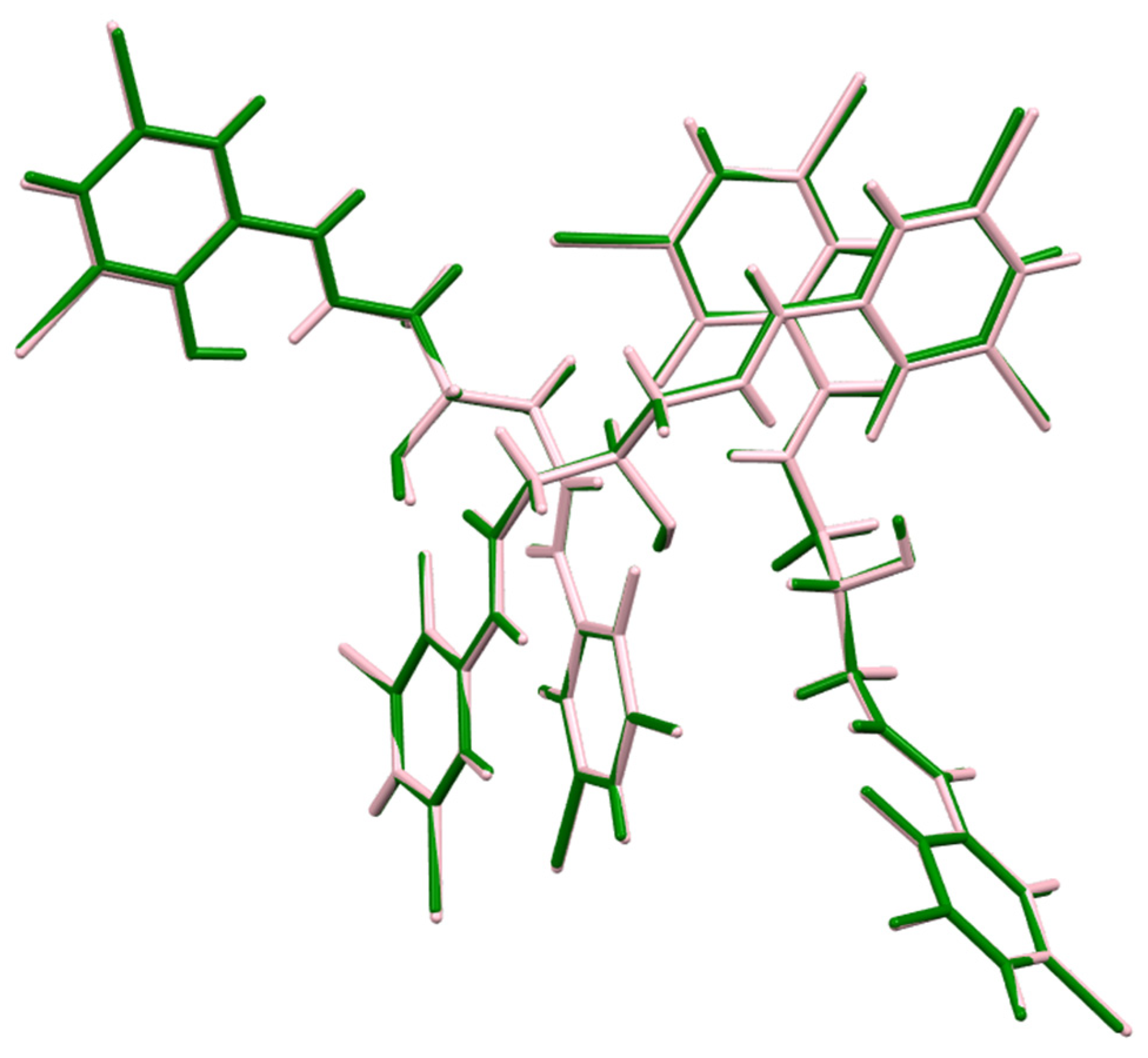
The crystal structure of 1,3-bis(3-bromo-5-chlorosalicylideneamino)-2-propanol (Figure S1) is very similar to that of the one already published, 1,3-bis(3,5-dibromosalicylideneamino)-2-propanol [27]. In both cases, the asymmetric parts of the appropriate unit cells consist of 3 independent molecules, which occupy analogous positions (Figure 2). The unit cell similarity index equals 0.0088, and the isostructurality index [28] is 0.996, which proves an extremely high degree of isostructurality between those two derivatives.
The only difference which has been found between those two compounds is the position of one certain, however important, hydrogen atom involved in the N32…O35 intermolecular hydrogen bond. It has to be admitted that establishing the position of the hydrogen atom by means of X-ray diffraction data may be quite a tricky issue. In the structure of H3L, some indications have been found, however, which implicate the possibility of a disordered hydrogen atom between atoms O35 and N32. The elongation of the C35-O35 bond (in comparison to the unprotonated oxygen atoms observed in the structure) indicates the possibility of protonation on the O34 atom; on the other hand, the C33-N32-C31 angle, of 121.54(4)°, also shows the N32 atom as a possible protonation center. (Table S1) The difference electron density map (Figure 3) shows the missing electron density concentrations, which can be connected with the hydrogen atom in question close to both O35 and N32 atoms, and two electron density maxima, of similar height (0.44/0.4 e). Therefore, it has been decided to model a disordered hydrogen atom; however, it should be noted that refining the shared hydrogen atom (at one position) yields the same refinement parameters.
Quite a detailed structural analysis of the molecular structure of 1,3-bis(3,5-dibromosalicylideneamino)-2-propanol has been described earlier [27], so, due to the almost perfect isostructurality, this discussion will not be repeated here. The crystal packing in 1,3-bis(3-bromo-5-chlorosalicylideneamino)-2-propanol is mostly determined by quite strong O-H··· hydrogen bonds and, to some extent, also by the stacking interactions between substituted aromatic rings. Additionally, there are weak C-H···Br and C-H···π hydrogen-bond-like interactions, which may also influence the arrangement of molecules in the crystal lattice. The geometry of selected hydrogen bonds has been summarized in Table 1, Table S2; the others can be found in the Supplementary Materials, together with bond distances, valence angles, and torsion angles for all three molecules of 1,3-bis(3-bromo-5-chlorosalicylideneamino)-2-propanol (Figure S2).

2.1. FT-IR Spectroscopy

FTIR studies were performed on the ligand H3L and the solid-state complexes [Eu(H3L)2(NO3)3], [Gd(H3L)2(NO3)3], [Tb(H3L)2(NO3)3] to investigate the coordination sphere in the obtained complexes. The spectra were recorded in the range of 4000–400 cm−1. The spectra of the complexes were compared to that of the ligand. FTIR spectroscopic analysis of the H3L ligand revealed the presence of bands with a wavelength of 1637 cm−1, which are from imine bonds formed as a result of the condensation reaction of amine and carbonyl groups. Comparing the position of this band in the spectra of the ligand and the complexes, a very small shift in this band was found: 1639 cm−1 (for Eu3+), 1640 cm−1 (for Gd3+), and 1640 cm−1 (for Tb3+), which may indicate that the azomethine nitrogen atoms do not participate in coordination with the metal ions. However, the decrease in the frequency of stretching of the C=N bond due to the reduction in the double bond character is usually attributed to the participation of the nitrogen atom in the coordination with the metal ion. Data collected from both X-ray crystallography and spectroscopy reveal that in the ligand, occurs a proton transfer from hydroxyl groups to nitrogen atoms, which causes the C=N double bond to elongate. Therefore, when the metal ion coordinates to the donor nitrogen atom, we observe a very small shift in C=N band. Additionally, FTIR spectra of the ligand and complexes present a broad band in the range 3254–3064 cm−1 attributed to the stretching vibration of OH groups. The FTIR spectra of the ligand and complexes show bands at 1133 cm−1 for the ligand and at 1142 cm−1 for the [Eu(H3L)2(NO3)3], 1146 cm−1 for [Gd(H3L)2(NO3)3], and 1440 cm−1 for [Tb(H3L)2(NO3)3] complexes attributed to the C-O bond. The shift in the band confirmed coordination of lanthanide ions to donor oxygen atoms. The FTIR spectra of the complexes exhibited bands in the ranges 1437–1206 cm−1 for Eu complex, 1444–1209 cm−1 for Gd complex, and 1440–1208 cm−1 for Tb complex characteristic of coordinated nitrate groups. The difference between these bands of about 233 cm−1 suggests that the nitrate counterions are coordinated in a bidentate fashion.

2.2. Equilibrium Study

The research involved the examination of the acid–base properties of 1,3-bis(3-bromo-5-chlorosalicylideneamino)-2-propanol (H3L) using potentiometric titration. The first stage of this study was to determine the protonation constants of the ligand, which were calculated corresponding to four possible protonation steps (Table 2). The obtained values reflect the presence of two phenolic groups, one hydroxyl group, and one nitrogen donor atom capable of binding protons in aqueous solution.
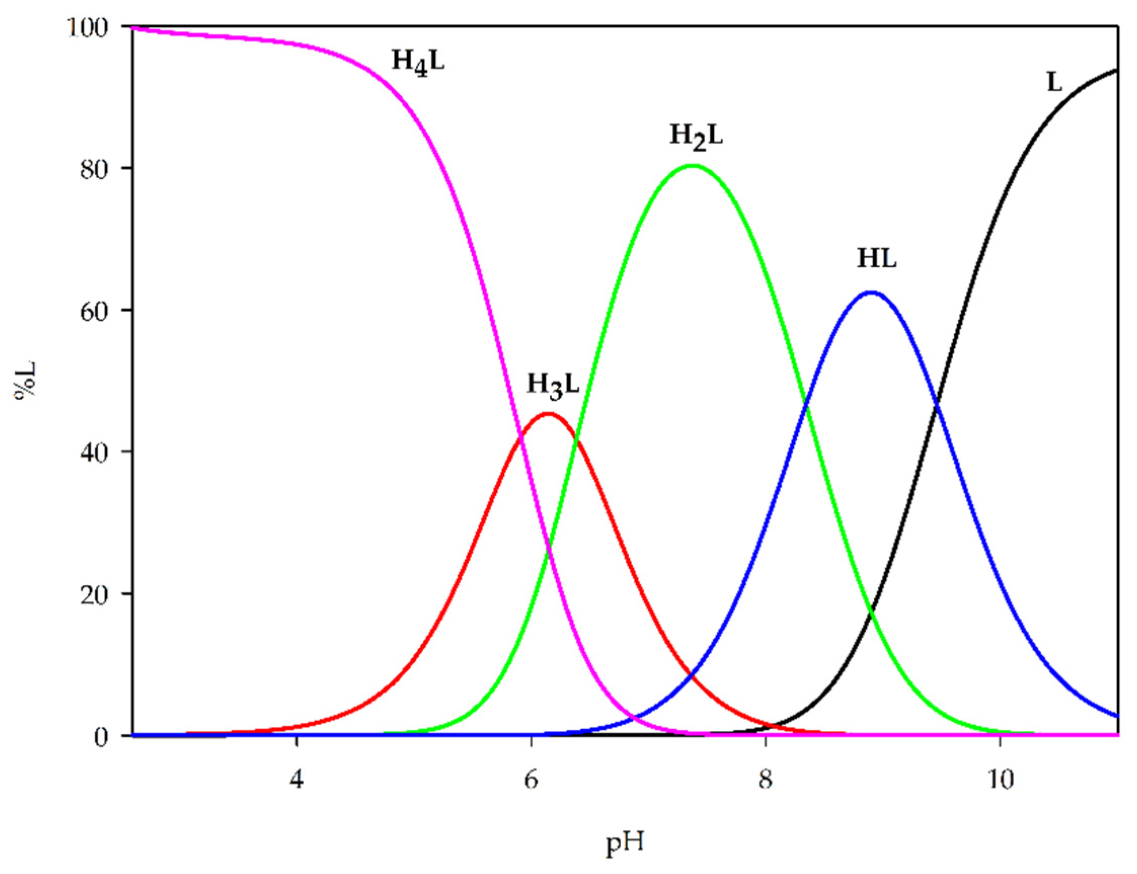
Analysis of these protonation constants revealed that the first deprotonation occurs at pH below 4.0 and is assigned to one of the phenolic groups, followed by subsequent protonation/deprotonation steps involving the second phenolic oxygen, hydroxyl group, and nitrogen atom. Subsequently, the obtained set of protonation constants was used to generate theoretical distribution diagrams of H3L species as a function of pH, providing detailed insight into the speciation profile and relative stability of the different forms of the ligand (Figure 4).
Potentiometric titrations were carried out to determine the stability of complexes formed between Eu(III), Gd(III), and Tb(III) ions and the Schiff base ligand H3L at metal-to-ligand ratios of 1:1 and 1:2. The overall stability constants (logβ) as well as stepwise equilibrium constants (logKe) were calculated for the formed species (Table 3). The results show that all three lanthanide ions form stable complexes with 1,3-bis(3-bromo-5-chlorosalicylideneamino)-2-propanol, with both 1:1 and 1:2 stoichiometries being detected depending on the metal ion. The formation of M(HxL), M(HxL)2, ML, ML2, ML(OH)x, and MLx(OH)x (where x = 1–3) was observed. With excess ligand concentration, the coordination shifts from simple 1:1 metal–ligand species toward complexes with two molecules of the studied ligand in the coordination sphere of the metal ion.
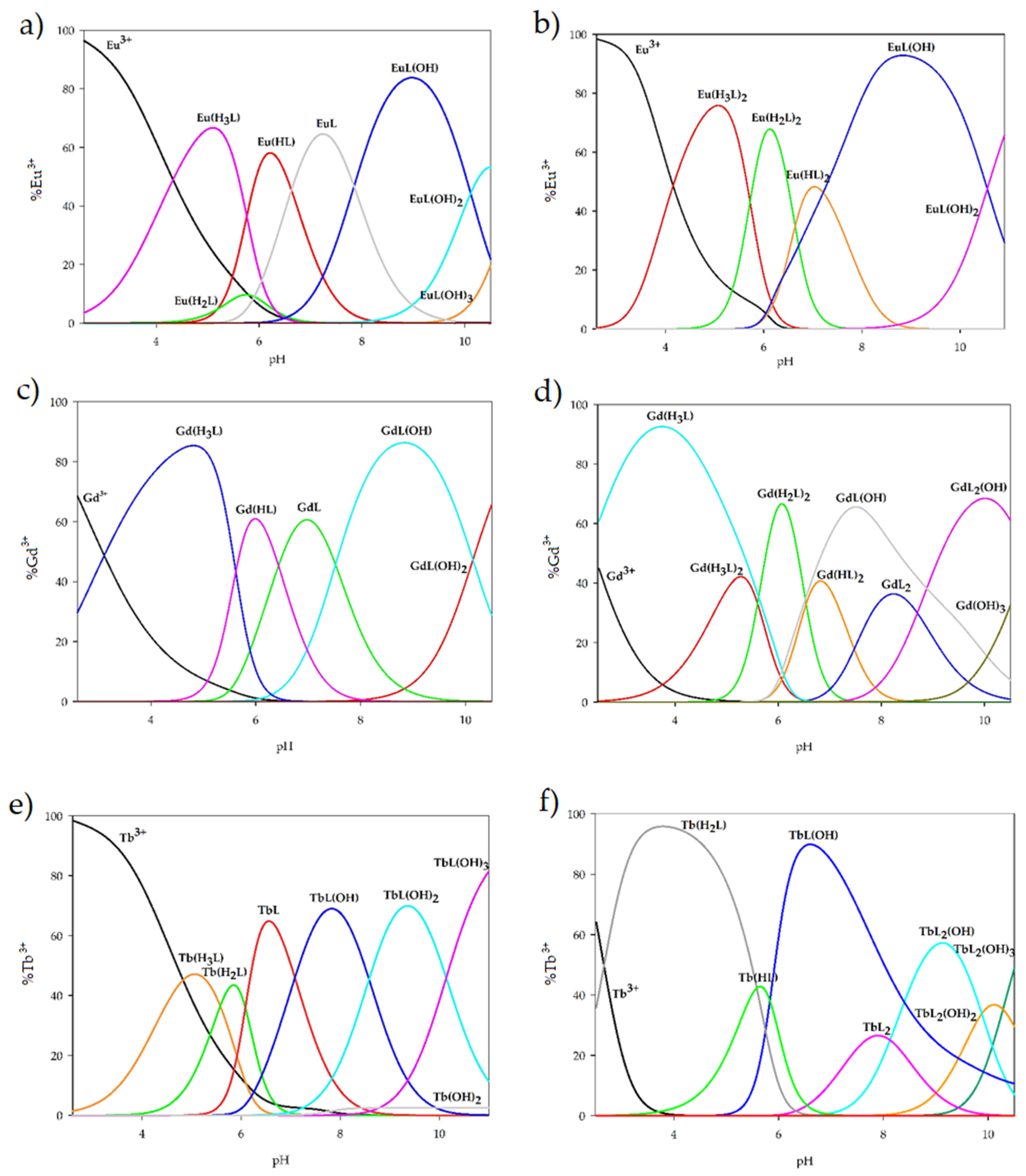
Based on the potentiometric titration data, species distribution diagrams were generated for the Ln(III)–H3L systems with metal-to-ligand ratios of 1:1 and 1:2, taking into account ligand protonation, metal ion hydrolysis, and the stability constants of the complexes formed (Figure 5). In the equimolar (1:1) systems, complexation starts at pH ≈ 2.5 with the formation of M(H3L), which reaches its maximum concentration above pH 5.0. The M(H2L) species was not detected for gadolinium(III); in the case of europium(III), it represents only a minor fraction (ca. 10% of the metal ions), whereas for terbium(III), it becomes the dominant form at pH 5.8, binding approximately 40% of the metal ions. With further increases in pH and progressive ligand deprotonation, M(HL) complexes appear for both gadolinium(III) and europium(III). Complete deprotonation of the ligand leads to the formation of ML species, which are dominant for all studied metal ions and at their maximum account for about 60% of all complexes present in solution. At alkaline pH, hydroxo-complexes such as ML(OH), ML(OH)2, and ML(OH)3 become predominant.
In systems with an excess of 1,3-bis(3-bromo-5-chlorosalicylideneamino)-2-propanol, differences in coordination were observed compared to systems with equal amounts of ligand. For terbium(III) ions in an acidic environment, the dominant forms are M(H2L) and the non-equimolar complex M(HL). At pH 6.0, the TbL2 form begins to form, which in alkaline pH participates in the formation of the hydroxyl complex TbL2(OH), whose maximum concentration is reached at pH 9.1 (about 60% of the terbium ions participate in coordination). Additionally, the formation of TbL2(OH)2 and TbL2(OH)3 hydroxocomplexes was observed. The complexation of ligands with gadolinium(III) ions at lower pH conditions is analogous to an equimolar system, where the formation of Gd(H3L) is observed. Unlike the equimolar system, the maximum concentration was achieved at pH 3.7, compared to pH 5.0 in the 1:1 system. With an increased pH value, the participation of two ligand molecules and the formation of Gd(H3L)2, Gd(H2L)2, Gd(HL)2, GdL2, and GdL2(OH) complexes were observed. The dominant forms are only Gd(H2L)2 and GdL2(OH) complexes, which bind approximately 65% of gadolinium(III) ions at their maximum concentration. Additionally, the GdL(OH) complex is present in the studied system, which is the dominant form in the pH range of 6.5–9.0. For europium(III) ions, the complexation process begins similarly to that in an equimolar system at a pH of approximately 2.5, but the complex formed is Eu(H3L)2. This complex dominates at a pH of 5.0, binding nearly 80% of europium(III) ions, and under the same pH conditions, the formation of the Eu(H2L)2 complex is observed. At a pH of approximately 7.0, the dominant complex is Eu(HL)2. A change in the conditions of the system to alkaline results in the presence of EuL(OH) hydroxy complexes (accounting for approximately 90% of all forms in the system at a pH of approximately 9.0) and EuL(OH)2 in the system.
The comparison of the stability of the tested complexes with our previous research [29,30,31] confirmed that the presence of Br atoms causes an increase in the equilibrium constants of formation (logKe) of the complexes, which is attributed to the effects of the heavy atom and more stable coordination in the metal environment.

2.3. UV-Vis Spectroscopy

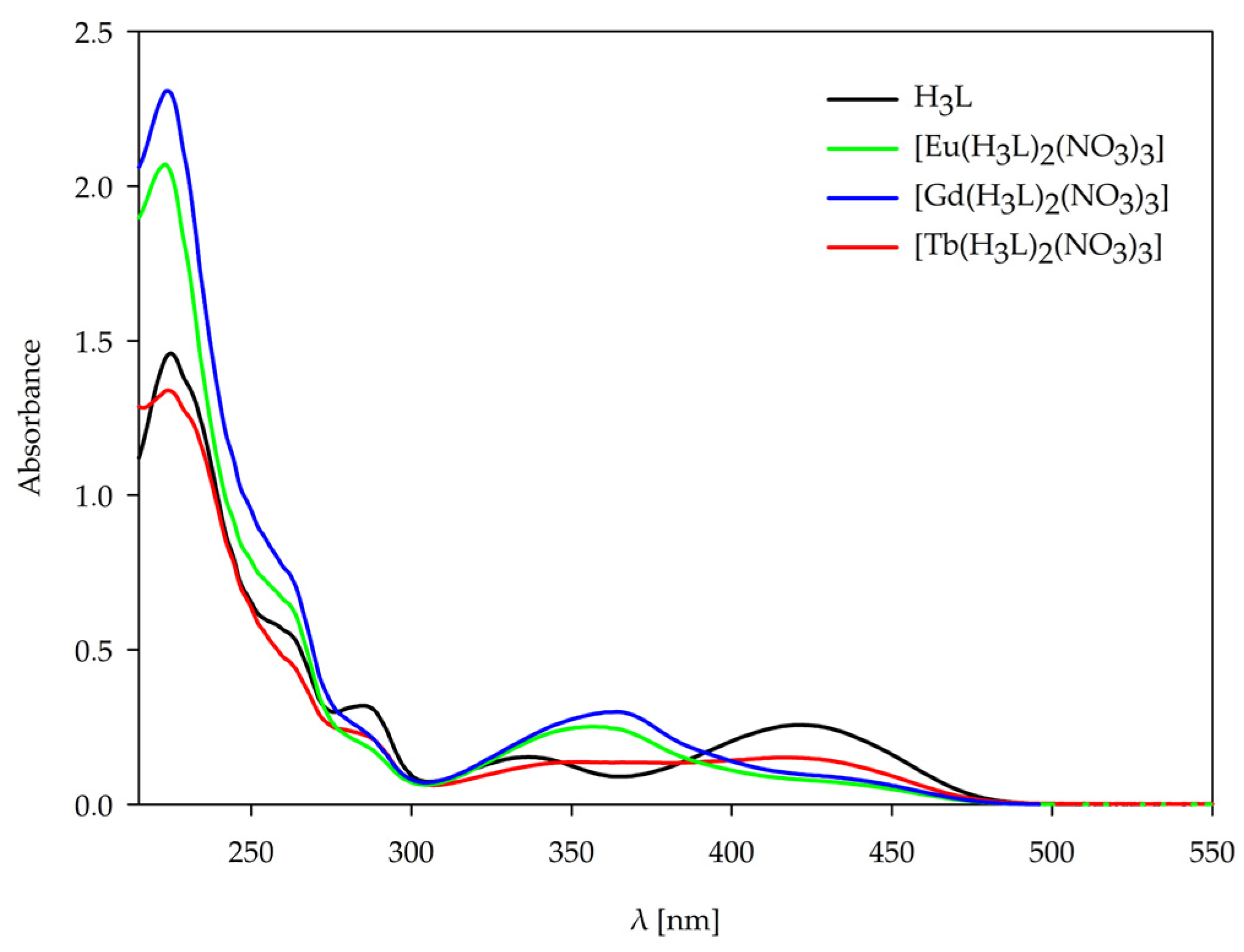
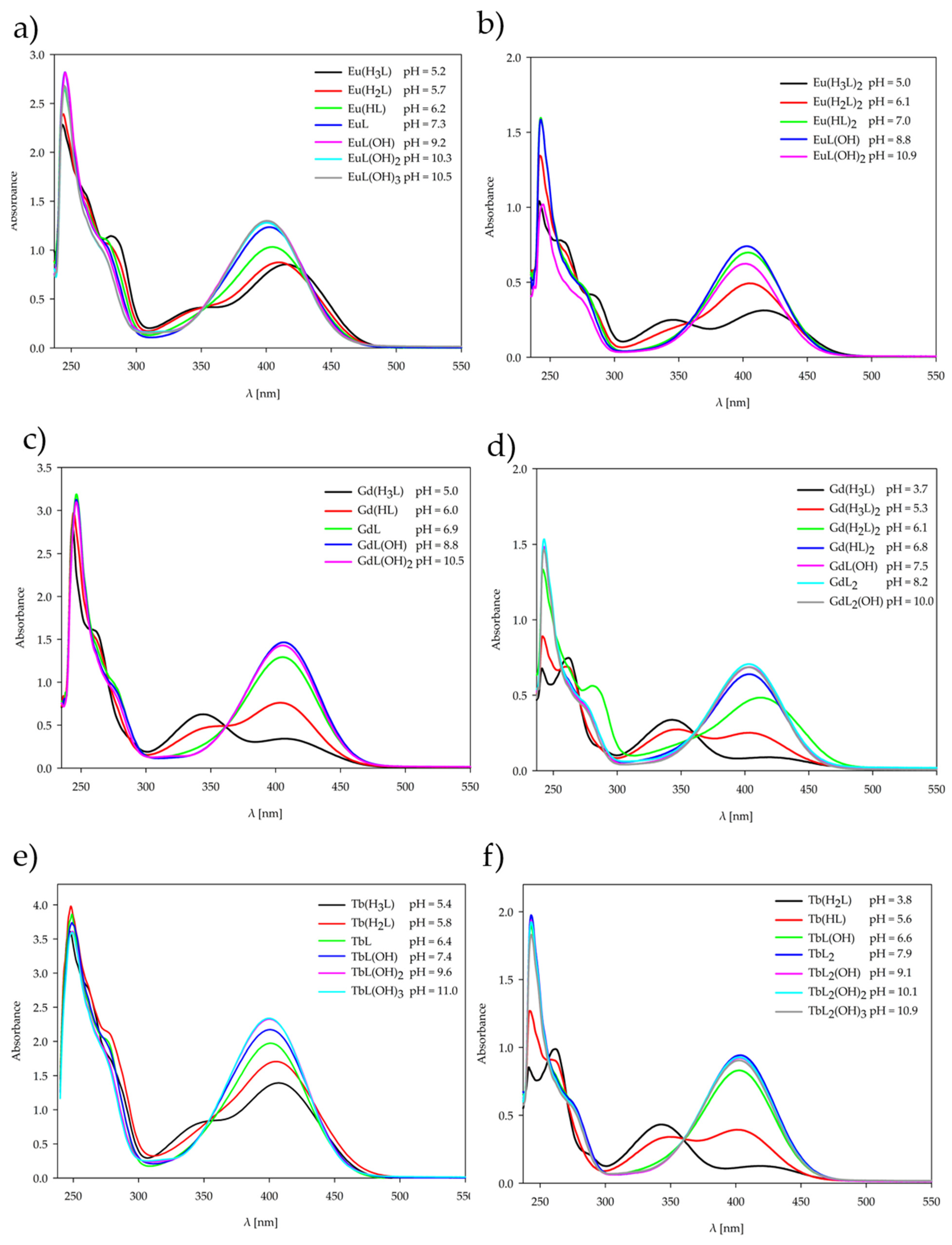
The behavior of the 1,3-bis(3-bromo-5-chlorosalicylideneamino)-2-propanol, H3L ligand, and the complexes [Eu(H3L)2(NO3)3], [Gd(H3L)2(NO3)3], [Tb(H3L)2(NO3)3] obtained in the solid state was studied using UV-Vis spectroscopy. The behavior of the H3L ligand and the complexes formed in the solution at various pH values was also studied using UV-Vis spectroscopy. The UV-Vis spectra were recorded in the range from 200 to 600 nm for all forms of the ligand and complexes in the solid state and at the pH value of dominance of each form determined by potentiometric titration. The coordination of the ligand H3L with the lanthanide ions is confirmed by the shifts in characteristic bands and changes in the absorption spectra in both solid-state and solution complexes. The observed changes for solid-state complexes and solution complexes are shown in Figure 6 and Figure 7, and all spectroscopic data are listed in Tables S3–S5. The H3L ligand spectrum is characterized by five main absorption bands that are assigned to the π-π* and n-π* transitions of the Schiff base H3L ligand. In the range of 220–300 nm, three bands occur due to the π-π* transition of the aromatic rings. The band at 337 nm is assigned to the n-π* transition of the C=N group. The last band at 423 nm may be assigned to the coupling of aromatic ring π-π* transition and an imine group n-π* transition. For the complexes, the bathochromic shifts in the last two bands are observed at 359–364 nm and 436–442 nm, which confirms the coordination of lanthanide ions with the donor nitrogen atoms of the C=N groups.
All forms of complexes formed in solution at the pH value of dominance of each form, determined by potentiometric titration, were studied by the UV-Vis spectra. For all the complexes forming in the solution, a decrease in the number of characteristic bands was observed with increasing pH values of the systems. Upon complex formation, the two higher-energy bands of the free ligand merge into a single band. The band shift, the emergence of new bands, and the variation in molar absorptivity (ε) provide evidence for the coordination of metal ions with ligands and the resulting complex formation [32]. Changes are observed above 350 nm, i.e., in the range of occurrence of characteristic bands assigned to the n-π* transition of the C=N group, for both Ln(III)–ligand 1:1 and 1:2 systems. The change in this characteristic band pattern is caused by deprotonation of the ligand with increasing pH, which correlates with a change in the internal structure of the coordination sphere. Absorption in the spectra can be ascribed to the organic ligands, as the contribution of lanthanide(III) ions is negligible owing to the Laporte-forbidden nature of their electronic transitions.
The long-term stability of the stable complexes of the tested compounds after 5 and 10 days of storage at room temperature was also examined. The changes were monitored using the UV–Vis method, and no significant differences in the spectra were observed, indicating the stability of the complexes.

2.4. Luminescence Spectroscopy

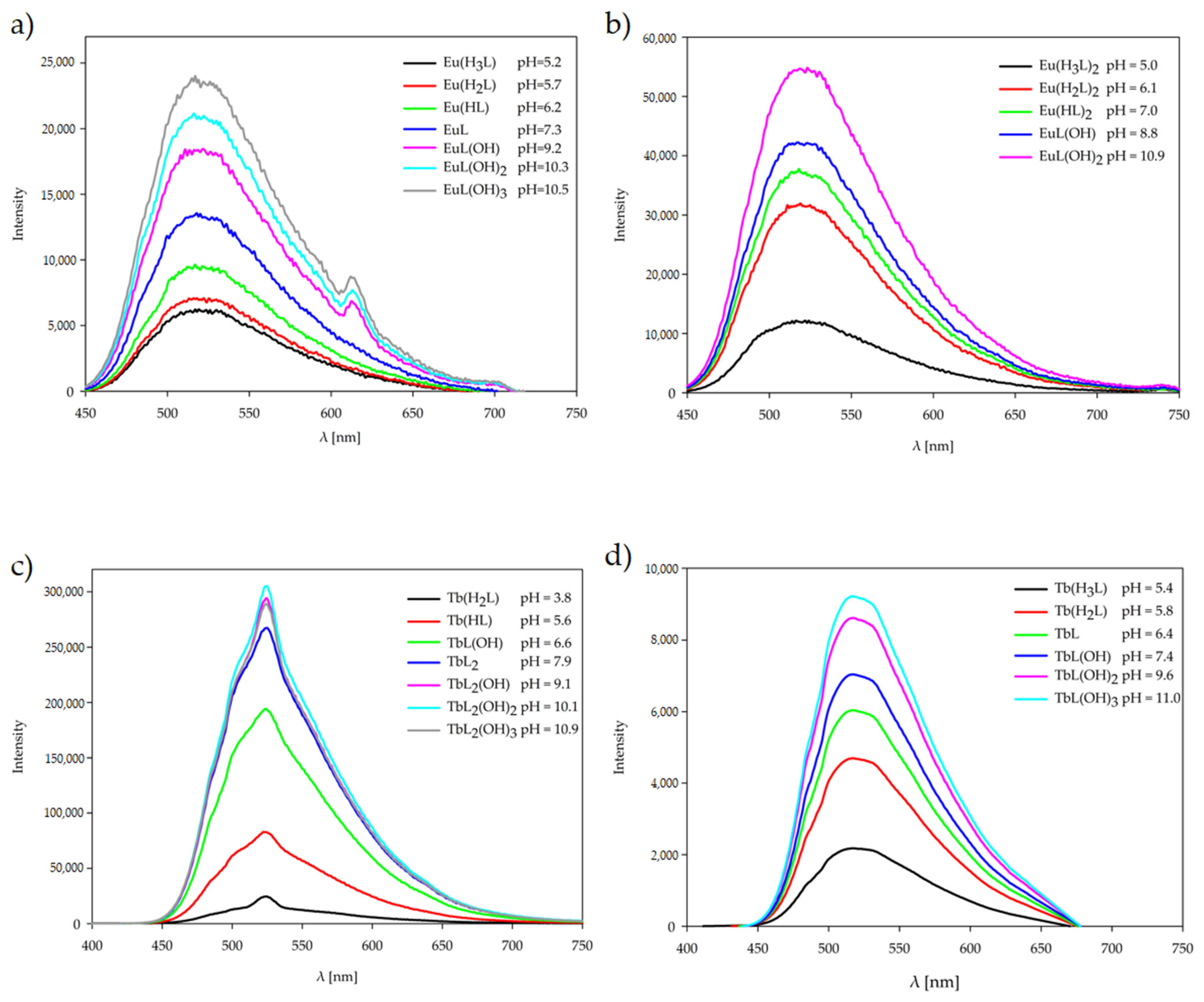
A photoluminescence study of the ligand and its Eu(III) and Tb(III) complexes has been carried out in solution at the pH value of dominance of each form, determined by potentiometric titration, at room temperature, Figure 8. Upon excitation into the UV region, the luminescence spectrum of the H3L ligand in solution displays broad emission bands with maxima at 529 nm for the protonated H4L form of ligand (pH 4.0), 518 nm for the neutral H3L form (pH 6.0), 519 nm, 523 nm, and 517 nm for the deprotonated H2L form (pH 7.2), HL (pH 9.0), and L (pH 10.5), respectively. Luminescence studies also have been carried out, giving the emission spectra of complexes Eu(III) and Tb(III) upon excitation at 243 nm at the pH value where each form of complexes predominates. Eu(III) complexes exhibit the main emission centered around 523–516 nm, while Tb(III) complexes at around 525–515 nm, which may be caused by ligand π-π* transition. Lanthanide(III) ions present in the systems increase the luminescence of the H3L ligand. The enhancement value depends on the type of Ln(III) ion, but also on the deprotonation of the ligand, which depends on the pH of the tested solution. The Eu(III) complex solutions showed a very weak emission band at approximately 613 nm, attributed to the 5D0–7F2 transitions. This band is visible for complexes forming at pH above 9.2 and only for the stoichiometric ratio Ln(III):H3L 1:1. The results of our luminescence experiments show that low efficiency of energy transfer within the molecule occurs only in Eu(III) complexes. No transfer of energy was observed in Tb(III) complexes at any pH value. The probable cause is the large energy difference between the lowest energy level of the H3L ligand triplet state and the lowest resonant energy level of the Ln(III) ions studied [33].

3. Materials and Methods

3.1. Materials

Europium(III) nitrate pentahydrate, gadolinium(III) nitrate hexahydrate, and terbium(III) nitrate pentahydrate, 3-bromo-5-chloro-2-hydroxybenzaldehyde, and 2-hydroxy-1,3-propanediamine were purchased from Aldrich Chemical Company and used without further purification.

3.2. Synthesis

3.2.1. Synthesis of 1,3-Bis(3-bromo-5-chlorosalicylideneamino)-2-propanol, H3L

To a 100 mL round-bottom flask were added 0.09095 g (1 mmol) of 2-hydroxy-1,3-propanediamine and 0.4706 g (2 mmol) of 3-bromo-5-chloro-2-hydroxybenzaldehyde in 40 mL of ethanol (EtOH). The reaction mixture was stirred at room temperature for 3 h. During this time, the solution gradually developed a yellow color, indicating the formation of a Schiff base. After completion of the reaction, the solvent was partially evaporated under reduced pressure to approximately half of the original volume, and the mixture was left to stand at room temperature. Upon standing, yellow single crystals suitable for X-ray diffraction analysis were formed by slow evaporation of ethanol at RT. The product, as yellow crystals, was filtered off and dried (0.2695 g, 98%). Elemental analysis for C17H14Br2Cl2N2O3 (525.02 g·mol−1): Found: C, 38.92; H, 2.70; N, 5.32; Calc.: C, 38.89; H, 2.69; N, 5.34%. Selected FT-IR (cm−1): 3254 (νOH), 3061, 2927 (νOH···N), 1637 (νC=N), 1133 (νC-O). 1H NMR (400 MHz, DMSO-d6) δ 14.64 (1;17) (s, 1H), 8.52 (7;11) (s, 1H), 7.71 (5;13) (dd, J = 2.6, 0.09 Hz, 1H), 7.5 (3;15) (d, J = 2.6 Hz, 1H), 5.64 (9 (O-H)) (d, J = 5.2 Hz, 1H), 4.06 (9) (tt, J = 7.6, 3.7 Hz, 1H), 3.83 (8;10) (dd, J = 12.9, 3.9 Hz, 1H), 3.62 (8;10) (dd, J = 12.8, 6.9 Hz, 1H) 13C NMR (101 MHz, DMSO-d6) δ 166.48 (7;11), 163.20 (1;17), 135.61 (3;15), 130.99 (5;13), 118.06 (4;14), 117.01 (6;12), 114.59 (2;16), 68.07 (9), 58.19 (8;11). ESI-MS: m/z 525 [C17H13Br2Cl2N2O3] 100%; 1084 [(C17H14Br2Cl2N2O3)2Cl].

3.2.2. Synthesis of Europium(III) Complex [Eu(H3L)2(NO3)3]

To a 50 mL round-bottom flask equipped with a magnetic stirrer, 0.09418 g (0.4 mmol) of 3-bromo-5-chloro-2-hydroxybenzaldehyde was dissolved in 10 mL of ethanol and heated to reflux (78 °C) under continuous stirring. After 15 min, 0.0135 g (0.15 mmol) of 2-hydroxy-1,3-propanediamine, dissolved in 5 mL of ethanol, was added dropwise. The solution gradually turned yellow, indicating the formation of a Schiff base. The reaction mixture was stirred at reflux for an additional 60 min. Subsequently, 0.021403 g (0.05 mmol) of europium(III) nitrate pentahydrate, dissolved in 5 mL of ethanol, was added dropwise, and the reaction was continued under the same conditions. After 24 h, the reaction mixture was concentrated under reduced pressure to approximately 3 mL and poured into an excess of diethyl ether to induce precipitation. The resulting yellow solid was collected by decantation and dried in air at room temperature. The product was obtained as a yellow solid (0.0548 g, 80.89%). Elemental analysis for [Eu(C17H14Br2Cl2N2O2)2(NO3)3] (1388.02 g·mol−1): Found: C, 29.39; H, 2.01; N, 7.08; Calc.: C, 29.42; H, 2.02; N, 7.06%. Selected FT-IR (cm−1): 3064 (νOH), 1639 (νC=N), 1142 (νC-O), 1437–1206 (νNO3). ESI-MS: m/z 1262 [Eu(C17H12Br2Cl2N2O3)(C17H13Br2Cl2N2O3)(NO3)], 1199 [Eu(C17H12Br2Cl2N2O3)2], 736 [Eu(C17H11Br2Cl2N2O3)(NO3)], 586 [(C17H14Br2Cl2N2O3)(NO3)], 525 [C17H13Br2Cl2N2O3], 706 [Eu2(C17H12Br2Cl2N2O3)(C17H13Br2Cl2N2O3)(NO3)]2+, 675 [Eu(C17H12Br2Cl2N2O3)]+.

3.2.3. Synthesis of Gadolinium(III) Complex [Gd(H3L)2(NO3)3]

To a 50 mL round-bottom flask equipped with a magnetic stirrer, 0.09484 g (0.4 mmol) of 3-bromo-5-chloro-2-hydroxybenzaldehyde was dissolved in 10 mL of absolute ethanol and heated to reflux (78 °C) under continuous stirring. After 15 min, a solution of 0.01307 g (0.15 mmol) of 2-hydroxy-1,3-propanediamine in 5 mL of ethanol was added dropwise. The reaction mixture was maintained at reflux, and the gradual development of a yellow color indicated the formation of a Schiff base. Sixty minutes after the addition of the diamine, a solution of 0.02234 g (0.05 mmol) of gadolinium(III) nitrate hexahydrate in 5 mL of ethanol was added slowly. Reflux and stirring were continued under the same conditions. After 24 h, the reaction mixture was concentrated under reduced pressure to approximately 3 mL and poured into cold diethyl ether to induce precipitation. The resulting yellow solid was collected by decantation and dried in air at room temperature. The gadolinium complex was obtained as a yellow solid (0.0353 g, 63.19%). Elemental analysis for [Gd(C17H14Br2Cl2N2O2)2(NO3)3] (1393.30 g·mol−1): Found: C, 29.29; H, 2.05; N, 7.03; Calc.: C, 29.31; H, 2.03; N, 7.04%. ESI-MS: m/z 1266 [Gd(C17H12Br2Cl2N2O3)(C17H13Br2Cl2N2O3)(NO3)], 1203 [Gd(C17H12Br2Cl2N2O3)2], 741 [Gd(C17H11Br2Cl2N2O3)(NO3)], 586 [(C17H14Br2Cl2N2O3)(NO3)], 525 [C17H13Br2Cl2N2O3], 712 [Gd2(C17H12Br2Cl2N2O3)(C17H13Br2Cl2N2O3)(NO3)]2+, 681 [Gd(C17H12Br2Cl2N2O3)]+. Selected FT-IR (cm−1): 3067 (νOH), 1640 (νC=N), 1146 (νC-O), 1444–1209 (νNO3).

3.2.4. Synthesis of Terbium(III) Complex [Tb(H3L)2(NO3)3]

In a 50 mL round-bottom flask, 0.091484 g (0.4 mmol) of 3-bromo-5-chloro-2-hydroxybenzaldehyde was dissolved in 10 mL of ethanol and heated to reflux (78 °C) under continuous stirring. After 15 min, a solution of 0.0135 g (0.15 mmol) of 2-hydroxy-1,3-propanediamine in 5 mL of ethanol was added dropwise. The reaction mixture gradually turned yellow, indicating the formation of the Schiff base ligand. After an additional 60 min of reflux, 0.021751 g (0.05 mmol) of terbium(III) nitrate pentahydrate, dissolved in 5 mL of ethanol, was added slowly. After 24 h, the solution was concentrated under reduced pressure to approximately 3 mL and poured into cold diethyl ether to induce precipitation. The resulting yellow solid was collected by decantation and dried in air at room temperature. The terbium complex was obtained as a yellow solid (0.0461 g, 63.75%). [Tb(C17H14Br2Cl2N2O2)2(NO3)3] (1394.98 g·mol−1): Found: C, 29.31; H, 2.00; N, 7.05; Calc.: C, 29.27; H, 2.02; N, 7.03%. Selected FT-IR (cm−1): 3065 (νOH), 1640 (νC=N), 1143 (νC-O), 1440-1208 (νNO3). ESI-MS: m/z 1267 [Tb(C17H12Br2Cl2N2O3)(C17H13Br2Cl2N2O3)(NO3)], 1204 [Tb(C17H12Br2Cl2N2O3)2], 742 [Tb(C17H11Br2Cl2N2O3)(NO3)], 586 [(C17H14Br2Cl2N2O3)(NO3)], 525 [C17H13Br2Cl2N2O3], 713 [Tb2(C17H12Br2Cl2N2O3)(C17H13Br2Cl2N2O3)(NO3)]2+, 681 [Tb(C17H12Br2Cl2N2O3)]+.

3.3. Physical Measurements

Elementary analysis (CHN) was conducted using an Elementar Analyzer Vario EL III. Mass (Elementar, Langenselbold, Germany) spectrometric analyses were carried out using electrospray ionization (ESI). Measurements were performed in acetonitrile with a Waters Micromass ZQ spectrometer (Waters, Milford, MA, USA), operated in positive-ion mode. The analyte concentration was approximately 1 × 10⁻⁴ mol dm−3. Spectra were recorded in the m/z range 100–1000 with a 6 s scan time, and the final spectrum was obtained by averaging 10 scans. NMR spectra were acquired in DMSO-d6 on a Bruker BioSpin GmbH MHz spectrometer (Bruker BioSpin GmbH, Ettlingen, Germany), calibrated against residual solvent signals (δ = 2.50 ppm). Infrared spectra were collected with an INVENIO R Bruker FT-IR spectrometer (Bruker, Ettlingen, Germany), and band positions are reported in cm−1. Electronic absorption spectra were measured on a Nicolet Evolution 300 UV–VIS ThermoFisher Scientific spectrophotometer (ThermoFisher Scientific, Waltham, MA, USA) in ethanol and a mixture of dimethyl sulfoxide and water. The luminescence studies were recorded on an RF−6000 spectrofluorophotometer (Shimadzu, Kyoto, Japan) using 5/5 nm slit widths.

3.4. Equilibrium Studies

All experiments were conducted in a dimethyl sulfoxide/water mixture (20:80, v/v) prepared with demineralized, CO2-free water. Potentiometric titrations were performed on a Metrohm Titrino 702 (Metrohm, Herisau, Switzerland) with an autoburette and a Metrohm Solvotrode combination pH electrode designed for nonaqueous titrations, calibrated prior to each run. pH readings were corrected before every measurement series using two standard buffers (pH 4.002 and 9.225). Titrations were carried out under helium (5.0) at constant ionic strength (0.1 M LiNO3) and temperature (20.0 °C), with CO2-free NaOH (0.1979 M) as titrant. The ligand concentration was 1 × 10⁻³ M, and HCl (1.5 mL, 0.1 M) was added to the systems. Metal-to-ligand ratios of 1:1 and 1:2 were investigated. The pKw value determined for the DMSO/water (20:80) mixture was 14.501 [34]. Protonation constants of 1,3-bis(3-bromo-5-chlorosalicylideneamino)-2-propanol, model selection, and complex stability constants were obtained using HYPERQUAD software (version 5.2.0.19), which applies a nonlinear least-squares approach [35]. Species distribution diagrams were generated with the HySS (Hyperquad Simulation and Speciation) program (version 4.0.31) [36]. Calculations were based on 150–350 experimental points, excluding sections of titrations where precipitation occurred. Model construction began with the simplest assumptions and was progressively expanded; species rejected during refinement were eliminated. The verification criteria followed those outlined in previous studies [37,38].

3.5. Crystal Structure

Single-crystal X-ray diffraction data were collected at 100(1) K on a Bruker D8 QUEST KAPPA diffractometer, equipped with a microfocus sealed tube (CuKα radiation, λ = 1.54178 Å), using a multilayer mirror as monochromator and a Bruker PHOTON III CPAD detector (Bruker, Ettlingen, Germany). The data collection temperature was controlled by an Oxford Cryostream 700 low temperature device (Oxford Cryosystems Ltd., Oxford, UK). All data were integrated with SAINT V8.41 [39]. A multi-scan absorption correction using SADABS 2016/2 was applied [40]. The primary computational tool used was OLEX2 (version 1.5) [41]. The structures were solved with ShelxT (version 2014/7) [42] and refined through full-matrix least-squares method on F2 using SHELXL-2015/2017, employing scattering factors from SHELXL [43]. All non-hydrogen atoms were refined with anisotropic displacement parameters. All hydrogen atoms were refined with isotropic displacement parameters. Some of their coordinates were refined freely, and some were calculated positions using a riding model, with their Uiso values constrained to 1.2 times their carrier atoms. One atom has been refined as disordered between O35 and N32 (cf. Discussion). Structure representation was prepared with Olec1.5 [41] and Mercury 4.3.0 software [44]. Table 4 lists the relevant experimental data and refinement details. Crystallographic data for the structural analysis have been deposited with the Cambridge Crystallographic Data Centre, No CCDC 2435763. Copies of this information may be obtained free of charge from: The Director, CCDC, 12 Union Road, Cambridge, CB2 1EZ, UK. e-mail: deposit@ccdc.cam.ac.uk or www: http://www.ccdc.cam.ac.uk.

4. Conclusions

The Schiff base ligand H3L, 1,3-bis(3-bromo-5-chlorosalicylideneamino)-2-propanol and its complexes of lanthanide(III) ions with formulas [Eu(H3L)2(NO3)3], [Gd(H3L)2(NO3)3], [Tb(H3L)2(NO3)3], where were synthesized. The solid-state complexes were obtained by template reaction of 3-bromo-5-chloro-2-hydroxybenzaldehyde with 2-hydroxy-1,3-propanediamine in the presence of an appropriate lanthanide(III) ion. It was found that the crystal structure of 1,3-bis(3-bromo-5-chlorosalicylideneamino)-2-propanol is very similar to that of the one already published 1,3-bis(3,5-dibromosalicylideneamino)-2-propanol. In both cases, the asymmetric parts of the appropriate unit cells consist of three independent molecules, which occupy analogous positions. Potentiometric titrations were carried out to determine the stability of complexes formed between Eu(III), Gd(III), and Tb(III) ions and the Schiff base ligand H3L at metal-to-ligand ratios of 1:1 and 1:2. The overall stability constants (logβ) as well as stepwise equilibrium constants (logKe) were calculated for the formed species. The results show that all three lanthanide(III) ions form stable complexes with 1,3-bis(3-bromo-5-chlorosalicylideneamino)-2-propanol, with both 1:1 and 1:2 stoichiometries being detected depending on the metal ion. Spectroscopic studies confirmed the coordination of the lanthanide(III) ion by the donor nitrogen and oxygen atoms of the H3L ligand and the complementing of the coordination sphere by the bidentate coordination of nitrate groups. Luminescence studies showed that low efficiency of energy transfer within the molecule occurs only in Eu(III) complexes. No energy transfer was observed in Tb(III) complexes at any pH value. The probable cause is the large energy difference between the lowest energy level of the H3L ligand triplet state and the lowest resonant energy level of the Ln(III) ions studied. Organic ligands can act as energy acceptors from lanthanide(III) ions, leading to a weakening or complete quenching of luminescence.

Supplementary Materials

The following supporting information can be downloaded at: https://www.mdpi.com/article/10.3390/ijms262110379/s1.

Author Contributions

Conceptualization, J.B., M.T.K. and K.K.-S.; methodology, J.B., M.T.K.; K.K.-S. and M.Z.; validation, M.T.K., M.Z. and R.J.; formal Analysis, J.B., M.S. and M.N.; investigation, A.G.; resources, M.T.K. and K.K.-S.; data curation, M.T.K. and J.B.; writing—original draft preparation, J.B., K.K.-S., M.T.K. and A.G.; writing—review and editing, J.B., K.K.-S., M.T.K. and A.G.; visualization, J.B. and M.T.K.; supervision, M.T.K., K.K.-S. and R.J.; project administration, M.T.K. All authors have read and agreed to the published version of the manuscript.

Funding

This research received no external funding.

Institutional Review Board Statement

Not applicable.

Informed Consent Statement

Not applicable.

Data Availability Statement

The original contributions presented in this study are included in the article/Supplementary Materials. Further inquiries can be directed to the corresponding author.

Acknowledgments

This research did not receive any specific grant from funding agencies in the public, commercial, or not-for-profit sectors.

Conflicts of Interest

The authors declare no conflicts of interest.

References

  1. Kaczmarek, M.T.; Pospieszna-Markiewicz, I.; Kubicki, M.; Radecka-Paryzek, W. Novel lanthanide salicylaldimine complexes with unusual coordination mode. Inorg. Chem. Commun. 2004, 7, 1247–1249. [Google Scholar] [CrossRef]
  2. Kaczmarek, M.T.; Kubicki, M.; Mondry, A.; Janicki, R.; Radecka-Paryzek, W. Self-assembled lanthanide salicylaldimines with a unique coordination mode. Eur. J. Inorg. Chem. 2010, 2010, 2193–2200. [Google Scholar] [CrossRef]
  3. Kaczmarek, M.T.; Zabiszak, M.; Nowak, M.; Jastrzab, R. Lanthanides: Schiff base complexes, applications in cancer diagnosis, therapy, and antibacterial activity. Coord. Chem. Rev. 2018, 370, 42–54. [Google Scholar] [CrossRef]
  4. Al-Ghamdi, K.; Hamed, A.M.; Ibrahem, M.A.; Alharbi, S.K.; Abu Al-Ola, K.A.; Amin, M.S.; Eskander, T.N.A.; Abdel-Latif, S.A.; Abu-Dief, A.M. A robust synthesis, physicochemical characterization, stability determination and potential biomedical applications of novel Salen complexes supported by theoretical approaches. J. Mol. Struct. 2025, 1336, 142060–142082. [Google Scholar] [CrossRef]
  5. Zou, X.; Du, C.; Dong, Y.; Li, G. Slow relaxation of two-dimensional salen type lanthanide coordination polymer. Inorg. Chim. Acta 2020, 507, 119455–119461. [Google Scholar] [CrossRef]
  6. Mediavilla, P.; Ribeiro, A.; Gutierrez, A.; Herrero, S.; Torralba, M.C. Novel 3-ethoxysalicylaldehyde lanthanide complexes obtained by decomposition of salen-type ligands. Inorganics 2025, 13, 93. [Google Scholar] [CrossRef]
  7. Zou, X.; Fei, B.; Li, G. Structural effect on NIR luminescence of three salen type heteropolynuclear 3d–4f erbium complexes. Polyhedron 2020, 192, 114811–114817. [Google Scholar] [CrossRef]
  8. Chakraborty, A.; Middya, P.; Chattapadhyay, S. An overview of the synthesis and properties of heterodinuclear europium(III)–transition metal complexes with salen type compartmental ligands having inner N2O2 and outer O4 cavities. Inorg. Chim. Acta 2025, 581, 122625–122654. [Google Scholar] [CrossRef]
  9. Shaw, S.; White, J.D. Asymmetric catalysis using chiral salen–metal complexes: Recent advances. Chem. Rev. 2019, 119, 9381–9426. [Google Scholar] [CrossRef]
  10. Pessoa, J.C.; Correia, I. Salan vs. salen metal complexes in catalysis and medicinal applications: Virtues and pitfalls. Coord. Chem. Rev. 2019, 388, 227–247. [Google Scholar] [CrossRef]
  11. Abu-Dief, A.M.; Mohamed, I.M.A. A review on versatile applications of transition metal complexes incorporating Schiff bases. Beni-Suef Univ. J. Basic Appl. Sci. 2015, 4, 119–133. [Google Scholar] [CrossRef]
  12. Mandal, S.S. Metallo-salen complexes show promise towards treatment of leukemia. Leuk. Res. 2011, 35, 571–572. [Google Scholar] [CrossRef]
  13. Binnemans, K. Lanthanide-based luminescent hybrid materials. Chem. Rev. 2009, 109, 4283–4374. [Google Scholar] [CrossRef]
  14. Qi, Y.; Liu, H.; Li, S. Lanthanide complexes with salen-type ligands: Synthesis, luminescence and catalytic applications. Coord. Chem. Rev. 2017, 352, 30–52. [Google Scholar]
  15. Zhang, Y.; Liu, Z. Lanthanide Schiff base complexes: Synthesis, structure, luminescence and applications. Dalton Trans. 2014, 43, 8036–8050. [Google Scholar]
  16. Zou, X.; Du, C.; Dong, Y.; Li, G. Luminescence and structure of a family of salen type dinuclear lanthanide complexes. Inorg. Chim. Acta 2020, 512, 119860–119867. [Google Scholar] [CrossRef]
  17. Li, S.; Jansone-Popova, S.; Jiang, D. Insights into coordination and ligand trends of lanthanide complexes from the Cambridge Structural Database. Sci. Rep. 2024, 14, 11301. [Google Scholar] [CrossRef]
  18. Mortensen, S.S.; Nielsen, V.R.M.; Sørensen, T.J. Contrasting impact of coordination polyhedra and site symmetry on the electronic energy levels in nine-coordinated Eu(III) and Sm(III) crystal structures determined from single crystal luminescence spectra. Dalton Trans. 2024, 53, 10079–10089. [Google Scholar] [CrossRef] [PubMed]
  19. Lin, L.-R.; Tang, H.-H.; Wang, Y.-G.; Wang, X.; Fang, X.-M.; Ma, L.-H. Functionalized lanthanide(III) complexes constructed from azobenzene derivative and β-diketone ligands: Luminescent, magnetic, and reversible trans-to-cis photoisomerization properties. Inorg. Chem. 2017, 56, 3889–3900. [Google Scholar] [CrossRef]
  20. Patra, K.; Pal, H. Lanthanide-based metal–organic frameworks (Ln-MOFs): Synthesis, properties and applications. RSC Sustain. 2025, 3, 629–660. [Google Scholar] [CrossRef]
  21. McHugh, D.; Tong, W.; Bezrukov, A.; Farras, P.; Zaworotko, M.J.; Mayans, J.; Skelton, J.M.; Barnett, S.; Pallipurath, A.R.; Papatriantafyllopoulou, C. Lanthanide(III) MOFs (Ln = Gd, Tb, Dy) based on a C3 symmetrical tricarboxylate linker. Eur. J. Inorg. Chem. 2025, 28, e202400541. [Google Scholar] [CrossRef]
  22. Roesky, P.W.; Bhunia, A.; Lan, Y.; Powell, A.K.; Kureti, S. Salen-based metal–organic frameworks of nickel and the lanthanides. Chem. Commun. 2011, 47, 2035–2037. [Google Scholar] [CrossRef]
  23. Yao, Y.; Yin, H.-Y.; Ning, Y.; Wang, J.; Meng, Y.-S.; Huang, X.; Zhang, W.; Kang, L.; Zhang, J.-L. Strong fluorescent lanthanide salen complexes: Photophysical properties, excited-state dynamics, and bioimaging. Inorg. Chem. 2019, 58, 1806–1814. [Google Scholar] [CrossRef]
  24. Summers, T.J.; Taylor, M.G.; Augustine, L.J.; Janssen, J.; Perez, D.; Batista, E.R.; Yang, P. On the importance of configuration search to the predictivity of lanthanide selectivity. JACS Au 2024, 5, 631–641. [Google Scholar] [CrossRef]
  25. Yarullin, D.N.; Slavova, S.O.; Abramova, E.O.; Zavalishin, M.N.; Tolstoy, P.M.; Gamov, G.A.; Grachova, E.V. Conformer-specific differences in solid-phase emission of pyridoxal 5′-phosphate hydrazones containing heteroaromatic cycles. Opt. Mater. 2025, 159, 116593–116602. [Google Scholar] [CrossRef]
  26. Khodov, I.A.; Belov, K.V.; Pogonin, A.E.; Savenkova, M.A.; Gamov, G.A. Spatial structure and conformations of hydrazones derived from pyridoxal 5′-phosphate and 2-, 3-pyridinecarbohydrazide in the light of NMR study and quantum chemical calculations. J. Mol. Liq. 2021, 342, 117372–117383. [Google Scholar] [CrossRef]
  27. Mikuriya, M.; Tsuchimoto, N.; Koyama, Y.; Mitsuhashi, R.; Tsuboi, M. Crystal structure of 1,3-bis(3,5-dibromosalicylideneamino)-2-propanol. X-ray Struct. Anal. Online 2022, 38, 3–5. [Google Scholar] [CrossRef]
  28. Kálmán, A.; Párkányi, L.; Argay, G. Classification of the isostructurality of organic molecules in the crystalline state. Acta Cryst. B 1993, 49, 1039–1049. [Google Scholar] [CrossRef]
  29. Kaczmarek, M.T.; Zabiszak, M.; Nowak, M.; Jastrzab, R. Potentiometric determination of novel complexes of selected lanthanide ions with N,N′-bis(5-methylsalicylidene)-4-methyl-1,3-phenylenediamine. J. Iran. Chem. Soc. 2018, 15, 407–414. [Google Scholar] [CrossRef]
  30. Kaczmarek, M.T.; Jastrząb, R.; Radecka-Paryzek, W. Potentiometric study of lanthanide salicylaldimine Schiff base complexes. J. Solut. Chem. 2013, 42, 18–26. [Google Scholar] [CrossRef]
  31. Kaczmarek, M.T.; Skrobanska, M.; Zabiszak, M.; Walesa-Chorab, M.; Kubicki, M.; Jastrzab, R. Coordination properties of N,N′-bis(5-methylsalicylidene)-2-hydroxy-1,3-propanediamine with d- and f-electron ions: Crystal structure, stability in the solution, spectroscopic and spectroelectrochemical studies. RSC Adv. 2018, 8, 30994–31007. [Google Scholar] [CrossRef]
  32. Sarwar, A.; Shamsuddin, M.; Kassim, K.; Kakar, E.; Iqbal, S. New luminescent Eu(III) and Er(III) Schiff base complexes: Synthesis, characterization and luminescence properties. J. Iran. Chem. Soc. 2024, 21, 2933–2942. [Google Scholar] [CrossRef]
  33. Kaczmarek, M.T.; Kubicki, M.; Hnatejko, Z. Two types of lanthanide Schiff base complexes: Synthesis, structure and spectroscopic studies. Polyhedron 2015, 102, 224–232. [Google Scholar] [CrossRef]
  34. Stańczak, P.; Łuczkowski, M.; Juszczyk, P.; Grzonka, Z.; Kozłowski, H. Interactions of Cu2+ ions with chicken prion tandem repeats. Dalton Trans. 2004, 14, 2102–2107. [Google Scholar] [CrossRef]
  35. Gans, P.; Sabatini, A.; Vacca, A. Investigation of equilibria in solution. Determination of equilibrium constants with the HYPERQUAD suite of programs. Talanta 1996, 43, 1739–1753. [Google Scholar] [CrossRef]
  36. Alderighi, L.; Gans, P.; Ienco, A.; Peters, D.; Sabatini, A.; Vacca, A. Hyperquad simulation and speciation (HySS): A utility program for the investigation of equilibria involving soluble and partially soluble species. Coord. Chem. Rev. 1999, 184, 311–318. [Google Scholar] [CrossRef]
  37. Jastrząb, R. Phosphoserine and specific types of its coordination in copper(II) and adenosine nucleotides systems—Potentiometric and spectroscopic studies. J. Inorg. Biochem. 2009, 103, 766–773. [Google Scholar] [CrossRef] [PubMed]
  38. Bregier-Jarzebowska, R.; Gasowska, A.; Jastrząb, R.; Łomozik, L. Noncovalent interactions and coordination reactions in the systems consisting of copper(II) ions, aspartic acid and diamines. J. Inorg. Biochem. 2009, 103, 1228–1235. [Google Scholar] [CrossRef]
  39. Bruker. SAINT, V8.41; Bruker AXS Inc.: Madison, WI, USA, 2025.
  40. Krause, L.; Herbst-Irmer, R.; Sheldrick, G.M.; Stalke, D. Comparison of silver and molybdenum microfocus X-ray sources for single-crystal structure determination. J. Appl. Cryst. 2015, 48, 3–10. [Google Scholar] [CrossRef]
  41. Dolomanov, O.V.; Bourhis, L.J.; Gildea, R.J.; Howard, J.A.K.; Puschmann, H. OLEX2: A complete structure solution, refinement and analysis program. J. Appl. Cryst. 2009, 42, 339–341. [Google Scholar] [CrossRef]
  42. Sheldrick, G.M. SHELXT—Integrated space-group and crystal-structure determination. Acta Cryst. A 2015, 71, 3–8. [Google Scholar] [CrossRef] [PubMed]
  43. Sheldrick, G.M. Crystal structure refinement with SHELXL. Acta Cryst. C 2015, 71, 3–8. [Google Scholar] [CrossRef] [PubMed]
  44. Macrae, C.F.; Sovago, I.; Cottrell, S.J.; Galek, P.T.A.; McCabe, P.; Pidcock, E.; Platings, M.; Shields, G.P.; Stevens, J.S.; Towler, M.; et al. Mercury 4.0: From visualization to analysis, design and prediction. J. Appl. Cryst. 2020, 53, 226–235. [Google Scholar] [CrossRef] [PubMed]
Figure 1. Chemical formula of 1,3-bis(3-bromo-5-chlorosalicylideneamino)-2-propanol, H3L.
Ijms 26 10379 g001
Figure 2. The molecular overlay of compound 1,3-bis(3-bromo-5-chlorosalicylideneamino)-2-propanol, H3L (green), and 1,3-bis(3,5-dibromosalicylideneamino)-2-propanol (pink).
Ijms 26 10379 g002
Figure 3. The contour map with the difference electron density map with q-peaks.
Ijms 26 10379 g003
Figure 4. Distribution diagram of 1,3-bis(3-bromo-5-chlorosalicylideneamino)-2-propanol (H3L).
Ijms 26 10379 g004
Figure 5. Distribution diagram of the complexes formed in the studied systems with 1,3-bis(3-bromo-5-chlorosalicylideneamino)-2-propanol and lanthanide(III) ions: (a) Eu(III) (1:1 ratio); (b) Eu(III) (1:2 ratio); (c) Gd(III) (1:1 ratio); (d) Gd(III) (1:2 ratio); (e) Tb(III) (1:1 ratio); (f) Tb(III) (1:2 ratio).
Ijms 26 10379 g005
Figure 6. UV-Vis spectra of solid-state H3L ligand and its complexes with Eu(III), Gd(III), and Tb(III).
Ijms 26 10379 g006
Figure 7. UV-Vis spectra of the complexes formed in the studied systems with 1,3-bis(3-bromo-5-chlorosalicylideneamino)-2-propanol and lanthanide(III) ions: (a) Eu(III) (1:1 ratio); (b) Eu(III) (1:2 ratio); (c) Gd(III) (1:1 ratio); (d) Gd(III) (1:2 ratio);(e) Tb(III) (1:1 ratio); (f) Tb(III) (1:2 ratio).
Ijms 26 10379 g007
Figure 8. Luminescence spectra of the complexes formed in the studied systems with 1,3-bis(3-bromo-5-chlorosalicylideneamino)-2-propanol and lanthanide(III) ions: (a) Eu(III) (1:1 ratio); (b) Eu(III) (1:2 ratio); (c) Tb(III) (1:1 ratio); (d) Tb(III) (1:2 ratio).
Ijms 26 10379 g008
Table 1. The geometry of selected hydrogen bonds.
D—H···AD—H (Å)H···A (Å)D···A (Å)D—H···A (°)
O10—H10···O210.842.012.836 (5)166.7
N12—H12···O150.881.882.599 (5)137.1
N48—H48···O410.881.942.619 (6)133.1
N28—H28···O210.881.962.630 (5)132.4
N28—H28···O540.882.272.917 (5)130.7
N8—H8···O10.881.872.569 (5)134.9
O30—H30···O10.841.962.774 (5)162.0
N32—H32···O350.881.822.532 (5)137.1
N52—H52···O210.882.593.222 (6)129.2
N52—H52···O540.881.832.557 (5)138.1
O50—H50···O150.841.912.754 (5)178.3
Table 2. Overall protonation constants (logβ) and successive protonation constants (logKe) of 1,3-bis(3-bromo-5-chlorosalicylideneamino)-2-propanol (H3L).
SpecieslogβlogKe
H4L30.09 (3)5.91
H3L24.19 (3)6.39
H2L17.79 (2)8.34
HL9.46 (2)9.46
Table 3. The overall stability constants (logβ) and the equilibrium constants of formation (logKe) of the complexes formed in the studied systems with 1,3-bis(3-bromo-5-chlorosalicylideneamino)-2-propanol and lanthanide ions (standard deviations are given in parentheses).
SpeciesEu(III)Gd(III)Tb(III)
logβlogKelogβlogKelogβlogKe
1:1M(H3L)28.65 (4)4.8629.88 (5)6.0928.26 (7)4.47
M(H2L)22.31 (1)4.78--22.68 (9)5.15
M(HL)17.13 (4)7.7718.64 (5)9.28--
ML10.50 (6)10.5012.20 (5)12.2010.51 (1)10.51
ML(OH)2.65 (4)6.664.68 (5)6.983.42 (4)7.41
ML(OH)2−7.46 (5)4.39−5.46 (6)4.36−5.16 (8)5.92
ML(OH)3−18.39 (7)3.57--−15.30 (9)4.36
1:2M(H3L)257.71 (7)10.1358.47 (1)10.90--
M(H2L)246.27 (7)11.2047.28 (8)12.22--
M(HL)232.99 (8)14.2734.19 (1)15.48--
ML2--19.25 (1)19.2523.36 (8)23.36
M(H3L)----30.43 (8)6.65
M(H2L)----27.47 (4)9.94
M(HL)----21.89 (5)12.53
ML2(OH)--10.55 (8)5.8015.23 (6)6.37
ML2(OH)2----5.35 (7)4.62
ML2(OH)3----−4.93 (7)4.22
ML(OH)4.46 (8)18.966.13 (1)20.6310.23 (5)24.73
ML(OH)2−6.09 (1)3.95----
Hydrolysis constants used in calulations: Eu(OH)2 = −15.15; Eu(OH)3 = −24.31; Gd(OH) = −9.30; Gd(OH)2 = −17.74; Gd(OH)3 = −28.12; Tb(OH)2 = −15.56.
Table 4. Experimental details.
1,3-Bis(3-bromo-5-chlorosalicylideneamino)-2-propanol
Crystal data
Chemical formula3(C17H14Br2Cl2N2O3)
Mr1575.06
Crystal system, space groupMonoclinic, P21/n
Temperature (K)100
a, b, c (Å)16.445 (3), 18.111 (3), 18.956 (4)
â (°)92.978 (9)
V3)5638.2 (17)
Z4
Radiation typeCu K
m (mm−1)8.27
Crystal size (mm)0.2 × 0.05 × 0.05
Data collection
DiffractometerBruker D8 QUEST
Absorption correctionMulti-scan
Tmin, Tmax0.250, 0.383
No. of measured, independent and
observed [I > 2ó(I)] reflections
42,240,
10,291,
8425
Rint0.064
(sinθ/λmax−1)0.603
Refinement
R[F2 > 2ó(F2)], wR(F2), S0.046, 0.113, 1.02
No. of reflections10,291
No. of parameters802
Δρmax, Δρmin (e Å−3)1.56, −1.17
Disclaimer/Publisher’s Note: The statements, opinions and data contained in all publications are solely those of the individual author(s) and contributor(s) and not of MDPI and/or the editor(s). MDPI and/or the editor(s) disclaim responsibility for any injury to people or property resulting from any ideas, methods, instructions or products referred to in the content.