Commentary

CDK4/6 Inhibitors in Breast Cancer—Who Should Receive Them?

1
Research Oncology, Bayer, Cambridge, MA 02142, USA
2
Lester and Sue Smith Breast Center, Baylor College of Medicine, Houston, TX 77030, USA
3
Department of Medicine, Baylor College of Medicine, Houston, TX 77030, USA
4
Department of Molecular and Cellular Biology, Baylor College of Medicine, Houston, TX 77030, USA
*
Author to whom correspondence should be addressed.
Int. J. Mol. Sci. 2025, 26(21), 10322; https://doi.org/10.3390/ijms262110322
Submission received: 24 September 2025 / Revised: 17 October 2025 / Accepted: 22 October 2025 / Published: 23 October 2025
(This article belongs to the Special Issue Hormone Receptor in Breast Cancer: 2nd Edition)

Abstract

More than 70% of breast cancers are estrogen receptor-positive (ER+). Endocrine therapy that blocks estrogen signaling remains the cornerstone of treatment, yet relapses continue to affect many patients. Cyclin-dependent kinases 4 and 6 (CDK4/6) regulate the G1-S phase transition in the cell cycle, and pharmacological inhibition of this pathway has been successfully leveraged to reduce recurrence. CDK4/6 inhibitors combined with endocrine therapy are now the standard of care, although determining the optimal patient population for treatment remains a key challenge. A newly published study provides important insight, showing that loss of the NF1/neurofibromin tumor suppressor confers greater sensitivity to CDK4/6 inhibition, as these tumors rely heavily on CDK4/6 activity for survival under endocrine therapy.

Cyclin-dependent kinases 4 and 6 (CDK4/6), in complex with D-type cyclins, are critical regulators of the G1-S phase transition of cell cycle [1,2]. In the resting state, the active, hypophosphorylated retinoblastoma (Rb) tumor suppressor binds and inactivates the E2F transcription factor, thereby blocking cell cycle progression. Following mitogenic stimulation, cyclin D levels rise and activate CDK4/6, which in turn phosphorylate Rb. This modification disables Rb’s inhibitory function, releasing E2F to drive transcription of genes required for DNA synthesis and irreversibly committing the cell to division. The importance of this pathway in cancer is underscored by its frequent subversion through cyclin D1 overexpression or loss of inhibitors such as p16. Although this CDK4/6-Rb pathway is frequently altered in cancers, it is not strictly essential for cell division, as deletion of Ccnd1 or CDK4/6 is not embryonically lethal in mice. These observations support the concept that CDK4/6 represent prime targets for anticancer therapies with limited toxic to normal cells [1,2].
Approximately 70–80% of breast cancers express estrogen receptor α (ER), a ligand-dependent transcription factor [3]. In the presence of estrogen, ER associates with co-activators to induce expression of growth-promoting genes, including CCND1, which encodes cyclin D1. ER+ breast cancers are treated with endocrine therapies that disrupt estrogen signaling. For example, tamoxifen is a selective ER modulator (SERM) that antagonizes ER signaling in the breast by recruiting co-repressors to chromatin [4]. Additional treatment options include fulvestrant, a selective ER degrader (SERD) and aromatase inhibitors, such as letrozole and anastrozole, which block estrogen synthesis in postmenopausal women. Despite advances in targeting ER with tamoxifen and aromatase inhibitors, outcomes for ER+ disease have not improved as rapidly as for ER or human epidermal growth factor receptor 2 positive (HER2+) breast cancer [5]. Moreover, ER+ tumors account for the majority of global breast cancer deaths, now exceeding half a million annually [6].
Over recent years, several CDK4/6 inhibitors (CDK4/6i), such as palbociclib, abemaciclib, and ribociclib, have been developed for the treatment of ER+ breast cancer, achieving significant clinical success. ER-positivity is currently the primary biomarker guiding CDK4/6i therapy, as ER+ breast cancer cell lines demonstrate the strongest response in preclinical studies [7]. Conversely, ER-negative cell lines generally show little response, partly because they lack functional Rb, underscoring the importance of Rb status in determining CDK4/6i response (see below).
In first-line metastatic settings [8], the PALOMA-2 (palbociclib, NCT01740427), MONALEESA-2 and -7 (ribociclib, NCT01958021 and NCT02278120, respectively), and MONARCH-3 (abemaciclib, NCT02246621) trials demonstrated that adding a CDK4/6i to endocrine therapy improved progression free survival (PFS) of in ER+/HER2 metastatic breast cancer by approximately 50%. Similar benefits were observed in the second-line PALOMA-3 (NCT01942135), MONALEESA-3 (NCT02422615) and MONARCH-2 (NCT02107703) trials. Notably, both ribociclib and abemaciclib have also demonstrated overall survival (OS) benefits in the metastatic setting. In early-stage ER+/HER2 disease, two years of adjuvant abemaciclib and three years of ribociclib modestly reduce recurrence rates in high-risk disease; however, neither agent has yet shown an OS advantage [8]. Health economic analyses indicate that CDK4/6i plus letrozole is not cost effective in the first-line metastatic setting [9], and concerns about toxicity and quality of life can also limit their use in the adjuvant context [10]. Together, these observations raise a fundamental question: are all ER+ breast cancers intrinsically resistant to CDK4/6 inhibition or does a subset of patients derive the greatest benefit?
Because CDK4/6 promote cell cycle progression through Rb inactivation, loss of functional Rb could identify patients intrinsically resistant to CDK4/6i therapy [11]. Indeed, RB1 mutations frequently emerge after CDK4/6i treatment, and their role in promoting resistance has been experimentally validated [12,13]. However, RB1 mutations are rare in untreated population (<1%) [12,14], and most ER+ tumors retain Rb expression (>90%), as determined by immunohistochemistry (IHC) [15]. This suggests that Rb has essential functions in ER+ breast cancer, consistent with the observation that Rb1-null mice are embryonically lethal. In addition, high level of cyclin E1 expression, which activates CDK2, has also been associated with resistance to CDK4/6i in PALOMA-3 study [16]. Other acquired resistance mechanisms include activation of RAS, AKT1, and AURKA [13].
Among patients whose tumors are resistant to endocrine therapy, CDK4/6i treatment remain represent a top priority to reduce recurrence. ESR1 mutations in the ligand-binding domain drive resistance to aromatase inhibitors; however, in the PALOMA-3 study, adding palbociclib to fulvestrant significantly improves PFS even in patients with baseline ESR1 mutations [17]. In the same vein, ESR1-fusions represent another class of aberrant ER with enhanced signaling capacity, and preclinical models carrying these features also respond to CDK4/6i [18]. Furthermore, a subset of ER+ breast cancers with defects in MutL mismatch repair [19] and DNA damage repair [20] exhibit resistance to all classes of endocrine therapy, yet preclinical data indicate that such tumors still respond to CDK4/6i.
Taking a precision medicine perspective, a recent study by Zheng et al. (2025) published in Science Translational Medicine demonstrated that a subset of ER+ breast cancers exhibit heightened sensitivity to CDK4/6i, owing to their pronounced reliance on CDK4/6 activity for survival in the context of endocrine therapy [21]. This study centers on the neurofibromin/NF1 tumor suppressor, which functions both as a GTPase activating protein (GAP) that represses RAS (rat sarcoma virus) signaling and as a canonical ER transcriptional co-repressor that inhibits ER signaling [22]. NF1 inactivation therefore leads to simultaneous activation of RAS and ER signaling (Figure 1). Consistent with its co-repressor activity, loss of NF1 enhances recruitment of ER to the chromatin, including at the CCND1 locus, resulting in increased cyclin D1 levels and Rb phosphorylation. To demonstrate the clinical relevance of this mechanism, the authors show that NF1 mRNA levels inversely correlate with cyclin D1 protein levels in TCGA’s Reverse-Phase Protein Array (RPPA) dataset. Furthermore, in two separate early-stage ER+/HER2 cohorts, tumors harboring both an NF1 shallow deletion and high CCND1 expression have worse relapse-free survival (RFS) than those with either abnormality alone.
While cyclin D1 is required for CDK4/6 activation, full activation of protein kinases also depends on phosphorylation of a threonine residue within the activation loop, which permits substrate access to the catalytic site. CDK4/6 are structurally distinct from other cyclin-dependent kinases in the activation loop [23]. For example, CDK2 is phosphorylated at T160, with an adjacent histidine at position 162. In contrast, the corresponding threonine in CDK4 is T172, which is followed by a proline rather than a histidine at position 173. Because proline imposes strong conformational constraints, this substitution can significantly alter the local structure of the activation loop and thereby influence binding of the CDK-activating kinase (CAK). The identity of the cognate CAK that phosphorylates CDK4 at T172 in ER+ breast cancer cells remains elusive. Although CDK7 is a known CAK for CDK2 and can phosphorylate CDK4 in vitro, it requires mM concentrations of ATP [24]. Speculating that CAK substrates possess distinct amino acid sequences in their activation loops, Zheng et al. used an unbiased Group-based Prediction System analysis [25] on the CDK4 sequence, which identified both CDK7 and CRAF/RAF1 (rapidly accelerated fibrosarcoma 1) as potential CAKs. The latter is particularly notable given that RAF activity is stimulated upon NF1 loss [21]. Indeed, in vitro kinase assays using purified components and 10 µM ATP showed that CRAF efficiently phosphorylates purified CDK4 at T172, whereas CDK7 and c-Jun N-terminal kinase (JNK) were excluded as relevant CAKs in the context of NF1-depleted ER+ breast cancer. Consistent with activation of CDK4/6 by NF1 loss, clinical data indicate that CDK4/6 kinase activities negatively correlate with NF1 protein levels in breast tumors [21].
One common assumption is that higher enzymatic activity confers resistance to drugs targeting that enzyme. Alternatively, elevated activity may reflect an ‘addiction’ essential for survival, thereby creating a therapeutic vulnerability. There are many examples of the latter: HER2+ cells are addicted to HER2 signaling, and BRCA-mutant cells to poly(ADP-ribose) polymerase (PARP) activity. These vulnerabilities were not initially recognized or exploited; consequently, neither trastuzumab nor olaparib demonstrated significant benefit when first tested without biomarker guidance. The data provided by Zheng et al. suggest that NF1-depleted ER tumors are also addicted to CDK4/6 activity when treated with endocrine therapy (Figure 1). In particular, two NF1-depleted ER+ patient-derived xenograft (PDX) models resistant to fulvestrant undergo marked regression when treated with the addition of palbociclib, whereas an NF1-normal model exhibits only cytostatic effects under palbociclib treatment. To validate their findings using a clinical dataset, they examined mRNA levels in biopsy samples from the neoadjuvant NeoPalAna trial (NCT01723774) [26]. In this trial, early-stage ER+/HER2 breast cancer patients first received anastrozole for 4 weeks, followed by palbociclib for another two weeks. Biopsies were obtained at baseline, after one month of anastrozole, and after six weeks of combined treatment. Zheng et al. show that NF1-normal ER+/HER2 tumors respond to anastrozole as expected, while NF1-low tumors are largely resistant to anastrozole alone but regain sensitivity with the anastrozole/palbociclib combination.
A major challenge in assessing the impact of NF1 on treatment response lies in reliably measuring NF1 inactivation in the tumors. While NF1 mutation analysis is commonly used, it underestimates cases in which NF1 is functionally inactivated. NF1 mutations are detected in only 2–5% early-stage ER+ breast cancers. However, by measuring mRNA expression, as well as protein levels by IHC [27], the frequency of NF1-loss is estimated to be around 25%. Zheng et al. reported another approach to assess NF1 functional status via copy number loss. Specifically, they report that about 20% of ER+/HER2 breast cancers in the METABRIC [28] dataset harbor a shallow NF1 deletion (−1 copy number) correlating with poorer RFS and disease-specific survival [21].
In solid tumors, a single tumor often carries multiple driver and passenger mutations. To determine whether NF1 inactivation acts alone or cooperates with other oncogenic events in early-stage breast cancer to promote progression to aggressive disease, we analyzed METABRIC data using Fisher’s exact test (Table 1). NF1 shallow deletions most frequently co-occur with tumor protein 53 (TP53) mutations, with an odds ratio of 5.3 (p = 1.10 × 10−58, Table 1), and both missense and “truncating” TP53 mutations behave similarly. Co-occurrence of TP53 mutation and NF1 loss has also been reported in other cancers, including acute myloid leukemia [29], non-small cell lung cancer [30], and ovarian serous carcinoma [31]. The next most frequent co-occurring events affecting NF1 itself (odds ratio = 3.3, p = 7.50 × 10⁻⁸, Table 1), suggesting strong selection for NF1 inactivation during progression toward metastasis. Mutations in USH2A and MUC16, encoding usherin and mucin, rank a distant 3rd and 4th by odds ratio (1.7 and 1.5, respectively, Table 1), but their roles in breast cancer remain unclear. The analysis also reveals mutual exclusivity between NF1 shallow deletions and mutations in MAP3K1, PIK3CA, and GATA3, which occur more commonly in luminal breast cancers [32]. In primary ER+ breast cancer, PIK3CA mutations correlate with better outcomes [32], whereas in the metastatic setting they predict poor prognosis [33]; accordingly, the PI3Kα-selective inhibitor alpelisib is FDA-approved for advanced ER+ breast cancer [34]. Notably, NF1 and PICK3CA mutations frequently co-occur in a metastatic breast cancer cohort (odds ratio = 1.75, p < 0.001) [21], suggesting convergent signaling pathways that drive metastatic progression. Importantly, CDK4/6 remain viable therapeutic targets in tumors resistant to alpelisib, as these tumors continue to respond to CDK4/6i in preclinical models [35].
Notabily, co-inactivation of NF1 and p53 is associated with the poorest RFS compared to tumors in which only one genetic defect could be detected (Table 2). Given NF1-p53 co-inactivation is also detected in metastatic breast cancer [21], we speculate that NF1 and p53 inactivation cooperate to plays a key role in promoting metastasis. Co-activation of ER and RAS, driven by NF1 loss, converges on CDK4/6 to promote endocrine resistance in ER+ tumors. Concurrent p53 inactivation may further increase CDK4/6 activity by suppressing CDKN1A expression (Figure 1), which encodes the CDK4/6 inhibitor p21 (also known as WAF1/CLP1) [36,37,38]. How p53 impacts treatment response to CDK4/6i remains inconclusive, however. Consistent with the concept that p53-inactive tumors require CDK4/6 activity to survive under endocrine therapy, both wild type and mutant TP53 tumors responded to palbociclib in NeoPalAna [26] and PALOMA-3 [17], as well as ribociclib in MONALEESA-2 [39] trials. In contrast, in an Asian cohort [40], baseline TP53 mutations correlate with worse outcomes following ribociclib treatment. In addition, genomic instability caused by p53 inactivation may foster additional oncogenic alterations that cooperate with NF1 inactivation or disrupt NF1 itself, which is a particularly large gene spanning more than 282 kb with 60 exons, making it prone to errors during cell division. In future clinical investigations, NF1 and p53 status may need to be co-analyzed to refine the determination of the subset of patients who could benefit most from CDK4/6i treatment. To this end, we suggest that rather than relying solely on NF1 mutations, which are rare, we should focus on detecting NF1 mRNA or protein levels to assess NF1 functional status. For the latter, an IHC assay using a commercially available monoclonal antibody [22] (MABN2557/A376G3) is available and can identify NF1-depleted tumors associated with letrozole resistance [27]. For p53, a combination of IHC and whole exome sequencing may be advisable as missense loss-of-function TP53 mutations can stabilize p53 protein [41].
CRAF recognizes the specific amino acid sequence in CDK4’s activation loop. This finding raises the possibility that a CDK4-specific inhibitor would be more on-target in driving clinical efficacy in early-stage ER+ breast cancer. Data from DepMap, where ER+ breast cancer cell lines underwent systematic gene silencing to identify genes essential for survival [42], show that CDK4 knockout yields much lower DepMap CRISPR CERES scores than CDK6 knockout [43], indicating stronger dependency on CDK4 than CDK6 for viability. In addition, amplification events are more common with CDK4 than CDK6 in primary luminal breast cancers: about 6% of luminal tumors carry CDK4 amplification, whereas CDK6 amplification is undetectable [44]. Wild type CRAF can control endocrine therapy by directly activating CDK4, and up to 8% in a cohort of metastatic breast cancers [45] harbor genetic alterations in CRAF (e.g., amplification). However, current FDA-approved RAF inhibitors target mutant BRAF, leaving wild type CRAF alterations unaddressed and highlighting an opportunity for future drug development.
In summary, CDK4/6i have fundamentally transformed the therapeutic landscape of ER+ breast cancer, yet their broad application is constrained by heterogeneous benefit, cost, and toxicity, underscoring the need for proper patient selection. Functional NF1 loss, present in ~25% of cases when shallow deletion and reduced mRNA/protein levels are considered, should be assessed both for prognostic significance and for predicting benefit from CDK4/6i in ER+ disease. Mechanistically, NF1 loss co-activates ER and RAS signaling, increasing CDK4 activity through cyclin D1 induction and activation-loop phosphorylation. This dual input creates a profound dependency, reframing CDK4/6 activity not as a resistance mechanism but as a therapeutically exploitable vulnerability under endocrine pressure. This framework is supported by clinical data, including observations from the NeoPalAna trial where NF1-low tumors that progressed on anastrozole were re-sensitized upon the addition of palbociclib.
These insights carry clear translational implications. First, biomarker strategies should prioritize robust assays that measure functional NF1 status (e.g., protein levels by IHC or mRNA expression) rather than relying solely on rare mutation analysis, in order to more accurately identify this patient subgroup. We should also develop clinical assays detecting CDK4-pT172 levels. Such assays should be integrated into prospective trials, including neoadjuvant designs with an endocrine lead-in, to evaluate NF1 as a predictive biomarker for risk-stratifying treatment. Second, elucidation of the RAS-CRAF-CDK4-pT172 axis, combined with the greater dependency on CDK4 versus CDK6 in ER+ disease, provides a compelling rationale for developing next-generation CDK4-selective inhibitors. In short, the field must pivot from a ‘CDK4/6i for all’ approach to biomarker-anchored, CDK4-centric therapy, with NF1 expression serving as one potential gateway to patient selection and mechanism-informed pharmacology guiding the next wave of precision oncology trials.

Author Contributions

Conceptualization, A.C., Z.-Y.Z., M.A., A.E., N.C.C. and E.C.C.; methodology, A.C., Z.-Y.Z. and M.A.; formal analysis, A.C., Z.-Y.Z. and M.A.; investigation, A.C., Z.-Y.Z. and M.A.; writing—original draft preparation, A.C.; writing—review and editing, A.E., N.C.C. and E.C.C.; supervision, A.E., N.C.C. and E.C.C.; project administration, E.C.C.; funding acquisition, M.A. and E.C.C. All authors have read and agreed to the published version of the manuscript.

Funding

Both E.C.C. and M.A. were supported by a Breast Cancer SPORE from the NIH, P50CA186784-06. E.C.C. is also supported by grants from the DoD: W81XWH-21-1-0106, W81XWH-21-1-0634, and HT94252410103.

Conflicts of Interest

All authors, except Anran Chen and Meenakshi Anurag, declare no conflicts of interest. Chen is currently an employer at Bayer, and Anurag received research support from AstraZeneca. This paper was written in the absence of any financial influence from Bayer and AstraZeneca, which play no role in this study.

References

  1. Klein, M.E.; Kovatcheva, M.; Davis, L.E.; Tap, W.D.; Koff, A. CDK4/6 Inhibitors: The Mechanism of Action May Not Be as Simple as Once Thought. Cancer Cell 2018, 34, 9–20. [Google Scholar] [CrossRef]
  2. Sherr, C.J.; Beach, D.; Shapiro, G.I. Targeting CDK4 and CDK6: From Discovery to Therapy. Cancer Discov. 2016, 6, 353–367. [Google Scholar] [CrossRef]
  3. Feng, Q.; O’Malley, B.W. Nuclear receptor modulation—Role of coregulators in selective estrogen receptor modulator (SERM) actions. Steroids 2014, 90, 39–43. [Google Scholar] [CrossRef]
  4. Shang, Y.; Hu, X.; DiRenzo, J.; Lazar, M.A.; Brown, M. Cofactor dynamics and sufficiency in estrogen receptor-regulated transcription. Cell 2000, 103, 843–852. [Google Scholar] [CrossRef]
  5. Cossetti, R.J.; Tyldesley, S.K.; Speers, C.H.; Zheng, Y.; Gelmon, K.A. Comparison of breast cancer recurrence and outcome patterns between patients treated from 1986 to 1992 and from 2004 to 2008. J. Clin. Oncol. 2015, 33, 65–73. [Google Scholar] [CrossRef]
  6. Global Burden of Disease Cancer Collaboration; Fitzmaurice, C.; Akinyemiju, T.F.; Al Lami, F.H.; Alam, T.; Alizadeh-Navaei, R.; Allen, C.; Alsharif, U.; Alvis-Guzman, N.; Amini, E.; et al. Global, Regional, and National Cancer Incidence, Mortality, Years of Life Lost, Years Lived With Disability, and Disability-Adjusted Life-Years for 29 Cancer Groups, 1990 to 2016: A Systematic Analysis for the Global Burden of Disease Study. JAMA Oncol. 2018, 4, 1553–1568. [Google Scholar] [CrossRef] [PubMed]
  7. Finn, R.S.; Dering, J.; Conklin, D.; Kalous, O.; Cohen, D.J.; Desai, A.J.; Ginther, C.; Atefi, M.; Chen, I.; Fowst, C.; et al. PD 0332991, a selective cyclin D kinase 4/6 inhibitor, preferentially inhibits proliferation of luminal estrogen receptor-positive human breast cancer cell lines in vitro. Breast Cancer Res. 2009, 11, R77. [Google Scholar] [CrossRef] [PubMed]
  8. O’Sullivan, C.C.; Clarke, R.; Goetz, M.P.; Robertson, J. Cyclin-Dependent Kinase 4/6 Inhibitors for Treatment of Hormone Receptor-Positive, ERBB2-Negative Breast Cancer: A Review. JAMA Oncol. 2023, 9, 1273–1282. [Google Scholar] [CrossRef] [PubMed]
  9. Masurkar, P.P.; Damgacioglu, H.; Deshmukh, A.A.; Trivedi, M.V. Cost Effectiveness of CDK4/6 Inhibitors in the First-Line Treatment of HR+/HER2- Metastatic Breast Cancer in Postmenopausal Women in the USA. Pharmacoeconomics 2023, 41, 709–718. [Google Scholar] [CrossRef]
  10. Tannock, I.F.; Khan, Q.J.; Fojo, T. Why We Do Not Recommend That Women With Breast Cancer Receive Adjuvant Treatment With a CDK4/6 Inhibitor. J. Clin. Oncol. 2025, 43, 2456–2460. [Google Scholar] [CrossRef]
  11. Asghar, U.S.; Kanani, R.; Roylance, R.; Mittnacht, S. Systematic Review of Molecular Biomarkers Predictive of Resistance to CDK4/6 Inhibition in Metastatic Breast Cancer. JCO Precis. Oncol. 2022, 6, e2100002. [Google Scholar] [CrossRef]
  12. O’Leary, B.; Cutts, R.J.; Liu, Y.; Hrebien, S.; Huang, X.; Fenwick, K.; Andre, F.; Loibl, S.; Loi, S.; Garcia-Murillas, I.; et al. The Genetic Landscape and Clonal Evolution of Breast Cancer Resistance to Palbociclib plus Fulvestrant in the PALOMA-3 Trial. Cancer Discov. 2018, 8, 1390–1403. [Google Scholar] [CrossRef]
  13. Wander, S.A.; Cohen, O.; Gong, X.; Johnson, G.N.; Buendia-Buendia, J.E.; Lloyd, M.R.; Kim, D.; Luo, F.; Mao, P.; Helvie, K.; et al. The Genomic Landscape of Intrinsic and Acquired Resistance to Cyclin-Dependent Kinase 4/6 Inhibitors in Patients with Hormone Receptor-Positive Metastatic Breast Cancer. Cancer Discov. 2020, 10, 1174–1193. [Google Scholar] [CrossRef]
  14. Schwartz, C.J.; Marra, A.; Selenica, P.; Gazzo, A.; Tan, K.; Ross, D.; Razavi, P.; Chandarlapaty, S.; Weigelt, B.; Reis-Filho, J.S.; et al. RB1 Genetic Alterations in Estrogen Receptor-Positive Breast Carcinomas: Correlation With Neuroendocrine Differentiation. Mod. Pathol. 2024, 37, 100541. [Google Scholar] [CrossRef] [PubMed]
  15. Finn, R.S.; Liu, Y.; Zhu, Z.; Martin, M.; Rugo, H.S.; Dieras, V.; Im, S.A.; Gelmon, K.A.; Harbeck, N.; Lu, D.R.; et al. Biomarker Analyses of Response to Cyclin-Dependent Kinase 4/6 Inhibition and Endocrine Therapy in Women with Treatment-Naive Metastatic Breast Cancer. Clin. Cancer Res. 2020, 26, 110–121. [Google Scholar] [CrossRef]
  16. Turner, N.C.; Liu, Y.; Zhu, Z.; Loi, S.; Colleoni, M.; Loibl, S.; DeMichele, A.; Harbeck, N.; Andre, F.; Bayar, M.A.; et al. Cyclin E1 Expression and Palbociclib Efficacy in Previously Treated Hormone Receptor-Positive Metastatic Breast Cancer. J. Clin. Oncol. 2019, 37, 1169–1178, Erratum in J. Clin. Oncol. 2019, 37, 2956. https://doi.org/10.1200/JCO.19.02416. [Google Scholar] [CrossRef]
  17. Cristofanilli, M.; Rugo, H.S.; Im, S.-A.; Slamon, D.J.; Harbeck, N.; Bondarenko, I.; Masuda, N.; Colleoni, M.; DeMichele, A.; Loi, S.; et al. Overall Survival with Palbociclib and Fulvestrant in Women with HR+/HER2− ABC: Updated Exploratory Analyses of PALOMA-3, a Double-blind, Phase III Randomized Study. Clin. Cancer Res. 2022, 28, 3433–3442. [Google Scholar] [CrossRef]
  18. Lei, J.T.; Shao, J.; Zhang, J.; Iglesia, M.; Chan, D.W.; Cao, J.; Anurag, M.; Singh, P.; He, X.; Kosaka, Y.; et al. Functional Annotation of ESR1 Gene Fusions in Estrogen Receptor-Positive Breast Cancer. Cell Rep. 2018, 24, 1434–1444 e1437. [Google Scholar] [CrossRef]
  19. Haricharan, S.; Punturi, N.; Singh, P.; Holloway, K.R.; Anurag, M.; Schmelz, J.; Schmidt, C.; Lei, J.T.; Suman, V.; Hunt, K.; et al. Loss of MutL Disrupts CHK2-Dependent Cell-Cycle Control through CDK4/6 to Promote Intrinsic Endocrine Therapy Resistance in Primary Breast Cancer. Cancer Discov. 2017, 7, 1168–1183. [Google Scholar] [CrossRef] [PubMed]
  20. Anurag, M.; Punturi, N.; Hoog, J.; Bainbridge, M.N.; Ellis, M.J.; Haricharan, S. Comprehensive Profiling of DNA Repair Defects in Breast Cancer Identifies a Novel Class of Endocrine Therapy Resistance Drivers. Clin. Cancer Res. 2018, 24, 4887–4899. [Google Scholar] [CrossRef] [PubMed]
  21. Zheng, Z.-Y.; Chen, A.; Jaehnig, E.J.; Anurag, M.; Lei, J.T.; Feng, L.; Wang, C.; Fandino, D.; Singh, P.; Kennedy, H.; et al. NF1-depleted ER+ breast cancers are differentially sensitive to CDK4/6 inhibitors. Sci. Transl. Med. 2025, 17, eadq5492. [Google Scholar] [CrossRef]
  22. Zheng, Z.Y.; Anurag, M.; Lei, J.T.; Cao, J.; Singh, P.; Peng, J.; Kennedy, H.; Nguyen, N.C.; Chen, Y.; Lavere, P.; et al. Neurofibromin Is an Estrogen Receptor-alpha Transcriptional Co-repressor in Breast Cancer. Cancer Cell 2020, 37, 387–402 e387. [Google Scholar] [CrossRef] [PubMed]
  23. Wood, D.J.; Endicott, J.A. Structural insights into the functional diversity of the CDK-cyclin family. Open Biol. 2018, 8, 180112. [Google Scholar] [CrossRef]
  24. Bockstaele, L.; Bisteau, X.; Paternot, S.; Roger, P.P. Differential regulation of cyclin-dependent kinase 4 (CDK4) and CDK6, evidence that CDK4 might not be activated by CDK7, and design of a CDK6 activating mutation. Mol. Cell Biol. 2009, 29, 4188–4200. [Google Scholar] [CrossRef] [PubMed]
  25. Wang, C.; Xu, H.; Lin, S.; Deng, W.; Zhou, J.; Zhang, Y.; Shi, Y.; Peng, D.; Xue, Y. GPS 5.0: An Update on the Prediction of Kinase-specific Phosphorylation Sites in Proteins. Genom. Proteom. Bioinform. 2020, 18, 72–80. [Google Scholar] [CrossRef] [PubMed]
  26. Ma, C.X.; Gao, F.; Luo, J.; Northfelt, D.W.; Goetz, M.; Forero, A.; Hoog, J.; Naughton, M.; Ademuyiwa, F.; Suresh, R.; et al. NeoPalAna: Neoadjuvant Palbociclib, a Cyclin-Dependent Kinase 4/6 Inhibitor, and Anastrozole for Clinical Stage 2 or 3 Estrogen Receptor-Positive Breast Cancer. Clin. Cancer Res. 2017, 23, 4055–4065. [Google Scholar] [CrossRef]
  27. Kim, B.J.; Zheng, Z.Y.; Lei, J.T.; Holt, M.V.; Chen, A.; Peng, J.; Fandino, D.; Singh, P.; Kennedy, H.; Dou, Y.; et al. Proteogenomic Approaches for the Identification of NF1/Neurofibromin-depleted Estrogen Receptor-positive Breast Cancers for Targeted Treatment. Cancer Res. Commun. 2023, 3, 1366–1377. [Google Scholar] [CrossRef]
  28. Curtis, C.; Shah, S.P.; Chin, S.F.; Turashvili, G.; Rueda, O.M.; Dunning, M.J.; Speed, D.; Lynch, A.G.; Samarajiwa, S.; Yuan, Y.; et al. The genomic and transcriptomic architecture of 2,000 breast tumours reveals novel subgroups. Nature 2012, 486, 346–352. [Google Scholar] [CrossRef]
  29. Tashakori, M.; Kadia, T.; Loghavi, S.; Daver, N.; Kanagal-Shamanna, R.; Pierce, S.; Sui, D.; Wei, P.; Khodakarami, F.; Tang, Z.; et al. TP53 copy number and protein expression inform mutation status across risk categories in acute myeloid leukemia. Blood 2022, 140, 58–72. [Google Scholar] [CrossRef]
  30. Redig, A.J.; Capelletti, M.; Dahlberg, S.E.; Sholl, L.M.; Mach, S.; Fontes, C.; Shi, Y.; Chalasani, P.; Janne, P.A. Clinical and Molecular Characteristics of NF1-Mutant Lung Cancer. Clin. Cancer Res. 2016, 22, 3148–3156. [Google Scholar] [CrossRef]
  31. Sangha, N.; Wu, R.; Kuick, R.; Powers, S.; Mu, D.; Fiander, D.; Yuen, K.; Katabuchi, H.; Tashiro, H.; Fearon, E.R.; et al. Neurofibromin 1 (NF1) defects are common in human ovarian serous carcinomas and co-occur with TP53 mutations. Neoplasia 2008, 10, 1362–1372. [Google Scholar] [CrossRef]
  32. Griffith, O.L.; Spies, N.C.; Anurag, M.; Griffith, M.; Luo, J.; Tu, D.; Yeo, B.; Kunisaki, J.; Miller, C.A.; Krysiak, K.; et al. The prognostic effects of somatic mutations in ER-positive breast cancer. Nat. Commun. 2018, 9, 3476, Erratum in Nat. Commun. 2018, 9, 850. [Google Scholar] [CrossRef]
  33. Fillbrunn, M.; Signorovitch, J.; Andre, F.; Wang, I.; Lorenzo, I.; Ridolfi, A.; Park, J.; Dua, A.; Rugo, H.S. PIK3CA mutation status, progression and survival in advanced HR+ /HER2- breast cancer: A meta-analysis of published clinical trials. BMC Cancer 2022, 22, 1002. [Google Scholar] [CrossRef] [PubMed]
  34. Andre, F.; Ciruelos, E.; Rubovszky, G.; Campone, M.; Loibl, S.; Rugo, H.S.; Iwata, H.; Conte, P.; Mayer, I.A.; Kaufman, B.; et al. Alpelisib for PIK3CA-Mutated, Hormone Receptor-Positive Advanced Breast Cancer. N. Engl. J. Med. 2019, 380, 1929–1940. [Google Scholar] [CrossRef]
  35. Vora, S.R.; Juric, D.; Kim, N.; Mino-Kenudson, M.; Huynh, T.; Costa, C.; Lockerman, E.L.; Pollack, S.F.; Liu, M.; Li, X.; et al. CDK 4/6 inhibitors sensitize PIK3CA mutant breast cancer to PI3K inhibitors. Cancer Cell 2014, 26, 136–149. [Google Scholar] [CrossRef] [PubMed]
  36. el-Deiry, W.S.; Tokino, T.; Velculescu, V.E.; Levy, D.B.; Parsons, R.; Trent, J.M.; Lin, D.; Mercer, W.E.; Kinzler, K.W.; Vogelstein, B. WAF1, a potential mediator of p53 tumor suppression. Cell 1993, 75, 817–825. [Google Scholar] [CrossRef]
  37. Harper, J.W.; Adami, G.R.; Wei, N.; Keyomarsi, K.; Elledge, S.J. The p21 Cdk-interacting protein Cip1 is a potent inhibitor of G1 cyclin-dependent kinases. Cell 1993, 75, 805–816. [Google Scholar] [CrossRef] [PubMed]
  38. Xiong, Y.; Hannon, G.J.; Zhang, H.; Casso, D.; Kobayashi, R.; Beach, D. p21 is a universal inhibitor of cyclin kinases. Nature 1993, 366, 701–704. [Google Scholar] [CrossRef]
  39. Hortobagyi, G.N.; Stemmer, S.M.; Burris, H.A.; Yap, Y.S.; Sonke, G.S.; Paluch-Shimon, S.; Campone, M.; Petrakova, K.; Blackwell, K.L.; Winer, E.P.; et al. Updated results from MONALEESA-2, a phase III trial of first-line ribociclib plus letrozole versus placebo plus letrozole in hormone receptor-positive, HER2-negative advanced breast cancer. Ann. Oncol. 2018, 29, 1541–1547, Erratum in Ann. Oncol. 2019, 30, 1842. [Google Scholar] [CrossRef]
  40. Chiu, J.; Su, F.; Joshi, M.; Masuda, N.; Ishikawa, T.; Aruga, T.; Zarate, J.P.; Babbar, N.; Balbin, O.A.; Yap, Y.S. Potential value of ctDNA monitoring in metastatic HR + /HER2- breast cancer: Longitudinal ctDNA analysis in the phase Ib MONALEESASIA trial. BMC Med. 2023, 21, 306. [Google Scholar] [CrossRef]
  41. Anderson, S.A.; Bartow, B.B.; Harada, S.; Siegal, G.P.; Wei, S.; Dal Zotto, V.L.; Huang, X. p53 protein expression patterns associated with TP53 mutations in breast carcinoma. Breast Cancer Res. Treat. 2024, 207, 213–222. [Google Scholar] [CrossRef] [PubMed]
  42. Tsherniak, A.; Vazquez, F.; Montgomery, P.G.; Weir, B.A.; Kryukov, G.; Cowley, G.S.; Gill, S.; Harrington, W.F.; Pantel, S.; Krill-Burger, J.M.; et al. Defining a Cancer Dependency Map. Cell 2017, 170, 564–576 e516. [Google Scholar] [CrossRef]
  43. Palmer, C.L.; Boras, B.; Pascual, B.; Li, N.; Li, D.; Garza, S.; Huser, N.; Yuan, J.T.; Cianfrogna, J.A.; Sung, T.; et al. CDK4 selective inhibition improves preclinical anti-tumor efficacy and safety. Cancer Cell 2025, 43, 464–481 e414. [Google Scholar] [CrossRef]
  44. Cancer Genome Atlas, N. Comprehensive molecular portraits of human breast tumours. Nature 2012, 490, 61–70. [Google Scholar] [CrossRef]
  45. Lanman, R.B.; Mortimer, S.A.; Zill, O.A.; Sebisanovic, D.; Lopez, R.; Blau, S.; Collisson, E.A.; Divers, S.G.; Hoon, D.S.; Kopetz, E.S.; et al. Analytical and Clinical Validation of a Digital Sequencing Panel for Quantitative, Highly Accurate Evaluation of Cell-Free Circulating Tumor DNA. PLoS ONE 2015, 10, e0140712. [Google Scholar] [CrossRef] [PubMed]
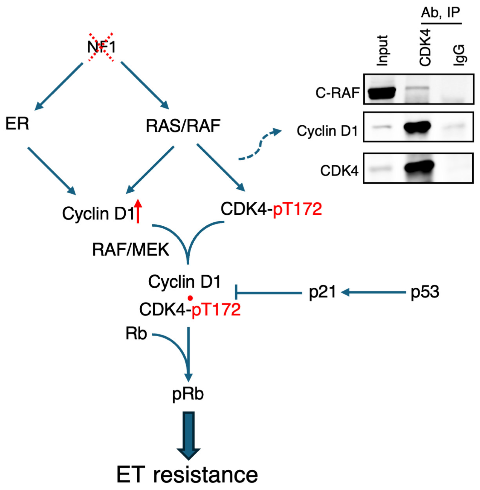
Figure 1. Model. NF1 depletion co-activates ER and RAS signaling, which converge on the CDK4/6-Rb pathway to bypass endocrine therapy by cyclin D1 induction (red upward arrow), and CDK4 phosphorylation at T172. p53 may modulate with this pathway via regulation of p21. Upper right: co-immunoprecipitation showed interaction between CDK4 and CRAF in ER+ T47D cells.
Ijms 26 10322 g001
Table 1. Co-occurring and mutually exclusive mutations associated with NF1 shallow deletion, relative to normal NF1 copy number, in the METABRIC ER+ breast cancer cohort. p value and odds ratio were calculated using Fisher’s exact test. FDR was adjusted across all mutations, including non-significant ones. Only mutations with FDR < 0.05 are reported.
Genep ValueFDROdds RatioTrend
TP531.1 × 10−581.9 × 10−565.3Co-occurring
NF17.5 × 10−86.5 × 10−63.2Co-occurring
USH2A1.3 × 10−33.3 × 10−21.7Co-occurring
MUC167.3 × 10−42.1 × 10−21.5Co-occurring
PIK3CA4.7 × 10−41.6 × 10−20.7Mutually Exclusive
GATA31.1 × 10−44.6 × 10−30.5Mutually Exclusive
MAP3K14.4 × 10−42.5 × 10−30.5Mutually Exclusive
Table 2. Relapse-free survival in relation to NF1 and TP53 genetic status in the ER+ breast cancer cohort in METABRIC. Hazard ratio with 95% confidence intervals (CI), and p values were calculated using the Cox proportional-hazards model, with patients having normal NF1 copy number and wild type TP53 (n = 886) as the reference group.
Tumor TypeHazard Ratio (HR)HR 95% CIp Value
NF1 Loss, TP53 Wildtype (n = 148)1.641.27–2.130.000185
NF1 Normal, TP53 Mutated (n = 127)1.621.22–2.150.00077
NF1 Loss, TP53 Mutated (n = 106)1.741.3–2.350.000248
Disclaimer/Publisher’s Note: The statements, opinions and data contained in all publications are solely those of the individual author(s) and contributor(s) and not of MDPI and/or the editor(s). MDPI and/or the editor(s) disclaim responsibility for any injury to people or property resulting from any ideas, methods, instructions or products referred to in the content.