Review

Beyond Folding: Expanding the Functional Landscape of Hsp90 Chaperone Machinery in Health and Disease

1
Department of Biochemistry and Molecular Biology, School of Medicine, Kyung Hee University, Seoul 02447, Republic of Korea
2
Biomedical Science Institute, Kyung Hee University, Seoul 02447, Republic of Korea
3
Department of Biomedical Science, Graduate School, Kyung Hee University, Seoul 02447, Republic of Korea
*
Authors to whom correspondence should be addressed.
Int. J. Mol. Sci. 2025, 26(21), 10279; https://doi.org/10.3390/ijms262110279
Submission received: 15 September 2025 / Revised: 16 October 2025 / Accepted: 22 October 2025 / Published: 22 October 2025
(This article belongs to the Section Macromolecules)

Abstract

Molecular chaperones are crucial for maintaining protein homeostasis by assisting in the proper folding, stabilization, and function of proteins. Among them, Heat shock protein 90 (Hsp90), represents a highly conserved protein family of molecular chaperones that plays an essential role in diverse biological processes and is fundamental to cellular health and survival. As a highly abundant molecular chaperone, Hsp90 comprises 1–2% of cellular proteins, increasing to 4–6% under stress conditions. It interacts with client proteins, assisting them in proper folding and stability. Unlike classical chaperonins, Hsp90 operates through a highly regulated, ATP-dependent cycle that involves multiple co-chaperones. This process allows Hsp90 to selectively engage with numerous client proteins, including signaling proteins, kinases, hormone receptors, and transcription factors. Recent discoveries have revealed its involvement in processes beyond protein folding, demonstrating its role in diverse cellular functions such as epigenetic regulation, immune signaling, and oncogenic transformation. This current review highlighted the specific characteristics of cytoplasmic and endoplasmic reticulum (ER) as well as mitochondrial paralogs and functions, focusing on its contribution to buffering genetic variation, facilitating oncogene addiction, and modulating disease phenotypes in conditions such as cancer, neurodegeneration, cardiovascular diseases (CVD), and diabetes. We also discuss the therapeutic potential of targeting Hsp90 and its co-chaperones, outlining the challenges and prospects in drug development. These insights not only reshape our understanding of chaperone biology but also present opportunities for precision medicine in various human diseases.

1. Introduction

The heat shock protein 90 (Hsp90), is a highly abundant and conserved molecular chaperone which plays an essential role in maintaining protein homeostasis across all organisms. In eukaryotic cells, Hsp90 is essential for cell viability under both normal and stress conditions. Among the heat shock proteins (HSPs), Hsp90 is the most prominent chaperone, accounting for approximately 1–2% of total cellular protein under normal physiological conditions, with levels increasing to 4–6% during stress [1]. Hsp90 comprises four main isoforms: the inducible cytosolic Hsp90α (Hsp90AA1), the constitutive cytosolic Hsp90β (Hsp90AB1), the ER resident Grp94/Gp96, and the mitochondrial paralog tumor necrosis factor receptor-associated protein-1 (TRAP1) [2]. Additionally, there is an alternative splice variant, Hsp90N, which is associated with cellular transformation. TRAP1 shares similarities with the bacterial homolog HtpG and is found in the mitochondrial matrix, while ER-resident Hsp90 is localized in the ER lumen (Table 1). This indicates a close evolutionary relationship between TRAP1 and eubacterial HtpG [3]. In plants, Hsp90 has been identified in the chloroplast (cHsp90). It plays a role in the import of proteins into the organelle as well as in the maturation of proteins related to photosynthesis [4].

1.1. Hsp90 Structure and Co-Chaperone and Client Interactions

Hsp90 is a cytosolic molecular chaperone that primarily exists as an elongated homodimer. In mammalian cells, two cytosolic isoforms of Hsp90 have been identified: Hsp90α and Hsp90β, encoded by distinct genes and sharing approximately 84% amino acid sequence identity [2]. In contrast, the bacterial homolog HtpG is largely dispensable under non-stress conditions [11]. In S. cerevisiae, two Hsp90 isoforms, Hsc82 and Hsp82, share 97% amino acid sequence homology. Hsc82 is constitutively expressed, whereas Hsp82 is induced in response to stress [12]. Higher eukaryotes contain both homodimers (α-α, β-β) and heterodimers (α-β) [13]. Each Hsp90 monomer consists of three structural domains: an N-terminal nucleotide-binding domain (NTD) responsible for ATP binding and hydrolysis, a middle domain (MD) that interacts with client proteins, and a C-terminal dimerization domain (CTD) featuring a conserved MEEVD recognition motif [14,15]. The N-terminal domain contains the canonical ATP-binding pocket, which also serves as a target for several natural product inhibitors, such as radicicol and geldanamycin. These compounds interfere with Hsp90’s function, destabilizing protein kinases and ultimately promoting their degradation [16]. The MD, comprising approximately 50 amino acid residues, is crucial for coordinating interactions between the NTD and CTD, linking ATP binding with conformational changes in Hsp90 [17]. Notably, the MD has a catalytic site that binds the γ-phosphate of ATP, classifying Hsp90 as a “split ATPase”. Structural similarities with DNA topoisomerase II (gyrase B) further underscore the MD’s significance as a primary docking site for client proteins such as AKT, Cdc37, p53, and eNOS [18,19,20]. Recent studies have revealed that human Hsp90 (hHsp90) adopts a more flexible structural confirmation, predominantly in an open/apo state. This structural plasticity allows Hsp90 to accommodate a broader range of clients and co-chaperones, providing additional regulatory mechanisms (Figure 1A,B) [21].
Moreover, the modulation of lid dynamics in hHsp90 represents an evolutionary adaptation that alters its functional properties and cellular requirements [22]. In vivo, Hsp90 function is regulated by a repertoire of co-chaperones that either directly modulate client proteins or fine-tune the chaperone cycle, resulting in a highly dynamic network of transient chaperone and co-chaperone assemblies [23]. Proteome-wide analyses have revealed that approximately 10% of human proteins are clients of Hsp90, and their proper folding and maturation critically rely on it. These client proteins encompass diverse functional categories, including transcription factors, ubiquitin-related proteins, and a significant number of kinases. Notably, nearly 60% of Hsp90-dependent kinases are part of the serine/threonine kinase family, such as Cdc37, eIF-2α, Src kinases, and Raf kinases. Hsp90 plays a crucial role in regulating essential cellular processes, such as signal transduction, apoptosis, and cell survival, through its interactions with these proteins [24,25]. Hsp90 co-chaperones exhibit substantial structural diversity, and their interactions are dynamically modulated by cofactors such as activator of Hsp90 ATPase homolog 1 (Aha1), Hop, Cdc37, and p23. Several co-chaperones regulate the ATPase activity of Hsp90 in a manner that is specific to both the client and the cellular context. For instance, Aha-1 and high copy Hsp90 suppressor 1 (Hch1), directly stimulate the basal ATPase activity of Hsp90 [26]. The interaction of the GR ligand-binding domain with Hsp90 has been shown to enhance Hsp90 ATPase activity by nearly 200-fold [27]. In contrast, other clients, including the mineralocorticoid receptor ligand-binding domain (MR-LBD), Tau, and the p53 DNA-binding domain (DBD), do not significantly alter Hsp90 ATPase activity [28]. Recognition of client proteins by Hsp90 is driven by conformational rearrangements that expose specific binding interfaces. While the initial modes of interaction vary among different clients, they ultimately converge on the formation of productive closed Hsp90 complexes. ATP-dependent allosteric transitions mediate communication between Hsp90 domains, a process further fine-tuned by co-chaperones such as p23, thereby adapting the chaperone cycle in a client-specific manner [28].
Another co-chaperone such as Hop/Sti1 possesses TPR motifs that allow simultaneously interaction with both Hsp70 and Hsp90, which stabilizes an open confirmation of Hsp90 and inhibits its ATPase activity [29]. Another TPR-containing co-chaperone, PP5/Ppt1, regulates the Hsp90 cycle by dephosphorylation of Hsp90 and the co-chaperone Cdc37, thus affecting the client maturation [30]. Other TPR-containing co-chaperones, including FK506-binding proteins such as FKBP52 (FKBP4), cyclophilin 40 (CPY40), as well as Cpr6 and Cpr7 in yeast, also modulate Hsp90 function through their peptidyl-prolyl cis-trans isomerase (PPIase) domains. Cdc37, which is specific to kinases, interacts with both client kinases and the NTD of Hsp90, partially inhibiting Hsp90’s ATPase activity. In contrast, p23/Sba1 binds directly to the NTD, stabilizing the closed conformation of Hsp90, which reduces ATPase activity and regulates the progression of the chaperone cycle and maturation of client proteins [31]. Beyond protein folding, the Hsp90-Hsp70 chaperone machinery is critical in RNA metabolism, particularly in loading small RNA duplexes into Argonaute (AGO) proteins during the assembly of the RNA-induced silencing complex (RISC). Pharmacological inhibition of Hsp90’s ATPase activity with geldanamycin and its derivatives significantly reduces RISC activity, underscoring the essential role of Hsp90 in small RNA processing pathways (Figure 2) [32].
Most client proteins are recruited via large multiprotein complexes formed by the Hsp90/Hsp70 chaperone machinery [33]. Under stress conditions, cells activate protective mechanisms by either inducing pro-survival kinases or inhibiting pro-apoptotic signaling pathways [34]. AKT is a well-characterized Hsp90-dependent kinase, whose active conformation is stabilized via intracellular complex formation with Cdc37. Hsp90 inhibition suppresses AKT phosphorylation, leading to increased apoptosis. In interstitial epithelial cells (IECs) exposed to hypoxic conditions, a reduction in Hsp90 decreases phosphorylated AKT (pAKT), triggering apoptosis, whereas an increase in Hsp90 enhances pAKT levels and reduces apoptosis. This highlights the role of AKT in Hsp90-dependent cell survival [35]. Specifically, Hsp90β protects hypoxic IECs from apoptosis by enhancing the activity of the pro-apoptotic protein Bad, which in turn reduces the release of cytochrome c, caspase-3 activation, and PARP cleavage [36]. Additionally, Hsp90 promotes AKT activity by inhibiting protein phosphatase 2A (PP2A) [18]. Emerging biophysical approaches, such as single-molecule fluorescence and force spectroscopy, are improving our understanding of these dynamic interactions (Figure 2) [37].
The stability of Hsp70-based complexes is further enhanced by Hsp70-interacting proteins (HIP), which facilitate ATP hydrolysis. For instance, Hsp40 initiates the chaperoning of progesterone receptor (PR) by regulating the ATPase activity of Hsp70 [38]. The PR then forms a complex with Hsp40 and Hsp70, which subsequently recruits Hop and Hsp90, resulting in formation of an intermediate complex. This complex ultimately matures into a PR: Hsp90: p23 complex. The co-chaperone p23 selectively binds to Hsp90 in its ATP-bound state, interacting with both the N-terminal and middle domains of dimeric Hsp90 [39]. This interaction stabilizes the closed conformation of Hsp90 and maintains the receptor’s ligand-binding domain in a configuration that allows for high-affinity hormone binding [40]. Characterizing the binding interfaces of Hsp40 on PR and exploring whether similar sites exist on other Hsp90 client proteins represents an important avenue for future research. A comprehensive list of Hsp90 interactors is available on the following link https://www.picard.ch/downloads/Hsp90interactors.pdf (accessed on 27 August 2025).
The glucocorticoid receptor (GR), a prototypical member of the steroid hormone receptor (SHR) family, requires interaction with Hsp90 to attain its hormone-binding conformation [41]. The interaction of GR and Hsp90 is ATP-dependent process that involves a prior activity of Hsp70 and the co-chaperone Hop, leading to the formation of an inactive “loading complex” comprising GR, Hsp90, Hsp70, and Hop. upon ATP hydrolysis by Hsp90, Hsp70, and Hop, the latter two components are released, permitting the incorporation of p23 and the assembly of the active GR: Hsp90: p23 “maturation complex”, which restores GR ligand-binding activity with enhanced affinity [42]. Notably, the C-terminal tail helix of p23 can directly interact with the GR ligand-binding domain (GR-LBD) in vitro, thereby promoting receptor maturation even in the absence of Hsp90 [43]. The highly conserved hydrophobic groove of GR interacts with the p23 tail helix, stabilizing the closed confirmation of Hsp90 and underscoring the critical role of p23-dependent chaperoning across all SHRs [44]. The conformational transition of the Hsp90 complex from an open-to-closed state is coordinated by ATPase activities of both Hsp70 and Hsp90 [45,46]. Recent Cryo-EM studies have revealed that GR binds within the lumen of Hsp90, with the complex further stabilized by p23 [44].

1.2. ER-Resident Hsp90 (Grp94)

Glucose-regulated protein 94 (Grp94, also known as gp96 or Hsp90B1) is an ER-resident paralog of Hsp90 that shares approximately 50% sequence homology with its cytoplasmic counterpart [47]. Like Hsp90s, Grp94 functions as a homodimer, composed of two identical promoters comprising an NTD, MD, and a CTD. A unique feature of Grp94 is the presence of a pre-N domain within the NTD, which regulates ATP hydrolysis and the maturation of client protein [48]. Grp94 exhibits a shorter, lysine-rich charged linker that contains multiple calcium-binding sites [49]. It also possesses a carboxy-terminal KDEL motif, which facilitates its retention in the ER, and it lacks the canonical carboxy-terminal MEEVD sequence. Due to its hydrophobic properties, Grp94 is associated with the membrane of the ER and Golgi complex [50]. Grp94 interacts with a variety of ER-resident proteins, including protein kinases, calmodulin, and molecular chaperones such as Grp78 (BiP), calnexin, calreticulin, and Grp170. The interaction between Grp94 and BIP is client-specific and does not exhibit a general chaperone mechanism. In vivo, the maturation of Toll-like receptors (TLRs) requires the client-specific co-chaperone canopy fibroblast growth factor signaling regulator 3 (CNPY3). CNPY3 forms a trimolecular complex with gp96 and TLR9 precursor 1, thereby synergistically promoting the tertiary folding of the TLR9 precursor 2. Once the TLR9 precursor 2 achieves its properly folded conformation, it dissociates from the gp96–CNPY3 complex and is subsequently translocated to the endolysosome for further processing [51]. These findings suggest that the interaction between Grp94 and the ER-resident chaperone BiP may become particularly critical in the absence of client-specific co-chaperones [52].
Grp94 is involved in protein folding, processing, and secretion, and it plays a significant role in the signaling pathways of steroid hormones, integrins, and TLRs, which are crucial for immunity and metastasis. ATP binding induces conformational changes in the lid region of Grp94, while BIP binding stabilizes a partially closed Grp94 intermediate. Conversely, impaired ATP binding disrupts the processing of client proteins. Under conditions of ER stress, Grp94 can adopt an ATP-independent “holdase” mode, stabilizing unfolded proteins until normal folding conditions are restored. This ATP-independent activity has been observed in the co-chaperone-independent processing of p53 and the stabilization of the client kinase v-Src under heat stress [53,54].
An emerging regulatory paradigm in chaperone biology is the concept of the “chaperone code,” wherein post-translational modifications (PTMs) dynamically modulate chaperone activity and client interactions [55]. Proteomic analyses have identified nearly 300 PTM sites on Grp94, most of which localize within the pre-N domain, suggesting a complex and largely uncharacterized layer of regulation. Functionally, Grp94 is indispensable for the stability and maturation of multiple cancer-associated proteins, including TLRs, insulin-like growth factors (IGFs), immunoglobulins, and p53 [56]. For instance, the E3 ubiquitin ligase FBXL2, a member of the conserved F-box protein family, targets both EGFR and tyrosine kinase inhibitor (TKI)-resistant EGFR mutants for proteasome-mediated degradation in EGFR-driven non-small cell lung cancer (NSCLC). FBXL2 itself serves as a client of Grp94, competing with EGFR for Grp94 binding. This competition can alter protein conformations, thereby disrupting the stable interaction between FBXL2 and EGFR. Consequently, the combined strategy of Grp94 inhibition and FBXL2 activation has been shown to exert potent growth-suppressive effects in EGFR-TKI-resistant NSCLC models [57]. Knockdown studies further implicate Grp94 in cell adhesion and migration, providing mechanistic insights into its role in regulating cancer cell motility (Figure 3) [58]. Despite its importance, the cellular functions of Grp94 remain less understood compared to cytosolic or mitochondrial Hsp90. Collectively, these findings highlight Grp94 as a central regulator of proteostasis and signaling in the ER, with particular relevance in cancer biology. Its unique structural features, ATP-dependent and -independent activities, and PTM-driven regulation underscore its therapeutic potential, warranting further exploration of client interactions and regulatory mechanisms for drug discovery.

1.3. Mitochondrial Hsp90 (TRAP1)

TRAP1, also known as Hsp75, is a mitochondrial molecular chaperone that plays a pivotal role in maintaining protein homeostasis and preserving mitochondrial integrity under cellular stress conditions. TRAP1 shares approximately 34% amino acid sequence identity with Hsp90β and contains an N-terminal mitochondrial targeting sequence that is cleaved upon import into the mitochondrial matrix. Unlike cytosolic Hsp90 isoforms, TRAP1 lacks the highly charged, flexible linker region typically located between the N- and middle (M) domains [7] as well as the C-terminal MEEVD motif required for the recruitment of co-chaperones. Consequently, TRAP1 can function independently of classical Hsp90 co-chaperones such as p23 and Hop [59]. The binding of client proteins to TRAP1 requires a confirmation transition into the open form. Clients are directly loaded via direct protein–protein interactions, which subsequently promote ATP binding and hydrolysis, driving conformational rearrangements that facilitate client remodeling [60]. Sequential ATP hydrolysis events may generate structural strain on client proteins positioned between TRAP1 protomers, thereby assisting their refolding and stabilizing their native conformations or enabling the formation of multiprotein complexes. Conversely, inhibition of TRAP1 with ATP-mimetic compounds such as Gamitrinibs (geldanamycin-based conjugates) or SMTIN-P01 leads to proteolytic degradation of unfolded client proteins [61]. Gamitrinibs rapidly accumulate within mitochondria, where they compromise mitochondrial integrity by influencing the mitochondrial permeability transition pore (mPTP), promoting cytochrome c release, and activating caspases in prostate cancer cells [62]. These findings highlight the therapeutic significance of mitochondria-targeted drugs as promising anticancer strategies.
Under certain conditions, client proteins bound to TRAP1 do not undergo classical folding but instead achieve structural stabilization through TRAP1’s “holdase” activity. For instance, the interaction between TRAP1 and SIRT3 stimulates ATPase activity, indicative of this holdase function, which enhances the enzymatic activity of client proteins. However, due to the absence of co-chaperones, e.g., p90, TRAP1 is unable to mediate the maturation of the progesterone receptor (PR). In contrast, cytosolic Hsp90 facilitates PR maturation through its cooperation with co-chaperones such as Hop and p23, a process primarily carried out by the Hsp90α and Hsp90β isoforms [63]. A distinct mitochondrial pool of Hsp90 has been identified in various cancer cells as well as in certain normal mouse tissues. This mitochondrial Hsp90 population appears to be regulated differently from its cytosolic counterpart and does not physically interact with TRAP1 [8]. Beyond its chaperoning function, TRAP1 regulates multiple mitochondrial processes, including bioenergetics, redox homeostasis, oxidative stress responses, mitochondrial dynamics, mitophagy, and resistance to apoptosis [64].
Functionally, TRAP1 exhibits a dual role: it acts as a cytoprotective factor under physiological stress while also promoting tumor progression. The metabolic reprogramming of cancer cells from oxidative phosphorylation (OXPHOS) to glycolysis, a hallmark of tumor metastasis is partially regulated by TRAP1 through its interaction with succinate dehydrogenase alpha (SHDα) under hypoxic and nutrient deficient conditions [65]. During nutrient deprivation, TRAP1 enhances glutamine metabolism to meet elevated energy demands, thereby supporting cancer cell survival and proliferation [66]. Conversely, TRAP1 overexpression and SDH inhibition can protect tumor cells from oxidative stress by preventing the opening of the mitochondrial permeability transition pore (mPTP), inhibiting cytochrome c (Cyt c) release, and suppressing apoptosis [67]. Cyclophilin D (CypD), another regulator of mPTP opening, maintains mitochondrial Ca2+ homeostasis. Excessive Ca2+ accumulation disrupts F1F0-ATP synthase activity, promotes ROS generation, and triggering mPTP opening, loss of mitochondrial membrane potential, and apoptosis. TRAP1 induction inhibits CypD-dependent mPTP opening, thereby preventing the mitochondrial-mediated apoptosis [68]. Moreover, TRAP1 stabilizes HIF1α via SDH inhibition, promoting the downstream genes that enhance angiogenesis and nutrient supply in proliferating tumor cells [69]. TRAP1 prevents the ubiquitin-mediated degradation of CDK1 and MAD2, supporting mitotic progression in cancer cells. This regulatory mechanism has been demonstrated in breast, colorectal, and lung carcinoma cell lines and tumor samples, highlighting TRAP1 as a promising therapeutic target in these malignancies [70].
Mitochondria-targeted Hsp90 inhibitors have emerged as promising agents for selectively modulating TRAP1 activity while minimizing off-target effects associated with pan-Hsp90 inhibition. The compound SMTIN-P01 demonstrated enhanced cytotoxicity against various cancer cell lines (22Rv1, A172, H460, and MDA-MB-231) compared with PU-H17, underscoring the therapeutic potential of mitochondrial targeting. Incorporation of a triphenylphosphonium (TPP) moiety facilitates the mitochondrial accumulation of these inhibitors and enables engagement with an allosteric site on TRAP1, thereby increasing its ATPase activity [61]. Similarly, the C-terminal inhibitor 6BrCaQ-C10-TPP, generated by linking 6BrCaQ to TPP through variable linkers exhibited potent nanomolar GI50 values against multiple cancer cell lines, including MDA-MB-231, HT-29, HCT-116, K562, and PC-3. This compound significantly affected mitochondrial membrane potential and preserved TRAP1 client proteins such as SDH without inducing a heat-shock response [71]. DN401, another TRAP1-selective inhibitor, also showed preferential binding and potent antiproliferative effects in cancer cells. Alternatively, the addition of cationic moieties such as TPP, rhodamine, cyanine, or cationic peptides into Hsp90 inhibitors enhances their mitochondrial accumulation by 100–1000-fold, as confirmed through fluorescence co-localization with MitoTracker dyes and mitochondrial isolation assays [72].
Natural compounds have also been identified as inhibitors of TRAP1. For instance, Honokiol lipophilic bisdichloroacetate ester (HDCA) exhibits potent antineoplastic activity by inducing oxidative stress and apoptosis through selective inhibition of TRAP1 ATPase activity at low micromolar concentrations, without affecting cytosolic Hsp90 isoforms [73]. Similarly, green tea extract (GTE) and its major polyphenolic component, epigallocatechin gallate (EGCG), broadly inhibit chaperone activity. Both GTE and EGCG suppress the expression of TRAP1, Hsp90, and Hsp27, thereby impairing tumor growth and promoting apoptosis [74]. Collectively, these findings indicate that both HDCA and GTE exert anticancer effects through inhibition of mitochondrial and cytosolic heat shock proteins, positioning TRAP1 as a promising therapeutic target for cancer treatment [65].

1.4. Post Translational Modifications (PTMs)

PTMs such as phosphorylation, acetylation, glycosylation, methylation, ubiquitination, and SUMOylation of Hsp90 have been identified as vital regulators of its interactions with co-chaperones and client proteins [75]. These modifications collectively regulate Hsp90 conformational dynamics, the maturation of client proteins, and interactions with co-chaperones [76]. Phosphorylation of Hsp90α at Thr90 by protein kinase A alters its interactions with cofactors and client proteins. This modification enhances binding to ATP and cochaperones such as Aha1, p23, PP5, and CHIP, while reducing association with Hsp70, Hop, and Cdc37. It also impairs the binding of client kinases including Src, Akt, and PKCγ. Notably, Thr90 phosphorylation is elevated in proliferating cells, suggesting a regulatory role in tumor metastasis [77]. Another prominent phosphorylation sites at serine residues Ser226 and Ser255, with additional sites found on tyrosine. This modification affects Hsp90’s conformational transitions, thereby influencing client protein folding and Hsp90 conformational transitions, modulating client protein folding and co-chaperone binding. The phosphorylation close to the TPR recognition site located in the CTD at Thr725 and Ser726 are adjacent phosphorylation, which influence the co-chaperone interaction [78]. Particularly, hyperphosphorylation disrupts these processes, whereas basal phosphorylation promotes Hsp90 dimerization and facilitates the assembly of higher-order oligomers [79].
Additionally, phosphorylation of Hsp90β at Ser226 and Ser255 stabilizes a closed conformation by orienting the linker in an upward direction, thereby exposing the middle domain of Hsp90 and promoting stable interactions with co-chaperones such as [80]. Methylation of Hsp90 at lysine 616 (K616) by Smyd2 is required for proper sarcomere function; however, this observation still requires validation in cardiac and skeletal muscle cells [81]. Additionally, S-nitrosylation of Hsp90 at cysteine 589 (C589) by nitric oxide (NO) stabilizes the TGF-β receptor and mitigates fibrosis by blocking the TGF-β/SMAD3 signaling pathway in cardiomyocytes [82]. Another report showed that SUMOylation of Hsp90 at lysine 191 (K191) has been shown to facilitate recruitment of the co-chaperone Aha1, which enhances Hsp90 ATPase activity [83], whereas Hsp90α-K283, and Hsp90β-K275 modulates drug binding to Hsp90 by influencing both adjacent and distal serine and threonine phosphorylation sites [84]. These conformational rearrangements disrupt Hsp90’s canonical ATPase-driven folding cycle but promote its incorporation into higher-order, multimeric epichaperome assemblies. Under stress conditions, N-glycosylation of Grp94 at Asn62, located within intrinsically disordered regions (IDRs), stabilizes a pathological conformation that facilitates its integration into epichaperomes [85]. In breast cancer, the Grp94 N62Q mutant, which fails to undergo glycosylation, exhibits reduced epichaperome formation, diminished plasma membrane localization, and attenuated EGFR signaling compared to N217A mutant, which retain EGFR signaling. This demonstrates that Grp94 glycosylation is critical for the pathological induction and stabilization of epichaperomes via extensive protein–protein interactions (PPIs). For instance, over 2481 proteins were sequestered in breast cancer cells associated with aggressive phenotypes. Notably, cells entering mitosis in the presence of epichaperome disruption fail to complete cell division and ultimately undergo cell death, underscoring their dependency on epichaperome-mediated mitotic rewiring [84]. In summary, these findings suggest that targeting epichaperomes to restore context-specific PPI network function may represent a promising therapeutic strategy for diseases such as cancer and Alzheimer’s disease (AD) [86].

1.5. Role of Hsp90 in Cellular Processs and Protein Homeostasis

Hsp90 has been reported in various cellular processes including protein folding, signal transduction, maturity and stability of a wide range of client proteins [87]. These client proteins encompass steroid hormone receptors, transcriptional factors, signaling kinases, and growth factors [88]. In humans, Hsp90 exerts a significant regulatory influence on kinase activity by facilitating the assembly of client proteins into multimeric complexes, such as those involving kinases and kinetochore components, and by promoting ligand binding, as observed in steroid hormone receptors [89]. The extent of Hsp90’s direct participation in de novo folding or refolding of denatured polypeptides remains a topic of debate. In S. cerevisiae, loss of Hsp90 function does not result in widespread folding defects [90,91]. Similarly, in vitro studies indicate that Hsp90 alone is insufficient to refold denatured proteins. Instead, it stabilizes non-native substrates in a “folding-competent” state, thereby allowing other factors to facilitate subsequent folding [50].
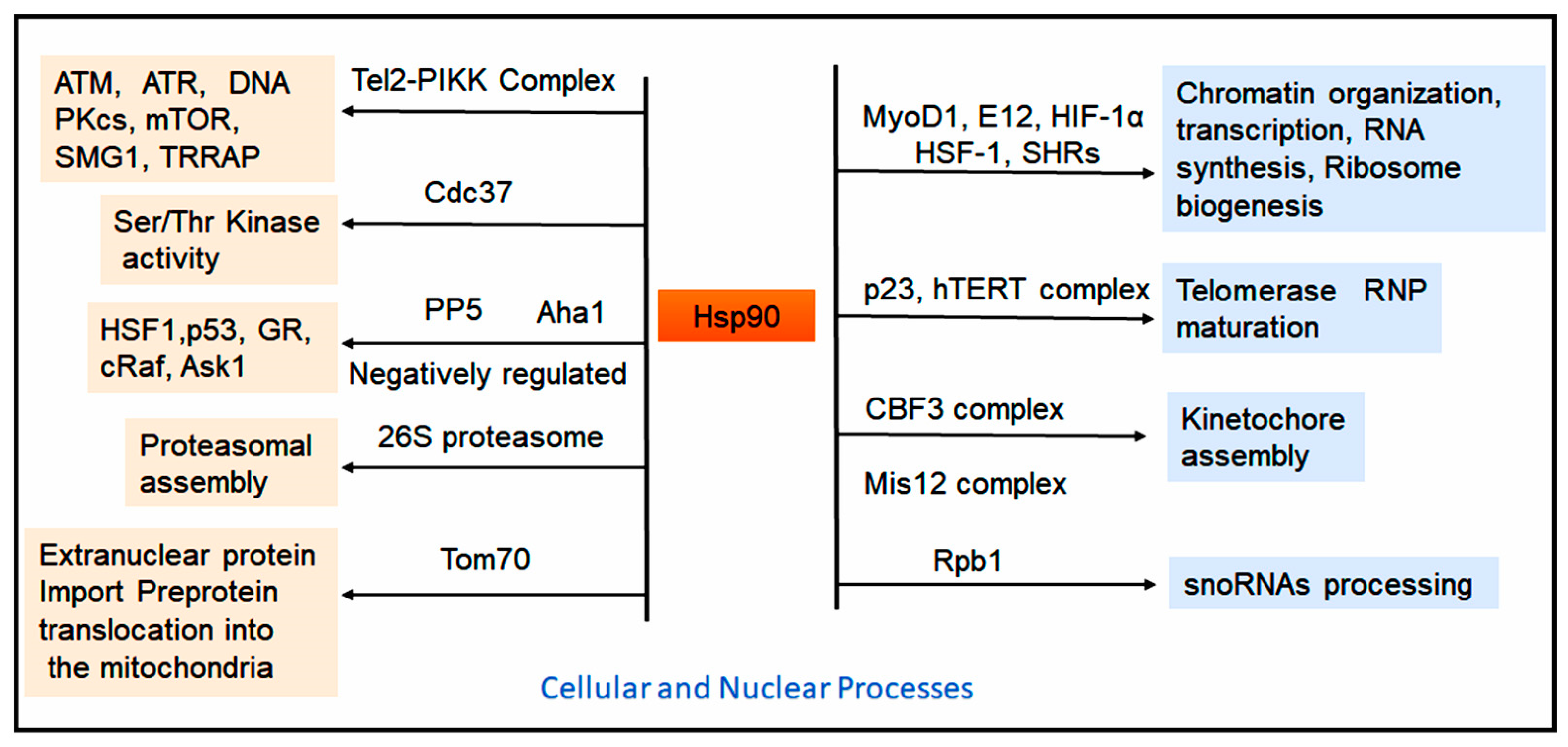
More than half of all human kinases require Hsp90 and its co-chaperone Cdc37 for proper folding and activation. Hsp90 also modulates kinase activity by influencing PTMs. For instance, the Hsp90-associated serine/threonine-protein phosphatase 5 (PP5) dephosphorylates and inactivates c-Raf [92]. PP5, a member of the PPP phosphatase family, contains an N-terminal-TPR domain preceding its catalytic domain and acts as a negative regulator of several Hsp90 clients, including apoptosis signal–regulating kinase 1 (Ask1) under oxidative stress [93]. Other Hsp90 client kinases include ATM, which is activated in response to DNA double-strand breaks, and Chk1, which responds to UV-induced DNA damage [94,95]. The co-chaperone Aha1 binds to the MD of Hsp90, stimulating ATP hydrolysis and inorganic phosphate release. This activity promotes client folding, dissociation of co-chaperones and immunophilins, and resetting of Hsp90 to its open conformation [96]. PP5 also regulates several transcriptional regulators, such as Hsf1, p53, and the GR in an Hsp90-dependent manner (Figure 4) [97,98]. Additionally, phosphorylation of Cdc37 at Ser13 facilitates client recruitment to the Hsp90:Cdc37:kinase complex, while PP5-mediated dephosphorylation of Cdc37 within the complex promotes client release and kinase modification [99]. Beyond kinases, approximately 30% of E3 ubiquitin ligases and 7% of the transcription factors, including p53, interact with Hsp90. An updated list of Hsp90 client proteins is available at https://www.picard.ch/downloads/Hsp90interactors.pdf (accessed on 27 August 2025). The TPR co-chaperone CHIP, which contains a U-box domain homologous to E4 ubiquitination factors, recognizes both Hsp90 and Hsc70/Hsp70, targeting chaperone-bound misfolded proteins for ubiquitination and degradation.
Although predominantly cytoplasmic, approximately 5–10% of Hsp90 resides within the nucleus under basal conditions, with nuclear localization markedly increased during cellular stress. Nuclear Hsp90 associates with nucleoli [100] and peri-chromatin ribonucleoprotein fibrils [101], facilitating the nuclear import of proteins such as FKBP52, steroid hormone receptors (SHRs), and kinases [102,103]. Notably, these import processes are independent of calcium, calmodulin, and ATP [104]. Hsp90 also interacts directly with DNA, RNA, and histones, thereby influencing chromatin organization, transcriptional regulation, and RNA synthesis. It promotes the subnuclear translocation of steroid receptors to chromatin [105] and forms complexes with various transcription factors, including zinc finger proteins, helix–loop–helix proteins, MyoD1, E12, hypoxia-inducible factor-1α (HIF-1α), and heat shock factor-1 (Hsf1) [50]. Reduction in Hsp90 levels activates Hsf1 in vitro [106,107], while pharmacological inhibition by geldanamycin (GA) derivatives induces Hsf1 activation in vivo, suggesting that Hsp90 represses Hsf1 activity under non-stress conditions. Under normal circumstances, Hsp90 forms a complex with Hsf1, which dissociates during stress, allowing Hsf1 to trigger the heat shock response [107]. Additionally, Hsp90 supports ribosome biogenesis by facilitating the nuclear export of the assembled 60S ribosomal subunit into the cytoplasm [108].
The role of Hsp90 in telomerase ribonucleoprotein (RNP) assembly and function is less clearly defined. The Hsp90–p23 complex has been reported to be indispensable for telomerase RNP maturation and activity [109]. Inhibition of Hsp90 by GA and its derivatives suppresses telomerase activity in vitro. The integrity of the hTERT–Hsp90–p23 complex is essential for nuclear localization of hTERT, whereas dissociation of p23 results in cytoplasmic retention of hTERT [109]. Another essential Hsp90 client is Tel2, a protein critical for the stability of all six members of the phosphatidylinositol 3-kinase–related kinase (PIKK) family: ATM, ATR, DNA-PKcs, mTOR, SMG1, and TRRAP. These kinases govern key cellular processes, including DNA damage response (ATM, ATR, DNA-PKcs), nutrient sensing (mTOR), nonsense-mediated mRNA decay (SMG1), and transcriptional regulation (TRRAP). Inhibition of Hsp90 disrupts the Tel2–PIKK complex, similar to the effects observed with loss of its co-chaperone Cdc37 [110]. Hsp90 also plays a pivotal role in kinetochore assembly, a dynamic multiprotein structure anchoring chromosome to spindle microtubules during mitosis. The chromatin-binding complex CBF3, composed of Cep3, Ctf13, Skp1, and Ndc10, depends on Hsp90 for maintaining transient interactions between Sgt1 and Skp1/Ctf13, thereby regulating the balance between CBF3 assembly and turnover (Figure 4) [111]. In vitro evidence further indicates that the Hsp90–Sgt1 complex stabilizes the Mis12 complex, which is vital for kinetochore organization [112].
Beyond nuclear functions, Hsp90 contributes to extranuclear protein import, particularly the translocation of preproteins into mitochondria through the translocase of the outer mitochondrial membrane (Tom70) complex (Figure 4). Inhibition of the Hsp90 C-terminal domain (CTD) by novobiocin prevents preprotein complex formation, while GA impairs the generation of translocation intermediates, underscoring Hsp90’s essential role in mitochondrial targeting and import [113].
Moreover, Hsp90 is crucial for the assembly and stability of several macromolecular complexes. A novel ATP-dependent function has been identified in the assembly of the 26S proteasome, where Hsp90 inhibition results in complete disassembly into the 20S core particle and lid component, demonstrating its necessity for proteasomal integrity [114]. In HER2-positive breast cancer cells, proteasome inhibitors induce degradation of ErbB family proteins by triggering Hsp90 inhibition through a chaperone stress response, thereby confirming these proteins as bona fide Hsp90 clients [115]. Similarly, Hsp90 contributes to the assembly of the R2TP complex in S. cerevisiae, which regulates the accumulation and processing of box C/D small nucleolar RNAs (snoRNAs). Similarly, in human cells, Rpb1 paralog of the R2TP complex associates with a prefoldin-like complex [116]. Additionally, Hsp90 stabilizes Rpb1 during cytoplasmic assembly of RNA polymerase II [117]. Collectively, these findings highlight Hsp90’s central role in maintaining protein homeostasis through the folding, activation, and assembly of diverse protein complexes. The evidence suggests that combined inhibition of Hsp90 and the proteasome represents a promising therapeutic approach for overcoming drug resistance in cancers.

2. Implications of Hsp90 in Health and Diseases

Hsp90 is a highly conserved and abundantly expressed molecular chaperone found in both eukaryotic and prokaryotic cells. It plays a pivotal role in numerous cellular processes, including protein folding, stabilization, degradation, and the regulation of signaling pathways involved in cell proliferation, differentiation, migration, angiogenesis, steroid hormone signaling, organelle-specific stress responses, and apoptosis. There is growing evidence that highlights the significant involvement of Hsp90 in the development of a wide range of human diseases, such as cancer, neurodegenerative disorders, cardiovascular diseases, inflammatory conditions, infectious diseases, and age-related pathologies [118,119]. As a result, several Hsp90 inhibitors are currently in clinical evaluation for the treatment of various malignancies [120,121]. Structurally, Hsp90 consists of distinct domains that allow it to interact with specific client proteins and oncogenic factors. This structural complexity opens up opportunities for developing therapeutic strategies aimed at selectively targeting its binding interfaces. Combining Hsp90 inhibition with client-specific therapeutic agents may represent a promising approach for preventing and treating cancer, as well as other diseases associated with Hsp90.

2.1. Hsp90, ER-Localized Grp94, and Mitochondrial Hsp90 in Cancers

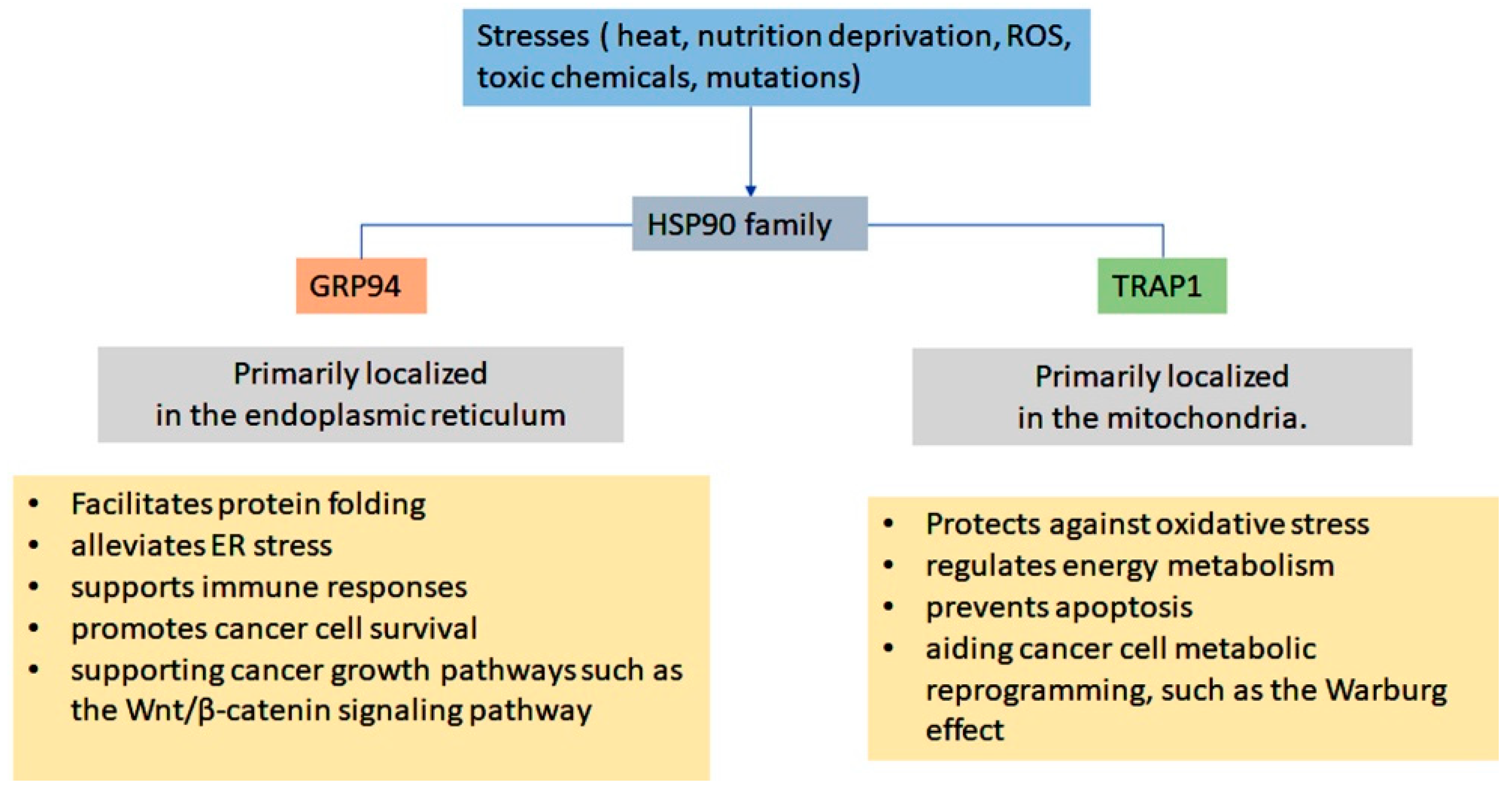
Tumor cells often encounter stressful conditions such as heat, nutrient deprivation, hypoxia, and aberrant angiogenesis. In response to these stressors, stress responsive genes are activated, leading to the induced expression of Hsp90 in tumor tissues [122]. The upregulated Hsp90 promotes cancer cell survival, proliferation, and metastasis [123]. Elevated Hsp90 expression has been consistently observed across various types of malignancies. For instance, nearly 90% of primary breast cancers exhibit high levels of Hsp90, which strongly correlate with poor prognosis [124]. Upregulation of Hsp90 has also been reported in other cancers, including multiple myeloma, leukemia, colorectal cancer, non–small cell lung cancer, and prostate cancer [122]. In addition to its intracellular functions, Hsp90 can be secreted by various cancer cell lines and human tumors [77]. Extracellular circulating Hsp90 (eHsp90) has emerged as an early and reliable pan-cancer biomarker for multiple cancers. eHsp90 interacts with cell-surface receptors, including low-density lipoprotein receptor-related protein-1 (LRP1) and toll-like receptors (TLR2 and TLR4), thereby promoting cancer cell proliferation, migration, and invasion [125]. Notably, the co-chaperone of eHsp90, Morgana, which is secreted via unconventional pathways, enhances cancer cell motility via TLR2, TLR4, and LRP1 signaling. Its overexpression in breast cancer has been associated with more aggressive tumor and unfavorable clinical outcomes [126,127]. Therapeutic strategies targeting eHsp90 or co-chaperones like Morgana—such as monoclonal antibodies—have demonstrated promise in suppressing tumor growth by stimulating macrophage-dependent phagocytosis and recruiting CD8+ T cells. These findings highlight novel avenues for anticancer therapy, although further validation is necessary [128].
Hsp90 plays a significant role in multidrug resistance (MDR) in cancer by positively regulating the expression of various resistance-associated proteins, including P-glycoprotein (P-gp/MDR1), breast cancer resistance protein (BCRP), survivin, and Bcl-2 [129,130]. Mechanistically, Hsp90 promotes the accumulation of β-catenin, a key transcriptional regulator of MDR genes, by activating the AKT/GSK3β signaling pathway. In ovarian cancer, this Hsp90-driven activation of β-catenin is a crucial factor underlying resistance to paclitaxel and cisplatin. Inhibiting Hsp90 sensitizes MDR ovarian cancer cells to chemotherapy by impairing the AKT/GSK3β/β-catenin signaling pathway, highlighting Hsp90 as a potential therapeutic target [129]. Additionally, Hsp90 stabilizes several oncogenic signaling proteins. For example, mutant B-Raf, an essential component of the Ras–Raf–ERK signaling cascade that regulates cell proliferation and survival, relies on Hsp90 for its stability and activity (Figure 2) [131]. In breast cancer, estrogen receptor-α (ERα)—a ligand-activated transcription factor that regulates genes associated with cell proliferation—forms a nuclear multiprotein complex with Hsp90 and co-chaperones in the absence of estradiol. Inhibiting Hsp90 promotes the proteasomal degradation of ERα, offering a promising therapeutic strategy in ER-positive breast cancers [123]. Similarly, Hsp90 is crucial for the stability and function of BCR-ABL, a constantly active fusion kinase that drives chronic myeloid leukemia (CML) by activating multiple oncogenic pathways [132]. Additionally, Hsp90 activates NF-κB, a master regulator of inflammation and cell survival, thereby counteracting apoptosis [133].
The tumor suppressor p53, which is mutated in over half of human cancers, plays a central role in apoptosis following DNA damage or oncogenic stress. Multiple studies demonstrated that three main pathways—DNA damage, growth signals (such as those from oncogenes like Ras or Myc), and exposure to chemotherapeutic drugs or ultraviolet light—contribute to the inhibition and degradation of p53, ultimately stabilizing p53 at high levels [134]. Hsp90, in collaboration with Hsp70, binds to mutant p53 and prevents its proteasomal degradation, thereby promoting resistance to apoptosis [135]. Additionally, Hsp90 regulates cell cycle progression by modulating cyclin D-associated kinase activity. Inhibition of Hsp90 disrupts PI3K/AKT signaling (Figure 2) [136], reduces cyclin D-dependent phosphorylation of RB, and induces G1-phase arrest [137]. These findings highlight the potential of Hsp90 inhibitors to target abnormal cell cycle regulation in cancer.
Hsp90 plays a crucial role in cancer cell immortality by supporting the proliferation and survival of cancer cells. It stabilizes telomerase, an enzyme critical for maintaining telomere length, which is essential for the immortality of these cells [138]. The catalytic subunit of telomerase, known as hTERT, interacts with Hsp90 and its co-chaperone p23, forming active telomerase complexes that help preserve telomere length [139]. Additionally, Hsp90 aids in angiogenesis by stabilizing vascular endothelial growth factor receptors (VEGFRs) and fibroblast growth factor receptor 3 (FGFR3) [138]. When Hsp90 is inhibited, these receptors become ubiquitinated and are targeted for degradation, which disrupts angiogenic signaling in tumors [140].

2.2. Grp94

Grp94 is essential for maintaining the ER protein-folding capacity, upholding ER stress sensors, and inhibiting ER-associated apoptosis, thus regulating the balance between cancer cell survival and death [141]. As a key mediator of the unfolded protein response (UPR), Grp94 supports plasma cell differentiation, illustrated by the absence of plasma cells in XBP1-deficient systems [142,143]. Additionally, Grp94 stabilizes and protects a wide range of client proteins critical for immune regulation and tumor progression. These include TLRs, the majority of α- and β-integrin subunits, the Wnt co-receptor LRP6, glycoprotein A repetitions predominant (GARP), insulin-like growth factors (IGFs), and the platelet glycoprotein Ib-IX-V complex [144]. Consistent with its functional significance, Grp94 overexpression has been documented in several cancers, including esophageal [145], lung [146], colon [147], gastric [148], and breast carcinomas [149].
Plasma cells, which play a pivotal role in the development of multiple myeloma (MM), rely heavily on the ER for immunoglobulin folding and processing [150]. The excessive accumulation of misfolded proteins leads to ER stress, necessitating robust chaperone activity. To maintain proteostasis, plasma cells depend on ER chaperones such as BiP/Grp78 and Grp94. In vivo studies show that the growth of both XBP1s-transgenic mice and human MM cell lines is dependent on gp96, indicating that inhibiting gp96 could represent a promising therapeutic strategy against MM [143]. Exploiting the unfolded protein response (UPR) has proven effective for treating MM. The combined use of the Hsp90 inhibitor such as, 17-AAG, and the proteasome inhibitor bortezomib induced strong apoptotic responses by activating all three major ER stress sensors [151,152].
Furthermore, Grp94 has also been implicated in the pathogenesis of several solid tumors. In breast cancer, Grp94 is significantly overexpressed compared to normal breast tissues. The genetic ablation of Grp78 and Grp94 in vivo has been shown to reduce tumor proliferation and migration while enhancing apoptosis [153,154]. In triple-negative breast cancer (TNBC), a highly aggressive subtype lacking estrogen receptor (ER), progesterone receptor (PR), and HER2 expression, Grp94 facilitates tumor progression by activating the Wnt/β-catenin pathway, which promotes brain metastasis in TNBC [155]. This adaptation allows cancer cells to survive in hostile microenvironments through the activation of survival-promoting autophagy [156]. Additionally, Grp94 directly stabilizes HER2, a receptor often overexpressed in aggressive breast cancers, thereby amplifying oncogenic signaling cascades that drive tumor growth [157]. Collectively, these findings suggest that dual blockade of Grp94 and autophagy may offer a novel therapeutic strategy for breast cancer.
Grp94 has also been reported to maintain intestinal homeostasis and colon tumorigenesis. Intestinal epithelial cells (IECs) are continuously regenerated by intestinal stem cells, which operate through the canonical Wnt signaling pathway. This regeneration requires Grp94 to work in conjunction with mesoderm development (MesD), an ER chaperone, to ensure the proper surface expression of Wnt co-receptors LRP5 and LRP6 [158]. In a mouse model of colitis-associated colon cancer, a macrophage-specific knockout (KO) of gp96 contributes to colitis and tumorigenesis by enhancing the production of inflammatory cytokines, such as IL-17, IL-23, IL-12, IL-16, and IFN-γ. Mechanistically, Grp94 in macrophages supports inflammation and tumor growth by promoting β-catenin mutations, activating Wnt signaling pathway in colonic epithelial cells, and creating a pro-tumorigenic cytokine environment [159].

2.3. Mitochondrial Chaperone TRAP1

Elevated expression of TRAP1 has been reported in various cancers, including hepatocellular carcinoma, breast cancer [160], glioblastoma, small-cell lung cancer [161], renal, prostate, ovarian, pancreatic, and colorectal cancers [160], and esophageal carcinomas. This overexpression is often associated with advanced metastatic tumors and poor prognosis [162]. It has been demonstrated that TRAP1 promotes cell survival by interacting with CypD, a peptidyl-prolyl isomerase that sensitizes the opening of the mPTP. The opening of mPTP, triggered by elevated mitochondrial ROS or Ca2+ ions, initiates apoptosis. By binding to CypD, TRAP1 inhibits the opening of PTP, thereby conferring resistance to apoptosis [162]. Additionally, TRAP1 plays a role in the metabolic reprogramming of cancer cells. Despite the availability of oxygen, many tumors rely on glycolysis for rapid ATP generation, a phenomenon known as the Warburg effect [163]. TRAP1 reduces the activity of SDH, which is involved in both the tricarboxylic acid (TCA) cycle and the electron transport chain (ETC). This inhibition leads to the stabilization of hypoxia-inducible factor 1α (HIF1α), promoting tumor growth, although the exact regulatory mechanisms remain incompletely defined [67]. Furthermore, TRAP1 stabilizes and regulates cytochrome c oxidase subunit II (COXII), a critical protein required for ATP synthesis within the ETC. When TRAP1 is inhibited, COXII becomes destabilized, resulting in disruption of the ETC, accumulation of ROS, and apoptosis due to impaired ATP production [164,165,166]. During tumor progression, TRAP1 interacts with NRF2, the master regulator of antioxidant defense, to lower ROS levels [167]. In precancerous cells, it enhances the activity of specific enzymes, like citrate synthase, which fuels the pentose phosphate pathway (PPP) to generate nucleotides and biosynthetic precursors needed for cell proliferation [167]. Moreover, TRAP1 interacts with TBP7, a protein involved in proteostasis and cell cycle regulation, helping to stabilize essential cell cycle regulators such as CDK1 [168]. Downregulation of TRAP1 leads to the degradation of proliferation markers, such as Ki67, CDK1, and MAD2, in breast, colorectal, and lung cancer models, sensitizing these cells to agents that target the cell cycle machinery. These findings suggest that elevated levels of TRAP1 may contribute to resistance against therapies aimed at disrupting cell cycle processes [70].
Overall, these findings underscore Hsp90’s role as a central regulator of various processes, including multidrug resistance, oncogenic signaling, evasion of apoptosis, cell cycle progression, telomerase activity, and angiogenesis. This makes Hsp90 an attractive therapeutic target for treating a wide range of malignancies.

2.4. Clinical Applications of Hsp90 Inhibitors in Cancer

Hsp90 inhibitors have emerged as promising agents for cancer treatment due to their ability to destabilize a wide range of oncogenic client proteins. Researchers have explored both natural products and synthetic derivatives in preclinical and clinical settings.

2.5. Curcumin-Based Hsp90 Inhibitors

Curcumin derivatives have shown potential as novel Hsp90 inhibitors. The compound, 4-(4-pyridinylmethylene) curcumin (C1206), suppresses the ATPase activity of Hsp90, leading to the inhibition of chronic myeloid leukemia (CML) cell proliferation [169]. Similarly, another derivative, C0818, binds to the C-terminal dimerization domain of Hsp90 and inhibits its ATPase activity as well [170]. Additionally, the compound (E)-2-(4-hydroxy-3-methoxybenzylidene)-5-(E)-3-(4-hydroxy-3-methoxyphenyl) acryloyl) cyclopentanone (CUR3d) downregulates the expression of both Hsp90AA1 and Hsp90AB1. This results in growth inhibition, increased cytotoxicity, activation of caspase-3, and DNA fragmentation in hepatocellular carcinoma (HCC) cells [171]. Moreover, combination therapy that incorporates curcumin along with histone deacetylase (HDAC) inhibitors like vorinostat and panobinostat has proven effective. This combination enhances the depletion of Hsp90 client proteins, producing a synergistic effect that leads to increased growth inhibition in squamous cell carcinoma (SCC) A431 and mesothelioma STO cells [172].

2.6. Geldanamycin and Derivatives

Geldanamycin is a natural benzoquinone ansamycin that binds to the N-terminal ATP-binding pocket of Hsp90. This interaction inhibits the chaperone function of Hsp90 and disrupts cell proliferation. However, the clinical development of geldanamycin was limited due to issues such as hepatotoxicity, metabolic instability, and poor solubility. As a result, researchers developed less toxic derivatives, including 17-allylamino-17-demethoxygeldanamycin (17-AAG) and 17-dimethylaminoethylamino-17-demethoxygeldanamycin (17-DMAG), which showed improved potency and tolerability [173]. 17-AAG retains potent anticancer activity while reducing hepatotoxicity and improving bioavailability [174]. It binds to the ATP-binding site of Hsp90, inhibiting the formation of multi-chaperone complexes and promoting the ubiquitin-proteasome-mediated degradation of client proteins [175]. In cases of HER2-positive breast cancer, combining trastuzumab with 17-AAG resulted in clinical benefits for 59% of patients. In neuroblastoma with the ALK gene mutation F1174L, which confers resistance to crizotinib, the combination of 17-AAG with the ALK inhibitor TAE684 restored sensitivity to treatment [176]. In thyroid cancer, 17-AAG induced the depletion of Raf-1 and AKT, which inhibited MEK1/2 phosphorylation, suppressed cell signaling, and reduced cell proliferation [177,178]. Additionally, in leukemia, 17-AAG facilitated the shift of BCR-ABL from Hsp90 to Hsp70, promoting its proteasomal degradation [132]. It also decreased Bcl-2 expression in acute promyelocytic leukemia (APL), triggering apoptosis. These findings underscore the potential of 17-AAG in combination therapies for both hematological malignancies and solid tumors [179].
17-DMAG, a water-soluble analog of 17-AAG, binds to the ATP-binding site of Hsp90, resulting in the misfolding and subsequent proteasomal degradation of client proteins [180]. In acute myeloid leukemia (AML), 17-DMAG decreased the expression levels of HIF-1 and VEGF, thereby inhibiting angiogenesis and tumor growth [181]. In case of metastatic pancreatic carcinoma and lung cancer, 17-DMAG exhibited superior efficacy compared to 17-AAG [182]. In breast and ovarian cancers, 17-DMAG efficiently targeted HER2, leading to its degradation [183]. In HCC, 17-DMAG promotes the degradation of Survivin, inhibits NF-κB, increases p53 levels, and reduces Cyclin D1 expression, collectively suppressing cell proliferation [184]. Phase I clinical trials have explored the use of 17-DMAG both as a monotherapy and in combination regimens for various hematological and solid malignancies. However, further validation in phase II and III trials is still needed to confirm the specificity and efficacy (Table 2) [185].

2.7. Purine-Based Hsp90 Inhibitors

Purine-based Hsp90 inhibitors have been extensively studied for their ability to target Hsp90 both in vitro and in vivo across various cancer cell lines and models. Zelavespib (PU-H71) has shown significant activity in several tumor types, including lymphoma, metastatic solid tumors, and myeloproliferative neoplasms (MPNs). Recently, it was evaluated in relation to acute myeloid leukemia (AML) that carries the PML–SYK fusion [186]. CUDC-305 has demonstrated potent antitumor effects in diverse preclinical models, including NSCLC, glioblastoma, triple-negative breast cancer, AML, and colorectal cancer. In an H1975 NSCLC xenograft model, CUDC-305 inhibited subcutaneous tumor growth and promoted degradation of multiple Hsp90 client proteins, such as mutant EGFR and key regulators of the RAF/MEK/ERK and PI3K/AKT signaling pathways (Figure 2) [187]. BIIB021 and BIIB028, both of which are orally bioavailable Hsp90 inhibitors, were developed for the treatment of advanced solid tumors (Table 2) [188].

2.8. Small Molecule Inhibitors of Hsp90

Newer generations of Hsp90 inhibitors include small molecules that target distinct domains. SL-145, a C-terminal inhibitor, has demonstrated potent antitumor and anti-metastatic activity without triggering the heat shock response in TNBC cells. This is achieved by suppressing the AKT, MEK/ERK, and JAK2/STAT3 signaling pathways both in vitro and in vivo [189]. Another innovative compound, Gamitrinib, a mitochondrial-targeted Hsp90 inhibitor, along with its derivatives such as G-G4, selectively disrupts Hsp90 and CypD in mitochondria. These compounds exhibit strong antitumor efficacy in multiple xenograft models, including the Transgenic Adenocarcinoma of the Mouse Prostate (TRAMP) model, by promoting the degradation of mitochondrial Hsp90 and CypD [190]. Epigallocatechin-3-gallate (EGCG) is another Hsp90 inhibitor that binds to the CTD and disrupts the activity of client proteins, such as telomerase and aryl hydrocarbon receptor (AhR) [191]. Similarly, HTS, also known as 3-propylpyrimido (5,4-e) (1,2,4) triazine-5,7-dione (C9), disrupts client binding and reduces Hsp90 activity [191]. Additionally, Sansalvamide A-amide (San A-amide) serves as a small molecule inhibitor of Hsp90, binding between the NTD and the MD to disrupt their functions in various cancers, including pancreatic, colon, breast, and prostate cancers, both in vivo and in vitro [192]. Ganetespib, which contains a resorcinol ring, is another Hsp90 inhibitor with reduced cardiac and liver toxicity. It exhibits robust cellular potency against drug-resistant tumors in various human malignancies [193]. SNX-2112 is an ATP-competitive Hsp90 inhibitor that has demonstrated antitumor activity in both hematological and solid tumor malignancies [194]. Pimitespib is an orally active Hsp90 inhibitor that selectively targets Hsp90α/β, while exhibiting minimal affinity for Grp94 and TRAP1. In Gastrointestinal stromal tumors (GIST), it demonstrated strong antitumor activity and has been approved for the treatment of GIST patients in Japan. Furthermore, combination therapy of pimitespib with the anti-PD-1 antibody nivolumab showed an acceptable safety profile and promising antitumor activity (ORR 16%) in patients with advanced solid tumors, particularly [195]. Furthermore, novel orally bioavailable ATP-competitive inhibitors, such as 2-aminothieno [2,3-d] pyrimidine and NVP-BEP800, have shown oral efficacy in animal models of cancer [196]. A list of these inhibitors is presented in Table 2. In addition to those mentioned, many other inhibitors have been demonstrated in various cancer cell lines and in vivo models, though they may be beyond the scope of this review.
Table 2. Hsp90 inhibitors, their mechanism of action, and therapeutic role in cancer.
S. No.Hsp90 InhibitorsDerivativesBinding SitesClinical Phase/TrailsAction MechanismDiseasesReference
1.Curcumin and derivativesC212Reduces Hsp90 levelPreclinicalTargets client proteinLeukemia cells[197]
C1206MDIn vivo/in vitroInhibits ATPase activityCML[169]
C0818CTDIn vivo/in vitroInhibits ATP hydrolysisIn vivo and in vitro tumor models[170]
CUR3dReduces Hsp90 expression Phase IModulates multiple signaling pathways and client proteinsHepG2, hepatocellular cancer[171]
2.Geldanamycin and derivatives17-AAGNTDPhase IIIInhibits ATPase activityBreast, Leukemia, Lung; thyroid cancer[177,179]
17-DMAGNTDPhase ITargets client proteinsHCC, HepG2, AML[180,181]
IPI-504NTDPhase IIIInhibits ATPase activityGIST[198]
IPI493Impairs signaling protein kinasesPhase IIIActs as tyrosine kinase inhibitorGIST[199]
3.Purine-based inhibitorsCUDC-305NTDPhase ITyrosine kinase inhibitionBreast cancer, NSCLC[187]
PU-H71NTDPhase ITargets client proteinsTNBC, myeloma; HCC[200]
PU3NTDIn vivo/in vitroCell cycle inhibitionBreast cancer[201]
MPC-3100NTDPhase IInhibits apoptosisBreast cancer[202]
BIIB021NTDPhase IIInhibits ATPase activityXenograft models[188]
4.Small-molecule inhibitors/benzamide-scaffold inhibitorsNovobiocin (KU135; A4)CTDPhase IDisrupts the dimerization and promotes release of client proteinsBreast cancer[203]
Epigallocatechin gallate (EGCG)CTDPhase IVTargets XAP2-bound Hsp90 complexPancreatic, prostate cancer[191,204]
SNX-2112, SNX-5422NTDPhase IInduces cell cycle arrest; inhibits eNOS and AKT signalingNSCLC, SCLC, multiple myeloma[194]
AT13387
(Onalespib)
NTDPhase I/IIDepletes Hsp90 client proteins Prostate cancer[205]
5.Tryptophan (TRP) analogsXL888NTDPhase IInhibits cell cycle; targets client proteinsColorectal, pancreatic cancer[206]
San A-amideN-middle domainPhase I/IIAllosterically disrupts CTD protein bindingPancreatic, breast, colon cancers[192]
6.Resorcinol-based inhibitorsNVP-AUY922 (Luminespib)NTDPhase I/IIInactivates apoptotic pathwaysMultiple myeloma, solid tumors[207]
GanetespibN-terminal ATP-binding sitePhase I/IIInduces cell cycle arrest and apoptosisOvarian, breast cancer[193]
Pimitespib (TAS-116)NTDPhase IIIInhibits growth and induces apoptosisGIST, colorectal cancer[195]

3. Hsp90 in Alzheimer’s Disease (AD) and Neurodegeneration

Hsp90 has emerged as a critical regulator in the pathogenesis of multiple neurodegenerative disorders, including AD, Parkinson’s disease (PD), and Huntington’s disease (HD). Numerous studies have demonstrated that Hsp90 provides neuroprotective effects by maintaining protein homeostasis and preventing the accumulation of misfolded proteins. Specifically, Hsp90 has been found to reduce amyloid-β (Aβ) aggregation and plaque formation. Further, extracellular Hsp90 influences immune responses by activating phagocytes via the Toll-like receptor 4 (TLR4) pathway, which helps promote Aβ clearance. Hsp90 also plays a role in the phosphorylation and dephosphorylation of tau by stabilizing tau kinases [208]. Despite this, several co-chaperones, such as protein phosphatase 5 (PP5), Cdc37, and CacyBP/SIP, are essential for tau dephosphorylation [209]. CacyBP/SIP, a Siah-interacting protein that is highly expressed in the brain, is predominantly localized to neuronal somata in AD patients compared to healthy controls. It associates with phosphorylated tau and β-tubulin and protects against α-synuclein aggregation in Lewy bodies within the substantia nigra, indicating its potential involvement in both AD and PD pathologies. Modulating Hsp90 activity may affect CacyBP/SIP function and, consequently, influence tau phosphorylation and α-synuclein aggregation. However, further validation through in vitro and in vivo studies is needed.
Cdc37 is a co-chaperone that consists of three domains: NTD, MD, and CTD. It interacts with Hsp90 through its stable middle region [210]. Cdc37 plays a crucial role in stabilizing tau kinases, including Cdk5 and Akt, and enhances their activity by facilitating interactions with Hsp90. Research has demonstrated that inhibiting Hsp90 not only helps clear tau but also regulates the activation of the tau kinase Cdk5 [211]. In AD, the abnormal hyperactivation of Cdk5 is known to contribute to tau pathology. Therefore, Hsp90 inhibition promotes tau clearance while also modulating Cdk5 activity, which reduces neurotoxic kinase signaling [212]. Disrupting the Hsp90–Cdc37 complex using small molecules, such as celastrol, withaferin A, platycodin D, and kongensin A, either individually or in combination with 17-AAG, can destabilize neurotoxic kinase assemblies. This offers a potential therapeutic strategy for treating neurodegenerative diseases [213].
The E3 ubiquitin ligase, carboxyl terminus of the Hsp70-interacting protein (CHIP) plays a critical role in the degradation of misfolded or aggregated tau proteins through the ubiquitin–proteasome system (UPS). Reduced levels of CHIP lead to the accumulation of pathological tau, and impairment of the co-chaperone STI1/Hop has been implicated in tauopathies. CHIP forms functional complexes with Hsp70, Hsp90, and tau, positioning Hop as a key regulator that facilitates tau clearance via the UPS [214]. These observations suggest that age-related decline in co-chaperone function may contribute to the development of neurodegenerative disorders, such as AD [215]. However, under pathological conditions, Hsp90 activity paradoxically promotes tau aggregation, which makes Hsp90 inhibition a promising strategy for reducing tau accumulation and mitigating amyloid-beta (Aβ) toxicity. Hsp90 inhibitors are generally classified into two categories: N-terminal and C-terminal inhibitors. N-terminal inhibitors such as geldanamycin and its less toxic derivatives, 17-AAG and 17-DMAG disrupt ATP binding, thereby promoting tau clearance [216]. Additionally, purine-based N-terminal inhibitors, such as EC102 and PU24FCI, have demonstrated effectiveness in reducing tau levels [211]. C-terminal inhibitors, including celastrol, novobiocin, and the derivatives KU-32 and A4, exert neuroprotective effects by modulating client protein interactions without inducing the heat shock response. These agents have shown potential in models of mitochondrial dysfunction and diabetic peripheral neuropathy in an Hsp70-dependent manner [217]. For instance, in the Tg2576 mouse model and cultured neurons, 17-AAG mitigated damage from soluble Aβ and enhanced synaptic protein expression through the activation of heat shock factor 1 (HSF1) [218]. Similarly, radicicol exhibits comparable neuroprotective effects and is considered a promising candidate for the treatment of neurodegenerative diseases [219].
Hsp90 co-chaperone complexes play dynamic and critical roles in tau metabolism, significantly impacting the progression of neurodegenerative disease. Various co-chaperones, such as ATPase homolog 1 (Aha1), Cdc37, and peptidyl-prolyl cis-trans isomerases FKBP51/52, along with other PPIase complexes, and the Hsp-organizing protein (Hop), are often overexpressed with aging and contribute to disease development. For instance, the overexpression of Aha1 accelerates tau fibrillization, while FKBP52 interferes with AMPA receptor binding, which impairs synaptic plasticity [83,219]. Pharmacological inhibition of Aha1 using KU-177 has been shown to reduce tau toxicity in models of AD. Additionally, elevated levels of FKBP51 correlate with increased tau oligomer accumulation. Given its strong affinity for Hsp90, targeting FKBP51 could represent a viable strategy to modulate tau phosphorylation and the pathology of AD [220].
Similarly, overexpression of HOP exacerbates α-synuclein toxicity in PD and amyloid toxicity in AD [221]. On the other hand, knocking down Hop facilitates tau accumulation, indicating that both Hsp70 and Hsp90 work together to chaperone tau, each relying on the other for efficient clearance [222]. Inhibition of cytosolic Hsp90 triggers a compensatory upregulation of Hsp70, which may confer neuroprotection. For example, 17-AAG has been shown to improve synaptic function in AD mouse models [218], while SNX-0723 reduces α-synuclein toxicity in PD rat models [223,224]. Targeting Hsp90 co-chaperones may enhance therapeutic specificity by selectively targeting tau and minimizing off-target effects. Although several pan-Hsp90 inhibitors have reached phase II clinical trials, their application is limited due to dose-dependent hepatotoxicity and ocular toxicity resulting from non-selective isoform inhibition [225]. Therefore, developing isoform-selective or co-chaperone-directed modulators represents a promising approach to harness Hsp90 biology therapeutically while reducing adverse effects in neurodegenerative diseases.

4. Hsp90 in Cardiac Disease and Diabetes

Hsp90 is essential for the proper functioning of eukaryotic cells, as it helps maintain proteostasis and supports cellular viability. However, aging is associated with a decline in Hsp90 levels, which can lead to proteotoxic stress, accelerated cellular senescence, and disrupted protein homeostasis. Experimental evidence has shown that cells need approximately 50–70% of normal Hsp90 levels to remain viable; levels below this threshold result in senescence, collapse of proteostasis, and ultimately cell death [226]. Hsp90, along with its co-chaperones and client proteins, is involved in signaling pathways linked to cardiac diseases, such as MAPK, PI3K/AKT/mTOR, and TNF-α signaling (Figure 2) [227]. In metabolic disorders like cardiovascular disease and diabetes, the chaperone functions of Hsp90 and its interactions with client proteins become particularly important, as cells face increased stress and pathological changes. Hsp90 plays a crucial role in signaling pathways associated with cardiomyopathy. Specifically, the expression of Hsp90α is elevated in cardiac tissues from patients with obstructive hypertrophic cardiomyopathy (HCM) and in mice subjected to transverse aortic constriction (TAC). The increased levels of secreted Hsp90α (eHsp90α) are linked to chronic pressure overload, which triggers cardiac hypertrophy and dysfunction. Research has indicated that pharmacological or genetic removal of Hsp90α or eHsp90α can reduce cardiac hypertrophy by inhibiting β-catenin/TCF7 signaling through N-cadherin-mediated pathways under pressure overload conditions. These findings suggest that the Hsp90α/N-cadherin axis could be a potential therapeutic target for regulating cardiac remodeling in pressure-overload situations and other cardiovascular diseases [228].
TGF-β-Smad2/3 signaling plays a crucial role in fibroblast-mediated cardiac fibrosis, myocardial infarction, and ischemia/reperfusion (I/R) injury [229]. Hsp90 is responsible for stabilizing Smad1/5 and facilitating their movement into the nucleus, thereby functioning downstream of both canonical and non-canonical TGF-β pathways [230]. When Smad2/3 or Tgfbr1/2 is knocked down in cardiac fibroblasts, there is a decrease in fibrosis and extracellular matrix remodeling. Notably, the knockdown of Tgfbr1/2 specifically alters the expression of genes critical to maintaining cardiomyocyte homeostasis and adaptive responses. This suggests that TGF-β–Smad2/3 signaling in activated fibroblasts is a central mediator of the fibrotic response [229]. Additionally, Hsp90 stabilizes various kinases and transcription factors that contribute to cardiac hypertrophy, inflammation, and fibrosis. For instance, Hsp90 stabilizes the IκB kinase (IKK) complex, which is a key regulator of NF-κB signaling. Inhibition of Hsp90 using geldanamycin disrupts TNFα-induced NF-κB activation in cardiomyocytes [231,232]. The activation of NF-κB leads to the expression of pro-inflammatory cytokines that contribute to hypertrophy, fibrosis, and tissue repair following I/R injury. Furthermore, Hsp90AA1 serves as a target of miR-1 in the context of I/R injury. In vivo, miR-1 negatively regulates the expression of Hsp90AA1, while restoring Hsp90AA1 levels helps to reduce cardiac damage induced by I/R [233]. Hsp90 also regulates the differentiation of fibroblasts into myofibroblasts (MF) and adventitial remodeling through calcineurin (CN) and dynamin-related protein 1 (Drp1) in response to angiotensin II (AngII).
NF-κB plays a role in AngII-induced cardiac hypertrophy through interactions with Hsp90. Inhibiting Hsp90 with geldanamycin prevents cardiac hypertrophy by lowering IKKα/β levels and blocking NF-κB activation [234]. In metabolic diseases like diabetes, cytosolic Hsp90 has complex regulatory functions. It is upregulated in diabetic tissues as part of the cellular stress response and interacts with key metabolic signaling proteins [235]. Notably, Hsp90 binds to components of the insulin signaling pathway. When Hsp90 is pharmacologically inhibited with AUY922 in insulin-resistant cells, JNK activation decreases, insulin signaling improves, and that mimics the beneficial effects of elevated Hsp70 [235,236,237]. Chronic treatment with Hsp90 inhibitors in obese or diabetic mouse models activates the Hsf1–Hsp70 stress pathway, leading to improved glucose control, reversal of hyperglycemia, and enhanced insulin sensitivity [235,238]. These findings highlight Hsp90 as a central regulator of cardiac fibrosis, hypertrophy, inflammation, and metabolic dysfunction, suggesting that targeted modulation of Hsp90 could be a promising therapeutic strategy for cardiovascular and metabolic diseases.

4.1. Grp94 in Metabolic Disease and Cardiac Hypertrophy

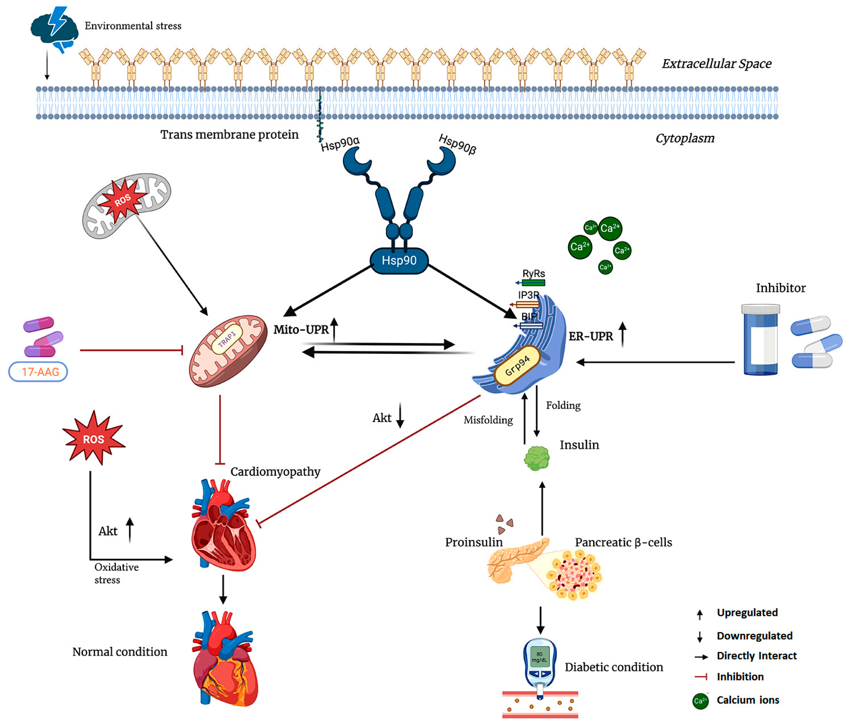
Grp94, also known as gp96, is a chaperone located in the ER that is essential for the proper folding and quality control of secreted and membrane proteins. Its importance in metabolic diseases is highlighted by its role in insulin production. Pancreatic β cells depend on Grp94 for the correct folding of proinsulin within the ER. Grp94 physically associates with proinsulin, and any inhibition or knockdown of Grp94 leads to misfolded insulin and a reduction in glucose-stimulated insulin secretion [239,240]. Experimental models have shown that inhibiting Grp94 shortens the half-life of proinsulin, decreases intracellular insulin levels, and triggers mild endoplasmic reticulum stress through PERK signaling in β cells (Figure 5) [240,241]. In type 2 diabetes, β cells upregulate Grp94 mRNA as an adaptive response, underscoring its critical role in maintaining protein balance during periods of high secretory demand. Thus, adequate Grp94 activity is vital to protecting β cells from failure by ensuring efficient management of insulin.
Grp94 plays a crucial role in the heart by contributing to the unfolded protein response and supporting the survival of cardiomyocytes during stress. When Grp94 is overexpressed in cardiomyocytes, it helps reduce cell death in situations such as calcium overload or simulated ischemia [241,242,243]. By acting as a chaperone for calcium-handling proteins and ER stress sensors, Grp94 protects cardiac muscle cells from injury. In cases of diabetic cardiomyopathy, chronic high blood sugar levels lead to ER stress, which can deplete adaptive chaperones like Grp94. Research on human hearts affected by diabetes has shown a notable decrease in Grp94 expression, indicating that ER dysfunction may increase cardiac vulnerability in these patients [244,245]. Conversely, studies have found that alleviating ER stress in diabetic rodents can restore Grp94 levels and improve cardiac function. In summary, Grp94 is a key mediator of proteostasis in both pancreatic beta cells and cardiomyocytes. By maintaining the protein-folding capacity of the ER, Grp94 serves as a protective factor under metabolic stress. Its dysfunction can lead to impaired insulin secretion and heightened susceptibility of the heart to injury.

4.2. Hsp90 Inhibitors and Their Role in Cardiac Disorder and Diabetes

Curcumin is a natural polyphenol derived from Curcuma longa, known for its potent anti-inflammatory and antioxidant properties. Numerous studies have shown that curcumin interacts with Hsp90, hindering its chaperone function and leading to the depletion of client proteins. While its effectiveness as a direct Hsp90 inhibitor is relatively modest, this interaction provides a mechanistic basis for some of its anti-inflammatory and metabolic effects [172]. In models of metabolic disease, curcumin has been found to improve glucose tolerance and enhance insulin sensitivity in obese and diabetic rodents. These benefits are attributed to increased Akt signaling, regulation of sterol regulatory element-binding proteins (SREBPs), and a reduction in inflammation and oxidative stress through the suppression of NF-κB and c-Jun N-terminal kinase (JNK) pathways (Figure 5) [246]. Additionally, in certain models, the metabolic benefits of curcumin are mediated through gut microbiota-dependent induction of fibroblast growth factor 15 (FGF15) signaling [247,248]. In summary, these findings support a systems-level mechanism in which partial modulation of Hsp90 activity, combined with the regulation of multiple pathways, contributes to the cardiometabolic advantages of curcumin.

4.3. Geldanamycin (GA) and Its Derivatives Role in Cardiac Disorders and Diabetes

Geldanamycin (GA) and its analogs represent a class of Hsp90 inhibitors that target Hsp90 by binding to its N-terminal ATP-binding pocket, disrupting its chaperoning functions. Studies have demonstrated that using GA or inhibitory peptides can attenuate profibrotic TGF-β signaling in both cardiomyocytes and cardiac fibroblasts [249]. However, the clinical use of GA has been limited due to dose-dependent hepatotoxicity, which is linked to the benzoquinone component of its structure. To address this issue, researchers have synthesized many low toxicity derivatives by modifying the quinone ring structure, including 17-AAG, 17-DMAG, and novobiocin. These analogs display improved pharmacological profiles with reduced toxicity, enhanced metabolic stability, and better tolerability [250].
In experimental models of cardiac disease, administering 17-AAG after myocardial infarction or pressure overload has been shown to reduce pathological remodeling. This effect is partly mediated by the accelerated degradation of Raf-1 and the subsequent attenuation of ERK/GATA4 signaling. Additionally, Hsp90 inhibition has been shown to suppress necroptotic signaling pathways (RIP1/RIP3/MLKL) and NF-κB activation, which helps to reduce cardiomyocyte death [227,251]. In a transverse aortic constriction (TAC) model, treatment with 17-AAG resulted in the downregulation of the Hsp90 client protein calcineurin and the transcription factor NFATc2 in cardiac fibroblasts, further supporting its anti-remodeling effects [252].
Similarly, 17-DMAG has demonstrated protective effects in vascular disease models. In case of atherosclerosis, 17-DMAG reduced ERK phosphorylation and suppressed oxidative stress [253]. Inhibition of Hsp90 decreases oxidative effects by disrupting its interaction with Nox1 [254]. In vivo, treatment with 17-DMAG in diabetic mice attenuated the progression of atherosclerosis. This effect was associated with enhanced autophagy, increased expression of antioxidant enzymes (such as catalase, superoxide dismutase, and heme oxygenase-1), and induction of Hsp70 [255]. In apolipoprotein E-deficient diabetic mice, 17-DMAG reduced atherosclerotic plaque burden by approximately 60% and promoted the development of a more stable plaque phenotype. These vascular benefits were linked to the suppression of NF-κB/STAT signaling and the activation of the Nrf2 antioxidant pathway, occurring independently of changes in glucose or lipid levels [256,257]. In the context of metabolic disease, selective Hsp90 inhibition using the synthetic inhibitor AUY922 has shown positive effects. AUY922 suppressed JNK1 phosphorylation, activated the Hsf1–Hsp70 cytoprotective axis, and improved insulin signaling in vitro. Chronic treatment with AUY922 in db/db homozygous mice reversed hyperglycemia, while in diet-induced obese mice, it enhanced insulin sensitivity [235,258]. Across multiple models of diabetic complications—including nephropathy, neuropathy, and atherosclerosis—Hsp90 inhibition consistently alleviated pathology by dampening inflammatory and stress-related signaling pathways.

4.4. Mitochondria-Directed and Extracellular Hsp90 Inhibition

Mitochondrial Hsp90 (mtHsp90), also referred to as TRAP1, has emerged as a promising therapeutic target in cardiopulmonary disease. Gamitrinib, a mitochondria-targeted Hsp90 inhibitor, selectively disrupts the function of mtHsp90. In models of pulmonary arterial hypertension (PAH), Gamitrinib has been shown to reduce the viability and proliferation of pulmonary artery smooth muscle cells (PASMCs) [259]. Inhibition of mtHsp90 with Gamitrinib effectively reversed pulmonary vascular remodeling and improved cardiac output in preclinical PAH models, with no evidence of systemic toxicity. Extracellular Hsp90 has also been implicated in cardiac pathology. Celastrol, a bioactive triterpenoid compound derived from Tripterygium wilfordii and Celastrus orbiculatus (both from the Celastraceae family), inhibits Hsp90 by blocking its interaction with CDC37 at the C-terminal domain. This action disrupts the Hsp90–CDC37 complex [213]. Celastrol exerts cardioprotective effects by inducing cytoprotective proteins such as Hsp70 and heme oxygenase-1 (HO-1). In ischemia–reperfusion (IR) injury models, Celastrol, when administered either as a pre-treatment or at the time of reperfusion, significantly reduced cardiac injury. This protective effect was associated with the suppression of STAT3 activation in angiotensin II-challenged mice, enhanced cardiomyocyte viability, and reduced mPTP opening, thereby preventing cardiac cell death [260].

5. Conclusions and Future Direction

HSPs can be considered the cellular “engines,” much like an engine drives a vehicle; without their proper function, cells are unable to maintain normal physiology. Hsps orchestrate diverse cellular activities, including protein folding and refolding of denatured or misfolded proteins [261], cell cycle regulation, survival pathways, protein homeostasis, and hormone and signal transduction [262]. As a result, they are essential for maintaining cellular homeostasis, and dysfunction in these proteins has been linked to a wide range of human diseases. Among the Hsp family, Hsp90 functions as a central molecular chaperone, orchestrating the stability, maturation, and activity of numerous client proteins with the help of co-chaperones. The repertoire of Hsp90 client proteins continues to expand, suggesting that additional client interactions remain to be identified. Beyond its canonical role in protein folding, Hsp90 has been extensively characterized as a key regulator in human diseases, particularly cancer, as well as in neurodegenerative disorders and metabolic pathologies such as cardiac dysfunction and diabetes. Since Hsp90 is vital for the maturation and stabilization of numerous oncogenic kinases, pharmacological inhibition of Hsp90 disrupts these clients, leading to their degradation. Despite the advancements in understanding Hsp90, several critical questions remain unanswered. It is still unclear how Hsp90 recognizes such a diverse array of client proteins and how co-chaperones refine client specificity and activity. Moreover, the relative contributions of distinct Hsp90 isoforms, including those localized to the ER and mitochondria, to client protein maturation are not fully understood. Elucidating these mechanisms will be crucial for the rational design of more selective Hsp90-targeted therapies for cancer and other human diseases.
Despite significant advances in our understanding of Hsp90 biology, several challenges remain. The molecular principles that govern Hsp90’s recognition of structurally diverse clients, the fine-tuning of specificity by co-chaperones, and the distinct roles of its isoforms—particularly those localized in the ER and mitochondria—are not yet fully elucidated. These gaps limit the rational development of selective Hsp90-targeted therapeutics. Inhibition of Hsp90 leads to destabilization of client proteins, which then undergo ubiquitination and are subsequently degraded by proteasomes [131]. To date, no Hsp90 inhibitor has achieved full clinical approval, primarily due to issues related to toxicity and compensatory cellular responses, such as the activation of Hsf1, leading to the subsequent upregulation of other Hsps. Emerging strategies are being developed to overcome these limitations, including the following:
  • Designing small molecules or peptide analogs of the TRP domain that disrupt Hsp90 complex assembly by competitively binding the CTD.
  • Targeting PTMs that regulate Hsp90-client or Hsp90–co-chaperone interactions, either genetically or pharmacologically, to enhance the effectiveness of existing inhibitors.
  • Co-inhibiting HSF1 to prevent stress-induced feedback responses that attenuate the effects of Hsp90 blockade.
  • Developing isoform-specific inhibitors, particularly targeting Hsp90α, which has been associated with cardiac hypertrophy and heart failure due to pressure overload.
  • Integration of organelle-specific targeting motifs into pan-Hsp90 inhibitors to promote their selective accumulation in target organelles, including mitochondria and the ER.
Additionally, PTMs that influence Hsp90’s interaction networks play a crucial role in cardiomyopathies and other pathologies. Small molecules designed to modulate these modifications may offer a promising therapeutic avenue. A deeper mechanistic understanding of Hsp90’s structural dynamics, client specificity, isoform biology, and regulatory PTMs will be essential for the development of next-generation therapies. Further, mutational studies that induce conformational changes in Hsp90, disrupting assemblies such as epichaperome without impairing its folding capability, could provide valuable insights. Integrating these insights with innovative inhibitor design could enable precise targeting of Hsp90 across a range of conditions, including malignancies, neurodegenerative disorders, cardiovascular diseases, and diabetes, thereby maximizing therapeutic efficacy while minimizing adverse effects.

Author Contributions

Conceptualization, M.K.S., H.R.Y., I.K. and S.S.K.; resources, M.K.S., M.F., H.R.Y., J.S.R. and S.H.; writing—original draft preparation, M.K.S., M.F. and S.J.; figure preparation M.K.S., J.S.R., M.F. and S.J.; review and editing, M.K.S., S.H., J.S.R. and W.C.; visualization, W.C. and H.R.Y.; supervision, W.C., I.K. and S.S.K.; project administration, I.K. and S.S.K.; funding acquisition, S.S.K. All authors have read and agreed to the published version of the manuscript.

Funding

This study was supported by National Research Foundation of Korea (NRF) grants funded by the Korean government (MEST) (grant NRF-2018R1A6A1A03025124).

Institutional Review Board Statement

Not Applicable.

Informed Consent Statement

Not applicable.

Data Availability Statement

No new data were created or analyzed in this study. Data sharing is not applicable to this article.

Conflicts of Interest

The authors declare no conflicts of interest.

Abbreviations

The following abbreviations are used in this manuscript:
17-AAG17-allylamino-17-demethoxygeldanamycin
17DMAG17-dimethylaminoethylamino-17-demethoxygeldanamycin
Amyloid-β
ADAlzheimer’s disease
AHA1Activator of Hsp90 ATPase homolog 1
AMLAcute Myeloid Leukemia
AGOArgonaute
Ask1Apoptosis signal–regulating kinase 1
CTDC-terminal dimerization domain
COXIICytochrome c oxidase subunit II
EREndoplasmic Reticulum
eHsp90Extracellular Hsp90
FGFR3Fibroblast growth factor receptor 3
GARPGlycoprotein A repetitions predominant
GISTGastrointestinal stromal tumors
GRGlucocorticoid receptor
Grp94Glucose-regulated protein 94
HCCHepatocellular carcinoma
HCMHypertrophic cardiomyopathy
HDHuntington’s disease
HIF-1αHypoxia-inducible factor 1α
Hch1High copy Hsp90 suppressor 1
HIPHsp70-interacting proteins
HSPsHeat shock proteins
Hsp90Heat shock protein 90
IGFInsulin-like growth factors
JNKc-Jun N-terminal kinase
LRP1Low-density lipoprotein receptor-related protein-1
MDMiddle domain
MDRMultidrug resistance
NTDN-terminal nucleotide-binding domain
PDParkinson’s disease
PP5Protein phosphatase 5
PTMsPost-translational modifications
PPPPentose phosphate pathway
PPIasePeptidyl-prolyl cis-trans isomerase
PIKKPhosphatidylinositol-3 kinase–related kinase
PRProgesterone receptor
PTPMitochondrial permeability transition pore
ROSReactive oxygen species
SREBPssterol regulatory element-binding proteins
SDHSuccinate dehydrogenase
SHRSteroid hormone receptor
TACTransverse aortic constriction
TCATricarboxylic acid
TRAP1Tumor necrosis factor receptor-associated protein
TPRTetratricopeptide repeat
TLRToll-like receptors
UPRUnfolded protein response
VEGFRsVascular endothelial growth factor receptors

References

  1. Taipale, M.; Jarosz, D.F.; Lindquist, S. HSP90 at the hub of protein homeostasis: Emerging mechanistic insights. Nat. Rev. Mol. Cell Biol. 2010, 11, 515–528. [Google Scholar] [CrossRef]
  2. Maiti, S.; Picard, D. Cytosolic Hsp90 isoform-specific functions and clinical significance. Biomolecules 2022, 12, 1166. [Google Scholar] [CrossRef] [PubMed]
  3. Chen, B.; Zhong, D.; Monteiro, A. Comparative genomics and evolution of the HSP90 family of genes across all kingdoms of organisms. BMC Genom. 2006, 7, 156. [Google Scholar] [CrossRef] [PubMed]
  4. Inoue, H.; Li, M.; Schnell, D.J. An essential role for chloroplast heat shock protein 90 (Hsp90C) in protein import into chloroplasts. Proc. Natl. Acad. Sci. USA 2013, 110, 3173–3178. [Google Scholar] [CrossRef] [PubMed]
  5. Biswas, C.; Ostrovsky, O.; Makarewich, C.A.; Wanderling, S.; Gidalevitz, T.; Argon, Y. The peptide-binding activity of GRP94 is regulated by calcium. Biochem. J. 2007, 405, 233–241. [Google Scholar] [CrossRef]
  6. Schleiff, E.; Becker, T. Common ground for protein translocation: Access control for mitochondria and chloroplasts. Nat. Rev. Mol. Cell Biol. 2011, 12, 48–59. [Google Scholar] [CrossRef]
  7. Kang, B.H. TRAP1 regulation of mitochondrial life or death decision in cancer cells and mitochondria-targeted TRAP1 inhibitors. BMB Rep. 2012, 45, 1–6. [Google Scholar] [CrossRef]
  8. Kang, B.H.; Plescia, J.; Dohi, T.; Rosa, J.; Doxsey, S.J.; Altieri, D.C. Regulation of tumor cell mitochondrial homeostasis by an organelle-specific Hsp90 chaperone network. Cell 2007, 131, 257–270. [Google Scholar] [CrossRef]
  9. Li, W.; Sahu, D.; Tsen, F. Secreted heat shock protein-90 (Hsp90) in wound healing and cancer. Biochim. Biophys. Acta (BBA)-Mol. Cell Res. 2012, 1823, 730–741. [Google Scholar] [CrossRef]
  10. Chaudhury, S.; D’Amico, T.; Blagg, B.S. The Hsp90β Isoform: An Attractive Target for Drug Development. Med. Res. Rev. 2025, 45, 1452–1465. [Google Scholar] [CrossRef]
  11. Wegele, H.; Müller, L.; Buchner, J. Hsp70 and Hsp90—A relay team for protein folding. Rev. Physiol. Biochem. Pharmacol. 2004, 151, 1–44. [Google Scholar] [PubMed]
  12. Rios, E.I.; Hunsberger, I.L.; Johnson, J.L. Insights into Hsp90 mechanism and In Vivo functions learned from studies in the yeast, Saccharomyces cerevisiae. Front. Mol. Biosci. 2024, 11, 1325590. [Google Scholar] [CrossRef]
  13. Nemoto, T.; Matsusaka, T.; Ota, M.; Takagi, T.; Collinge, D.B.; Walther-Larsen, H. Dimerization characteristics of the 94-kDa glucose-regulated protein. J. Biochem. 1996, 120, 249–256. [Google Scholar] [CrossRef]
  14. Henot, F.; Rioual, E.; Favier, A.; Macek, P.; Crublet, E.; Josso, P.; Brutscher, B.; Frech, M.; Gans, P.; Loison, C. Visualizing the transiently populated closed-state of human HSP90 ATP binding domain. Nat. Commun. 2022, 13, 7601. [Google Scholar] [CrossRef]
  15. Schulze, A.; Beliu, G.; Helmerich, D.A.; Schubert, J.; Pearl, L.H.; Prodromou, C.; Neuweiler, H. Cooperation of local motions in the Hsp90 molecular chaperone ATPase mechanism. Nat. Chem. Biol. 2016, 12, 628–635. [Google Scholar] [CrossRef] [PubMed]
  16. Roe, S.M.; Prodromou, C.; O’Brien, R.; Ladbury, J.E.; Piper, P.W.; Pearl, L.H. Structural basis for inhibition of the Hsp90 molecular chaperone by the antitumor antibiotics radicicol and geldanamycin. J. Med. Chem. 1999, 42, 260–266. [Google Scholar] [CrossRef]
  17. Meyer, P.; Prodromou, C.; Hu, B.; Vaughan, C.; Roe, S.M.; Panaretou, B.; Piper, P.W.; Pearl, L.H. Structural and functional analysis of the middle segment of hsp90: Implications for ATP hydrolysis and client protein and cochaperone interactions. Mol. Cell 2003, 11, 647–658. [Google Scholar] [CrossRef]
  18. Sato, S.; Fujita, N.; Tsuruo, T. Modulation of Akt kinase activity by binding to Hsp90. Proc. Natl. Acad. Sci. USA 2000, 97, 10832–10837. [Google Scholar] [CrossRef]
  19. Müller, L.; Schaupp, A.; Walerych, D.; Wegele, H.; Buchner, J. Hsp90 regulates the activity of wild type p53 under physiological and elevated temperatures. J. Biol. Chem. 2004, 279, 48846–48854. [Google Scholar] [CrossRef] [PubMed]
  20. Fontana, J.; Fulton, D.; Chen, Y.; Fairchild, T.A.; McCabe, T.J.; Fujita, N.; Tsuruo, T.; Sessa, W.C. Domain mapping studies reveal that the M domain of hsp90 serves as a molecular scaffold to regulate Akt-dependent phosphorylation of endothelial nitric oxide synthase and NO release. Circ. Res. 2002, 90, 866–873. [Google Scholar] [CrossRef]
  21. Tsutsumi, S.; Mollapour, M.; Graf, C.; Lee, C.-T.; Scroggins, B.T.; Xu, W.; Haslerova, L.; Hessling, M.; Konstantinova, A.A.; Trepel, J.B. Hsp90 charged-linker truncation reverses the functional consequences of weakened hydrophobic contacts in the N domain. Nat. Struct. Mol. Biol. 2009, 16, 1141–1147. [Google Scholar] [CrossRef]
  22. Riedl, S.; Bilgen, E.; Agam, G.; Hirvonen, V.; Jussupow, A.; Tippl, F.; Riedl, M.; Maier, A.; Becker, C.F.; Kaila, V.R. Evolution of the conformational dynamics of the molecular chaperone Hsp90. Nat. Commun. 2024, 15, 8627. [Google Scholar] [CrossRef]
  23. Röhl, A.; Rohrberg, J.; Buchner, J. The chaperone Hsp90: Changing partners for demanding clients. Trends Biochem. Sci. 2013, 38, 253–262. [Google Scholar] [CrossRef]
  24. Gracia, L.; Lora, G.; Blair, L.J.; Jinwal, U.K. Therapeutic potential of the Hsp90/Cdc37 interaction in neurodegenerative diseases. Front. Neurosci. 2019, 13, 1263. [Google Scholar] [CrossRef]
  25. Taipale, M.; Krykbaeva, I.; Koeva, M.; Kayatekin, C.; Westover, K.D.; Karras, G.I.; Lindquist, S. Quantitative analysis of HSP90-client interactions reveals principles of substrate recognition. Cell 2012, 150, 987–1001. [Google Scholar] [CrossRef] [PubMed]
  26. Meyer, P. Structural basis for recruitment of the ATPase activator Aha1 to the Hsp90 chaperone machinery. EMBO J. 2004, 23, 1402–1410. [Google Scholar] [CrossRef] [PubMed]
  27. McLaughlin, S.H.; Smith, H.W.; Jackson, S.E. Stimulation of the weak ATPase activity of human hsp90 by a client protein. J. Mol. Biol. 2002, 315, 787–798. [Google Scholar] [CrossRef] [PubMed]
  28. Lopez, A.; Dahiya, V.; Delhommel, F.; Freiburger, L.; Stehle, R.; Asami, S.; Rutz, D.; Blair, L.; Buchner, J.; Sattler, M. Client binding shifts the populations of dynamic Hsp90 conformations through an allosteric network. Sci. Adv. 2021, 7, eabl7295. [Google Scholar] [CrossRef]
  29. Li, J.; Richter, K.; Buchner, J. Mixed Hsp90–cochaperone complexes are important for the progression of the reaction cycle. Nat. Struct. Mol. Biol. 2011, 18, 61–66. [Google Scholar] [CrossRef]
  30. Soroka, J.; Wandinger, S.K.; Mäusbacher, N.; Schreiber, T.; Richter, K.; Daub, H.; Buchner, J. Conformational switching of the molecular chaperone Hsp90 via regulated phosphorylation. Mol. Cell 2012, 45, 517–528. [Google Scholar] [CrossRef]
  31. Weaver, A.J.; Sullivan, W.P.; Felts, S.J.; Owen, B.A.; Toft, D.O. Crystal structure and activity of human p23, a heat shock protein 90 co-chaperone. J. Biol. Chem. 2000, 275, 23045–23052. [Google Scholar] [CrossRef] [PubMed]
  32. Iwasaki, S.; Kobayashi, M.; Yoda, M.; Sakaguchi, Y.; Katsuma, S.; Suzuki, T.; Tomari, Y. Hsc70/Hsp90 chaperone machinery mediates ATP-dependent RISC loading of small RNA duplexes. Mol. Cell 2010, 39, 292–299. [Google Scholar] [CrossRef] [PubMed]
  33. Pratt, W.B.; Toft, D.O. Regulation of signaling protein function and trafficking by the hsp90/hsp70-based chaperone machinery. Exp. Biol. Med. 2003, 228, 111–133. [Google Scholar] [CrossRef]
  34. Lanneau, D.; Brunet, M.; Frisan, E.; Solary, E.; Fontenay, M.; Garrido, C. Heat shock proteins: Essential proteins for apoptosis regulation. J. Cell. Mol. Med. 2008, 12, 743–761. [Google Scholar] [CrossRef]
  35. Liu, K.; Jin, H.; Guo, Y.; Liu, Y.; Wan, Y.; Zhao, P.; Zhou, Z.; Wang, J.; Wang, M.; Zou, C. CFTR interacts with Hsp90 and regulates the phosphorylation of AKT and ERK1/2 in colorectal cancer cells. FEBS Open Bio 2019, 9, 1119–1127. [Google Scholar] [CrossRef]
  36. Zhang, S.; Sun, Y.; Yuan, Z.; Li, Y.; Li, X.; Gong, Z.; Peng, Y. Heat shock protein 90β inhibits apoptosis of intestinal epithelial cells induced by hypoxia through stabilizing phosphorylated Akt. BMB Rep. 2013, 46, 47. [Google Scholar] [CrossRef]
  37. Silbermann, L.-M.; Vermeer, B.; Schmid, S.; Tych, K. The known unknowns of the Hsp90 chaperone. eLife 2024, 13, e102666. [Google Scholar] [CrossRef]
  38. Cintron, N.S.; Toft, D. Defining the requirements for Hsp40 and Hsp70 in the Hsp90 chaperone pathway. J. Biol. Chem. 2006, 281, 26235–26244. [Google Scholar] [CrossRef]
  39. Chadli, A.; Bouhouche, I.; Sullivan, W.; Stensgard, B.; McMahon, N.; Catelli, M.G.; Toft, D.O. Dimerization and N-terminal domain proximity underlie the function of the molecular chaperone heat shock protein 90. Proc. Natl. Acad. Sci. USA 2000, 97, 12524–12529. [Google Scholar] [CrossRef]
  40. Hernández, M.P.; Chadli, A.; Toft, D.O. HSP40 binding is the first step in the HSP90 chaperoning pathway for the progesterone receptor. J. Biol. Chem. 2002, 277, 11873–11881. [Google Scholar] [CrossRef] [PubMed]
  41. Ricketson, D.; Hostick, U.; Fang, L.; Yamamoto, K.; Darimont, B. A conformational switch in the ligand-binding domain regulates the dependence of the glucocorticoid receptor on Hsp90. J. Mol. Biol. 2007, 368, 729–741. [Google Scholar] [CrossRef]
  42. Wang, R.Y.-R.; Noddings, C.M.; Kirschke, E.; Myasnikov, A.G.; Johnson, J.L.; Agard, D.A. Structure of Hsp90–Hsp70–Hop–GR reveals the Hsp90 client-loading mechanism. Nature 2022, 601, 460–464. [Google Scholar] [CrossRef]
  43. Biebl, M.M.; Lopez, A.; Rehn, A.; Freiburger, L.; Lawatscheck, J.; Blank, B.; Sattler, M.; Buchner, J. Structural elements in the flexible tail of the co-chaperone p23 coordinate client binding and progression of the Hsp90 chaperone cycle. Nat. Commun. 2021, 12, 828. [Google Scholar] [CrossRef]
  44. Noddings, C.M.; Wang, R.Y.-R.; Johnson, J.L.; Agard, D.A. Structure of Hsp90–p23–GR reveals the Hsp90 client-remodelling mechanism. Nature 2022, 601, 465–469. [Google Scholar] [CrossRef]
  45. Schopf, F.H.; Biebl, M.M.; Buchner, J. The HSP90 chaperone machinery. Nat. Rev. Mol. Cell Biol. 2017, 18, 345–360. [Google Scholar] [CrossRef] [PubMed]
  46. Krukenberg, K.A.; Street, T.O.; Lavery, L.A.; Agard, D.A. Conformational dynamics of the molecular chaperone Hsp90. Q. Rev. Biophys. 2011, 44, 229–255. [Google Scholar] [CrossRef]
  47. Gupta, R.S. Phylogenetic analysis of the 90 kD heat shock family of protein sequences and an examination of the relationship among animals, plants, and fungi species. Mol. Biol. Evol. 1995, 12, 1063–1073. [Google Scholar] [CrossRef] [PubMed]
  48. Huck, J.D.; Que, N.L.; Hong, F.; Li, Z.; Gewirth, D.T. Structural and functional analysis of GRP94 in the closed state reveals an essential role for the pre-N domain and a potential client-binding site. Cell Rep. 2017, 20, 2800–2809. [Google Scholar] [CrossRef] [PubMed]
  49. Jackson, S.E. Hsp90: Structure and function. In Molecular Chaperones; Springer: Berlin/Heidelberg, Germany, 2012; pp. 155–240. [Google Scholar]
  50. Csermely, P.; Schnaider, T.; So, C.; Prohászka, Z.; Nardai, G. The 90-kDa molecular chaperone family: Structure, function, and clinical applications. A comprehensive review. Pharmacol. Ther. 1998, 79, 129–168. [Google Scholar] [CrossRef]
  51. Li, X.; Yin, M.-Y.; Zhang, S.-T.; Xie, S.-A. The role of canopy family proteins: Biological mechanism and disease function. Mol. Biol. Rep. 2025, 52, 164. [Google Scholar] [CrossRef]
  52. Amankwah, Y.S.; Fleifil, Y.; Unruh, E.; Collins, P.; Wang, Y.; Vitou, K.; Bates, A.; Obaseki, I.; Sugoor, M.; Alao, J.P. Structural transitions modulate the chaperone activities of Grp94. Proc. Natl. Acad. Sci. USA 2024, 121, e2309326121. [Google Scholar] [CrossRef]
  53. Amankwah, Y.S.; Collins, P.; Fleifil, Y.; Unruh, E.; Marquez, K.J.R.; Vitou, K.; Kravats, A.N. Grp94 works upstream of BiP in protein remodeling under heat stress. J. Mol. Biol. 2022, 434, 167762. [Google Scholar] [CrossRef]
  54. Boczek, E.E.; Reefschläger, L.G.; Dehling, M.; Struller, T.J.; Häusler, E.; Seidl, A.; Kaila, V.R.; Buchner, J. Conformational processing of oncogenic v-Src kinase by the molecular chaperone Hsp90. Proc. Natl. Acad. Sci. USA 2015, 112, E3189–E3198. [Google Scholar] [CrossRef]
  55. Yan, P.; Patel, H.J.; Sharma, S.; Corben, A.; Wang, T.; Panchal, P.; Yang, C.; Sun, W.; Araujo, T.L.; Rodina, A. Molecular stressors engender protein connectivity dysfunction through aberrant N-glycosylation of a chaperone. Cell Rep. 2020, 31, 107840. [Google Scholar] [CrossRef] [PubMed]
  56. Mirikar, D.; Bushman, Y.; Truman, A.W. Structural transitions modulate the chaperone activities of Grp94. Trends Biochem. Sci. 2024, 49, 752–753. [Google Scholar] [CrossRef] [PubMed]
  57. Niu, M.; Xu, J.; Liu, Y.; Li, Y.; He, T.; Ding, L.; He, Y.; Yi, Y.; Li, F.; Guo, R. FBXL2 counteracts Grp94 to destabilize EGFR and inhibit EGFR-driven NSCLC growth. Nat. Commun. 2021, 12, 5919. [Google Scholar] [CrossRef]
  58. Ghosh, S.; Shinogle, H.E.; Galeva, N.A.; Dobrowsky, R.T.; Blagg, B.S. Endoplasmic reticulum-resident heat shock protein 90 (HSP90) isoform glucose-regulated protein 94 (GRP94) regulates cell polarity and cancer cell migration by affecting intracellular transport. J. Biol. Chem. 2016, 291, 8309–8323. [Google Scholar] [CrossRef]
  59. Zuehlke, A.; Johnson, J.L. Hsp90 and co-chaperones twist the functions of diverse client proteins. Biopolym. Orig. Res. Biomol. 2010, 93, 211–217. [Google Scholar] [CrossRef]
  60. Kang, S.; Kang, B.H. Structure, function, and inhibitors of the mitochondrial chaperone TRAP1. J. Med. Chem. 2022, 65, 16155–16172. [Google Scholar] [CrossRef] [PubMed]
  61. Lee, C.; Park, H.-K.; Jeong, H.; Lim, J.; Lee, A.-J.; Cheon, K.Y.; Kim, C.-S.; Thomas, A.P.; Bae, B.; Kim, N.D. Development of a mitochondria-targeted Hsp90 inhibitor based on the crystal structures of human TRAP1. J. Am. Chem. Soc. 2015, 137, 4358–4367. [Google Scholar] [CrossRef] [PubMed]
  62. Kang, B.H.; Siegelin, M.D.; Plescia, J.; Raskett, C.M.; Garlick, D.S.; Dohi, T.; Lian, J.B.; Stein, G.S.; Languino, L.R.; Altieri, D.C. Preclinical characterization of mitochondria-targeted small molecule hsp90 inhibitors, gamitrinibs, in advanced prostate cancer. Clin. Cancer Res. 2010, 16, 4779–4788. [Google Scholar] [CrossRef] [PubMed]
  63. Park, H.-K.; Hong, J.-H.; Oh, Y.T.; Kim, S.S.; Yin, J.; Lee, A.-J.; Chae, Y.C.; Kim, J.H.; Park, S.-H.; Park, C.-K. Interplay between TRAP1 and sirtuin-3 modulates mitochondrial respiration and oxidative stress to maintain stemness of glioma stem cells. Cancer Res. 2019, 79, 1369–1382. [Google Scholar] [CrossRef]
  64. Ramos Rego, I.; Santos Cruz, B.; Ambrósio, A.F.; Alves, C.H. TRAP1 in oxidative stress and neurodegeneration. Antioxidants 2021, 10, 1829. [Google Scholar] [CrossRef]
  65. Gutierrez, A.; Archdeacon, J.; Blagg, B.S. TRAP1 and its therapeutic potential. Bioorg. Med. Chem. Lett. 2025, 129, 130395. [Google Scholar] [CrossRef]
  66. Dharaskar, S.P.; Subbarao, S.A. The mitochondrial chaperone TRAP-1 regulates the glutamine metabolism in tumor cells. Mitochondrion 2023, 69, 159–170. [Google Scholar] [CrossRef] [PubMed]
  67. Guzzo, G.; Sciacovelli, M.; Bernardi, P.; Rasola, A. Inhibition of succinate dehydrogenase by the mitochondrial chaperone TRAP1 has anti-oxidant and anti-apoptotic effects on tumor cells. Oncotarget 2014, 5, 11897. [Google Scholar] [CrossRef] [PubMed]
  68. Landriscina, M.; Amoroso, M.R.; Piscazzi, A.; Esposito, F. Heat shock proteins, cell survival and drug resistance: The mitochondrial chaperone TRAP1, a potential novel target for ovarian cancer therapy. Gynecol. Oncol. 2010, 117, 177–182. [Google Scholar] [CrossRef]
  69. Hu, K.; Babapoor-Farrokhran, S.; Rodrigues, M.; Deshpande, M.; Puchner, B.; Kashiwabuchi, F.; Hassan, S.J.; Asnaghi, L.; Handa, J.T.; Merbs, S. Hypoxia-inducible factor 1 upregulation of both VEGF and ANGPTL4 is required to promote the angiogenic phenotype in uveal melanoma. Oncotarget 2016, 7, 7816, Erratum in Oncotarget 2016, 12, 519–520. [Google Scholar] [CrossRef]
  70. Sisinni, L.; Maddalena, F.; Condelli, V.; Pannone, G.; Simeon, V.; Li Bergolis, V.; Lopes, E.; Piscazzi, A.; Matassa, D.S.; Mazzoccoli, C. TRAP1 controls cell cycle G2–M transition through the regulation of CDK1 and MAD2 expression/ubiquitination. J. Pathol. 2017, 243, 123–134. [Google Scholar] [CrossRef]
  71. Mathieu, C.; Chamayou, Q.; Luong, T.T.H.; Naud, D.; Mahuteau-Betzer, F.; Alami, M.; Fattal, E.; Messaoudi, S.; Vergnaud-Gauduchon, J. Synthesis and antiproliferative activity of 6BrCaQ-TPP conjugates for targeting the mitochondrial heat shock protein TRAP1. Eur. J. Med. Chem. 2022, 229, 114052. [Google Scholar] [CrossRef]
  72. Zielonka, J.; Joseph, J.; Sikora, A.; Hardy, M.; Ouari, O.; Vasquez-Vivar, J.; Cheng, G.; Lopez, M.; Kalyanaraman, B. Mitochondria-targeted triphenylphosphonium-based compounds: Syntheses, mechanisms of action, and therapeutic and diagnostic applications. Chem. Rev. 2017, 117, 10043–10120. [Google Scholar] [CrossRef]
  73. Sanchez-Martin, C.; Menon, D.; Moroni, E.; Ferraro, M.; Masgras, I.; Elsey, J.; Arbiser, J.L.; Colombo, G.; Rasola, A. Honokiol bis-dichloroacetate is a selective allosteric inhibitor of the mitochondrial chaperone TRAP1. Antioxid. Redox Signal. 2021, 34, 505–516. [Google Scholar] [CrossRef]
  74. Zhang, L.; Pang, E.; Loo, R.R.O.; Rao, J.; Go, V.L.W.; Loo, J.A.; Lu, Q.Y. Concomitant inhibition of HSP90, its mitochondrial localized homologue TRAP1 and HSP27 by green tea in pancreatic cancer HPAF-II cells. Proteomics 2011, 11, 4638–4647. [Google Scholar] [CrossRef]
  75. Backe, S.J.; Sager, R.A.; Woodford, M.R.; Makedon, A.M.; Mollapour, M. Post-translational modifications of Hsp90 and translating the chaperone code. J. Biol. Chem. 2020, 295, 11099–11117. [Google Scholar] [CrossRef] [PubMed]
  76. Chu, F.; Sharma, S.; Ginsberg, S.D.; Chiosis, G. PTMs as molecular encoders: Reprogramming chaperones into epichaperomes for network control in disease. Trends Biochem. Sci. 2025, 50, 892–905. [Google Scholar] [CrossRef]
  77. Wang, X.; Song, X.; Zhuo, W.; Fu, Y.; Shi, H.; Liang, Y.; Tong, M.; Chang, G.; Luo, Y. The regulatory mechanism of Hsp90α secretion and its function in tumor malignancy. Proc. Natl. Acad. Sci. USA 2009, 106, 21288–21293. [Google Scholar] [CrossRef]
  78. Gajsek, O.; Becker, C.F.; Conibear, A.C. Site-specifically Phosphorylated Hsp90C-terminal Domain Variants Provide Access to Deciphering the Chaperone Code. Chem. Eur. J. 2025, 31, e202403676. [Google Scholar] [CrossRef]
  79. Jakob, U.; Meyer, I.; Bügl, H.; André, S.; Bardwell, J.C.; Buchner, J. Structural organization of procaryotic and eucaryotic Hsp90. Influence of divalent cations on structure and function. J. Biol. Chem. 1995, 270, 14412–14419. [Google Scholar] [CrossRef] [PubMed]
  80. Roychowdhury, T.; McNutt, S.W.; Pasala, C.; Nguyen, H.T.; Thornton, D.T.; Sharma, S.; Botticelli, L.; Digwal, C.S.; Joshi, S.; Yang, N. Phosphorylation-driven epichaperome assembly is a regulator of cellular adaptability and proliferation. Nat. Commun. 2024, 15, 8912. [Google Scholar] [CrossRef] [PubMed]
  81. Voelkel, T.; Andresen, C.; Unger, A.; Just, S.; Rottbauer, W.; Linke, W.A. Lysine methyltransferase Smyd2 regulates Hsp90-mediated protection of the sarcomeric titin springs and cardiac function. Biochim. Biophys. Acta (BBA)-Mol. Cell Res. 2013, 1833, 812–822. [Google Scholar] [CrossRef]
  82. Zhang, X.; Zhang, Y.; Miao, Q.; Shi, Z.; Hu, L.; Liu, S.; Gao, J.; Zhao, S.; Chen, H.; Huang, Z. Inhibition of HSP90 S-nitrosylation alleviates cardiac fibrosis via TGFβ/SMAD3 signalling pathway. Br. J. Pharmacol. 2021, 178, 4608–4625. [Google Scholar] [CrossRef]
  83. Shelton, L.B. Targeting the Hsp90/Aha1 Complex for the Treatment of Tauopathies. Ph.D. Thesis, University of South Florida, Tampa, FL, USA, 2018. [Google Scholar]
  84. Ginsberg, S.D.; Sharma, S.; Norton, L.; Chiosis, G. Targeting stressor-induced dysfunctions in protein–protein interaction networks via epichaperomes. Trends Pharmacol. Sci. 2023, 44, 20–33. [Google Scholar] [CrossRef]
  85. Magni, A.; Sciva, C.; Castelli, M.; Digwal, C.S.; Rodina, A.; Sharma, S.; Ochiana, S.; Patel, H.J.; Shah, S.; Chiosis, G. N-Glycosylation-Induced Pathologic Protein Conformations as a Tool to Guide the Selection of Biologically Active Small Molecules. Chem. Eur. J. 2024, 30, e202401957. [Google Scholar] [CrossRef]
  86. Pasala, C.; Digwal, C.S.; Sharma, S.; Wang, S.; Bubula, A.; Chiosis, G. Epichaperomes: Redefining chaperone biology and therapeutic strategies in complex diseases. RSC Chem. Biol. 2025, 6, 678–698. [Google Scholar] [CrossRef]
  87. Young, J.C.; Moarefi, I.; Hartl, F.U. Hsp90: A specialized but essential protein-folding tool. J. Cell Biol. 2001, 154, 267. [Google Scholar] [CrossRef]
  88. Echeverría, P.C.; Bernthaler, A.; Dupuis, P.; Mayer, B.; Picard, D. An interaction network predicted from public data as a discovery tool: Application to the Hsp90 molecular chaperone machine. PLoS ONE 2011, 6, e26044. [Google Scholar] [CrossRef] [PubMed]
  89. Pratt, W.B.; Dittmar, K.D. Studies with purified chaperones advance the understanding of the mechanism of glucocorticoid receptor–hsp90 heterocomplex assembly. Trends Endocrinol. Metab. 1998, 9, 244–252. [Google Scholar] [CrossRef] [PubMed]
  90. Nathan, D.F.; Vos, M.H.; Lindquist, S. In Vivo functions of the Saccharomyces cerevisiae Hsp90 chaperone. Proc. Natl. Acad. Sci. USA 1997, 94, 12949–12956. [Google Scholar] [CrossRef]
  91. Caplan, A.J. Hsp90’s secrets unfold: New insights from structural and functional studies. Trends Cell Biol. 1999, 9, 262–268. [Google Scholar] [CrossRef] [PubMed]
  92. Von Kriegsheim, A.; Pitt, A.; Grindlay, G.J.; Kolch, W.; Dhillon, A.S. Regulation of the Raf–MEK–ERK pathway by protein phosphatase 5. Nat. Cell Biol. 2006, 8, 1011–1016. [Google Scholar] [CrossRef]
  93. Morita, K.-i.; Saitoh, M.; Tobiume, K.; Matsuura, H.; Enomoto, S.; Nishitoh, H.; Ichijo, H. Negative feedback regulation of ASK1 by protein phosphatase 5 (PP5) in response to oxidative stress. EMBO J. 2001, 20, 6028–6036. [Google Scholar] [CrossRef] [PubMed]
  94. Zhang, J.; Bao, S.; Furumai, R.; Kucera, K.S.; Ali, A.; Dean, N.M.; Wang, X.-F. Protein phosphatase 5 is required for ATR-mediated checkpoint activation. Mol. Cell. Biol. 2005, 25, 9910–9919. [Google Scholar] [CrossRef]
  95. Amable, L.; Grankvist, N.; Largen, J.W.; Ortsäter, H.; Sjöholm, Å.; Honkanen, R.E. Disruption of serine/threonine protein phosphatase 5 (PP5: PPP5c) in mice reveals a novel role for PP5 in the regulation of ultraviolet light-induced phosphorylation of serine/threonine protein kinase Chk1 (CHEK1). J. Biol. Chem. 2011, 286, 40413–40422. [Google Scholar] [CrossRef]
  96. Li, J.; Soroka, J.; Buchner, J. The Hsp90 chaperone machinery: Conformational dynamics and regulation by co-chaperones. Biochim. Biophys. Acta (BBA)-Mol. Cell Res. 2012, 1823, 624–635. [Google Scholar] [CrossRef]
  97. Zuo, Z.; Urban, G.; Scammell, J.G.; Dean, N.M.; McLean, T.K.; Aragon, I.; Honkanen, R.E. Ser/Thr protein phosphatase type 5 (PP5) is a negative regulator of glucocorticoid receptor-mediated growth arrest. Biochemistry 1999, 38, 8849–8857. [Google Scholar] [CrossRef]
  98. Zuo, Z.; Dean, N.M.; Honkanen, R.E. Serine/threonine protein phosphatase type 5 acts upstream of p53 to regulate the induction of p21WAF1/Cip1 and mediate growth arrest. J. Biol. Chem. 1998, 273, 12250–12258. [Google Scholar] [CrossRef]
  99. Jaime-Garza, M.; Nowotny, C.A.; Coutandin, D.; Wang, F.; Tabios, M.; Agard, D.A. Hsp90 provides a platform for kinase dephosphorylation by PP5. Nat. Commun. 2023, 14, 2197. [Google Scholar] [CrossRef] [PubMed]
  100. Gasc, J.-M.; Renoir, J.-M.; Faber, L.E.; Delahaye, F.; Baulieu, E.-E. Nuclear localization of two steroid receptor-associated proteins, hsp90 and p59. Exp. Cell Res. 1990, 186, 362–367. [Google Scholar] [CrossRef] [PubMed]
  101. Vazquez-Nin, G.; Echeverria, O.; Carbajal, M.; Tanguay, R.; Diez, J.; Fakan, S. Immunoelectron microscope localization of Mr 90000 heat shock protein and Mr 70000 heat shock cognate protein in the salivary glands of Chironomus thummi. Chromosoma 1992, 102, 50–59. [Google Scholar] [CrossRef]
  102. Schlatter, L.K.; Howard, K.J.; Parker, M.G.; Distelhorst, C.W. Comparison of the 90-kilodalton heat shock protein interaction with In Vitro translated glucocorticoid and estrogen receptors. Mol. Endocrinol. 1992, 6, 132–140. [Google Scholar]
  103. Chambraud, B.; Berry, M.; Redeuilh, G.; Chambon, P.; Baulieu, E. Several regions of human estrogen receptor are involved in the formation of receptor-heat shock protein 90 complexes. J. Biol. Chem. 1990, 265, 20686–20691. [Google Scholar] [CrossRef]
  104. Sweitzer, T.D.; Hanover, J.A. Calmodulin activates nuclear protein import: A link between signal transduction and nuclear transport. Proc. Natl. Acad. Sci. USA 1996, 93, 14574–14579. [Google Scholar] [CrossRef]
  105. Defranco, D.B. Role of molecular chaperones in subnuclear trafficking of glucocorticoid receptors. Kidney Int. 2000, 57, 1241–1249. [Google Scholar] [CrossRef]
  106. Santoro, M.G. Heat shock factors and the control of the stress response. Biochem. Pharmacol. 2000, 59, 55–63. [Google Scholar] [CrossRef]
  107. Zou, J.; Guo, Y.; Guettouche, T.; Smith, D.F.; Voellmy, R. Repression of heat shock transcription factor HSF1 activation by HSP90 (HSP90 complex) that forms a stress-sensitive complex with HSF1. Cell 1998, 94, 471–480. [Google Scholar] [CrossRef] [PubMed]
  108. Schlatter, H.; Langer, T.; Rosmus, S.; Onneken, M.-L.; Fasold, H. A novel function for the 90 kDa heat-shock protein (Hsp90): Facilitating nuclear export of 60 S ribosomal subunits. Biochem. J. 2002, 362, 675–684. [Google Scholar] [CrossRef]
  109. Keppler, B.R.; Grady, A.T.; Jarstfer, M.B. The biochemical role of the heat shock protein 90 chaperone complex in establishing human telomerase activity. J. Biol. Chem. 2006, 281, 19840–19848. [Google Scholar] [CrossRef] [PubMed]
  110. Gray Jr, P.J.; Prince, T.; Cheng, J.; Stevenson, M.A.; Calderwood, S.K. Targeting the oncogene and kinome chaperone CDC37. Nat. Rev. Cancer 2008, 8, 491–495. [Google Scholar] [CrossRef] [PubMed]
  111. Zhang, M.; Boter, M.; Li, K.; Kadota, Y.; Panaretou, B.; Prodromou, C.; Shirasu, K.; Pearl, L.H. Structural and functional coupling of Hsp90-and Sgt1-centred multi-protein complexes. EMBO J. 2008, 27, 2789–2798. [Google Scholar] [CrossRef]
  112. Davies, A.E.; Kaplan, K.B. Hsp90–Sgt1 and Skp1 target human Mis12 complexes to ensure efficient formation of kinetochore–microtubule binding sites. J. Cell Biol. 2010, 189, 261–274. [Google Scholar] [CrossRef]
  113. Fan, A.C.; Bhangoo, M.K.; Young, J.C. Hsp90 functions in the targeting and outer membrane translocation steps of Tom70-mediated mitochondrial import. J. Biol. Chem. 2006, 281, 33313–33324. [Google Scholar] [CrossRef]
  114. Imai, J.; Maruya, M.; Yashiroda, H.; Yahara, I.; Tanaka, K. The molecular chaperone Hsp90 plays a role in the assembly and maintenance of the 26S proteasome. EMBO J. 2003, 22, 3557–3567. [Google Scholar] [CrossRef]
  115. Martins, C.D.; Da Pieve, C.; Burley, T.A.; Smith, R.; Ciobota, D.M.; Allott, L.; Harrington, K.J.; Oyen, W.J.; Smith, G.; Kramer-Marek, G. HER3-mediated resistance to Hsp90 inhibition detected in breast cancer xenografts by affibody-based PET imaging. Clin. Cancer Res. 2018, 24, 1853–1865. [Google Scholar] [CrossRef]
  116. Makhnevych, T.; Houry, W.A. The role of Hsp90 in protein complex assembly. Biochim. Biophys. Acta (BBA)-Mol. Cell Res. 2012, 1823, 674–682. [Google Scholar] [CrossRef]
  117. Boulon, S.; Pradet-Balade, B.; Verheggen, C.; Molle, D.; Boireau, S.; Georgieva, M.; Azzag, K.; Robert, M.-C.; Ahmad, Y.; Neel, H. HSP90 and its R2TP/Prefoldin-like cochaperone are involved in the cytoplasmic assembly of RNA polymerase II. Mol. Cell 2010, 39, 912–924. [Google Scholar] [CrossRef] [PubMed]
  118. Luo, W.; Sun, W.; Taldone, T.; Rodina, A.; Chiosis, G. Heat shock protein 90 in neurodegenerative diseases. Mol. Neurodegener. 2010, 5, 24. [Google Scholar] [CrossRef]
  119. Trepel, J.; Mollapour, M.; Giaccone, G.; Neckers, L. Targeting the dynamic HSP90 complex in cancer. Nat. Rev. Cancer 2010, 10, 537–549. [Google Scholar] [CrossRef] [PubMed]
  120. Blagg, B.S.; Kerr, T.D. Hsp90 inhibitors: Small molecules that transform the Hsp90 protein folding machinery into a catalyst for protein degradation. Med. Res. Rev. 2006, 26, 310–338. [Google Scholar] [CrossRef]
  121. Powers, M.V.; Workman, P. Targeting of multiple signalling pathways by heat shock protein 90 molecular chaperone inhibitors. Endocr. Relat. Cancer 2006, 13 (Suppl. S1), S125–S135. [Google Scholar] [CrossRef] [PubMed]
  122. Barrott, J.J.; Haystead, T.A. Hsp90, an unlikely ally in the war on cancer. FEBS J. 2013, 280, 1381–1396. [Google Scholar] [CrossRef]
  123. Whitesell, L.; Lindquist, S.L. HSP90 and the chaperoning of cancer. Nat. Rev. Cancer 2005, 5, 761–772. [Google Scholar] [CrossRef]
  124. Cheng, Q.; Chang, J.T.; Geradts, J.; Neckers, L.M.; Haystead, T.; Spector, N.L.; Lyerly, H.K. Amplification and high-level expression of heat shock protein 90 marks aggressive phenotypes of human epidermal growth factor receptor 2 negative breast cancer. Breast Cancer Res. 2012, 14, R62. [Google Scholar] [CrossRef]
  125. Calderwood, S.K. Heat shock proteins and cancer: Intracellular chaperones or extracellular signalling ligands? Philos. Trans. R. Soc. B Biol. Sci. 2018, 373, 20160524. [Google Scholar] [CrossRef]
  126. Fusella, F.; Seclì, L.; Busso, E.; Krepelova, A.; Moiso, E.; Rocca, S.; Conti, L.; Annaratone, L.; Rubinetto, C.; Mello-Grand, M. The IKK/NF-κB signaling pathway requires Morgana to drive breast cancer metastasis. Nat. Commun. 2017, 8, 1636. [Google Scholar] [CrossRef]
  127. Seclì, L.; Avalle, L.; Poggio, P.; Fragale, G.; Cannata, C.; Conti, L.; Iannucci, A.; Carrà, G.; Rubinetto, C.; Miniscalco, B. Targeting the extracellular HSP90 co-chaperone morgana inhibits cancer cell migration and promotes anticancer immunity. Cancer Res. 2021, 81, 4794–4807. [Google Scholar] [CrossRef] [PubMed]
  128. Tian, Y.; Wang, C.; Chen, S.; Liu, J.; Fu, Y.; Luo, Y. Extracellular Hsp90α and clusterin synergistically promote breast cancer epithelial-to-mesenchymal transition and metastasis via LRP1. J. Cell Sci. 2019, 132, jcs228213. [Google Scholar] [CrossRef] [PubMed]
  129. Yin, L.; Yang, Y.; Zhu, W.; Xian, Y.; Han, Z.; Huang, H.; Peng, L.; Zhang, K.; Zhao, Y. Heat shock protein 90 triggers multi-drug resistance of ovarian cancer via AKT/GSK3β/β-catenin signaling. Front. Oncol. 2021, 11, 620907. [Google Scholar] [CrossRef]
  130. Fernández, J.G.; Rodríguez, D.A.; Valenzuela, M.; Calderon, C.; Urzúa, U.; Munroe, D.; Rosas, C.; Lemus, D.; Díaz, N.; Wright, M.C. Survivin expression promotes VEGF-induced tumor angiogenesis via PI3K/Akt enhanced β-catenin/Tcf-Lef dependent transcription. Mol. Cancer 2014, 13, 209. [Google Scholar] [CrossRef] [PubMed]
  131. Mahalingam, D.; Swords, R.; Carew, J.S.; Nawrocki, S.; Bhalla, K.; Giles, F. Targeting HSP90 for cancer therapy. Br. J. Cancer 2009, 100, 1523–1529. [Google Scholar] [CrossRef]
  132. Nimmanapalli, R.; O’Bryan, E.; Bhalla, K. Geldanamycin and its analogue 17-allylamino-17-demethoxygeldanamycin lowers Bcr-Abl levels and induces apoptosis and differentiation of Bcr-Abl-positive human leukemic blasts. Cancer Res. 2001, 61, 1799–1804. [Google Scholar]
  133. Lewis, J.; Devin, A.; Miller, A.; Lin, Y.; Rodriguez, Y.; Neckers, L.; Liu, Z.-g. Disruption of hsp90 function results in degradation of the death domain kinase, receptor-interacting protein (RIP), and blockage of tumor necrosis factor-induced nuclear factor-κB activation. J. Biol. Chem. 2000, 275, 10519–10526. [Google Scholar] [CrossRef] [PubMed]
  134. Vogelstein, B.; Lane, D.; Levine, A.J. Surfing the p53 network. Nature 2000, 408, 307–310. [Google Scholar] [CrossRef]
  135. Whitesell, L.; Sutphin, P.D.; Pulcini, E.J.; Martinez, J.D.; Cook, P.H. The physical association of multiple molecular chaperone proteins with mutant p53 is altered by geldanamycin, an hsp90-binding agent. Mol. Cell. Biol. 1998, 18, 1517–1524. [Google Scholar] [CrossRef]
  136. Muise-Helmericks, R.C.; Grimes, H.L.; Bellacosa, A.; Malstrom, S.E.; Tsichlis, P.N.; Rosen, N. Cyclin D expression is controlled post-transcriptionally via a phosphatidylinositol 3-kinase/Akt-dependent pathway. J. Biol. Chem. 1998, 273, 29864–29872. [Google Scholar] [CrossRef]
  137. Srethapakdi, M.; Liu, F.; Tavorath, R.; Rosen, N. Inhibition of Hsp90 function by ansamycins causes retinoblastoma gene product-dependent G1 arrest. Cancer Res. 2000, 60, 3940–3946. [Google Scholar]
  138. Miyata, Y.; Nakamoto, H.; Neckers, L. The therapeutic target Hsp90 and cancer hallmarks. Curr. Pharm. Des. 2013, 19, 347–365. [Google Scholar] [CrossRef] [PubMed]
  139. Toogun, O.A.; DeZwaan, D.C.; Freeman, B.C. The hsp90 molecular chaperone modulates multiple telomerase activities. Mol. Cell. Biol. 2008, 28, 457–467. [Google Scholar] [CrossRef]
  140. Laederich, M.B.; Degnin, C.R.; Lunstrum, G.P.; Holden, P.; Horton, W.A. Fibroblast growth factor receptor 3 (FGFR3) is a strong heat shock protein 90 (Hsp90) client: Implications for therapeutic manipulation. J. Biol. Chem. 2011, 286, 19597–19604. [Google Scholar] [CrossRef]
  141. Fu, Y.; Lee, A.S. Glucose regulated proteins in cancer progression, drug resistance and immunotherapy. Cancer Biol. Ther. 2006, 5, 741–744. [Google Scholar] [CrossRef]
  142. Reimold, A.M.; Iwakoshi, N.N.; Manis, J.; Vallabhajosyula, P.; Szomolanyi-Tsuda, E.; Gravallese, E.M.; Friend, D.; Grusby, M.J.; Alt, F.; Glimcher, L.H. Plasma cell differentiation requires the transcription factor XBP-1. Nature 2001, 412, 300–307. [Google Scholar] [CrossRef] [PubMed]
  143. Shaffer, A.; Shapiro-Shelef, M.; Iwakoshi, N.N.; Lee, A.-H.; Qian, S.-B.; Zhao, H.; Yu, X.; Yang, L.; Tan, B.K.; Rosenwald, A. XBP1, downstream of Blimp-1, expands the secretory apparatus and other organelles, and increases protein synthesis in plasma cell differentiation. Immunity 2004, 21, 81–93. [Google Scholar] [CrossRef]
  144. Duan, X.; Iwanowycz, S.; Ngoi, S.; Hill, M.; Zhao, Q.; Liu, B. Molecular chaperone GRP94/GP96 in cancers: Oncogenesis and therapeutic target. Front. Oncol. 2021, 11, 629846. [Google Scholar] [CrossRef] [PubMed]
  145. Chen, X.; Ding, Y.; Liu, C.-G.; Mikhail, S.; Yang, C.S. Overexpression of glucose-regulated protein 94 (Grp94) in esophageal adenocarcinomas of a rat surgical model and humans. Carcinogenesis 2002, 23, 123–130. [Google Scholar] [CrossRef] [PubMed]
  146. Wang, Q.; An, L.; Chen, Y.; Yue, S. Expression of endoplasmic reticulum molecular chaperon GRP94 in human lung cancer tissues and its clinical significance. Chin. Med. J. 2002, 115, 1615–1619. [Google Scholar]
  147. Wang, X.-P.; Qiu, F.-R.; Liu, G.-Z.; Chen, R.-F. Correlation between clinicopathology and expression of heat shock protein 70 and glucose-regulated protein 94 in human colonic adenocarcinoma. World J. Gastroenterol. WJG 2005, 11, 1056. [Google Scholar] [CrossRef]
  148. Wang, X.-P.; Wang, Q.-X.; Ying, X.-P. Correlation between clinicopathology and expression of heat shock protein 72 and glycoprotein 96 in human gastric adenocarcinoma. Tohoku J. Exp. Med. 2007, 212, 35–41. [Google Scholar] [CrossRef] [PubMed]
  149. Bini, L.; Magi, B.; Marzocchi, B.; Arcuri, F.; Tripodi, S.; Cintorino, M.; Sanchez, J.C.; Frutiger, S.; Hughes, G.; Pallini, V. Protein expression profiles in human breast ductal carcinoma and histologically normal tissue. Electrophoresis 1997, 18, 2832–2841. [Google Scholar] [CrossRef]
  150. Bagratuni, T.; Wu, P.; Gonzalez de Castro, D.; Davenport, E.L.; Dickens, N.J.; Walker, B.A.; Boyd, K.; Johnson, D.C.; Gregory, W.; Morgan, G.J. XBP1s levels are implicated in the biology and outcome of myeloma mediating different clinical outcomes to thalidomide-based treatments. Blood J. Am. Soc. Hematol. 2010, 116, 250–253. [Google Scholar] [CrossRef]
  151. Davenport, E.L.; Moore, H.E.; Dunlop, A.S.; Sharp, S.Y.; Workman, P.; Morgan, G.J.; Davies, F.E. Heat shock protein inhibition is associated with activation of the unfolded protein response pathway in myeloma plasma cells. Blood J. Am. Soc. Hematol. 2007, 110, 2641–2649. [Google Scholar] [CrossRef]
  152. White-Gilbertson, S.; Hua, Y.; Liu, B. The role of endoplasmic reticulum stress in maintaining and targeting multiple myeloma: A double-edged sword of adaptation and apoptosis. Front. Genet. 2013, 4, 109. [Google Scholar] [CrossRef]
  153. Dejeans, N.; Glorieux, C.; Guenin, S.; Beck, R.; Sid, B.; Rousseau, R.; Bisig, B.; Delvenne, P.; Calderon, P.B.; Verrax, J. Overexpression of GRP94 in breast cancer cells resistant to oxidative stress promotes high levels of cancer cell proliferation and migration: Implications for tumor recurrence. Free Radic. Biol. Med. 2012, 52, 993–1002. [Google Scholar] [CrossRef]
  154. Pan, Z.; Erkan, M.; Streit, S.; Friess, H.; Kleeff, J. Silencing of GRP94 expression promotes apoptosis in pancreatic cancer cells. Int. J. Oncol. 2009, 35, 823–828. [Google Scholar] [CrossRef]
  155. Smid, M.; Wang, Y.; Zhang, Y.; Sieuwerts, A.M.; Yu, J.; Klijn, J.G.; Foekens, J.A.; Martens, J.W. Subtypes of breast cancer show preferential site of relapse. Cancer Res. 2008, 68, 3108–3114. [Google Scholar] [CrossRef]
  156. Santana-Codina, N.; Muixí, L.; Foj, R.; Sanz-Pamplona, R.; Badia-Villanueva, M.; Abramowicz, A.; Marcé-Grau, A.; Cosialls, A.M.; Gil, J.; Archilla, I. GRP94 promotes brain metastasis by engaging pro-survival autophagy. Neuro-Oncology 2020, 22, 652–664. [Google Scholar] [CrossRef]
  157. Li, X.; Sun, L.; Hou, J.; Gui, M.; Ying, J.; Zhao, H.; Lv, N.; Meng, S. Cell membrane gp96 facilitates HER 2 dimerization and serves as a novel target in breast cancer. Int. J. Cancer 2015, 137, 512–524. [Google Scholar] [CrossRef]
  158. Liu, B.; Staron, M.; Hong, F.; Wu, B.X.; Sun, S.; Morales, C.; Crosson, C.E.; Tomlinson, S.; Kim, I.; Wu, D. Essential roles of grp94 in gut homeostasis via chaperoning canonical Wnt pathway. Proc. Natl. Acad. Sci. USA 2013, 110, 6877–6882. [Google Scholar] [CrossRef]
  159. Morales, C.; Rachidi, S.; Hong, F.; Sun, S.; Ouyang, X.; Wallace, C.; Zhang, Y.; Garret-Mayer, E.; Wu, J.; Liu, B. Immune chaperone gp96 drives the contributions of macrophages to inflammatory colon tumorigenesis. Cancer Res. 2014, 74, 446–459. [Google Scholar] [CrossRef] [PubMed]
  160. Zhang, B.; Wang, J.; Huang, Z.; Wei, P.; Liu, Y.; Hao, J.; Zhao, L.; Zhang, F.; Tu, Y.; Wei, T. Aberrantly upregulated TRAP1 is required for tumorigenesis of breast cancer. Oncotarget 2015, 6, 44495. [Google Scholar] [CrossRef] [PubMed]
  161. Lee, J.H.; Kang, K.W.; Kim, J.-E.; Hwang, S.W.; Park, J.H.; Kim, S.-H.; Ji, J.H.; Kim, T.G.; Nam, H.-Y.; Roh, M.S. Differential expression of heat shock protein 90 isoforms in small cell lung cancer. Int. J. Clin. Exp. Pathol. 2015, 8, 9487. [Google Scholar] [PubMed]
  162. Masgras, I.; Laquatra, C.; Cannino, G.; Serapian, S.A.; Colombo, G.; Rasola, A. The molecular chaperone TRAP1 in cancer: From the basics of biology to pharmacological targeting. Semin. Cancer Biol. 2021, 76, 45–53. [Google Scholar] [CrossRef]
  163. Wengert, L.A.; Backe, S.J.; Bourboulia, D.; Mollapour, M.; Woodford, M.R. TRAP1 chaperones the metabolic switch in cancer. Biomolecules 2022, 12, 786. [Google Scholar] [CrossRef]
  164. Xiang, F.; Ma, S.-y.; Lv, Y.-l.; Zhang, D.-x.; Song, H.-p.; Huang, Y.-s. Tumor necrosis factor receptor-associated protein 1 regulates hypoxia-induced apoptosis through a mitochondria-dependent pathway mediated by cytochrome c oxidase subunit II. Burn. Trauma 2019, 7, 16. [Google Scholar] [CrossRef]
  165. Xiang, F.; Ma, S.-Y.; Zhang, D.-X.; Zhang, Q.; Huang, Y.-S. Tumor necrosis factor receptor-associated protein 1 improves hypoxia-impaired energy production in cardiomyocytes through increasing activity of cytochrome c oxidase subunit II. Int. J. Biochem. Cell Biol. 2016, 79, 239–248. [Google Scholar] [CrossRef] [PubMed]
  166. Yoshida, S.; Tsutsumi, S.; Muhlebach, G.; Sourbier, C.; Lee, M.-J.; Lee, S.; Vartholomaiou, E.; Tatokoro, M.; Beebe, K.; Miyajima, N. Molecular chaperone TRAP1 regulates a metabolic switch between mitochondrial respiration and aerobic glycolysis. Proc. Natl. Acad. Sci. USA 2013, 110, E1604–E1612. [Google Scholar] [CrossRef] [PubMed]
  167. Kowalik, M.A.; Guzzo, G.; Morandi, A.; Perra, A.; Menegon, S.; Masgras, I.; Trevisan, E.; Angioni, M.M.; Fornari, F.; Quagliata, L. Metabolic reprogramming identifies the most aggressive lesions at early phases of hepatic carcinogenesis. Oncotarget 2016, 7, 32375. [Google Scholar] [CrossRef]
  168. Amoroso, M.R.; Matassa, D.S.; Laudiero, G.; Egorova, A.V.; Polishchuk, R.S.; Maddalena, F.; Piscazzi, A.; Paladino, S.; Sarnataro, D.; Garbi, C. TRAP1 and the proteasome regulatory particle TBP7/Rpt3 interact in the endoplasmic reticulum and control cellular ubiquitination of specific mitochondrial proteins. Cell Death Differ. 2012, 19, 592–604. [Google Scholar] [CrossRef] [PubMed]
  169. Fan, Y.-j.; Zhou, Y.-x.; Zhang, L.-r.; Lin, Q.-f.; Gao, P.-z.; Cai, F.; Zhu, L.-p.; Liu, B.; Xu, J.-h. C1206, a novel curcumin derivative, potently inhibits Hsp90 and human chronic myeloid leukemia cells In Vitro. Acta Pharmacol. Sin. 2018, 39, 649–658. [Google Scholar] [CrossRef]
  170. Fan, Y.; Liu, Y.; Zhang, L.; Cai, F.; Zhu, L.; Xu, J. C0818, a novel curcumin derivative, interacts with Hsp90 and inhibits Hsp90 ATPase activity. Acta Pharm. Sin. B 2017, 7, 91–96. [Google Scholar] [CrossRef]
  171. Bhullar, K.S.; Jha, A.; Rupasinghe, H.V. Novel carbocyclic curcumin analog CUR3d modulates genes involved in multiple apoptosis pathways in human hepatocellular carcinoma cells. Chem. Biol. Interact. 2015, 242, 107–122. [Google Scholar] [CrossRef]
  172. Giommarelli, C.; Zuco, V.; Favini, E.; Pisano, C.; Dal Piaz, F.; De Tommasi, N.; Zunino, F. The enhancement of antiproliferative and proapoptotic activity of HDAC inhibitors by curcumin is mediated by Hsp90 inhibition. Cell. Mol. Life Sci. 2010, 67, 995–1004. [Google Scholar] [CrossRef]
  173. Liew, H.Y.; Tan, X.Y.; Chan, H.H.; Khaw, K.Y.; Ong, Y.S. Natural HSP90 inhibitors as a potential therapeutic intervention in treating cancers: A comprehensive review. Pharmacol. Res. 2022, 181, 106260. [Google Scholar] [CrossRef]
  174. Krishnamoorthy, G.P.; Guida, T.; Alfano, L.; Avilla, E.; Santoro, M.; Carlomagno, F.; Melillo, R.M. Molecular mechanism of 17-allylamino-17-demethoxygeldanamycin (17-AAG)-induced AXL receptor tyrosine kinase degradation. J. Biol. Chem. 2013, 288, 17481–17494. [Google Scholar] [CrossRef] [PubMed]
  175. Pedersen, K.S.; Kim, G.P.; Foster, N.R.; Wang-Gillam, A.; Erlichman, C.; McWilliams, R.R. Phase II trial of gemcitabine and tanespimycin (17AAG) in metastatic pancreatic cancer: A Mayo Clinic Phase II Consortium study. Investig. New Drugs 2015, 33, 963–968. [Google Scholar] [CrossRef] [PubMed]
  176. Sasaki, T.; Okuda, K.; Zheng, W.; Butrynski, J.; Capelletti, M.; Wang, L.; Gray, N.S.; Wilner, K.; Christensen, J.G.; Demetri, G. The neuroblastoma-associated F1174L ALK mutation causes resistance to an ALK kinase inhibitor in ALK-translocated cancers. Cancer Res. 2010, 70, 10038–10043. [Google Scholar] [CrossRef]
  177. Braga-Basaria, M.; Hardy, E.; Gottfried, R.; Burman, K.D.; Saji, M.; Ringel, M.D. 17-Allylamino-17-demethoxygeldanamycin activity against thyroid cancer cell lines correlates with heat shock protein 90 levels. J. Clin. Endocrinol. Metab. 2004, 89, 2982–2988. [Google Scholar] [CrossRef]
  178. Zhang, J.; Zheng, Z.; Zhao, Y.; Zhang, T.; Gu, X.; Yang, W. The heat shock protein 90 inhibitor 17-AAG suppresses growth and induces apoptosis in human cholangiocarcinoma cells. Clin. Exp. Med. 2013, 13, 323–328. [Google Scholar] [CrossRef] [PubMed]
  179. Meyer, P.N.; Roychowdhury, S.; Kini, A.R.; Alkan, S. HSP90 inhibitor 17AAG causes apoptosis in ATRA-resistant acute promyelocytic leukemia cells. Leuk. Res. 2008, 32, 143–149. [Google Scholar] [CrossRef]
  180. Egorin, M.J.; Lagattuta, T.F.; Hamburger, D.R.; Covey, J.M.; White, K.D.; Musser, S.M.; Eiseman, J.L. Pharmacokinetics, tissue distribution, and metabolism of 17-(dimethylaminoethylamino)-17-demethoxygeldanamycin (NSC 707545) in CD2F1 mice and Fischer 344 rats. Cancer Chemother. Pharmacol. 2002, 49, 7–19. [Google Scholar] [CrossRef]
  181. Panarsky, R.; Reichert, F.; Ben-Ishay, Z. Effectiveness of 17DMAG, a geldanamycin derivative, in murine acute myeloid leukemia. Acta Haematol. 2009, 121, 32–36. [Google Scholar] [CrossRef]
  182. Hollingshead, M.; Alley, M.; Burger, A.M.; Borgel, S.; Pacula-Cox, C.; Fiebig, H.-H.; Sausville, E.A. In Vivo antitumor efficacy of 17-DMAG (17-dimethylaminoethylamino-17-demethoxygeldanamycin hydrochloride), a water-soluble geldanamycin derivative. Cancer Chemother. Pharmacol. 2005, 56, 115–125. [Google Scholar] [CrossRef]
  183. Niu, G.; Li, Z.; Cao, Q.; Chen, X. Monitoring therapeutic response of human ovarian cancer to 17-DMAG by noninvasive PET imaging with 64Cu-DOTA-trastuzumab. Eur. J. Nucl. Med. Mol. Imaging 2009, 36, 1510–1519. [Google Scholar] [CrossRef]
  184. Leng, A.-m.; Liu, T.; Yang, J.; Cui, J.-f.; Li, X.-h.; Zhu, Y.-n.; Xiong, T.; Zhang, G.; Chen, Y. The apoptotic effect and associated signalling of HSP90 inhibitor 17-DMAG in hepatocellular carcinoma cells. Cell Biol. Int. 2012, 36, 893–899. [Google Scholar] [CrossRef]
  185. Mellatyar, H.; Talaei, S.; Pilehvar-Soltanahmadi, Y.; Barzegar, A.; Akbarzadeh, A.; Shahabi, A.; Barekati-Mowahed, M.; Zarghami, N. Targeted cancer therapy through 17-DMAG as an Hsp90 inhibitor: Overview and current state of the art. Biomed. Pharmacother. 2018, 102, 608–617. [Google Scholar] [CrossRef] [PubMed]
  186. Sugita, M.; Wilkes, D.C.; Bareja, R.; Eng, K.W.; Nataraj, S.; Jimenez-Flores, R.A.; Yan, L.; De Leon, J.P.; Croyle, J.A.; Kaner, J. Targeting the epichaperome as an effective precision medicine approach in a novel PML-SYK fusion acute myeloid leukemia. npj Precis. Oncol. 2021, 5, 44. [Google Scholar] [CrossRef] [PubMed]
  187. Bao, R.; Lai, C.-J.; Wang, D.-G.; Qu, H.; Yin, L.; Zifcak, B.; Tao, X.; Wang, J.; Atoyan, R.; Samson, M. Targeting heat shock protein 90 with CUDC-305 overcomes erlotinib resistance in non–small cell lung cancer. Mol. Cancer Ther. 2009, 8, 3296–3306. [Google Scholar] [CrossRef]
  188. Lundgren, K.; Biamonte, M.A. The Discovery of BIIB021 and BIIB028; Royal Society of Chemistry: London, UK, 2013. [Google Scholar]
  189. Kim, J.Y.; Cho, T.-M.; Park, J.M.; Park, S.; Park, M.; Nam, K.D.; Ko, D.; Seo, J.; Kim, S.; Jung, E. A novel HSP90 inhibitor SL-145 suppresses metastatic triple-negative breast cancer without triggering the heat shock response. Oncogene 2022, 41, 3289–3297. [Google Scholar] [CrossRef]
  190. Kang, B.H.; Tavecchio, M.; Goel, H.; Hsieh, C.; Garlick, D.S.; Raskett, C.; Lian, J.; Stein, G.; Languino, L.R.; Altieri, D.C. Targeted inhibition of mitochondrial Hsp90 suppresses localised and metastatic prostate cancer growth in a genetic mouse model of disease. Br. J. Cancer 2011, 104, 629–634. [Google Scholar] [CrossRef]
  191. Palermo, C.M.; Westlake, C.A.; Gasiewicz, T.A. Epigallocatechin gallate inhibits aryl hydrocarbon receptor gene transcription through an indirect mechanism involving binding to a 90 kDa heat shock protein. Biochemistry 2005, 44, 5041–5052. [Google Scholar] [CrossRef] [PubMed]
  192. Vasko, R.C.; Rodriguez, R.A.; Cunningham, C.N.; Ardi, V.C.; Agard, D.A.; McAlpine, S.R. Mechanistic studies of Sansalvamide A-amide: An allosteric modulator of Hsp90. ACS Med. Chem. Lett. 2010, 1, 4–8. [Google Scholar] [CrossRef]
  193. Ying, W.; Du, Z.; Sun, L.; Foley, K.P.; Proia, D.A.; Blackman, R.K.; Zhou, D.; Inoue, T.; Tatsuta, N.; Sang, J. Ganetespib, a unique triazolone-containing Hsp90 inhibitor, exhibits potent antitumor activity and a superior safety profile for cancer therapy. Mol. Cancer Ther. 2012, 11, 475–484. [Google Scholar] [CrossRef]
  194. Okawa, Y.; Hideshima, T.; Steed, P.; Vallet, S.; Hall, S.; Huang, K.; Rice, J.; Barabasz, A.; Foley, B.; Ikeda, H. SNX-2112, a selective Hsp90 inhibitor, potently inhibits tumor cell growth, angiogenesis, and osteoclastogenesis in multiple myeloma and other hematologic tumors by abrogating signaling via Akt and ERK. Blood J. Am. Soc. Hematol. 2009, 113, 846–855. [Google Scholar] [CrossRef] [PubMed]
  195. Kurokawa, Y.; Honma, Y.; Sawaki, A.; Naito, Y.; Iwagami, S.; Komatsu, Y.; Takahashi, T.; Nishida, T.; Doi, T. Pimitespib in patients with advanced gastrointestinal stromal tumor (CHAPTER-GIST-301): A randomized, double-blind, placebo-controlled phase III trial. Ann. Oncol. 2022, 33, 959–967. [Google Scholar] [CrossRef]
  196. Brough, P.A.; Barril, X.; Borgognoni, J.; Chene, P.; Davies, N.G.; Davis, B.; Drysdale, M.J.; Dymock, B.; Eccles, S.A.; Garcia-Echeverria, C. Combining hit identification strategies: Fragment-based and in silico approaches to orally active 2-aminothieno [2, 3-d] pyrimidine inhibitors of the Hsp90 molecular chaperone. J. Med. Chem. 2009, 52, 4794–4809. [Google Scholar] [CrossRef]
  197. Liu, B.; Shen, Y.; Huang, H.; Croce, K.D.; Wu, M.; Fan, Y.; Liu, Y.; Xu, J.; Yao, G. Curcumin derivative C212 inhibits Hsp90 and eliminates both growing and quiescent leukemia cells in deep dormancy. Cell Commun. Signal. 2020, 18, 159. [Google Scholar] [CrossRef]
  198. Wagner, A.J.; Chugh, R.; Rosen, L.S.; Morgan, J.A.; George, S.; Gordon, M.; Dunbar, J.; Normant, E.; Grayzel, D.; Demetri, G.D. A phase I study of the HSP90 inhibitor retaspimycin hydrochloride (IPI-504) in patients with gastrointestinal stromal tumors or soft-tissue sarcomas. Clin. Cancer Res. 2013, 19, 6020–6029. [Google Scholar] [CrossRef]
  199. Floris, G.; Sciot, R.; Wozniak, A.; Van Looy, T.; Wellens, J.; Faa, G.; Normant, E.; Debiec-Rychter, M.; Schöffski, P. The Novel HSP90 inhibitor, IPI-493, is highly effective in human gastrostrointestinal stromal tumor xenografts carrying heterogeneous KIT mutations. Clin. Cancer Res. 2011, 17, 5604–5614. [Google Scholar] [CrossRef]
  200. Saber, S.; Abdelhady, R.; Elhemely, M.A.; Elmorsy, E.A.; Hamad, R.S.; Abdel-Reheim, M.A.; El-Kott, A.F.; AlShehri, M.A.; Morsy, K.; AlSheri, A.S. PU-H71 (NSC 750424): A molecular masterpiece that targets HSP90 in cancer and beyond. Front. Pharmacol. 2024, 15, 1475998. [Google Scholar] [CrossRef]
  201. Taldone, T.; Chiosis, G. Purine-scaffold Hsp90 inhibitors. Curr. Top. Med. Chem. 2009, 9, 1436–1446. [Google Scholar] [CrossRef]
  202. Gökşen Tosun, N. Enhancing therapeutic efficacy in breast cancer: A study on the combined cytotoxic effects of doxorubicin and MPC-3100. Naunyn-Schmiedeberg’s Arch. Pharmacol. 2024, 397, 3249–3259. [Google Scholar] [CrossRef] [PubMed]
  203. Marcu, M.G.; Schulte, T.W.; Neckers, L. Novobiocin and related coumarins and depletion of heat shock protein 90-dependent signaling proteins. J. Natl. Cancer Inst. 2000, 92, 242–248. [Google Scholar] [CrossRef] [PubMed]
  204. Li, Y.; Zhang, T.; Jiang, Y.; Lee, H.-F.; Schwartz, S.J.; Sun, D. (−)-Epigallocatechin-3-gallate inhibits Hsp90 function by impairing Hsp90 association with cochaperones in pancreatic cancer cell line Mia Paca-2. Mol. Pharm. 2009, 6, 1152–1159. [Google Scholar] [CrossRef]
  205. Smyth, T.; Paraiso, K.H.; Hearn, K.; Rodriguez-Lopez, A.M.; Munck, J.M.; Haarberg, H.E.; Sondak, V.K.; Thompson, N.T.; Azab, M.; Lyons, J.F. Inhibition of HSP90 by AT13387 delays the emergence of resistance to BRAF inhibitors and overcomes resistance to dual BRAF and MEK inhibition in melanoma models. Mol. Cancer Ther. 2014, 13, 2793–2804. [Google Scholar] [CrossRef] [PubMed]
  206. Haarberg, H.E.; Paraiso, K.H.; Wood, E.; Rebecca, V.W.; Sondak, V.K.; Koomen, J.M.; Smalley, K.S. Inhibition of Wee1, AKT, and CDK4 underlies the efficacy of the HSP90 inhibitor XL888 in an In Vivo model of NRAS-mutant melanoma. Mol. Cancer Ther. 2013, 12, 901–912. [Google Scholar] [CrossRef]
  207. Yeramian, A.; Vea, A.; Benítez, S.; Ribera, J.; Domingo, M.; Santacana, M.; Martinez, M.; Maiques, O.; Valls, J.; Dolcet, X. 2-phenylethynesulphonamide (PFT-μ) enhances the anticancer effect of the novel hsp90 inhibitor NVP-AUY922 in melanoma, by reducing GSH levels. Pigment Cell Melanoma Res. 2016, 29, 352–371. [Google Scholar] [CrossRef]
  208. Batko, J.; Antosz, K.; Miśków, W.; Pszczołowska, M.; Walczak, K.; Leszek, J. Chaperones—A new class of potential therapeutic targets in Alzheimer’s disease. Int. J. Mol. Sci. 2024, 25, 3401. [Google Scholar] [CrossRef] [PubMed]
  209. Wasik, U.; Schneider, G.; Mietelska-Porowska, A.; Mazurkiewicz, M.; Fabczak, H.; Weis, S.; Zabke, C.; Harrington, C.R.; Filipek, A.; Niewiadomska, G. Calcyclin binding protein and Siah-1 interacting protein in Alzheimer’s disease pathology: Neuronal localization and possible function. Neurobiol. Aging 2013, 34, 1380–1388. [Google Scholar] [CrossRef]
  210. Smith, J.R.; Workman, P. Targeting CDC37: An alternative, kinase-directed strategy for disruption of oncogenic chaperoning. Cell Cycle 2009, 8, 362–372. [Google Scholar] [CrossRef] [PubMed]
  211. Luo, W.; Dou, F.; Rodina, A.; Chip, S.; Kim, J.; Zhao, Q.; Moulick, K.; Aguirre, J.; Wu, N.; Greengard, P. Roles of heat-shock protein 90 in maintaining and facilitating the neurodegenerative phenotype in tauopathies. Proc. Natl. Acad. Sci. USA 2007, 104, 9511–9516. [Google Scholar] [CrossRef]
  212. Cruz, J.C.; Tseng, H.-C.; Goldman, J.A.; Shih, H.; Tsai, L.-H. Aberrant Cdk5 activation by p25 triggers pathological events leading to neurodegeneration and neurofibrillary tangles. Neuron 2003, 40, 471–483. [Google Scholar] [CrossRef]
  213. Zhang, T.; Li, Y.; Yu, Y.; Zou, P.; Jiang, Y.; Sun, D. Characterization of celastrol to inhibit hsp90 and cdc37 interaction. J. Biol. Chem. 2009, 284, 35381–35389. [Google Scholar] [CrossRef]
  214. Grelle, G.; Kostka, S.; Otto, A.; Kersten, B.; Genser, K.F.; Müller, E.-C.; Wälter, S.; Böddrich, A.; Stelzl, U.; Hänig, C. Identification of VCP/p97, carboxyl terminus of Hsp70-interacting protein (CHIP), and amphiphysin II interaction partners using membrane-based human proteome arrays. Mol. Cell. Proteom. 2006, 5, 234–244. [Google Scholar] [CrossRef] [PubMed]
  215. Ambegaokar, S.S.; Jackson, G.R. Functional genomic screen and network analysis reveal novel modifiers of tauopathy dissociated from tau phosphorylation. Hum. Mol. Genet. 2011, 20, 4947–4977. [Google Scholar] [CrossRef]
  216. Blair, L.J.; Sabbagh, J.J.; Dickey, C.A. Targeting Hsp90 and its co-chaperones to treat Alzheimer’s disease. Expert Opin. Ther. Targets 2014, 18, 1219–1232. [Google Scholar] [CrossRef]
  217. Ma, J.; Farmer, K.L.; Pan, P.; Urban, M.J.; Zhao, H.; Blagg, B.S.; Dobrowsky, R.T. Heat shock protein 70 is necessary to improve mitochondrial bioenergetics and reverse diabetic sensory neuropathy following KU-32 therapy. J. Pharmacol. Exp. Ther. 2014, 348, 281–292. [Google Scholar] [CrossRef]
  218. Chen, Y.; Wang, B.; Liu, D.; Li, J.J.; Xue, Y.; Sakata, K.; Zhu, L.-q.; Heldt, S.A.; Xu, H.; Liao, F.-F. Hsp90 chaperone inhibitor 17-AAG attenuates Aβ-induced synaptic toxicity and memory impairment. J. Neurosci. 2014, 34, 2464–2470. [Google Scholar] [CrossRef]
  219. Pillay, I.; Nakano, H.; Sharma, S. Radicicol inhibits tyrosine phosphorylation of the mitotic Src substrate Sam68 and retards subsequent exit from mitosis of Src-transformed cells. Cell Cycle 1996, 20, 21. [Google Scholar]
  220. Blair, L.J.; Nordhues, B.A.; Hill, S.E.; Scaglione, K.M.; O’Leary, J.C.; Fontaine, S.N.; Breydo, L.; Zhang, B.; Li, P.; Wang, L. Accelerated neurodegeneration through chaperone-mediated oligomerization of tau. J. Clin. Investig. 2013, 123, 4158–4169. [Google Scholar] [CrossRef]
  221. Lackie, R.E.; de Miranda, A.S.; Lim, M.P.; Novikov, V.; Madrer, N.; Karunatilleke, N.C.; Rutledge, B.S.; Tullo, S.; Brickenden, A.; Maitland, M.E. Stress-inducible phosphoprotein 1 (HOP/STI1/STIP1) regulates the accumulation and toxicity of α-synuclein In Vivo. Acta Neuropathol. 2022, 144, 881–910. [Google Scholar] [CrossRef] [PubMed]
  222. K Jinwal, U.; Koren III, J.; A Dickey, C. Reconstructing the Hsp90/tau machine. Curr. Enzym. Inhib. 2013, 9, 41–45. [Google Scholar] [CrossRef]
  223. McFarland, N.R.; Dimant, H.; Kibuuka, L.; Ebrahimi-Fakhari, D.; Desjardins, C.A.; Danzer, K.M.; Danzer, M.; Fan, Z.; Schwarzschild, M.A.; Hirst, W. Chronic treatment with novel small molecule Hsp90 inhibitors rescues striatal dopamine levels but not α-synuclein-induced neuronal cell loss. PLoS ONE 2014, 9, e86048. [Google Scholar] [CrossRef] [PubMed]
  224. Silvestro, S.; Raffaele, I.; Mazzon, E. Modulating stress proteins in response to therapeutic interventions for Parkinson’s disease. Int. J. Mol. Sci. 2023, 24, 16233. [Google Scholar] [CrossRef]
  225. Yu, J.; Zhang, C.; Song, C. Pan-and isoform-specific inhibition of Hsp90: Design strategy and recent advances. Eur. J. Med. Chem. 2022, 238, 114516. [Google Scholar] [CrossRef] [PubMed]
  226. Bhattacharya, K.; Maiti, S.; Zahoran, S.; Weidenauer, L.; Hany, D.; Wider, D.; Bernasconi, L.; Quadroni, M.; Collart, M.; Picard, D. Translational reprogramming in response to accumulating stressors ensures critical threshold levels of Hsp90 for mammalian life. Nat. Commun. 2022, 13, 6271. [Google Scholar] [CrossRef]
  227. Qi, S.; Yi, G.; Yu, K.; Feng, C.; Deng, S. The role of HSP90 inhibitors in the treatment of cardiovascular diseases. Cells 2022, 11, 3444. [Google Scholar] [CrossRef]
  228. Pan, L.; Huang, C.; Jin, X.; Wu, J.; Jin, K.; Lin, J.; Wang, Y.; Li, J.; Yin, C.; Wang, X. Cardiac secreted HSP90α exacerbates pressure overload myocardial hypertrophy and heart failure. Redox Biol. 2025, 79, 103466. [Google Scholar] [CrossRef]
  229. Khalil, H.; Kanisicak, O.; Prasad, V.; Correll, R.N.; Fu, X.; Schips, T.; Vagnozzi, R.J.; Liu, R.; Huynh, T.; Lee, S.-J. Fibroblast-specific TGF-β–Smad2/3 signaling underlies cardiac fibrosis. J. Clin. Investig. 2017, 127, 3770–3783. [Google Scholar] [CrossRef] [PubMed]
  230. Lee, J.; An, Y.S.; Kim, M.R.; Kim, Y.A.; Lee, J.K.; Hwang, C.S.; Chung, E.; Park, I.C.; Yi, J.Y. Heat shock protein 90 regulates subcellular localization of smads in Mv1Lu cells. J. Cell. Biochem. 2016, 117, 230–238. [Google Scholar] [CrossRef] [PubMed]
  231. Kanamoto, M.; Tsuchiya, Y.; Nakao, Y.; Suzuki, T.; Motohashi, H.; Yamamoto, M.; Kamata, H. Structural instability of IκB kinase β promotes autophagic degradation through enhancement of Keap1 binding. PLoS ONE 2018, 13, e0203978. [Google Scholar] [CrossRef]
  232. Thangjam, G.S.; Dimitropoulou, C.; Joshi, A.D.; Barabutis, N.; Shaw, M.C.; Kovalenkov, Y.; Wallace, C.M.; Fulton, D.J.; Patel, V.; Catravas, J.D. Novel mechanism of attenuation of LPS-induced NF-κB activation by the heat shock protein 90 inhibitor, 17-N-allylamino-17-demethoxygeldanamycin, in human lung microvascular endothelial cells. Am. J. Respir. Cell Mol. Biol. 2014, 50, 942–952. [Google Scholar] [CrossRef]
  233. Zhu, W.S.; Guo, W.; Zhu, J.N.; Tang, C.M.; Fu, Y.H.; Lin, Q.X.; Tan, N.; Shan, Z.X. Hsp90aa1: A novel target gene of miR-1 in cardiac ischemia/reperfusion injury. Sci. Rep. 2016, 6, 24498. [Google Scholar] [CrossRef]
  234. Lee, K.H.; Jang, Y.; Chung, J.H. Heat shock protein 90 regulates IκB kinase complex and NF-κB activation in angiotensin II-induced cardiac cell hypertrophy. Exp. Mol. Med. 2010, 42, 703–711. [Google Scholar] [CrossRef]
  235. Lee, J.-H.; Gao, J.; Kosinski, P.A.; Elliman, S.J.; Hughes, T.E.; Gromada, J.; Kemp, D.M. Heat shock protein 90 (HSP90) inhibitors activate the heat shock factor 1 (HSF1) stress response pathway and improve glucose regulation in diabetic mice. Biochem. Biophys. Res. Commun. 2013, 430, 1109–1113. [Google Scholar] [CrossRef]
  236. Archer, A.E.; Von Schulze, A.T.; Geiger, P.C. Exercise, heat shock proteins and insulin resistance. Philos. Trans. R. Soc. B Biol. Sci. 2018, 373, 20160529. [Google Scholar] [CrossRef]
  237. Rowles, J.E.; Keane, K.N.; Gomes Heck, T.; Cruzat, V.; Verdile, G.; Newsholme, P. Are Heat Shock Proteins an Important Link between Type 2 Diabetes and Alzheimer Disease? Int. J. Mol. Sci. 2020, 21, 8204. [Google Scholar] [CrossRef]
  238. Jing, E.; Sundararajan, P.; Majumdar, I.D.; Hazarika, S.; Fowler, S.; Szeto, A.; Gesta, S.; Mendez, A.J.; Vishnudas, V.K.; Sarangarajan, R.; et al. Hsp90β knockdown in DIO mice reverses insulin resistance and improves glucose tolerance. Nutr. Metab. 2018, 15, 11. [Google Scholar] [CrossRef]
  239. Ghiasi, S.M.; Dahlby, T.; Hede Andersen, C.; Haataja, L.; Petersen, S.; Omar-Hmeadi, M.; Yang, M.; Pihl, C.; Bresson, S.E.; Khilji, M.S.; et al. Endoplasmic Reticulum Chaperone Glucose-Regulated Protein 94 Is Essential for Proinsulin Handling. Diabetes 2019, 68, 747–760. [Google Scholar] [CrossRef] [PubMed]
  240. Sun, J.; Cui, J.; He, Q.; Chen, Z.; Arvan, P.; Liu, M. Proinsulin misfolding and endoplasmic reticulum stress during the development and progression of diabetes. Mol. Asp. Med. 2015, 42, 105–118. [Google Scholar] [CrossRef]
  241. Kim, D.-S.; Song, L.; Gou, W.; Kim, J.; Liu, B.; Wei, H.; Muise-Helmericks, R.C.; Li, Z.; Wang, H. GRP94 is an IGF-1R chaperone and regulates beta cell death in diabetes. Cell Death Dis. 2024, 15, 374. [Google Scholar] [CrossRef]
  242. Özcan, U.; Cao, Q.; Yilmaz, E.; Lee, A.-H.; Iwakoshi, N.N.; Özdelen, E.; Tuncman, G.; Görgün, C.; Glimcher, L.H.; Hotamisligil, G.S. Endoplasmic Reticulum Stress Links Obesity, Insulin Action, and Type 2 Diabetes. Science 2004, 306, 457–461. [Google Scholar] [CrossRef] [PubMed]
  243. Vitadello, M.; Penzo, D.; Petronilli, V.; Michieli, G.; Gomirato, S.; Menabò, R.; Di Lisa, F.; Gorza, L. Overexpression of the stress-protein Grp94 reduces cardiomyocyte necrosis due to calcium overload and simulated ischemia. FASEB J. 2003, 17, 1–20. [Google Scholar] [CrossRef] [PubMed]
  244. Kaur, N.; Ruiz-Velasco, A.; Raja, R.; Howell, G.; Miller, J.M.; Abouleisa, R.R.E.; Ou, Q.; Mace, K.; Hille, S.S.; Frey, N.; et al. Paracrine signal emanating from stressed cardiomyocytes aggravates inflammatory microenvironment in diabetic cardiomyopathy. iScience 2022, 25, 103973. [Google Scholar] [CrossRef]
  245. Bugger, H.; Abel, E.D. Molecular mechanisms of diabetic cardiomyopathy. Diabetologia 2014, 57, 660–671. [Google Scholar] [CrossRef]
  246. Ding, L.; Li, J.; Song, B.; Xiao, X.; Zhang, B.; Qi, M.; Huang, W.; Yang, L.; Wang, Z. Curcumin rescues high fat diet-induced obesity and insulin sensitivity in mice through regulating SREBP pathway. Toxicol. Appl. Pharmacol. 2016, 304, 99–109. [Google Scholar] [CrossRef]
  247. Kim, Y.; Rouse, M.; González-Mariscal, I.; Egan, J.M.; O’Connell, J.F. Dietary curcumin enhances insulin clearance in diet-induced obese mice via regulation of hepatic PI3K-AKT axis and IDE, and preservation of islet integrity. Nutr. Metab. 2019, 16, 48. [Google Scholar] [CrossRef]
  248. Zhong, Y.; Xiao, Y.; Gao, J.; Zheng, Z.; Zhang, Z.; Yao, L.; Li, D. Curcumin improves insulin sensitivity in high-fat diet-fed mice through gut microbiota. Nutr. Metab. 2022, 19, 76. [Google Scholar] [CrossRef]
  249. Datta, R.; Bansal, T.; Rana, S.; Datta, K.; Datta Chaudhuri, R.; Chawla-Sarkar, M.; Sarkar, S. Myocyte-derived Hsp90 modulates collagen upregulation via biphasic activation of STAT-3 in fibroblasts during cardiac hypertrophy. Mol. Cell. Biol. 2017, 37, e00611-16. [Google Scholar] [CrossRef]
  250. Fukuyo, Y.; Hunt, C.R.; Horikoshi, N. Geldanamycin and its anti-cancer activities. Cancer Lett. 2010, 290, 24–35. [Google Scholar] [CrossRef] [PubMed]
  251. Marunouchi, T.; Ito, T.; Onda, S.; Kyo, L.; Takahashi, K.; Uchida, M.; Yano, E.; Tanonaka, K. Effects of 17-AAG on the RIP1/RIP3/MLKL pathway during the development of heart failure following myocardial infarction in rats. J. Pharmacol. Sci. 2021, 147, 192–199. [Google Scholar] [CrossRef]
  252. Kim, J.; Jang, S.-W.; Park, E.; Oh, M.; Park, S.; Ko, J. The role of heat shock protein 90 in migration and proliferation of vascular smooth muscle cells in the development of atherosclerosis. J. Mol. Cell. Cardiol. 2014, 72, 157–167. [Google Scholar] [CrossRef] [PubMed]
  253. Wang, Y.-L.; Shen, H.-H.; Cheng, P.-Y.; Chu, Y.-J.; Hwang, H.-R.; Lam, K.-K.; Lee, Y.-M. 17-DMAG, an HSP90 inhibitor, ameliorates multiple organ dysfunction syndrome via induction of HSP70 in endotoxemic rats. PLoS ONE 2016, 11, e0155583. [Google Scholar] [CrossRef] [PubMed]
  254. Madrigal-Matute, J.; Fernandez-Garcia, C.E.; Gomez-Guerrero, C.; Lopez-Franco, O.; Munoz-Garcia, B.; Egido, J.; Blanco-Colio, L.M.; Martin-Ventura, J.L. HSP90 inhibition by 17-DMAG attenuates oxidative stress in experimental atherosclerosis. Cardiovasc. Res. 2012, 95, 116–123. [Google Scholar] [CrossRef]
  255. Pant, A.; Krishnakumar, K.C.; Dileep, N.C.; Yamana, M.; Alamelu, N.M.; Paithankar, K.; Amash, V.; Subbarao, S.A. Hsp90 and its mitochondrial homologue TRAP-1 independently regulate hypoxia adaptations in Caenorhabditis elegans. Mitochondrion 2021, 60, 101–111. [Google Scholar] [CrossRef] [PubMed]
  256. Lazaro, I.; Oguiza, A.; Recio, C.; Mallavia, B.; Madrigal-Matute, J.; Blanco, J.; Egido, J.; Martin-Ventura, J.-L.; Gomez-Guerrero, C. Targeting HSP90 Ameliorates Nephropathy and Atherosclerosis Through Suppression of NF-κB and STAT Signaling Pathways in Diabetic Mice. Diabetes 2015, 64, 3600–3613. [Google Scholar] [CrossRef] [PubMed]
  257. Lazaro, I.; Oguiza, A.; Recio, C.; Lopez-Sanz, L.; Bernal, S.; Egido, J.; Gomez-Guerrero, C. Interplay between HSP90 and Nrf2 pathways in diabetes-associated atherosclerosis. Clínica Investig. Arterioscler. (Engl. Ed.) 2017, 29, 51–59. [Google Scholar]
  258. Neckers, L.; Blagg, B.; Haystead, T.; Trepel, J.B.; Whitesell, L.; Picard, D. Methods to validate Hsp90 inhibitor specificity, to identify off-target effects, and to rethink approaches for further clinical development. Cell Stress Chaperones 2018, 23, 467–482. [Google Scholar] [CrossRef]
  259. Boucherat, O.; Peterlini, T.; Bourgeois, A.; Nadeau, V.; Breuils-Bonnet, S.; Boilet-Molez, S.; Potus, F.; Meloche, J.; Chabot, S.; Lambert, C. Mitochondrial HSP90 accumulation promotes vascular remodeling in pulmonary arterial hypertension. Am. J. Respir. Crit. Care Med. 2018, 198, 90–103. [Google Scholar] [CrossRef]
  260. Aceros, H.; Der Sarkissian, S.; Borie, M.; Stevens, L.-M.; Mansour, S.; Noiseux, N. Celastrol-type HSP90 modulators allow for potent cardioprotective effects. Life Sci. 2019, 227, 8–19. [Google Scholar] [CrossRef]
  261. Saibil, H. Chaperone machines for protein folding, unfolding and disaggregation. Nat. Rev. Mol. Cell Biol. 2013, 14, 630–642. [Google Scholar] [CrossRef] [PubMed]
  262. Echeverria, P.C.; Picard, D. Molecular chaperones, essential partners of steroid hormone receptors for activity and mobility. Biochim. Biophys. Acta (BBA)-Mol. Cell Res. 2010, 1803, 641–649. [Google Scholar] [CrossRef]
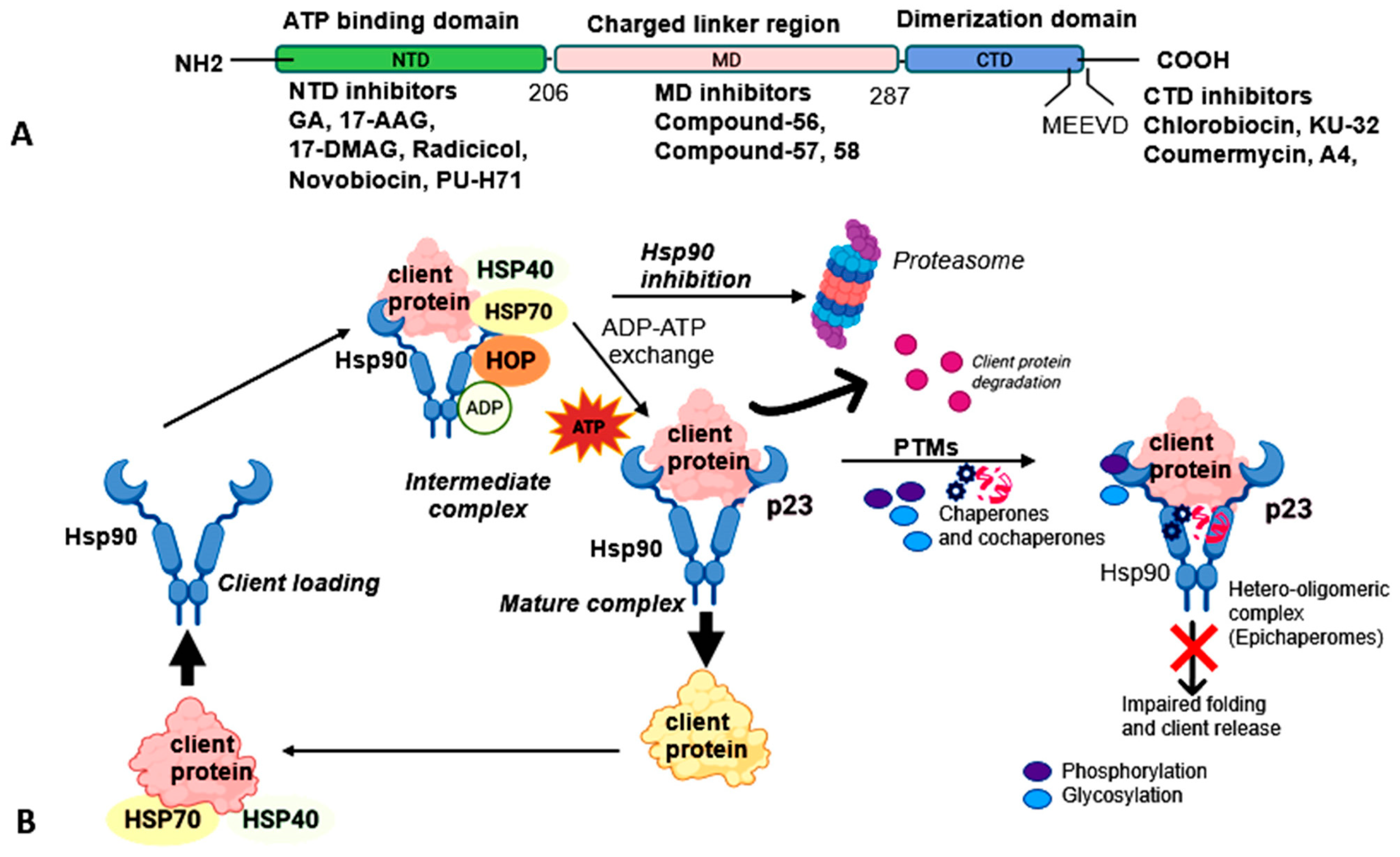
Figure 1. Schematic representation of structural organization of Hsp90 and its interactions with co-chaperones and client proteins. (A) Organization of human Hsp90 gene and its functional domains: N-terminal domain (NTD), middle domain (MD), and C-terminal domain (CTD) (adapted from the NCBI Gene database). (B) Illustration of the Hsp90 chaperone cycle, showing how ATP binding and hydrolysis drive conformational changes between open and closed states, facilitating the recruitment, stabilization, and maturation of co-chaperones and client proteins. The post-translational modifications (PTMs) promote the formation of hetero-oligomeric complex and inhibit the release of client proteins.
Ijms 26 10279 g001
Figure 2. The schematic illustration of Hsp90 interactions with signaling pathways and client protein kinases. Schematic representation of Hsp90 involvement in diverse cellular pathways, including cancer, steroid hormone signaling, and cell death. In oncogenic signaling, Hsp90 associates with key client proteins, such as AKT, Ras/Raf, p23, ATM, ATR, and HSF1—supporting downstream cascades that promote tumor cell growth, survival, and adaptation.
Ijms 26 10279 g002
Figure 3. The schematic representation of organelle-specific Hsp90 homologs in cancer and oxidative stress. Schematic representation of ER and mitochondrial specific Hsp90 functions in diverse cancers and reactive oxygen species (ROS) regulation, highlighting the roles of its endoplasmic reticulum (Grp94) and mitochondrial (TRAP1) homologs under stress conditions. The figure also depicts ER- and mitochondria-mediated processes that support cancer cell growth, survival, and metastasis.
Ijms 26 10279 g003
Figure 4. Schematic illustration of Hsp90-mediated cellular and nuclear processes. Hsp90 interacts with co-chaperones such as Cdc37, PP5, Aha1, and Tom70 to regulate diverse client proteins and complexes. In the cytoplasm, Hsp90 supports Ser/Thr kinases (ATM, ATR, DNA-PKcs, mTOR, SMG1, TRRAP), transcription factors (HSF1, p53, GR), and proteasomal assembly, as well as mitochondrial protein translocation. In the nucleus, Hsp90 facilitates the assembly and function of complexes involved in chromatin organization, transcription, RNA synthesis, ribosome biogenesis (MyoD1, E12, HIF-1α, HSF-1, SHRs), telomerase maturation (p23, hTERT), kinetochore assembly (CBF3, Mis12), and snoRNA processing (Rpb1).
Ijms 26 10279 g004
Figure 5. A schematic illustration of roles of the ER and mitochondria in cardiomyopathy and diabetes, and potential therapeutic strategies. The schematic illustrates how ER and mitochondria act as primary targets under diverse cellular stress conditions. Pharmacological inhibition of Hsp90 and its client proteins with agents such as 17-AAG, 17-DMAG, and novobiocin can disrupt ER stress responses and client protein degradation, affecting pathways including reactive oxygen species (ROS) regulation, oxidative phosphorylation (OXPHOS), and the unfolded protein responses (UPRs). Small-molecule inhibitors, particularly natural compounds, and antioxidants, may help alleviate cellular dysfunctions associated with cardiomyopathy and diabetes, especially in the early stages of these diseases.
Ijms 26 10279 g005
Table 1. Hsp90 isoforms, main characteristics, and biological functions.
LocalizationGene/Protein CharacteristicsHomologyFunctionsReferences
CytosolHsp90α (Hsp90AA1)Stress inducible ~86% amino acid sequence similarity between Hsp90α and Hsp90β; >95% homology within N-terminal and ATP-binding domainsChaperoning and maturation of client proteins; regulation of steroid hormone receptor signaling; facilitation of protein trafficking; cell-cycle control[2]
Hsp90β (Hsp90 AB1)Constitutively expressed
Endoplasmic reticulum (ER) Grp94/Gp96
(Hsp90B1)
Ca2+-binding chaperone; ATP hydrolysis triggers conformational activation>50% amino acid sequence homology with cytosolic Hsp90; possesses a KDEL retention motif; lacks MEEVD sequenceFolding and maturation of secretory and membrane proteins; quality control of ER clients such as TLR, insulin-like growth factors[5]
Mitochondria TRAP1 (TNF receptor-associated protein 1)Cytoplasmic and imported into mitochondria; mitochondrial targeting sequence cleaved upon import>85% identity with N-terminal ATP-binding domain of cytosolic Hsp90; lacks charged linker region and MEEVD motifMaintains mitochondrial integrity; protects against ROS and oxidative stress; overexpressed in several cancers[6,7,8]
Extracellular medium eHsp90α (plasma and serum); eHsp90β (exosome secretion dependent on p53 activation in cancer cells)Cell surface-associated and secreted forms of Hsp90High homology with cytosolic Hsp90; contains a distinct 115-amino-acid middle and charged linker binding domainFacilitates tumor cell invasion, migration, and extracellular matrix remodeling[9,10]
Disclaimer/Publisher’s Note: The statements, opinions and data contained in all publications are solely those of the individual author(s) and contributor(s) and not of MDPI and/or the editor(s). MDPI and/or the editor(s) disclaim responsibility for any injury to people or property resulting from any ideas, methods, instructions or products referred to in the content.