Article

Next-Generation Sequencing in Congenital Eye Malformations: Identification of Genetic Causes and Comparison of Different Panel-Based Diagnostic Strategies

1
Department of Ophthalmology, LMU University Hospital, LMU Munich, 80336 Munich, Germany
2
Medizinisch Genetisches Zentrum (MGZ) München, 80335 Munich, Germany
*
Author to whom correspondence should be addressed.
Int. J. Mol. Sci. 2025, 26(20), 9854; https://doi.org/10.3390/ijms26209854
Submission received: 10 September 2025 / Revised: 23 September 2025 / Accepted: 9 October 2025 / Published: 10 October 2025
(This article belongs to the Special Issue Molecular Research and Advances in Ocular Disease)

Abstract

Congenital eye malformations like microphthalmia–anophthalmia–coloboma (MAC), anterior segment dysgenesis (ASD), primary congenital glaucoma (PCG) and congenital cataracts (CC) are significant causes of childhood visual impairment. Phenotypic heterogeneity often complicates diagnosis. The goal of this study was to optimize the diagnostic strategy for next-generation sequencing (NGS)-based procedures, thereby aiming to identify genetic causes of congenital eye malformations. Forty patients with congenital eye malformations were included. A primary diagnostic testing (PD) of a limited number of genes was followed by multigene panel (MGP) testing, including 186 eye-related genes, and exome sequencing. Causative variants were identified in 17 patients (43%) and clinically relevant variants of uncertain significance (VUS) in 6 patients (15%). PD had a diagnostic yield (DY) of 15%, MGP of 29% and exome sequencing of 4%, leading to a cumulative DY of 43%. Diagnostic rates were highest in CC (75%), bilateral cases (46%), complex ocular phenotypes (78%), patients with extraocular manifestations (55%) and positive family history (70%). Rare and possible new genotype–phenotype correlations and candidate genes (FAT1, POGZ) could be identified. In total, eight (likely) pathogenic variants in six genes (CYP1B1, ADAMTS18, MAB21L2, NHS, MFRP, CRYBB1) were not yet reported. A stepwise genetic testing approach starting with a broad multigene panel followed by exome sequencing provides higher diagnostic yield than limited phenotype-specific testing. Comprehensive genetic diagnosis is essential for prognosis, treatment and genetic counseling, underscoring the need for routine genetic testing and interdisciplinary collaboration in managing congenital eye malformations.

1. Introduction

Congenital eye malformations pose significant diagnostic and therapeutic challenges and are a major cause of early-onset visual impairment, possibly impacting normal childhood development. The most common malformations include the microphthalmia–anophthalmia–coloboma (MAC) spectrum, anterior segment dysgenesis (ASD), primary congenital glaucoma (PCG) and congenital cataracts (CC). Although individually rare (e.g., MAC: 0.3–0.66 per 10,000 [1]; CC: 3.5–15 per 10,000 [2,3]), these anomalies collectively account for a substantial proportion of childhood blindness globally [4].
The etiology is highly heterogeneous [3,4]. While environmental factors (e.g., intrauterine infections) may be involved in isolated cases, most malformations are genetically driven [5]. They may present isolated or as first manifestation of complex syndromes involving extraocular manifestations [2,6]. Over 100 genes have been associated with congenital ocular anomalies, but in around 50% of cases, no genetic cause is identified despite extensive testing [6].
An accurate molecular diagnosis is essential for prognosis, recurrence risk assessment [2], targeted surveillance (e.g., stroke risk in COL4A1 [7]) and eligibility for emerging gene therapies (e.g., RPE65-associated retinal dystrophy [8]). Prior to next-generation sequencing (NGS), diagnostics relied on phenotype-based single-gene testing, often missing the underlying cause due to overlapping or atypical presentations [2,9]. NGS now allows broader, parallel analysis of multiple genes or exomes, improving diagnostic efficiency [6,10]. However, challenges remain due to high phenotype–genotype heterogeneity and unknown genetic causes [10,11]. This study investigates the effectiveness of a cross-phenotype multigene panel (MGP) and exome sequencing in patients with MAC, ASD/PCG and CC, comparing diagnostic yields (DY) with small phenotype-specific panels routinely applied in clinical practice, and aims to identify novel or rare variants to guide future diagnostic strategies.

2. Results

2.1. Study Population Characteristics and Sequencing Results

This cohort included 40 individuals with congenital ocular malformations: 22 with MAC, 14 with ASD and/or PCG and 4 with CC. Of these, 77% had a simple ocular phenotype, 23% a complex one; 72% had isolated eye involvement, while 28% showed syndromic features. Most cases (92%) were bilateral. A positive family history was present in 25%, with sequence data available for relatives in half of the cases. The cohort was 63% female and 37% male. For analysis, ASD and PCG were grouped and mixed MAC/CC cases were classified under MAC.
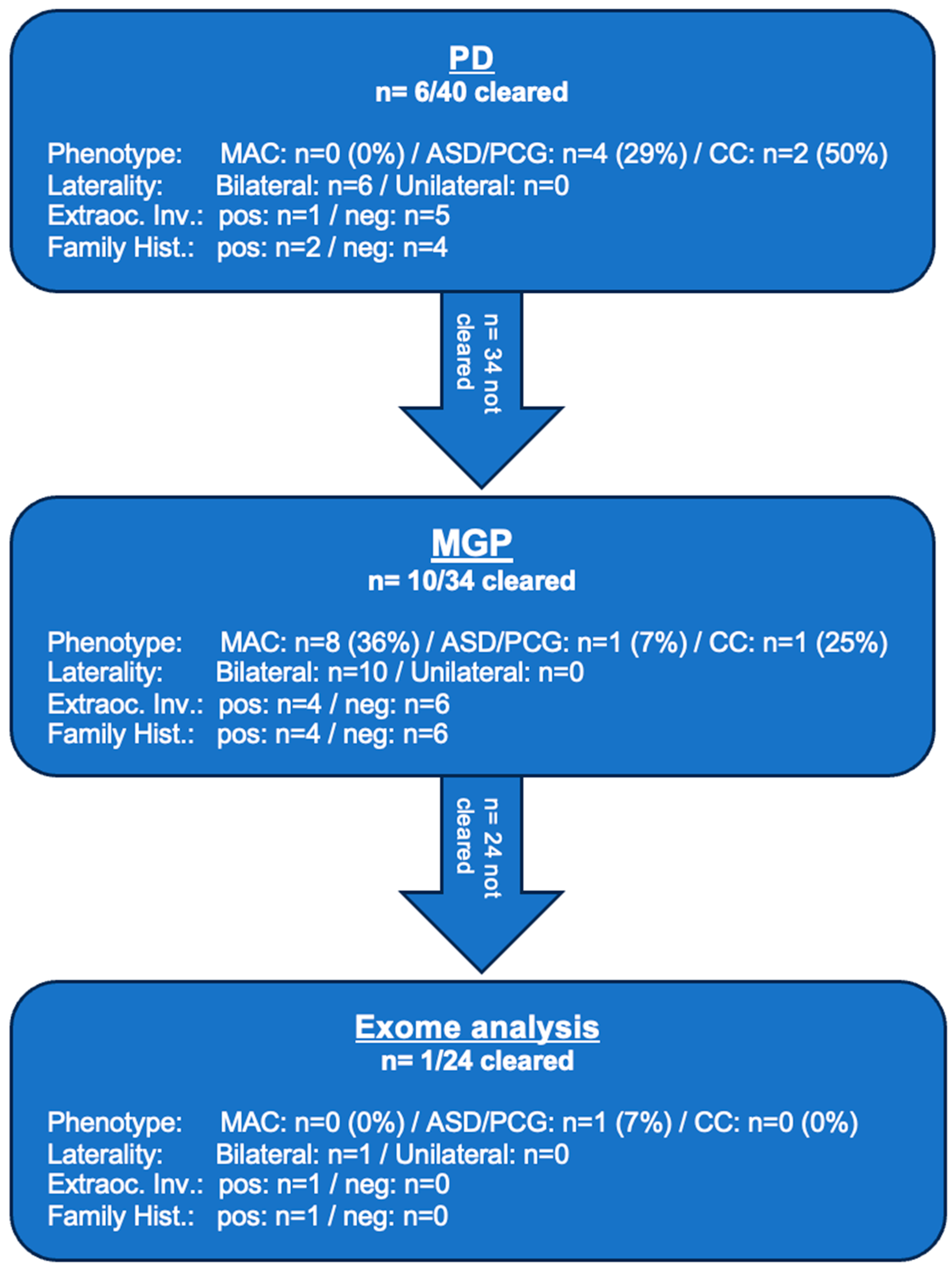
As illustrated in Table 1, across all three diagnostic levels—PD, MGP and exome—causative variants (ACMG class 4 or 5) were identified in 43% (17/40), including two cases where NGS suggested deletions confirmed by Multiplex ligation-dependent probe amplification (MLPA). Six causative variants were not previously described. PD found causative variants in 15% (6/40): four in ASD/PCG, two in CC and none in MAC. Among the 34 unresolved cases, the MGP identified causative variants in 29% (10/34): 36% in MAC (8 cases), 7% in ASD/PCG (1 case) and 25% in CC (1 case). Exome analysis of the remaining 24 unsolved cases revealed one additional novel variant (2.5% overall) in ASD/PCG. No further variants were found in MAC or CC.

2.2. Phenotype-Subgroup Results

2.2.1. MAC

This was the largest clinical subgroup, including 22 patients. Of these, 36% had coloboma, 36% microphthalmia and 28% both (no patient with anophthalmia). Most cases (91%) were bilateral and 23% had additional ocular findings, such as cataracts or persistent hyperplastic primary vitreous. Syndromic features occurred in 36% and 18% had a positive family history (Figure 1A,B). Causative variants (ACMG class 4 or 5) were identified in 36% (8/22), all with bilateral phenotypes, while none were found in unilateral cases. Five genes were involved, all previously associated with MAC. The most common was MAB21L2 (3 cases) followed by NHS (2 cases) and BCOR, MFRP and PRSS56 (1 case each). Four variants were novel: MAB21L2 c.58del (p.Cys20Valfs*37), NHS c.320del (p.Gly107Alafs*89) and c.3659dup (p.Asn1220Lysfs*5) and MFRP c.1534T>G (p.Cys512Gly).

2.2.2. ASD/PCG

This subgroup included 14 patients. Eight (57%) had isolated PCG, six (43%) had various forms of ASD, four of which (67%) developed glaucoma in early childhood. Two patients had ASD without glaucoma. Other ocular abnormalities, such as microcornea, high myopia and corneal changes, were noted in two patients (14%) and two patients had extraocular features (Figure 1C,D). Thirteen patients (93%) had bilateral disease and four (29%) had a positive family history. Causative variants (ACMG class 4 or 5) were identified in six patients (43%), all in bilateral cases and none in unilateral cases. Four genes, all previously associated with ASD/PCG, were involved: FOXC1 (two cases), CYP1B1 (two cases), PITX2 (one case) and ADAMTS18 (one case). Two variants were novel: ADAMTS18 c.1399G>T (p.Gly467*) and CYP1B1 c.1076_1082dup (p.Asp361Glufs*16). In patient 22, a deletion of the 3‘-region of exon 1 in FOXC1 was confirmed by MLPA. This specific deletion had not been previously reported.

2.2.3. CC

All four recruited patients with CC had bilateral involvement. Two also had microcornea and one had extraocular abnormalities. Two patients had a positive family history. Causative variants (ACMG class 4 or 5) were found in three of four patients (75%) in COL4A1, PAX6 and GCNT2—all genes known to be associated with CC and all previously described in the literature.

2.3. Diagnostic Yield

Diagnostic yield (DY) refers to the proportion of patients in which a causative genetic variant (ACMG class 4 or 5) was identified. It varied significantly by diagnostic approach and phenotype.
PD identified causative variants in 6 of 40 patients (15%). The multigene panel (MGP) resolved 10 additional cases, raising the cumulative yield to 40%, as it included all PD genes. Exome sequencing of 24 unresolved cases revealed one more variant (4%), bringing the overall DY to 43% (Figure 2A).
Among phenotype groups, the highest yield was in CC (75%), followed by ASD/PCG (43%) and MAC (36%) (Figure 2B). No causative variants were found in unilateral cases, while bilateral phenotypes had a 46% yield (CC: 75%, ASD/PCG: 46%, MAC: 40%). Patients with simple ocular phenotypes had a 32% DY (CC: 50%, ASD/PCG: 33%, MAC: 29%) versus 78% in those with additional ocular findings (CC: 100%, MAC: 60%, ASD/PCG: 100%). Syndromic or extraocular features were associated with a 55% yield (CC: 100%, ASD/PCG: 100%, MAC: 38%) compared to 38% (CC: 67%, ASD/PCG: 33%, MAC: 36%) in isolated ocular cases. A positive family history increased DY to 70% (MAC: 75%, ASD/PCG: 75%, CC: 50%), while cases without it had a 33% DY (CC: 100%, ASD/PCG: 30%, MAC: 28%) (Figure 2C). Figure 3 illustrates the results obtained in each diagnostic step.

2.4. Variants of Unknown Significance and Candidate Genes

In six patients, variants of uncertain significance (VUS; ACMG class 3) with possible relevance to the observed phenotypes were identified in five genes (POGZ, COL18A1, FAT1, MFRP, CRYBB1) (Table 2).
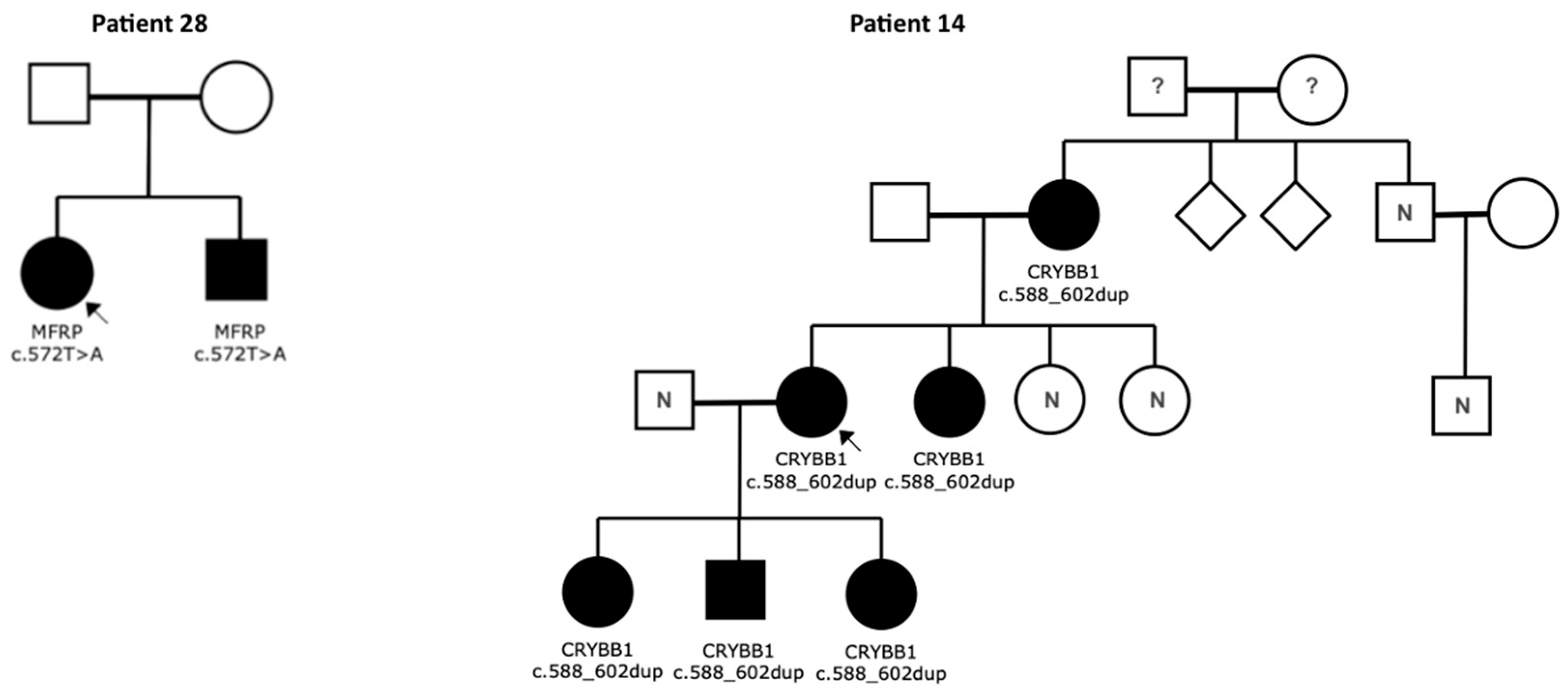
In patient 14, a novel duplication c.588_602dup (p.Gln197_Tyr201dup) in CRYBB1 was found in a patient with bilateral cataracts. The variant segregated in all affected family members (Figure 4). Despite its formal classification as VUS, when viewed in the context of clinical and genetic data, this variant is to be considered as highly likely causative, especially since no other causative variant could be identified. Patient 28 with bilateral microphthalmia carries a novel homozygous variant c.572T>A (p.Ile191Lys) in MFRP, which currently is classified as VUS. However, this variant is very likely to be pathogenic, according to the clinical and genetic findings, especially since the same variant was confirmed in the similarly affected sibling (Figure 4).

3. Discussion

The study aimed to identify genetic causes of congenital eye malformations using extended analysis of NGS data and to compare the DY across different testing strategies. In 17 of 40 patients, pathogenic or likely pathogenic variants (ACMG class 4 or 5) were found. Additionally, two patients carried VUS (ACMG class 3) in CRYBB1 and MFRP that were likely causative based on clinical correlation, raising the DY to 48% overall.
The MGP showed the highest standalone yield and increased the detection rate by 2.5-fold compared to PD, while WES added minimal additional findings.
These results align with previous studies showing detection rates between 24.5% and 75% for broad eye panels [10,22] and support the conclusion that phenotype-spanning MGP and WES significantly outperform limited, phenotype-specific gene panels in detecting genetic causes of congenital eye malformations.
Consistent with findings from other studies [10,23], the DY in this study was highest for CC (100%), followed by ASD/PCG (43%) and MAC (41%), including VUS in MFRP (MAC) and CRYBB1 (CC), which can clinically be considered causative.
No pathogenic variants were found unilateral cases, while the DY in bilateral cases was 46%, consistent with previous research and suggesting that unilateral cases may be more influenced by non-genetic factors, such as intrauterine infection, disruption or undetectable mosaicism [24,25].
DY was higher in cases with additional ocular abnormalities, extraocular or syndromic features and/or positive family history than in isolated or non-syndromic cases and/or negative family history. In ASD/PCG and CC, DY was higher in patients with extraocular features, whereas in MAC, it was comparable regardless of extraocular involvement. This contrasts with previous results suggesting a higher detection rate in isolated CC and in MAC cases with syndromic presentation [6,10].
Overall, the results of this study are largely consistent with existing literature, though differences in study design, patient selection, gene panel composition and variant classification limit direct comparisons. Nevertheless, the data indicate that the highest likelihood of identifying a causative variant occurs in patients with bilateral disease, a positive family history and complex or syndromic ocular phenotypes. In contrast, patients with unilateral involvement and no family history are less likely to receive a molecular diagnosis.
Biallelic variants in CYP1B1 are the most common cause of autosomal recessive PCG and often linked to corneal opacities presenting as edema, Haab’s striae or buphthalmos [26]. However, patient 27 with CYP1B1 variants showed a different primary phenotype with diffuse corneal opacity and iris hypoplasia, while glaucoma was only diagnosed secondarily. This matches reports of a rare frosted-glass-like opacity affecting the entire cornea, independent of intraocular pressure [27], expanding the phenotypic spectrum of CYP1B1-related disease. In patient 8 with a classical presentation of PCG, a novel frameshift variant c.1076_1082dup (p.Asp361Glufs*16) was identified.
The ADAMTS18 gene plays an essential role in embryonic eye development and biallelic variants have been linked to MMCAT syndrome (microcornea, myopic chorioretinal atrophy, telecanthus) microcornea and rod–cone dystrophy, early childhood cataracts and/or ectopia lentis and retinal dystrophy without anterior segment features [28]. In patient 20 and the similarly affected sister, exome sequencing revealed a novel homozygous pathogenic variant c.1399G>T (p.Gly467*). The phenotype of the patients included bilateral Axenfeld–Rieger Syndrome (ARS), which has not been previously reported in ADAMTS18-related eye disorders, as well as microcornea, high myopia, macular changes, taurodontism and craniofacial anomalies. These findings extend the known clinical spectrum and support the role of ADAMTS18 in combined anterior and posterior segment malformations.
The COL4A1 variant c.2317G>A (p.Gly773Arg), previously reported in patients with bilateral CC and neurological features [14], was found in patient 30, who primarily showed a purely ocular phenotype with bilateral CC and microcornea. However, further work-up revealed a past cerebral hemorrhage contributing to a minor motor delay. This highlights the phenotypic variability of COL4A1-related disease and underlines the importance of genetic testing in children with CC. Patient 33 carried the missense variant c.113G>A (p.Arg38Gln) in PAX6, which is critical for eye and CNS development [29]. While this variant was previously described with bilateral CC and additional features like nystagmus or ASD and microphthalmia [15,29], we report a particularly mild expression with only bilateral CC and microcornea.
MAB21L2 is involved in ocular and skeletal development and has been linked to MAC spectrum disorders with or without extraocular abnormalities [11,18]. Primarily missense variants are associated with autosomal dominant eye malformations. In this study, the novel heterozygous frameshift variant c.58del (p.Cys20Valfs*37) was found in two unrelated patients with isolated bilateral iris/retina/choroid colobomas, supporting monoallelic loss-of-function variants as a cause of milder, non-syndromic phenotypes. These results expand the phenotypic spectrum of MAB21L2 and confirm its relevance in isolated ocular malformations without systemic involvement.
CRYBB1 encodes a lens protein involved in non-syndromic CC [6]. The heterozygous variant c.588_602dup (p.GIn197_Tyr201dup) was identified in patient 14 and five affected family members. Although classified as a VUS, its segregation, conservation and absence in databases suggest likely pathogenicity. This variant is reported here for the first time.
A recent study described a homozygous frameshift variant in FAT1 in connection with microphthalmia or coloboma and syndromic features in five families and demonstrated its implication in optic cup development in a mouse model [30]. However, a definite gene-disease correlation is not yet established. Patient 26 with bilateral colobomas carried a novel heterozygous variant c.1025G>C (p.Gly342Ala). While its pathogenicity remains uncertain (VUS), bioinformatic analysis suggests a damaging effect. This supports FAT1 as a promising candidate gene for MAC spectrum disorders.
POGZ is linked to White–Sutton syndrome, a rare neurodevelopmental disorder with variable features including ocular anomalies, such as retinal dystrophies; colobomas have only described in isolated (n = 3) patients [21,31]. Patients 1 and 5 with POGZ variants both presented with coloboma and mild developmental delays. Patient 1 carried a novel missense variant(c.3271C>T (p.His1091Tyr)), while the missense variant found in patient 5 (c.4086A>C (p.Glu1362Asp)) has been previously described as a VUS in a patient with isolated congenital diaphragmatic hernia [21]. Although classified as VUS, bioinformatic analysis and rarity in databases underscore POGZ as an interesting candidate gene for congenital eye malformations and the possible association of White–Sutton Syndrome with coloboma.
NGS significantly improves the detection of genetic causes in congenital eye malformations. Cross-phenotype multigene panels (MGP) and exome sequencing consistently outperform phenotype-specific single-gene tests, especially given the high clinical and genetic heterogeneity. This study highlights that rare variants in genes like MAB21L2—missed in initial small phenotype-based panels—were the actual cause in several MAC cases, while in former times commonly implicated genes (e.g., SOX2, OTX2) [32] were not involved. Comprehensive MGP or WES avoid miss-selection of subpanels, save time and are increasingly cost-effective. A stepwise evaluation based on WES offers a practical balance between MGP and WES. While an MGP possibly provides better analysis of the included gene in these specific regions, including alleviation of the assessment of dosage alterations, WES allows broader testing of rare cause including novel associated genes not yet included in panels [22,33]. Therefore, in clinical routine diagnostics it is especially advisable to use a combined approach with primary analysis of an MGP on the base of exome sequencing with possible sequential exome analysis in selected cases. Since no diagnostic approach can detect all causative variants, in specific cases additional genetic methods need to be considered, as was demonstrated in two patients in our cohort where deletions were confirmed by MLPA after this pathogenic mechanism was suggested by NGS. Thus, in unresolved cases, complementary methods like genome sequencing and techniques to detect copy number variations (CNVs) (including deletions and duplications), such as MLPA, may be important.
However, challenges remain using NGS, especially in rare syndromes where HPO-based gene prioritization is difficult due to poor description of ocular findings. Precise ophthalmological and genetic assessment remain crucial, but in many syndromic conditions, the ocular phenotype is vaguely described—e.g., “visual impairment” instead of specific findings like coloboma. For example, in POGZ-associated White–Sutton syndrome, ocular anomalies or visual impairment are frequently mentioned, yet rarely detailed. This lack of specificity reduces diagnostic sensitivity, especially when the ocular findings are the primary reason for referral. It underscores the need for detailed phenotype documentation and reanalysis of prior negative genetic results if more specific phenotypic description is available.
In 17 cases, no causative variants were found, possibly due to non-genetic causes, non-coding mutations, or undiscovered genotype–phenotype associations [34].
Accurate genetic diagnosis in congenital eye malformations has significant implications for prognosis, therapy, comorbidities and family planning. It also enables access to emerging gene therapies. In several cases, similar clinical phenotypes were linked to different genetic causes, each requiring distinct medical management. For example, COL4A1 mutations may indicate risk for cerebral hemorrhage, as seen in patient 30, and a timely diagnosis could facilitate personalized risk adapted management. On the other hand, X-linked variants in NHS or BCOR carry specific familial risks [3,16].
Our study has several limitations. Some clinical data from external reports were incomplete, limiting phenotype accuracy and segregation analysis. Due to evolving sequencing methods and software, in earlier cases newly recognized variants may have been missed. Thus, reported yields likely represent a minimum and support re-testing over time.
Exome-based NGS proved highly effective for diagnosing congenital eye malformations, confirming causative variants in 43% of cases and suspected variants in 15%. Broader panels outperformed phenotype-specific diagnostics and our findings highlight the importance of interdisciplinary cooperation and routine genetic testing. Eight novel pathogenic variants in six genes, rare potential genotype–phenotype associations and candidate genes were identified, advancing understanding of genetic mechanisms in congenital eye malformations.

4. Materials and Methods

4.1. Patient Selection and Clinical Data Collection

We included patients who presented at MGZ Medical Genetics Center, Munich, for consultation and diagnostics between 2016 and 2021 due to congenital eye malformations with the MAC, ASD, PGC or CC phenotypes. Informed consent was obtained from all participants or their legal guardians and the study was approved by the institutional review board of Ludwig-Maximilians University Munich, Germany (approval number: 19-317). All principles of the Declaration of Helsinki were followed. Clinical data were collected retrospectively, including ophthalmologic findings, family and prenatal history, prior genetic testing and extraocular anomalies.

4.2. Genetic Testing Workflow

Blood samples were collected from patients and available relatives. DNA was extracted using standard protocols. Sequencing libraries were prepared using SureSelectXT Target Enrichment System for the Illumina Platform (Agilent Technologies, Santa Clara, CA, USA) for patients 1–26 and the Human Comprehensive Exome + Mitochondrial Panel (Twist Bioscience, San Francisco, CA, USA) for patients 27–40. Sequencing was performed on MiSeq, NextSeq, or NovaSeq platforms (Illumina, San Diego, CA, USA). A stepwise NGS-based diagnostic approach was used. Initial testing (primary diagnostics, PD) targeted small, phenotype-specific virtual panels (<25 kb coding sequence) selected based on clinical presentation. If no pathogenic variants were found, a broader multigene panel (MGP) of 186 eye-related genes was analyzed. For unresolved cases, full exome data were filtered using Human Phenotype Ontology (HPO) terms to prioritize known gene–disease associations (including phenotype-overlapping genes), syndromic genes with ocular involvement and novel candidate genes with ocular expression and loss-of-function (LOF) variants.

4.3. Bioinformatic Analysis, Variant Classification and Interpretation

For patients 1–26, sequencing data were analyzed following a standardized in-house pipeline with Burrows–Wheeler Aligner (BWA, Version 0.7.8-r455), SAMtools (Version 1.1), SnpEff (Version 4.0e) and Alamut-Batch (Version 1.3.1, Interactive Biosoftware, Rouen, France). Population (dbSNP, 1000 Genomes, ESP, GnomAD/ExAC) and clinical databases (ClinVar, Emory, MGZ internal, HGMD, Mastermind, COSMIC) supported variant annotation.
For patients 27 to 40, the Varvis software (Version 2.4.0, Limbus Technologies, Rostock, Germany) was used. Variants were classified by a team of genetic and ophthalmological specialists according to the American College of Medical Genetics (ACMG) system [35]. Only variants classified as pathogenic or likely pathogenic (ACMG classes 4 and 5) were reported as causative. Variants of Unknown Significance (VUS, ACMG class 3) were discussed if there was strong phenotype–genotype correlation.

Author Contributions

Conceptualization, L.N. and T.N.; methodology, L.N. and T.N.; software, L.N. and A.L.; validation, A.L., T.N. and E.H.-F.; formal analysis, L.N., T.N. and A.L.; investigation, L.N. and T.N.; resources, T.N. and E.H.-F.; data curation, L.N., A.L. and T.N.; writing—original draft preparation, L.N.; writing—review and editing, T.N. and E.H.-F.; visualization, L.N. and T.N.; supervision, T.N. and E.H.-F.; project administration, L.N., T.N. and E.H.-F. All authors have read and agreed to the published version of the manuscript.

Funding

This research received no external funding.

Institutional Review Board Statement

The study was conducted in accordance with the Declaration of Helsinki, and approved by the Institutional Review Board of Ludwig-Maximilians University Munich, Germany (approval number: 19-317; approval date: 4 July 2019).

Informed Consent Statement

Informed consent was obtained from all subjects involved in the study.

Data Availability Statement

The raw data supporting the conclusions of this article will be made available by the authors on request.

Conflicts of Interest

A.L., E.H.-F. and T.N. are employed at MGZ—Medizinisch Genetisches Zentrum. This affiliation with a medical practice had no influence on the preparation or content of this manuscript. The remaining authors declare that the research was conducted in the absence of any commercial or financial relationships that could be construed as a potential conflict of interest.

References

  1. Hu, Z.; Yu, C.; Li, J.; Wang, Y.; Liu, D.; Xiang, X.; Su, W.; Pan, Q.; Xie, L.; Xia, K. A Novel Locus for Congenital Simple Microphthalmia Family Mapping to 17p12-q12. Investig. Ophthalmol. Vis. Sci. 2011, 52, 3425–3429. [Google Scholar] [CrossRef]
  2. Haer-Wigman, L.; van Zelst-Stams, W.A.; Pfundt, R.; Born, L.I.v.D.; Klaver, C.C.; Verheij, J.B.; Hoyng, C.B.; Breuning, M.H.; Boon, C.J.; Kievit, A.J.; et al. Diagnostic exome sequencing in 266 Dutch patients with visual impairment. Eur. J. Hum. Genet. 2017, 25, 591–599. [Google Scholar] [CrossRef]
  3. Sun, W.; Xiao, X.; Li, S.; Guo, X.; Zhang, Q.; Leung, Y.F. Exome Sequencing of 18 Chinese Families with Congenital Cataracts: A New Sight of the NHS Gene. PLoS ONE 2014, 9, e100455. [Google Scholar] [CrossRef] [PubMed]
  4. Wang, P.; Li, S.; Sun, W.; Xiao, X.; Jia, X.; Liu, M.; Xu, L.; Long, Y.; Zhang, Q. An Ophthalmic Targeted Exome Sequencing Panel as a Powerful Tool to Identify Causative Mutations in Patients Suspected of Hereditary Eye Diseases. Transl. Vis. Sci. Technol. 2019, 8, 21. [Google Scholar] [CrossRef]
  5. Chassaing, N.; Causse, A.; Vigouroux, A.; Delahaye, A.; Alessandri, J.; Boespflug-Tanguy, O.; Boute-Benejean, O.; Dollfus, H.; Duban-Bedu, B.; Gilbert-Dussardier, B.; et al. Molecular findings and clinical data in a cohort of 150 patients with anophthalmia/microphthalmia. Clin. Genet. 2013, 86, 326–334. [Google Scholar] [CrossRef]
  6. Gillespie, R.L.; O’Sullivan, J.; Ashworth, J.; Bhaskar, S.; Williams, S.; Biswas, S.; Kehdi, E.; Ramsden, S.C.; Clayton-Smith, J.; Black, G.C.; et al. Personalized Diagnosis and Management of Congenital Cataract by Next-Generation Sequencing. Ophthalmology 2014, 121, 2124–2137. [Google Scholar] [CrossRef] [PubMed]
  7. Magnin, E.; Ayrignac, X.; Berger, E.; Mine, M.; Tournier-Lasserve, E.; Labauge, P. Late Diagnosis of COL4A1 Mutation and Problematic Vascular Risk Fac-tor Management. Eur. Neurol. 2014, 72, 150–152. [Google Scholar] [CrossRef]
  8. Méjécase, C.; Malka, S.; Guan, Z.; Slater, A.; Arno, G.; Moosajee, M. Practical guide to genetic screening for inherited eye diseases. Ther. Adv. Ophthalmol. 2020, 12, 2515841420954592. [Google Scholar] [CrossRef]
  9. Deng, H.; Yuan, L. Molecular genetics of congenital nuclear cataract. Eur. J. Med. Genet. 2014, 57, 113–122. [Google Scholar] [CrossRef]
  10. Patel, A.; Hayward, J.D.; Tailor, V.; Nyanhete, R.; Ahlfors, H.; Gabriel, C.; Jannini, T.B.; Abbou-Rayyah, Y.; Henderson, R.; Nischal, K.K. The Oculome Panel Test: Next-Generation Sequencing to Diagnose a Di-verse Range of Genetic Developmental Eye Disorders. Ophthalmology 2019, 126, 888–907. [Google Scholar] [CrossRef]
  11. Harding, P.; Gore, S.; Malka, S.; Rajkumar, J.; Oluonye, N.; Moosajee, M. Real-world clinical and molecular management of 50 prospective patients with microphthalmia, anophthalmia and/or ocular coloboma. Br. J. Ophthalmol. 2022, 107, 1925–1935. [Google Scholar] [CrossRef] [PubMed]
  12. Hilal, L.; Boutayeb, S.; Serrou, A.; Refass-Buret, L.; Shisseh, H.; Bencherifa, F.; El Mzibri, M.; Benazzouz, B.; Berraho, A. Screening of CYP1B1 and MYOC in Moroccan families with primary congenital glaucoma: Three novel mutations in CYP1B1. Mol. Vis. 2010, 16, 1215–1226. [Google Scholar] [PubMed]
  13. Zhang, Y.; Chen, X.; Wang, L.; Sun, X.; Chen, Y. Heterogeneity of Axenfeld–Rieger Syndrome: Molecular and Clinical Findings in Chinese Patients. Front. Genet. 2021, 12, 732170. [Google Scholar] [CrossRef]
  14. Deml, B.; Reis, L.; Maheshwari, M.; Griffis, C.; Bick, D.; Semina, E. Whole exome analysis identifies dominant COL4A1 mutations in patients with complex ocular phenotypes involving microphthalmia. Clin. Genet. 2014, 86, 475–481. [Google Scholar] [CrossRef]
  15. Li, J.; Leng, Y.; Han, S.; Yan, L.; Lu, C.; Luo, Y.; Zhang, X.; Cao, L. Clinical and genetic characteristics of Chinese patients with familial or sporadic pe-diatric cataract. Orphanet J. Rare Dis. 2018, 13, 94. [Google Scholar] [CrossRef]
  16. Ng, D.; Thakker, N.; Corcoran, C.M.; Donnai, D.; Perveen, R.; Schneider, A.; Hadley, D.W.; Tifft, C.; Zhang, L.; Wilkie, A.O.M.; et al. Oculofaciocardiodental and Lenz microphthalmia syndromes result from distinct classes of mutations in BCOR. Nat. Genet. 2004, 36, 411–416. [Google Scholar] [CrossRef]
  17. Mukhopadhyay, R.; Sergouniotis, P.I.; Mackay, D.S.; Day, A.C.; Wright, G.; Devery, S.; Leroy, B.P.; Robson, A.G.; Holder, G.E.; Li, Z.; et al. A detailed phenotypic assessment of individuals affected by MFRP-related oculopathy. Mol. Vis. 2010, 16, 540–548. [Google Scholar]
  18. Rainger, J.; Pehlivan, D.; Johansson, S.; Bengani, H.; Sanchez-Pulido, L.; Williamson, K.A.; Ture, M.; Barker, H.; Rosendahl, K.; Spranger, J.; et al. Monoallelic and Biallelic Mutations in MAB21L2 Cause a Spectrum of Major Eye Malformations. Am. J. Hum. Genet. 2014, 94, 915–923. [Google Scholar] [CrossRef]
  19. Gal, A.; Rau, I.; El Matri, L.; Kreienkamp, H.-J.; Fehr, S.; Baklouti, K.; Chouchane, I.; Li, Y.; Rehbein, M.; Fuchs, J.; et al. Autosomal-Recessive Posterior Microphthalmos Is Caused by Mutations in PRSS56, a Gene Encoding a Trypsin-Like Serine Protease. Am. J. Hum. Genet. 2011, 88, 382–390. [Google Scholar] [CrossRef]
  20. Reis, L.M.; Tyler, R.C.; Weh, E.; Hendee, K.E.; Kariminejad, A.; Abdul-Rahman, O.; Ben-Omran, T.; Manning, M.A.; Yesilyurt, A.; McCarty, C.A.; et al. Analysis of CYP1B1 in pediatric and adult glaucoma and other ocular pheno-types. Mol. Vis. 2016, 22, 1229–1238. [Google Scholar] [PubMed]
  21. Assia Batzir, N.; Posey, J.E.; Song, X.; Akdemir, Z.C.; Rosenfeld, J.A.; Brown, C.W.; Chen, E.; Holtrop, S.G.; Mizerik, E.; Nieto Moreno, M.; et al. Phenotypic expansion of POGZ-related intellectual disability syndrome (White-Sutton syndrome). Am. J. Med. Genet. A 2020, 182, 38–52. [Google Scholar] [CrossRef]
  22. Kamenarova, K.; Mihova, K.; Veleva, N.; Mermeklieva, E.; Mihaylova, B.; Dimitrova, G.; Oscar, A.; Shandurkov, I.; Cherninkova, S.; Kaneva, R. Panel-based next-generation sequencing identifies novel mutations in Bulgarian patients with inherited retinal dystrophies. Mol. Genet. Genom. Med. 2022, 10, e1997. [Google Scholar] [CrossRef]
  23. Jackson, D.; Malka, S.; Harding, P.; Palma, J.; Dunbar, H.; Moosajee, M. Molecular diagnostic challenges for non-retinal developmental eye disorders in the United Kingdom. Am. J. Med. Genet. C Semin. Med. Genet. 2020, 184, 578–589. [Google Scholar] [CrossRef]
  24. Chacon Fonseca, I.; Wong, J.; Mireskandari, K.; Chitayat, D. Newborn with bilateral congenital cataracts: Never forget congenital rubella syndrome. Paediatr. Child Health 2020, 25, 72–76. [Google Scholar] [CrossRef] [PubMed]
  25. Prontera, P.; Stangoni, G.; Ardisia, C.; Rogaia, D.; Mencarelli, A.; Donti, E. Trisomy 2 mosaicism with caudal dysgenesis, Hirschsprung disease, and micro-anophthalmia. Am. J. Med. Genet. A 2011, 155, 928–930. [Google Scholar] [CrossRef] [PubMed]
  26. Coêlho, R.E.; Sena, D.R.; Cruz, F.S.; Moura, B.C.; Han, C.C.; Andrade, F.N.; Lira, R.P. CYP1B1 Gene and Phenotypic Correlation in Patients From Northeastern Brazil With Primary Congenital Glaucoma. Eur. J. Gastroenterol. Hepatol. 2019, 28, 161–164. [Google Scholar] [CrossRef] [PubMed]
  27. Nischal, K.K. A new approach to the classification of neonatal corneal opacities. Curr. Opin. Ophthalmol. 2012, 23, 344–354. [Google Scholar] [CrossRef]
  28. Chandra, A.; Arno, G.; Williamson, K.; Sergouniotis, P.I.; Preising, M.N.; Charteris, D.G.; Thompson, D.A.; Holder, G.E.; Borman, A.D.; Davagnanam, I.; et al. Expansion of Ocular Phenotypic Features Associated With Mutations in ADAMTS18. JAMA Ophthalmol. 2014, 132, 996–1001, Erratum in JAMA Ophthalmol. 2014, 132, 1153. [Google Scholar] [CrossRef]
  29. Williamson, K.A.; Hall, H.N.; Owen, L.J.; Livesey, B.J.; Hanson, I.M.; Adams, G.; Bodek, S.; Calvas, P.; Castle, B.; Clarke, M.; et al. Recurrent heterozygous PAX6 missense variants cause severe bilateral microphthalmia via predictable effects on DNA–protein interaction. Anesth. Analg. 2019, 22, 598–609. [Google Scholar] [CrossRef]
  30. Lahrouchi, N.; George, A.; Ratbi, I.; Schneider, R.; Elalaoui, S.C.; Moosa, S.; Bharti, S.; Sharma, R.; Abu-Asab, M.; Onojafe, F.; et al. Homozygous frameshift mutations in FAT1 cause a syndrome characterized by colobomatous-microphthalmia, ptosis, nephropathy and syndactyly. Nat. Commun. 2019, 10, 1180. [Google Scholar] [CrossRef]
  31. Murch, O.; Jain, V.; Benneche, A.; Metcalfe, K.; Hobson, E.; Prescott, K.; Chandler, K.; Ghali, N.; Carmichael, J.; Foulds, N.C.; et al. Further delineation of the clinical spectrum of White–Sutton syndrome: 12 new individuals and a review of the literature. Eur. J. Hum. Genet. 2021, 30, 95–100. [Google Scholar] [CrossRef] [PubMed]
  32. Williamson, K.A.; FitzPatrick, D.R. The genetic architecture of microphthalmia, anophthalmia and coloboma. Eur. J. Med. Genet. 2014, 57, 369–380. [Google Scholar] [CrossRef]
  33. Moon, D.; Park, H.W.; Surl, D.; Won, D.; Lee, S.-T.; Shin, S.; Choi, J.R.; Han, J. Precision Medicine through Next-Generation Sequencing in Inherited Eye Diseases in a Korean Cohort. Genes 2021, 13, 27. [Google Scholar] [CrossRef]
  34. Rechsteiner, D.; Issler, L.; Koller, S.; Lang, E.; Bähr, L.; Feil, S.; Rüegger, C.M.; Kottke, R.; Toelle, S.P.; Zweifel, N.; et al. Genetic Analysis in a Swiss Cohort of Bilateral Congenital Cataract. JAMA Ophthalmol. 2021, 139, 691–700. [Google Scholar] [CrossRef] [PubMed]
  35. Richards, S.; Aziz, N.; Bale, S.; Bick, D.; Das, S.; Gastier-Foster, J.; Grody, W.W.; Hegde, M.; Lyon, E.; Spector, E.; et al. Standards and guidelines for the interpretation of sequence variants: A joint consensus recommendation of the American College of Medical Genetics and Genomics and the Association for Molecular Pathology. Genet. Med. 2015, 17, 405–424. [Google Scholar] [CrossRef] [PubMed]
Figure 1. Distribution of clinical features in patients with MAC (A,B) and ASD/PCG (C,D). COL = coloboma, MCO = microphthalmia, PCG = primary congenital glaucoma, ASD = anterior segment dysgenesis.
Ijms 26 09854 g001
Figure 2. Diagnostic yield—(A) for each diagnostic step, (B) by phenotypic groups and (C) by different clinical feature subgroups. PD = primary diagnostics, MGP = multigene panel, n = number of patients, iso = isolated, cum = cumulative, FH +/− = family history positive/negative.
Ijms 26 09854 g002
Figure 3. Flowchart of results obtained through each subsequent diagnostic step. Extraoc. Inv.: = extraocular involvement, Family Hist. = Family History, pos = positive, neg = negative.
Ijms 26 09854 g003
Figure 4. Segregation analysis in patients with Variants of Unknown Significance. ? = phenotype not known, N = variant not present, arrow = index patient.
Ijms 26 09854 g004
Table 1. Clinical details and sequencing results cases with causative variants. het = heterozygote, hom = homozygote, hem = hemizygote, - = not applicable, Known = previously described in the literature/databases, Novel = not previously described in the literature/databases, ø = unknown/not performed, de novo = negative segregation analysis.
Diagnostic LevelPatient IDDescribed Ocular PhenotypeUnilateral/BilateralSystemic FeaturesFamily History (Phenotype)GeneSequence VariantAmino Acid ChangeVariant TypeACMG ClassVariant Known/NovelSegregation/Variant in Family Members?
PDASD/PCG-8Congenital glaucomaBilateral-NegativeCYP1B1c.1168C>A; het/c.1076_1082dup; hetp.Arg390Ser/p.Asp361Glufs*16Missense/FrameshiftClass 5/Class 5Known [12]/Novelø
PDASD/PCG-22ARS, early childhood glaucomaBilateral-Positive (mother, aunt, grandmother, daughter)FOXC1Deletion 3’ Probe Exon 1; het-DeletionClass 4-ø
PDASD/PCG-23Anterior segment dysgenesis (Rieger-anomaly)BilateralTeeth anomalies, excessive umbilical skinPositive (daughter)PITX2Deletion of all Probes (Intron 3, Exon 4, Exon 5); het-DeletionClass 4-ø
PDASD/PCG-34Anterior segment dysgenesis/congenital glaucomaBilateral-NegativeFOXC1c.247T>C; hetp.Tyr83HisMissenseClass 4Known [13]de novo
PDCC-30Congenital cataract, microcorneaBilateralCerebral hemorrhages NegativeCOL4A1c.2317G>A; hetp.Gly773ArgMissenseClass 5Known [14]de novo
PDCC-33Congenital cataract, microcorneaBilateral-NegativePAX6c.113G>A; hetp.Arg38GlnMissenseClass 4Known [15]ø
MGPMAC-9Microphthalmia, congenital cataractBilateralRadiculomegaly, oligodontia, toe syndactyly, broad nasal tipPositive (mother, sister)BCORc.4038_4039del; hetp.Glu1348Ilefs*26FrameshiftClass 4Known [16]Mother affected
MGPMAC-10Microphthalmia, congenital cataractBilateral-Positive (sister)NHSc.320del; hemp.Gly107Alafs*89FrameshiftClass 5Novelø
MGPMAC-11Iris/retinal/choroidal colobomaBilateral-NegativeMAB21L2c.58del; hetp.Cys20Valfs*37FrameshiftClass 4Novelø
MGPMAC-15Iris/retinal/choroidal colobomaBilateral-NegativeMAB21L2c.58del; hetp.Cys20Valfs*37FrameshiftClass 4Novelø
MGPMAC-19Microphthalmia, glaucoma, extreme hyperopiaBilateral-Positive (sister)MFRPc.201G>A; het/c.1534T>G; hetp.Trp67*/p.Cys512GlyNonsense/MissenseClass 4/Class 4Known [17]/
Novel
ø
MGPMAC-37Microphthalmia, congenital cataractBilateralUreteral stricture, psoriasis, dental duplicationNegativeNHSc.3659dup; hemp.Asn1220Lysfs*5FrameshiftClass 4Novelø
MGPMAC-39Iris/retinal/choroidal/optic disk colobomaBilateralSandal gap deformityNegativeMAB21L2c.145G>A; hetp.Glu49LysMissenseClass 5Known [18]de novo
MGPMAC-40MicrophthalmiaBilateral-NegativePRSS56c.1066dup; homp.Gln356Profs*152FrameshiftClass 5Known [19]ø
MGPASD/PCG-27Corneal opacity, iris hypoplasia, optic disk changes, congenital glaucomaBilateral-NegativeCYP1B1c.1159G>A; het/c.1064_1076del; hetp.Glu387Lys/p.Arg355Hisfs*69Missense/FrameshiftClass 5/Class 5Known [20]ø
MGPCC-4Congenital cataractBilateralLanguage-related developmental delay/microcephalyPositive (brother)GCNT2c.1148G>A; homp.Arg383HisMissenseClass 4Known [15]ø
ExomeASD/PCG-20ARS, high myopia, microcornea, maculopathy,
glaucoma
BilateralTaurodontism, conspicuous auriclePositive (sister, brother)ADAMTS18c.1399G>T; homp.Gly467*NonsenseClass 5NovelSister affected
Table 2. Clinical Variants of Unclear Significance (VUS) and candidate genes. het = heterozygous, hom = homozygous, - = not applicable, Known = previously described in the literature/databases, Novel = not previously described in the literature/databases, ø = unknown/not performed, * = variant published as VUS in the context of diaphragmatic hernia.
Patient IDDescribed Ocular PhenotypeUnilateral/BilateralSystemic FeaturesFamily History (Phenotype)GeneSequence VariantAmino Acid ChangeVariant TypeACMG ClassVariant Known/NovelSegregation/Variant in Family Members?
MAC-1Iris/choroidal coloboma OD, optic disk coloboma OSBilateralMotor development delayNegativePOGZc.3271C>T; hetp.His1091TyrMissenseClass 3Novelø
MAC-5Iris/choroidal colobomaUnilateralDevelopmental delayNegativePOGZc.4086A>C; hetp.Glu1362AspMissenseClass 3Known [21] *ø
MAC-26Iris/retinal/choroidal colobomaBilateral-NegativeFAT1c.1025G>C; hetp.Gly342AlaMissenseClass 3Novelø
MAC-28Microphthalmia, extreme hyperopia,
glaucoma
Bilateral-Positive (brother)MFRPc.572T>A; homp.Ile191LysMissenseClass 3NovelBrother affected
ASD/PCG-12Early childhood glaucomaUnilateral-NegativeCOL18A1c.2156C>T; hetp.Thr719MetMissenseClass 3Novelø
CC-14Congenital cataractBilateral-Positive (mother, sister, 2 daughters, son)CRYBB1c.588_602dup; hetp.Gln197_Tyr201dupIntragenic DuplicationClass 3NovelMother, Sister, 2 Daughters, Son affected
Disclaimer/Publisher’s Note: The statements, opinions and data contained in all publications are solely those of the individual author(s) and contributor(s) and not of MDPI and/or the editor(s). MDPI and/or the editor(s) disclaim responsibility for any injury to people or property resulting from any ideas, methods, instructions or products referred to in the content.