Article

In Silico Analysis of MiRNA Regulatory Networks to Identify Potential Biomarkers for the Clinical Course of Viral Infections

by
Elena V. Mikheeva
,
Kseniya S. Aulova
,
Georgy A. Nevinsky
and
Anna M. Timofeeva
*
SB RAS Institute of Chemical Biology and Fundamental Medicine, Novosibirsk 630090, Russia
*
Author to whom correspondence should be addressed.
Int. J. Mol. Sci. 2025, 26(20), 10100; https://doi.org/10.3390/ijms262010100
Submission received: 8 September 2025 / Revised: 7 October 2025 / Accepted: 11 October 2025 / Published: 16 October 2025

Abstract

MiRNA expression profiles exhibit notable alterations in numerous diseases, particularly viral infections. Consequently, miRNAs may be regarded as both therapeutic targets and markers for the development of complications. MiRNAs can significantly influence the modulation of immune responses, offering an extra layer of regulation during viral infections. In this study, miRNAs associated with viral infections were analyzed using an in silico approach. Computer modeling predicted a number of miRNAs capable of influencing the functionality of specific components of the immune system. As a result, 242 miRNAs common to the three types of infections were identified. A network of miRNA-gene regulatory interactions, encompassing 502 nodes (224 miRNAs and 278 genes) and 2236 interactions, was developed. Within this network, subnetworks were identified that are involved in the operation of specific connections in the immune response to viruses. For each step of the immune response, the miRNAs involved in governing these processes were examined. These predicted miRNAs are of particular interest for further analysis aimed at establishing the relationship between their differential expression and disease symptom severity. The obtained data lay the foundation for identifying the most promising molecules as predictive biomarkers and the subsequent development of a diagnostic system.

1. Introduction

RNA viruses are characterized by high evolutionary rates due to their frequent replication cycles and low fidelity of RNA polymerase, resulting in mutations that may be linked to high pathogenicity in humans. Thus, it is not unexpected that RNA viruses are responsible for the most recent significant epidemics. SARS-CoV, SARS-CoV-2, and MERS-CoV are highly pathogenic coronaviruses that induce severe acute respiratory illness [1,2]. Presently, HIV-1 continues to be a considerable worldwide public health issue. According to UNAIDS, approximately 88.4 million individuals have been infected with HIV, and approximately 42.3 million have died from AIDS-related illnesses since the beginning of the epidemic (https://www.un.org/en/global-issues/aids accessed on 22 August 2025). Over the past few years, there have been significant threats to global health because of consistent outbreaks of infectious diseases from the Flaviviridae family, including Dengue virus (DENV), Zika virus (ZIKV), West Nile virus (WNV), and tick-borne encephalitis virus (TBEV). The DENV is transmitted to humans by mosquitoes, causing roughly 400 million infections and tens of thousands of deaths annually [3,4]. Over the last decade, ZIKV has infected millions, causing substantial epidemics in the tropics and extending to other regions [5,6].
The advent of the COVID-19 pandemic, an unanticipated event, severely tested the medical field and exposed multiple systemic weaknesses in the treatment and prevention of infectious diseases and their secondary effects. It is plausible that humanity may confront a new viral threat in the future, which could trigger another epidemic. Therefore, a search for predictive markers regarding the clinical course of viral infections is highly relevant. MiRNAs can be considered as representative markers. These elements control numerous biological mechanisms, thus contributing substantially to various processes [7]. Notable changes in the miRNA expression profile are observed in diverse diseases, encompassing viral infections [8,9,10,11,12,13,14]. Certain miRNAs can be assessed as important regulators of immune responses, inflammatory mediators, and cytokines [15,16]. Consequently, miRNAs can be considered as therapeutic targets in viral infection, as well as markers for the progression of complications. Identifying shared miRNA regulatory pathways across various infections is crucial not only for understanding the infections themselves but also for addressing novel diseases that may emerge in the future.
The objective of this work was to identify miRNAs with the potential to predict the severity of viral infections. The miRWalk database was used to identify miRNAs associated with viral infections of various etiologies, specifically flavivirus, coronavirus, and HIV. A total of 242 miRNAs were found to have altered profiles in the three viral infections investigated. The correlation between the identified miRNAs and genes was analyzed. Genes associated with the functioning of the immune system during viral infection were identified by gene ontology analysis. A miRNA-gene regulatory interaction network, encompassing 502 nodes (224 miRNAs and 278 genes), with 2236 interactions, has been established. Subnetworks connected to the function of single links in the immune response to viral infection were detected in this network. The involvement of miRNAs in regulating these processes has been assessed for each facet of the immune response. The research has successfully identified miRNAs that impact the functions of individual immune system components and can be employed to predict the severity of clinical progression in viral infections, including potential infections that humanity may face in the future.

2. Results

2.1. miRNAs Associated with Viral Infections

The miRWalk database was used to explore miRNAs linked to viral infections of different origins [17,18,19]. Here, the focus is on flavivirus, coronavirus, and HIV infections. The miRNAs were filtered according to the following selection criteria: (1) a predicted probability of binding to the target greater than 0.95, (2) binding of the miRNAs to the 3′-UTR, and (3) presence in at least one database: targetScan [20], miRDB [21], and miRTarBase [22]. In animal models, a fraction of the miRNA sequence is known to bind to the 3′-untranslated region (3′-UTR) of the target mRNA, which results in diminished gene expression [23,24]. Consequently, our focus was directed towards predicted miRNAs that interact with this region. Given the availability of over 10 miRNA target prediction programs that utilize different computational methods [25], miRNAs present in a minimum of one of the other three databases were selected.
The miRWalk database was used to extract the data on miRNAs associated with flavivirus infections caused by DENV, TBEV, WNV, and ZIKV. Of the four members of the Flaviviridae family, DENV has been the subject of the most extensive research. Consequently, it is unsurprising that the greatest number of miRNAs has been documented for this virus. Figure 1A illustrates the quantity of filtered miRNAs (miRWalk data) across the four flaviviruses. Eight miRNAs were found to be common to DENV and TBEV (miR-12118, miR-22-5p, miR-4437), DENV and WNV (miR-4443, miR-671-5p, miR-8085), DENV and ZIKV (miR-12131, miR-1273h-5p) infections. Our hypothesis is that the infrequent overlap in miRNAs across flavivirus infections stems from the relative lack of miRNA research in these viral infections. The high prevalence of miRNAs associated with Dengue fever detection results from intensive investigations into DENV infection, a prominent flavivirus. As an illustration, a search in PubMed (https://pubmed.ncbi.nlm.nih.gov/, accessed on 17 April 2025) revealed 20,357 publications for the query “dengue fever,” whereas the query “tick-borne encephalitis” yielded a result of only 2073 publications. Therefore, the absence of miRNA crossovers in the four flavivirus infections is explained by the lack of investigation into the miRNAs that regulate various genes in infections caused by viruses within the Flaviviridae family. For subsequent analysis, all miRNAs were combined and designated as “miRNAs associated with flavivirus infections.”
A similar approach was used to study miRNAs related to coronavirus infections, employing three search terms in the miRWalk database: “Coronavirus infectious disease,” “Middle East respiratory syndrome (MERS),” and “Severe acute respiratory syndrome.”
The search results for “Coronavirus infectious disease” and “Severe acute respiratory syndrome” were consolidated and termed SARS. Only eleven miRNAs have been associated with MERS, a likely consequence of the limited number of existing studies. The number of miRNAs associated with SARS was found to be significantly higher, accounting for 685. Only one miRNA (miR-3173-5p) was identified for both diseases (Figure 1B). For subsequent analysis, all miRNAs were aggregated and designated as “miRNAs associated with coronavirus infections.”
A search of the miRWalk database with the phrase “human immunodeficiency virus infection disease” was conducted to analyze miRNAs relevant to HIV infection, which resulted in the identification of 1152 miRNAs.
Consequently, three lists of miRNAs were generated.

2.2. Characterization of miRNAs in the Context of Different Viral Infections

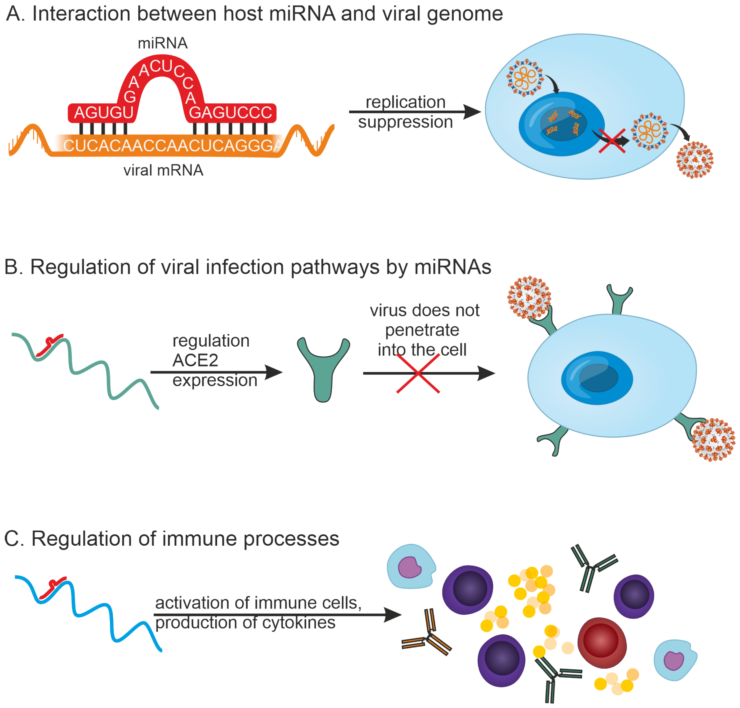
Viral infections are known to be associated with changes in the expression of various miRNAs, with several mechanisms of host antiviral response by miRNAs having been identified [26] (Figure 2). Viruses are also known to encode miRNAs, but a discussion of viral miRNAs is beyond the scope of this study.
(1)
MiRNAs can inhibit viral replication by directly binding to the viral RNA genome. For example, 873 miRNAs that can target the SARS-CoV-2 genome have been detected by in silico methods [27].
(2)
MiRNAs can participate in regulating infection pathways. For example, it has been shown that a number of miRNAs can regulate the expression of ACE2, a cell membrane-associated protein that is required for SARS-CoV-2 virus entry into the host cell. miR-125b directly targets the 3′-UTR of ACE2 mRNA, thereby regulating its expression [28].
(3)
MiRNAs can play a role in regulating the innate and adaptive immune systems.
The first two mechanisms on the list are virus-specific. A third mechanism indicates that several of these regulatory pathways may be common among viral infections. For example, large amounts of miRNAs can regulate the expression of IL-1β, IL-6, and IL-8 because there are numerous miRNA binding sites in the 3′-UTR of IL-1β, IL-6, and IL-8 mRNAs [29]. MiRNAs may also modulate the expression of cytokine signaling pathways and inflammatory responses during viral infection [30]. It is thus of interest to investigate miRNAs involved in regulating the immune response cascade in diverse viral infections.
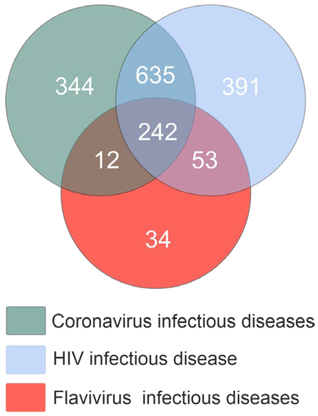
Next, we compared the three miRNA lists to identify common miRNAs. The study identified 242 miRNAs among the three infectious disease types (Figure 3). A total of 877 common miRNAs were identified for coronavirus infection and HIV infection. Regarding flavivirus infectious diseases, a significantly smaller number of miRNAs were found in common with coronavirus infectious diseases (254 miRNAs) and HIV-infection disease (295 miRNAs). The dataset used for further analysis comprised 242 miRNAs.
Next, using miRTarBase [31] and TargetScan [32,33] databases, target genes for 242 miRNAs common to different viruses were identified, and their gene ontology (GO) was analyzed. These miRNAs can influence viral infection through one of the three pathways listed in Figure 2. The correlation between the viral genome (Figure 2A) and the control of infection pathways (Figure 2B) may vary substantially depending on the specific virus. Thus, given the similarities in immune responses to viral infections, the focus was directed to miRNAs involved in regulating various components of the human immune system.

2.3. Analysis of miRNA-Gene Interactions and Gene Ontology

Analysis of miRNA-gene interactions was performed using Cytoscape 3.10.2. It has a modular structure, and applications (plugins) allow for additional functions to enhance analysis. By employing the CyTargetLinker 4.0.0+ plugin [34,35] for analyzing 242 miRNAs, a miRNA-gene regulatory interaction network was constructed, comprising 14,507 nodes (242 miRNAs and 14,265 genes) and 124,211 interactions.
Subsequently, a Gene Ontology (GO) analysis was performed. Selection was made of genes associated with diverse elements of the immune response to viral infection. Consequently, a network comprising 502 nodes (224 miRNAs and 278 genes) and 2236 interactions was generated. Next, separate subnetworks were identified, enabling the characterization of miRNAs influencing the regulation of distinct components of the immune response to viral infection.

2.3.1. MiRNAs Associated with the B-Cell Response

B-cell development involves several stages: from the early stages of B-cell differentiation in the bone marrow in the absence of antigen to later stages after interaction with antigen in the periphery [36]. Differential miRNA expression of B cell development indicates a regulatory role for these miRNAs at each developmental stage. A number of studies have demonstrated the involvement of miRNAs in early B cell development coordination [37,38,39]. This is succeeded by the release of B lymphocytes to the periphery and their interaction with antigens. Upon exposure to an antigen, some activated naïve B cells differentiate directly into short-lived cells that primarily produce IgM antibodies. Other B cells enter the follicle of secondary lymphoid organs such as lymph nodes and spleen to establish a germinal center and eventually differentiate into high-affinity IgG-producing plasma cells and memory cells [40]. Therefore, subsequent stages of differentiation are crucial for characterizing the function of miRNAs in regulating immune processes during viral infections.
GO analysis allowed 29 miRNAs to be identified that are involved in the regulation of B cell activation related to immune response (GO: 2312) (Figure 4). Included among these miRNAs are miR-29a-3p, miR-29b-3p, miR-486-3p, and others. In vitro deletion of miR-29a-3p has been shown to correlate with reduced B cell proliferation and antibody production [41].
The generation of antibodies is strongly associated with the advanced stages of B-cell development. Antibodies circulating in the blood and lymph neutralize pathogens, enhance phagocytosis, and participate in the destruction of cells affected by viruses. The study examined gene ontologies related to the production (GO: 2381) and regulation (GO: 2889) of the immunoglobulin-mediated immune response, as well as the immune response mediated by circulating immunoglobulin (GO: 2455). A gene ontology analysis identified 42 genes regulated by 144 miRNAs, including miR-146a-5p, miR-181a-5p, miR-486-3p, miR-650, miR-4689, miR-3175, miR-766-3p, miR-29a-3p, and others (Figure S1). Earlier findings established the expression of miR-181a-3p to be elevated simultaneously with B-cell activation [42,43].

2.3.2. MiRNAs Associated with T-Cell Response

The immune response mediated by T cells is central to protecting the host from invading pathogens, such as viruses. Pathogen-infected cells can be directly eliminated by cytotoxic CD8+ T lymphocytes, which are the predominant type of T cell [44]. CD4+ T lymphocytes orchestrate the immune response by activating diverse immune cells, comprising B lymphocytes, CD8+ T lymphocytes, and phagocytic antigen-presenting cells [45]. Treg cells, specifically CD4+CD25+FoxP3+ cells, also can suppress unwanted immune activation and prevent detrimental immune-mediated inflammation and tissue injury [46].
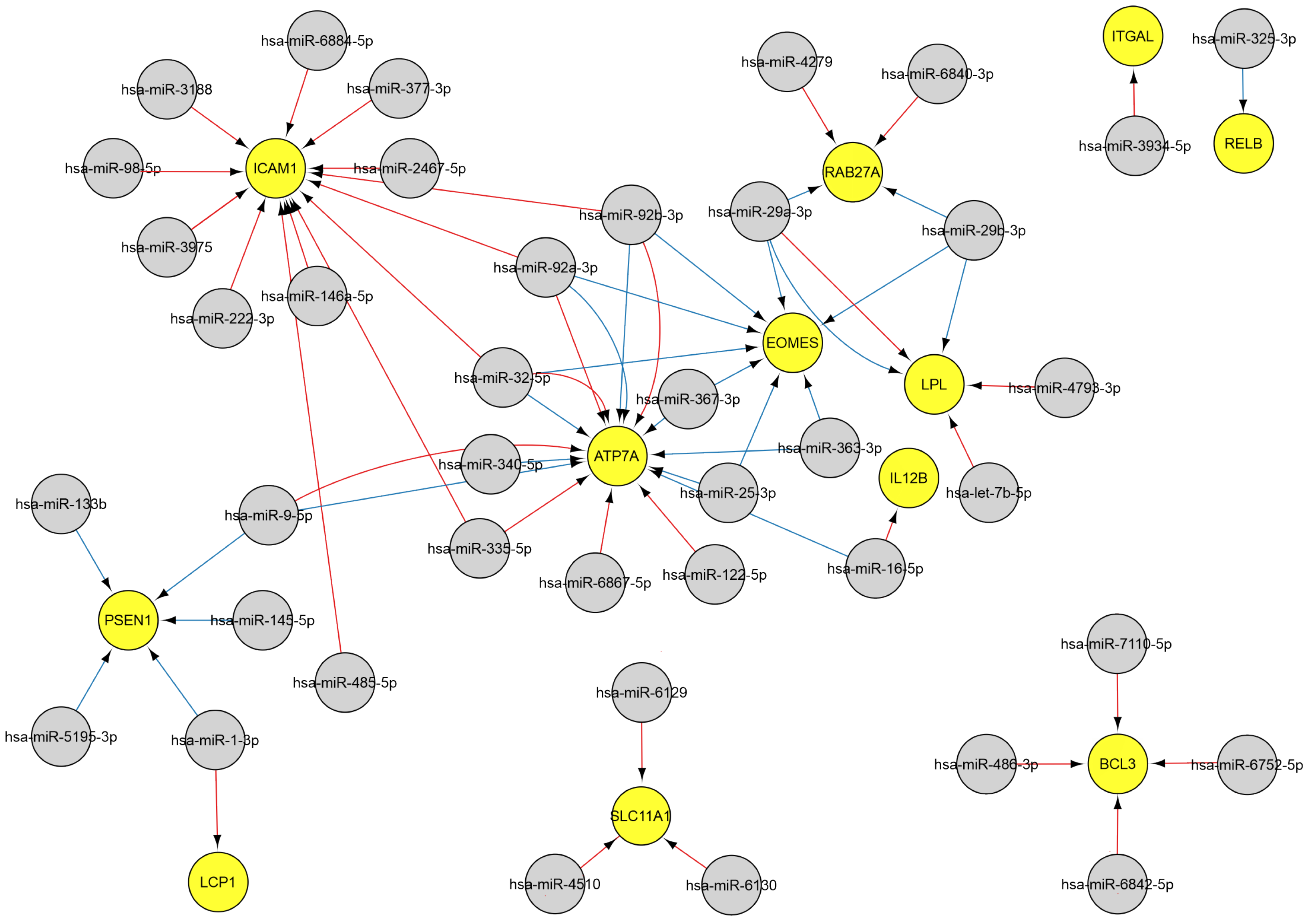
Twelve genes govern T cell activation associated with the immune response, and three genes are involved in T cell proliferation. These genes are regulated miRNAs, including miR-146a-5p, miR-486-3p, miR-25-3p, miR-29a-3p, miR-29b-3p, miR-325-3p, miR-9-5p, miR-92b-3p, miR-98-5p (Figure 5).
MiR-29b-3p, miR-25-3p, miR-29a-3p, miR-32-5p, miR-363-3p, miR-367-3p, miR-92a-3p, and miR-92b-3p impact T cell function by controlling the expression of the EOMES transcription factor, which is essential for the differentiation of cytotoxic CD8+ T cells during immune responses by upregulating lytic effector genes [47,48].
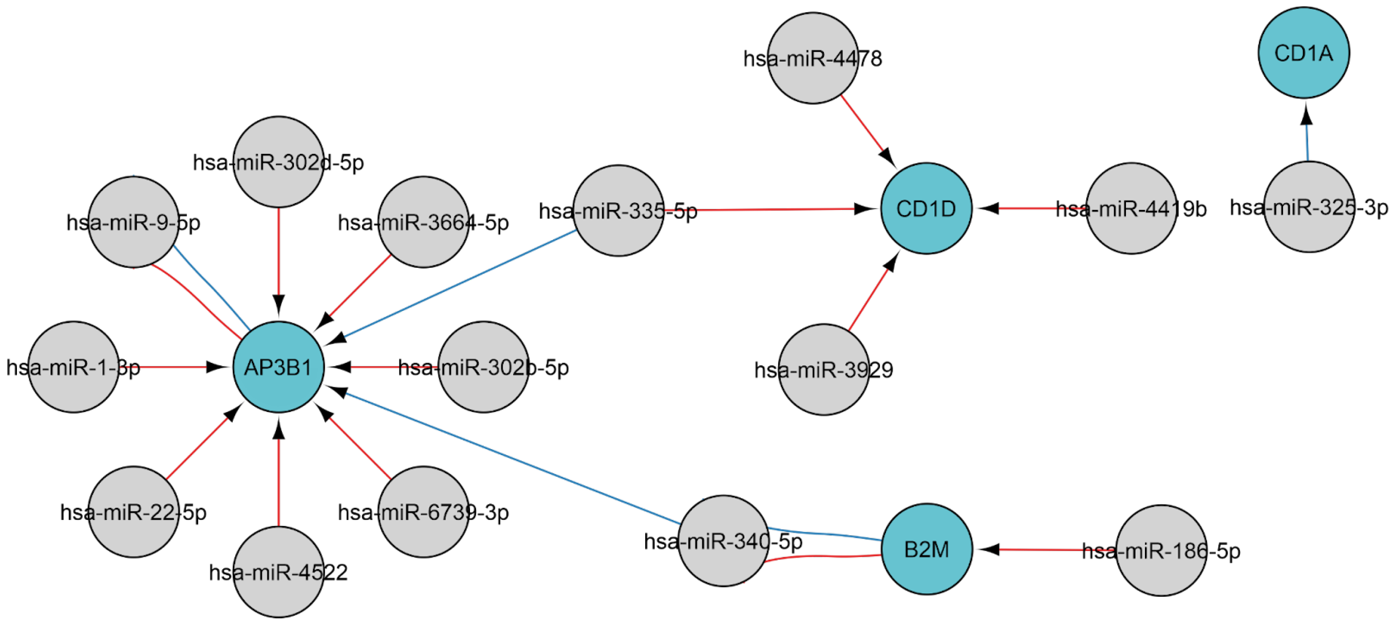
Native antigens undergo fragmentation by antigen-presenting cells, followed by display of the fragments on the cell surface via major histocompatibility complex (MHC) molecules, enabling T-cell receptor recognition [49]. Infected cells present intracellular antigens, including viral ones, as part of the MHC class I complex. The regulation of MHC antigen processing and presentation during viral infection involves four genes: AP3B1, B2M, CD1A, and CD1D. The genes are, in turn, regulated by the following 15 miRNAs: miR-1-3p, miR-186-5p, miR-22-5p, miR-302b-5p, miR-302d-5p, miR-325-3p, miR-335-5p, miR-340-5p, miR-3664-5p, miR-3929, miR-4419b, miR-4478, miR-4522, miR-6739-3p, and miR-9-5p (Figure 6). Elevated miR-1-3p levels were correlated with the severity of COVID-19, with the highest levels corresponding to extended hospitalizations and reduced survival [50].

2.3.3. MiRNAs Associated with the Natural Killer Cell Response

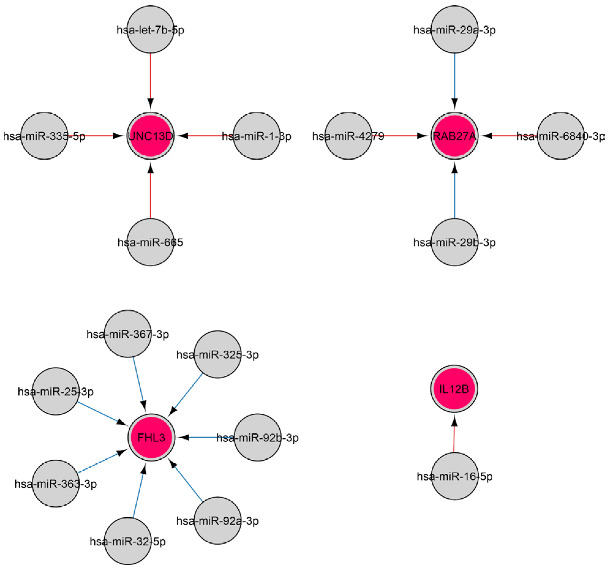
Natural killer (NK) cells are members of innate immunity that provide, among other things, defense against viruses by secreting a number of cytokines and killing virus-infected cells by initiating their apoptosis [51]. Over 400 and 300 miRNAs have previously been identified in human and murine NK cells, respectively, with the expression of a number of miRNAs found to alter upon NK activation [52,53]. The findings of this study indicate that, during viral infection, NK (GO: 2323) cell activation is governed by four genes, the regulation of which is influenced by sixteen miRNAs, including let-7b-5p, miR-25-3p, miR-29a-3p, miR-29b-3p, miR-363-3p, miR-367-3p miR-92a-3p, and others (Figure 7).
miR-29b has previously been shown to exhibit elevated expression in NK cells and to be critical for both the transformation of NK precursors into immature NK cells and the terminal maturation and function of NK cells [54,55,56]. Therefore, miRNAs could potentially regulate innate immunity by modulating natural killer cells.

2.3.4. MiRNAs Regulating the Cytokine Response

MiRNAs have been computationally analyzed [57] and shown to predominantly target transcription factors, cofactors, and chromatin modifiers. In contrast, molecules within the innate immune pathway, such as Toll-like receptors (TLRs), chemokines, and cytokines, are generally not targeted by miRNAs. MiRNA binding sites were predicted in nine of the interleukin (IL1-29) genes under analysis. Examination of twenty interleukin receptor genes revealed high-probability miRNA target sites in just two. However, another study revealed that clusters of adenine- and uridine-rich elements (AREs) were identified in the 3′ untranslated regions (UTRs) of mRNA molecules encoding cytokines [58]. AREs recruit various ARE-binding proteins (ARE-BPs) that modulate mRNA stability and/or translation [59]. Despite the absence of miRNA target sites in the 3′-UTRs of most cytokines, miRNA can regulate cytokine expression by targeting ARE binding proteins, thus influencing the half-life of numerous cytokines [58]. Moreover, miRNAs can also modulate the expression of a range of cytokines through regulating transcription factors and elements of signaling pathways, which results in cytokine mRNA transcription. The current research established a comprehensive network of miRNA-gene interactions involved in modulating cytokine responses during diverse viral infections. We have identified 48 genes and 395 interactions related to the cytokine-mediated signaling pathway (GO: 19221). Due to the visualization and interpretation challenges presented by the resulting network, three subnetworks were identified: (1) regulation cytokine gene, (2) regulation cytokine receptor gene, and (3) cytokine signaling pathways regulation.
Regulation of Cytokine Gene
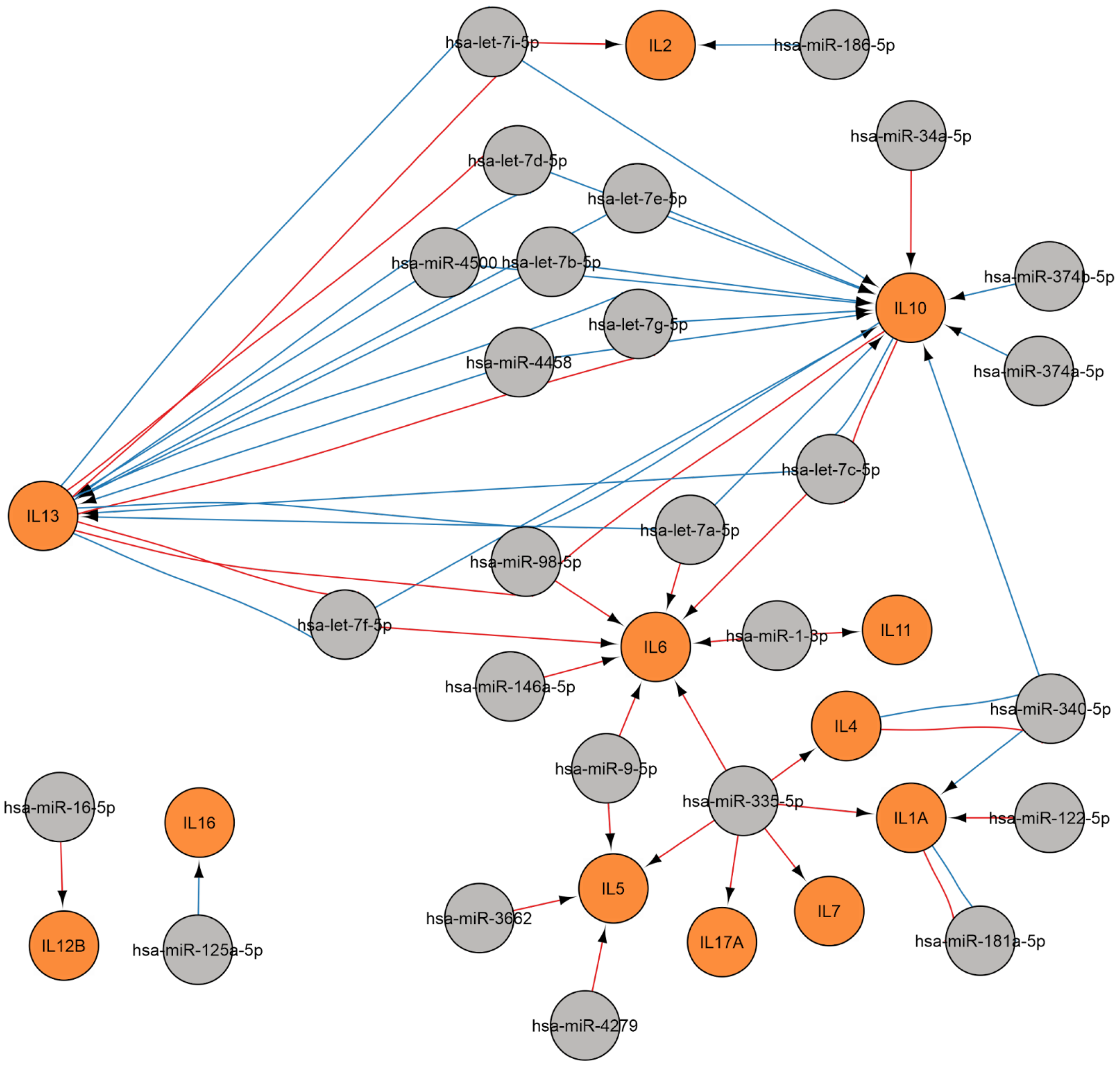
A regulatory network was constructed to show the regulation of cytokine gene during viral infection (Figure 8). Twelve cytokine genes are subject to modulation by twenty-six miRNAs, including let-7 family miRNAs, as well as miR-1-3p, miR-122-5p, miR-125a-5p, miR-146a-5p, miR-16-5p, miR-181a-5p, miR-186-5p, miR-335-5p, miR-340-5p, miR-34a-5p, miR-3662, miR-374a-5p, miR-374b-5p, miR-4279, miR-4458, miR-4500, miR-9-5p, and miR-98-5p.
IL-6 is implicated in the mechanisms of both acute and chronic inflammation, such as those observed in autoimmune conditions [60]. This cytokine promotes plasma cell survival and immunoglobulin secretion through the JAK/STAT3 signaling pathway, also promoting the differentiation of CD4+ cells into Th17 lymphocytes while inhibiting differentiation into Treg [61,62,63]. The regulation of IL-6 involves let-7a-5p, let-7c-5p, let-7f-5p, miR-146a-5p, miR-98-5p, miR-335-5p, miR-9-5p, and miR-1-3p.
The data presented here indicate that the IL-10 gene is regulated by the let-7 family miRNAs, as well as miR-34-5p, miR-374b-5p, and miR-374a-5p. The cytokine in question suppresses myeloid cells, thereby modulating inflammation to minimize tissue damage, which is also significant for immune defense against viral infections [64]. At the same time, IL-10 stimulates proliferation, B lymphocyte differentiation, and antibody secretion and participates in the pathogenesis of autoimmune diseases such as systemic lupus erythematosus [65].
Regulation of Cytokine Receptor Gene
A regulatory network was constructed to illustrate the regulation of cytokine receptor gene in the context of viral infection (Figure 9). 16 cytokine receptor genes are modulated by 69 miRNAs, including the let-7 family, and also miR-1228-3p, miR-125a-5p, miR-146a-5p, miR-16-5p, miR-29a-3p, miR-29b-3p, miR-325-3p, miR-335-5p, miR-34a-5p, miR-34b-3p, miR-377-3p, miR-485-5p, miR-508-5p, miR-515-5p, miR-766-3p, miR-9-5p, miR-92a-3p, miR-98-5p, and others. Therefore, the function of several cytokines (IL-1, IL-6, IL-10, IL-17, and others) is also influenced by miRNAs through regulating receptor gene for these signaling molecules.
Regulation of Cytokine Signaling Pathways
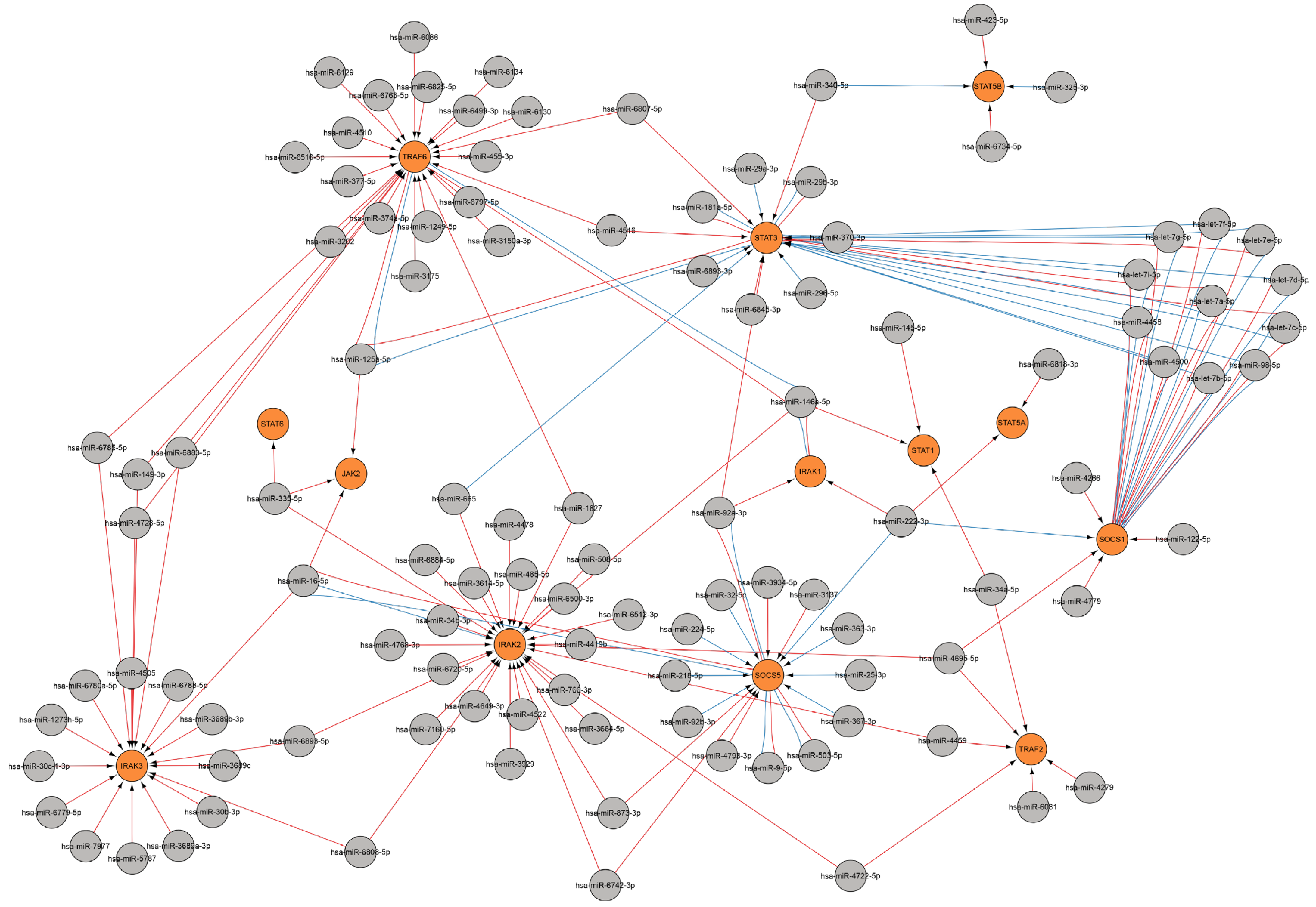
The products of several genes are involved in the direct function and regulation of cytokine signaling pathways, including the JAK, STAT, SOCS, TRAF, IRAK, and other protein families. MiRNAs are involved in the regulation of these genes, which in turn leads to the regulation of the cytokine response. A miRNA network was constructed for a number of major genes involved in regulating cytokine signaling pathways (Figure 10).
The JAK/STAT signaling pathway can activate over 50 cytokines and growth factors. When activated, JAKs are responsible for cytokine receptor phosphorylation and then recruit STAT proteins [66]. The JAK2-dependent signaling pathway regulates crucial pro-inflammatory cytokines such as IL-6, IFN-γ, and GM-CSF [67]. JAK2 is regulated by three miRNAs: miR-125a-5p, miR-16-5p, and miR-335-5p.
STAT proteins are implicated in regulating transcription and mediate cellular responses to signals from cytokine and growth factor receptors [68,69,70,71]. STAT3, for example, functions as a regulator of the inflammatory response by modulating the differentiation of naive CD4+ T cells into Th17 or Treg T-helper cells [72,73]. The regulation of the STAT3 gene during viral infection involves twenty-four miRNAs, including let-7 family miRNAs, miR-181a-5p, miR-29a-3p, miR-29b-3p, miR-98-5p, miR-125a-5p, miR-296-5p, miR-340-5p, miR-370-3p, miR-4458, miR-4500, miR-4516, miR-665, miR-6807-5p, miR-6845-3p, miR-6893-3p, miR-92a-3p and others. The STAT1 gene is regulated by three miRNAs: miR-145-5p, miR-146a-5p, and miR-34a-5p.
Upon activation, STATs dimerize and enter the nucleus, prompting the expression of suppressor of cytokine signaling (SOCS), and SOCS proteins bind to phosphorylated JAK and its receptor, thereby negatively regulating the JAK-STAT signaling pathway [66]. SOCS1 acts as a crucial negative regulator of IFN-I, IFN-II, and other cytokine signaling pathways [74,75]. SOCS1 downregulates cytokine signaling through the inhibition of the JAK/STAT signaling pathway. During viral infection, the SOCS1 gene is regulated by sixteen miRNAs, including the let-7 family, miR-98-5p, miR-122-5p, miR-222-3p, miR-4266, miR-4458, miR-4500, miR-4695-5p, miR-4779, and others.
The current findings are consistent with prior research, which indicated that miRNA let-7 and miR-98 binding sites were detected within the 3′UTR regions of SOCS1, STAT3, IL-6, IL-10, and IL-13 transcripts [57].
TRAF molecules are involved in various signaling cascades, functioning as central regulators of immunity and inflammation. TRAFs exhibit the ability to interact with the cytoplasmic domains of cytokine receptors, such as IL-1β, IL-2, IL-6, IL-17, IL-18, IL-33, IFN type I, IFN type III, GM-CSF, M-CSF, and TGF-β, and also with TLRs, NLRs, RIG-I-like receptors, and C-type lectin receptors [76]. TRAF2 is a prerequisite for TNFα to activate MAPK8/JNK and NF-κB. Mice with a global TRAF2 deficiency (TRAF2 −/−) experience early lethality as a result of an inflammatory phenotype, which is marked by increased serum TNFα levels [77]. TRAF2 is regulated by the following six miRNAs: miR-34a-5p, miR-4279, miR-4459, miR-4695-5p, miR-4722-5p, and miR-6081. TRAF6 modulates cellular proliferation, differentiation, and apoptosis via the activation of NF-κB, JNK/p38, PI3K/AKT, and AP-1 pathways, as well as regulating innate and adaptive immunity, oxidative stress, and inflammation [78,79]. TRAF6 is regulated by 24 miRNAs, including miR-146a-5p. miR-146a-5p can suppress IL-6 and IL-1β secretion by modulating the IRAK1/TRAF6 pathway [80].
IRAK kinases serve as downstream signal transducers subsequent to receptor activation, exemplified by TLR pathogen recognition receptors or IL-1 receptors (IL-1R) [81]. Mutations in the IRAK1 protein have been detected in patients who are highly susceptible to severe infections [82]. The IRAK1 gene is modulated by three miRNAs: miR-146a-5p, miR-222-3p, miR-92a-3p, while IRAK2 is modulated by twenty-eight miRNAs, including miR-146a-5p, miR-16-5p, miR-34b-3p, miR-485-5p, miR-508-5p, miR-665, miR-766-3p, miR-873-3p, and other miRNAs.
Given the above, it can be assumed that a number of miRNAs may be involved in regulation of both the genes of cytokines themselves and the genes of participants of signaling pathways that are triggered by these cytokines. Alterations in cytokine profiles, including those stemming from diverse infections, may result in various complications, such as cytokine storms, and the initiation of autoimmune processes [60,83,84].

3. Discussion

Successful binding of a miRNA to its target typically results in translational repression and/or degradation of the mRNA [85]. Human miRNA is thought to be capable of regulating several hundred different mRNAs. Alternatively, a single mRNA can be the target of several miRNAs simultaneously [20]. MiRNAs offer a method of modulating rather than completely suppressing protein levels within the cell. The reduction in transcript levels resulting from the targeting of miRNAs to a specific mRNA typically does not exceed threefold, and this modest variation seems to correlate with translation levels [86]. The association between miRNAs and various diseases is being actively investigated because they are currently considered promising diagnostic markers and therapies.
MiRNAs offer multiple advantages when employed as biomarkers. First, miRNAs are stable molecules that are resistant to degradation [87]. The stability of miRNAs makes them appropriate for routine clinical analysis. Second, miRNAs can be detected in various biological fluids, including blood and saliva [88]. Third, miRNA quantification is feasible through techniques commonly employed in clinical laboratories, including quantitative real-time polymerase chain reaction (RT-PCR) [89]. Integration into present clinical workflows is enabled by compatibility with standard laboratory protocols, thereby reducing the need for specialized equipment. Furthermore, the characterization of circulating miRNAs holds considerable promise for enhancing the management of patients with viral infections, thus providing novel perspectives on disease progression and treatment outcomes. The application of this method could facilitate more precise therapeutic measures and a better understanding of the fundamental processes of viral infection.
MiRNAs are capable of significantly affecting the regulation of immune responses, offering an extra dimension in the organization of immune responses during viral infections [58]. The molecular targets of miRNAs are identified through computational studies. In animals, complementarity between miRNAs and mRNA knockdown is imperfect [90]. The structure of mRNA has also been demonstrated to identify miRNA targets [91,92]. STarMir accounts for mRNA-miRNA binding features based on complex sequence, thermodynamic, and structural characteristics [93].
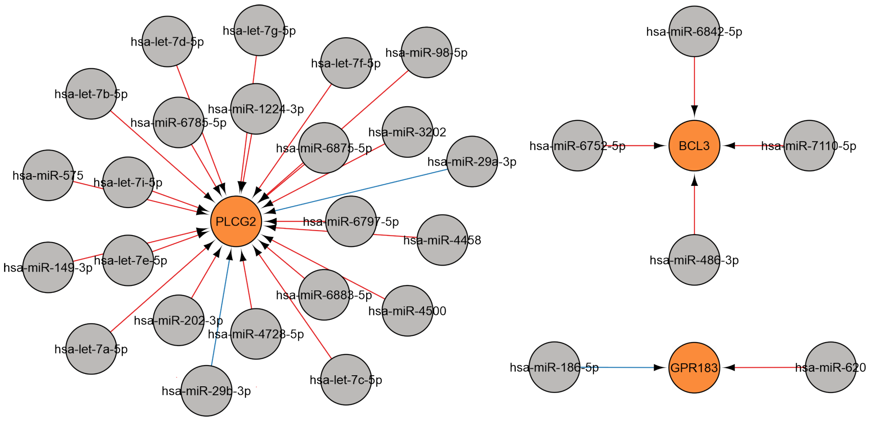
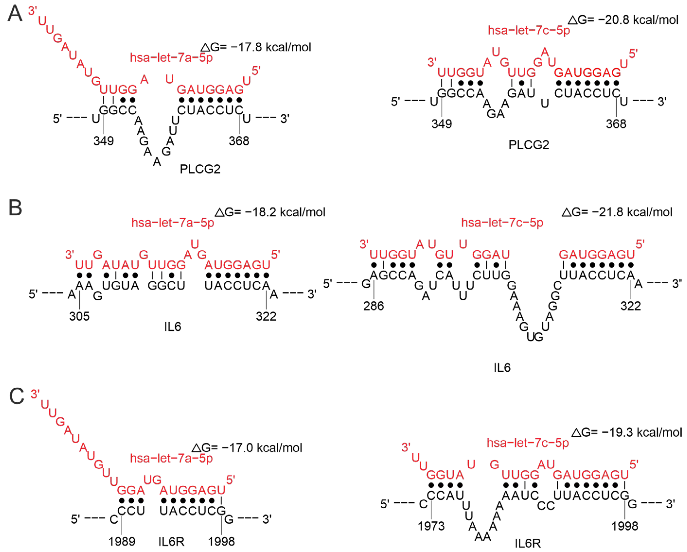
Numerous studies have indicated that let-7 expression is altered in a variety of viral diseases compared to healthy controls [94]. This work illustrates the involvement of let-7 family miRNAs in regulating multiple mechanisms of immune responses to viral infection, including B-cell responses, and the regulation of both cytokine gene expression and its receptors. Let-7 is involved in the regulation of PLCG2 (Figure 11A). This gene encodes phospholipase Cg2, which is essential for the function of hematopoietic cells and plays a key role in the regulation of immune responses. Genetic abnormalities affecting this region lead to impaired B-cell differentiation, increased myeloid cell proliferation, and enhanced synthesis of pro-inflammatory cytokines [95,96]. Let-7 is also involved in the regulation of both cytokines and their receptors by interacting with the 3′-UTR region of the corresponding mRNA (Figure 11B,C). According to STarMir data, both let-7a-5p and let-7c-5p bind to transcripts of the PLCG2, IL6, and its receptor genes at the same site with a high probability of finding the true binding site (LogitProb parameter, Table S1). let-7c-5p exhibits lower binding energy compared to let-7a-5p for all three transcripts (ΔGhybrid parameter, Table S1), which may indicate a potentially more stable interaction.
Our data are consistent with literature demonstrating a role for let-7 in viral infections. Let-7 was shown to affect effector CD8+ T cell differentiation and is also associated with B-cell activation and antibody production [97]. Reduced expression levels of let-7a-5p, let-7b-5p, let-7c-5p were reported in COVID-19 patients, spanning both mild and severe cases [98,99]. Additionally, the expression levels of let-7b-5p were observed to be increased in a manner that correlated with the viral load [100]. A significant reduction in let-7e-5p expression was reported in COVID-19 patients requiring oxygenation [101]. The expression of let-7c was observed to increase in the context of ZIKV infection [102].
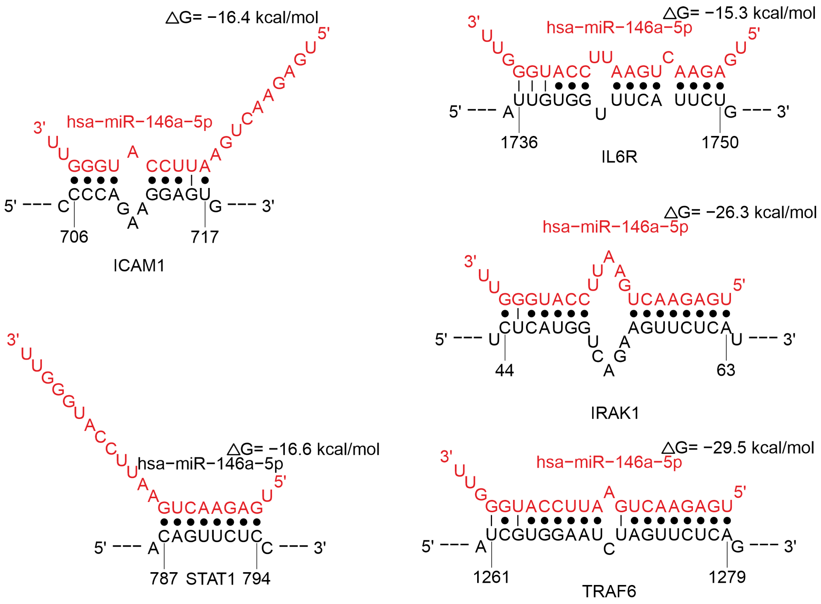
MiR-146a-5p is involved in the regulation of T-cell response activation during viral infection, regulation of cytokine gene and its receptors, regulation of cytokine signaling pathways. MiR-146a-5p is involved in the regulation of ICAM1 (Figure 12), which plays a role in T cell activation, migration, and function [103]. This miRNA affects STAT1, IRAK1, and TRAF6 (Figure 12). IRAK1 and TRAF6 form a complex in response to signals from the Toll-like receptor/interleukin-1 receptor superfamily (TLR/IL-1R). This leads to the activation of signaling pathways such as NF-κB and MAPK, which in turn can activate STAT1, influencing of genes associated with the immune response and inflammation [104,105]. Also, miR-146a-5p has a direct effect on IL6 (Figure 12). Among these interactions, miR-146a-5p binding to the TRAF6 transcript is characterized by the highest probability of predicting the true binding site. Moreover, the binding site in the target IL6 mRNA has the highest structural accessibility (Site_Access parameter, Table S1).
Earlier studies have demonstrated that numerous viruses can induce the expression of miR-146a-5p, including HIV-1 [106], vesicular stomatitis virus [107], the Epstein–Barr virus [108], and DENV [109]. The molecule miR-146a-5p, also known as inflamma-miR, was shown to regulate inflammation [110,111,112,113,114]. This is corroborated by data on the correlation between miR-146a-5p and IL-6 levels in COVID-19 patients [115]. Clinical data analysis revealed an upregulation of miR-146a-5p during the acute phase of COVID-19 [116], with a notable increase in its expression observed in severe cases [16,117]. DENV infection has also been demonstrated to upregulate miR-146a-5p. This miRNA diminished IFN-β production, thereby promoting the proviral effect of miRNA [109]. HIV-1 infection is associated with increased miR-146a expression in the peripheral blood of infected patients. Higher levels of miRNA have been reported to correlate positively with depletion markers [118]. The induction of miR-146a in enterovirus [119], DENV [109], and Japanese encephalitis virus [120] infected cells has been shown to negatively regulate TRAF6, an IFN signaling protein, and consequently, prevents IFN-α/β induction.
MiR-181a-5p is known to be involved in regulating immunoglobulin production, cytokines (IL1A), and the STAT3 gene-related cytokine signaling pathway (Figure 13). Reduced levels of miR-181a-5p have been demonstrated in cases of severe COVID-19 [121,122]. Additional research indicates that diminished miR-181a-5p levels diminish renin secretion, potentially causing more acute hypotensive episodes in patients [122,123,124]. A decrease in miR-181a expression has been documented in the elderly population [125,126]. It is also noteworthy that the elderly are more vulnerable to viral infections, for example, influenza or COVID-19.
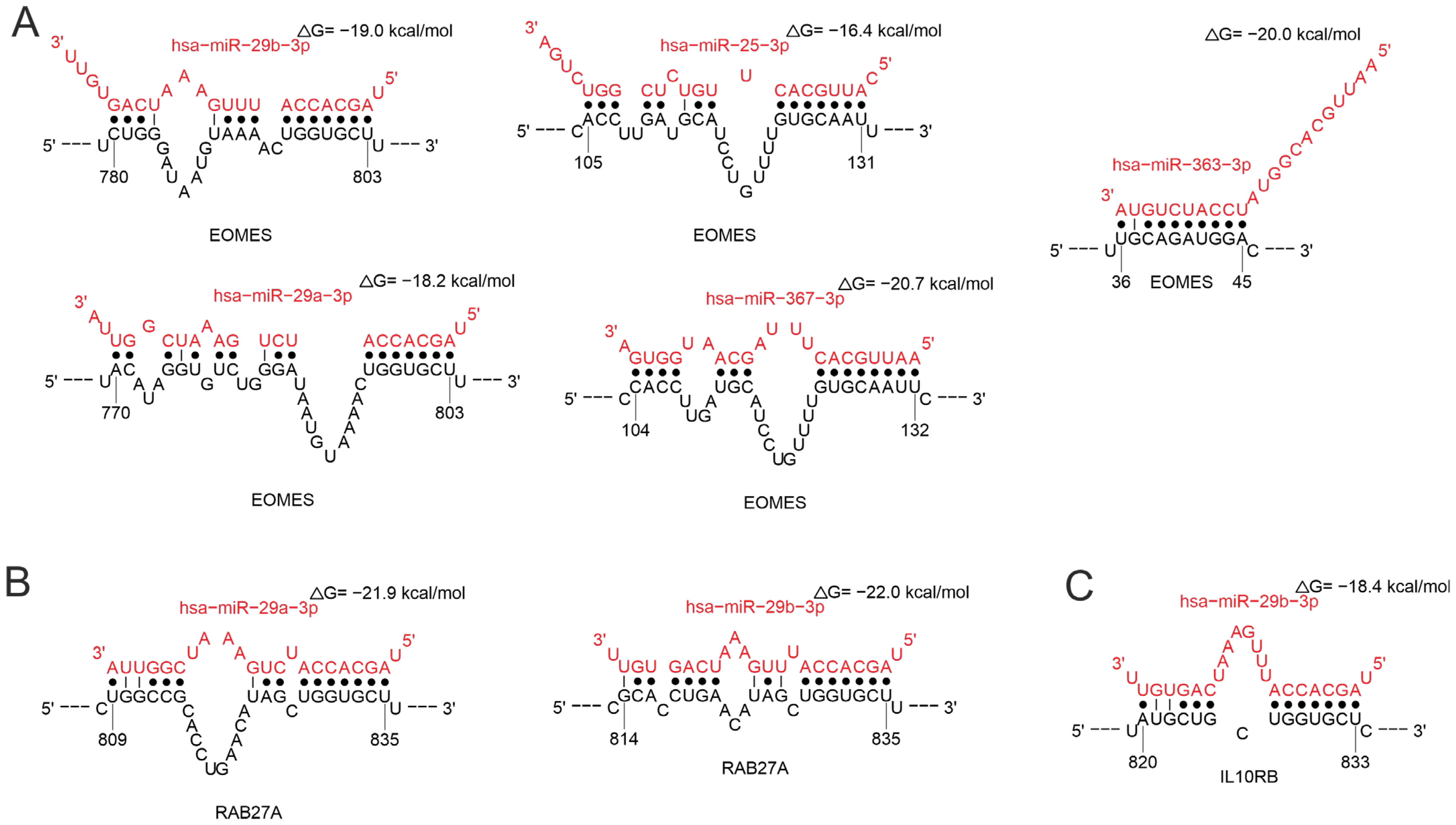
MiR-25-3p, miR-29a-3p, miR-29b-3p, miR-363-3p, and miR-367-3p effect EOMES (Figure 14A), which plays a key role in regulating the development and function of T cells, especially CD8+ T cells and memory T cells. EOMES maintains the function of effector T cells by stimulating the expression of cytokines such as IFN-γ and TNF-α [127,128,129,130]. MiR-29b-3p and miR-29b-3p regulate RAB27A (Figure 14B). This protein is required for the functioning of natural killer (NK) cells [131,132]. MiR-29b-3p regulates the interleukin-10 receptor beta subunit (IL10RB), which is required for interleukin-10 signaling (Figure 14C). The interaction of miR-367-3p with the EOMES transcript is characterized by the highest binding site prediction reliability and structural accessibility values. Furthermore, the binding sites on the EOMES and RAB27 transcripts are the same for miR-29a-3p and miR-29b-3p, and the parameters of these interactions are similar (Table S1). Serum miR-29a-3p levels have been observed to be inversely proportional to the severity of COVID-19 [133,134]. Decreased miR-29b-3p expression was also observed in lung biopsies from most COVID-19 patients compared to controls [135]. Another study revealed a significant correlation between elevated levels of miR-29a-3p and miR-29b-3p and ZIKV infection [136]. IL-21 produced by CD4+ T cells has been shown to be a significant immunomodulator and promotes miR-29 production during HIV infection through the STAT3 pathway [137,138].
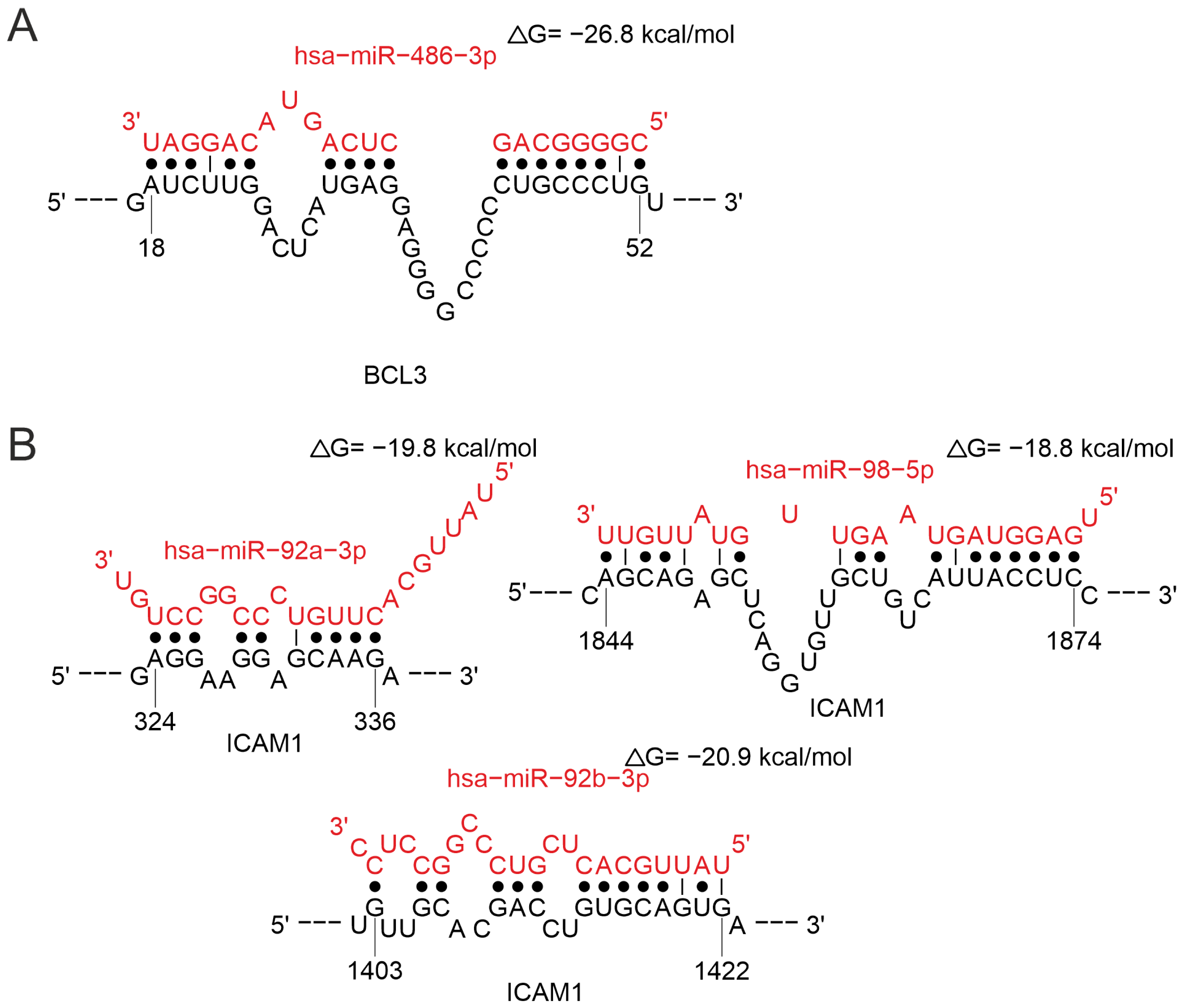
MiR-486-3p is involved in the regulation of BCL3 (Figure 15A). Bcl-3 is a member of the IκB protein family and an essential modulator of NF-κB activity. It is well established that Bcl-3 is critical for the normal development, survival and differentiation of adaptive immune cells, especially T cells [139]. The predicted binding site of miR-486-3p to the BCL3 transcript has low confidence (LogitProb < 0.5) and should be interpreted with caution (Table S1). MiR-486-3p is assumed to modulate T cell activation and differentiation [140], thus corroborating the role of this miRNA in regulating T cell responses demonstrated in this study. The upregulation of miR-486-3p has been demonstrated solely in individuals with severe clinical manifestations of COVID-19 [122,141]. Also, miR-486-5p has been reported to contribute to acute lung injury in COVID-19 [142].
MiR-92a-3p, miR-92b-3p, and miR-98-5p regulate ICAM1 gene (Figure 15B). MiR-98-5p is involved in regulating of genes important for the immune response, such as PLCG2, IL6, IL13, and IL10, and miR-92b-3p-SOCS5, IRAK1, and STST3. The interactions of the studied miRNAs with the ICAM1 transcript are characterized by similar values of binding site prediction reliability and binding energy. Moreover, the predicted binding site for miR-98-5p has a slightly higher structural accessibility compared to the sites for miR-92a-3p and miR-92b-3p (Table S1).
Reduced plasma concentrations of miR-92a-3p have been identified in COVID-19 patients with pulmonary fibrosis [143]. A reduction in miR-98-5p expression, prompted by viral elements, has been documented to promote infection initiation [144].
Thus, miRNA profiling permitted the identification of miRNAs implicated in modulating segments of the immune response to viral infection. Presented here is an intricate regulatory network that miRNAs mediate. Variations in the expression of a single miRNA can lead to a cascade of effects that impact various immune system processes.
Integrating our data with existing literature regarding the connection between specific miRNAs and the clinical progression of viral infections enabled us to pinpoint several miRNAs with potential utility as predictive indicators of the clinical course of viral infection. Additionally, spatial omics analysis of various immune cells may illuminate the alterations in gene expression under the influence of miRNAs [145].

Limitations and Prospects of the Study

The primary limitation of this work is its exclusively computational nature. The results obtained represent valuable predictions rather than experimentally confirmed data. While our analysis identified a number of miRNAs that influence the functioning of the immune system during viral infections. The specific changes in their expression across different diseases and the relationship between the differential expression of these miRNAs and symptom severity remain to be established through further research.
Despite this limitation, the conducted study opens clear avenues for future work. Unlike previous studies, we did not focus on a specific disease, but rather analyzed miRNAs that are involved in regulating the immune response to different types of viral infections. The identified set of miRNAs represents promising candidates for in-depth analysis and experimental validation. The main directions for future research should include:
  • Experimental validation: Validation of the expression of predicted miRNAs using PCR methods [146,147,148], sequencing [149,150], multiplex analysis using optically encoded particles [151], or another method on clinical samples from patients with various viral infections.
  • Clinical Correlation: Analysis of the relationship between the expression levels of the selected miRNAs and clinical parameters, including symptom severity, viral load, and disease outcome.
  • Development of a Diagnostic Panel: Based on the confirmed data—the creation and testing of a diagnostic system or biomarker panel to assess the status of the immune response and predict the course of the disease.
Thus, this work forms an important theoretical foundation and sets a strategic direction for subsequent research. The miRNAs identified in silico are high-probability candidates for the development of new predictive biomarkers and future diagnostic solutions.

4. Materials and Methods

4.1. Analysis of miRNAs Associated with Viral Infections

The miRWalk database [17] was utilized to identify miRNAs relevant to the pathogenesis of three viral infections: flaviviruses, HIV-1, and coronaviruses (accessed on 18 April 2025). The search was performed using the following identifiers:
  • for flavivirus infections: DOID:0050175#tick-borne encephalitis, DOID:12205#dengue disease, DOID:2366#West Nile fever, DOID:0060478#Zika fever;
  • for coronavirus infections: DOID:0080599#Coronavirus infectious disease, DOID: 0080642#Middle East respiratory syndrome, DOID:2945#Severe acute respiratory syndrome (given the absence of data on other coronavirus infections within the miRWalk database, we deem the analysis to be exhaustive);
  • for HIV infection: DOID:526#human immunodeficiency virus infection disease.
MiRNAs were selected if (1) the predicted probability of functional interaction between the miRNA and the target mRNA was greater than 0.95, (2) the miRNAs bound to the 3′-UTR, and (3) the miRNAs were present in at least one of the databases TargetScan [20], miRDB [21], and miRTarBase [22]. The dataset was processed utilizing the R software version 4.4.2.

4.2. Target Predictions of the miRNAs Analyzed

The miRTarBase [31] and TargetScan [32,33] databases were utilized to search for target miRNA genes. This was performed through the CyTargetLinker 4.0.0+ application [34,35] (accessed on 11 April 2025). Visualization of the interaction network was performed using Cytoscape 3.10.2. Gene Ontology (GO) analysis was conducted utilizing the GOlorize 1.0.0 plugin [152]. The gene ontology identifiers (GO_ID) 2312, 2381, 2889, 2286, 2309, 2475, 19883, 19221 and 2323, which are associated with immune system function, were selected. The Benjamini–Hochberg procedure was applied to account for multiple comparisons. False discovery rate (FDR) < 0.05 were deemed statistically significant.

4.3. Prediction of miRNA Binding Sites on Target mRNAs

The STarMir web server (https://sfold.wadsworth.org/cgi-bin/starmirWeb.pl accessed on 30 September 2025) was used to predict miRNA binding sites on target mRNAs. The current version of STarMir is an implementation of logistic prediction models developed using high-throughput miRNA binding data obtained by cross-linked immunoprecipitation. The models take into account comprehensive thermodynamics, structure, and sequence [153]. Binding of −15 kcal/mol or lower was considered stable [154].

5. Conclusions

This study employed computational approaches to elucidate the significant role of numerous miRNAs in regulating immune responses to viral infections, with several RNA viruses serving as illustrative examples. We examined the regulation of T- and B-cell responses, focusing on processes such as MHC antigen presentation and immunoglobulin production. Detailed consideration was given to the miRNA regulation of cytokines, their receptors, and related signaling pathways. A relationship has already been reported between the severity of a disease and the varying expression levels of the following miRNAs: miR-146a-5p, miR-181a-5p, miR-29a-3p, miR-29a and miR-29b, miR-486-3p, and miR-574-5p. The miRNAs in question are not specific to one infection, but may be associated with a wide range of viral infections. The identified set of miRNAs represents a promising candidate for in-depth analysis and experimental validation as prognostic indicators. Additionally, an understanding of the regulatory pathways of these miRNAs will facilitate the identification of critical connections in the body’s immune system for targeted therapeutic interventions.
Further research should concentrate on defining miRNA expression profiles that are unique to different viruses, infection stages, and the development of complications following infection. The findings will be instrumental in the identification of trustworthy biomarkers for disease diagnosis and observation. A more thorough understanding of the influence of miRNAs on immune responses has the potential to facilitate the development of new therapeutic interventions and proactive strategies in clinical practice.

Supplementary Materials

The supporting information can be downloaded at: https://www.mdpi.com/article/10.3390/ijms262010100/s1.

Author Contributions

Conceptualization, A.M.T.; methodology, E.V.M., K.S.A.; validation, A.M.T.; investigation, E.V.M., K.S.A. and A.M.T.; resources, A.M.T.; writing—original draft preparation, A.M.T.; writing—review and editing, A.M.T. and G.A.N.; visualization, A.M.T.; project admin-istration, A.M.T.; funding acquisition A.M.T. All authors have read and agreed to the published version of the manuscript.

Funding

This research was funded by the Russian Science Foundation grant no. 25-15-00010 (https://rscf.ru/en/project/25-15-00010/).

Institutional Review Board Statement

Not applicable.

Informed Consent Statement

Not applicable.

Data Availability Statement

The original contributions presented in this study are included in the article/Supplementary Material. Further inquiries can be directed to the corresponding authors.

Conflicts of Interest

The authors declare no conflicts of interest.

Abbreviations

The following abbreviations are used in this manuscript:
AREClusters of elements rich in adenine and uridine
DENVDengue virus
GOGene ontology
MHCMajor histocompatibility complex
TBEVTick-borne encephalitis virus
TLRToll-like receptors
WNVWest Nile virus
ZIKVZika virus

References

  1. Cherry, J.D.; Krogstad, P. SARS: The First Pandemic of the 21st Century. Pediatr. Res. 2004, 56, 1–5. [Google Scholar] [CrossRef] [PubMed]
  2. Straub, J.; Estrada Lobato, E.; Paez, D.; Langs, G.; Prosch, H. Artificial Intelligence in Respiratory Pandemics—Ready for Disease X? A Scoping Review. Eur. Radiol. 2024, 35, 1583–1593. [Google Scholar] [CrossRef] [PubMed]
  3. Aguirre, S.; Luthra, P.; Sanchez-Aparicio, M.T.; Maestre, A.M.; Patel, J.; Lamothe, F.; Fredericks, A.C.; Tripathi, S.; Zhu, T.; Pintado-Silva, J.; et al. Dengue Virus NS2B Protein Targets CGAS for Degradation and Prevents Mitochondrial DNA Sensing during Infection. Nat. Microbiol. 2017, 2, 17037. [Google Scholar] [CrossRef]
  4. Ashour, J.; Laurent-Rolle, M.; Shi, P.-Y.; García-Sastre, A. NS5 of Dengue Virus Mediates STAT2 Binding and Degradation. J. Virol. 2009, 83, 5408–5418. [Google Scholar] [CrossRef]
  5. Kumar, A.; Hou, S.; Airo, A.M.; Limonta, D.; Mancinelli, V.; Branton, W.; Power, C.; Hobman, T.C. Zika Virus Inhibits Type-I Interferon Production and Downstream Signaling. EMBO Rep. 2016, 17, 1766–1775. [Google Scholar] [CrossRef]
  6. Fanunza, E.; Carletti, F.; Quartu, M.; Grandi, N.; Ermellino, L.; Milia, J.; Corona, A.; Capobianchi, M.R.; Ippolito, G.; Tramontano, E. Zika Virus NS2A Inhibits Interferon Signaling by Degradation of STAT1 and STAT2. Virulence 2021, 12, 1580–1596. [Google Scholar] [CrossRef]
  7. Molinero, M.; Perez-Pons, M.; González, J.; Barbé, F.; de Gonzalo-Calvo, D. Decoding Viral and Host MicroRNA Signatures in Airway-Derived Biosamples: Insights for Biomarker Discovery in Viral Respiratory Infections. Biomed. Pharmacother. 2024, 177, 116984. [Google Scholar] [CrossRef]
  8. Fayyad-Kazan, M. MicroRNAs in SARS-CoV-2 Infection: Emerging Modulators of Inflammation, Pathogenesis, and Therapeutic Potential. Inflammopharmacology 2025, 33, 4895–4910. [Google Scholar] [CrossRef]
  9. Paval, N.-E.; Căliman-Sturdza, O.A.; Lobiuc, A.; Dimian, M.; Sirbu, I.-O.; Covasa, M. MicroRNAs in Long COVID: Roles, Diagnostic Biomarker Potential and Detection. Hum. Genom. 2025, 19, 90. [Google Scholar] [CrossRef]
  10. Tahamtan, A.; Inchley, C.S.; Marzban, M.; Tavakoli-Yaraki, M.; Teymoori-Rad, M.; Nakstad, B.; Salimi, V. The Role of MicroRNAs in Respiratory Viral Infection: Friend or Foe? Rev. Med. Virol. 2016, 26, 389–407. [Google Scholar] [CrossRef]
  11. Paul, S.; Bravo Vázquez, L.A.; Reyes-Pérez, P.R.; Estrada-Meza, C.; Aponte Alburquerque, R.A.; Pathak, S.; Banerjee, A.; Bandyopadhyay, A.; Chakraborty, S.; Srivastava, A. The Role of MicroRNAs in Solving COVID-19 Puzzle from Infection to Therapeutics: A Mini-Review. Virus Res. 2022, 308, 198631. [Google Scholar] [CrossRef]
  12. Rezaei, T.; Amini, M.; Hashemi, Z.S.; Mansoori, B.; Rezaei, S.; Karami, H.; Mosafer, J.; Mokhtarzadeh, A.; Baradaran, B. MicroRNA-181 Serves as a Dual-Role Regulator in the Development of Human Cancers. Free Radic. Biol. Med. 2020, 152, 432–454. [Google Scholar] [CrossRef]
  13. Gao, X.; Qiao, Y.; Han, D.; Zhang, Y.; Ma, N. Enemy or Partner: Relationship between Intronic Micrornas and Their Host Genes. IUBMB Life 2012, 64, 835–840. [Google Scholar] [CrossRef] [PubMed]
  14. Głobińska, A.; Pawełczyk, M.; Kowalski, M.L. MicroRNAs and the Immune Response to Respiratory Virus Infections. Expert Rev. Clin. Immunol. 2014, 10, 963–971. [Google Scholar] [CrossRef] [PubMed]
  15. Fernández-Pato, A.; Virseda-Berdices, A.; Resino, S.; Ryan, P.; Martínez-González, O.; Pérez-García, F.; Martin-Vicente, M.; Valle-Millares, D.; Brochado-Kith, O.; Blancas, R.; et al. Plasma MiRNA Profile at COVID-19 Onset Predicts Severity Status and Mortality. Emerg. Microbes Infect. 2022, 11, 676–688. [Google Scholar] [CrossRef] [PubMed]
  16. Giannella, A.; Riccetti, S.; Sinigaglia, A.; Piubelli, C.; Razzaboni, E.; Di Battista, P.; Agostini, M.; Dal Molin, E.; Manganelli, R.; Gobbi, F.; et al. Circulating MicroRNA Signatures Associated with Disease Severity and Outcome in COVID-19 Patients. Front. Immunol. 2022, 13, 968991. [Google Scholar] [CrossRef]
  17. Dweep, H.; Sticht, C.; Pandey, P.; Gretz, N. MiRWalk–Database: Prediction of Possible MiRNA Binding Sites by “Walking” the Genes of Three Genomes. J. Biomed. Inform. 2011, 44, 839–847. [Google Scholar] [CrossRef]
  18. Sticht, C.; De La Torre, C.; Parveen, A.; Gretz, N. MiRWalk: An Online Resource for Prediction of MicroRNA Binding Sites. PLoS ONE 2018, 13, e0206239. [Google Scholar] [CrossRef]
  19. Dweep, H.; Gretz, N.; Sticht, C. MiRWalk Database for MiRNA–Target Interactions. Methods Mol. Biol. 2014, 1182, 289–305. [Google Scholar]
  20. Lewis, B.P.; Burge, C.B.; Bartel, D.P. Conserved Seed Pairing, Often Flanked by Adenosines, Indicates That Thousands of Human Genes Are MicroRNA Targets. Cell 2005, 120, 15–20. [Google Scholar] [CrossRef]
  21. Chen, Y.; Wang, X. MiRDB: An Online Database for Prediction of Functional MicroRNA Targets. Nucleic Acids Res. 2020, 48, D127–D131. [Google Scholar] [CrossRef]
  22. Papadopoulos, G.L.; Reczko, M.; Simossis, V.A.; Sethupathy, P.; Hatzigeorgiou, A.G. The Database of Experimentally Supported Targets: A Functional Update of TarBase. Nucleic Acids Res. 2009, 37, D155–D158. [Google Scholar] [CrossRef]
  23. Didiano, D.; Hobert, O. Molecular Architecture of a MiRNA-Regulated 3′ UTR. RNA 2008, 14, 1297–1317. [Google Scholar] [CrossRef] [PubMed]
  24. Lee, I.; Ajay, S.S.; Yook, J.I.; Kim, H.S.; Hong, S.H.; Kim, N.H.; Dhanasekaran, S.M.; Chinnaiyan, A.M.; Athey, B.D. New Class of MicroRNA Targets Containing Simultaneous 5′-UTR and 3′-UTR Interaction Sites. Genome Res. 2009, 19, 1175–1183. [Google Scholar] [CrossRef] [PubMed]
  25. Tan, S.; Guo, J.; Huang, Q.; Chen, X.; Li-Ling, J.; Li, Q.; Ma, F. Retained Introns Increase Putative MicroRNA Targets within 3′ UTRs of Human MRNA. FEBS Lett. 2007, 581, 1081–1086. [Google Scholar] [CrossRef] [PubMed]
  26. Trobaugh, D.W.; Klimstra, W.B. MicroRNA Regulation of RNA Virus Replication and Pathogenesis. Trends Mol. Med. 2017, 23, 80–93. [Google Scholar] [CrossRef]
  27. Fulzele, S.; Sahay, B.; Yusufu, I.; Lee, T.J.; Sharma, A.; Kolhe, R.; Isales, C.M. COVID-19 Virulence in Aged Patients Might Be Impacted by the Host Cellular MicroRNAs Abundance/Profile. Aging Dis. 2020, 11, 509. [Google Scholar] [CrossRef]
  28. Widiasta, A.; Sribudiani, Y.; Nugrahapraja, H.; Hilmanto, D.; Sekarwana, N.; Rachmadi, D. Potential Role of ACE2-Related MicroRNAs in COVID-19-Associated Nephropathy. Non-Coding RNA Res. 2020, 5, 153–166. [Google Scholar] [CrossRef]
  29. Chou, C.-H.; Shrestha, S.; Yang, C.-D.; Chang, N.-W.; Lin, Y.-L.; Liao, K.-W.; Huang, W.-C.; Sun, T.-H.; Tu, S.-J.; Lee, W.-H.; et al. MiRTarBase Update 2018: A Resource for Experimentally Validated MicroRNA-Target Interactions. Nucleic Acids Res. 2018, 46, D296–D302. [Google Scholar] [CrossRef]
  30. Chow, J.T.-S.; Salmena, L. Prediction and Analysis of SARS-CoV-2-Targeting MicroRNA in Human Lung Epithelium. Genes 2020, 11, 1002. [Google Scholar] [CrossRef]
  31. Huang, H.-Y.; Lin, Y.-C.-D.; Cui, S.; Huang, Y.; Tang, Y.; Xu, J.; Bao, J.; Li, Y.; Wen, J.; Zuo, H.; et al. MiRTarBase Update 2022: An Informative Resource for Experimentally Validated MiRNA–Target Interactions. Nucleic Acids Res. 2022, 50, D222–D230. [Google Scholar] [CrossRef] [PubMed]
  32. McGeary, S.E.; Lin, K.S.; Shi, C.Y.; Pham, T.M.; Bisaria, N.; Kelley, G.M.; Bartel, D.P. The Biochemical Basis of MicroRNA Targeting Efficacy. Science 2019, 366, 6472. [Google Scholar] [CrossRef] [PubMed]
  33. Agarwal, V.; Bell, G.W.; Nam, J.-W.; Bartel, D.P. Predicting Effective MicroRNA Target Sites in Mammalian MRNAs. eLife 2015, 4, e05005. [Google Scholar] [CrossRef]
  34. Kutmon, M.; Ehrhart, F.; Willighagen, E.L.; Evelo, C.T.; Coort, S.L. CyTargetLinker App Update: A Flexible Solution for Network Extension in Cytoscape. F1000Research 2018, 7, 743. [Google Scholar] [CrossRef]
  35. Kutmon, M.; Kelder, T.; Mandaviya, P.; Evelo, C.T.A.; Coort, S.L. CyTargetLinker: A Cytoscape App to Integrate Regulatory Interactions in Network Analysis. PLoS ONE 2013, 8, e82160. [Google Scholar] [CrossRef]
  36. Zheng, B.; Xi, Z.; Liu, R.; Yin, W.; Sui, Z.; Ren, B.; Miller, H.; Gong, Q.; Liu, C. The Function of MicroRNAs in B-Cell Development, Lymphoma, and Their Potential in Clinical Practice. Front. Immunol. 2018, 9, 936–946. [Google Scholar] [CrossRef]
  37. Borbet, T.C.; Hines, M.J.; Koralov, S.B. MicroRNA Regulation of B Cell Receptor Signaling. Immunol. Rev. 2021, 304, 111–125. [Google Scholar] [CrossRef]
  38. Brandl, A.; Daum, P.; Brenner, S.; Schulz, S.R.; Yap, D.Y.; Bösl, M.R.; Wittmann, J.; Schuh, W.; Jäck, H. The Microprocessor Component, DGCR8, Is Essential for Early B-cell Development in Mice. Eur. J. Immunol. 2016, 46, 2710–2718. [Google Scholar] [CrossRef]
  39. Coffre, M.; Benhamou, D.; Rieß, D.; Blumenberg, L.; Snetkova, V.; Hines, M.J.; Chakraborty, T.; Bajwa, S.; Jensen, K.; Chong, M.M.W.; et al. MiRNAs Are Essential for the Regulation of the PI3K/AKT/FOXO Pathway and Receptor Editing during B Cell Maturation. Cell Rep. 2016, 17, 2271–2285. [Google Scholar] [CrossRef]
  40. Shapiro-Shelef, M.; Calame, K. Regulation of Plasma-Cell Development. Nat. Rev. Immunol. 2005, 5, 230–242. [Google Scholar] [CrossRef]
  41. van Nieuwenhuijze, A.; Dooley, J.; Humblet-Baron, S.; Sreenivasan, J.; Koenders, M.; Schlenner, S.M.; Linterman, M.; Liston, A. Defective Germinal Center B-Cell Response and Reduced Arthritic Pathology in MicroRNA-29a-Deficient Mice. Cell. Mol. Life Sci. 2017, 74, 2095–2106. [Google Scholar] [CrossRef]
  42. de Yébenes, V.G.; Belver, L.; Pisano, D.G.; González, S.; Villasante, A.; Croce, C.; He, L.; Ramiro, A.R. MiR-181b Negatively Regulates Activation-Induced Cytidine Deaminase in B Cells. J. Exp. Med. 2008, 205, 2199–2206. [Google Scholar] [CrossRef] [PubMed]
  43. Kuchen, S.; Resch, W.; Yamane, A.; Kuo, N.; Li, Z.; Chakraborty, T.; Wei, L.; Laurence, A.; Yasuda, T.; Peng, S.; et al. Regulation of MicroRNA Expression and Abundance during Lymphopoiesis. Immunity 2010, 32, 828–839. [Google Scholar] [CrossRef] [PubMed]
  44. Sun, L.; Su, Y.; Jiao, A.; Wang, X.; Zhang, B. T Cells in Health and Disease. Signal Transduct. Target. Ther. 2023, 8, 235. [Google Scholar] [CrossRef] [PubMed]
  45. Swain, S.L.; McKinstry, K.K.; Strutt, T.M. Expanding Roles for CD4+ T Cells in Immunity to Viruses. Nat. Rev. Immunol. 2012, 12, 136–148. [Google Scholar] [CrossRef]
  46. Lin, H.; Xu, Y.; Lin, C. Heterogeneity and Subtypes of CD4+ Regulatory T Cells: Implications for Tumor Therapy. Front. Immunol. 2024, 14, 1291796. [Google Scholar] [CrossRef]
  47. Atreya, I.; Schimanski, C.C.; Becker, C.; Wirtz, S.; Dornhoff, H.; Schnurer, E.; Berger, M.R.; Galle, P.R.; Herr, W.; Neurath, M.F. The T-Box Transcription Factor Eomesodermin Controls CD8 T Cell Activity and Lymph Node Metastasis in Human Colorectal Cancer. Gut 2007, 56, 1572–1578. [Google Scholar] [CrossRef]
  48. Wells, A.C.; Daniels, K.A.; Angelou, C.C.; Fagerberg, E.; Burnside, A.S.; Markstein, M.; Alfandari, D.; Welsh, R.M.; Pobezinskaya, E.L.; Pobezinsky, L.A. Modulation of Let-7 MiRNAs Controls the Differentiation of Effector CD8 T Cells. eLife 2017, 6, e26398. [Google Scholar] [CrossRef]
  49. Blum, J.S.; Wearsch, P.A.; Cresswell, P. Pathways of Antigen Processing. Annu. Rev. Immunol. 2013, 31, 443–473. [Google Scholar] [CrossRef]
  50. Di Pietro, P.; Abate, A.C.; Izzo, C.; Toni, A.L.; Rusciano, M.R.; Folliero, V.; Dell’Annunziata, F.; Granata, G.; Visco, V.; Motta, B.M.; et al. Plasma MiR-1-3p Levels Predict Severity in Hospitalized COVID-19 Patients. Br. J. Pharmacol. 2025, 182, 451–467. [Google Scholar] [CrossRef]
  51. Letafati, A.; Ardekani, O.S.; Naderisemiromi, M.; Norouzi, M.; Shafiei, M.; Nik, S.; Mozhgani, S.-H. Unraveling the Dynamic Mechanisms of Natural Killer Cells in Viral Infections: Insights and Implications. Virol. J. 2024, 21, 18. [Google Scholar] [CrossRef]
  52. Liu, X.; Wang, Y.; Sun, Q.; Yan, J.; Huang, J.; Zhu, S.; Yu, J. Identification of MicroRNA Transcriptome Involved in Human Natural Killer Cell Activation. Immunol. Lett. 2012, 143, 208–217. [Google Scholar] [CrossRef] [PubMed]
  53. Fehniger, T.A.; Wylie, T.; Germino, E.; Leong, J.W.; Magrini, V.J.; Koul, S.; Keppel, C.R.; Schneider, S.E.; Koboldt, D.C.; Sullivan, R.P.; et al. Next-Generation Sequencing Identifies the Natural Killer Cell MicroRNA Transcriptome. Genome Res. 2010, 20, 1590–1604. [Google Scholar] [CrossRef]
  54. Mundy-Bosse, B.L.; Scoville, S.D.; Chen, L.; McConnell, K.; Mao, H.C.; Ahmed, E.H.; Zorko, N.; Harvey, S.; Cole, J.; Zhang, X.; et al. MicroRNA-29b Mediates Altered Innate Immune Development in Acute Leukemia. J. Clin. Investig. 2016, 126, 4404–4416. [Google Scholar] [CrossRef] [PubMed]
  55. Steiner, D.F.; Thomas, M.F.; Hu, J.K.; Yang, Z.; Babiarz, J.E.; Allen, C.D.C.; Matloubian, M.; Blelloch, R.; Ansel, K.M. MicroRNA-29 Regulates T-Box Transcription Factors and Interferon-γ Production in Helper T Cells. Immunity 2011, 35, 169–181. [Google Scholar] [CrossRef] [PubMed]
  56. Qi, Y.; Huang, Y.; Pang, L.; Gu, W.; Wang, N.; Hu, J.; Cui, X.; Zhang, J.; Zhao, J.; Liu, C.; et al. Prognostic Value of the Micro RNA—29 Family in Multiple Human Cancers: A Meta—Analysis and Systematic Review. Clin. Exp. Pharmacol. Physiol. 2017, 44, 441–454. [Google Scholar] [CrossRef]
  57. Asirvatham, A.J.; Gregorie, C.J.; Hu, Z.; Magner, W.J.; Tomasi, T.B. MicroRNA Targets in Immune Genes and the Dicer/Argonaute and ARE Machinery Components. Mol. Immunol. 2008, 45, 1995–2006. [Google Scholar] [CrossRef]
  58. Asirvatham, A.J.; Magner, W.J.; Tomasi, T.B. MiRNA Regulation of Cytokine Genes. Cytokine 2009, 45, 58–69. [Google Scholar] [CrossRef]
  59. Anderson, P. Post-Transcriptional Control of Cytokine Production. Nat. Immunol. 2008, 9, 353–359. [Google Scholar] [CrossRef]
  60. Hirano, T. IL-6 in Inflammation, Autoimmunity and Cancer. Int. Immunol. 2021, 33, 127–148. [Google Scholar] [CrossRef]
  61. Rodríguez-Bayona, B.; Ramos-Amaya, A.; López-Blanco, R.; Campos-Caro, A.; Brieva, J.A. STAT-3 Activation by Differential Cytokines Is Critical for Human In Vivo–Generated Plasma Cell Survival and Ig Secretion. J. Immunol. 2013, 191, 4996–5004. [Google Scholar] [CrossRef] [PubMed]
  62. Veldhoen, M.; Hocking, R.J.; Atkins, C.J.; Locksley, R.M.; Stockinger, B. TGFβ in the Context of an Inflammatory Cytokine Milieu Supports De Novo Differentiation of IL-17-Producing T Cells. Immunity 2006, 24, 179–189. [Google Scholar] [CrossRef] [PubMed]
  63. Bettelli, E.; Carrier, Y.; Gao, W.; Korn, T.; Strom, T.B.; Oukka, M.; Weiner, H.L.; Kuchroo, V.K. Reciprocal Developmental Pathways for the Generation of Pathogenic Effector TH17 and Regulatory T Cells. Nature 2006, 441, 235–238. [Google Scholar] [CrossRef] [PubMed]
  64. Ouyang, W.; O’Garra, A. IL-10 Family Cytokines IL-10 and IL-22: From Basic Science to Clinical Translation. Immunity 2019, 50, 871–891. [Google Scholar] [CrossRef]
  65. Menon, M.; Blair, P.A.; Isenberg, D.A.; Mauri, C. A Regulatory Feedback between Plasmacytoid Dendritic Cells and Regulatory B Cells Is Aberrant in Systemic Lupus Erythematosus. Immunity 2016, 44, 683–697. [Google Scholar] [CrossRef]
  66. Hu, X.; Li, J.; Fu, M.; Zhao, X.; Wang, W. The JAK/STAT Signaling Pathway: From Bench to Clinic. Signal Transduct. Target. Ther. 2021, 6, 402. [Google Scholar] [CrossRef]
  67. Amoozgar, B.; Bangolo, A.; Mohamed, A.; Mansour, C.; Elias, D.; Cho, C.; Reddy, S. JAK2 Inhibitors and Emerging Therapies in Graft-Versus-Host Disease: Current Perspectives and Future Directions. Biomedicines 2025, 13, 1527. [Google Scholar] [CrossRef]
  68. Tsai, Y.-T.; Su, Y.-H.; Fang, S.-S.; Huang, T.-N.; Qiu, Y.; Jou, Y.-S.; Shih, H.; Kung, H.-J.; Chen, R.-H. Etk, a Btk Family Tyrosine Kinase, Mediates Cellular Transformation by Linking Src to STAT3 Activation. Mol. Cell. Biol. 2000, 20, 2043–2054. [Google Scholar] [CrossRef]
  69. Yamamoto, T.; Sekine, Y.; Kashima, K.; Kubota, A.; Sato, N.; Aoki, N.; Matsuda, T. The Nuclear Isoform of Protein-Tyrosine Phosphatase TC-PTP Regulates Interleukin-6-Mediated Signaling Pathway through STAT3 Dephosphorylation. Biochem. Biophys. Res. Commun. 2002, 297, 811–817. [Google Scholar] [CrossRef]
  70. Hou, T.; Ray, S.; Lee, C.; Brasier, A.R. The STAT3 NH2-Terminal Domain Stabilizes Enhanceosome Assembly by Interacting with the P300 Bromodomain. J. Biol. Chem. 2008, 283, 30725–30734. [Google Scholar] [CrossRef]
  71. Saxena, N.K.; Vertino, P.M.; Anania, F.A.; Sharma, D. Leptin-Induced Growth Stimulation of Breast Cancer Cells Involves Recruitment of Histone Acetyltransferases and Mediator Complex to CYCLIN D1 Promoter via Activation of Stat3. J. Biol. Chem. 2007, 282, 13316–13325. [Google Scholar] [CrossRef]
  72. Cheung, K.L.; Zhang, F.; Jaganathan, A.; Sharma, R.; Zhang, Q.; Konuma, T.; Shen, T.; Lee, J.-Y.; Ren, C.; Chen, C.-H.; et al. Distinct Roles of Brd2 and Brd4 in Potentiating the Transcriptional Program for Th17 Cell Differentiation. Mol. Cell 2017, 65, 1068–1080.e5. [Google Scholar] [CrossRef]
  73. Ma, L.; Huang, C.; Wang, X.-J.; Xin, D.E.; Wang, L.; Zou, Q.C.; Zhang, Y.S.; Tan, M.; Wang, Y.; Zhao, T.C.; et al. Lysyl Oxidase 3 Is a Dual-Specificity Enzyme Involved in STAT3 Deacetylation and Deacetylimination Modulation. Mol. Cell 2017, 65, 296–309. [Google Scholar] [CrossRef]
  74. Thaventhiran, J.E.D.; Lango Allen, H.; Burren, O.S.; Rae, W.; Greene, D.; Staples, E.; Zhang, Z.; Farmery, J.H.R.; Simeoni, I.; Rivers, E.; et al. Whole-Genome Sequencing of a Sporadic Primary Immunodeficiency Cohort. Nature 2020, 583, 90–95. [Google Scholar] [CrossRef] [PubMed]
  75. Hadjadj, J.; Castro, C.N.; Tusseau, M.; Stolzenberg, M.-C.; Mazerolles, F.; Aladjidi, N.; Armstrong, M.; Ashrafian, H.; Cutcutache, I.; Ebetsberger-Dachs, G.; et al. Early-Onset Autoimmunity Associated with SOCS1 Haploinsufficiency. Nat. Commun. 2020, 11, 5341. [Google Scholar] [CrossRef] [PubMed]
  76. Swaidani, S.; Liu, C.; Zhao, J.; Bulek, K.; Li, X. TRAF Regulation of IL-17 Cytokine Signaling. Front. Immunol. 2019, 10, 1293. [Google Scholar] [CrossRef] [PubMed]
  77. Yeh, W.-C.; Shahinian, A.; Speiser, D.; Kraunus, J.; Billia, F.; Wakeham, A.; de la Pompa, J.L.; Ferrick, D.; Hum, B.; Iscove, N.; et al. Early Lethality, Functional NF-ΚB Activation, and Increased Sensitivity to TNF-Induced Cell Death in TRAF2-Deficient Mice. Immunity 1997, 7, 715–725. [Google Scholar] [CrossRef]
  78. Yan, F.; Wufuer, D.; Ding, J.; Wang, J. MicroRNA MiR-146a-5p Inhibits the Inflammatory Response and Injury of Airway Epithelial Cells via Targeting TNF Receptor-Associated Factor 6. Bioengineered 2021, 12, 1916–1926. [Google Scholar] [CrossRef]
  79. Arcipowski, K.M.; Stunz, L.L.; Bishop, G.A. TRAF6 Is a Critical Regulator of LMP1 Functions in Vivo. Int. Immunol. 2014, 26, 149–158. [Google Scholar] [CrossRef]
  80. Pan, J.; Du, M.; Cao, Z.; Zhang, C.; Hao, Y.; Zhu, J.; He, H. MiR-146a-5p Attenuates IL-1β-induced IL-6 and IL-1β Expression in a Cementoblast-derived Cell Line. Oral Dis. 2020, 26, 1308–1317. [Google Scholar] [CrossRef]
  81. Soytürk, H.; Önal, C.; Kılıç, Ü.; Türkoğlu, Ş.A.; Ayaz, E. The Effect of the HMGB1/RAGE/TLR4/NF-κB Signalling Pathway in Patients with Idiopathic Epilepsy and Its Relationship with Toxoplasmosis. J. Cell. Mol. Med. 2024, 28, e18542. [Google Scholar] [CrossRef]
  82. Amali, A.A.; Paramasivam, K.; Ravikumar, S.; Tan, Z.; Loong, S.S.E.; Chong, K.H.C.; Ang, A.; Zhu, J.; Rao, S.S.; Chai, L.Y.A. Severe Invasive Infections Linked to IRAK2 Immune Variants. Int. J. Infect. Dis. 2024, 148, 107245. [Google Scholar] [CrossRef] [PubMed]
  83. García, L.F. Immune Response, Inflammation, and the Clinical Spectrum of COVID-19. Front. Immunol. 2020, 11, 1441. [Google Scholar] [CrossRef] [PubMed]
  84. Timofeeva, A.M.; Sedykh, S.E.; Nevinsky, G.A. SARS-CoV-2 Infection as a Risk Factor for the Development of Autoimmune Pathology. Mol. Med. 2022, 20, 3–10. [Google Scholar] [CrossRef]
  85. Fabian, M.R.; Sonenberg, N. The Mechanics of MiRNA-Mediated Gene Silencing: A Look under the Hood of MiRISC. Nat. Struct. Mol. Biol. 2012, 19, 586–593. [Google Scholar] [CrossRef]
  86. Belver, L.; Papavasiliou, F.N.; Ramiro, A.R. MicroRNA Control of Lymphocyte Differentiation and Function. Curr. Opin. Immunol. 2011, 23, 368–373. [Google Scholar] [CrossRef]
  87. Mitchell, P.S.; Parkin, R.K.; Kroh, E.M.; Fritz, B.R.; Wyman, S.K.; Pogosova-Agadjanyan, E.L.; Peterson, A.; Noteboom, J.; O’Briant, K.C.; Allen, A.; et al. Circulating MicroRNAs as Stable Blood-Based Markers for Cancer Detection. Proc. Natl. Acad. Sci. USA 2008, 105, 10513–10518. [Google Scholar] [CrossRef]
  88. Mitchell, M.I.; Ben-Dov, I.Z.; Ye, K.; Liu, C.; Shi, M.; Sadoughi, A.; Shah, C.; Siddiqui, T.; Okorozo, A.; Gutierrez, M.; et al. Exhaled Breath Condensate Contains Extracellular Vesicles (EVs) That Carry MiRNA Cargos of Lung Tissue Origin That Can Be Selectively Purified and Analyzed. J. Extracell. Vesicles 2024, 13, e12440. [Google Scholar] [CrossRef]
  89. Timofeeva, A.M.; Nikitin, A.O.; Nevinsky, G.A. Circulating MiRNAs in the Plasma of Post-COVID-19 Patients with Typical Recovery and Those with Long-COVID Symptoms: Regulation of Immune Response-Associated Pathways. Non-Coding RNA 2024, 10, 48. [Google Scholar] [CrossRef]
  90. Liu, C.; Mallick, B.; Long, D.; Rennie, W.A.; Wolenc, A.; Carmack, C.S.; Ding, Y. CLIP-Based Prediction of Mammalian MicroRNA Binding Sites. Nucleic Acids Res. 2013, 41, e138. [Google Scholar] [CrossRef]
  91. Long, D.; Lee, R.; Williams, P.; Chan, C.Y.; Ambros, V.; Ding, Y. Potent Effect of Target Structure on MicroRNA Function. Nat. Struct. Mol. Biol. 2007, 14, 287–294. [Google Scholar] [CrossRef]
  92. Liu, C.; Rennie, W.A.; Mallick, B.; Kanoria, S.; Long, D.; Wolenc, A.; Carmack, C.S.; Ding, Y. MicroRNA Binding Sites in C. Elegans 3′ UTRs. RNA Biol. 2014, 11, 693–701. [Google Scholar] [CrossRef]
  93. Liu, B.; Li, J.; Cairns, M.J. Identifying MiRNAs, Targets and Functions. Brief. Bioinform. 2014, 15, 1–19. [Google Scholar] [CrossRef]
  94. Letafati, A.; Najafi, S.; Mottahedi, M.; Karimzadeh, M.; Shahini, A.; Garousi, S.; Abbasi-Kolli, M.; Sadri Nahand, J.; Tamehri Zadeh, S.S.; Hamblin, M.R.; et al. MicroRNA Let-7 and Viral Infections: Focus on Mechanisms of Action. Cell. Mol. Biol. Lett. 2022, 27, 14. [Google Scholar] [CrossRef] [PubMed]
  95. Leontyeva, M.E.; Bogdanova, D.V.; Moiseeva, A.A.; Burlakov, V.I.; Nesterenko, Z.A.; Merkushov, A.Y.; Kan, N.Y.; Khoreva, A.L.; Rodina, Y.A.; Shvets, O.A.; et al. A Case Report of Autoinflammation and PLCy2-Associated Antibody Deficiency and Immune Dysregulation. Pediatr. Hematol. Immunopathol. 2022, 21, 163–168. [Google Scholar] [CrossRef]
  96. Baysac, K.; Sun, G.; Nakano, H.; Schmitz, E.G.; Cruz, A.C.; Fisher, C.; Bailey, A.C.; Abbott, J.K.; Aldave Becerra, J.C.; Allenspach, E.J.; et al. PLCG2-Associated Immune Dysregulation (PLAID) Comprises Broad and Distinct Clinical Presentations Related to Functional Classes of Genetic Variants. J. Allergy Clin. Immunol. 2024, 153, 230–242. [Google Scholar] [CrossRef] [PubMed]
  97. Jiang, S. Recent Findings Regarding Let-7 in Immunity. Cancer Lett. 2018, 434, 130–131. [Google Scholar] [CrossRef]
  98. Wang, Y.; Li, J.; Zhang, L.; Sun, H.-X.; Zhang, Z.; Xu, J.; Xu, Y.; Lin, Y.; Zhu, A.; Luo, Y.; et al. Plasma Cell-Free RNA Characteristics in COVID-19 Patients. Genome Res. 2022, 32, 228–241. [Google Scholar] [CrossRef]
  99. Saulle, I.; Garziano, M.; Cappelletti, G.; Limanaqi, F.; Strizzi, S.; Vanetti, C.; Lo Caputo, S.; Poliseno, M.; Santantonio, T.A.; Clerici, M.; et al. Salivary MiRNA Profiles in COVID-19 Patients with Different Disease Severities. Int. J. Mol. Sci. 2023, 24, 10992. [Google Scholar] [CrossRef]
  100. Devadoss, D.; Acharya, A.; Manevski, M.; Houserova, D.; Cioffi, M.D.; Pandey, K.; Nair, M.; Chapagain, P.; Mirsaeidi, M.; Borchert, G.M.; et al. Immunomodulatory LncRNA on Antisense Strand of ICAM-1 Augments SARS-CoV-2 Infection-Associated Airway Mucoinflammatory Phenotype. iScience 2022, 25, 104685. [Google Scholar] [CrossRef]
  101. Farr, R.J.; Rootes, C.L.; Rowntree, L.C.; Nguyen, T.H.O.; Hensen, L.; Kedzierski, L.; Cheng, A.C.; Kedzierska, K.; Au, G.G.; Marsh, G.A.; et al. Altered MicroRNA Expression in COVID-19 Patients Enables Identification of SARS-CoV-2 Infection. PLoS Pathog. 2021, 17, e1009759. [Google Scholar] [CrossRef]
  102. Ramphan, S.; Chumchanchira, C.; Sornjai, W.; Chailangkarn, T.; Jongkaewwattana, A.; Assavalapsakul, W.; Smith, D.R. Strain Variation Can Significantly Modulate the MiRNA Response to Zika Virus Infection. Int. J. Mol. Sci. 2023, 24, 16216. [Google Scholar] [CrossRef]
  103. Stanciu, L.; Djukanovic, R. The Role of ICAM-1 on T-Cells in the Pathogenesis of Asthma. Eur. Respir. J. 1998, 11, 949–957. [Google Scholar] [CrossRef] [PubMed]
  104. Bezbradica, J.S.; Schroder, K. TRAF6 Is a Nexus for TLR-STAT1 Crosstalk. Immunol. Cell Biol. 2014, 92, 737–738. [Google Scholar] [CrossRef] [PubMed]
  105. Muroi, M.; Tanamoto, K. TRAF6 Distinctively Mediates MyD88- and IRAK-1-Induced Activation of NF-ΚB. J. Leukoc. Biol. 2008, 83, 702–707. [Google Scholar] [CrossRef] [PubMed]
  106. Rom, S.; Rom, I.; Passiatore, G.; Pacifici, M.; Radhakrishnan, S.; Del Valle, L.; Piña-Oviedo, S.; Khalili, K.; Eletto, D.; Peruzzi, F. CCL8/MCP-2 Is a Target for Mir-146a in HIV-1-infected Human Microglial Cells. FASEB J. 2010, 24, 2292–2300. [Google Scholar] [CrossRef]
  107. Hou, J.; Wang, P.; Lin, L.; Liu, X.; Ma, F.; An, H.; Wang, Z.; Cao, X. MicroRNA-146a Feedback Inhibits RIG-I-Dependent Type I IFN Production in Macrophages by Targeting TRAF6, IRAK1, and IRAK2. J. Immunol. 2009, 183, 2150–2158. [Google Scholar] [CrossRef]
  108. Motsch, N.; Pfuhl, T.; Mrazek, J.; Barth, S.; Grässer, F.A. Epstein-Barr Virus-Encoded Latent Membrane Protein 1 (LMP1) Induces the Expression of the Cellular MicroRNA MiR-146a. RNA Biol. 2007, 4, 131–137. [Google Scholar] [CrossRef]
  109. Wu, S.; He, L.; Li, Y.; Wang, T.; Feng, L.; Jiang, L.; Zhang, P.; Huang, X. MiR-146a Facilitates Replication of Dengue Virus by Dampening Interferon Induction by Targeting TRAF6. J. Infect. 2013, 67, 329–341. [Google Scholar] [CrossRef]
  110. Gaytán-Pacheco, N.; Ibáñez-Salazar, A.; Herrera-Van Oostdam, A.S.; Oropeza-Valdez, J.J.; Magaña-Aquino, M.; Adrián López, J.; Monárrez-Espino, J.; López-Hernández, Y. MiR-146a, MiR-221, and MiR-155 Are Involved in Inflammatory Immune Response in Severe COVID-19 Patients. Diagnostics 2022, 13, 133. [Google Scholar] [CrossRef]
  111. Cheng, H.S.; Sivachandran, N.; Lau, A.; Boudreau, E.; Zhao, J.L.; Baltimore, D.; Delgado-Olguin, P.; Cybulsky, M.I.; Fish, J.E. MicroRNA-146 Represses Endothelial Activation by Inhibiting Pro-inflammatory Pathways. EMBO Mol. Med. 2013, 5, 1017–1034. [Google Scholar] [CrossRef]
  112. Ramkaran, P.; Khan, S.; Phulukdaree, A.; Moodley, D.; Chuturgoon, A.A. MiR-146a Polymorphism Influences Levels of MiR-146a, IRAK-1, and TRAF-6 in Young Patients with Coronary Artery Disease. Cell Biochem. Biophys. 2014, 68, 259–266. [Google Scholar] [CrossRef] [PubMed]
  113. Mussbacher, M.; Salzmann, M.; Brostjan, C.; Hoesel, B.; Schoergenhofer, C.; Datler, H.; Hohensinner, P.; Basílio, J.; Petzelbauer, P.; Assinger, A.; et al. Cell Type-Specific Roles of NF-ΚB Linking Inflammation and Thrombosis. Front. Immunol. 2019, 10, 85. [Google Scholar] [CrossRef] [PubMed]
  114. Mortazavi-Jahromi, S.S.; Aslani, M.; Mirshafiey, A. A Comprehensive Review on MiR-146a Molecular Mechanisms in a Wide Spectrum of Immune and Non-Immune Inflammatory Diseases. Immunol. Lett. 2020, 227, 8–27. [Google Scholar] [CrossRef] [PubMed]
  115. Sabbatinelli, J.; Giuliani, A.; Matacchione, G.; Latini, S.; Laprovitera, N.; Pomponio, G.; Ferrarini, A.; Svegliati Baroni, S.; Pavani, M.; Moretti, M.; et al. Decreased Serum Levels of the Inflammaging Marker MiR-146a Are Associated with Clinical Non-Response to Tocilizumab in COVID-19 Patients. Mech. Ageing Dev. 2021, 193, 111413. [Google Scholar] [CrossRef]
  116. Gronau, L.; Duecker, R.P.; Jerkic, S.-P.; Eickmeier, O.; Trischler, J.; Chiocchetti, A.G.; Blumchen, K.; Zielen, S.; Schubert, R. Dual Role of MicroRNA-146a in Experimental Inflammation in Human Pulmonary Epithelial and Immune Cells and Expression in Inflammatory Lung Diseases. Int. J. Mol. Sci. 2024, 25, 7686. [Google Scholar] [CrossRef]
  117. Bautista-Becerril, B.; Nava-Quiroz, K.J.; Muñoz-Soria, E.; Camarena, Á.; Fricke-Galindo, I.; Buendia-Roldan, I.; Pérez-Rubio, G.; Chavez-Galán, L.; Pérez-Torres, K.; Téllez-Quijada, F.; et al. High Expression Levels of MiR-21-5p in Younger Hospitalized COVID-19 Patients Are Associated with Mortality and Critical Disease. Int. J. Mol. Sci. 2023, 24, 10112. [Google Scholar] [CrossRef]
  118. Wang, P.; Qu, X.; Zhou, X.; Shen, Y.; Ji, H.; Fu, Z.; Deng, J.; Lu, P.; Yu, W.; Lu, H.; et al. Two Cellular MicroRNAs, MiR-196b and MiR-1290, Contribute to HIV-1 Latency. Virology 2015, 486, 228–238. [Google Scholar] [CrossRef]
  119. Ho, B.-C.; Yu, I.-S.; Lu, L.-F.; Rudensky, A.; Chen, H.-Y.; Tsai, C.-W.; Chang, Y.-L.; Wu, C.-T.; Chang, L.-Y.; Shih, S.-R.; et al. Inhibition of MiR-146a Prevents Enterovirus-Induced Death by Restoring the Production of Type I Interferon. Nat. Commun. 2014, 5, 3344. [Google Scholar] [CrossRef]
  120. Sharma, N.; Verma, R.; Kumawat, K.L.; Basu, A.; Singh, S.K. MiR-146a Suppresses Cellular Immune Response during Japanese Encephalitis Virus JaOArS982 Strain Infection in Human Microglial Cells. J. Neuroinflamm. 2015, 12, 30. [Google Scholar] [CrossRef]
  121. Salem, S.; Lotfy, R.; Eltaweel, N.; Elbadry, M. Association of Plasma MicroRNAs with COVID-19 Severity and Outcome. J. Genet. Eng. Biotechnol. 2024, 22, 100433. [Google Scholar] [CrossRef]
  122. Tang, H.; Gao, Y.; Li, Z.; Miao, Y.; Huang, Z.; Liu, X.; Xie, L.; Li, H.; Wen, W.; Zheng, Y.; et al. The Noncoding and Coding Transcriptional Landscape of the Peripheral Immune Response in Patients with COVID-19. Clin. Transl. Med. 2020, 10, e200. [Google Scholar] [CrossRef]
  123. Aslani, M.; Mortazavi-Jahromi, S.S.; Mirshafiey, A. Cytokine Storm in the Pathophysiology of COVID-19: Possible Functional Disturbances of MiRNAs. Int. Immunopharmacol. 2021, 101, 108172. [Google Scholar] [CrossRef] [PubMed]
  124. Gangemi, S.; Tonacci, A. AntagomiRs: A Novel Therapeutic Strategy for Challenging COVID-19 Cytokine Storm. Cytokine Growth Factor Rev. 2021, 58, 111–113. [Google Scholar] [CrossRef] [PubMed]
  125. Kim, C.; Jin, J.; Ye, Z.; Jadhav, R.R.; Gustafson, C.E.; Hu, B.; Cao, W.; Tian, L.; Weyand, C.M.; Goronzy, J.J. Histone Deficiency and Accelerated Replication Stress in T Cell Aging. J. Clin. Investig. 2021, 131, e143632. [Google Scholar] [CrossRef] [PubMed]
  126. Knarr, M.; Avelar, R.A.; Sekhar, S.C.; Kwiatkowski, L.J.; Dziubinski, M.L.; McAnulty, J.; Skala, S.; Avril, S.; Drapkin, R.; DiFeo, A. MiR-181a Initiates and Perpetuates Oncogenic Transformation through the Regulation of Innate Immune Signaling. Nat. Commun. 2020, 11, 3231. [Google Scholar] [CrossRef]
  127. Dejean, A.S.; Joulia, E.; Walzer, T. The Role of Eomes in Human CD4 T Cell Differentiation: A Question of Context. Eur. J. Immunol. 2019, 49, 38–41. [Google Scholar] [CrossRef]
  128. Li, J.; He, Y.; Hao, J.; Ni, L.; Dong, C. High Levels of Eomes Promote Exhaustion of Anti-Tumor CD8+ T Cells. Front. Immunol. 2018, 9, 2981. [Google Scholar] [CrossRef]
  129. Llaó-Cid, L.; Roessner, P.M.; Chapaprieta, V.; Öztürk, S.; Roider, T.; Bordas, M.; Izcue, A.; Colomer, D.; Dietrich, S.; Stilgenbauer, S.; et al. EOMES Is Essential for Antitumor Activity of CD8+ T Cells in Chronic Lymphocytic Leukemia. Leukemia 2021, 35, 3152–3162. [Google Scholar] [CrossRef]
  130. Emerson, D.A.; Rolig, A.S.; Redmond, W.L. Enhancing the Generation of Eomeshi CD8+ T Cells Augments the Efficacy of OX40- and CTLA-4–Targeted Immunotherapy. Cancer Immunol. Res. 2021, 9, 430–440. [Google Scholar] [CrossRef]
  131. Medlyn, M.J.; Maeder, E.; Bradley, C.; Phatarpekar, P.; Ham, H.; Billadeau, D.D. MADD Regulates Natural Killer Cell Degranulation through Rab27a Activation. J. Cell Sci. 2024, 137, jcs261582. [Google Scholar] [CrossRef]
  132. Liu, D.; Meckel, T.; Long, E.O. Distinct Role of Rab27a in Granule Movement at the Plasma Membrane and in the Cytosol of NK Cells. PLoS ONE 2010, 5, e12870. [Google Scholar] [CrossRef] [PubMed]
  133. Ergün, S.; Sankaranarayanan, R.; Petrović, N. Clinically Informative Micrornas for SARS-CoV-2 Infection. Epigenomics 2023, 15, 705–716. [Google Scholar] [CrossRef] [PubMed]
  134. Keikha, R.; Hashemi-Shahri, S.M.; Jebali, A. The Relative Expression of MiR-31, MiR-29, MiR-126, and MiR-17 and Their MRNA Targets in the Serum of COVID-19 Patients with Different Grades during Hospitalization. Eur. J. Med. Res. 2021, 26, 75. [Google Scholar] [CrossRef]
  135. Centa, A.; Fonseca, A.S.; Da Silva Ferreira, S.G.; Azevedo, M.L.V.; De Paula, C.B.V.; Nagashima, S.; MacHado-Souz, C.; Dos Santos Miggiolaro, A.F.R.; Baena, C.P.; De Noronha, L.; et al. Deregulated MiRNA Expression Is Associated with Endothelial Dysfunction in Post-Mortem Lung Biopsies of COVID-19 Patients. Am. J. Physiol. Lung Cell. Mol. Physiol. 2021, 320, L405–L412. [Google Scholar] [CrossRef]
  136. Azouz, F.; Arora, K.; Krause, K.; Nerurkar, V.R.; Kumar, M. Integrated MicroRNA and MRNA Profiling in Zika Virus-Infected Neurons. Viruses 2019, 11, 162. [Google Scholar] [CrossRef]
  137. Adoro, S.; Cubillos-Ruiz, J.R.; Chen, X.; Deruaz, M.; Vrbanac, V.D.; Song, M.; Park, S.; Murooka, T.T.; Dudek, T.E.; Luster, A.D.; et al. IL-21 Induces Antiviral MicroRNA-29 in CD4 T Cells to Limit HIV-1 Infection. Nat. Commun. 2015, 6, 7562. [Google Scholar] [CrossRef]
  138. Ortega, P.A.S.; Saulle, I.; Mercurio, V.; Ibba, S.V.; Lori, E.M.; Fenizia, C.; Masetti, M.; Trabattoni, D.; Caputo, S.L.; Vichi, F.; et al. Interleukin 21 (IL-21)/MicroRNA-29 (MiR-29) Axis Is Associated with Natural Resistance to HIV-1 Infection. AIDS 2018, 32, 2453–2461. [Google Scholar] [CrossRef]
  139. Liu, H.; Zeng, L.; Pan, M.; Huang, L.; Li, H.; Liu, M.; Niu, X.; Zhang, C.; Wang, H. Bcl-3 Regulates T Cell Function through Energy Metabolism. BMC Immunol. 2023, 24, 35. [Google Scholar] [CrossRef]
  140. Bautista-Becerril, B.; Pérez-Dimas, G.; Sommerhalder-Nava, P.C.; Hanono, A.; Martínez-Cisneros, J.A.; Zarate-Maldonado, B.; Muñoz-Soria, E.; Aquino-Gálvez, A.; Castillejos-López, M.; Juárez-Cisneros, A.; et al. MiRNAs, from Evolutionary Junk to Possible Prognostic Markers and Therapeutic Targets in COVID-19. Viruses 2021, 14, 41. [Google Scholar] [CrossRef]
  141. Farr, R.J.; Rootes, C.L.; Stenos, J.; Foo, C.H.; Cowled, C.; Stewart, C.R. Detection of SARS-CoV-2 Infection by MicroRNA Profiling of the Upper Respiratory Tract. PLoS ONE 2022, 17, e0265670. [Google Scholar] [CrossRef]
  142. Luo, Q.; Zhu, J.; Zhang, Q.; Xie, J.; Yi, C.; Li, T. MicroRNA-486-5p Promotes Acute Lung Injury via Inducing Inflammation and Apoptosis by Targeting OTUD7B. Inflammation 2020, 43, 975–984. [Google Scholar] [CrossRef] [PubMed]
  143. Wuttge, D.M.; Carlsen, A.L.; Teku, G.; Steen, S.O.; Wildt, M.; Vihinen, M.; Hesselstrand, R.; Heegaard, N.H.H. Specific Autoantibody Profiles and Disease Subgroups Correlate with Circulating Micro-RNA in Systemic Sclerosis. Rheumatology 2015, 54, 2100–2107. [Google Scholar] [CrossRef] [PubMed]
  144. Mallick, B.; Ghosh, Z.; Chakrabarti, J. MicroRNome Analysis Unravels the Molecular Basis of SARS Infection in Bronchoalveolar Stem Cells. PLoS ONE 2009, 4, e7837. [Google Scholar] [CrossRef] [PubMed]
  145. Li, Y.; Wang, Y.; Chen, S.; Liu, L. The Landscape of MiRNA-MRNA Regulatory Network and Cellular Sources in Inflammatory Bowel Diseases: Insights from Text Mining and Single Cell RNA Sequencing Analysis. Front. Immunol. 2024, 15. [Google Scholar] [CrossRef]
  146. Kramer, M.F. Stem-Loop RT-qPCR for MiRNAs. Curr. Protoc. Mol. Biol. 2011, 95. [Google Scholar] [CrossRef]
  147. Chen, C. Real-Time Quantification of MicroRNAs by Stem-Loop RT-PCR. Nucleic Acids Res. 2005, 33, e179. [Google Scholar] [CrossRef]
  148. Forero, D.A.; González-Giraldo, Y.; Castro-Vega, L.J.; Barreto, G.E. QPCR-Based Methods for Expression Analysis of MiRNAs. Biotechniques 2019, 67, 192–199. [Google Scholar] [CrossRef]
  149. He, X.-H.; Xiao, Y.-T.; Chen, W.-Y.; Wang, M.-J.; Wu, X.-D.; Mei, L.-Y.; Gao, K.-X.; Huang, Q.-C.; Huang, R.-Y.; Chen, X.-M. In Silico Analysis of Serum MiRNA Profiles in Seronegative and Seropositive Rheumatoid Arthritis Patients by Small RNA Sequencing. PeerJ 2023, 11, e15690. [Google Scholar] [CrossRef]
  150. Gautam, A.; Kumar, R.; Dimitrov, G.; Hoke, A.; Hammamieh, R.; Jett, M. Identification of Extracellular MiRNA in Archived Serum Samples by Next-Generation Sequencing from RNA Extracted Using Multiple Methods. Mol. Biol. Rep. 2016, 43, 1165–1178. [Google Scholar] [CrossRef]
  151. Sorensen, K. Individualized MiRNA Assay Panels Using Optically Encoded Beads. In Next-Generation MicroRNA Expression Profiling Technology; Humana Press: Totowa, NJ, USA, 2012; pp. 131–141. [Google Scholar]
  152. Garcia, O.; Saveanu, C.; Cline, M.; Fromont-Racine, M.; Jacquier, A.; Schwikowski, B.; Aittokallio, T. GOlorize: A Cytoscape Plug-in for Network Visualization with Gene Ontology-Based Layout and Coloring. Bioinformatics 2007, 23, 394–396. [Google Scholar] [CrossRef]
  153. Kanoria, S.; Rennie, W.; Liu, C.; Carmack, C.S.; Lu, J.; Ding, Y. STarMir Tools for Prediction of MicroRNA Binding Sites. Methods Mol. Biol. 2016, 1490, 73–82. [Google Scholar]
  154. Rehmsmeier, M.; Steffen, P.; Höchsmann, M.; Giegerich, R. Fast and Effective Prediction of MicroRNA/Target Duplexes. RNA 2004, 10, 1507–1517. [Google Scholar] [CrossRef]
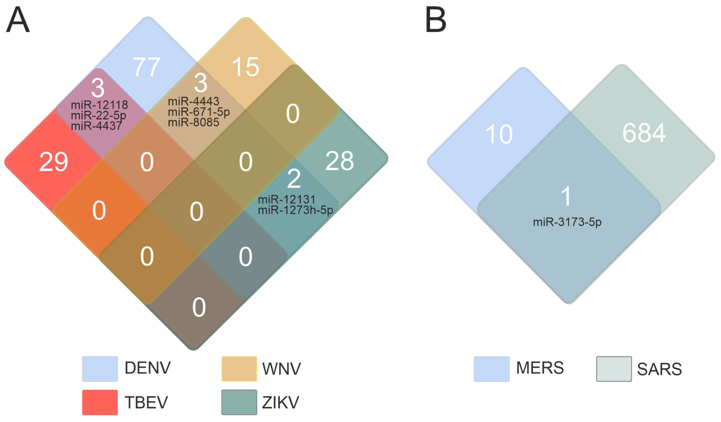
Figure 1. Quantitation of miRNAs associated with flavivirus (A) and coronavirus (B) infections. The data from the miRWalk database [17] (accessed on 18 April 2025). The figure presents a list of common miRNAs.
Ijms 26 10100 g001
Figure 2. A possible role of host miRNAs during infection. (A) MiRNAs can directly bind to the viral RNA genome and inhibit viral replication. (B) MiRNAs can be involved in regulating infection pathways, such as regulating the expression of ACE2, which is required for the SARS-CoV-2 virus entry into the host cell. MiRNAs can play a role in regulating multiple components of the immune system. (C) MiRNAs play a role in regulating the innate and adaptive immune systems.
Ijms 26 10100 g002
Figure 3. Quantitation of miRNAs associated with three types of viral infections.
Ijms 26 10100 g003
Figure 4. A regulatory network of miRNAs affecting B cell activation involved in the immune response in viral infections. MiRNA data were acquired from miRTarBase [22] (red edges) and TargetScan [20] (blue edges) (accessed on 11 April 2025). Visualization of target genes was performed using Cytoscape 3.10.2. MiRNAs are denoted in gray, and genes are indicated in orange.
Ijms 26 10100 g004
Figure 5. The regulatory network of miRNA-gene interactions during viral infections. MiRNA data were acquired from miRTarBase [22] (red edges) and TargetScan [20] (blue edges) (accessed on 11 April 2025). MiRNAs are indicated by gray, while GO genes are indicated by yellow and blue: 2286 and 2309, T cell activation involved in immune response and T cell proliferation involved in immune response.
Ijms 26 10100 g005
Figure 6. The regulatory network of miRNA-gene interactions in viral infections. MiRNA data were acquired from miRTarBase [22] (red edges) and TargetScan [20] (blue edges) (accessed on 11 April 2025). Selected GO genes: 2475, antigen processing and presentation via MHC. Gray denotes miRNAs, and blue represents genes.
Ijms 26 10100 g006
Figure 7. The regulatory network of miRNA-gene interactions in viral infections. MiRNA data were acquired from miRTarBase [22] (red edges) and TargetScan [20] (blue edges) (accessed on 11 April 2025). The selection involved genes associated with natural killer cell activation and immune response (GO: 2323). MiRNAs are denoted in gray, and genes are denoted in red.
Ijms 26 10100 g007
Figure 8. A regulatory network of miRNAs involved in the regulation of cytokine gene during viral infection. MiRNA data were acquired from miRTarBase [22] (red edges) and TargetScan [20] (blue edges) (accessed on 11 April 2025). MiRNAs are indicated in gray, and genes are indicated in orange.
Ijms 26 10100 g008
Figure 9. A regulatory network of miRNAs involved in regulating cytokine receptor gene during viral infection. MiRNA data were acquired from miRTarBase [22] (red edges) and TargetScan [20] (blue edges) (accessed on 11 April 2025). MiRNAs are represented in gray, and genes are represented in orange.
Ijms 26 10100 g009
Figure 10. A regulatory network of miRNAs involved in the regulation of cytokine signaling pathways during viral infection. MiRNA data were acquired from miRTarBase [22] (red edges) and TargetScan [20] (blue edges) (accessed on 11 April 2025). MiRNAs are designated in gray, while orange designates genes.
Ijms 26 10100 g010
Figure 11. Analysis of let-7 binding in the 3′-UTR region mRNAs of PLCG2 (A), IL6 (B), IL6R (C) genes.
Ijms 26 10100 g011
Figure 12. Analysis of miR-146a-5p binding in the 3′-UTR region mRNAs of ICAM1, IL6, STAT1, IRAK1, and TRAF6 gene.
Ijms 26 10100 g012
Figure 13. Analysis of miR-181a-5p binding in the 3′-UTR region mRNAs of IL1A and STAT3 gene.
Ijms 26 10100 g013
Figure 14. Analysis of miRNA binding in the 3′-UTR region mRNAs of EOMES (A), RAB27A (B) and IL10RB (C) gene.
Ijms 26 10100 g014
Figure 15. Analysis of miRNA binding in the 3′-UTR region mRNAs of EOMES (A) and IL10RB (B).
Ijms 26 10100 g015
Disclaimer/Publisher’s Note: The statements, opinions and data contained in all publications are solely those of the individual author(s) and contributor(s) and not of MDPI and/or the editor(s). MDPI and/or the editor(s) disclaim responsibility for any injury to people or property resulting from any ideas, methods, instructions or products referred to in the content.