Review

Pseudomonas aeruginosa Pathogenicity and Its Interaction with Other Microorganisms During the Skin Wound Healing Process

by
Inti Yamberla
1,†,
Carla Pupiales
1,†,
Andrea Jazmín Chiliquinga
1,
Tania Sulca-Villamarín
1,
Alejandra Plasencia
1,
Francisco Cabrera Aulestia
2,3,4,
Ramiro F. Díaz
2,5,
Andrés Caicedo
2,3,6,7 and
Pedro Miguel Barba
1,*
1
Carrera de Biotecnología, Universidad Técnica del Norte, Ibarra 100105, Ecuador
2
Instituto de Investigaciones en Biomedicina iBiomed, Universidad San Francisco de Quito, Quito 170901, Ecuador
3
Mito-Act Research Consortium, Quito 170901, Ecuador
4
Laboratorio de Salud Animal, Instituto de Biodiversidad Tropical, Escuela de Medicina Veterinaria, Universidad San Francisco de Quito, Quito 170901, Ecuador
5
Escuela de Medicina Veterinaria, Universidad San Francisco de Quito, Quito 170901, Ecuador
6
Escuela de Medicina, Colegio de Ciencias de la Salud, Universidad San Francisco de Quito, Quito 170901, Ecuador
7
USFQ Space Front, Universidad San Francisco de Quito, Quito 170901, Ecuador
*
Author to whom correspondence should be addressed.
These authors contributed equally to this work.
Int. J. Mol. Sci. 2025, 26(19), 9677; https://doi.org/10.3390/ijms26199677
Submission received: 16 July 2025 / Revised: 18 August 2025 / Accepted: 20 August 2025 / Published: 4 October 2025
(This article belongs to the Special Issue Skin Microbiome and Skin Health: Molecular Interactions)

Abstract

Pseudomonas aeruginosa is a Gram-negative opportunistic pathogen frequently associated with delayed wound healing, particularly in chronic skin injuries. Its capability to form biofilms, secrete virulence factors, and the faculty to compete with other microorganisms makes it a major challenge in clinical wound management. Recent literature reveals different molecular and cellular mechanisms through which P. aeruginosa disrupts the wound healing process. Findings highlight that it interferes with key phases of healing by modulating host immune responses, degrading extracellular matrix components, and inhibiting keratinocyte migration. Its quorum-sensing systems regulate the expression of critical virulence factors such as exotoxin A, elastases, pyocyanin, and rhamnolipids. Additionally, the production of the biofilm matrix components alginate, and polysaccharides provide protection against host defenses and antibiotics. Interactions with other microorganisms, including antagonistic effects on Staphylococcus epidermidis and synergistic relationships with Staphylococcus aureus, modify the wound microbiota. Promising therapeutic alternatives have shown efficacy in disrupting biofilms and reducing virulence. These insights remark the importance of targeting both P. aeruginosa and its ecological interactions to enhance wound healing outcomes and develop more effective treatments. This review aimed to highlight the pathogenic role of P. aeruginosa and its interactions with other microbial species in the context of skin wound healing.

1. Introduction

Wound healing is a complex and dynamic biological process involving a coordinated cascade of cellular and molecular events that restore the integrity of damaged skin. This process occurs in overlapping stages, including hemostasis, inflammation, proliferation, and remodeling, all of which depend on a tightly regulated environment and the interplay of local and systemic factors [1,2].
When skin integrity is compromised, the balance of the local microbiota is disrupted, making the wound more susceptible to colonization by opportunistic and pathogenic microorganisms [3,4]. Among these pathogens, Pseudomonas aeruginosa is particularly notable for its ability to colonize open wounds and interfere with the wound healing trajectory. Its presence not only alters the local microbial community but also interferes with the progression of healing by influencing immune responses and delaying resolution of inflammation [5,6]. The microbial shift caused by P. aeruginosa colonization often leads to competitive interactions with other microorganisms such as Staphylococcus aureus, Klebsiella pneumoniae, and Streptococcus pyogenes, further complicating infection outcomes [7,8].
The wound environmental conditions particularly elevated iron concentrations, contributing significantly to the pathogenic potential of P. aeruginosa. High iron levels in the wound microenvironment have been shown to inhibit the repressor proteins of Psl (Polysaccharide of the biofilm matrix) biosynthesis, molecule crucial for biofilm development, and bacterial aggregation [9]. Moreover, iron availability itself plays a fundamental role in improving the ability of the pathogen to evade host immune responses and persist in chronic wound settings [10,11].
Understanding how P. aeruginosa interacts with both host tissues and co-infecting microorganisms is essential for developing new therapeutic approaches. Despite numerous studies on its virulence, there is still limited integrative knowledge regarding how P. aeruginosa dynamically interacts with other microbes within the wound niche and how this affects healing outcomes. Therefore, this study aims to investigate the pathogenic role of P. aeruginosa and its interactions with other microorganisms in skin wound healing.

2. Brief Overview of the Normal Skin Wound Healing Process

Wound healing is a natural physiological process that takes place to repair damaged tissues and involves multiple molecular, cellular, and clinical facts. This process includes hemostasis, inflammation, proliferation, and remodeling phases [1]. Wound healing begins immediately after tissue injury with the activation of hemostasis, which results in the formation of a fibrin mesh, a protein-based scaffold that halts bleeding and provides a structural matrix for cell adhesion and signal integration. This fibrin network also facilitates the release and concentration of chemical mediators known as growth factors, which are signaling proteins that regulate key cellular behaviors such as proliferation, migration, and differentiation [12,13].
The inflammatory phase follows the hemostasis stage and is characterized by the infiltration of neutrophils and macrophages into the wounded area [14,15]. These immune cells initially try to eliminate the pathogens by phagocytosis, also the neutrophils build an extracellular matrix to capture opportunistic pathogens such as Pseudomonas aeruginosa [16]. On the other hand, the macrophages have more versatile actions, such as contributing to the secretion of pro-inflammatory cytokines such as IL-1, IL-1β, IL-6, and TNF (Tumor Necrosis Factor); macrophages also recruit other leukocytes to the wound area and prepare the zone for tissue regeneration by TGF (Transforming Growth Factor) excretion [17].
During the proliferative phase, keratinocytes, fibroblasts, and myofibroblasts migrate and proliferate to restore epithelial integrity and synthesize extracellular matrix components essential for tissue repair [18,19].
In the remodeling phase, the granulation tissue is reorganized and replaced by a more structured extracellular matrix, leading to revascularization, reduction in cellularity, and partial restoration of tissue function. Although these stages occur in a sequential order, they are not entirely distinct and may overlap temporally depending on the nature and severity of the injury [20]. Proper resolution of each phase is critical to achieving functional tissue regeneration and preventing chronic wound formation.
The pathogenic potential of P. aeruginosa becomes particularly relevant during the inflammatory and proliferative phases by secreting exotoxins and forming biofilms, P. aeruginosa can disrupt immune responses, prolong inflammation, and hinder keratinocyte migration, ultimately delaying wound closure and promoting chronic infection [21,22].

3. The Pseudomonas aeruginosa Role in Skin Wound Healing

Pseudomonas aeruginosa is a Gram-negative opportunistic pathogen capable of adapting to a wide range of environmental and host-associated niches. It possesses a broad arsenal of virulence mechanisms that enable colonization, immune evasion, and tissue destruction.
One of the key adaptive traits of P. aeruginosa is the ability to interact with host defense systems, particularly by binding to vitronectin glycoprotein. Normally, vitronectin is located on the extracellular matrix (ECM) and regulates the complement immune pathway and the recruitment of defense cells. This interaction between the bacteria with the vitronectin affects the immune cell adhesion to the damage zone and facilitates the immune response evasion and contributes to bacterial adhesion to host tissues [23,24].
In acute and chronic wounds, P. aeruginosa thrives by producing a complex biofilm structure constituted by polysaccharides such as alginate, Pel, and Psl (Table 1). These biofilms are further stabilized by extracellular DNA, structural proteins, fimbriae and type IV pili protein, this context facilitates surface adherence and microcolony formation by the pathogen [25,26]. The biofilm not only enhances antibiotic resistance but also physically shields bacteria from immune cells such as neutrophils and macrophages, and finally interferes with complement pathway, exacerbating chronic inflammation [7,27].
P. aeruginosa also secretes an array of extracellular proteins and toxins that intensify its virulence. Among them are pyocyanin, elastase, and alkaline protease, all of which contribute to oxidative stress, tissue degradation, and modulation of host immune signaling [28]. A pivotal contributor to pathogenicity is the Type III Secretion System (T3SS), a needle-like apparatus that injects effector proteins directly into host cells. T3SS system delivers specific exotoxins such as ExoS, ExoT, ExoU, and ExoY, each of which disrupts distinct cellular processes like cytoskeletal integrity, intracellular signaling, and apoptosis (Figure 1) [6,29].
Additional virulence factors include phospholipase C, which lyses host cell membranes, the ferripyochelin-binding protein involved in iron acquisition, and the surface-expressed lipopolysaccharide (LPS), which stimulates strong inflammatory responses through Toll-like receptor 4 activation [30]. These components together enable P. aeruginosa to manipulate both the immune landscape and the structural integrity of wounded tissue, thereby delaying resolution and contributing to chronic wound pathology (Table 1).
Table 1. Main molecules secreted by Pseudomonas aeruginosa during the wound healing process.
CellsSecreted MoleculesDescriptionReference
Pseudomonas aeruginosa
Planktonic cellExotoxin AExotoxin that inhibits protein synthesis and causes cytopathic effects in immune cells.[31]
Sessile cells
(Biofilm)
PyocyaninVirulence factor that induces eryptosis. It has a potential role in biofilm formation by promoting eDNA release due to cell lysis.[32]
Alkaline proteaseExtracellular protease, that prevents bacterial elimination by degrading the immune C2 complement protein, can also degrade flagellin and is a known pro-inflammatory responses activator.[33]
Di-rhamnolipidGlycolipid biosurfactant that lyse neutrophils, macrophages, and different
animal cells rapidly. Acts in swarming motility and shape the biofilm structure, also possess different antimicrobial activity.
[32]
Cyclic diguanosine-5′-monophosphate (c-di-GMP)Nucleotide on which the lifestyle of P. aeruginosa depends; low levels favor the motility factors expression, promoting the planktonic state and high levels favor the sessile lifestyle by increasing the extracellular matrix components and adhesion factors expression.[32]
AlginateMannuronic acid and glucuronic acid linear polymer, a biofilm component and acts as a cell evasion mechanism, blocking the antibodies and phagocytosis immune action.[34]
Lectin BMembrane protein that coats the bacterial cells together and promotes the adhesion of P. aeruginosa to both host cell and exopolysaccharide matrix. On epithelial cells, it inhibits cell migration and proliferation during re-epithelization phase.[35]
QuinolonesAntimicrobial molecules with selective toxicity to inhibit the synthesis of other bacterial DNA.[36]
PelPolysaccharide involved binding initiation on the surface and maintenance the integrity of biofilm. It crosslinks the eDNA in the biofilm matrix and maintains cell–cell interactions.[25]
PslPolysaccharide involved in cell–cell adhesion. It reduces the immune system attacks because it inhibits opsonization and reduces the neutrophil’s reactive oxygen species (ROS). In addition, it reduces the matrix phagocytosis.[25]
AcetateMolecules produced by Pseudomonas aeruginosa, useful to bind to the LPS side chains or alginate by ester bond for preventing the complement immune system activation.[34]

4. Pathogenicity of Pseudomonas aeruginosa in Skin Wound Healing

The pathogenicity of Pseudomonas aeruginosa in cutaneous wounds is supported by a diverse set of molecular and physiological strategies that enhance its ability to persist, evade host defenses, and disrupt the healing environment. One important factor is the presence of outer membrane porins, particularly the OprF porin, which functions as a nonspecific diffusion channel involved in nutrient acquisition and environmental sensing [37]. The OprF porin has also been linked to the regulation and export of virulence molecules such as pyocyanin, a redox-active metabolite that generates oxidative stress in host cells and inhibits the growth of competing microorganisms [38].
Quorum sensing (QS) is a central mechanism in P. aeruginosa pathogenesis that modulates the expression of genes associated with virulence and biofilm maturation, it is a cell-density-dependent regulatory system. P. aeruginosa possesses at least two canonical QS circuits based on acyl homoserine lactones: the “las” and “rhl” systems, composed of the lasI/lasR and rhlI/rhlR gene pairs, respectively [39,40]. These QS pathways activate the expression and release of numerous virulence factors, including toxA (exotoxin A), aprA (alkaline protease), and lasA and lasB (elastases), which contribute to tissue destruction and immune interference [41]. The rhl system also regulates motility and biosurfactant production by activating the rhlAB operon, which controls the expression and synthesis of molecules as di-rhamnolipids that reduce surface tension in the tissues and facilitate bacterial spreading [35,42].
Another key regulatory mechanism is the intracellular signaling molecule cyclic diguanosine monophosphate (c-di-GMP), which modulates the transition between planktonic and biofilm lifestyles of P. aeruginosa. High intracellular concentrations of c-di-GMP promote sessile behavior by enhancing the production of extracellular polymeric substances, including alginate, Pel, and Psl polysaccharides, and by inducing cell aggregation and adhesion to surfaces [43]. Conversely, low levels of c-di-GMP increase motility and reduce biofilm formation, facilitating dispersal and new colonization [44]. This regulatory system plays a pivotal role in modulating virulence in response to environmental cues within the wound (Figure 1) [45,46].
The expression of these virulence factors enables P. aeruginosa not only to resist phagocytic clearance and the immune antimicrobial activity but also to manipulate the wound microenvironment. This includes disruption of immune signaling, degradation of extracellular matrix components, and modulation of host cell migration and proliferation. As such, understanding these interlinked regulatory networks provides a baseline for designing targeted therapies directed to disrupt quorum sensing, inhibiting biofilm formation, or restoring immune balance in infected wounds [6,8].

5. Pseudomonas aeruginosa Biofilm Effect on Skin Wounds

The P. aeruginosa biofilm formation is an aggravating key factor for the inflammatory response and impairing skin wound healing [5,35,47]. The biofilm produced by P. aeruginosa promotes the bacterial persistence ability, facilitating the bacterial wound colonization [39,47]. P. aeruginosa biofilms are generally constituted by exopolysaccharides like alginate, Psl and Pel polysaccharides, exogenous DNA, and lipids, which determine the biofilm stability and protect the bacteria from the host immune response [48,49].
On the other hand, P. aeruginosa mature biofilms often form a central core surrounded by a distinct shell-like wall of non-motile cells [50]. In the biofilm, there are two different bacterial phenotypes, highly motile cells in the central core, and planktonic non-motile cells on shell-like wall [51]. At the same time P. aeruginosa produce alginate that confers bacterial resistance by lipopolysaccharide (LPS) side chains elimination to obtain acetate substitutes [34,52]. The acetate residues are attached via ester bonds to hydroxyl groups that act as covalent bond acceptors for opsonins on the bacteria surface, preventing the activation of the immune complement system.
These biofilms adhere to devitalized tissue and wound bed surfaces, often forming stratified layers in which metabolically active bacteria reside in the outer regions while dormant or persistent cells occupy the deeper layers, enhancing antibiotic tolerance [50,51]. In the wound microenvironment, the biofilm matrix not only provides a physical barrier to immune cell penetration but also retains pro-inflammatory mediators, perpetuating tissue damage and delaying re-epithelialization [52]. This structural adaptation to the skin wound milieu differs from flow-cell or in vitro models, as wound biofilms interact closely with host extracellular matrix components and inflammatory exudates, which influence their architecture and stability [25,50].
Furthermore, the Pel and Psl polysaccharides are essential for subpopulation interactions and microcolony formation in the later stages of biofilm formation. Pel inhibits opsonization, resulting in reduced production of neutrophil reactive oxygen species (ROS) and decreased matrix destruction by phagocytes cells [25].
The release of Lectin B is another important virulence factor that causes loss of epithelial polarity. It has a high binding affinity for L-fucose and its derivatives [47]. The process that LecB binds to fucose-bearing lipids induces membrane invaginations and locates integrin’s in these invaginations. As consequence, LecB causes the inhibition of cell migration, facilitates the establishment and stabilization of bacterial infection, and prevents proper healing [53].
The LecB not only induces membrane invaginations that trap integrins but also disrupts essential signaling pathways involved in wound healing. By prolonging the inflammatory phase, it impairs neutrophil and macrophage transition to pro-repair phenotypes, which delays angiogenesis and tissue regeneration. Additionally, LecB contributes to excessive NET formation and inflammasome activation in macrophages, further exacerbating inflammation and preventing resolution, especially in chronic wounds such as diabetic ulcers [16].

6. Pseudomonas aeruginosa Interaction with the Immune System

A successful bacterial infection develops when the immune response fails. Also, it is well known that persistent inflammation in wounds is accompanied by an inefficient healing process [35,54]. P. aeruginosa may alter the inflammatory response by secreting pro-apoptotic factors, which are correlated with an unresolved inflammation [55]. For example, exotoxin A modifies the gene expression in mammalian cells and causes polymorphonuclear neutrophils (PMNs) and macrophages apoptosis [56]. When PMNs die, the cellular components are released, increasing inflammation and causing host collateral damage. Similarly, in macrophages absence, the fragmented neutrophils will eventually release their pro-inflammatory content [57] (Table 2).
It is known that M1-macrophages act in the early inflammation phase and M2-macrophages interfere in the proliferation phase. Particularly, M1-macrophages produce pro-inflammatory cytokines such as tumor necrosis factor (TNFα), interleukin 6 (IL-6), interleukin IL-12, and antibacterial mediators such as reactive nitrogen and oxygen species (NO and ROS); M2-macrophages produce anti-inflammatory cytokines such as interleukin IL-4, interleukin IL-10, TGF, and arginase that promote tissue regeneration (Figure 2) [57].
P. aeruginosa in the wound healing environment promotes the pro-inflammatory genes expression in M1 macrophages because P. aeruginosa produces changes in the phenotype of macrophages and prolongs the M1-cells markers release, resulting in insufficient M2-macrophage signals, reduction in anti-inflammatory cytokines and growth factors TGFβ, and finally, the extension of the inflammation phase and delays healing [5,21].

7. Interaction of Pseudomonas aeruginosa with Other Pathogens on Skin Wound Healing Process

The skin is a physical barrier that offers protective niches and nutrients for different microorganisms facilitating their survival and at the same time generating competition and cooperation among them; but when this physical barrier is deteriorated, inflammatory conditions occur in the skin. Therapies focusing only on a primary pathogen are not usually successful due to the typical polymicrobial communities of the skin [64,65,66] where P. aeruginosa can interact with both pathogenic and commensal skin microorganisms. The common bacteria interacting with P. aeruginosa are Staphylococcus aureus, Klebsiella pneumoniae and Streptococcus spp., Staphylococcus epidermidis, and Micrococcus luteus (Table 3).
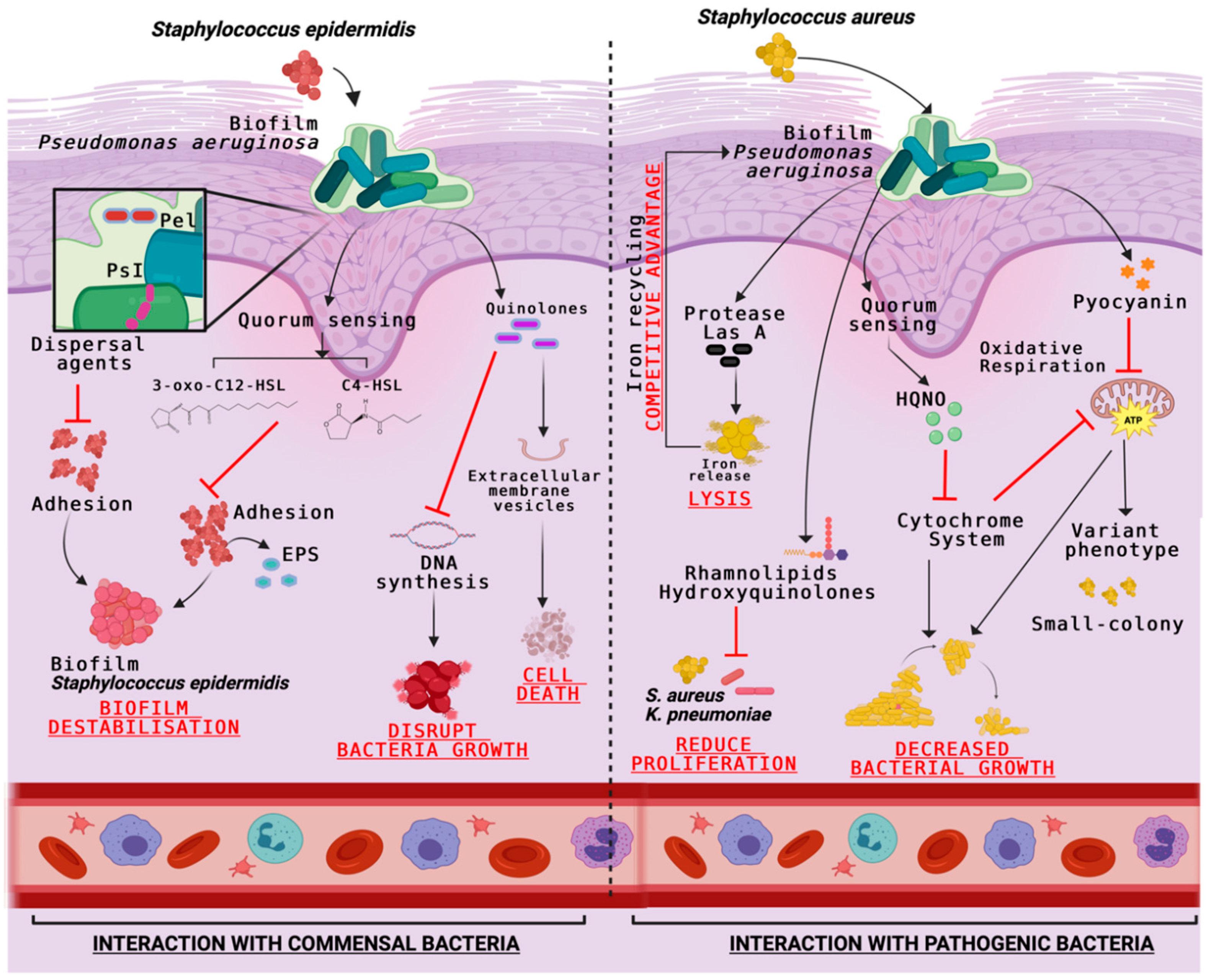
Under normal conditions, P. aeruginosa can suppress S. aureus growth by releasing a variety of toxins such as 4-hydroxy-2-heptylquinoline N-oxide (HQNO), pyocyanin, or LasA protease which inhibits the cytochrome system, the oxidative respiration reactions, and lyses S. aureus cells [30]. However, when the quorum sensing (QS) is deficient, a commensal-like interaction occurs between S. aureus and P. aeruginosa. It is also known that in these polymicrobial communities, one species always predominates. Using an antimicrobial method, Wilkinson y Hardman [20] found that P. aeruginosa becomes dominant when resources are limited, taking advantage of vital cofactors such as iron released by S. aureus when it is lysed by P. aeruginosa. They have also demonstrated that P. aeruginosa has a competitive advantage over S. aureus and K. pneumoniae by releasing toxic metabolites such as rhamnolipids or hydroxyquinolone. However, there is the possibility that at a certain point in the infection, when any strain of P. aeruginosa suffers an enrichment, a divergence of its population occurs and gives rise to the survival of pathogens such as S. aureus and K. pneumoniae (Table 3) [64].
Pseudomonas aeruginosa uses quorum-sensing molecules such as 3-oxo-C12-HSL and C4-HSL to regulate key virulence factors such as elastase, pyocyanin, rhamnolipids, and exotoxin A. These signals not only control their own pathogenicity but also inhibit the adhesion and biofilm formation of Staphylococcus epidermidis, giving P. aeruginosa a competitive advantage in polymicrobial wound environments [71]. This is achieved through the production of quorum-sensing molecules such as 2-heptyl-3-hydroxy-4-quinolone (PQS) and N-acyl homoserine lactones (AHLs), which interfere with the gene expression and metabolic activity of competing bacteria, thereby suppressing their colonization and enhancing the dominance of P. aeruginosa in chronic wounds [72].
Therefore, it can also help P. aeruginosa dominate under coinfection conditions. The P. aeruginosa extracellular polysaccharides (Pel and Ps1) have been shown to exert anti-staphylococcal activity by dispersing agents of S. epidermidis biofilms [73]. This occurs through a dual mechanism: Pel and Psl interfere with the structural integrity of the S. epidermidis biofilm matrix while also enhancing the penetration of antimicrobial compounds produced by P. aeruginosa, such as rhamnolipids and pyocyanin, which further inhibit staphylococcal viability and colonization in polymicrobial environments [40]. On the other hand, P. aeruginosa produces around 55 quinolones/quinolines molecules with significant antibiotic activity against Gram-positive bacteria [74]. Antimicrobial quinolines can be packaged into extracellular membrane vesicles (MVs) to cause direct S. epidermidis lysis, for example (Table 3) [73].
Interactions between Pseudomonas aeruginosa and Streptococcus species are frequently observed in cystic fibrosis-associated infections. Notably, Streptococcus parasanguinis has been shown to penetrate P. aeruginosa biofilms and disrupt their structure, thereby reducing the expression of key virulence genes. This occurs through the secretion of hydrogen peroxide (H2O2) and other diffusible inhibitory molecules that compromise the integrity of the P. aeruginosa exopolysaccharide matrix, leading to biofilm destabilization and downregulation of quorum-sensing systems such as lasR and rhlR, which are essential for the regulation of virulence factors including elastase and pyocyanin [75].
This interference impairs biofilm stability and limits the production of pathogenic factors, ultimately attenuating P. aeruginosa pathogenesis and enhancing the host’s ability to control infection [76]. S. parasanguinis is atypical on skin, however a similar interaction could occur between P. aeruginosa and another Streptococcus genus pathogens. In its interaction with Anginosus Group Streptococci, P. aeruginosa enhances the production of virulence factors such as pyocyanin and elastase, important molecules for biofilm formation. These virulence factors’ activity increase, causing considerable damage in the affected area of the patient (Figure 3) [77].
P. aeruginosa secretes acyl homoserine lactones (AHLs) with variable fatty acid side chains, which interfere with Streptococcus pyogenes hemolytic capacity. The mechanism includes the negative regulation of the sag operon expression, involved in the streptolysin S production, important for erythrocytes, leukocytes, and platelets lysing [70].
Moreover, the ability of P. aeruginosa to inhibit or promote the growth of other microorganisms significantly shapes the trajectory of wound healing. Inhibitory interactions, such as the production of HQNO, pyocyanin, or LasA protease against Staphylococcus aureus, may transiently suppress competing pathogens but often intensify local tissue damage, inflammation, and biofilm formation, ultimately delaying re-epithelialization [68,69,70]. Conversely, in certain polymicrobial contexts, cooperative interactions can increase overall virulence, leading to more severe infections and prolonged healing times [64,77].

8. Pseudomonas aeruginosa Skin Wound Infection: Current and Emerging Therapies

The best option to avoid damage by P. aeruginosa in epithelium lesions is to eliminate the bacterial cells. As a result, some treatments are using antibiotics such as levofloxacin, ciprofloxacin, gentamicin, amikacin or tobramycin, as the first line of treatment. Because P. aeruginosa has high resistance levels to most of the commonly used antibiotics, including aminoglycosides, quinolones, and β-lactams, and also to the common disinfectants, this makes it difficult to definitively eradicate from the infected tissue [78,79]. Furthermore, antibiotics can target only vegetative cells but poorly penetrate the biofilm matrix [80]. At present, aminoglycoside antibiotics and azithromycin are widely used to treat P. aeruginosa infections [81]. However, the treatment effectiveness depends on the correct dose to eliminate all bacterial cells and avoid side effects. Antibiotics synergy has been considered as a good alternative because it requires a lower dose of each antibiotic. For example, combining azithromycin and gentamicin with ceftolozane and tazobactam have had significant results in the eradication of P. aeruginosa [82,83].
The relentless rise in multidrug-resistant Pseudomonas aeruginosa underscores the need for adjuvant or alternative therapies that do not rely solely on conventional antibiotics. Several antimicrobial peptides, cold atmospheric plasma, non-classical modalities—photodynamic therapy, mesenchymal stromal cells (MSCs) and their extracellular vesicles (EVs), as well as mitochondrial transplantation [84,85] have emerged as promising tools to disrupt biofilms and accelerate wound closure.
Antimicrobial peptides are promising therapeutic options to treat P. aeruginosa infections that reduce the negative impact of virulence factors on host cells by preventing biofilm formation and also favors skin epithelial repair by inducing the migration of keratinocytes to the affected area that promote wound healing [84]. P. aeruginosa facilitates this process through the secretion of specific bioactive molecules, such as lipoxygenase-derived oxylipins, which modulate host cell signaling pathways, including MAPK and PI3K/Akt, thereby stimulating keratinocyte migration and proliferation essential for re-epithelialization [86].
Cold plasma used in skin injury therapy inhibits microbes in chronic wounds due to its antiseptic effects, since it has been used as a sterilizing agent. Its efficacy arises from the generation of a mixture of reactive oxygen and nitrogen species (RONS), UV photons, and transient electric fields, which together disrupt microbial cell walls, damage nucleic acids, and inactivate essential enzymes. Unlike conventional antiseptics, cold plasma acts non-thermally and can penetrate biofilms without harming surrounding healthy tissue, making it particularly effective against multidrug-resistant bacteria commonly found in chronic wounds. Moreover, it has been shown to promote wound healing by enhancing local microcirculation and stimulating keratinocyte and fibroblast proliferation [87].
In addition, it is a source of gaseous nitric oxide which works as a powerful microbicide agent that destroys different kinds of bacteria, while it promotes healing by proliferation cell stimulation [88]. The aim of the topical therapy is to reduce bacteria count in the wound and remove soluble debris without adversely affecting cellular activities during the wound healing process.
Furthermore, photodynamic therapy (PDT) has shown considerable potential in inhibiting Pseudomonas aeruginosa biofilm formation and accelerating the wound healing process. PDT functions through the administration of a photosensitizing agent that selectively localizes within bacterial cells. Upon exposure to a specific wavelength of light, this agent becomes activated and produces reactive oxygen species (ROS), such as singlet oxygen and free radicals. These ROS induce oxidative stress that damages bacterial membranes, proteins, and DNA, ultimately leading to cell death. In the case of P. aeruginosa, oxidative damage disrupts the biofilm matrix and inhibits its growth, allowing the host immune system and tissue repair mechanisms to function more effectively [89].
New methods are currently being studied to improve tissue repair, such as the use of mitochondrial transfer due to its potential to revitalize senescent cells. The mitochondria could stop the production of free radicals, thus reducing the recovery timing from a wound and possible complications [90]. Mitochondria are the main source of reactive oxygen species (ROS) that are involved in all stages of the tissue repair process. At low concentrations, they are essential participants in cell signaling, induction of the myogenic response, and defense against infectious agents [90]. In pre-clinical models, MSC-EVs enhance standard antibiotic regimens by (i) delivering microbicidal molecules that curb bacterial replication, (ii) re-programming macrophages toward a pro-resolution (M2) phenotype, and (iii) releasing pro-angiogenic and anti-apoptotic factors that stimulate keratinocyte migration and tissue regeneration [91,92,93,94]. For example, in a rabbit model of renal tuberculosis, MSC-EVs potentiated first-line anti-tubercular drugs, tempered inflammation, and minimized parenchymal damage [93]. NADPH leukocyte oxidase (Nox) is one of the main sources of ROS involved in pathogen destruction as well as vascular endothelial growth factor (VEGF) signaling, and TNF response [90]. However, when concentration exceeds the balance between production and uptake, a phenomenon known as oxidative stress is generated, which is a key factor that delays healing because it significantly alters the microbiome by promoting the colonization of biofilm-forming bacteria [95,96].
In addition to pharmacological approaches, wound bed preparation through debridement is a cornerstone in chronic wound management. Debridement physically removes necrotic tissue, bacterial biofilms, and cellular debris, reducing the bioburden and facilitating the penetration of topical or systemic antimicrobial agents [91]. Various methods, such as surgical, enzymatic, autolytic, and mechanical debridement, have been shown to enhance granulation tissue formation and create a favorable microenvironment for subsequent healing phases [91].
Equally important is promoting epithelial repair, which involves stimulating keratinocyte migration and proliferation to restore the skin barrier. Strategies include the application of bioactive dressings enriched with growth factors, extracellular matrix components, or antimicrobial peptides, which not only inhibit P. aeruginosa colonization but also accelerate re-epithelialization [91,92,93]. By integrating these regenerative interventions alongside infection control, wound healing outcomes can be significantly improved.
Further approaches have been revised like the restoration of microbial balance. Strategies include the use of probiotics, bacteriophage therapy, and topical antimicrobials with selective activity that modulate the microbiota without completely eradicating beneficial species [75]. Additionally, quorum-sensing inhibitors and biofilm-disrupting agents can reduce P. aeruginosa dominance, allowing commensal species to re-establish and support wound repair [77]. These approaches aim to shift the microbial community toward a non-pathogenic state, thereby reducing inflammation, enhancing granulation tissue formation, and accelerating closure of chronic wounds.

9. Conclusions

Pseudomonas aeruginosa plays a critical pathogenic role in skin wound healing by interfering with multiple phases of the repair process, especially inflammation and proliferation. Through the secretion of exotoxins and the formation of structurally complex biofilms composed of alginate, Pel, Psl, and extracellular DNA, the pathogen delays wound closure and promotes chronic infection.
The virulence of P. aeruginosa is regulated by quorum-sensing systems (las and rhl) and intracellular signaling molecules such as cyclic di-GMP, which coordinate the expression of key factors including pyocyanin, exotoxin A, elastases, and rhamnolipids. These molecules disrupt host immune responses, degrade extracellular matrix components, and prevent effective epithelial regeneration.
The capacity of the pathogen to evade immune defenses is reinforced by its interaction with host proteins such as vitronectin, suppression of complement activation, and modulation of macrophage phenotypes, prolonging the pro-inflammatory M1 phase and delaying the anti-inflammatory M2 transition required for tissue regeneration.
The P. aeruginosa maintains a competitive advantage in polymicrobial environments by inhibiting adhesion and biofilm development of other pathogens like Staphylococcus epidermidis and Streptococcus pyogenes through the release of AHL signals, quinolones, and other antimicrobial compounds. However, certain streptococcal species, such as Streptococcus parasanguinis, can disrupt P. aeruginosa biofilms and reduce its virulence.
Therapies such as antibiotics, peptides, and other novel alternatives such as mitochondrial application help to control the pathogenic potential of P. aeruginosa in the wound healing environment.

Author Contributions

Conceptualization, writing—original draft, preparation, and investigation, I.Y. and C.P.; conceptualization and writing—review and editing, A.J.C., T.S.-V., F.C.A., A.C. and R.F.D.; writing—review and editing, A.P.; conceptualization, project administration, supervision, and writing—review and editing, P.M.B. All authors have read and agreed to the published version of the manuscript.

Funding

This research received no external funding.

Data Availability Statement

No new data were created or analyzed in this study. Data sharing is not applicable to this article.

Conflicts of Interest

AC is the scientific founder and advisor of Dragon Biomed and Homeos, two entrepreneurial initiatives at Universidad San Francisco de Quito (USFQ). He also serves as a scientific advisor in the Research and Development department at Luvigix. In these roles, he provides scientific guidance and expertise but does not engage in decision-making processes or operational responsibilities. The remaining authors declare no conflicts of interest.

References

  1. Gonzalez, A.C.D.O.; Andrade, Z.A.D.; Costa, T.F.; Medrado, A.R.A.P. Wound healing—A literature review. An. Bras. Dermatol. 2016, 91, 614–620. [Google Scholar] [CrossRef] [PubMed]
  2. Tomic-Canic, M.; Burgess, J.L.; O’Neill, K.E.; Strbo, N.; Pastar, I. Skin microbiota and its interplay with wound healing. Am. J. Clin. Dermatol. 2020, 21, 36–43. [Google Scholar] [CrossRef]
  3. Byrd, A.L.; Belkaid, Y.; Segre, J.A. The human skin microbiome. Nat. Rev. Microbiol. 2018, 16, 143–155. [Google Scholar] [CrossRef]
  4. Daeschlein, G.; von Woedtke, T.; Kindel, E.; Brandenburg, R.; Weltmann, K.D. In vitro susceptibility of multidrug resistant skin and wound pathogens against low temperature atmospheric pressure plasma jet (APPJ) and dielectric barrier discharge plasma (DBD). Plasma Process. Polym. 2014, 11, 175–183. [Google Scholar] [CrossRef]
  5. Brandenburg, K.S.; Weaver, A.J., Jr.; Karna, S.L.; You, T.; Chen, P.; Van Stryk, S.; Leung, K.P. Formation of Pseudomonas aeruginosa biofilms in full-thickness scald burn wounds in rats. Sci. Rep. 2019, 9, 13627. [Google Scholar] [CrossRef]
  6. Wood, S.J.; Kuzel, T.M.; Shafikhani, S.H. Pseudomonas aeruginosa: Infections, animal modeling, and therapeutics. Cells 2023, 12, 199. [Google Scholar] [CrossRef]
  7. Vestweber, P.K.; Wächter, J.; Planz, V.; Jung, N.; Windbergs, M. The interplay of Pseudomonas aeruginosa and Staphylococcus aureus in dual-species biofilms impacts development, antibiotic resistance and virulence of biofilms in in vitro wound infection models. PLoS ONE 2023, 19, e0304491. [Google Scholar] [CrossRef] [PubMed]
  8. Guzzo, F.; Scognamiglio, M.; Fiorentino, A.; Buommino, E.; D’Abrosca, B. Plant-derived natural products against Pseudomonas aeruginosa and Staphylococcus aureus: Antibiofilm activity and molecular mechanisms. Molecules 2020, 25, 5024. [Google Scholar] [CrossRef]
  9. Yu, S.; Wei, Q.; Zhao, T.; Guo, Y.; Ma, L.Z. A Survival Strategy for Pseudomonas aeruginosa that uses exopolysaccharides to sequester and store iron to stimulate Psl-dependent biofilm formation. Appl. Environ. Microbiol. 2016, 82, 6403–6413. [Google Scholar] [CrossRef]
  10. Haschka, D.; Hoffmann, A.; Weiss, G. Iron in immune cell function and host defense. Semin. Cell Dev. Biol. 2021, 115, 27–36. [Google Scholar] [CrossRef] [PubMed]
  11. Nairz, M.; Weiss, G. Iron in infection and immunity. Mol. Asp. Med. 2020, 75, 100864. [Google Scholar] [CrossRef]
  12. Barrientos, S.; Stojadinovic, O.; Golinko, M.S.; Brem, H.; Tomic-Canic, M. Growth factors and cytokines in wound healing. Wound Repair Regen. 2008, 16, 585–601. [Google Scholar] [CrossRef]
  13. Schäfer, M.; Werner, S. Oxidative stress in normal and impaired wound repair. Pharmacol. Res. 2008, 58, 165–171. [Google Scholar] [CrossRef]
  14. Peña, O.A.; Martin, P. Cellular and molecular mechanisms of skin wound healing. Nat. Rev. Mol. Cell Biol. 2024, 25, 599–616. [Google Scholar] [CrossRef]
  15. Rodrigues, M.; Kosaric, N.; Bonham, C.A.; Gurtner, G.C. Wound healing: A cellular perspective. Physiol. Rev. 2019, 99, 665–706. [Google Scholar] [CrossRef] [PubMed]
  16. Phillipson, M.; Kubes, P. The healing power of neutrophils. Trends Immunol. 2019, 40, 635–647. [Google Scholar] [CrossRef] [PubMed]
  17. Koh, T.J.; DiPietro, L.A. Inflammation and wound healing: The role of the macrophage. Expert Rev. Mol. Med. 2011, 13, e23. [Google Scholar] [CrossRef]
  18. Mäemets-Allas, K.; Klaas, M.; Cárdenas-León, C.G.; Arak, T.; Kankuri, E.; Jaks, V. Stimulation with THBS4 activates pathways that regulate proliferation, migration and inflammation in primary human keratinocytes. Biochem. Biophys. Res. Commun. 2023, 642, 97–106. [Google Scholar] [CrossRef]
  19. Eming, S.A.; Murray, P.J.; Pearce, E.J. Metabolic orchestration of the wound healing response. Cell Metab. 2021, 33, 1726–1743. [Google Scholar] [CrossRef]
  20. Wilkinson, H.N.; Hardman, M.J. Wound healing: Cellular mechanisms and pathological outcomes. Open Biol. 2020, 10, 200223. [Google Scholar] [CrossRef] [PubMed]
  21. Chen, L.; Tredget, E.E.; Wu, P.Y.G.; Wu, Y. Paracrine factors of mesenchymal stem cells recruit macrophages and endothelial lineage cells and enhance wound healing. PLoS ONE 2008, 3, e1886. [Google Scholar] [CrossRef]
  22. Uberoi, A.; McCready-Vangi, A.; Grice, E.A. The wound microbiota: Microbial mechanisms of impaired wound healing and infection. Nat. Rev. Microbiol. 2024, 22, 210–225. [Google Scholar] [CrossRef]
  23. Paulsson, M.; Su, Y.C.; Ringwood, T.; Uddén, F.; Riesbeck, K. Pseudomonas aeruginosa uses multiple receptors for adherence to laminin during infection of the respiratory tract and skin wounds. Sci. Rep. 2019, 9, 18168. [Google Scholar] [CrossRef]
  24. Ibberson, C.B.; Whiteley, M. The social life of microbes in chronic infection. Curr. Opin. Microbiol. 2020, 53, 44–50. [Google Scholar] [CrossRef] [PubMed]
  25. Jennings, L.K.; Storek, K.M.; Ledvina, H.E.; Coulon, C.; Marmont, L.S.; Sadovskaya, I.; Secor, P.R.; Tseng, B.S.; Scian, M.; Filloux, A.; et al. Pel is a cationic exopolysaccharide that cross-links extracellular DNA in the Pseudomonas aeruginosa biofilm matrix. Proc. Natl. Acad. Sci. USA 2015, 112, 11353–11358. [Google Scholar] [CrossRef] [PubMed]
  26. Moradali, M.F.; Ghods, S.; Rehm, B.H.A. Pseudomonas aeruginosa lifestyle: A paradigm for adaptation, survival, and persistence. Front. Cell. Infect. Microbiol. 2017, 7, 39. [Google Scholar] [CrossRef] [PubMed]
  27. Watters, C.M.; Burton, T.; Dickson, D.B.; Kelly, K.A. The role of Pseudomonas aeruginosa biofilms in delaying wound healing through evasion of innate immunity. Adv. Wound Care 2017, 6, 373–382. [Google Scholar] [CrossRef]
  28. Qin, S.; Xiao, W.; Zhou, C.; Pu, Q.; Deng, X.; Lan, L.; Liang, H.; Song, X.; Wu, M. Pseudomonas aeruginosa: Pathogenesis, virulence factors, antibiotic resistance, interaction with host, technology advances and emerging therapeutics. Signal Transduct. Target. Ther. 2022, 7, 199. [Google Scholar] [CrossRef]
  29. Hauser, A.R. The type III secretion system of Pseudomonas aeruginosa: Infection by injection. Nat. Rev. Microbiol. 2009, 7, 654–665. [Google Scholar] [CrossRef]
  30. Hotterbeekx, A.; Kumar-Singh, S.; Goossens, H.; Malhotra-Kumar, S. In vivo and in vitro interactions between Pseudomonas aeruginosa and Staphylococcus spp. Front. Cell. Infect. Microbiol. 2017, 7, 106. [Google Scholar] [CrossRef]
  31. Morlon-Guyot, J.; Méré, J.; Bonhoure, A.; Beaumelle, B. Processing of Pseudomonas aeruginosa exotoxin A is dispensable for cell intoxication. Infect. Immun. 2009, 77, 3090–3099. [Google Scholar] [CrossRef] [PubMed]
  32. Jakobsen, T.H.; Tolker-Nielsen, T.; Givskov, M. Bacterial biofilm control by perturbation of bacterial signaling processes. Int. J. Mol. Sci. 2017, 18, 1970. [Google Scholar] [CrossRef] [PubMed]
  33. Casilag, F.; Lorenz, A.; Krueger, J.; Klawonn, F.; Weiss, S.; Häussler, S. The LasB elastase of Pseudomonas aeruginosa acts in concert with alkaline protease AprA to prevent flagellin-mediated immune recognition. Infect. Immun. 2016, 84, 162–171. [Google Scholar] [CrossRef]
  34. Leid, J.G.; Willson, C.J.; Shirtliff, M.E.; Hassett, D.J.; Parsek, M.R.; Jeffers, A.K. The exopolysaccharide alginate protects Pseudomonas aeruginosa biofilm bacteria from IFN-γ–mediated macrophage killing. J. Immunol. 2005, 175, 7512–7518. [Google Scholar] [CrossRef]
  35. Thuenauer, R.; Landi, A.; Trefzer, A.; Altmann, S.; Wehrum, S.; Eierhoff, T.; Diedrich, B.; Dengjel, J.; Nyström, A.; Imberty, A.; et al. The Pseudomonas aeruginosa Lectin LecB causes integrin internalization and inhibits epithelial wound healing. mBio 2020, 11, e03260-19. [Google Scholar] [CrossRef]
  36. Andriole, V.T. The quinolones: Past, present, and future. Clin. Infect. Dis. 2005, 41 (Suppl. S2), S120–S126. [Google Scholar] [CrossRef]
  37. Fito-Boncompte, L.; Chapalain, A.; Bouffartigues, E.; Chaker, H.; Lesouhaitier, O.; Gicquel, G.; Bazire, A.; Madi, A.; Connil, N.; VérOn, W.; et al. Full virulence of Pseudomonas aeruginosa requires OprF. Infect. Immun. 2011, 79, 1176–1186. [Google Scholar] [CrossRef]
  38. Bukhari, S.I.; Aleanizy, F.S. Association of OprF mutant and disturbance of biofilm and pyocyanin virulence in Pseudomonas aeruginosa. Saudi Pharm. J. 2020, 28, 196–200. [Google Scholar] [CrossRef]
  39. Luo, J.; Dong, B.; Wang, K.; Cai, S.; Liu, T.; Cheng, X.; Lei, D.; Chen, Y.; Li, Y.; Kong, J.; et al. Baicalin inhibits biofilm formation, attenuates the quorum sensing–controlled virulence and enhances Pseudomonas aeruginosa clearance in a mouse peritoneal implant infection model. PLoS ONE 2017, 12, e0176883. [Google Scholar] [CrossRef]
  40. De Kievit, T.R. Quorum sensing in Pseudomonas aeruginosa biofilms. Environ. Microbiol. 2009, 11, 279–288. [Google Scholar] [CrossRef] [PubMed]
  41. Khosravi, A.D.; Hoveizavi, H.; Mohammadian, A.; Farahani, A.; Jenabi, A. Genotyping of multidrug-resistant strains of Pseudomonas aeruginosa isolated from burn and wound infections by ERIC-PCR. Acta Cir. Bras. 2016, 31, 206–211. [Google Scholar] [CrossRef]
  42. Shrout, J.D.; Chopp, D.L.; Just, C.L.; Hentzer, M.; Givskov, M.; Parsek, M.R. The impact of quorum sensing and swarming motility on Pseudomonas aeruginosa biofilm formation is nutritionally conditional. Mol. Microbiol. 2006, 62, 1264–1277. [Google Scholar] [CrossRef] [PubMed]
  43. Wei, Q.; Zhang, W.; Zhang, Q. Cyclic-di-GMP signaling and regulation of bacterial virulence. Microbiol. China 2019, 46, 2265–2273. [Google Scholar] [CrossRef]
  44. Wei, Q.; Zhang, Z.; Luo, J.; Kong, J.; Ding, Y.; Chen, Y.; Wang, K. Insulin treatment enhances Pseudomonas aeruginosa biofilm formation by increasing intracellular cyclic di-GMP levels, leading to chronic wound infection and delayed wound healing. Am. J. Transl. Res. 2019, 11, 3261–3279. [Google Scholar]
  45. Valentini, M.; Filloux, A. Biofilms and cyclic di-GMP (c-di-GMP) signaling: Lessons from Pseudomonas aeruginosa and other bacteria. J. Biol. Chem. 2019, 294, 7193–7203. [Google Scholar] [CrossRef]
  46. Trøstrup, H.; Lerche, C.J.; Christophersen, L.J.; Thomsen, K.; Jensen, P.Ø.; Hougen, H.P.; Høiby, N.; Moser, C. Chronic Pseudomonas aeruginosa biofilm infection impairs murine S100A8/A9 and neutrophil effector cytokines—implications for delayed wound closure? Pathog. Dis. 2017, 75, ftx068. [Google Scholar] [CrossRef]
  47. Karna, S.L.R.; Leung, K.P.; Karna, R.S.; You, T.; Chen, P.; Van Stryk, S.; Brandenburg, K.S. The impact of simultaneous inoculation of Pseudomonas aeruginosa, Staphylococcus aureus, and Candida albicans on burn-wound biofilms and host response in a rat model. Burns 2021, 47, 331–341. [Google Scholar] [CrossRef]
  48. Mishra, R.; Panda, A.K.; De Mandal, S.; Shakeel, M.; Bisht, S.S.; Khan, J. Natural anti-biofilm agents: Strategies to control biofilm-forming pathogens. Front. Microbiol. 2020, 11, 566325. [Google Scholar] [CrossRef]
  49. James, G.A.; Swogger, E.; Wolcott, R.; Pulcini, E.; Secor, P.; Sestrich, J.; Costerton, J.W.; Stewart, P.S. Biofilms in chronic wounds. Wound Repair Regen. 2008, 16, 37–44. [Google Scholar] [CrossRef] [PubMed]
  50. Bjarnsholt, T.; Kirketerp-Møller, K.; Jensen, P.Ø.; Madsen, K.G.; Phipps, R.; Krogfelt, K.; Høiby, N.; Givskov, M. Why chronic wounds will not heal: A novel hypothesis. Wound Repair Regen. 2008, 16, 2–10. [Google Scholar] [CrossRef] [PubMed]
  51. Malone, M.; Goeres, D.M.; Gosbell, I.B.; Vickery, K.; Jensen, S.; Stoodley, P. Approaches to biofilm-associated infections in chronic wounds. Wound Pract Res. 2017, 25, 78–88. [Google Scholar]
  52. Pier, G.B. Pseudomonas aeruginosa lipopolysaccharide: A major virulence factor, initiator of inflammation and target for effective immunity. Int. J. Med. Microbiol. 2007, 297, 277–295. [Google Scholar] [CrossRef]
  53. Cott, C.; Thuenauer, R.; Landi, A.; Kühn, K.; Juillot, S.; Imberty, A.; Madl, J.; Eierhoff, T.; Römer, W. Pseudomonas aeruginosa Lectin LecB inhibits tissue repair processes by triggering β-Catenin degradation. BBA 2016, 1863 Pt A, 1106–1118. [Google Scholar] [CrossRef] [PubMed]
  54. Jeffery Marano, R.; Branski, L.K.; Boehning, D.F.; McCauley, R.L. Mitochondrial dysfunction and reactive oxygen species in thermal injury: A review. J. Burn. Care Res. 2015, 36, e1–e9. [Google Scholar] [CrossRef]
  55. Trøstrup, H.; Thomsen, K.; Christophersen, L.J.; Hougen, H.P.; Bjarnsholt, T.; Jensen, P.Ø.; Kirkby, N.; Calum, H.; Høiby, N.; Moser, C. Pseudomonas aeruginosa biofilm aggravates skin inflammatory response in BALB/c mice in a novel chronic wound model. Wound Repair Regen. 2013, 21, 292–299. [Google Scholar] [CrossRef]
  56. Pena, R.T.; Blasco, L.; Ambroa, A.; González-Pedrajo, B.; Fernández-García, L.; López, M.; Bou, G.; García-Contreras, R.; Wood, T.K.; Tomás, M. Relationship between quorum sensing and secretion systems. Front Microbiol. 2019, 10, 1100. [Google Scholar] [CrossRef]
  57. Nakagami, G.; Minematsu, T.; Harada, Y.; Shigematsu, T.; Ohta, Y.; Sanada, H. Risk factors and prediction model for intractable pressure ulcers. Wound Repair Regen. 2011, 19, 505–515. [Google Scholar] [CrossRef]
  58. Weaver AJJr Brandenburg, K.S.; Karna, S.L.R.; You, T.; Chen, P.; Leung, K.P. Differential macrophage polarization and cytokine profiles in response to oral and wound biofilm isolates of Pseudomonas aeruginosa. Microorganisms 2019, 7, 676. [Google Scholar] [CrossRef]
  59. Parameswaran, N.; Patial, S. Tumor necrosis factor-α signaling in macrophages. Crit. Rev. Eukaryot. Gene Expr. 2010, 20, 87–103. [Google Scholar] [CrossRef]
  60. Fernando, M.R.; Reyes, J.L.; Iannuzzi, J.; Leung, G.; McKay, D.M. The pro-inflammatory cytokine, interleukin-6, enhances the polarization of alternatively activated macrophages. PLoS ONE. 2014, 9, e94188. [Google Scholar] [CrossRef]
  61. Xing, Z.; Zganiacz, A.; Santosuosso, M. Role of IL-12 in protection and immunopathology of tuberculosis. Cell Mol. Biol. 2000, 46, 537–546. [Google Scholar]
  62. Ryan, R.P. Cyclic di-GMP signaling and the regulation of bacterial virulence. Microbiology 2013, 159 Pt 7, 1286–1297. [Google Scholar] [CrossRef]
  63. Wynn, T.A.; Barron, L. Macrophages: Master regulators of inflammation and fibrosis. Sem. Liver Dis. 2010, 30, 245–257. [Google Scholar] [CrossRef] [PubMed]
  64. Kawabata, K.; Hagio, T.; Matsuoka, S. The role of neutrophil elastase in acute lung injury. Eur. J. Pharmacol. 2002, 451, 1–10. [Google Scholar] [CrossRef]
  65. Shapouri-Moghaddam, A.; Mohammadian, S.; Vazini, H.; Taghadosi, M.; Esmaeili, S.A.; Mardani, F.; Seifi, B.; Mohammadi, A.; Afshari, J.T.; Sahebkar, A. Macrophage plasticity, polarization, and function in health and disease. J. Cell Physiol. 2018, 233, 6425–6440. [Google Scholar] [CrossRef]
  66. Anju, V.T.; Busi, S.; Imchen, M.; Kumavath, R.; Mohan, M.S.; Salim, S.A.; Subhaswaraj, P.; Dyavaiah, M. Polymicrobial infections and biofilms: Clinical Significance and Eradication Strategies. Antibiotics 2022, 11, 1731. [Google Scholar] [CrossRef]
  67. Nguyen, T.; Soulika, A.M. The dynamics of the skin’s immune system and microbial environment. Clin. Dermatol. 2019, 37, 619–627. [Google Scholar] [CrossRef]
  68. Qin, Z.; Yang, L.; Qu, D.; Molin, S.; Tolker-Nielsen, T. Pseudomonas aeruginosa and Staphylococcus aureus interaction in biofilm development and disease. Mol. Microbiol. 2009, 71, 442–455. [Google Scholar] [CrossRef]
  69. Singh, V.; Sharma, J.; Kumari, A.; Halliwell, R.F.; Goyal, R. Quorum sensing and virulence regulation in polymicrobial infections of Pseudomonas aeruginosa. Microb. Pathog. 2017, 111, 14–20. [Google Scholar] [CrossRef]
  70. Hotterbeekx, A.; Kumar-Singh, S.; Goossens, H.; Malhotra-Kumar, S. Interaction of Pseudomonas aeruginosa with other pathogens and the host. Int. J. Med. Microbiol. 2017, 307, 409–419. [Google Scholar] [CrossRef]
  71. Saroj, S.D.; Shashidhar, R.; Karani, M.; Bandekar, J.R. Acyl homoserine lactones interfere with Streptococcus pyogenes hemolytic activity by modulating sag operon expression. J. Med. Microbiol. 2017, 66, 775–783. [Google Scholar] [CrossRef]
  72. Murray, E.J.; Dubern, J.-F.; Chan, W.C.; Chhabra, S.R.; Williams, P. A Pseudomonas aeruginosa PQS quorum-sensing system inhibitor with anti-staphylococcal activity sensitizes polymicrobial biofilms to tobramycin. Cell Chem. Biol. 2022, 29, 1187–1199.e6. [Google Scholar] [CrossRef]
  73. Korgaonkar, A.; Trivedi, U.; Rumbaugh, K.P.; Whiteley, M. Community surveillance enhances Pseudomonas aeruginosa virulence during polymicrobial infection. Proc. Natl. Acad. Sci. USA 2013, 110, 1059–1064. [Google Scholar] [CrossRef]
  74. Vacca, I. Modified sugar compound can clear intestinal colonization by UPEC. Nat. Rev. Microbiol. 2017, 15, 449. [Google Scholar] [CrossRef]
  75. Déziel, E.; Lépine, F.; Milot, S.; He, J.; Mindrinos, M.N.; Tompkins, R.G.; Rahme, L.G. Analysis of Pseudomonas aeruginosa 4-Hydroxy-2-Alkylquinolines (HAQs) reveals a role for 4-Hydroxy-2-Heptylquinoline in cell-to-cell communication. Proc. Natl. Acad. Sci. USA 2004, 101, 1339–1344. [Google Scholar] [CrossRef] [PubMed]
  76. Ramsey, M.M.; Rumbaugh, K.P.; Whiteley, M. Metabolite cross-feeding enhances virulence in a model polymicrobial infection. PLoS Pathog. 2016, 12, e1005465. [Google Scholar] [CrossRef]
  77. Scoffield, J.A.; Duan, D.; Zhu, F.; Wu, H. A commensal Streptococcus hijacks a Pseudomonas aeruginosa exopolysaccharide to promote biofilm formation. PLoS Pathog. 2017, 13, e1006300. [Google Scholar] [CrossRef] [PubMed]
  78. Turner, K.H.; Wessel, A.K.; Palmer, G.C.; Murray, J.L.; Whiteley, M. Essential genome of Pseudomonas aeruginosa in cystic fibrosis sputum. Proc. Natl. Acad. Sci. USA 2014, 111, 5109–5114. [Google Scholar] [CrossRef] [PubMed]
  79. Livermore, D.M. Multiple mechanisms of antimicrobial resistance in Pseudomonas aeruginosa: Our worst nightmare? Clin. Infect. Dis. 2002, 34, 634–640. [Google Scholar] [CrossRef] [PubMed]
  80. Mensa, J.; Barberán, J.; Soriano, A.; Llinares, P.; Grau, S. Guía de terapéutica antimicrobiana 2018; Antares: Barcelona, Spain, 2018. [Google Scholar]
  81. Limoli, D.H.; Jones, C.J.; Wozniak, D.J. Bacterial extracellular polysaccharides in biofilm formation and function. Nat. Rev. Microbiol. 2015, 13, 461–469. [Google Scholar] [CrossRef]
  82. Lin, L.; Nonejuie, P.; Munguia, J.; Hollands, A.; Olson, J.; Dam, Q.; Kumaraswamy, M.; Rivera, H.; Corriden, R.; Rohde, M.; et al. Azithromycin synergizes with cationic antimicrobial peptides to exert bactericidal and therapeutic activity against highly multidrug-resistant Gram-negative bacterial pathogens. EBioMedicine 2015, 2, 690–698. [Google Scholar] [CrossRef]
  83. Hagihara, M.; Aitken, S.L.; Hasegawa, Y.; Nicolau, D.P. In vitro evaluation of azithromycin combinations with ceftolozane/tazobactam and other antibiotics against Pseudomonas aeruginosa. Antimicrob. Agents Chemother. 2025, 69, e01234-24. [Google Scholar] [CrossRef]
  84. Nambiar, S.; Laessig, K.A.; Toerner, J.G.; Farley, J.; Cox, E. Antibacterial drug development: Challenges, recent developments, and future considerations. Clin. Pharmacol. Ther. 2019, 106, 1298–1311. [Google Scholar] [CrossRef]
  85. Chung, P.Y.; Khanum, R. Antimicrobial peptides as potential anti-biofilm agents against multidrug-resistant bacteria. J. Microbiol. Immunol. Infect. 2017, 50, 405–410. [Google Scholar] [CrossRef] [PubMed]
  86. Kumar, R.; Shimizu, T.; Yokoyama, T.; Takabe, W.; Takahashi, T. Lipoxygenase-derived oxylipins modulate keratinocyte migration via PI3K/Akt signaling. J. Dermatol. Sci. 2019, 93, 180–189. [Google Scholar] [CrossRef]
  87. Heinlin, J.; Isbary, G.; Stolz, W.; Zeman, F.; Landthaler, M.; Shimizu, T.; Steffes, B.; Nosenko, T.; Zimmermann, J.; Karrer, S. Plasma applications in medicine with a special focus on dermatology. J. Eur. Acad. Dermatol. Venereol. 2011, 25, 1–11. [Google Scholar] [CrossRef] [PubMed]
  88. Cao, J.; Su, M.; Hasan, N.; Lee, J.; Kwak, D.; Kim, D.Y.; Kim, K.; Lee, E.H.; Jung, J.H.; Yoo, J.-W. Nitric oxide-releasing thermoresponsive pluronic F127/alginate hydrogel for enhanced antibacterial activity and accelerated healing of infected wounds. Pharmaceutics 2020, 12, 926. [Google Scholar] [CrossRef]
  89. Fila, G.; Krychowiak, M.; Rychlowski, M.; Bielawski, K.P.; Grinholc, M. Antimicrobial blue light photoinactivation of Pseudomonas aeruginosa: Quorum sensing signaling molecules, biofilm formation and pathogenicity. J. Biophotonics 2018, 11, e201800079. [Google Scholar] [CrossRef]
  90. Bedard, K.; Krause, K.-H. The NOX family of ROS-generating NADPH oxidases: Physiology and pathophysiology. Physiol. Rev. 2007, 87, 245–313. [Google Scholar] [CrossRef] [PubMed]
  91. Dunnill, C.; Patton, T.; Brennan, J.; Barrett, J.; Dryden, M.; Cooke, J.; Leaper, D.; Georgopoulos, N.T. Reactive oxygen species (ROS) and wound healing: The functional role of ROS and emerging ROS-modulating technologies for augmentation of the healing process. Int. Wound J. 2017, 14, 89–96. [Google Scholar] [CrossRef]
  92. Miliotis, S.; Nicolalde, B.; Ortega, M.; Yepez, J.; Caicedo, A. Forms of extracellular mitochondria and their impact in health. Mitochondrion 2019, 48, 16–30. [Google Scholar] [CrossRef]
  93. Balcázar, M.; Cañizares, S.; Borja, T.; Pontón, P.; Bisiou, S.; Carabasse, E.; Bacilieri, A.; Canavese, C.; Diaz, R.F.; Cabrera, F.; et al. Bases for treating skin aging with artificial mitochondrial transfer/transplant (AMT/T). Front. Bioeng. Biotechnol. 2020, 8, 919. [Google Scholar] [CrossRef] [PubMed]
  94. You, J.; Fu, Z.; Zou, L. Mechanism and potential of extracellular vesicles derived from mesenchymal stem cells for the treatment of infectious diseases. Front. Microbiol. 2021, 12, 761338. [Google Scholar] [CrossRef] [PubMed]
  95. Manzoor, T.; Saleem, A.; Farooq, N.; Dar, L.A.; Nazir, J.; Saleem, S.; Ismail, S.; Gugjoo, M.B.; Shiekh, P.A.; Ahmad, S.M. Extracellular vesicles derived from mesenchymal stem cells—A novel therapeutic tool in infectious diseases. Inflamm. Regen. 2023, 43, 17. [Google Scholar] [CrossRef] [PubMed]
  96. Martins-Green, M.; Kim, J.; Aziz, K. The impact of the skin microbiome and oxidative stress on the initiation and development of cutaneous chronic wounds. Antioxidants 2025, 14, 682. [Google Scholar] [CrossRef]
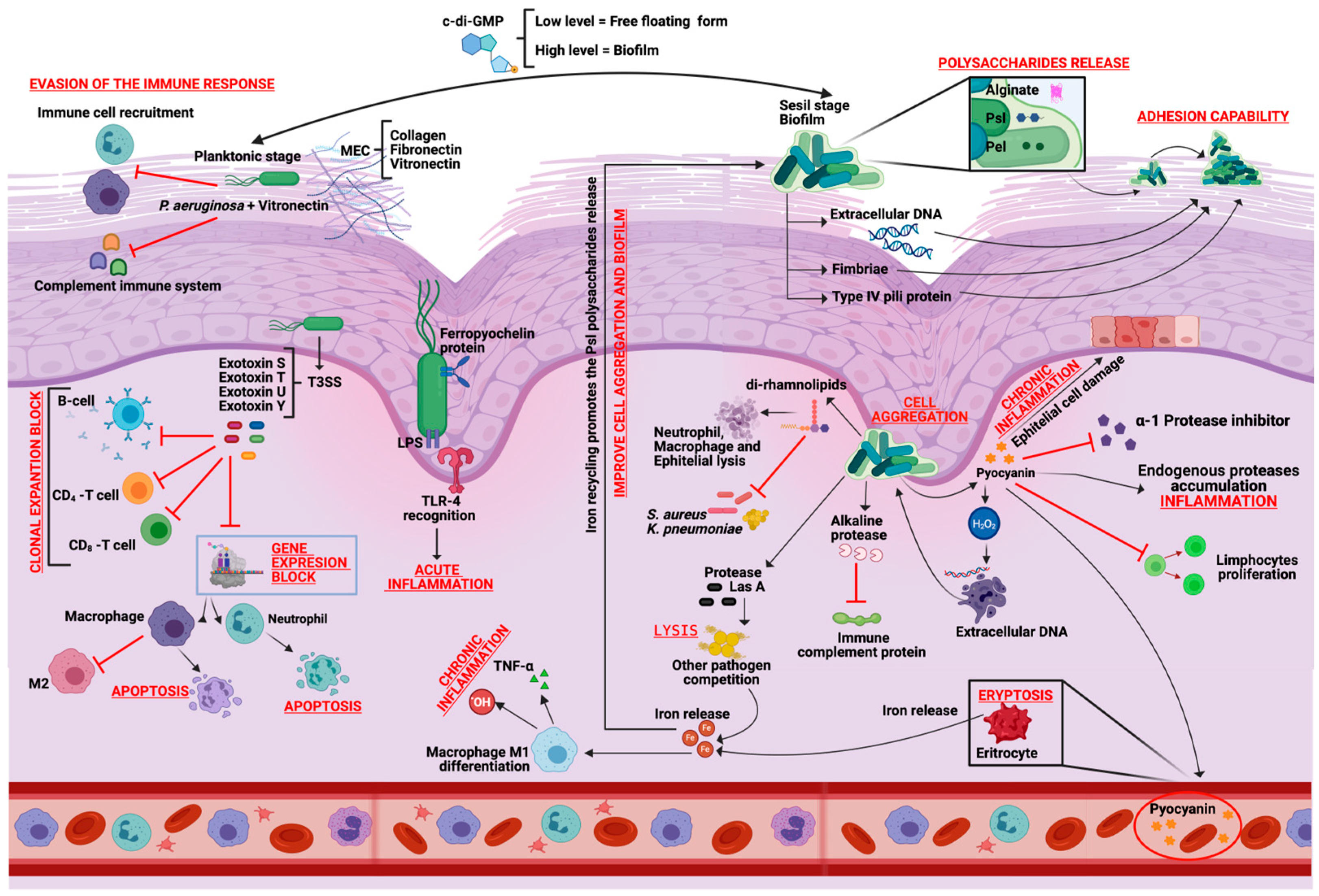
Figure 1. Pathogenicity of Pseudomonas aeruginosa in wounds healing process.
Ijms 26 09677 g001
Figure 2. Normal immune response to Pseudomonas aeruginosa.
Ijms 26 09677 g002
Figure 3. Interactions of Pseudomonas aeruginosa with commensal and pathogenic bacteria.
Ijms 26 09677 g003
Table 2. Cells and molecules interacting between Pseudomonas aeruginosa and the immune system.
CellsSecreted MoleculesDescriptionReference
Immune System
M1
macrophages
Pro-inflammatory cytokinesTumor Necrosis Factor (TNFα)Cytokine released immediately after any damage by exposure to bacterial LPS. Acts through two transmembrane receptors: TNF receptor 1 (TNFR1) induces programmed cell death and TNF receptor 2 (TNFR2) is responsible for cell proliferation. Therefore, depending on the cell type, TNFR1 and TNFR2 may have distinct roles in signal transduction and gene expression.[58]
IL-6Interleukin that promotes T and B lymphocytes differentiation and maturation, stimulates immunoglobulin release by B cells and pro-inflammatory cytokines inhibition such as TNF-α that participates in the macrophage M1 to M2 maturation.[59]
IL-12Interleukin that activates T CD4+ (H1) type 1 cells and stimulates the NK cells and T CD8+ lymphocytes production.[60]
ROS/NOSReactive oxygen and nitrogen species that cause significant cell structures damage causing cell lysis.[61]
M2
macrophages
Anti-inflammatory cytokinesTGF-βInterleukin that protects the collagen expression by some protease activity inhibition.[62]
IL-14Interleukin that inhibits the pro-inflammatory cytokines synthesis such as TNF-alpha and IL-6.[60]
IL-10Interleukin that inhibits the pro-inflammatory cytokines synthesis such as IFN-γ, IL-2, IL-3, and TNFα.[60]
ArginaseEnzyme responsible for NOS synthesis regulation and tissue regeneration.[62]
NeutrophilsElastasesProtease that is released as a defense mechanism to remove NOS and ROS tissue degradation products.[63]
Pseudomonas aeruginosaExotoxin AModifies macrophage gene expression and inhibits the maturation of M1 macrophages to M2.[56]
AlginateInhibits bacterial uptake during phagocytosis in Macrophages and Neutrophils.[34]
Exotoxin AModify gene expression to cause apoptosis in Neutrophils.[56]
Table 3. Molecular and cellular interactions during wound healing process in the P. aeruginosa presence.
Pseudomonas
aeruginosa Secreted Molecules
Cells
Interaction with
DescriptionReference
QuinolonesStaphylococcus epidermidisMolecule packaged in extracellular
membrane vesicles (MVs) to cause cell
lysis.
[67]
Pel and PsIBoth acts as dispersing agents to inhibit biofilm formation and adhesion.[67]
3-Oxo-C12-HSLInhibits the bacterial growth and EPS
secretion that hinders the initial adhesion and the biofilms formation.
[67]
4-hydroxy-2-heptylquinoline N-oxide (HQNO)Staphylococcus aureusQuinolone signal system component acts as an inhibitor of S. aureus electron transport chain (ETC). Prolonged exposure to this compound leads to the small colony variants selection.[68]
PyocyaninIncreases the H2O2 formation and leads to cell lysis.[68]
Las AProtease that lyses S. aureus cells.[68]
PyocyaninStreptococcus spp.Increases the H2O2 formation and leads to cell lysis.[69]
Acyl homoserine
lactone (AHL)
Streptococcus pyogenesModifies hemolytic activity and reduces pathogenicity of S. pyogenes.[70]
Disclaimer/Publisher’s Note: The statements, opinions and data contained in all publications are solely those of the individual author(s) and contributor(s) and not of MDPI and/or the editor(s). MDPI and/or the editor(s) disclaim responsibility for any injury to people or property resulting from any ideas, methods, instructions or products referred to in the content.