Article

Missense Variants in Nutrition-Related Genes: A Computational Study

by
Giovanni Maria De Filippis
1,†,
Maria Monticelli
2,3,†,
Bruno Hay Mele
2,* and
Viola Calabrò
2
1
Department of Electrical Engineering and Information Technology, University of Naples Federico II, Naples, via Claudio 21, 80125 Napoli, Italy
2
Biology Department, University of Naples Federico II, Complesso Universitario Monte Sant’Angelo, Via Cinthia, 80126 Napoli, Italy
3
Institute of Biomolecular Chemistry ICB, Consiglio Nazionale delle Ricerche (CNR), Via Campi Flegrei 34, 80078 Pozzuoli, Italy
*
Author to whom correspondence should be addressed.
These authors contributed equally to this work.
Int. J. Mol. Sci. 2025, 26(19), 9619; https://doi.org/10.3390/ijms26199619
Submission received: 5 September 2025 / Revised: 29 September 2025 / Accepted: 30 September 2025 / Published: 2 October 2025

Abstract

Genetic variants in nutrition-related genes exhibit variable functional consequences; however, systematic characterization across different nutritional domains remains limited. This highlights the need for detailed exploration of variant distribution and functional effects across nutritional gene categories. Therefore, the main objective of this computational study is to delve deeper into the distribution and functional impact of missense variants in nutrition-related genes. We analyzed Genetic polymoRphism variants using Personalized Medicine (GRPM) dataset, focusing on ten groups of nutrition-related genes. Missense variants were characterized using ProtVar for functional/structural impact, Pharos for functional classification, network analysis for pathway identification, and Gene Ontology enrichment for biological process annotation. The analysis of 63,581 Single Nucleotide Polymorphisms (SNP) revealed 27,683 missense variants across 1589 genes. Food intolerance (0.23) and food allergy (0.15) groups showed the highest missense/SNP ratio, while obesity-related genes showed the lowest (0.04). Enzymes predominated in xenobiotic and vitamin metabolism groups, while G-protein-coupled receptors were enriched in eating behavior genes. The vitamin metabolism group had the highest proportion of pathogenic variants. Network analysis identified apolipoproteins as central hubs in metabolic groups and inflammatory proteins in allergy-related groups. These findings offer insights into personalized nutrition approaches and underscore the utility of computational variant analysis in elucidating gene-diet interactions.

1. Introduction

Understanding the relationship between genetic variation and nutrition-related phenotypes is fundamental to advancing personalized medicine and nutrition. Genetic variants affecting nutritional metabolism, food tolerance, and dietary response exhibit highly variable functional consequences, yet systematic characterization of these effects across different nutritional domains remains limited.
The evolutionary perspective provides crucial insights into nutrition-related genetic variation. Many variants implicated in food intolerance, allergy, and metabolic disorders represent common polymorphisms shaped by historical selective pressures rather than recent deleterious mutations. This observation raises a fundamental question: why do disease-associated variants persist at appreciable frequencies in human populations? Evidence suggests these variants often represent evolutionary trade-offs, where historical advantages balanced current risks. For example, alleles in the APOL1 gene, which confer resistance to Trypanosoma brucei infection, also predispose carriers to kidney disease [1]. Similarly, immune-related genes such as HLA, IL4, or chemokine receptors (CCR3, CXCR5), frequently implicated in food allergies, are known to be subject to balancing selection driven by historical pathogen exposures [2]. In these cases, variants that may today contribute to allergy or autoimmunity could have conferred survival benefits in infectious environments, explaining their persistence and relatively high frequencies [3]. This pattern contrasts with the evolutionary dynamics of rare, highly deleterious variants—such as those causing familial hypercholesterolemia—which are efficiently removed from the population by purifying selection [4]. As such, a broader principle emerges: variants associated with immune-mediated food responses or micronutrient handling are often evolutionarily tolerated, either because they posed minimal reproductive cost or because they were once adaptive. Mechanistically, these variants often act not by abolishing protein function outright, but by subtly modifying protein stability, folding, or regulation.
Missense mutations in enzymes involved in vitamin metabolism, for example, frequently impair conformational stability rather than active site chemistry [5,6]. Structural studies and protein modeling have shown that such mutations can destabilize the native fold, reduce thermal tolerance, or impair subunit interactions—leading to partial loss-of-function. This insight has guided the development of pharmacological chaperones for some inborn errors of metabolism, illustrating the therapeutic relevance of structural interpretation [7,8]. Likewise, G-protein-coupled receptors (GPCRs)—which mediate taste perception, gut–brain hunger signaling, and food reward—harbor common variants affecting individual differences in dietary preference and metabolic response. Taste receptor polymorphisms (e.g., T1R2/T1R3, TAS2R) influence sweetness or bitterness perception, while genetic variation in dopaminergic or opioid GPCRs can modulate the hedonic response to food, potentially contributing to compulsive eating or obesity [9,10].
The integration of omics technologies has transformed our understanding of complex biological systems, enabling large-scale investigations across genomics, proteomics, and metabolomics [11,12]. In the context of personalized nutrition, genetic data are key to uncovering how individual variability influences dietary responses and health outcomes. Progress in this field increasingly depends on the availability of curated, interoperable datasets that bridge genetics and nutrition. One such resource is GRPM (Gene-RsID-PMID-MeSH), a publicly available dataset of genetic polymorphisms associated with nutrition-related traits (https://zenodo.org/records/14052302, accessed on 31 March 2025), constructed through the integration of multiple data sources and structured using the MeSH ontology [13]. Its standardized, ontology-driven design facilitates systematic analysis of gene–diet interactions and supports the development of targeted interventions in nutrigenetics and personalized nutrition.
Despite this progress, a comprehensive understanding of how missense variants impact nutrition-related genes remains limited. We hypothesize that genes involved in different nutritional processes exhibit distinct patterns of evolutionary constraint and functional variation, reflecting their varying biological importance and the selective pressures they have experienced. Therefore, the main objective of this computational study is to delve deeper into the distribution and functional impact of missense variants in nutrition-related genes.
We developed a comprehensive computational pipeline (Figure 1) that integrates multiple data sources and analytical approaches. Our specific objectives were to (1) quantify the distribution of missense variants across different nutrition-related gene groups; (2) assess the functional and structural impact of these variants using computational prediction tools; (3) identify functional patterns and protein classes enriched in each category; and (4) characterize protein interaction networks and biological pathways associated with each gene group.

2. Results

2.1. Missense/SNP Ratio and Functional Constraint

Our analysis included 63,581 Single Nucleotide Polymorphisms from the GRPM dataset, of which approximately 40% were successfully mapped to ProtVar for detailed structural and functional predictions. This mapping yielded 27,683 missense variants across 1589 unique genes distributed among the ten defined nutritional categories: (1) Cardiovascular Health and Lipid Metabolism (CHLM), (2) Diabetes Mellitus Type II and Metabolic Syndrome (DM&MS), (3) Diet-induced Oxidative Stress (DiOS), (4) Eating Behavior and Taste Sensation (EBTS), (5) Food Allergies (FAs), (6) Food Intolerances (FIs), (7) General Nutrition (GN), (8) Obesity, Weight Control and Compulsive Eating (OWG&CE), (9) Vitamin and Micronutrient Metabolism and Deficiency-Related Diseases (VMM&DRD), and (10) Xenobiotic Metabolism (XM) (Table 1). The complete list of genes containing the rsIDs analyzed is available as Supplementary Material (File S1). A comprehensive description of the ten nutritional categories is available in [13].
The distribution of variants across the ten nutritional categories revealed significant differences in missense/SNP ratios, ranging from 0.04 to 0.23 (Table 1). Food Intolerances showed the highest ratio (0.23), followed by Food Allergies (FAs, 0.15), suggesting these gene groups tolerate more missense variation. Conversely, Obesity, Weight Control and Compulsive Eating exhibited the lowest ratio (0.04), indicating stronger evolutionary constraints.

2.2. Variant Composition Across Nutritional Gene Groups

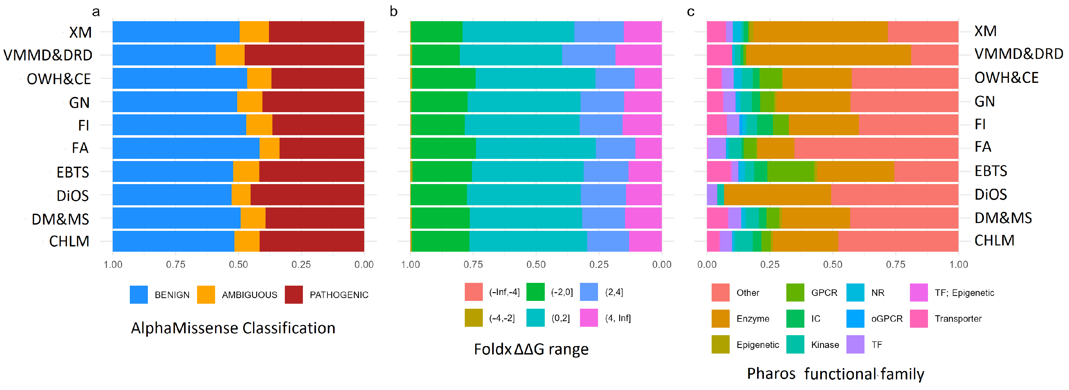
The analysis of the relative distribution of functional families associated with missense variants revealed a predominance of enzymes in the Xenobiotic Metabolism and Vitamin and Micronutrients Metabolism and Deficiency-Related Diseases groups. The Eating Behavior and Taste Sensation group showed a clear prevalence of GPCRs, while an unexpected predominance of “other” protein classes was observed in the Food Allergies group (Figure 2).
An evaluation of the distribution of variants based on predicted destabilization effects showed a generally homogeneous pattern between groups, with a slight increase in highly destabilizing mutations within the Vitamin and Micronutrient Metabolism and Deficiency-Related Diseases category (Figure 2). Very few highly stabilizing mutations seem to be predicted. This same group also showed the highest number of variants classified as “pathogenic” (Figure 2). The number of Variants of Uncertain Significance (VUS) was extremely small in all groups.

2.3. Network and Gene Ontology Analysis

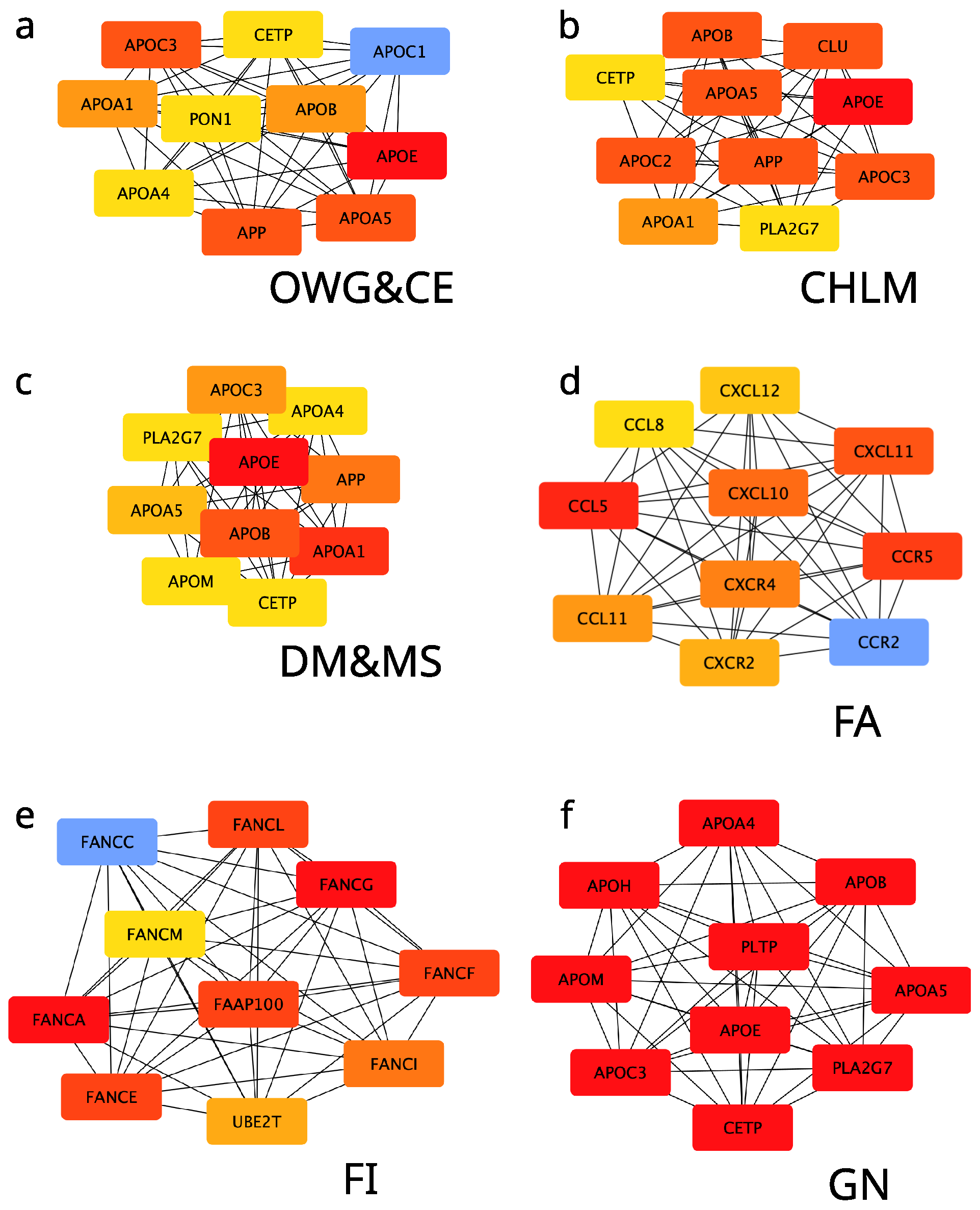
Protein–protein interaction (PPi) network analysis revealed that four out of ten groups are mainly composed of singletons and form few, scarcely populated clusters, while the other six produced dense clusters. Identification of central nodes (hubs) for genes implied in each group revealed interesting results (Figure 3). Apolipoproteins (APOs) were identified as central nodes in the non-food-related groups: Obesity, Weight Control and Compulsive Eating (panel a); Cardiovascular Health and Lipid Metabolism (panel b); Diabetes Mellitus Type II and Metabolic Syndrome (panel c); and General Nutrition (panel d). Notably, the General Nutrition group, broad yet less specific, still highlighted APOs as key players, emerging as a cross-category encompassing diverse aspects of other groups. Food allergies were mainly related to inflammatory state proteins (panel d), while the Food Intolerances group was surprisingly characterized by the centrality of Fanconi anemia (FA) proteins. The four out of ten groups comprising a low amount of proteins and showing many isolated nodes or pairs did not lead to a hub search (Supplementary Figure S1).
Gene Ontology (GO) analysis revealed overlapping patterns, as expected from network analysis (Additional Table A1). Cellular components (CC) GO as significantly enriched by far in extracellular context (i.e., extracellular space, extracellular region, plasma membrane region, external side of plasma membrane, and vescicle) in all the analyzed groups. Most of the biological processes (BPs) could be subsets of three major groups: (i) involvement in homeostasis (i.e., cellular homeostasis and homeostatic process); (ii) specialized metabolism (modified amino acid metabolic process; nitric oxide synthase biosynthetic process; organic acid metabolic process; secondary alcohol metabolic process; small molecule metabolic process; and vitamin metabolic process); and (iii) response to external stimuli (i.e., response to xenobiotic stimulus, response to chemical, response to interleukine-15, response to oxygen-containing compound, response to stress, and response to toxic substance). Interestingly, molecular functions (MFs) were largely related to molecule binding (cytokine binding, cytokine receptor binding, hormone binding, identical protein binding, lipid binding, signaling receptor binding, small molecule binding, tetrapyrrole binding, and vitamin binding). Besides this, a significant involvement of oxidoreductase and antioxidant activity was observed.

3. Discussion

This study provides an integrated, large-scale computational examination of missense variants in nutrition-related genes, focusing on evolutionary constraint and functional impact across ten systematically defined nutritional categories. These categories balance breadth (general nutrition, metabolic disease) with specificity (food intolerances, eating behavior, oxidative stress), ensuring clinical and mechanistic relevance (see [13] for methodological detail).
At the outset, this study contributes in three complementary ways: (1) it presents the first large-scale comparative analysis of missense variation across ten categories of nutrition-related genes, with systematic evaluation of functional consequences; (2) it integrates multiple computational pipelines (structural/functional predictions, network centrality, enrichment analyses) to generate a comprehensive, multi-level variant impact map; and (3) it offers a bioinformatics resource that can support personalized nutrition, disease risk stratification, and experimental follow-up, aligning with precision medicine initiatives. Together, these contributions establish novelty, scope, and translational relevance for the ensuing interpretation of constraint and function.
Our comparative findings reveal stratified tolerance to missense variation: genes implicated in food allergies and intolerances display higher missense/SNP ratios (0.15–0.23), consistent with relaxed constraint or diversifying pressures, whereas genes central to core metabolic homeostasis (e.g., obesity and cardiovascular-related pathways) show markedly lower ratios (0.04–0.08), indicative of stronger purifying selection. This gradient supports the hypothesis that distinct nutritional gene subclasses have been shaped by heterogeneous selective regimes across human evolution and provides a functional scaffold for prioritizing variants for experimental or clinical follow-up.

3.1. Variant Classification and Evolutionary Insights

The number of VUS was extremely small in all groups, highlighting the effectiveness of the GRPM method in variant classification. Such a low prevalence of VUS underscores the reliability and precision of the GRPM approach, as it successfully categorizes variants with clear effects, minimizing ambiguity in classification.
Interestingly, in our analysis, gene groups related to food allergies and food intolerances exhibited the highest missense-to-SNP ratios (0.15 and 0.23, respectively), consistent with previous findings suggesting that immune and digestive system genes are more tolerant of common variation. This likely reflects historical balancing selection or environmental adaptation [3]. In contrast, genes associated with cardiometabolic traits—such as cardiovascular disease and type 2 diabetes—showed lower missense-to-SNP ratios, suggesting stronger evolutionary constraints. The severe consequences of dysfunction in these pathways likely lead to stronger purifying selection, reducing the prevalence of deleterious variants in the population [4]. Notably, missense variants implicated in food allergies were generally not shared with other disease categories, supporting their relative specificity. The especially low missense/SNP ratio observed in the “Obesity, Weight Control, and Compulsive Eating” category further underscores the influence of selective pressures in shaping gene variant distributions.
The high frequency of missense variants in genes related to food intolerance and allergy likely reflects reduced reproductive penalties or even potential evolutionary advantages, such as improved pathogen resistance or dietary adaptability. Conversely, variants causing severe cardiometabolic disease have remained rare due to the fitness costs they impose.
These findings underscore the non-uniform distribution of variant tolerance across nutrition-related pathways and suggest that genes involved in food perception and immune interaction have evolved under different selective regimes than those governing core metabolic and cardiovascular processes.

3.2. Inflammation and Cardiovascular Risks from Food Allergies

While most food allergy-related variants appear to be relatively benign, our analysis found that food intolerance-associated variants may intersect with pathways implicated in cardiovascular disease. Food allergies are mediated by abnormal immune responses to specific dietary proteins. Chronic exposure to allergens can lead to persistent inflammation, in which the CCL and CXCR chemokine families play a central role [16]. This prolonged inflammatory state may extend systemically, influencing cardiovascular health.
Chronic inflammation is a known driver of atherosclerosis and endothelial dysfunction, with cytokines such as IL-1 β , IL-6, and TNF- α , and chemokines like CCL2/MCP-1 promoting immune cell infiltration and plaque formation [17]. A 2017 review by Castan et al. highlighted the role of chemokine networks in orchestrating immune responses in food allergy, asthma, and atopic dermatitis [16]. A 2024 longitudinal study (NHANES and MESA cohorts) found that individuals sensitized to common foods exhibited significantly increased cardiovascular mortality, particularly those with IgE sensitization to milk (HR 2.0–3.8) [18]. These findings support a systemic model of food allergy pathophysiology, wherein sustained immune activation contributes to vascular inflammation and cardiovascular risk.

3.3. Enzymes, GPCRs, and Eating Behavior

In our dataset, enzymes were prominently represented in the category of vitamin and micronutrient metabolism. Missense mutations in this group predominantly caused mild to severe enzyme destabilization, consistent with prior mechanistic studies. GPCRs were highly enriched in categories related to taste sensation and eating behavior, reflecting their established role in detecting food-derived chemical cues and modulating physiological and emotional responses. T1R and T2R GPCRs mediate sweet, umami, and bitter taste perception, influencing food preferences and aversions [9]. Taste receptors are expressed in several extra-oral tissues, where they play important physiological roles [19], including the respiratory system [20], the gastrointestinal tract [21], and reproductive tissues, especially testis [22]. Beyond sensory input, other GPCRs regulate hunger and satiety through pathways involving ghrelin and leptin, and also participate in brain reward systems, such as dopamine signaling [23,24,25,26]. Studies such as the one from Lee and co-workers have shown that sweet taste receptors (T1R2/T1R3) integrate nutrient sensing with behavioral outputs [27]. The role of dopamine D1 and D2 receptors in the nucleus accumbens and striatum further links eating behavior with reward and reinforcement mechanisms [28,29,30]. Notably, research by Johnson and Kenny demonstrated that rats exposed to hyperpalatable diets developed compulsive eating and reward deficits akin to addiction, accompanied by downregulation of D2 receptors [31]. Similar patterns have been observed in human neuroimaging studies, where reduced striatal D2 receptor availability correlates with obesity and altered reward sensitivity. Given these broader functions, taste receptor genes may be under stronger negative selection than expected from their role in taste or food intolerance alone.

3.4. Fanconi Anemia Proteins and Food Intolerance

One of the most intriguing findings of our analysis is the central role of Fanconi anemia (FA) proteins in food intolerance—and to a lesser extent, food allergy. FA proteins are primarily known for their role in DNA repair and genome stability. In patients with Fanconi anemia, poor digestive tolerance has been documented and may underlie the observed association with food intolerance [32]. Moreover, the underlying defect in the DNA repair system is known to predispose individuals to different cancer types, including gastrointestinal malignancies [33,34,35]. Although not directly linked to dietary responses, we hypothesize that exposure to intolerance-inducing foods may cause cellular stress or damage, triggering DNA repair pathways. UBE2T, which mono-ubiquitinates FANCD2, is a key activator of the FA pathway, facilitating the repair of DNA interstrand crosslinks. Furthermore, intestinal barrier dysfunction in food intolerance may involve epithelial stress responses where FA proteins contribute to cell maintenance. It is also possible that FA proteins participate in modulating immune function, given the immune component of food intolerance. While the precise mechanism remains unclear, these observations warrant further investigation into the role of FA genes in gut-immune homeostasis and food-related immune responses.

3.5. Role of Apolipoproteins in Metabolic Health

Lastly, apolipoproteins (APOs) emerged as critical hubs linking obesity, cardiovascular disease, and general nutrition. APO variants influence lipid metabolism, impacting how lipids are absorbed, transported, and cleared from the bloodstream [36]. Due to their central metabolic roles, APO genes exhibit high natural variability, likely shaped by evolutionary pressures such as dietary shifts or infectious disease exposure. For instance, the ApoE gene has three common alleles ( ε 2 , ε 3 , ε 4 ), each associated with different disease risks [37]. While the ε 4 allele increases cardiovascular and Alzheimer’s disease risk [38,39], it may have offered adaptive benefits in certain ancestral environments [40]. Similarly, the APOL1 risk alleles—common in African populations—confer resistance to Trypanosoma infection at the cost of increased kidney disease risk [41]. These examples illustrate the trade-offs inherent in human genetic diversity. Personalized approaches, such as tailoring diets based on APOE genotype, highlight the potential clinical utility of understanding this variability. Moreover, the rarity of highly pathogenic alleles in core metabolic genes (e.g., LDLR mutations in familial hypercholesterolemia) reinforces the principle that life-threatening variants are tightly constrained by natural selection.

3.6. Implications for Precision Nutrition and Personalized Medicine

Together, these findings illustrate the complex interplay of genetic variation, evolutionary pressure, and disease risk across nutritionally relevant genes. Genes involved in food perception, immune interaction, and metabolism exhibit distinct patterns of variation shaped by both historical and modern environments. This has significant implications for precision nutrition and personalized medicine.
Our key findings demonstrate that (1) genes involved in food allergies and intolerances exhibit higher tolerance to missense variation, likely reflecting historical balancing selection; (2) functional protein classes are differentially enriched across categories, with enzymes predominating in metabolic groups and GPCRs in sensory/behavioral groups; and (3) network analysis identifies apolipoproteins as central hubs in multiple metabolic categories. These findings contribute to our understanding of the functional landscape of nutrition-related genetic variation and provide a foundation for developing more precise and effective personalized nutrition interventions. The differential patterns of variant tolerance across nutritional categories suggest that genetic testing strategies should be tailored to the specific nutritional domain of interest. For instance, screening for variants in food allergy and intolerance genes may require different interpretation criteria than variants in core metabolic pathways. The identification of apolipoproteins as central network hubs across multiple metabolic categories highlights their potential as targets for nutrigenetic interventions.

3.7. Limitations and Future Directions

While our study provides valuable insights into nutrition-related genetic variation, several limitations should be acknowledged. The GRPM dataset aggregates genetic variants identified across diverse studies and populations, thereby capturing stable genetic polymorphisms that are not influenced by age or sex stratification and are reported as. However, interpreting their functional and clinical implications in specific populations may require consideration of demographic factors. The analysis was limited to variants with available rsIDs mappable to ProtVar, which may introduce selection bias. Future studies should incorporate population-specific variant databases and experimental validation of computational predictions. Additionally, the functional consequences of variants should be validated through in vitro and in vivo studies to confirm the computational predictions presented here.

4. Materials and Methods

4.1. Study Design and Data Source

This computational study employed a cross-sectional analysis of genetic variants using publicly available databases. We utilized the GRPM dataset, a curated collection of genetic polymorphisms associated with nutrition-related traits [13]. This dataset integrates multiple data sources and is structured using the Medical Subject Headings (MeSH) ontology to facilitate systematic analysis of gene–diet interactions.
From the GRPM dataset, we selected variants annotated with reference SNP cluster ID (rsID) numbers for comprehensive analysis. The dataset categorizes genes into ten nutrition-related groups as defined in [13]: (1) Cardiovascular Health and Lipid Metabolism, (2) Diabetes Mellitus Type II and Metabolic Syndrome, (3) Diet-induced Oxidative Stress, (4) Eating Behavior and Taste Sensation, (5) Food Allergies (FAs), (6) Food Intolerances, (7) General Nutrition, (8) Obesity, Weight Control and Compulsive Eating, (9) Vitamin and Micronutrient Metabolism and Deficiency-Related Diseases, and (10) Xenobiotic Metabolism. These categories reflect clinically and nutritionally relevant pathways, including nutrition physiology, nutrition-related diseases, disease prevention through diet, and eating behavior. For more details, we refer the reader to [13].

4.2. Data Retrieval and Annotation

We wrote an R script to download and unzip the zenodo record associated with the core dataset (https://zenodo.org/records/14052302, accessed on 31 March 2025). The dataset was imported into R using the read_parquet() function. From these datasets we extracted the list of unique reference SNP identifiers (rsids), which serve as variant makers. To annotate these SNPs, we utilized the Ensembl BioMart interface via the biomaRt package. We queried the hsapiens_snp dataset within the Ensembl SNP mart to retrieve variant-level annotations including rsid, chromosomal position, and alleles. We focused on variants for which the consequence_type_tv was “missense_variant” (i.e., variants that have the potential to alter protein structure/function through amino acid substitution). We enriched the SNP list by manually uploading the rsid list to the ProtVar web platform (https://www.ebi.ac.uk/ProtVar/, accessed on 31 March 2025) [42], which provides comprehensive variant effect predictions including structural and functional annotations. Using ProtVar’s “Mapping with Annotations” functionality, we generated an annotated dataset that includes a variety of functional and structural annotations relevant to the protein context of each variant. Finally, to facilitate subsequent integration and processing, the ProtVar-annotated CSV file was converted to Parquet format and saved for subsequent analyses.

4.3. Functional Classification and Pathway Analysis

Functional categorization of proteins containing missense variants was performed using Pharos version 3.19 (https://pharos.nih.gov/, accessed on 31 March 2025), which classifies proteins based on their therapeutic relevance and functional properties. A custom GraphQL interface was used to query Pharos based on gene symbols. The data retrieved contained functional family annotations, including enzymes, GPCRs, ion channels, kinases, transcription factors, and other less-characterized categories. These classifications were used to stratify the variants based on their likely biological roles and potential therapeutic relevance.

4.4. Structural Impact Prediction

Structural impact of missense variants was assessed using complementary metrics retrieved from the protvar query: AlphaMissense pathogenicity scores and FoldX Δ Δ G energy perturbation predictions. In the first case (alphamissense), variants were classified as pathogenic, likely pathogenic, VUS, likely benign, or benign based on AlphaMissense scores. Alphamissense is based on a deep learning model trained on evolutionary and clinical variant data [14]. As for FoldX, it estimates the changes in Gibbs free energy caused by the substitution, where higher-magnitude changes denote protein destabilization. Analyses were performed separately, and in both cases, variants were retained only if the specific score was available. In both cases, values were binned to reflect increasing levels of perturbation (pathogenicity or stability).

4.5. Network Analysis

Protein–protein interaction networks were constructed using the STRING database (https://string-db.org/, accessed on 31 March 2025) to identify hub proteins and functional clusters within each gene group. Network analysis was performed using Cytoscape software (version 3.10.4) with the CytoHubba plugin, employing the MCC algorithm to identify highly connected, essential nodes within scale-free networks. We used Cytoscape [43] to further investigate the genes associated with each topic. The STRINGdb plugin [44] was used to explore high confidence (Score > 0.7) protein–protein physical interaction networks for each of the ten nutrigenetic fields. To identify central proteins, we applied the CytoHubba plugin [45] within CytoScape, using the MCC algorithm. The top ten ranked hub proteins were selected for further interpretation. Maximal Clique Centrality ranks a node by how many fully connected “cliques” it belongs to—boosting the score further when those cliques are large—so the nodes sitting in the center of numerous, tightly knit groups stand out. A clique is a group where every member is directly connected to every other member.

4.6. Gene Ontology Analysis

To further explore the biological implications of the variant-bearing genes, we performed Gene Ontology (GO) enrichment analysis. This included overrepresentation testing for biological process, molecular function, and cellular component terms. GO terms enriched within each topical gene group helped highlight common pathways, cellular compartments, or enzymatic functions that may be particularly relevant to nutrient response or metabolism.

4.7. Statistical Analysis

For each gene group, we calculated the missense/SNP ratio as an indicator of evolutionary constraint and functional tolerance. We quantified the total number of SNPs, number of missense variants, and number of affected genes for each category. These metrics were compiled into a summary table and visualized for comparative interpretation. All computational analyses were performed using standard bioinformatics tools and databases as described above.
All code, including dataset handling, annotation parsing, and visualization routines, was implemented in R using a combination of tidyverse [46], jsonlite [47], arrow [48], httr [49] and GraphQL APIs (for Pharos). Visualization for non-network-related data was performed using ggplot2. R scripts are available as Supplementary Material (File S2) and in the project repository (https://github.com/johndef64/grpm_missense, accessed on 31 March 2025).
All queries were performed throughout the month of March 2025.

5. Conclusions

This comprehensive computational analysis of missense variants in nutrition-related genes reveals distinct evolutionary and functional patterns across different nutritional categories. Genes involved in food allergies and intolerances show increased tolerance to missense variation, and functional protein classes are differentially enriched across categories. Enzymes predominate in metabolic groups, and G protein-coupled receptors in sensory/behavioral groups. In our study, network analysis identifies apolipoproteins as central hubs in multiple metabolic categories.
The computational framework presented here demonstrates the value of integrating multiple data sources and prediction tools for comprehensive variant analysis in nutrigenetics research.

Supplementary Materials

The following supporting information can be downloaded at https://www.mdpi.com/article/10.3390/ijms26199619/s1.

Author Contributions

Conceptualization, B.H.M. and G.M.D.F.; methodology, B.H.M. and G.M.D.F.; software, B.H.M. and G.M.D.F.; formal analysis, B.H.M. and G.M.D.F.; data curation, B.H.M.; writing—original draft preparation, B.H.M. and M.M.; writing—review and editing, G.M.D.F. and V.C.; visualization, B.H.M. and M.M.; supervision, B.H.M. and V.C. All authors have read and agreed to the published version of the manuscript.

Funding

This research received no external funding.

Institutional Review Board Statement

Not applicable.

Informed Consent Statement

Not applicable.

Data Availability Statement

The original contributions presented in this study are included in the article/Supplementary Materials. Further inquiries can be directed to the corresponding author(s).

Conflicts of Interest

The authors declare no conflicts of interest.

Appendix A

Table A1. Gene ontology analysis (top three terms per each category within each nutrigenetic group). Group abbreviation as per (Table 1).
GroupGO CategoryGO Term IDGO Term Namep-Value
CHLMGO:MFGO:0005102Signaling receptor binding 5.79 × 10 51
CHLMGO:MFGO:0042802Identical protein binding 3.125 × 10 49
CHLMGO:MFGO:0097367Carbohydrate derivative binding 3.02 × 10 23
CHLMGO:BPGO:1901700Response to oxygen-containing compound 1.32 × 10 137
CHLMGO:BPGO:0044281Small molecule metabolic process 1.06 × 10 67
CHLMGO:BPGO:0030198Extracellular matrix organization 1.79 × 10 23
CHLMGO:CCGO:0071944Cell periphery 6.39 × 10 57
CHLMGO:CCGO:0034358Plasma lipoprotein particle 4.02 × 10 26
CHLMGO:CCGO:0005739Mitochondrion 3.99 × 10 19
DM&MSGO:MFGO:0005102Signaling Receptor Binding 2.77 × 10 25
DM&MSGO:MFGO:0008289Lipid binding 3.40 × 10 19
DM&MSGO:MFGO:0042802Identical Protein Binding 5.61 × 10 19
DM&MSGO:BPGO:1901700Response to oxygen-containing compound 2.07 × 10 101
DM&MSGO:BPGO:0042592Homeostatic process 3.26 × 10 97
DM&MSGO:BPGO:0010876Lipid localization 6.7 × 10 68
DM&MSGO:CCGO:0005615Extracellular space 1.40 × 10 36
DM&MSGO:CCGO:0005783Endoplasmic reticulum 1.75 × 10 26
DM&MSGO:CCGO:0045177Apical part of cell 1.87 × 10 25
GNGO:MFGO:0042802Identical protein bidning 3.68 × 10 29
GNGO:MFGO:0005102Signaling receptor binding 1.04 × 10 27
GNGO:MFGO:0046906Tetrapyrrole binding 2.51 × 10 19
GNGO:BPGO:0042221Response to chemical 9.83 × 10 106
GNGO:BPGO:0003013Circulatory system process 5.89 × 10 60
GNGO:BPGO:0019216Regulation of lipid metabolic process 7.08 × 10 46
GNGO:CCGO:0005615Extracellular space 1.05 × 10 35
GNGO:CCGO:0012505Endomembrane system 1.88 × 10 32
GNGO:CCGO:0045177Apical part of cell 1.39 × 10 23
DiOSGO:MFGO:0016209Antioxidant activity 4.015 × 10 23
DiOSGO:MFGO:0016491Oxidoreductase activity 1.66 × 10 15
DiOSGO:BPGO:0009636Response to toxic substance 1.85 × 10 29
DiOSGO:BPGO:0006950Response to stress 6.71 × 10 29
DiOSGO:BPGO:0019725Cellular homeostasis 1.52 × 10 14
DiOSGO:CCGO:0005615Extracellular space 2.1 × 10 13
EBTSGO:MFGO:0008527Taste receptor activity 1.10 × 10 28
EBTSGO:MFGO:0004930G protein-coupled receptor activity 3.55 × 10 14
EBTSGO:BPGO:0044281Small molecule metabolic process 2.31 × 10 45
EBTSGO:BPGO:0042221Response to chemical 8.87 × 10 44
EBTSGO:BPGO:0007631Feeding behavior 7.97 × 10 11
EBTSGO:CCGO:0071944Cell periphery 1.715 × 10 12
XMGO:MFGO:0016491Oxidoreductase activity 2.95 × 10 47
XMGO:MFGO:0046906Tetrapyrrole binding 8.09 × 10 29
XMGO:MFGO:0036094Small molecule binding 4.49 × 10 15
XMGO:BPGO:0009410Responde to xenobiotic stimulus 1.32 × 10 71
XMGO:BPGO:0044281Small molecule metabolic process 2.78 × 10 64
XMGO:BPGO:0010876Lipid localization 1.29 × 10 25
XMGO:CCGO:0005789Endoplasmic reticulum membrane 6.16 × 10 23
OWG&CEGO:MFGO:0005102Signaling receptor binding 3.87 × 10 21
OWG&CEGO:MFGO:0042562Hormone binding 1.95 × 10 13
OWG&CEGO:MFGO:0120020Cholesterol transfer activity 8.205 × 10 11
OWG&CEGO:BPGO:0042592Homeostati process 4.17 × 10 64
OWG&CEGO:BPGO:1901700Response to oxygen-containing compound 2.025 × 10 62
OWG&CEGO:BPGO:1902652Secondary alcohol metabolic process 3.12 × 10 21
OWG&CEGO:CCGO:0005615Extracellular space 1.11 × 10 13
VMM&DRDGO:MFGO:0016491Oxidoreductase activity 7.25 × 10 21
VMM&DRDGO:MFGO:0019842Vitamin binding 1.65 × 10 18
VMM&DRDGO:MFGO:0046906Tetrapyrrole binding 7.76 × 10 11
VMM&DRDGO:BPGO:0006766Vitamin metabolic process 5.69 × 10 46
VMM&DRDGO:BPGO:0006575Modified amino acid metabolic process 6.58 × 10 32
VMM&DRDGO:BPGO:0006082Organic acid metabolic process 5.845 × 10 24
FAGO:MFGO:0005126Cytokine receptor binding 2.90 × 10 54
FAGO:MFGO:0140375Immune receptor activity 2.74 × 10 45
FAGO:MFGO:0019955Cytokine binding 4.60 × 10 31
FAGO:BPGO:0006955Immune response 4.41 × 10 174
FAGO:BPGO:0051767Nitric oxide synthase biosynthetic process 5.19 × 10 12
FAGO:BPGO:0070672Response to interleukine-15 5.89 × 10 11
FAGO:CCGO:0009897External side of plasma membrane 4.14 × 10 64
FAGO:CCGO:0005576Extracellular region 1.81 × 10 22
FAGO:CCGO:0045121Membrane raft 5.56 × 10 15
FIGO:MFGO:0042802Identical protein binding 3.07 × 10 16
FIGO:BPGO:0070887Cellular response to chemical stimulus 5.62 × 10 32
FIGO:BPGO:0042592Homeostatic process 2.28 × 10 29
FIGO:BPGO:0036297Interstrand cross-link repair 6.51 × 10 11
FIGO:CCGO:0043240Fanconi anemia nuclear complex 7.01 × 10 13
FIGO:CCGO:0098590Plasma membrane region 1.17 × 10 11
FIGO:CCGO:0031982Vescicle 1.19 × 10 11

References

  1. Thomson, R.; Genovese, G.; Canon, C.; Kovacsics, D.; Higgins, M.K.; Carrington, M.; Winkler, C.A.; Kopp, J.; Rotimi, C.; Adeyemo, A.; et al. Evolution of the primate trypanolytic factor APOL1. Proc. Natl. Acad. Sci. USA 2014, 111, E2130–E2139. [Google Scholar] [CrossRef] [PubMed]
  2. Chen, Q.; Abdi, A.M.; Luo, W.; Yuan, X.; Dent, A.L. T follicular regulatory cells in food allergy promote IgE via IL-4. JCI Insight 2024, 9, e171241. [Google Scholar] [CrossRef] [PubMed]
  3. Ramos, P.S.; Shedlock, A.M.; Langefeld, C.D. Genetics of autoimmune diseases: Insights from population genetics. J. Hum. Genet. 2015, 60, 657–664. [Google Scholar] [CrossRef] [PubMed]
  4. Raychaudhuri, S. Mapping rare and common causal alleles for complex human diseases. Cell 2011, 147, 57–69. [Google Scholar] [CrossRef]
  5. Pey, A.L.; Stricher, F.; Serrano, L.; Martinez, A. Predicted effects of missense mutations on native-state stability account for phenotypic outcome in phenylketonuria, a paradigm of misfolding diseases. Am. J. Hum. Genet. 2007, 81, 1006–1024. [Google Scholar] [CrossRef]
  6. Liew, S.C.; Gupta, E.D. Methylenetetrahydrofolate reductase (MTHFR) C677T polymorphism: Epidemiology, metabolism and the associated diseases. Eur. J. Med Genet. 2015, 58, 1–10. [Google Scholar] [CrossRef]
  7. Pey, A.L.; Ying, M.; Cremades, N.; Velazquez-Campoy, A.; Scherer, T.; Thöny, B.; Sancho, J.; Martinez, A. Identification of pharmacological chaperones as potential therapeutic agents to treat phenylketonuria. J. Clin. Investig. 2008, 118, 2858–2867. [Google Scholar] [CrossRef]
  8. Liguori, L.; Monticelli, M.; Allocca, M.; Hay Mele, B.; Lukas, J.; Cubellis, M.V.; Andreotti, G. Pharmacological chaperones: A therapeutic approach for diseases caused by destabilizing missense mutations. Int. J. Mol. Sci. 2020, 21, 489. [Google Scholar] [CrossRef]
  9. Morini, G. The taste for health: The role of taste receptors and their ligands in the complex food/health relationship. Front. Nutr. 2024, 11, 1396393. [Google Scholar] [CrossRef]
  10. Baik, J.H. Dopamine signaling in food addiction: Role of dopamine D2 receptors. BMB Rep. 2013, 46, 519. [Google Scholar] [CrossRef]
  11. Gutierrez Reyes, C.D.; Alejo-Jacuinde, G.; Perez Sanchez, B.; Chavez Reyes, J.; Onigbinde, S.; Mogut, D.; Hernández-Jasso, I.; Calderón-Vallejo, D.; Quintanar, J.L.; Mechref, Y. Multi omics applications in biological systems. Curr. Issues Mol. Biol. 2024, 46, 5777–5793. [Google Scholar] [CrossRef] [PubMed]
  12. Vitorino, R. Transforming clinical research: The power of high-throughput omics integration. Proteomes 2024, 12, 25. [Google Scholar] [CrossRef] [PubMed]
  13. De Filippis, G.M.; Monticelli, M.; Pollice, A.; Angrisano, T.; Hay Mele, B.; Calabrò, V. Computational strategies in nutrigenetics: Constructing a reference dataset of nutrition-associated genetic polymorphisms. J. Biomed. Inform. 2025, 167, 104845. [Google Scholar] [CrossRef] [PubMed]
  14. Cheng, J.; Novati, G.; Pan, J.; Bycroft, C.; Žemgulytė, A.; Applebaum, T.; Pritzel, A.; Wong, L.H.; Zielinski, M.; Sargeant, T.; et al. Accurate proteome-wide missense variant effect prediction with AlphaMissense. Science 2023, 381, eadg7492. [Google Scholar] [CrossRef]
  15. Worth, C.L.; Preissner, R.; Blundell, T.L. SDM—a server for predicting effects of mutations on protein stability and malfunction. Nucleic Acids Res. 2011, 39, W215–W222. [Google Scholar] [CrossRef]
  16. Castan, L.; Magnan, A.; Bouchaud, G. Chemokine receptors in allergic diseases. Allergy 2017, 72, 682–690. [Google Scholar] [CrossRef]
  17. Henein, M.Y.; Vancheri, S.; Longo, G.; Vancheri, F. The role of inflammation in cardiovascular disease. Int. J. Mol. Sci. 2022, 23, 12906. [Google Scholar] [CrossRef]
  18. Keet, C.; McGowan, E.C.; Jacobs, D.; Post, W.S.; Richards, N.E.; Workman, L.J.; Platts-Mills, T.A.; Manichaikul, A.; Wilson, J.M. IgE to common food allergens is associated with cardiovascular mortality in the National Health and Examination Survey and the Multi-Ethnic Study of Atherosclerosis. J. Allergy Clin. Immunol. 2024, 153, 471–478. [Google Scholar] [CrossRef]
  19. Lee, S.J.; Depoortere, I.; Hatt, H. Therapeutic potential of ectopic olfactory and taste receptors. Nat. Rev. Drug Discov. 2019, 18, 116–138. [Google Scholar] [CrossRef]
  20. Deshpande, D.A.; Wang, W.C.; McIlmoyle, E.L.; Robinett, K.S.; Schillinger, R.M.; An, S.S.; Sham, J.S.; Liggett, S.B. Bitter taste receptors on airway smooth muscle bronchodilate by localized calcium signaling and reverse obstruction. Nat. Med. 2010, 16, 1299–1304. [Google Scholar] [CrossRef]
  21. Rozengurt, E. Taste receptors in the gastrointestinal tract. I. Bitter taste receptors and α-gustducin in the mammalian gut. Am. J. Physiol.-Gastrointest. Liver Physiol. 2006, 291, G171–G177. [Google Scholar] [CrossRef]
  22. Luddi, A.; Governini, L.; Wilmskötter, D.; Gudermann, T.; Boekhoff, I.; Piomboni, P. Taste receptors: New players in sperm biology. Int. J. Mol. Sci. 2019, 20, 967. [Google Scholar] [CrossRef] [PubMed]
  23. DiLeone, R.J. The influence of leptin on the dopamine system and implications for ingestive behavior. Int. J. Obes. 2009, 33, S25–S29. [Google Scholar] [CrossRef] [PubMed]
  24. Gross, J.D.; Zhou, Y.; Barak, L.S.; Caron, M.G. Ghrelin receptor signaling in health and disease: A biased view. Trends Endocrinol. Metab. 2023, 34, 106–118. [Google Scholar] [CrossRef] [PubMed]
  25. Qin, J.; Cai, Y.; Xu, Z.; Ming, Q.; Ji, S.Y.; Wu, C.; Zhang, H.; Mao, C.; Shen, D.D.; Hirata, K.; et al. Molecular mechanism of agonism and inverse agonism in ghrelin receptor. Nat. Commun. 2022, 13, 300. [Google Scholar] [CrossRef]
  26. Luengo, N.; Goldfield, G.S.; Obregón, A.M. Association between dopamine genes, adiposity, food addiction, and eating behavior in Chilean adult. Front. Nutr. 2024, 11, 1466384. [Google Scholar] [CrossRef]
  27. Lee, A.A.; Owyang, C. Sugars, sweet taste receptors, and brain responses. In Molecular Nutrition: Carbohydrates; Academic Press: Cambridge, MA, USA, 2019; pp. 265–283. [Google Scholar] [CrossRef]
  28. Lex, A.; Hauber, W. Dopamine D1 and D2 receptors in the nucleus accumbens core and shell mediate Pavlovian-instrumental transfer. Learn. Mem. 2008, 15, 483–491. [Google Scholar] [CrossRef]
  29. Gagnon, D.; Petryszyn, S.; Sanchez, M.; Bories, C.; Beaulieu, J.; De Koninck, Y.; Parent, A.; Parent, M. Striatal neurons expressing D1 and D2 receptors are morphologically distinct and differently affected by dopamine denervation in mice. Sci. Rep. 2017, 7, 41432. [Google Scholar] [CrossRef]
  30. Gallo, E.F.; Meszaros, J.; Sherman, J.D.; Chohan, M.O.; Teboul, E.; Choi, C.S.; Moore, H.; Javitch, J.A.; Kellendonk, C. Accumbens dopamine D2 receptors increase motivation by decreasing inhibitory transmission to the ventral pallidum. Nat. Commun. 2018, 9, 1086. [Google Scholar] [CrossRef]
  31. Johnson, P.M.; Kenny, P.J. Dopamine D2 receptors in addiction-like reward dysfunction and compulsive eating in obese rats. Nat. Neurosci. 2010, 13, 635–641. [Google Scholar] [CrossRef]
  32. Garza, J.; Schwarzenberg, S. Chapter 4: Gastrointestinal, hepatic, and nutritional problems. In Fanconi Anemia: Guidelines for Diagnosis and Management; Fanconi Anemia Research Fund, Inc.: Eugene, Oregon, 2014; pp. 74–98. [Google Scholar]
  33. Nepal, M.; Che, R.; Zhang, J.; Ma, C.; Fei, P. Fanconi anemia signaling and cancer. Trends Cancer 2017, 3, 840–856. [Google Scholar] [CrossRef] [PubMed]
  34. Wang, W.; Baral, S.; Liu, B.; Sun, Q.; Wang, L.; Ren, J.; Tang, D.; Wang, D. FANCA facilitates G1/S cell cycle advancement, proliferation, migration and invasion in gastric cancer: FANCA’s role in G1/S cell cycle progression and beyond in GC. Acta Biochim. Et Biophys. Sin. 2024, 56, 973. [Google Scholar] [CrossRef] [PubMed]
  35. Namikawa, T.; Tanaka, T.; Utsunomiya, M.; Yokota, K.; Munekage, M.; Maeda, H.; Kitagawa, H.; Kurioka, Y.; Satake, H.; Kobayashi, M.; et al. Gastric cancer with Fanconi anemia in adolescent and young adult patient diagnosed by comprehensive genome profiling using next-generation sequencing. Clin. J. Gastroenterol. 2024, 17, 12–17. [Google Scholar] [CrossRef] [PubMed]
  36. Feingold, K.R. Introduction to Lipids and Lipoproteins. In Endotext [Internet]; MDText.com, Inc.: South Dartmouth, MA, USA, 2024. [Google Scholar]
  37. Rebeck, G.W. The role of APOE on lipid homeostasis and inflammation in normal brains: Thematic Review Series: ApoE and Lipid Homeostasis in Alzheimer’s Disease. J. Lipid Res. 2017, 58, 1493–1499. [Google Scholar] [CrossRef]
  38. Dunk, M.M.; Li, J.; Liu, S.; Casanova, R.; Chen, J.C.; Espeland, M.A.; Hayden, K.M.; Manson, J.E.; Rapp, S.R.; Shadyab, A.H.; et al. Associations of dietary cholesterol and fat, blood lipids, and risk for dementia in older women vary by APOE genotype. Alzheimer’s Dement. 2023, 19, 5742–5754. [Google Scholar] [CrossRef]
  39. Fortea, J.; Pegueroles, J.; Alcolea, D.; Belbin, O.; Dols-Icardo, O.; Vaqué-Alcázar, L.; Videla, L.; Gispert, J.D.; Suárez-Calvet, M.; Johnson, S.C.; et al. APOE4 homozygosity represents a distinct genetic form of Alzheimer’s disease. Nat. Med. 2024, 30, 1284–1291. [Google Scholar] [CrossRef]
  40. van Exel, E.; Koopman, J.J.; Bodegom, D.v.; Meij, J.J.; Knijff, P.d.; Ziem, J.B.; Finch, C.E.; Westendorp, R.G. Effect of APOE ε4 allele on survival and fertility in an adverse environment. PLoS ONE 2017, 12, e0179497. [Google Scholar] [CrossRef]
  41. Genovese, G.; Friedman, D.J.; Ross, M.D.; Lecordier, L.; Uzureau, P.; Freedman, B.I.; Bowden, D.W.; Langefeld, C.D.; Oleksyk, T.K.; Knob, A.L.U.; et al. Association of Trypanolytic ApoL1 Variants with Kidney Disease in African Americans. Science 2010, 329, 841–845. [Google Scholar] [CrossRef]
  42. Stephenson, J.D.; Totoo, P.; Burke, D.F.; Jänes, J.; Beltrao, P.; Martin, M.J. ProtVar: Mapping and contextualizing human missense variation. Nucleic Acids Res. 2024, 52, W140–W147. [Google Scholar] [CrossRef]
  43. Shannon, P.; Markiel, A.; Ozier, O.; Baliga, N.S.; Wang, J.T.; Ramage, D.; Amin, N.; Schwikowski, B.; Ideker, T. Cytoscape: A Software Environment for Integrated Models of Biomolecular Interaction Networks. Genome Res. 2003, 13, 2498–2504. [Google Scholar] [CrossRef]
  44. Doncheva, N.T.; Morris, J.H.; Gorodkin, J.; Jensen, L.J. Cytoscape StringApp: Network Analysis and Visualization of Proteomics Data. J. Proteome Res. 2019, 18, 623–632. [Google Scholar] [CrossRef]
  45. Chin, C.H.; Chen, S.H.; Wu, H.H.; Ho, C.W.; Ko, M.T.; Lin, C.Y. cytoHubba: Identifying hub objects and sub-networks from complex interactome. BMC Syst. Biol. 2014, 8, S11. [Google Scholar] [CrossRef]
  46. Wickham, H.; Averick, M.; Bryan, J.; Chang, W.; McGowan, L.D.; François, R.; Grolemund, G.; Hayes, A.; Henry, L.; Hester, J.; et al. Welcome to the tidyverse. J. Open Source Softw. 2019, 4, 1686. [Google Scholar] [CrossRef]
  47. Ooms, J. The jsonlite Package: A Practical and Consistent Mapping Between JSON Data and R Objects. arXiv 2014, arXiv:1403.2805. [Google Scholar] [CrossRef]
  48. Richardson, N.; Cook, I.; Crane, N.; Dunnington, D.; François, R.; Keane, J.; Moldovan-Grünfeld, D.; Ooms, J.; Wujciak-Jens, J.; Apache Arrow. Arrow: Integration to ’Apache’ ’Arrow’, R Package Version 19.0.1; 2025. Available online: https://CRAN.R-project.org/package=arrow (accessed on 31 March 2025).
  49. Wickham, H. httr: Tools for Working with URLs and HTTP, R Package Version 1.4.7; 2023. Available online: https://CRAN.R-project.org/package=httr (accessed on 31 March 2025).
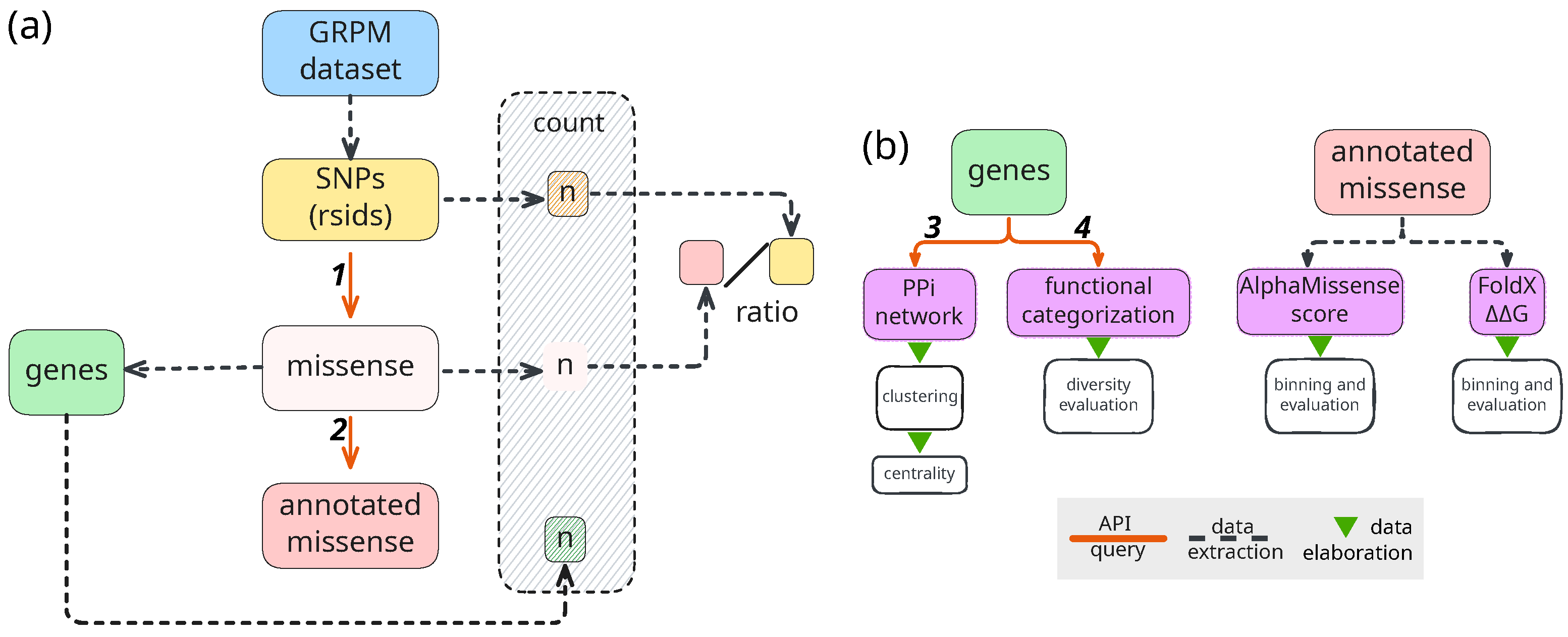
Figure 1. Schematic representation of the pipeline. (a) Data retrieval and extraction; (b) data analysis. Dashed lines mark data produced through extraction processes (i.e., subsetting and filtering files), green triangles mark data elaboration (analysis and visualization), solid orange lines mark API queries to the following services: 1. BioMart, 2. ProtVar, 3. Pharos, 4. StringDb. All services are referenced in the text. Abbreviations: GRPM: source dataset (Gene-RsID-PMID-MeSH) [13]; SNPs: Single Nucleotide Polymorphisms; PPI: protein–protein interaction.
Ijms 26 09619 g001
Figure 2. (a) Pathogenicity score distribution across nutrigentic topics. AlphaMissense classification, 0 to 0.34 means benign, 0.34 to 0.564 uncertain, 0.564 to 1 likely pathogenic [14]. (b) Energy perturbation (FoldX | Δ Δ | G) distribution, values > 2.0 kcal/mol and 4.0 kcal/mol are considered destabilizing and severely destabilizing, respectively [15]. (c) Functional family assignment distribution (Pharos annotation) within each nutrigenetic topic. Abbreviations: Cardiovascular Health and Lipid Metabolism (CHLM), Diabetes Mellitus Type II and Metabolic Syndrome (DM&MS), Diet-induced Oxidative Stress (DiOS), Eating Behavior and Taste Sensation (EBTS), Food Allergies (FAs), Food Intolerances (FI), General Nutrition (GN), Obesity, Weight Control and Compulsive Eating (OWG&CE), Vitamin and Micronutrient Metabolism and Deficiency-Related Diseases (VMM&DRD), and Xenobiotic Metabolism (XM).
Ijms 26 09619 g002
Figure 3. Top 10 hub genes identified on the richest networks by the CytoHubba plugin in Cytoscape, using the Maximal Clique Centrality algorithm. These hub genes are highly connected, essential nodes within a scale-free PPI network, integrating diverse functional partners and linking multiple network components. Red intensity marks the importance, going from blue (lowest) to full red (highest). Group acronyms are as follows: (a) OWG&CE Obesity—Weight Control and Compulsive Eating; (b) CHLM—Cardiovascular Health and Lipid Metabolism; (c) DM&MS—Diabetes Mellitus Type II and Metabolic Syndrome; (d) FAs—Food Allergies; (e) FIs—Food Intolerances; (f) GN—General Nutrition.
Ijms 26 09619 g003
Table 1. GRPM variants with an rsid found in ProtVar, grouped by topics. SNP Count refers to the total number of SNPs with rsid found in the GRMP dataset for each group. Missense Count refers to the number of variants having the “missense” value under the consequence key attribute in the Ensembl BiomaRt; Missense Gene Count is the number of genes containing at least one missense variant; The Missense/SNP Ratio is the ratio between the Missense and SNP counts. The group acronyms are as follows: CHLM—Cardiovascular Health and Lipid Metabolism; DM&MS—Diabetes Mellitus Type II and Metabolic Syndrome; DiOS—Diet-induced Oxidative Stress; EBTS—Eating Behavior and Taste Sensation; FAs—Food Allergies; FI—Food Intolerances; GN—General Nutrition; OWG&CE Obesity, Weight Control and Compulsive Eating; VMM&DRD—Vitamin and Micronutrient Metabolism and Deficiency-Related Diseases; XM—Xenobiotic Metabolism.
GroupSNP CountMissense CountMissense Gene CountMissense/SNP Ratio
CHLM365,24328,2949380.08
DM&MS215,86111,7525240.05
DiOS34,5101686730.05
EBTS28,29624061920.09
FA27,28042273710.15
FI16,66537662550.23
GN268,30114,8406230.06
OWG&CE118,19846612820.04
VMM&DRD34,6002457900.07
XM62,29558781850.09
Disclaimer/Publisher’s Note: The statements, opinions and data contained in all publications are solely those of the individual author(s) and contributor(s) and not of MDPI and/or the editor(s). MDPI and/or the editor(s) disclaim responsibility for any injury to people or property resulting from any ideas, methods, instructions or products referred to in the content.