Article

Hormone Receptor Positive/HER2 Negative Breast Carcinoma: Association of PIK3CA Mutational Status with PD-L1 and Tumor Cell Microenvironment and Their Prognostic Significance

1
Clinic for Orthopedic Surgery, 51415 Lovran, Croatia
2
Faculty of Medicine, University of Rijeka, 51000 Rijeka, Croatia
3
Clinical Department of Pathology and Cytology, Clinical Hospital Center Rijeka, 51000 Rijeka, Croatia
4
Clinical Department of Diagnostic and Intervetional Radiology, Clinical Hospital Center Rijeka, 51000 Rijeka, Croatia
5
Department of Surgery, Clinical Hospital Center Rijeka, 51000 Rijeka, Croatia
*
Author to whom correspondence should be addressed.
Int. J. Mol. Sci. 2025, 26(19), 9489; https://doi.org/10.3390/ijms26199489
Submission received: 20 August 2025 / Revised: 17 September 2025 / Accepted: 25 September 2025 / Published: 28 September 2025

Abstract

Novel research data in different cancer types indicate that mutations within PIK3CA might serve as a biomarker of an improved response to immune therapy. Therefore, the aim of this study was to evaluate and examine possible differences in the tumor microenvironment composition and PD-L1 expression as well the prognostic significance of CD4, CD8, CD68, and CD163 in PIK3CA mutated and non-mutated hormone receptor positive and HER2 negative (HR+/HER2−) breast carcinoma. Breast carcinoma tissue was analyzed by Cobas PIK3CA mutation test for the presence of PIK3CA mutation and immunohistochemistry was applied to assess PD-L1 expression and CD4, CD8, CD68, and CD163 infiltration within tumor. Statistically significant association was observed between PD-L1 expression and the presence of PIK3CA exon 20 mutation (p = 0.044), with PD-L1–positive patients predominantly harboring this mutation. Tumors harboring PIK3CA mutations exhibited moderate to strong statistically significant positive correlations between PD-L1 expression and infiltration by CD8 cells (rs = 0.462, p = 0.0027), CD68 cells (rs = 0.398, p = 0.0134), and CD163 cells (rs = 0.617, p < 0.0001). In patients with PIK3CA mutation and exon 20 PIK3CA mutation there was statistically significant longer survival without recurrence (p = 0.026 and p = 0.041, respectively). Research regarding PD-L1 expression, immune cells and PIK3CA mutations might have an impact on how to determine therapeutic approaches for patients with HR+/HER2− breast carcinoma.

1. Introduction

Breast cancer is the leading cause of cancer-related death among women despite early diagnosis and many advancements in treatment. Additionally, many breast cancer patients are unable to achieve durable and effective treatment response. Immune therapy is emerging as one of the promising options for management of many types of cancer and one of them is breast carcinoma, although it was considered to be a malignancy of low immunogenicity. Immune infiltration within the tumor microenvironment (TME) significantly influences the outcome of immunotherapy and other anti-tumor treatments [1,2]. Based on the composition and functional state of the immune cells within the TME, they can either boost or restrain tumor growth. High levels of tumor-infiltrating lymphocytes (TILs) are often associated with better responses to immune checkpoint inhibitors and improved patient outcomes [1,3]. Concerning the predictive and prognostic role of TILs in early hormone receptor positive/human epidermal growth factor-2 negative (HR+/HER2−) breast carcinoma, conflicting results have been highlighted: a significant association between high TILs and a worse prognosis has emerged in some studies, while other authors failed to demonstrate the prognostic significance of TILs [4,5].
In breast cancer, programmed cell death ligand 1 (PD-L1) expression levels are typically lower than in other solid tumors, with an expression rate of approximately 10–20% [6,7]. PD-L1 expression in early breast carcinoma is variable, based on tumor stage and molecular subtypes, where triple negative breast carcinomas (TNBC) show the highest percentage of PD-L1 positivity (around 45–55%), followed by HER2+ tumors (around 30%) while, PD-L1 is rarely expressed in HR+/HER2− breast carcinomas (around 0–10% of cases) [5]. Due to different immunohistochemistry (IHC) clones, cut-off points and scoring systems, the prognostic role of PD-L1 expression in breast carcinoma is still controversial. Based on TIL count and PD-L1 expression, primary breast cancer tumors show higher immunogenicity than the metastatic tumor samples [8]. PIK3CA is the most frequently mutated gene in breast cancer but its relevance to the breast cancer prognosis remains controversial [9]. Increasing evidence suggests that in addition to the direct proliferative effects on tumor cells, the PI3K-AKT-mTOR pathway is involved in creating an immunosuppressive TME by enhanced expression of PD-L1, recruitment and differentiation of myeloid-derived suppressor cells (MDSCs) and Tregs into the tumor, and secretion of suppressive cytokines to impair stimulation of macrophages and dendritic cells and the migration, expansion, functionality, and memory development of T cells [10].
PIK3CA-mutations are considered an early event in breast cancer development since they were detected even in small tumors as well as in non-invasive precursor lesions, like ductal carcinoma in situ (DCIS) [11]. Reported mutation rates for PIK3CA in breast carcinoma range from 18% to 40% [12]. The highest incidence of mutations, in the PIK3CA gene, is found in the most common subtype of breast cancer, in the HR+/HER2− breast carcinoma [13]. Approximately 80% of PIK3CA mutations occur in the helical and kinase domains, with E542K and E545K in exon 9 as well as H1047R and H1047L in exon 20, as the most common variants [14]. PIK3CA exon 20 mutations are associated with higher p-ERK1/2 levels that belong to the mitogen activated protein (MAP) kinases pathway, and tumors with a PIK3CA exon 9 mutation are associated with higher p-AKT and p-ERK1/2, but not with p-p70S6K [15]. Studies indicate poorer survival and development of resistance to hormonal therapy in patients with metastatic breast cancer who are HR+/HER2− and have a PIK3CA mutations [16]. Some studies indicate that exon 9 mutations are associated with a slightly worse prognosis [17]. While PIK3CA-mutated HR+/HER2− metastatic breast carcinoma is generally associated with poor outcome and resistance to chemotherapy Mosele F et al. showed that patients with PIK3CA mutated TNBC had improved overall survival [18]. They proposed that this finding might be explained by enrichment of PIK3CA mutations in the luminal breast carcinomas that subsequently lost hormone expression in the metastatic setting [18]. Also, mutations of the PIK3CA gene can influence changes in the TME and alter the body’s immune response. For example, in colon and stomach cancer, studies have shown the connection of PIK3CA gene mutation with increased infiltration by T cytotoxic lymphocytes, higher expression of PD-L1 on tumor cells and greater effectiveness of immunotherapy [19]. A study by Sobral-Leite et al. indicated that luminal breast cancer with high CD8 infiltration was associated with unfavorable outcome and that PI3K pathway alterations might be associated with the composition of the tumor microenvironment [20]. Also, a study by Jiang W et al. showed that PIK3CA mutation in cervical carcinoma creates an immune-suppression environment by increasing PD-L1 transcriptional expression and repressing the differentiation of CD8 T cells [21]. They also showed that PI3Kα inhibitor significantly enhances the anti-tumor efficacy of PD-1 blockade in cell derived xenografts and patient derived xenografts indicating that PIK3CA mutations may be predictors of cervical cancer response to PD-1 blockade [21].
Several characteristics of PIK3CA mutations in breast cancer have been observed, including a strong association with expression of the estrogen receptor (ER), a lack of an association with robust activation of the classical PI3K pathway, as well as a relatively good prognosis for patients with mutations compared with their wild-type counterparts [22,23].
Because there is increasing evidence that PIK3CA mutation alone or in combination with PD-L1 positivity may better predict the efficacy of immune checkpoint blockades, there is need to better characterize relationship between PIK3CA mutation, PD-L1 expression and TME in the HR+/HER2−, the most frequent type of breast carcinoma. Therefore, the aim of this study is to evaluate and examine possible differences in the TME composition and PD-L1 expression as well the prognostic significance of CD4, CD8, CD68, and CD163 in PIK3CA mutated and non-mutated HR+/HER2− breast carcinoma.

2. Results

2.1. Study Cohort

In this study group, out of 123 samples, 43.1% (53/123) had PIK3CA mutation with the majority having mutations in exon 20 (26.8%) and exon 9 (16.3%). Mutations in other exons covered by the used real time PCR assay were not found. Also, when looking at the PD-L1 expression in our study group, we found 9.8% (12/123) positive cases (Table 1). In the group with the PIK3CA mutation, there was 19% (8/42) of PD-L1 positive tumors, while tumors without a PIK3CA mutation were PD-L1 positive in only 6.7% (3/45) of cases (Table 3). Also, more tumors with exon 20 PIK3CA mutation were PD-L1 positive 25.9% (7/27) in comparison to 6.7% (1/15) PD-L1 positive tumors, which had exon 9 PIK3CA mutation (Table 3). The more detailed tumor characteristics of the study group are shown in Table 1.

2.2. Association of Clinical and Pathological Characteristics with PIK3CA Mutational Status

When looking into tumor characteristics in comparison to PIK3CA mutational status, we only found association with the multifocally and bilateral presence of breast carcinoma, and that was at the level of statistical trend (p = 0.089 and p = 0.059, respectively) (Table 2). However, when looking at the tumor characteristics depending on the exon 9 and exon 20 PIK3CA mutation, there was statistically significant association with Ki-67 proliferating index with much more cases with exon 20 mutation with a Ki-67 lower than 20% (p = 0.041). Also, there was an association of exon 9 and exon 20 PIK3CA mutation distribution with more frequently found tumors having low and intermediate histological grades at the level of statistical trend (p = 0–055). In addition, more tumors with exon 20 PIK3CA mutation were without metastasis/recurrence, but this finding was also at the level of the statistical trend (p = 0.097) (Table 2).

2.3. CD4, CD8, CD68, CD163 Distribution and PD-L1 Expression in Non-Mutated and Mutated PIK3CA Carcinomas

Comparison of the TME composition and the infiltration of CD4, CD8, CD68, and CD163 between tumor tissues with and without PIK3CA mutation revealed no statistically significant differences in their distribution. The comparison was performed using both median values and receiver operating characteristic (ROC) curve-derived cut-off points for the evaluated immune cell populations, yielded no statistically significant results (Table 3). Further on, a comparison of the PD-L1 expression between PIK3CA mutated and non-mutated tumors did not show a statistically significant difference (p = 0.110). A statistically significant association was observed between PD-L1 expression and the presence of a PIK3CA exon 20 mutation (p = 0.044), with PD-L1–positive patients predominantly harboring this mutation (Table 3). When comparing the composition of immune cells in the whole group, depending on metastasis/recurrence, there was statistically significant distribution of CD163 macrophages where the majority of patients with low CD163 infiltrate had metastasis/recurrence (p = 0.002) (Table 4). Also, a statistical trend was noticed in CD4 and PD-L1 distribution between these groups where tumors with high CD4 infiltrate and positive PD-L1 did not have metastasis/recurrence (p = 0.096 and p = 0.065, respectively) (Table 4). However, depending on PIK3CA mutational status, tumors without PIK3CA mutation and high CD4 infiltrate were less likely to have metastasis (p = 0.039) and low CD163 tumors with PIK3CA mutation more frequently had metastasis (p = 0.036) (Table 4). All other comparisons were not statistically significant. In addition, the statistical analysis and comparison of immune cell composition in luminal A and luminal B breast carcinoma with and without PIK3CA mutation was performed but there was no significant result (Supplementary Data Table S1).

2.4. Correlation of PD-L1 Expression Depending on PIK3CA Mutational Status

In the overall study cohort, PD-L1 expression demonstrated a weak statistically significant positive correlation with infiltration by CD4 cells (rs = 0.203, p = 0.051), CD8 cells (rs = 0.312, p = 0.0026), and CD68 cells (rs = 0.238, p = 0.023). In tumors lacking PIK3CA mutations, PD-L1 expression correlated negatively with CD163 cell infiltration (rs = –0.317, p = 0.052), while all other correlations were not statistically significant. In contrast, tumors harboring PIK3CA mutations exhibited moderate to strong statistically significant positive correlations between PD-L1 expression and infiltration by CD8 cells (rs = 0.462, p = 0.0027), CD68 cells (rs = 0.398, p = 0.0134), and CD163 cells (rs = 0.617, p < 0.0001) (Table 5).

2.5. Survival Analysis

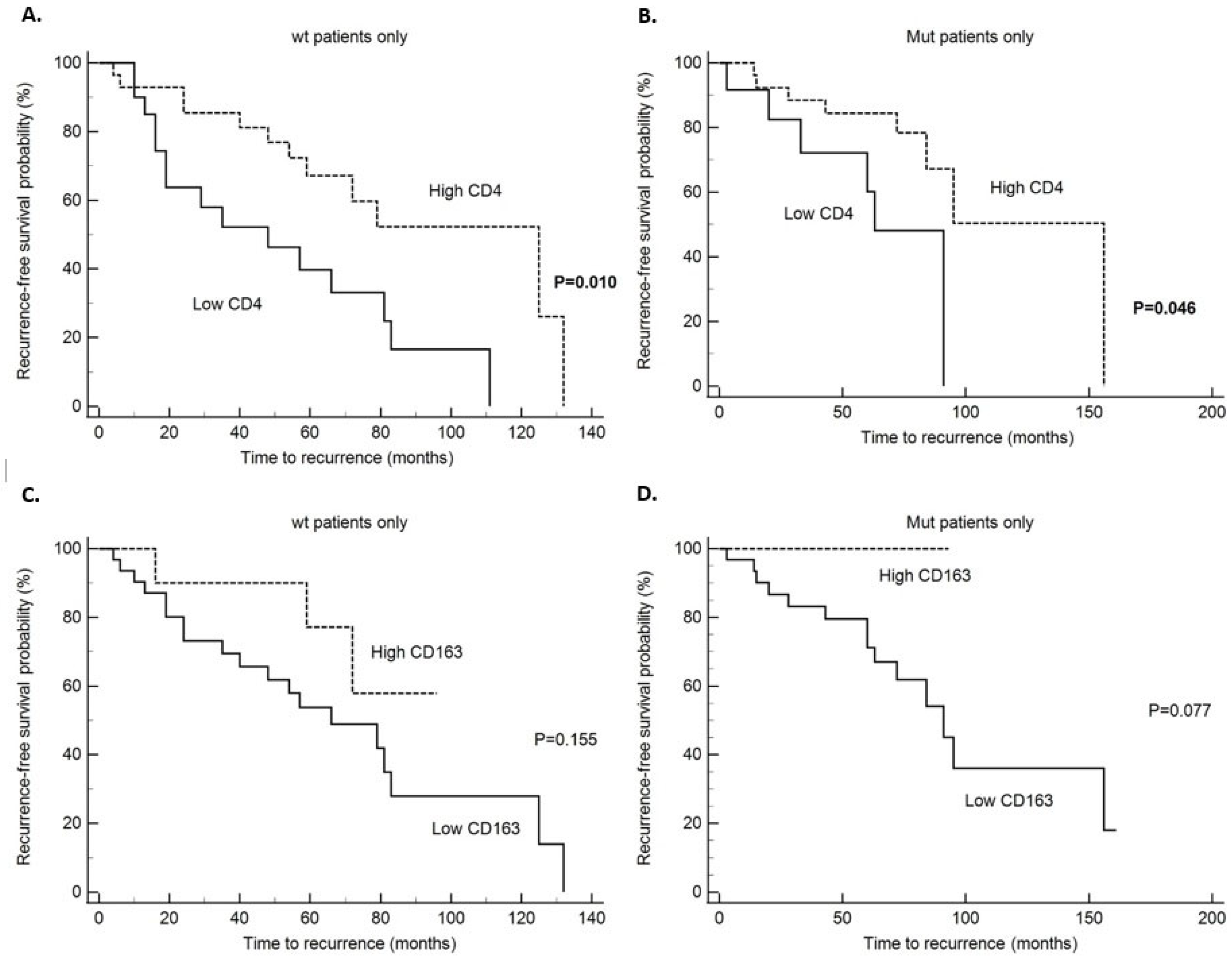
Disease specific survival (DSS) analysis has shown that patients without and with PIK3CA mutation with high CD4 tumor infiltrate had significantly better survival in comparison to patients with low CD4 infiltration (p = 0.015 and p = 0.0017, respectively) (Table 6). Also, we found that patients without PIK3CA mutation and high CD8 infiltrate had significantly better survival (p = 0.024), but the same was not found in patients with PIK3CA mutation (0.864) (Table 6 and Figure 1). All other survival analysis for DSS did not reach statistical significance in the PIK3CA non-mutated and mutated tumors (Table 6). When looking at the whole study group, patients with high CD4 and CD8 tumor infiltrates had a better prognosis with significantly longer DSS (p = 0.0003 and p = 0.041, respectively) (Table 6) and statistical trend to a better survival in the PD-L1 positive patients (p = 0.098) (Table 6). When looking at disease-free survival (DFS) in the non-mutated PIK3CA group and tumors with high CD4 infiltration, there was a statistically significant five-year survival without recurrences of 78.5% (p = 0.01) (Table 7). Also, in the PIK3CA mutated group and tumors with high CD4 infiltration, the five-year DFS rate was longer, (85% for CD4 high and 72% for CD4 low), and difference was statistically significant (p = 0.0458) (Table 7). Also, a statistical trend toward longer DFS was noticed in the PIK3CA mutated group and CD163 high infiltration (p = 0.077 and Figure 2). In the whole study group, when looking at DFS, statistical significance was shown in the patients with tumors with high CD4 (p = 0.005) and CD163 (p = 0–009) infiltrate (Table 7). In the patients with PIK3CA mutation and specifically exon 20 PIK3CA mutation, there was statistically significant longer survival without recurrence (p = 0.026 and p = 0.041, respectively) and statistical trend to a better DFS in PD-L1 positive patients (p = 0.072) (Table 7 and Figure 3). Also, we performed multivariate analysis for all the variables that reached statistical significance in the univariate analysis, and only PIK3CA in DFS retained statistical significance (p = 0.0488) (Table 8).

3. Discussion

In the early breast carcinoma, PIK3CA mutations have been detected in 37% of HR+/HER2− and associated with improved DFS but contrary PIK3CA mutations have been detected in 28% of HR+/HER2− metastatic breast carcinoma patients and correlated with worse overall survival as well as resistance to chemo- and endocrine therapy [18,24]. Results from our research are in agreement with previous studies because we also found 43.1% PIK3CA mutated tumors in HR+/HER2− breast carcinomas with majority having mutations in exon 20 (26.8%) and exon 9 (16.3%). Also, in our study group we found that PIK3CA mutations are associated with better DFS, but we did not find correlation between mutational status and DSS which corresponds to earlier findings. In addition, in this research we found that tumors with exon 20 PIK3CA mutations were more frequently associated with histologically more favorable characteristics like lower grade tumors, tumors with Ki67 < 20%, and patients had less frequently metastasis/recurrence during follow up. In this study, tumors with PIK3CA mutation had higher expression of PD-L1 in comparison to tumors without PIK3CA mutation but the difference did not reach statistical significance. However, the statistical significance was found in the distribution of PD-L1 positive tumors, when we compared the exon 9 and exon 20 PIK3CA mutated to the wild type (non-mutated) group, where patients with exon 20 mutations had higher number of PD-L1 positive tumors. This correlates with the literature data where PIK3CA mutations enhance transcription of PD-L1 and through AKT-mediated phosphorylation of β-catenin activate PD-L1 expression [25]. Also finding is opposite to research of Mosele et al. because they could not find any association between PD-L1 expression and PIK3CA mutations, but this is in TNBC, and they suggested that there is a population in which PI3K inhibitors could be developed independently from anti-PD-L1 agents [18]. However, data from the literature indicate that PIK3CA mutation might serve as a biomarker of an improved response to immune therapy in different types of carcinomas, like bladder, gastric, and cervical cancer [21,26,27]. Jiang W et al. showed a case where continuous pembrolizumab monotherapy treatment induced complete remission of a recurrent cervical cancer patient with systemic metastasis and PIK3CA-E545K mutation, implying that PIK3CA mutation is potentially a biomarker for pembrolizumab treatment in cervical cancer [21]. Having this in mind, data from our study might indicate that HR+/HER2− breast cancer patients with PIK3CA exon 20 mutation and PD-L1 expression might have the most benefit from immune checkpoint blockade. Also, all of the above findings from this study suggest that mutations in PIK3CA gene particularly in exon 20 are prognostically good characteristic in HR+/HER-2- breast carcinoma. Ruffell et al. observed that breast cancer tissues contained infiltrates dominated by CD8+ and CD4+ lymphocytes, with minor populations of NK cells and B lymphocytes, whereas in the normal breast tissue, myeloid-lineage cells including macrophages, mast cells, and neutrophils were more evident [28,29]. Despite the presence of many investigations, it is not possible to reach a clear and uniform conclusion about the role of each T-cell subset as well as M2-polarized (CD163+) macrophages in the breast TME or its association with breast carcinoma outcome [30]. Sobral-Leite et al. suggest that PI3K pathway alterations might be associated with the composition of the TME in luminal breast cancer, including the attraction of CD8-positive T-cells [20]. In their study, ER-positive breast cancer, with high tumor CD8 infiltration was associated with PIK3CA mutations and worse recurrence free survival and these associations were more pronounced among patients with grade 1 or 2 tumors [20]. In our study, we did not find statistical significance regarding DFS and CD8 lymphocyte infiltration in the non-mutated and mutated PIK3CA group but DSS was statistically significantly longer in CD8 high infiltration group in non-mutated PIK3CA patients indicating that wild type and mutated PIK3CA HR+/HER2− tumors have different functional state of the immune cells within TME indirectly supporting the findings of Sobral-Leite et al. Additionally, we did not find difference in the distribution of CD4, CD8, CD68, and CD163 infiltration between wild type (non-mutated) and PIK3CA mutated HR+/HER2− tumors. We thought that reason for this might be the fact that HR+/HER2− tumors are biologically heterogeneous group comprised from luminal A and B breast carcinoma. Thus, we additionally compared the immune cells composition between non-mutated and mutated PIK3CA luminal A and B breast carcinoma, but we did not find a statistically significant difference. However, when we compared the distribution of immune cells in wild type and PIK3CA mutated tumors who had metastasis/recurrence we found a statistically significant distribution of CD4 infiltration in wild type tumors (non-mutated) and CD163 infiltration in PIK3CA mutated tumors. Also, in the group of tumors with PIK3CA mutation and without metastasis/recurrence, there was a higher number of PD-L1 positive tumors but the result was not statistically significant (Table 4). The interesting finding is that when we analyzed CD4 infiltration and DSS or DFS, CD4 high infiltration in non-mutated and mutated PIK3CA group had better five -year survival, while the same was not found for CD8 infiltration in non-mutated and mutated PIK3CA tumors. These findings suggest that CD4 and CD8 TILs might have different functions and responses in PIK3CA mutated and non-mutated HR+/HER2− tumors status, so the final result of their complex interplay has a different impact on recurrence and overall survival. Within TME, tumor derived signals recruit monocytes and induce their polarization into tumor associated macrophages (TAMs) (M2 macrophages), promoting tumor cell proliferation, epithelial–mesenchymal transition (EMT), and suppression of CD8+ T-cell mediated anti-tumor effects [31]. So M2 macrophages facilitate tumor progression [31]. In this study, we found that PIK3CA mutated and non-mutated HR+/HER2− breast carcinomas had different correlations between PD-L1 expression and immune cells within TME (Table 5). An interesting finding is that CD163 as a marker of infiltration with M2 polarized macrophages (pro-tumor effect) in non-mutated PIK3CA group had a negative correlation with PD-L1 expression, while in the PIK3CA mutated group, this correlation was positive (Table 5). And also, when looking at the DFS in our study, patients with the PIK3CA mutation and high CD163 infiltration did not have disease recurrence (result was at the level of statistical trend, Table 7). The immunosuppressive properties of TAMs are generally believed to be dependent on the phosphatidylinositol 3-kinase (PI3K) signaling. High levels of M2 macrophages, particularly in the TME, are generally associated with poorer DFS but, nevertheless, a prognostic association in the opposite direction has also been suggested in breast cancer patients, thus further highlighting the need for a more in-depth evaluation of the TAM prognostic value [32]. Our result that shows high CD163 infiltrates to be associated with better DFS is contradictory, but at the same time it can be explained by new data in the literature where TAMs with dual characteristics of M1 and M2 have been identified, indicating the binary classification may be oversimplified. For example, Caronni et al. performed single-cell RNA-sequencing (scRNA-seq) on pancreatic ductal adenocarcinomas biopsies from cancer patients and revealed that inflammatory IL-1β + TAMs were shown to co-express both inflammatory (MHCII, CD80, and CD86) and immune inhibitory markers (CD206, arg-1, and PD-L1) [33]. This further emphasizes the complex interactions and crosstalk between immune cells and need for more precise TAM classification methods to help with the understanding of their dynamic functions. Also, this might further support the notion that PIK3CA mutations in HR+/HER2− breast tumors might influence and induce different functions of immune cells with prognostic result opposite from one expected knowing the pro-tumorigenic effects of M2 macrophages. Finding the explanation and mechanism on how mutations in PIK3CA achieve opposite effect of favorable versus poor prognosis and their effect on TME will be important in understanding the pathogenesis and progression of breast carcinoma. Also, this might have an impact on how to determine therapeutic approaches for patients with PIK3CA mutations in different stages and subtypes of disease. This emphasizes the need for more research regarding immune cells and PIK3CA mutations, especially in the era when there is available targeted therapy.
Limitations of this research would be the retrospective aspect of the study leading to heterogenic systemic treatment, but since all patients were treated at same clinical hospital within certified breast center, treatment decisions were made according to the national guidelines. Other limitations are the use of TMAs and PIK3CA mutation analysis using allele specific PCR detecting only “hot-spot” mutations. Using this type of assay, PIK3CA gene mutations, not covered by the kit, would not be detected, even though it remains difficult to interpret the functional consequences of new genetic mutations. Also, in order to compensate usage of TMA immunohistochemically stained slides, we used three to four 1 mm tissue cores as well as serial sections of the same TMA.
In conclusion, the focus of this research was HR+/HER2− breast carcinoma because studies examining PIK3CA, PD-L1 expression and immune cells within TME in this subtype are scarce. This study showed statistically significant distribution of PD-L1 expression in HR+/HER2−tumors with exon 20 PIK3CA mutation with different correlations of PD-L1 expression and immune cells infiltration within TME in mutated and non-mutated PIK3CA tumors. Currently, breast cancer with PIK3CA mutation comes into focus because novel research data in different cancer types indicate that mutations within PIK3CA might serve as a biomarker of an improved response to immune therapy.

4. Materials and Methods

4.1. Patients and Tumor Specimens

This retrospective study included 123 breast cancer samples over the period from 2010 to 2015 obtained from the archives of the Clinical Department of Pathology and Cytology, Clinical Hospital Center Rijeka, Croatia. The biopsy samples included hormone receptor (HR) positive/HER-2 negative breast cancer tissue from female patients that were not previously treated with radio or chemotherapy. The American Society of Clinical Oncology (ASCO)/College of American Pathologists (CAP) guideline recommendations were used as references for categorizing Ki-67, ER, progesterone receptor (PR), and HER2 status as part of the routine pathologic evaluation [34,35]. Also, the majority of material was surgical biopsy samples, but few were core biopsies that had enough tumor material. Clinicopathological parameters including age at initial diagnosis, tumor size, histologic grade, histologic subtype, lymphovascular and perineural invasion, axillary lymph node status, and clinical stage were obtained from the patient’s medical records. All carcinomas were classified according to the criteria of the WHO [36]. All of the patient’s clinicopathological characteristics are shown in more detail in Table 1. The study was conducted in accordance with the Declaration of Helsinki. The study was approved by the Institutional Ethical Committees of the Clinical Hospital Center Rijeka, Croatia class No:003-05/21-1/105, Ur.No:2170-29-02/1-21-2 and Faculty of Medicine, University of Rijeka, Croatia class No:003-08/21-01/75, Ur.No:2170-24-04-3-21-8 on 28 December 2021. Individual consent for this retrospective analysis was waived in accordance with the national legislation and the institutional requirements.

4.2. Immunohistochemical Staining

The tissue microarrays (TMAs) were constructed using three or four 1 mm cores of the above-mentioned archived biopsy samples. Also, to compensate for the spatial distribution of examined markers, we used serial sections of the same TMA cores. During immunohistochemical procedures, some cores were either lost, fragmented, or showed suboptimal staining; therefore, the number of examined samples sometimes differed between analyses. The antibodies used in this research were as follows: rabbit monoclonal antibody (IgG) anti-PD-L1 (clone SP142, Ventana, Tucson, AZ, USA), mouse monoclonal antibody (IgG) anti-CD4 (clone SP35, Cell Marque, Rocklin, CA, USA), mouse monoclonal antibody (IgG1) anti-CD8 (clone C8/144B, DakoAgilent, Santa Clara, CA, USA), mouse monoclonal antibody (IgG) anti-CD68 (clone PG-M1, DakoAgilent, Santa Clara, CA, USA), mouse monoclonal antibody (IgG1) anti-CD163 (clone 10D6, Leica Biosystems, Buffalo Grove, IL, USA). The antigen retrieval protocol, incubation, and other procedural steps included in the immunohistochemical analysis were conducted according to the guidelines provided by the manufacturer.

4.3. Immunohistochemical Evaluation

The independent evaluation of the expression of investigated biomarkers was conducted by two pathologists. Percentage of PD-L1 expression in tumor infiltrating immune cells (IC) was assessed as the proportion of tumor area occupied by PD-L1 positive immune cells of any intensity and in any cell compartment (membrane or cytoplasmic) [37]. TMA cores that contained less than 100 viable tumor cells were excluded from evaluation. For each of these percentages 1% or greater (≥1%) was considered positive score and less than 1% (<1%) as negative score. Results were evaluated with known positive and negative tissue controls.
The expression of CD4, CD8, CD68, CD163 was evaluated in three separate „hot spots” containing the highest density of immune cells and the number of immunoreactive cells per 0.4 mm × 0.4 mm was counted at ×200 magnification (×20 objective) [38]. The final number was calculated based on the average number of cells in three areas and expressed as the number of positive cells. For the statistical analyses, the number of CD4, CD8, CD68, and CD163 positive cells was divided into lower and higher groups based on ROC calculated cut-off values.

4.4. DNA Isolation from FFPE

Total DNA was isolated from formalin fixed paraffin embedded (FFPE) tumor tissue. Sample sectioning was conducted under great care, using procedures to avoid the risk of cross contamination between the samples. Depending on the amount of biopsy material embedded in paraffin, 4–10 sections (5 µm thick) were placed on microscopic slides for macrodisection. On a corresponding HE slide, the area with sufficient amount of tumor tissue was marked with a pen. The selected area of interest was then transferred and circled on unstained sections for macrodissection. The pieces of selected tumor tissue were scraped from the microscopic slides and put into microcentrifuge tube. The scrapped fragments were deparaffinized by adding 1 mL of xylene and heating at 55 °C for 30 min, followed by centrifugation and subsequent removal of the supernatant. Upon dewaxing with three washes of xylene, 1 mL of 100% ethanol was added to remove residual xylene. The tissues were dried at 37 °C for 30 min and DNA was isolated using NucleoSpin Tissue kit (Macharey-Nagel, Duren, Germany) according to the manufacturer’s instructions. Yield and the quality of isolated DNA was determined using Qubit 3.0 (ThermoFisher, Waltham, MA, USA).

4.5. PIK3CA Mutation Analysis

The isolated tumor DNA was analyzed for PIK3CA mutations using the Cobas PIK3CA mutation test (Roche Molecular Systems, Inc., Branchburg, NJ, USA) and Cobas z480 analyzer (Roche Diagnostics, Indianapolis, IN, USA) according to the manufacturer’s instructions. This real-time PCR method can detect mutations in 5 exons of the PIK3CA gene. The following mutations were detected using this mutation test in PIK3CA gene: exon 1: R88Q; exon 4: N345K; exon 7: C420R; exon 9: E542K, E545X (E545A, E545D, E545G, E545K), Q546X (Q546E, Q546K, Q546L, Q546R) and exon 20: M1043I, H1047X (H1047L, H1047R, H1047Y), G1049R. In 11 samples, due to poor quality of DNA, the status of PIK3CA gene could not be determined so the result was marked as invalid.

4.6. Statistical Analysis

The statistical analysis was conducted using MedCalc for Windows, version 23.3.4 (Med-Calc Statistical Software bvba in Ostend, Belgium). Frequency differences between nominal variables were assessed using Fisher’s exact test and chi-square test. Spearman’s rank correlation analysis was used to determine the association between PD-L1 and immune cells. The analysis of tumor recurrence prediction was performed using logistic regression. The Kaplan–Meier method was used to compute cumulative survival probability. The disparities in survival rates were assessed using a log-rank test. Multivariate analysis was performed using the Cox multiple-hazards model and p-value of 0.05 in the univariate survival analysis was adopted as the limit for inclusion in the multivariate model. All tests conducted were two-tailed, and a statistically significant result was defined when p < 0.05.
A receiver operating characteristic (ROC) curve was generated to evaluate the efficacy of CD4, CD8, CD68, and CD163 as biomarkers for predicting patient outcomes and determining the most effective statistical cut-off values. Hence, the ROC curve and Youden index were computed to optimize the sensitivity and specificity of the individual marker in predicting overall DSS and DFS in the univariate model. The area under the ROC curve (AUC) was calculated to assess the prediction model’s quality, along with a 95% confidence interval (CI). ROC analysis showed statistically significant cut-off values for CD4 (low CD4 versus high CD4 cut off value ≤6) (p = 0.001, AUC 0.711) and CD8 (low CD8 versus high CD8 cut off value < 8) (p = 0.014, AUC = 0.646). ROC calculated cut off values for CD68 (cut off value ≤ 23) and CD163 (cut off value > 12) did not show statistical significance (Supplementary Data Table S2). DSS was expressed as the number of months from diagnosis to the occurrence of a breast cancer related death. DFS was defined as the time interval from the date of diagnosis to the date of documented first recurrence of disease. If there was no recurrence, disease-free survival was determined as the date of last follow-up.

Supplementary Materials

The following supporting information can be downloaded at: https://www.mdpi.com/article/10.3390/ijms26199489/s1.

Author Contributions

Conceptualization, D.L. and I.H.; Data curation, D.L., J.H., E.B. and I.H.; Formal analysis, E.B.; Investigation, D.L., J.H. and I.H.; Resources, I.H.; Validation, D.L., E.B., P.V.Z. and D.G.; Visualization, P.V.Z., E.B., D.G. and I.H.; Writing—original draft, D.L. and I.H.; Writing—review and editing, D.L., E.B., J.H., P.V.Z., D.G. and I.H.; Funding acquisition, E.B. and I.H. All authors have read and agreed to the published version of the manuscript.

Funding

This work was supported by the project of the University of Rijeka (uniri-iskusni-biomed-23-213).

Institutional Review Board Statement

The study was conducted according to the guidelines of the Declaration of Helsinki and approved by the Institutional Ethical Committee of the Clinical Hospital Center Rijeka, Croatia class No:003-05/21-1/105, Ur.No:2170-29-02/1-21-2 on 23 July 2021 and Institutional Ethical Committee of the Faculty of Medicine, University of Rijeka, Croatia class No:003-08/21-01/75, Ur.No:2170-24-04-3-21-8 on 28 December 2021, with formal waiver statement.

Informed Consent Statement

Patient consent was omitted with formal statement from ethical committees because this was retrospective study and informed consent was waived in accordance with the national legislation and the institutional requirements.

Data Availability Statement

The datasets analyzed during the current study are available from the corresponding author upon reasonable request.

Acknowledgments

The authors would like to thank the technicians from the Clinical Department of Pathology and Cytology for their technical support.

Conflicts of Interest

The authors declare no conflicts of interest.

Abbreviations

The following abbreviations are used in this manuscript:
DCISDuctal carcinoma in situ
DFSDisease-free survival
DSSDisease-specific survival
FFPEFormalin fixed paraffin embedded
HR+/HER2−Hormone receptor positive/human epidermal growth factor-2 negative
IHCImmunohistochemistry
MAPMitogen activated protein
MDSCsMyeloid-derived suppressor cells
NSTNo special type
PD-L1Programmed cell death ligand 1
ROCReceiver operating characteristic
TAMsTumor associated macrophages
TILsTumor-infiltrating lymphocytes
TMAsTissue microarrays
TMETumor microenvironment
TNBCTriple negative breast carcinomas

References

  1. Zhang, W.; Lee, A.; Tiwari, A.K.; Yang, M.Q. Characterizing the Tumor Microenvironment and Its Prognostic Impact in Breast Cancer. Cells 2024, 13, 1518. [Google Scholar] [CrossRef] [PubMed] [PubMed Central]
  2. Stanton, S.E.; Disis, M.L. Clinical significance of tumor-infiltrating lymphocytes in breast cancer. J. Immunother. Cancer 2016, 4, 59. [Google Scholar] [CrossRef] [PubMed] [PubMed Central]
  3. Brummel, K.; Eerkens, A.L.; de Bruyn, M.; Nijman, H.W. Tumour-infiltrating lymphocytes: From prognosis to treatment selection. Br. J. Cancer 2023, 128, 451–458. [Google Scholar] [CrossRef] [PubMed] [PubMed Central]
  4. Valenza, C.; Taurelli Salimbeni, B.; Santoro, C.; Trapani, D.; Antonarelli, G.; Curigliano, G. Tumor Infiltrating Lymphocytes across Breast Cancer Subtypes: Current Issues for Biomarker Assessment. Cancers 2023, 15, 767. [Google Scholar] [CrossRef] [PubMed] [PubMed Central]
  5. Angelico, G.; Broggi, G.; Tinnirello, G.; Puzzo, L.; Vecchio, G.M.; Salvatorelli, L.; Memeo, L.; Santoro, A.; Farina, J.; Mulé, A.; et al. Tumor Infiltrating Lymphocytes (TILS) and PD-L1 Expression in Breast Cancer: A Review of Current Evidence and Prognostic Implications from Pathologist’s Perspective. Cancers 2023, 15, 4479. [Google Scholar] [CrossRef] [PubMed] [PubMed Central]
  6. Karnik, T.; Kimler, B.F.; Fan, F.; Tawfik, O. PD-L1 in breast cancer: Comparative analysis of 3 different antibodies. Hum. Pathol. 2018, 72, 28–34. [Google Scholar] [CrossRef] [PubMed]
  7. Jin, M.; Fang, J.; Peng, J.; Wang, X.; Xing, P.; Jia, K.; Hu, J.; Wang, D.; Ding, Y.; Wang, X.; et al. PD-1/PD-L1 immune checkpoint blockade in breast cancer: Research insights and sensitization strategies. Mol. Cancer 2024, 23, 266. [Google Scholar] [CrossRef] [PubMed] [PubMed Central]
  8. Szekely, B.; Bossuyt, V.; Li, X.; Wali, V.B.; Patwardhan, G.A.; Frederick, C.; Silber, A.; Park, T.; Harigopal, M.; Pelekanou, V.; et al. Immunological differences between primary and metastatic breast cancer. Ann. Oncol. 2018, 29, 2232–2239. [Google Scholar] [CrossRef] [PubMed]
  9. Ren, X.; Cui, H.; Dai, L.; Chang, L.; Liu, D.; Yan, W.; Zhao, X.; Kang, H.; Ma, X. PIK3CA mutation-driven immune signature as a prognostic marker for evaluating the tumor immune microenvironment and therapeutic response in breast cancer. J. Cancer Res. Clin. Oncol. 2024, 150, 119. [Google Scholar] [CrossRef] [PubMed] [PubMed Central]
  10. Zhang, Z.; Richmond, A. The Role of PI3K Inhibition in the Treatment of Breast Cancer, Alone or Combined With Immune Checkpoint Inhibitors. Front. Mol. Biosci. 2021, 8, 648663. [Google Scholar] [CrossRef] [PubMed] [PubMed Central]
  11. Dunlap, J.; Le, C.; Shukla, A.; Patterson, J.; Presnell, A.; Heinrich, M.C.; Corless, C.L.; Troxell, M.L. Phosphatidylinositol-3-kinase and AKT1 mutations occur early in breast carcinoma. Breast Cancer Res. Treat. 2010, 120, 409–418. [Google Scholar] [CrossRef] [PubMed]
  12. Bachman, K.E.; Argani, P.; Samuels, Y.; Silliman, N.; Ptak, J.; Szabo, S.; Konishi, H.; Karakas, B.; Blair, B.G.; Lin, C.; et al. The PIK3CA gene is mutated with high frequency in human breast cancers. Cancer Biol. Ther. 2004, 3, 772–775. [Google Scholar] [CrossRef] [PubMed]
  13. Mollon, L.E.; Anderson, E.J.; Dean, J.L.; Warholak, T.L.; Aizer, A.; Platt, E.A.; Tang, D.H.; Davis, L.E. A Systematic Literature Review of the Prognostic and Predictive Value of PIK3CA Mutations in HR+/HER2 Metastatic Breast Cancer. Clin. Breast Cancer 2020, 20, e232–e243. [Google Scholar] [CrossRef] [PubMed]
  14. Dirican, E.; Akkiprik, M.; Özer, A. Mutation distributions and clinical correlations of PIK3CA gene mutations in breast cancer. Tumor Biol. 2016, 37, 7033–7045. [Google Scholar] [CrossRef] [PubMed]
  15. Beelen, K.; Opdam, M.; Severson, T.M.; Koornstra, R.H.; Vincent, A.D.; Wesseling, J.; Muris, J.J.; Berns, E.M.; Vermorken, J.B.; van Diest, P.J.; et al. PIK3CA mutations, phosphatase and tensin homolog, human epidermal growth factor receptor 2, and insulin-like growth factor 1 receptor and adjuvant tamoxifen resistance in postmenopausal breast cancer patients. Breast Cancer Res. 2014, 16, R13. [Google Scholar] [CrossRef] [PubMed] [PubMed Central]
  16. André, F.; Ciruelos, E.; Rubovszky, G.; Campone, M.; Loibl, S.; Rugo, H.S.; Iwata, H.; Conte, P.; Mayer, I.A.; Kaufman, B.; et al. Alpelisib for PIK3CA-Mutated, Hormone Receptor-Positive Advanced Breast Cancer. N. Engl. J. Med. 2019, 380, 1929–1940. [Google Scholar] [CrossRef] [PubMed]
  17. Guo, S.; Loibl, S.; von Minckwitz, G.; Darb-Esfahani, S.; Lederer, B.; Denkert, C. PIK3CA H1047R Mutation Associated with a Lower Pathological Complete Response Rate in Triple-Negative Breast Cancer Patients Treated with Anthracycline-Taxane-Based Neoadjuvant Chemotherapy. Cancer Res. Treat. 2020, 52, 689–696. [Google Scholar] [CrossRef] [PubMed] [PubMed Central]
  18. Mosele, F.; Stefanovska, B.; Lusque, A.; Tran Dien, A.; Garberis, I.; Droin, N.; Le Tourneau, C.; Sablin, M.P.; Lacroix, L.; Enrico, D.; et al. Outcome and molecular landscape of patients with PIK3CA-mutated metastatic breast cancer. Ann. Oncol. 2020, 31, 377–386. [Google Scholar] [CrossRef] [PubMed]
  19. Ugai, T.; Zhao, M.; Shimizu, T.; Akimoto, N.; Shi, S.; Takashima, Y.; Zhong, R.; Lau, M.C.; Haruki, K.; Arima, K.; et al. Association of PIK3CA mutation and PTEN loss with expression of CD274 (PD-L1) in colorectal carcinoma. Oncoimmunology 2021, 10, 1956173. [Google Scholar] [CrossRef] [PubMed] [PubMed Central]
  20. Sobral-Leite, M.; Salomon, I.; Opdam, M.; Kruger, D.T.; Beelen, K.J.; van der Noort, V.; van Vlierberghe, R.L.P.; Blok, E.J.; Giardiello, D.; Sanders, J.; et al. Cancer-immune interactions in ER-positive breast cancers: PI3K pathway alterations and tumor-infiltrating lymphocytes. Breast Cancer Res. 2019, 21, 90. [Google Scholar] [CrossRef] [PubMed] [PubMed Central]
  21. Jiang, W.; Ouyang, X.; Li, C.; Long, Y.; Chen, W.; Ji, Z.; Shen, X.; Xiang, L.; Yang, H. Targeting PI3Kα increases the efficacy of anti-PD-1 antibody in cervical cancer. Immunology 2023, 170, 419–438. [Google Scholar] [CrossRef] [PubMed]
  22. Zardavas, D.; Phillips, W.A.; Loi, S. PIK3CA mutations in breast cancer: Reconciling findings from preclinical and clinical data. Breast Cancer Res. 2014, 16, 201. [Google Scholar] [CrossRef] [PubMed] [PubMed Central]
  23. Lee, M.H.; Cho, J.H.; Kwon, S.Y.; Jung, S.J.; Lee, J.H. Clinicopathological Characteristics of PIK3CA Mutation and Amplification in Korean Patients with Breast Cancers. Int. J. Med. Sci. 2020, 17, 1131–1135. [Google Scholar] [CrossRef] [PubMed] [PubMed Central]
  24. Zardavas, D.; Te Marvelde, L.; Milne, R.L.; Fumagalli, D.; Fountzilas, G.; Kotoula, V.; Razis, E.; Papaxoinis, G.; Joensuu, H.; Moynahan, M.E.; et al. Tumor PIK3CA Genotype and Prognosis in Early-Stage Breast Cancer: A Pooled Analysis of Individual Patient Data. J. Clin. Oncol. 2018, 36, 981–990. [Google Scholar] [CrossRef] [PubMed]
  25. Wang, Y.; Rozen, V.; Zhao, Y.; Wang, Z. Oncogenic activation of PIK3CA in cancers: Emerging targeted therapies in precision oncology. Genes Dis. 2024, 12, 101430. [Google Scholar] [CrossRef] [PubMed] [PubMed Central]
  26. Pan, Y.H.; Zhang, J.X.; Chen, X.; Liu, F.; Cao, J.Z.; Chen, Y.; Chen, W.; Luo, J.H. Predictive Value of the TP53/PIK3CA/ATM Mutation Classifier for Patients With Bladder Cancer Responding to Immune Checkpoint Inhibitor Therapy. Front. Immunol. 2021, 12, 643282. [Google Scholar] [CrossRef] [PubMed] [PubMed Central]
  27. Mishima, S.; Kawazoe, A.; Nakamura, Y.; Sasaki, A.; Kotani, D.; Kuboki, Y.; Bando, H.; Kojima, T.; Doi, T.; Ohtsu, A.; et al. Clinicopathological and molecular features of responders to nivolumab for patients with advanced gastric cancer. J. Immunother. Cancer 2019, 7, 24. [Google Scholar] [CrossRef] [PubMed] [PubMed Central]
  28. Ruffell, B.; Au, A.; Rugo, H.S.; Esserman, L.J.; Hwang, E.S.; Coussens, L.M. Leukocyte composition of human breast cancer. Proc. Natl. Acad. Sci. USA 2012, 109, 2796–2801. [Google Scholar] [CrossRef] [PubMed] [PubMed Central]
  29. Goff, S.L.; Danforth, D.N. The Role of Immune Cells in Breast Tissue and Immunotherapy for the Treatment of Breast Cancer. Clin. Breast Cancer 2021, 21, e63–e73. [Google Scholar] [CrossRef] [PubMed] [PubMed Central]
  30. Zareinejad, M.; Mehdipour, F.; Roshan-Zamir, M.; Faghih, Z.; Ghaderi, A. Dual Functions of T Lymphocytes in Breast Carcinoma: From Immune Protection to Orchestrating Tumor Progression and Metastasis. Cancers 2023, 15, 4771. [Google Scholar] [CrossRef] [PubMed] [PubMed Central]
  31. Jing, H.; Gao, Y.; Sun, Z.; Liu, S. Recent advances in novel tumor immunotherapy strategies based on regulating the tumor microenvironment and immune checkpoints. Front. Immunol. 2025, 16, 1529403. [Google Scholar] [CrossRef] [PubMed] [PubMed Central]
  32. Pelekanou, V.; Villarroel-Espindola, F.; Schalper, K.A.; Pusztai, L.; Rimm, D.L. CD68, CD163, and matrix metalloproteinase 9 (MMP-9) co-localization in breast tumor microenvironment predicts survival differently in ER-positive and -negative cancers. Breast Cancer Res. 2018, 20, 154. [Google Scholar] [CrossRef] [PubMed] [PubMed Central]
  33. Caronni, N.; La Terza, F.; Vittoria, F.M.; Barbiera, G.; Mezzanzanica, L.; Cuzzola, V.; Barresi, S.; Pellegatta, M.; Canevazzi, P.; Dunsmore, G.; et al. IL-1β+ macrophages fuel pathogenic inflammation in pancreatic cancer. Nature 2023, 623, 415–422. [Google Scholar] [CrossRef] [PubMed]
  34. Hammond, M.E.; Hayes, D.F.; Dowsett, M.; Allred, D.C.; Hagerty, K.L.; Badve, S.; Fitzgibbons, P.L.; Francis, G.; Goldstein, N.S.; Hayes, M.; et al. American Society of Clinical Oncology/College of American Pathologists guideline recommendations for immunohistochemical testing of estrogen and progesterone receptors in breast cancer. Arch. Pathol. Lab. Med. 2010, 134, 907–922. [Google Scholar] [CrossRef] [PubMed] [PubMed Central]
  35. Wolff, A.C.; Hammond, M.E.; Hicks, D.G.; Dowsett, M.; McShane, L.M.; Allison, K.H.; Allred, D.C.; Bartlett, J.M.; Bilous, M.; Fitzgibbons, P.; et al. Recommendations for human epidermal growth factor receptor 2 testing in breast cancer: American Society of Clinical Oncology/College of American Pathologists clinical practice guideline update. Arch. Pathol. Lab. Med. 2014, 138, 241–256. [Google Scholar] [CrossRef] [PubMed] [PubMed Central]
  36. WHO Classification of Tumours Editorial Board. Breast Tumours, 5th ed.; WHO Classification of Tumors Series; Lyon (France) International Agency for Research on Cancer: Lyon, France, 2019; Volume 2. [Google Scholar]
  37. Guo, H.; Ding, Q.; Gong, Y.; Gilcrease, M.Z.; Zhao, M.; Zhao, J.; Sui, D.; Wu, Y.; Chen, H.; Liu, H.; et al. Comparison of three scoring methods using the FDA-approved 22C3 immunohistochemistry assay to evaluate PD-L1 expression in breast cancer and their association with clinicopathologic factors. Breast Cancer Res. 2020, 22, 69. [Google Scholar] [CrossRef] [PubMed] [PubMed Central]
  38. Matsumoto, H.; Thike, A.A.; Li, H.; Yeong, J.; Koo, S.L.; Dent, R.A.; Tan, P.H.; Iqbal, J. Increased CD4 and CD8-positive T cell infiltrate signifies good prognosis in a subset of triple-negative breast cancer. Breast Cancer Res. Treat. 2016, 156, 237–247. [Google Scholar] [CrossRef] [PubMed]
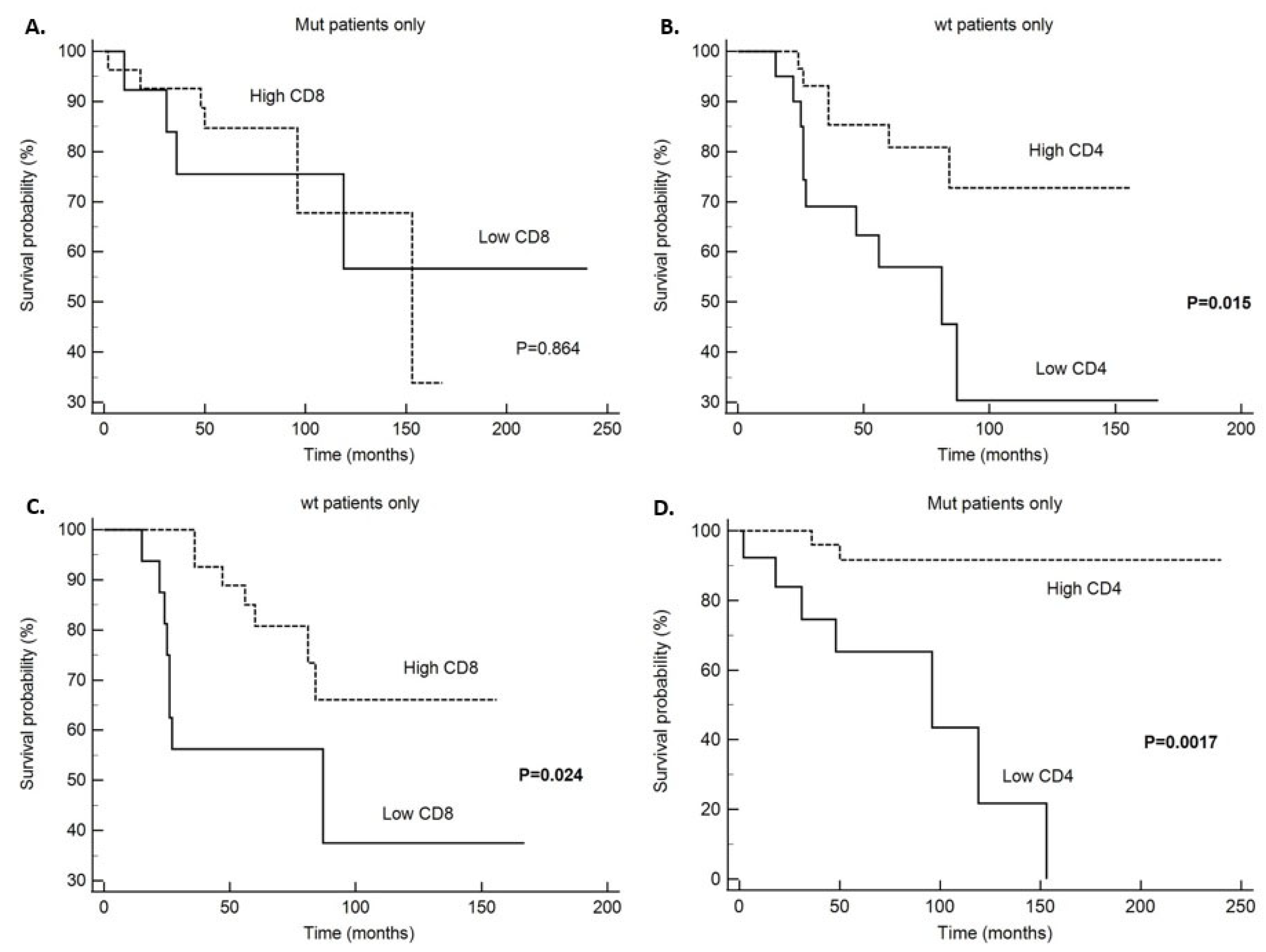
Figure 1. Disease specific survival (DSS); (A)—CD4 infiltrate in tumors without PIK3CA mutation (wt tumors), (B)—CD4 infiltrate in PIK3CA mutated tumors, (C)—CD8 infiltrate in tumors without PIK3CA mutation (wt tumors), (D)—CD8 infiltrate in PIK3CA mutated tumors.
Ijms 26 09489 g001
Figure 2. Disease-free survival (DFS); (A)—CD4 infiltrate in tumors without PIK3CA mutation (wt tumors), (B)—CD4 infiltrate in PIK3CA mutated tumors, (C)—CD163 infiltrate in tumors without PIK3CA mutation (wt tumors), (D)—CD163 infiltrate in PIK3CA mutated tumors.
Ijms 26 09489 g002
Figure 3. Disease specific survival (DSS), (A)—DSS depending on PD-L1 expression, (B)—DSS depending on presence of exon 9 and exon 20 PIK3CA mutation, (C)—DSS depending on presence of PIK3CA mutation; Disease-free survival (DFS), (D)—DFS depending on PD-L1 expression, (E)—DFS depending on presence of exon 9 and exon 20 PIK3CA mutation, (F)—DFS depending on presence of PIK3CA mutation (whole study group).
Ijms 26 09489 g003
Table 1. Patient and tumor characteristics.
Clinicopathological ParametersNumber of Patients (%)
Age (years)
≤50
>50

27 (22.0)
96 (78.0)
Histological grade
150 (40.7)
261 (49.4)
34 (3.3)
N/A8 (6.4)
Tumor size (cm)
<2
≥2
N/A

39 (31.7)
79 (64.2)
5 (4.1)
Multifocality
Absent
Present
N/A

105 (85.4)
14 (11.4)
4 (3.2)
Exulcerated tumor
Absent
Present
N/A

112 (91.1)
7 (5.7)
4 (3.2)
Breast
Right
Left
Both

63 (51.6)
54 (44.3)
5 (4.1)
Inflammatory carcinoma
Absent
Present
N/A

116 (94.3)
2 (1.6)
5 (4.1)
Histological type
NST
Spec.type (lobular)
Other spec. type (papillary, mucinous, cribriform)
Mixed NST + mucinous
Mixed NST + lobular

90 (73.2)
20 (16.3)
8 (6.5)
4 (3.3)
1 (0.7)
Histological subtype
Luminal A
Luminal B
Multifocality

65 (52.8)
57 (46.3)
1 (0.9)
Clinical stage
1
2
3
4
N/A

32 (26.0)
39 (31.7)
28 (22.8
21 (17.1)
3 (2.4)
Lymphovascular invasion
Absent
Present
N/A

36 (29.3)
82 (66.7)
5 (4.0)
Perineural invasion
Absent
Present
N/A

59 (48.8)
55 (45.5)
7 (5.7)
Necrosis
Absent
Present
N/A

88 (71.5)
33 (26.9)
2 (1.6)
Calcifications
Absent
Present
N/A

82 (66.7)
39 (31.7)
2 (1.6)
Ki-67
<20%
≥20%

72 (58.5)
51 (41.5)
Lymph node status
Negative
Positive
N/A

51 (41.5)
54 (43.9)
18 (14.6)
Extra-nodal tumor spreading
Absent
Present
N/A

21 (38.9)
32 (59.3)
1 (1.8)
Metastasis present at the diagnosis
Absent
Present
N/A


99 (80.5)
22 (17.9)
2 (1.6)
Metastasis and recurrence
Absent
Present, local
Present, distant metastasis
N/A

63 (51.2)
8 (6.5)
46 (37.4)
6 (4.9)
Months until the appearance of recurrence or metastasis
Median (range)

41.5 (3–180)
Follow up (months)
Median (range)

73 (12–240)
Number of died patients
N (%)

38 (30.9)
PIK3CA mutations
Wild type
Mutations
Invalid

59 (48.0)
53 (43.1)
11 (8.9)
PIK3CA mutations
Wild type
Exon 9
Exon 20
N/A

59 (48.0)
20 (16.3)
33 (26.8)
11 (8.9)
PD-L1
<1%
≥1%
N/A

85 (69.1)
12 (9.8)
26 (21.1)
CD4 median (range)
CD8 median (range)
CD68 median (range)
CD163 median (range)
8.0 (0.0–232.0)
10 (0.0–142.0)
7 (1.0–75.0)
6 (0.0–68.0)
NST-no special type; N/A-not available.
Table 2. Comparison of HR+/HER2− tumor characteristics depending on PIK3CA mutational status.
Characteristics (HR+/HER-2-)PIK3CAp-ValuePIK3CAp-Value
Wt
N (%)
Mt
N (%)
Wt
N (%)
Ex9
N (%)
Ex20
N (%)
Age (years)
≤50
>50

14 (23.7)
45 (76.3)

12 (22.6)
41 (77.4)

1.00

14 (23.7)
45 (76.3)

6 (30.0)
14 (70.0)

6 (18.2)
27 (81.8)

0.608
Clinical stage
I
II
III
IV

12 (20.3)
19 (32.2)
17 (28.8)
11 (18.6)

15 (30.0)
17 (34.0)
8 (16.0)
10 (20.0)


0.390 *

12 (20.3)
19 (32.2)
17 (28.8)
11 (18.6)

4 (21.1)
5 (26.3)
4 (21.1)
6 (31.6)

11 (35.5)
12 (38.7)
4 (12.9)
4 (12.9)


0.319 *
Tumor size
<2 cm
≥2 cm

18 (30.5)
41 (69.5)

17 (35.4)
31 (64.6)

0.679

18 (30.5)
41 (69.5)

5 (29.4)
12 (70.6)

12 (38.7)
19 (61.3)

0.697
Multifocality
Present
Absent

48 (82.8)
10 (17.2)

47 (94.0)
3 (6.0)

0.084

10 (17.2)
48 (82.8)

1 (5.3)
18 (94.7)

2 (6.5)
29 (93.5)

0.199
Exulcerated
Present
Absent

54 (93.1)
4 (6.9)

47 (94.0)
3 (6.0)

1.00

4 (6.9)
54 (93.1)

1 (5.3)
18 (94.7)

2 (6.5)
29 (93.5)

0.969
Inflammatory carcinoma
Present
Absent

55 (96.5)
2 (3.5)

50 (100)
0 (0)

0.497

2 (3.5)
55 (96.5)

0 (0)
19 (100)

0 (0)
31 (100)

0.409
Bilateral tumor
Present
Absent

54 (91.5)
5 (8.5)

52 (100)
0 (0)

0.059

5 (8.5)
54 (91.5)

0 (0)
20 (100)

0 (0)
32 (100)

0.099
Histological type
Ductal NST
Lobular
Other spec. type
Mixed (duct.+lob.)

44 (74.6)
9 (15.3)
5 (8.5)
1 (1.7)

42 (79.2)
9 (17.0)
2 (3.8)
0 (0)


0.569 *

44 (74.6)
9 (15.3)
5 (8.5)
1 (1.7)

14 (70.0)
6 (30.0)
(0)
0 (0)

28 (84.8)
3 (9.1)
2 (6.1)
0 (0)


0.370 *
Histological subtype
Luminal A
Luminal B

27 (45.8)
32 (54.2)

31 (58.5)
22 (41.5)


0.191

27 (45.8)
32 (54.2)

10 (50.0)
10 (50.0)

21 (63.6)
12 (36.4)


0.254
Histological grade
1
2
3

21 (36.8)
33 (57.9)
3 (5.3)

24 (51.1)
23 (48.9)
0 (0)


0.131 *

21 (36.8)
33 (57.9)
3 (5.3)

5 (29.4)
12 (70.6)
0 (0)

19 (63.3)
11 (36.7)
0 (0)


0.055 *
Lymph node status
Negative
Positive

22 (43.1)
29 (56.9)

23 (53.5)
20 (46.5)

0.408

22 (43.1)
29 (56.9)

7 (43.7)
9 (56.2)

16 (59.3)
11 (40.7)

0.373
Extranodal tumor spreading
Absent
Present

11 (37.9)
18 (62.1)

7 (36.8)
12 (63.2)

1.00

11 (37.9)
18 (62.1)

3 (33.3)
6 (66.7)

4 (40.0)
6 (60.0)

0.953
Metastasis present at diagnosis
Absent
Present

46 (79.3)
12 (20.7)

42 (80.8)
10 (19.2)

1.00

46 (79.3)
12 (20.7)

14 (70.0)
6 (30.0)

28 (87.5)
4 (12.5)

0.302
Metastasis and recurrence
Absent
Present

26 (46.4)
30 (53.6)

31 (59.6)
21 (40.4)


0.183

26 (46.4)
30 (53.6)

9 (45.0)
11 (55.0)

22 (68.7)
10 (31.2)

0.097
Ki-67
<20%
≥20%

31 (52.5)
28 (47.5)

35 (66.0)
18 (34.0)

0.179

31 (52.5)
28 (47.5)

10 (50.0)
10 (50.0)

25 (75.8)
8 (24.2)

0.041
N—number; Wt—no PIK3CA mutation; Mt—with PIK3CA mutation; Ex9—exon 9; Ex20—exon 20, NST—no special type; —Fischer’s exact test; *—χ2-test.
Table 3. Comparison of tumor microenvironment composition depending on PIK3CA mutational status.
VariablePIK3CAp-Value
WtMutation
CD4 median (range)

CD4
Low (≤6)
High (>6)
8.0 (0.0–94.0)


21 (41.2)
30 (58.8)
9.0 (0.0–232.0)


14 (34.1)
27 (65.9)
0.436


0.524
CD8 median (range)

CD8
Low (≤8)
High (>8)
10.0 (0.0–100.0)


17 (34.7)
32 (65.3)
14.5 (1.0–142.0)


15 (35.7)
27 (64.3)
0.655


1.00
CD68 median (range)

CD68
Low (≤23)
High (>23)
8.0 (1.0–30.0)


43 (84.3)
8 (15.7)
8.0 (1.0–75.0)


37 (92.5)
3 (7.5)
0.794


0.336
CD163 median (range)

CD163
Low (≤12)
High (>12)
8.0 (1.0–68.0)


32 (72.7)
12 (27.3)
6.0 (0.0–41.0)


32 (82.1)
7 (17.9)
0.143


0.433
PD-L1
<1%
≥1%

42 (93.3)
3 (6.7)

34 (81.0)
8 (19.0)

0.110
PD-L1
<1%
≥1%

42 (93.3)
3 (6.7)
Ex9
14 (93.3)
1 (6.7)
Ex20
20 (74.1)
7 (25.9)

0.044
Wt—without PIK3CA mutation.
Table 4. Comparison of microenvironment cellular composition between HR+/HER2− tumors with and without metastasis/recurrence depending on PIK3CA mutational status.
Variable
PIK3CA Wt
Metastasis/Recurrencep-Value
NoYes
CD4
Low (≤6)5 (23.8)15 (55.6)0.039
High (>6)16 (76.2)12 (44.4)
CD8
Low (≤8)6 (27.3)9 (37.5)0.539
High (>8)16 (72.7)15 (62.5)
CD68
Low (≤23)17 (81.0)24 (88.9)0.683
High (>23)4 (19.0)3 (11.1)
CD163
Low (≤12)12 (63.2)19 (86.4)0.144
High (>12)7 (36.8)3 (13.6)
PD-L1
<1%17 (89.5)22 (95.7)0.581
≥1%2 (10.5)1 (4.3)
PIK3CA mt
CD4
Low (≤6)7 (26.9)6 (42.9)0.480
High (>6)19 (73.1)8 (57.1)
CD8
Low (≤8)8 (28.6)6 (46.2)0.307
High (>8)20 (71.4)7 (53.8)
CD68
Low (≤23)25 (96.2)11 (84.6)0.253
High (>23)1 (3.8)2 (15.4)
CD163
Low (≤12)18 (72.0)14 (100)0.036
High (>12)7 (28.0)0 (0)
PDL-1
<1%21 (75.0)12 (92.3)0.398
≥1%7 (25.0)1 (7.7)
Whole study group
CD4
Low (≤6)17 (31.5)22 (50.0)0.096
High (>6)37 (68.5)22 (50.0)
CD8
Low (≤8)17 (29.8)17 (42.5)0.279
High (>8)40 (70.2)23 (57.5)
CD68
Low (≤23)46 (90.2)38 (88.4)1.00
High (>23)5 (9.8)5 (11.6)
CD163
Low (≤12)32 (64.0)36 (92.3)0.002
High (>12)18 (36.0)3 (7.7)
PD-L1
<1%43 (81.1)37 (94.9)0.065
≥1%10 (18.9)2 (5.1)
Wt—without PIK3CA mutation; Mt—with PIK3CA mutation.
Table 5. Correlation of PD-L1 expression in the whole group and in non-mutated (wt) and mutated (mt) PIK3CA HR+/HER2− tumors.
VariableCD4CD8CD68CD163
PD-L1 wtrs0.0250.0630.053−0.317
P0.8720.6930.7300.052
PD-L1 mtrs0.2850.4620.3980.617
P0.0780.00270.0134<0.0001
PD-L1 whole grouprs0.2030.3120.2380.205
P0.0510.00260.0230.061
Wt—no PIK3CA mutation; Mt—with PIK3CA mutation; Correlations were evaluated using Spearman rank correlation coefficient.
Table 6. Disease specific survival in patients without and with PIK3CA mutation depending on immune cell infiltration and for the whole study group.
VariableNDied from Underlying Disease (N)Five-Year Survival (%)Average Value ± SD95% CIχ2Log-Rank Test (p Value)
Patients without PIK3CA mutation (wt)
CD4
Low (≤6)20106386.7 ± 16.254.9–118.65.920.015
High (>6)30685127.5 ± 10.2107.5–147.6
CD8
Low (<8)1685689.3 ± 18.952.1–126.45.090.024
High (≥8)32785124.3 ± 10.1104.5–144.2
CD68
Low (≤23)421575111.0 ± 11.289.1–132.91.2560.263
High (>23)8185132.3 ± 16.4100.1–164.4
CD163
Low (≤12)31107699.3 ± 13.373.3–125.40.7220.395
High (>12)12375120.5 ± 14.891.4–149.5
PD-L1
<1%411377119.2 ± 10.798.1–140.20.5530.457
≥1%3010055.0 ± 0.055.0–55.0
Patients with PIK3CA mutation (mt)
CD4
Low (≤6)1376389.1 ± 17.954.4–123.89.880.0017
High (>6)26285223.6 ± 11.2201.7–245.4
CD8
Low (<8)13475164.8 ± 30.1105.9–223.70.0290.864
High (≥8)27685129.6 ± 13.4103.4–155.8
CD68
Low (≤23)36979158.3 ± 22.9113.2–203.31.2190.269
High (>23)30100168.0 ± 0.0168.0–168.0
CD163
Low (≤12)32884164.2 ± 21.6121.9–206.50.0450.831
High (>12)718786.9 ± 5.775.7 – 98.0
PD-L1
<1%32980148.8 ± 23.0103.7–193.90.8590.354
≥1%8189153.3 ± 13.8126.2–180.3
Whole study group
CD4
Low (≤6)39206285.8 ± 10.565.3–106.413.290.0003
High (>6)611185196.6 ± 12.1172.9- 220.2
CD8
Low (<8)341562129.7 ± 20.789.2–170.24.190.041
High (≥8)651685128.5 ± 8.3112.2–144.7
CD68
Low (≤23)862872148.6 ± 14.6120.1–177.12.140.143
High (>23)11190153.8 ± 13.5127.4–180.2
CD163
Low (≤12)692277144.5 ± 16.0113.1–175.90.8290.363
High (>12)23587124.7 ± 9.9105.2–144.2
PD-L1
<1%822775144.4 ± 15.2114.7–174.22.7410.098
≥1%12190156.2 ± 11.2134.3–178.1
PIK3CA specific mutation
Wt581972132.6 ± 11.7109.7–155.61.7880.409
Exon 920485140.8 ± 12.8115.8–165.9
Exon 2031880159.4 ± 29.9110.6–208.2
PIK3CA mutation
Wt581972132.6 ± 11.7109.7–155.61.6970.195
Mutation511281173.5 ± 16.7140.7–206.3
Wt—without PIK3CA mutation.
Table 7. Disease-free survival in patients without and with PIK3CA mutation and whole study group.
VariableNDisease Recurrence
(N)
Five Year DFS (%)Mean Value ± SD95% CIχ2Log-Rank Test (p-Value)
Patients without PIK3CA mutation (wt)
CD4 6.640.01
Low (≤6)20154551.95 ± 8.8434.63–69.3
High (>6)281278.589.82 ± 7.2570.35–109.3
CD8 1.840.175
Low (<8)1595051.68 ± 8.9034.24–69.12
High (≥8)31156584.37 ± 8.7867.16–101.59
CD68 0.0070.932
Low (≤23)41245572.4 ± 7.8157.09–87.7
High (>23)734166.21 ± 12.3442.02–90.4
CD163 2.020.155
Low (≤12)31195569.3 ± 9.151.5–87.1
High (>12)1037878.61 ± 8.5661.8–95.4
PD-L1 0.070.786
<1%39225972.45 ± 7.3857.99–86.91
≥1%316540.0 ± 12.2515.99–64.01
Patients with PIK3CA mutation (mt)
CD4 3.980.0458
Low (≤6)1367264.1 ± 10.443.7–84.4
High (>6)26885112.3 ± 14.484.1–140.4
CD8 0.3130.576
Low (<8)13675101.6 ± 18.9264.52–138.7
High (≥8)2778883.41 ± 4.6274.32–92.45
CD68 1.7140.190
Low (≤23)361182109.2 ± 12.2985.13–133.29
High (>23)326664.67 ± 8.9947.03–82.30
CD163 3.1360.077
Low (≤12)32148096.1 ± 11.7773.02–119.17
High (>12)7010093.0 ± 0.093.0–93.0
PD-L1 1.9430.163
<1%32128097.75 ± 12.872.58–122.9
≥1%8110092.0 ± 3.6584.84–99.16
Whole study group
CD4 8.090.005
Low (≤6)39224361.9 ± 6.649.1–74.8
High (>6)58227898.4 ± 8.581.8–115.0
CD8 1.4930.222
Low (≤8)33176085.1 ± 12.460.8–109.2
High (>8)63237390.8 ± 5.273.6–103.0
CD68 0.3670.545
Low (≤23)84386886.9 ± 7.272.9–101.0
High (>23)1055066.3 ± 9.248.3–84.3
CD163 6.8740.009
Low (≤12)683660.580.3 ± 7.565.7–94.9
High (>12)2138987.7 ± 4.678.8–96.7
PD-L1 3.2290.072
<1%79377383.9 ± 7.569.3–98.5
≥1%1229185.7 ± 7.271.6–99.8
PIK3CA specific mutation 6.3750.041
Wt56306577.9 ± 7.762.9–93.0
Exon 920117285.8 ± 13.559.5–112.2
Exon 20311082125.0 ± 14.097.7 ± 152.4
PIK3CA mutation 4.9710.026
Wt56306277.9 ± 7.762.9–93.0
Mutation512179.5107.4 ± 10.985.9–128.8
DFS—disease-free survival; wt—without PIK3CA mutation; mt—with PIK3CA mutation.
Table 8. Multivariate analysis of disease specific and disease-free survival.
Disease Specific Survival
Variables (Cut Off)HR95% CIp-Value
CD4 (≤6)0.510.19–1.300.159
CD8 (<8)0.610.23–0.1.590.313
PD-L1 0.200.02–1.620.133
Disease-free survival
CD4 (≤6)0.540.24–1.210.134
CD163 (>12)0.350.09–1.240.351
PD-L10.830.18–3.760.835
PIK3CA mutation0.450.21–0.990.0488
HR—hazards ratio; CI—confidence interval.
Disclaimer/Publisher’s Note: The statements, opinions and data contained in all publications are solely those of the individual author(s) and contributor(s) and not of MDPI and/or the editor(s). MDPI and/or the editor(s) disclaim responsibility for any injury to people or property resulting from any ideas, methods, instructions or products referred to in the content.