Review

Gene Therapy Strategies for the Treatment of Bestrophinopathies

by
Silja B. Haldrup
1,2,*,
Michelle E. McClements
2,3,
Jasmina Cehajic-Kapetanovic
2,3,
Thomas J. Corydon
1,4 and
Robert E. MacLaren
2,3
1
Department of Biomedicine, Aarhus University, 8000 Aarhus C, Denmark
2
Nuffield Department of Clinical Neurosciences, University of Oxford, Oxford OX3 9DU, UK
3
Oxford Eye Hospital, Oxford Hospitals NHS Foundation Trust, Oxford OX3 9DU, UK
4
Department of Ophthalmology, Aarhus University Hospital, 8200 Aarhus N, Denmark
*
Author to whom correspondence should be addressed.
Int. J. Mol. Sci. 2025, 26(19), 9421; https://doi.org/10.3390/ijms26199421
Submission received: 24 July 2025 / Revised: 23 August 2025 / Accepted: 29 August 2025 / Published: 26 September 2025
(This article belongs to the Special Issue Development of AAV-Based Gene Therapies: Unmet Needs and Solutions)

Abstract

The BEST1 gene encodes a transmembrane protein in the retinal pigment epithelium (RPE) in the eye, that functions as a calcium-dependent chloride channel (CaCC). Pathogenic variants in BEST1 are the underlying cause for bestrophinopathies, a group of inherited retinal disorders that vary in their pattern of inheritance, clinical appearance, and underlying molecular disease mechanisms. Currently, there are no treatments available for any of the bestrophinopathies, and gene therapy represents an attractive strategy due to the accessibility of the eye and slow disease progression. While gene augmentation may be effective for a subset of bestrophinopathies, others require allele-specific silencing or correction of the disease-causing variant to reconstitute expression of the BEST1 protein. This review aims to give an overview of the clinical diversity of bestrophinopathies and proposes the molecular disease mechanism of the pathogenic BEST1 variant as an important parameter for the choice of treatment strategy. Furthermore, we discuss the potential of different mutation-specific and mutation-independent CRISPR/Cas9-based gene editing strategies as a future treatment approach for bestrophinopathies.

1. Introduction

Gene therapy has the potential to improve the therapeutic landscape of inherited diseases, and treatment approaches for eye conditions are at the forefront of this development due to the eye’s appealing characteristics as a target organ. This includes the eye’s relative immune privilege, compartmentalization, and visual and surgical accessibility [1]. This review focuses on pathogenic variants in the BEST1 gene, which are causative for bestrophinopathies, a clinically and genetically heterogenous group of retinal degenerative disorders. Phenotypical hallmarks of bestrophinopathies include subretinal vitelliform lesions in the central retina, which progress over time and lead to vision loss and blindness in late stages of the diseases [2]. To date, there are no treatment options available for any of the bestrophinopathies, and novel approaches are therefore urgently needed. Gene therapy strategies may be a feasible treatment option, as the slow disease progression of bestrophinopathies provides a large therapeutic window. Encouragingly, gene augmentation has already proven to be successful for the treatment of autosomal recessive inherited retinal diseases and experienced a major breakthrough with the FDA/EMA approval of Voretigene neparvovec [3,4]. Hence, this approach is particularly promising for autosomal recessive bestrophinopathies with loss-of-function mechanism where supplementation with a wildtype BEST1 gene may restore sufficient protein function. In contrast, gene augmentation may be applicable to only a subset of autosomal dominant inherited bestrophinopathies, while others require silencing or correction of the disease-causing variant to achieve functional rescue. Here, we provide an overview of the clinical and genetic heterogeneity of BEST1-related disorders and highlight the importance of the molecular disease mechanism of the distinct pathogenic variant for the choice of a therapeutic strategy. Furthermore, we discuss the potential of different mutation-specific and mutation-independent CRISPR/Cas9-based gene editing strategies as a treatment approach for bestrophinopathies.

2. The BEST1 Gene and the Bestrophin-1 Channel

The BEST1 gene is positioned on chromosome 11q13 and consists of 11 exons, of which 10 encode bestrophin-1 (BEST1), a transmembrane protein comprising 585 amino acids (Figure 1A) [2,5]. In the human retina, BEST1 is predominantly localized to the basolateral membrane of the retinal pigment epithelium (RPE). Here, it functions as a calcium-dependent chloride channel (CaCC) that controls the chloride-ion current and maintains the transepithelial potential of the RPE (Figure 1B) [6,7]. Interestingly, research on the high-resolution crystal structure of the eukaryotic BEST1 channel revealed a pentameric configuration with five subunits surrounding a central ion pore [6,8]. Previous studies indicate that the ion pore is “flower vase-shaped” and consists of (i) a funnel-shaped outer entryway, (ii) a slender neck region, (iii) a large inner cavity, and (iv) a narrow aperture [6]. Multiple anion-binding sites within the ion pore facilitate the chloride current of the channel protein and both the neck and aperture region are involved in the gating mechanism that prevents channel leakage [9]. Importantly, the Ca2+-clasps near the neck region of each subunit regulate the calcium-dependent opening and closing of the gating apparatus through conformational alterations of the channel protein, thus being crucial for proper channel function. Indeed, especially pathogenic variants associated with dominant inherited bestrophinopathies are known to be clustered around the Ca2+-clasp and gating apparatus of the ion pore, while variants leading to recessive inherited bestrophinopathies have been identified outside of this region [6,10,11]. In addition to its function as an CaCC, BEST1 may be involved in (i) intracellular calcium signaling [12], (ii) the regulation of intracellular calcium by influencing the release of Ca2+ from endoplasmic reticulum stores [13], and (iii) the regulation of cell volume and homeostasis in RPE cells [14].

3. The Clinical Spectrum of Bestrophinopathies

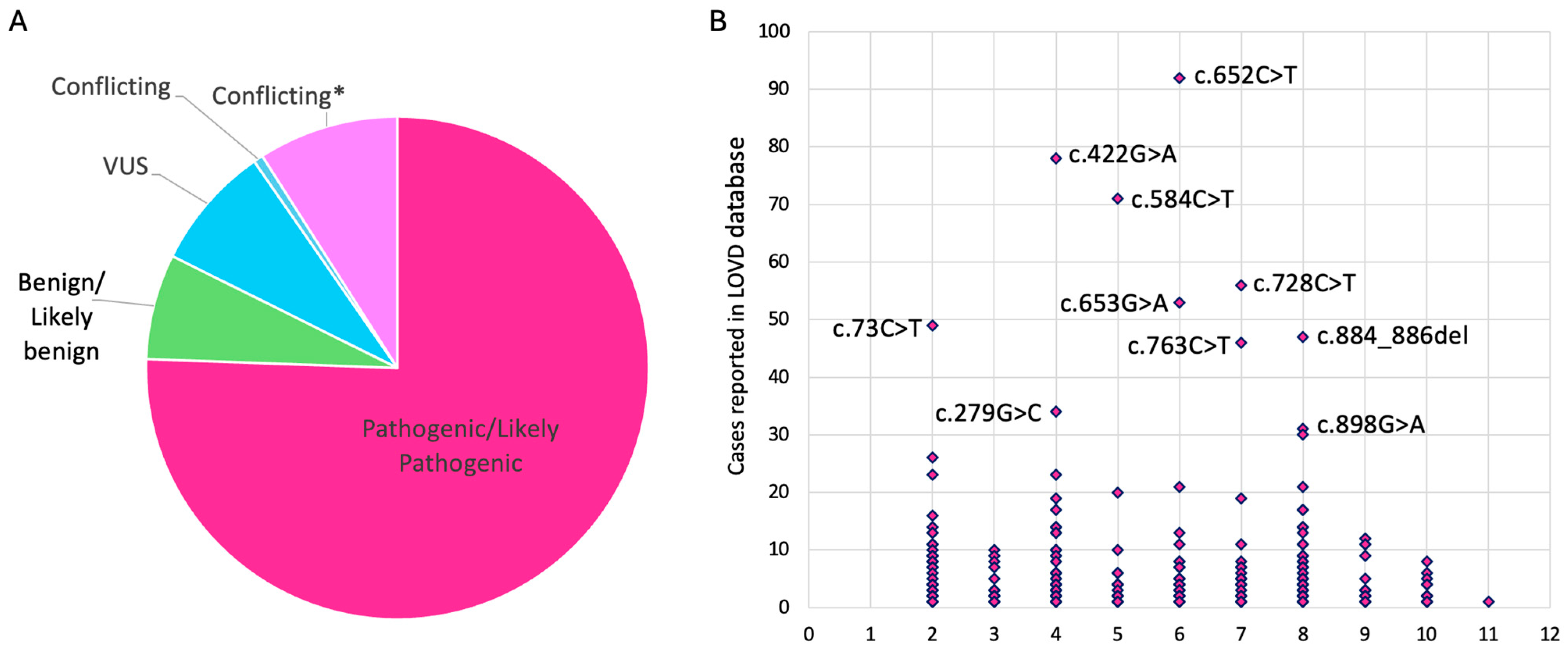
To date, more than 300 distinct variants have been associated with bestrophinopathies [10]. In the Leiden open-source variation database (LOVD) a total of 485 different variants can be found (accessed on 12 January 2024), of which 410 are labeled pathogenic/likely pathogenic or conflicting (Pathogenic/Likely pathogenic vs. Benign/Likely benign vs. uncertain significance) (Figure 2A,B).
The initial diagnosis of bestrophinopathies is based on the phenotypic appearance and a significantly reduced Arden ratio in the electrooculogram (EOG), a sign of comprised RPE function that is considered pathognomonic for these conditions [2].
Notably, bestrophinopathies present in at least five different distinct retinal phenotypes. This includes Best vitelliform macular dystrophy (BVMD), Adult vitelliform macular dystrophy (AVMD) [15,16], autosomal dominant vitreoretinochoroidopathy (ADVIRC), autosomal recessive bestrophinopathy (ARB) [17], and possibly retinitis pigmentosa (RP) [18], which are further described below.

3.1. Best Vitelliform Macular Dystrophy

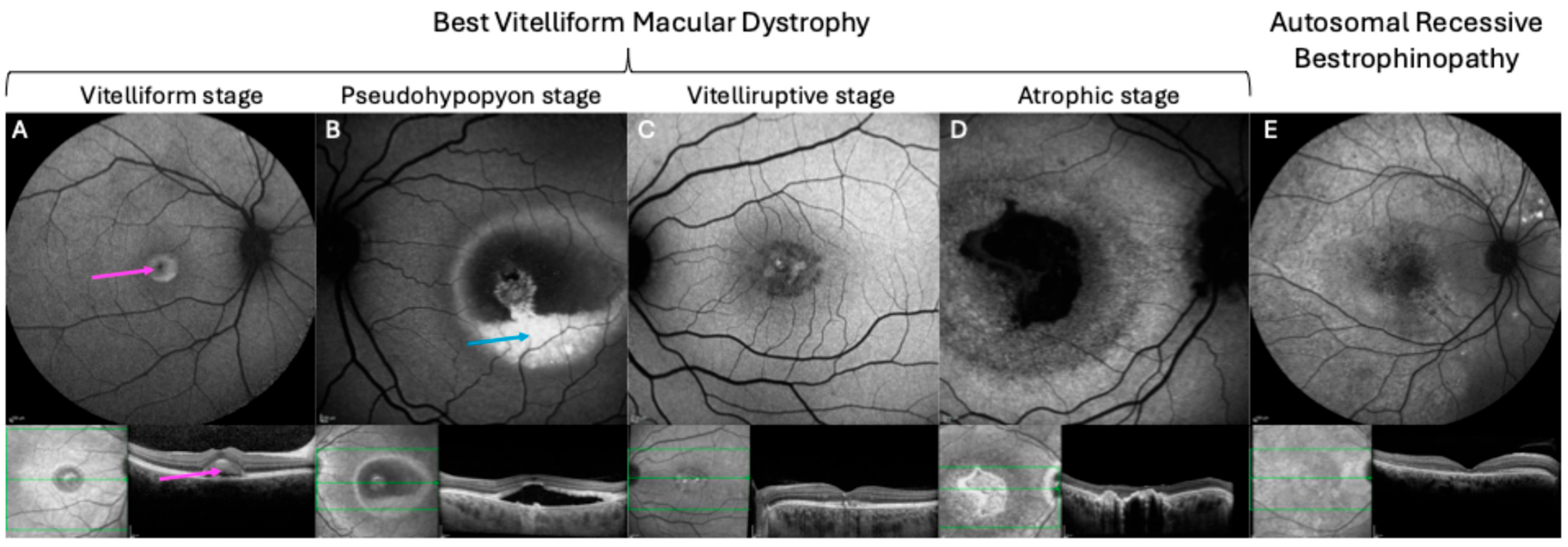
Best vitelliform macular dystrophy (BVMD), or Best disease, is the most common bestrophinopathy with an estimated prevalence between 1:5000 and 1:67,000 [19,20]. The disease was first described by Dr. Friedrich Best in 1905 and follows a dominant inheritance pattern with variable expression [21]. Clinically, BVMD is characterized by the formation of vitelliform lesions within the macula, that slowly progress over time [21,22]. Previous studies have outlined five different stages of disease progression, though they do not occur in all patients (Figure 3A–D).
In the first stage (pre-vitelliform stage), the fundus appears normal, aside from slight changes in the RPE, and the vision is often unaffected [23]. The second stage (vitelliform stage) is characterized by the formation of well-defined yellow, “egg yolk” vitelliform lesions in the macula. At this stage, visual acuity can be slightly decreased, and symptoms including photophobia, metamorphosis, and night blindness may occur [21,24]. In stage three (pseudohypopyon stage), layering of lipofuscin creates the appearance of a pseudohypopyon. However, visual acuity is often not dramatically affected. Further progression to the fourth stage (vitelliruptive stage) occurs when partial resorption and breakup of the vitelliform egg yolk lesion cause a “scrambled egg” appearance of the fundus. This stage is typically accompanied by the substantial loss of vision. The last stage of the disease (atrophic stage) is marked by the death of RPE and photoreceptor loss that causes severe, irreversible vision loss [23]. Notably, in most patients, vision loss progresses slowly, with 75% of the patients younger than 40 years maintaining a visual acuity of 6/12 or better in at least one eye [25]. However, malfunction of the RPE can trigger the growth of choroidal neovascularization (CNV), and subsequent rupture and bleeding of CNV may lead to a rapid decline of vision [26]. Typically, BVMD affects both eyes and presents with unifocal lesions. However, unilateral [26,27], multifocal [28], and asymmetric disease presentation have previously been described. Additionally, some studies have reported a link to other ophthalmological features including hyperopia, potentially resulting from reduced axial length linked to the role of BEST1 in ocular growth, as well as astigmatism and abnormalities of the anterior segment [22,29,30].

3.2. Adult Vitelliform Macular Dystrophy

Adult vitelliform macular dystrophy (AVMD) is an autosomal dominant inherited macula dystrophy with a similar phenotypic appearance to BVMD. However, AVMD is associated with a later age of onset (30–50 years), a slower disease progression, and a generally milder loss of vision. Importantly, due to the later age of onset, it is frequently misdiagnosed as age-related macular degeneration (AMD) [31].

3.3. Autosomal Recessive Bestrophinopathy

In contrast to the latter two conditions, autosomal recessive bestrophinopathy (ARB) follows an autosomal recessive inheritance pattern. Hence, ARB patients harbor pathogenic bi-allelic homozygous or compound heterozygous variants in the BEST1 gene and their parents often present with a normal fundus, EOG, and vision [17,21]. ARB was first described by Burgess et al. and proposed as the null phenotype of BEST1 in humans [17]. The authors identified two patients with a bi-allelic homozygous nonsense variant (Arg200X) as well as five patients with compound heterozygous mutations with following combinations: p.R141H and p.V317M (one patient), p.L41P and p.P152A (one patient), p.R141H and p.L41P (one patient), and p.D312N and p.M325T (two patients) [17]. More recently, a retrospective case series investigating 18 ARB patients from nine families identified bi-allelic homozygous BEST1 variants in 16 of the patients, while 2 patients harbored compound heterozygote variants [32]. Clinically, the fundus commonly displays multiple small vitelliform lesions, irregular RPE alterations, and whitish subretinal deposits, mainly found in the macula and midperiphery [21]. Further findings include, drusen and RPE atrophy in the retinal periphery, as well as subretinal fluid accumulation and macular edema [11,33].

3.4. Autosomal Dominant Vitreoretinochoroidopathy

Autosomal dominant vitreoretinochoroidopathy (ADVIRC) is a very rare condition (prevalence 1:1,000,000) that follows an autosomal dominant pattern of inheritance with intra-familial phenotypic variability [34,35]. The disease typically manifests during childhood and presents with a circumferential hyperpigmented band between the vortex veins and the ora serrata, a hallmark of the disease. Further findings include, preretinal punctate white opacities, retinal arteriolar narrowing, cystoid macular edema, and fibrillar condensation of the vitreous [36,37]. ADVIRC has also been associated with developmental eye conditions, such as nanophthalmos, microcornea, hyperopia, presenile cataract, a shallow anterior chamber, and optic nerve dysplasia [21,35,37]. Interestingly, previous studies have shown that ADVIRC is associated with BEST1 variants that cause exon skipping and ultimately lead to shortened and internally deleted isoforms [38].

3.5. Retinitis Pigmentosa

Mutations in the BEST1 gene that are associated with retinitis pigmentosa were initially described by Davidson et al. in 2009 [18]. The authors reported four missense variants in five unrelated families, of which three were autosomal dominant and one was autosomal recessive inherited [18]. The fundus of these patients exhibited dense pigmentary changes and bone spicules in the peripheral retina, retinal gliosis, and vascular attenuation [18]. However, it was recently proposed that these cases of BEST1-related retinitis pigmentosa may be misdiagnosed ADVIRC cases [32,39,40]. Alternatively, Dalvin et al. hypothesized that BEST1-associated RP may be a multigenic condition, thus requiring additional mutations in other genes [41].
However, despite each of the five BEST1-related conditions displaying a characteristic phenotype, bestrophinopathies present with a significant inter- and intra-familiar phenotypic variability, variable expression, and different age of disease onset. Hence, these factors challenge the diagnosis of bestrophinopathies and highlight the importance of genetic testing and thereby identification of the disease-causing variant.

4. Molecular Disease Mechanism of Pathogenic BEST1 Variants

The effect of a specific disease-causing variant on protein level, also known as their molecular disease mechanism, has recently been reviewed thoroughly by Backwell and Marsh [42], and are concisely summarized in the section below.
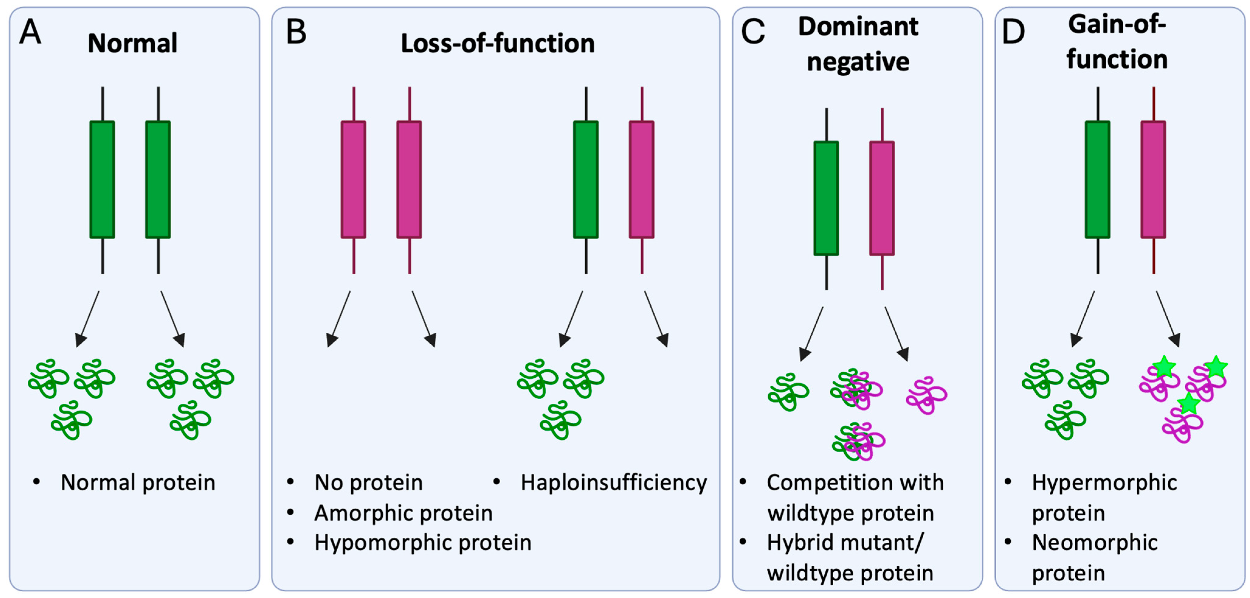
Briefly, molecular disease mechanisms of pathogenic variants can be divided into loss-of-function, dominant negative, and gain-of-function (Figure 4) [42].
Loss-of-function variants lead to (i) abolished protein synthesis, (ii) a non-functional protein (amorphic), or (iii) a protein with reduced function (hypomorphic). In most cases, loss-of-function variants are recessively inherited, as for instance BEST1 variants associated with ARB [10]. However, loss-of-function variants may be dominant in cases where haploinsufficiency is at play, hence where both alleles are required to produce a sufficient amount of functional protein. For dominant negative variants, the mutant protein impairs normal protein function by (i) competing with the wildtype protein or (ii) creating a hybrid wildtype–mutant complex, which inhibits the function of the wildtype protein. Notably, for pathogenic BEST1 variants, assembly of a mutated subunit into the pentameric channel complex has been shown to result in a non-functional or unstable protein, which is subjected to lysosomal degradation or decreases BEST1 channel function [10]. Furthermore, allelic expression imbalance, which is the higher transcription level of mutant allele compared to wildtype allele, has previously been described at the BEST1 locus of human RPE cells and has shown to promote the dominant negative effect of pathogenic BEST1 variants [43,44]. Pathogenic variants with a gain-of-function effect are mostly dominantly inherited and alter the encoded protein by either increasing its activity (hypermorphic) or introducing a novel function (neomorphic). Previous studies have provided evidence for a gain-of-function mechanism in ADVIRC-associated variants. For instance, Nachtigal et al. showed increased BEST1-related anion transport for induced pluripotent stem cell (iPSC)-RPE derived from ADVIRC patients (harboring an ADVIRC-associated variant) compared with control cell lines [10].

5. Gene Augmentation for the Treatment of Bestrophinopathies

To date, there are no treatment options available for any of the bestrophinopathies, and novel approaches are therefore urgently needed. Notably, the eye offers unique advantages for gene therapy, such as relative immune privilege, compartmentalization, and visual and surgical accessibility. Additionally, several characteristics appoint bestrophinopathies a suitable target for gene therapy: Firstly, given the slow progression of the disease and preservation of central photoreceptor function for a long period, a considerable therapeutic time window exists. Importantly, this treatment window may vary between patients, necessitating careful clinical evaluation of individual disease progression as well as a thorough evaluation of risks and benefits, particularly for patients with retained vision [45]. Secondly, quantifiable therapeutic endpoints like subretinal fluid and vitelliform lesions can readily be assessed with established methods such as optical coherence tomography (OCT) and autofluorescence.
Gene augmentation has already proven to be successful for the treatment of autosomal recessive inherited retinal diseases (IRD) and has experienced a major breakthrough with the FDA approval of Voretigene neparvovec for the treatment of bi-allelic RPE65 variants [3,4]. While gene augmentation for bestrophinopathies has not yet been evaluated in clinical trials, encouraging preclinical studies in cell and animal models suggest that this strategy may be feasible for recessive inherited bestrophinopathies. For instance, Li and colleagues demonstrated that baculovirus-mediated gene supplementation of wildtype BEST1 rescued the impaired Ca2+-dependent Cl current in iPSC-RPE cells derived from ARB patients [46] (Table 1). Furthermore, Guziewicz et al. showed that adeno-associated virus (AAV) 2-mediated BEST1 gene augmentation successfully reversed subretinal lesions and microdetachments and corrected structural alterations within the RPE/photoreceptor interface in a canine BEST1 disease model (Table 1) [47]. Recently, our group developed a quantitative chloride channel conductance assay that enables evaluation of AAV-delivered BEST1 gene augmentation, thereby offering a novel potency assay for future retinal gene therapy trials [48].
However, since most pathogenic BEST1 variants follow an autosomal dominant inheritance pattern, it is crucial to determine whether gene augmentation alone is sufficient for these conditions or whether simultaneous disruption of the mutant allele is required. Recently, Sinha et al. hypothesized that increasing the wildtype–mutant BEST1 ratio via gene augmentation may alleviate the disease phenotype in autosomal dominant inherited bestrophinopathies and tested this hypothesis in three iPSC-RPE models harboring various dominant BEST1 variants. Interestingly, while gene augmentation increased the BEST1 protein levels in all models, calcium-activated chloride channel activity was only restored in two of the three variants. However, CRISPR-Cas9-mediated silencing of the mutant allele established normal channel activity in all variants. This led to the authors proposal to approach treatment of autosomal dominant bestrophinopathies with a two-step strategy: (i) evaluation of gene augmentation in variant-specific patient iPSC-RPEs, and if not successful, (ii) evaluation of gene editing with CRISPR/Cas9-based suppression of the mutant allele (Table 1) [49].
However, given the labor-intensive nature of this approach, identifying characteristics of recessive and dominant bestrophinopathies that may benefit from either gene augmentation or gene editing would be of great interest.
Notably, several studies have proposed the molecular mechanism of pathogenic BEST1 variants as essential for the choice of the gene therapeutic treatment strategy. For instance, Ji and colleagues demonstrated that AAV2-delivered BEST1 gene augmentation restored protein expression levels and rescued the impaired Ca2+-dependent Cl current in iPSC-RPEs from patients with dominantly inherited BEST1 variants with a loss-of-function disease mechanism [50]. Intriguingly, the rescue efficacy of Ca2+-dependent Cl currents was similar to previously treated recessive variants (Table 1) [50].
Furthermore, a recent study by Zhao et al. investigated the therapeutic strategies for BEST1 loss-of-function and gain-of-function variants in human pluripotent stem cell-derived RPE (hPSC-RPE). Importantly, the results demonstrated that BEST1 gain-of-function variants in hPSC-RPE could not be rescued with gene augmentation alone (Table 1). However, the authors observed that autosomal BEST1 variants, previously thought to follow a loss-of-function mechanism, exhibited dominant negative behavior, primarily promoted by allelic expression imbalance [44]. Hence, these variants may be treatable with gene augmentation but potentially require a higher dose to dilute the effect of the mutant protein. Alternatively, including a Woodchuck hepatitis virus posttranscriptional regulatory element (WPRE) in the vector construct could be an option to boost BEST1 expression sufficiently [48].
Thus, dominant and recessive inherited variants with a loss-of-function or dominant negative molecular disease mechanisms may be candidates for gene augmentation, while variants acting through a gain-of-function mechanism require a different treatment strategy (Figure 5).

6. CRISPR-Based Treatment Strategies for Pathogenic BEST1 Variants

The clustered regularly interspaced short palindromic repeats (CRISPR)/Cas9 technology, holds enormous potential for the correction of genetic conditions, and has revolutionized the field of genome engineering and molecular medicine during the last decade. Originally, CRISPR/Cas9 is a natural defense system in bacteria, which provides adaptive immunity against foreign evaders [51]. Since its discovery, researchers have repurposed this system as a versatile tool for gene editing therapy, and in 2020 Jennifer Doudna and Emmanuele Charpentier were awarded the Nobel Prize in Chemistry for its discovery [52]. Encouragingly, CRISPR/Cas 9-based gene editing is showing significant potential to correct genetic disorders directly in the retina, and a phase I/II clinical trial for Edit-101; a CRISPR-based treatment for Leber congenital amaurosis (LCA) type 10 has shown a promising safety profile and improvement in photoreceptor function [53,54].
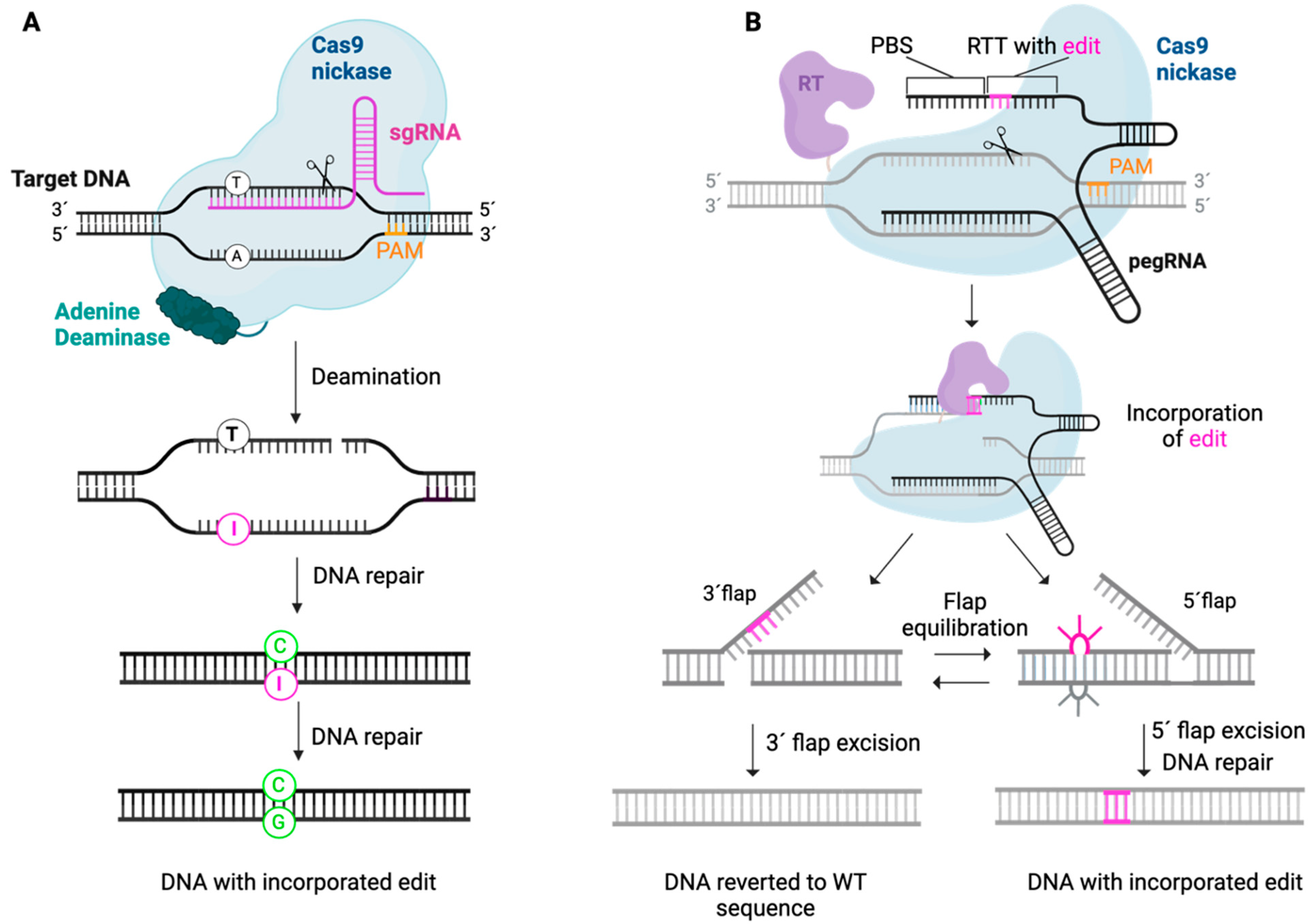
Essentially, the CRISPR/Cas9 technology consists of two critical elements: (i) the Cas9 nuclease and (ii) a single-guide RNA (sgRNA) that defines the DNA target by a 17–20 nucleotide complementary sequence (Figure 6). Upon reaching the targeted region, the Cas9/sgRNA complex promotes a double-strand break (DSB) in correlation to a protospacer adjacent motif (PAM) site. Subsequently, the cellular DNA repair machinery may attempt to repair the DSB by two major pathways: non-homologous end-joining pathway (NHEJ) or homology-directed repair (HDR) [55]. NHEJ rapidly ligates the DNA ends in a donor template-independent manner, thereby producing unpredictable insertions and deletions (indels). These indels can cause transcriptional frameshifts that induce premature stop codons and/or activate nonsense-mediated decay (NMD), which ultimately lead to degradation of the transcript and silencing of the targeted gene [56]. In contrast, the HDR pathway requires a donor template, thereby enabling the precise correction of a mutation of interest. Recently, the CRISPR toolbox has been further expanded by the discovery of base editing and prime editing, which enable the precise correction of variants in a DSB- and donor template-independent manner (Figure 7).
To address the dominant effect of gain-of-function variants, three different CRISPR-based strategies can be employed: (i) specific silencing of the disease-causing allele, (ii) silencing/knock down of both endogenous alleles and augmentation of the wildtype gene, or (iii) precise correction of the disease-causing variant. These approaches are further elaborated in the section below.

6.1. Specific Silencing of Disease-Causing Allele

6.1.1. CRISPR/Cas9-Mediated Gene Silencing

Recently, Sinha et al. utilized the CRISPR/Cas9 technology to specifically silence the disease-causing allele of three different dominant inherited BEST1 variants. Following lentiviral-mediated delivery of Cas9 and sgRNA targeting the relevant mutant loci, editing rates of up to 80% were detected. Notably, up to 98% of the indels resulted in desired frameshift mutations and thereby targeted silencing. Furthermore, single-cell patch-clamp experiments demonstrated restoration of CaCC activity and thereby functional rescue of BEST1 channel activity (Table 1) [49]. However, despite the potential of this approach for some pathogenic variants, it may not be feasible in cases where the presence of both healthy alleles is crucial and haploinsufficiency is at play. Furthermore, as a mutation-specific strategy, it would require experimental evaluation of a working sgRNA for each variant.

6.1.2. Targeting of Allele-Specific Single Nucleotide Polymorphisms

To achieve mutation independence and allele specificity, a CRISPR/Cas9 strategy targeting single nucleotide polymorphisms (SNPs) specific for the disease allele could be applied. Recently, Salman et al. used this strategy to target AMD-related SNPs in differentiated ARPE-19 cells and demonstrated allele-specific targeting with efficacies of up to 50% [57]. To the best of our knowledge, this strategy has not yet been explored for BEST1 but may represent an appealing option.

6.2. Ablation-And-Replace Strategy

To circumvent the issue of haploinsufficiency, the “ablate and replace strategy” can be employed. This strategy involves (i) non-selective silencing of both endogenous alleles and (ii) simultaneous supplementation of the exogenous wildtype gene. Encouragingly, this strategy has shown promising results in preclinical animal studies as a treatment strategy for autosomal dominant inherited IRDs such as autosomal dominant retinitis pigmentosa [58].
Recently, Zhao et al. utilized a similar strategy that combined transcriptional repression of endogenous BEST1 with augmentation of wildtype BEST1 for the treatment of BEST1 gain-of-function variants. For this purpose, a nuclease-dead Cas9 with a bipartite KRAB-MeCP2 repressor domain (dCas9-KRAB-MeCP2) and a sgRNA targeting both endogenous alleles were delivered to human pluripotent stem cells (hPSC)-derived RPE (hPSC-RPE) cells using two separate baculovirus vectors. The authors demonstrated sufficient repression of the endogenous BEST1 gene and restoration of the Ca2+-dependent Cl currents by the exogenous wildtype BEST1 gene (Table 1) [44]. Similarly, our research group has developed a novel mirtron-based “knockdown and replace strategy” for the treatment of rhodopsin-related dominant retinitis pigmentosa (ADRP). Mirtrons are atypical RNA interference (RNAi) effectors that are spliced from transcripts of short introns, thereby bypassing the Drosha/DGCR8 processing step of canonical micro RNAs (miRNAs). Importantly, compared to canonical RNAi effectors, mirtrons have several advantages: (i) Drosha/DGCR8-independent procession results in greater accuracy and may lower the risk of cellular toxicity related to oversaturation of the endogenous microRNA pathway and (ii) expression by Pol-II promotors enables cell-specific transgene expression. In this study, a single vector encoding both rhodopsin-targeting mirtrons and a mirtron-resistant coon-modified version of the rhodopsin coding sequence were delivered via subretinal injection and demonstrated slowing of retinal degeneration in a Rho23 knock-in mouse model of ADRP [59]. To the best of our knowledge, this strategy has not been investigated for BEST1 but could be an attractive option. Further benefits of the “ablate/knockdown and replace strategy” include its potential as a universal treatment strategy for all bestrophinopathies, as it is a mutation-independent approach.

6.3. Precise Correction of Disease-Causing Variant

6.3.1. Homology-Directed Repair

The homology-directed repair (HDR)-based CRISPR/Cas9 strategy enables the precise correction of a disease-causing variant in the presence of a DNA donor template. Notably, recent studies have demonstrated precise correction of pathogenic variants in USH2A [60] and ABCA4 [61] in human iPSCs, utilizing a single-stranded oligodeoxynucleotide (ssODN) donor template. However, for many of the preclinical studies conducted in the retina, the HDR-based approach yielded low efficacy. This is possibly due to the postmitotic nature of the retinal cells and the fact that HDR is exclusively active in the late S and G2 phases of the cell cycle [62,63].

6.3.2. Homology-Independent Targeted Integration

An attractive alternative is provided by the homology-independent targeted integration (HITI) strategy, which is based on the NHEJ-repair pathway and enables effective knock-in of a DNA template in postmitotic cells [64]. In contrast to HDR, HITI employs a donor plasmid without homology arms, instead featuring Cas9 cleavage sites that flank the DNA template. This allows for the simultaneous cleavage of both the target DNA sequence and the donor sequence, followed by the integration of donor DNA into the targeted loci (Figure 6) [65].
In the eye, this strategy has been shown to successfully improve vision in a rat model for retinitis pigmentosa [64]. Recently, Meng et al. used HITI to facilitate the precise knock-in of exon 7–11 of the CYP4V2 gene, a mutation hotspot for Bietti crystalline corneoretinal dystrophy (BCD) [66]. The HITI donor was designed to integrate in intron 6 and contained part of intro 6, the correct sequence of exon 7–11 and a stop codon to mitigate transcription of the disease-causing variants. Encouragingly, the authors showed precise DNA knock-in in BCD-derived patient iPSCs and restauration of viability in iPSC-RPE. Furthermore, HITI-based editing was applied in a humanized Cyp4v3 mouse model, using dual delivery by subretinal injection. Notably, this led to improved morphology, number and metabolism of RPE and photoreceptors, and improvement of visual function. To our knowledge, the HITI strategy has not been evaluated for the correction of pathogenic BEST1 variants but could be applied to treat mutation hotspots clustering to specific exons.
However, the Cas9-based gene editing strategies described above rely on the formation of a DSB, which can cause several genotoxic events, including off-target cleavage, chromosomal rearrangement, unintended deletions, chromothripsis, and activation of the p53 pathway [67,68,69,70]. These risks may be overcome by the novel CRISPR-based gene editing tools: base editing and prime editing (Figure 7).

6.3.3. Base Editing

Besides a target-site-defining sgRNA, the base editing technology consists of two components that differ from the above described CRISPR/Cas9-based methods: (i) the Cas9 (D10A) nickase (nCas9), which harbors the RuvC inactivating D10A mutation that promotes nicking of the complementary strand and (ii) a deaminase enzyme inducing the desired base substitution (Figure 7A) [71,72]. Jointly, cytosine base editors (CBEs) and adenosis base editors (ABEs) enable the correction of all four transition mutations and 2 of the 12 transversion mutations [73]. Encouragingly, two recent studies from our group nominated base editing a relevant strategy to correct pathogenic variants in large genes associated with IRDs [74] and RHO variants causative for autosomal dominant retinitis pigmentosa [75]. Furthermore, Suh et al. employed base editing to correct a nonsense mutation in Rpe65 in a murine model in vivo. Subretinal injection of a lentiviral vector encoding base editing components resulted in editing efficacies of 29%, restored RPE65 protein expression, and improved visual function [76]. Recently, Muller and colleagues utilized base editing to correct the most common ABCA4 mutation (c.5882G > A, p.G1961E), causative for Stargardt disease. Firstly, the authors optimized this strategy in relevant models in vitro, including retinal organoids, induced pluripotent stem cell-derived RPE cells, human retinal explants, and RPE/choroid explants using a dual AAV delivery strategy. Subsequently, they demonstrated high levels of gene correction in mice and non-human primates in vivo, reaching an average editing efficacy of 75% in cones and 87% in RPE cells [77]. To our knowledge, the base editing strategy has not been evaluated for the correction of pathogenic BEST1 variants. Encouragingly, base editing could theoretically be used to correct a large number of pathogenic BEST1 point mutations, as for instance the three most frequently reported pathogenic variants from the LOVD (p.R218C, p.R142H and p.A195V). However, the availability of PAM sites within 15 +/− 2 nt from the targeted nucleotide, the target window, and potential bystander edits will need to be taken into consideration and provide a limitation of this technology [78].

6.3.4. Prime Editing

The prime editing technology enables correction of all point mutations, smaller insertions and deletions, and mitigates the risk for bystander edits (Figure 7B) [79]. Intriguingly, prime editing has already been employed to target IRD-causing variants in several preclinical studies. For instance, Jang et al. demonstrated editing efficacies of 6.4% and functional rescue of the disease phenotype in the rd12 mouse model, after lentiviral-mediated delivery of the prime editing components [80]. Furthermore, Qin and colleagues developed PESpRY, a novel prime editor with unconstrained PAM requirements [81]. Encouragingly, the authors achieved preservation of photoreceptors and rescue of visual function in a mouse model of retinitis pigmentosa, after delivery via a dual AAV system. With its large target scope, prime editing has the potential to correct the majority of pathogenic BEST1 variants including deletions (i.e., p.I295del). However, due to its large size (>6.3 kB), delivery of the prime editor to the retina provides a challenge, and further evaluation of efficacy and safety are required before moving forward to clinical application [70].
Figure 7. Schematic overview of the base editing and prime editing technology. (A) Base editors consist of a sgRNA, a Cas9 nickase, and a deaminase. Upon cleavage of the complementary strand by the Cas9 nickase, the adenine deaminase mediates the transition from A to I (indicated in pink), which is recognized by the cell as G. DNA repair leads to installation of C on the opposite strand (indicated in green) and subsequently installation of G on the deaminated strand (indicated in green). (B) Prime editors consist of a Cas9 nickase fused to a reverse transcriptase (RT) and a pegRNA that encodes the desired edit (indicated in pink). Upon cleavage of the non-complementary strand of the target DNA, the PBS of the pegRNA binds to the target DNA and enables the RT to start reverse transcription and thereby incorporation of the edit in this strand. Subsequently, cellular repair mechanisms enable incorporation of the edited DNA 3’flap or the unedited 5’flap, thereby installing the desired edit or reversion to the WT sequence, respectively. A, adenine; C, cytidine; G, guanine; I, inosine; RT, reverse transcriptase; RTT, RT-template; PBS, primer-binding site.
Ijms 26 09421 g007

7. Conclusions

Bestrophinopathies form a group of inherited retinal diseases with a high complexity regarding their genetic inheritance and clinical appearance. Gene therapy could be a potential treatment strategy for these conditions, and the molecular disease mechanism of the distinct variant could help to further inform the choice of treatment strategy, as illustrated in the proposed workflow from clinical diagnosis to treatment selection in Figure 5. Investigating BEST1-related pathogenesis and its correlation to disease phenotypes may further help to choose a relevant treatment. For instance, Nachtigal et al. characterized the cellular and molecular mechanisms underlying the distinct pathologies of ARB, BVMD, and ADVIRC in patient-derived hiPSC-RPE, and developed a classification system for bestrophinopathies, based on these results [10]. Encouragingly, gene augmentation may be a feasible treatment option for BEST1 variants with loss-of-function disease mechanisms, which constitute the majority of all identified variants [44]. Additionally, preclinical evidence suggests that BEST1 variants, in which the dominant negative effect is promoted by allelic expression imbalance, may be treatable with gene augmentation, potentially at a higher dose. Lastly, for variants with a gain-of-function mechanism, CRISPR/Cas9 gene editing strategies may provide an interesting alternative. Mutation-independent CRISPR-based strategies are applicable to a wide range of BEST1 mutations, thereby providing universal treatment for several bestrophinopathies and making them potentially attractive from a commercial perspective. In contrast, mutation-specific strategies require customization for the pathogenic variant of interest, but they offer the potential to restore native gene function.
Collectively, the spectrum of these novel gene therapeutic strategies may provide a useful toolbox to treat a broad range of bestrophinopathies, conditions which are currently untreatable. However, further studies are needed to evaluate their efficacy and safety and optimize retinal delivery. This includes investigation of efficacy in model systems for bestrophinopathies, as for instance patient-derived iPSC-RPE, which enable disease-in-a dish modeling for a specific variant [22]. Furthermore, a thorough off-target analysis should be performed, to ensure high specificity of the gene therapeutic approach. To enable efficient and safe delivery of the therapeutic cargo to the RPE, delivery vehicles should be optimized and tested for the gene therapeutic strategy of choice. AAV-mediated gene augmentation has demonstrated high transduction efficacy and sustained transgene expression in the retina in several clinical trials [82,83] and may be suitable for BEST1 gene augmentation. In contrast, the large size of gene editing tools including prime editors (>6.3 kb) and base editors (approx. 5 kb) exceed the packaging capacity of a single AAV vector. Alternative strategies to circumvent this limitation include delivery via (i) dual AAV vectors [77,84], (ii) lentiviral vectors [85], (iii) ribonucleoproteins via engineered DNA-free virus-like particles [86,87], or (iv) non-viral delivery vehicles i.e., lipid nanoparticle formations [88]. These approaches have been extensively reviewed by others and are beyond the scope of this work [89,90].
In conclusion, the expanding repertoire of gene therapeutic approaches holds a significant potential to address the treatment of bestrophinopathies, and the molecular disease mechanism of the pathogenic variant may serve as an important parameter for the choice of treatment strategy. However, further research is necessary to enable safe and efficient clinical translation of these novel methods.

Author Contributions

Conceptualization, S.B.H., M.E.M., T.J.C., J.C.-K. and R.E.M.; writing—original draft preparation, S.B.H.; writing—review and editing, S.B.H., M.E.M., T.J.C., J.C.-K. and R.E.M.; supervision M.E.M., T.J.C., J.C.-K. and R.E.M.; project administration, S.B.H., M.E.M., T.J.C., J.C.-K. and R.E.M.; funding acquisition, S.B.H., T.J.C. and R.E.M. All authors have read and agreed to the published version of the manuscript.

Funding

This research was funded by the Aarhus University, Fight for Sight Denmark, Synoptik Fonden, Maskinfabrikant Jochum Jensen og hustru Mette Marie Jensen, f. Poulsens Mindelegat (fond), A.P. Møller Foundation, Mette Warburgs Fond, APT Holding, the VELUX Foundation [00038189 and 48176], the Novo Nordisk Foundation [Grant No. 24OC0088426], the NIHR Oxford Biomedical Research Centre and the Wellcome Discovery Award [304408/Z/23/Z].

Institutional Review Board Statement

Not applicable.

Informed Consent Statement

Not applicable.

Acknowledgments

Figures are created with BioRender.com (accessed on 23 July 2025) and powerpoint. Figure 1A Created in BioRender. Hansen, S. (2025) https://BioRender.com/r81wvt5; Figure 1B Created in BioRender. Hansen, S. (2025) https://BioRender.com/1h4tdil; Figure 4 Created in BioRender. Hansen, S. (2025) https://BioRender.com/sse1taj; Figure 5 Created in BioRender. Hansen, S. (2025) https://BioRender.com/i3s8ln4; Figure 6, Created in BioRender. Hansen, S. (2025) https://BioRender.com/bdq6y80; Figure 7 Created in BioRender. Hansen, S. (2025) https://BioRender.com/6b8b2l7 (all accessed on 23 July 2025).

Conflicts of Interest

The authors declare no conflicts of interest.

References

  1. Amato, A.; Arrigo, A.; Aragona, E.; Manitto, M.P.; Saladino, A.; Bandello, F.; Battaglia Parodi, M. Gene Therapy in Inherited Retinal Diseases: An Update on Current State of the Art. Front. Med. 2021, 8, 750586. [Google Scholar] [CrossRef]
  2. Johnson, A.A.; Guziewicz, K.E.; Lee, C.J.; Kalathur, R.C.; Pulido, J.S.; Marmorstein, L.Y.; Marmorstein, A.D. Bestrophin 1 and retinal disease. Prog. Retin. Eye Res. 2017, 58, 45–69. [Google Scholar] [CrossRef]
  3. Chung, D.C.; Lee, V.; Maguire, A.M. Recent advances in ocular gene therapy. Curr. Opin. Ophthalmol. 2009, 20, 377–381. [Google Scholar] [CrossRef]
  4. Maguire, A.M.; Russell, S.; Chung, D.C.; Yu, Z.-F.; Tillman, A.; Drack, A.V.; Simonelli, F.; Leroy, B.P.; Reape, K.Z.; High, K.A.; et al. Durability of Voretigene Neparvovec for Biallelic RPE65-Mediated Inherited Retinal Disease: Phase 3 Results at 3 and 4 Years. Ophthalmology 2021, 128, 1460–1468. [Google Scholar] [CrossRef]
  5. Marmorstein, A.D.; Marmorstein, L.Y.; Rayborn, M.; Wang, X.; Hollyfield, J.G.; Petrukhin, K. Bestrophin, the product of the Best vitelliform macular dystrophy gene (VMD2), localizes to the basolateral plasma membrane of the retinal pigment epithelium. Proc. Natl. Acad. Sci. USA 2000, 97, 12758–12763. [Google Scholar] [CrossRef]
  6. Kane Dickson, V.; Pedi, L.; Long, S.B. Structure and insights into the function of a Ca2+-activated Cl channel. Nature 2014, 516, 213–218. [Google Scholar] [CrossRef]
  7. Marmorstein, A.D.; Kinnick, T.R.; Stanton, J.B.; Johnson, A.A.; Lynch, R.M.; Marmorstein, L.Y. Bestrophin-1 influences transepithelial electrical properties and Ca2+ signaling in human retinal pigment epithelium. Mol. Vis. 2015, 21, 347–359. [Google Scholar]
  8. Owji, A.P.; Wang, J.; Kittredge, A.; Clark, Z.; Zhang, Y.; Hendrickson, W.A.; Yang, T. Structures and gating mechanisms of human bestrophin anion channels. Nat. Commun. 2022, 13, 3836. [Google Scholar] [CrossRef]
  9. Ji, C.; Kittredge, A.; Hopiavuori, A.; Ward, N.; Chen, S.; Fukuda, Y.; Zhang, Y.; Yang, T. Dual Ca2+-dependent gates in human Bestrophin1 underlie disease-causing mechanisms of gain-of-function mutations. Commun. Biol. 2019, 2, 240. [Google Scholar] [CrossRef]
  10. Nachtigal, A.L.; Milenkovic, A.; Brandl, C.; Schulz, H.L.; Duerr, L.M.J.; Lang, G.E.; Reiff, C.; Herrmann, P.; Kellner, U.; Weber, B.H.F. Mutation-Dependent Pathomechanisms Determine the Phenotype in the Bestrophinopathies. Int. J. Mol. Sci. 2020, 21, 1597. [Google Scholar] [CrossRef]
  11. Casalino, G.; Khan, K.N.; Armengol, M.; Wright, G.; Pontikos, N.; Georgiou, M.; Webster, A.R.; Robson, A.G.; Grewal, P.S.; Michaelides, M. Autosomal Recessive Bestrophinopathy: Clinical Features, Natural History, and Genetic Findings in Preparation for Clinical Trials. Ophthalmology 2021, 128, 706–718. [Google Scholar] [CrossRef]
  12. Strauß, O.; Müller, C.; Reichhart, N.; Tamm, E.R.; Gomez, N.M. The role of bestrophin-1 in intracellular Ca2+ signaling. Adv. Exp. Med. Biol. 2014, 801, 113–119. [Google Scholar] [CrossRef]
  13. Gómez, N.M.; Tamm, E.R.; Strauβ, O. Role of bestrophin-1 in store-operated calcium entry in retinal pigment epithelium. Pflugers Arch. 2013, 465, 481–495. [Google Scholar] [CrossRef]
  14. Fischmeister, R.; Hartzell, H.C. Volume sensitivity of the bestrophin family of chloride channels. J. Physiol. 2005, 562, 477–491. [Google Scholar] [CrossRef]
  15. Rahman, N.; Georgiou, M.; Khan, K.N.; Michaelides, M. Macular dystrophies: Clinical and imaging features, molecular genetics and therapeutic options. Br. J. Ophthalmol. 2020, 104, 451–460. [Google Scholar] [CrossRef]
  16. Krämer, F.; White, K.; Pauleikhoff, D.; Gehrig, A.; Passmore, L.; Rivera, A.; Rudolph, G.; Kellner, U.; Andrassi, M.; Lorenz, B.; et al. Mutations in the VMD2 gene are associated with juvenile-onset vitelliform macular dystrophy (Best disease) and adult vitelliform macular dystrophy but not age-related macular degeneration. Eur. J. Hum. Genet. 2000, 8, 286–292. [Google Scholar] [CrossRef]
  17. Burgess, R.; Millar, I.D.; Leroy, B.P.; Urquhart, J.E.; Fearon, I.M.; De Baere, E.; Brown, P.D.; Robson, A.G.; Wright, G.A.; Kestelyn, P.; et al. Biallelic mutation of BEST1 causes a distinct retinopathy in humans. Am. J. Hum. Genet. 2008, 82, 19–31. [Google Scholar] [CrossRef]
  18. Davidson, A.E.; Millar, I.D.; Urquhart, J.E.; Burgess-Mullan, R.; Shweikh, Y.; Parry, N.; O’Sullivan, J.; Maher, G.J.; McKibbin, M.; Downes, S.M.; et al. Missense Mutations in a Retinal Pigment Epithelium Protein, Bestrophin-1, Cause Retinitis Pigmentosa. Am. J. Hum. Genet. 2009, 85, 581–592. [Google Scholar] [CrossRef]
  19. Nordström, S. Hereditary macular degeneration—A population survey in the country of Vsterbotten, Sweden. Hereditas 1974, 78, 41–62. [Google Scholar] [CrossRef]
  20. Bitner, H.; Schatz, P.; Mizrahi-Meissonnier, L.; Sharon, D.; Rosenberg, T. Frequency, genotype, and clinical spectrum of best vitelliform macular dystrophy: Data from a national center in Denmark. Am. J. Ophthalmol. 2012, 154, 403–412.e404. [Google Scholar] [CrossRef]
  21. Boon, C.J.; Klevering, B.J.; Leroy, B.P.; Hoyng, C.B.; Keunen, J.E.; den Hollander, A.I. The spectrum of ocular phenotypes caused by mutations in the BEST1 gene. Prog. Retin. Eye Res. 2009, 28, 187–205. [Google Scholar] [CrossRef]
  22. Singh Grewal, S.; Smith, J.J.; Carr, A.F. Bestrophinopathies: Perspectives on clinical disease, Bestrophin-1 function and developing therapies. Ther. Adv. Ophthalmol. 2021, 13, 2515841421997191. [Google Scholar] [CrossRef]
  23. Marmorstein, A.D.; Cross, H.E.; Peachey, N.S. Functional roles of bestrophins in ocular epithelia. Prog. Retin. Eye Res. 2009, 28, 206–226. [Google Scholar] [CrossRef]
  24. Renner, A.B.; Tillack, H.; Kraus, H.; Kohl, S.; Wissinger, B.; Mohr, N.; Weber, B.H.; Kellner, U.; Foerster, M.H. Morphology and functional characteristics in adult vitelliform macular dystrophy. Retina 2004, 24, 929–939. [Google Scholar] [CrossRef]
  25. Fishman, G.A.; Baca, W.; Alexander, K.R.; Derlacki, D.J.; Glenn, A.M.; Viana, M. Visual acuity in patients with best vitelliform macular dystrophy. Ophthalmology 1993, 100, 1665–1670. [Google Scholar] [CrossRef]
  26. Arora, R.; Khan, K.; Kasilian, M.L.; Strauss, R.W.; Holder, G.E.; Robson, A.G.; Thompson, D.A.; Moore, A.T.; Michaelides, M. Unilateral BEST1-Associated Retinopathy. Am. J. Ophthalmol. 2016, 169, 24–32. [Google Scholar] [CrossRef]
  27. Kaden, T.R.; Tan, A.C.; Feiner, L.; Freund, K.B. UNILATERAL BEST DISEASE: A CASE REPORT. Retin. Cases Brief. Rep. 2017, 11 (Suppl. S1), S191–S196. [Google Scholar] [CrossRef]
  28. Querques, G.; Regenbogen, M.; Soubrane, G.; Souied, E.H. High-resolution spectral domain optical coherence tomography findings in multifocal vitelliform macular dystrophy. Surv. Ophthalmol. 2009, 54, 311–316. [Google Scholar] [CrossRef] [PubMed]
  29. Wittström, E.; Ekvall, S.; Schatz, P.; Bondeson, M.L.; Ponjavic, V.; Andréasson, S. Morphological and functional changes in multifocal vitelliform retinopathy and biallelic mutations in BEST1. Ophthalmic Genet. 2011, 32, 83–96. [Google Scholar] [CrossRef] [PubMed]
  30. Coussa, R.G.; Binkley, E.M.; Wilkinson, M.E.; Andorf, J.L.; Tucker, B.A.; Mullins, R.F.; Sohn, E.H.; Yannuzzi, L.A.; Stone, E.M.; Han, I.C. Predominance of hyperopia in autosomal dominant Best vitelliform macular dystrophy. Br. J. Ophthalmol. 2022, 106, 522–527. [Google Scholar] [CrossRef] [PubMed]
  31. Menchini, U.; Giacomelli, G.; Cappelli, S.; Giansanti, F.; Romani, A. Photodynamic Therapy in Adult-Onset Vitelliform Macular Dystrophy Misdiagnosed as Choroidal Neovascularization. Arch. Ophthalmol. 2002, 120, 1761–1763. [Google Scholar]
  32. Shah, M.; Broadgate, S.; Shanks, M.; Clouston, P.; Yu, J.; MacLaren, R.E.; Németh, A.H.; Halford, S.; Downes, S.M. Association of Clinical and Genetic Heterogeneity With BEST1 Sequence Variations. JAMA Ophthalmol. 2020, 138, 544–551. [Google Scholar] [CrossRef]
  33. Gerth, C.; Zawadzki, R.J.; Werner, J.S.; Héon, E. Detailed analysis of retinal function and morphology in a patient with autosomal recessive bestrophinopathy (ARB). Doc. Ophthalmol. 2009, 118, 239–246. [Google Scholar] [CrossRef]
  34. Landrum, M.J.; Lee, J.M.; Benson, M.; Brown, G.R.; Chao, C.; Chitipiralla, S.; Gu, B.; Hart, J.; Hoffman, D.; Jang, W.; et al. ClinVar: Improving access to variant interpretations and supporting evidence. Nucleic Acids Res. 2018, 46, D1062–D1067. [Google Scholar] [CrossRef] [PubMed]
  35. Vincent, A.; McAlister, C.; Vandenhoven, C.; Héon, E. BEST1-related autosomal dominant vitreoretinochoroidopathy: A degenerative disease with a range of developmental ocular anomalies. Eye 2011, 25, 113–118. [Google Scholar] [CrossRef] [PubMed]
  36. Kaufman, S.J.; Goldberg, M.F.; Orth, D.H.; Fishman, G.A.; Tessler, H.; Mizuno, K. Autosomal dominant vitreoretinochoroidopathy. Arch. Ophthalmol. 1982, 100, 272–278. [Google Scholar] [CrossRef]
  37. Yardley, J.; Leroy, B.P.; Hart-Holden, N.; Lafaut, B.A.; Loeys, B.; Messiaen, L.M.; Perveen, R.; Reddy, M.A.; Bhattacharya, S.S.; Traboulsi, E.; et al. Mutations of VMD2 splicing regulators cause nanophthalmos and autosomal dominant vitreoretinochoroidopathy (ADVIRC). Investig. Ophthalmol. Vis. Sci. 2004, 45, 3683–3689. [Google Scholar] [CrossRef]
  38. Burgess, R.; MacLaren, R.E.; Davidson, A.E.; Urquhart, J.E.; Holder, G.E.; Robson, A.G.; Moore, A.T.; Keefe, R.O.; Black, G.C.; Manson, F.D. ADVIRC is caused by distinct mutations in BEST1 that alter pre-mRNA splicing. J. Med. Genet. 2009, 46, 620–625. [Google Scholar] [CrossRef]
  39. Traboulsi, E. Genetic Diseases of the Eye, 2nd ed.; Oxford University Press: New York, NY, USA, 2012. [Google Scholar]
  40. Boulanger-Scemama, E.; Sahel, J.A.; Mohand-Said, S.; Antonio, A.; Condroyer, C.; Zeitz, C.; Audo, I. AUTOSOMAL DOMINANT VITREORETINOCHOROIDOPATHY: When Molecular Genetic Testing Helps Clinical Diagnosis. Retina 2019, 39, 867–878. [Google Scholar] [CrossRef] [PubMed]
  41. Dalvin, L.A.; Abou Chehade, J.E.; Chiang, J.; Fuchs, J.; Iezzi, R.; Marmorstein, A.D. Retinitis pigmentosa associated with a mutation in BEST1. Am. J. Ophthalmol. Case Rep. 2016, 2, 11–17. [Google Scholar] [CrossRef]
  42. Backwell, L.; Marsh, J.A. Diverse Molecular Mechanisms Underlying Pathogenic Protein Mutations: Beyond the Loss-of-Function Paradigm. Annu. Rev. Genom. Hum. Genet. 2022, 23, 475–498. [Google Scholar] [CrossRef]
  43. Llavona, P.; Pinelli, M.; Mutarelli, M.; Marwah, V.S.; Schimpf-Linzenbold, S.; Thaler, S.; Yoeruek, E.; Vetter, J.; Kohl, S.; Wissinger, B. Allelic Expression Imbalance in the Human Retinal Transcriptome and Potential Impact on Inherited Retinal Diseases. Genes. 2017, 8, 283. [Google Scholar] [CrossRef]
  44. Zhao, Q.; Kong, Y.; Kittredge, A.; Li, Y.; Shen, Y.; Zhang, Y.; Tsang, S.H.; Yang, T. Distinct expression requirements and rescue strategies for BEST1 loss- and gain-of-function mutations. Elife 2021, 10, e67622. [Google Scholar] [CrossRef]
  45. Yang, T.; Justus, S.; Li, Y.; Tsang, S.H. BEST1: The Best Target for Gene and Cell Therapies. Mol. Ther. 2015, 23, 1805–1809. [Google Scholar] [CrossRef]
  46. Li, Y.; Zhang, Y.; Xu, Y.; Kittredge, A.; Ward, N.; Chen, S.; Tsang, S.H.; Yang, T. Patient-specific mutations impair BESTROPHIN1’s essential role in mediating Ca2+-dependent Cl currents in human RPE. Elife 2017, 6, e29914. [Google Scholar] [CrossRef]
  47. Guziewicz, K.E.; Cideciyan, A.V.; Beltran, W.A.; Komáromy, A.M.; Dufour, V.L.; Swider, M.; Iwabe, S.; Sumaroka, A.; Kendrick, B.T.; Ruthel, G.; et al. BEST1 gene therapy corrects a diffuse retina-wide microdetachment modulated by light exposure. Proc. Natl. Acad. Sci. USA 2018, 115, E2839–E2848. [Google Scholar] [CrossRef]
  48. Wood, S.R.; McClements, M.E.; Martinez-Fernandez de la Camara, C.; Patrício, M.I.; Uggenti, C.; Sekaran, S.; Barnard, A.R.; Manson, F.D.; MacLaren, R.E. A Quantitative Chloride Channel Conductance Assay for Efficacy Testing of AAV.BEST1. Hum. Gene Ther. Methods 2019, 30, 44–52. [Google Scholar] [CrossRef]
  49. Sinha, D.; Steyer, B.; Shahi, P.K.; Mueller, K.P.; Valiauga, R.; Edwards, K.L.; Bacig, C.; Steltzer, S.S.; Srinivasan, S.; Abdeen, A.; et al. Human iPSC Modeling Reveals Mutation-Specific Responses to Gene Therapy in a Genotypically Diverse Dominant Maculopathy. Am. J. Hum. Genet. 2020, 107, 278–292. [Google Scholar] [CrossRef]
  50. Ji, C.; Li, Y.; Kittredge, A.; Hopiavuori, A.; Ward, N.; Yao, P.; Fukuda, Y.; Zhang, Y.; Tsang, S.H.; Yang, T. Investigation and Restoration of BEST1 Activity in Patient-derived RPEs with Dominant Mutations. Sci. Rep. 2019, 9, 19026. [Google Scholar] [CrossRef]
  51. Jinek, M.; Chylinski, K.; Fonfara, I.; Hauer, M.; Doudna, J.A.; Charpentier, E. A programmable dual-RNA-guided DNA endonuclease in adaptive bacterial immunity. Science 2012, 337, 816–821. [Google Scholar] [CrossRef]
  52. Doudna, J.A.; Charpentier, E. Genome editing. The new frontier of genome engineering with CRISPR-Cas9. Science 2014, 346, 1258096. [Google Scholar] [CrossRef]
  53. Maeder, M.L.; Stefanidakis, M.; Wilson, C.J.; Baral, R.; Barrera, L.A.; Bounoutas, G.S.; Bumcrot, D.; Chao, H.; Ciulla, D.M.; DaSilva, J.A.; et al. Development of a gene-editing approach to restore vision loss in Leber congenital amaurosis type 10. Nat. Med. 2019, 25, 229–233. [Google Scholar] [CrossRef]
  54. Pierce, E.A.; Aleman, T.S.; Jayasundera, K.T.; Ashimatey, B.S.; Kim, K.; Rashid, A.; Jaskolka, M.C.; Myers, R.L.; Lam, B.L.; Bailey, S.T.; et al. Gene Editing for CEP290-Associated Retinal Degeneration. N. Engl. J. Med. 2024, 390, 1972–1984. [Google Scholar] [CrossRef]
  55. Ran, F.A.; Hsu, P.D.; Wright, J.; Agarwala, V.; Scott, D.A.; Zhang, F. Genome engineering using the CRISPR-Cas9 system. Nat. Protoc. 2013, 8, 2281–2308. [Google Scholar] [CrossRef]
  56. Davis, A.J.; Chen, D.J. DNA double strand break repair via non-homologous end-joining. Transl. Cancer Res. 2013, 2, 130–143. [Google Scholar] [CrossRef]
  57. Salman, A.; Song, W.K.; Storm, T.; McClements, M.E.; MacLaren, R.E. CRISPR targeting of SNPs associated with age-related macular degeneration in ARPE-19 cells: A potential model for manipulating the complement system. Gene Ther. 2025, 32, 132–141. [Google Scholar] [CrossRef]
  58. Tsai, Y.-T.; Wu, W.-H.; Lee, T.-T.; Wu, W.-P.; Xu, C.L.; Park, K.S.; Cui, X.; Justus, S.; Lin, C.-S.; Jauregui, R.; et al. Clustered Regularly Interspaced Short Palindromic Repeats-Based Genome Surgery for the Treatment of Autosomal Dominant Retinitis Pigmentosa. Ophthalmology 2018, 125, 1421–1430. [Google Scholar] [CrossRef]
  59. Orlans, H.O.; McClements, M.E.; Barnard, A.R.; Martinez-Fernandez de la Camara, C.; MacLaren, R.E. Mirtron-mediated RNA knockdown/replacement therapy for the treatment of dominant retinitis pigmentosa. Nat. Commun. 2021, 12, 4934. [Google Scholar] [CrossRef]
  60. Sanjurjo-Soriano, C.; Erkilic, N.; Baux, D.; Mamaeva, D.; Hamel, C.P.; Meunier, I.; Roux, A.F.; Kalatzis, V. Genome Editing in Patient iPSCs Corrects the Most Prevalent USH2A Mutations and Reveals Intriguing Mutant mRNA Expression Profiles. Mol. Ther. Methods Clin. Dev. 2020, 17, 156–173. [Google Scholar] [CrossRef]
  61. Siles, L.; Ruiz-Nogales, S.; Navinés-Ferrer, A.; Méndez-Vendrell, P.; Pomares, E. Efficient correction of ABCA4 variants by CRISPR-Cas9 in hiPSCs derived from Stargardt disease patients. Mol. Ther. Nucleic Acids 2023, 32, 64–79. [Google Scholar] [CrossRef]
  62. Botto, C.; Rucli, M.; Tekinsoy, M.D.; Pulman, J.; Sahel, J.A.; Dalkara, D. Early and late stage gene therapy interventions for inherited retinal degenerations. Prog. Retin. Eye Res. 2022, 86, 100975. [Google Scholar] [CrossRef]
  63. Hustedt, N.; Durocher, D. The control of DNA repair by the cell cycle. Nat. Cell Biol. 2016, 19, 1–9. [Google Scholar] [CrossRef]
  64. Suzuki, K.; Tsunekawa, Y.; Hernandez-Benitez, R.; Wu, J.; Zhu, J.; Kim, E.J.; Hatanaka, F.; Yamamoto, M.; Araoka, T.; Li, Z.; et al. In vivo genome editing via CRISPR/Cas9 mediated homology-independent targeted integration. Nature 2016, 540, 144–149. [Google Scholar] [CrossRef] [PubMed]
  65. Jang, H.K.; Song, B.; Hwang, G.H.; Bae, S. Current trends in gene recovery mediated by the CRISPR-Cas system. Exp. Mol. Med. 2020, 52, 1016–1027. [Google Scholar] [CrossRef] [PubMed]
  66. Meng, X.; Jia, R.; Zhao, X.; Zhang, F.; Chen, S.; Yu, S.; Liu, X.; Dou, H.; Feng, X.; Zhang, J.; et al. In vivo genome editing via CRISPR/Cas9-mediated homology-independent targeted integration for Bietti crystalline corneoretinal dystrophy treatment. Nat. Commun. 2024, 15, 3773. [Google Scholar] [CrossRef]
  67. Kosicki, M.; Tomberg, K.; Bradley, A. Repair of double-strand breaks induced by CRISPR-Cas9 leads to large deletions and complex rearrangements. Nat. Biotechnol. 2018, 36, 765–771. [Google Scholar] [CrossRef]
  68. Cullot, G.; Boutin, J.; Toutain, J.; Prat, F.; Pennamen, P.; Rooryck, C.; Teichmann, M.; Rousseau, E.; Lamrissi-Garcia, I.; Guyonnet-Duperat, V.; et al. CRISPR-Cas9 genome editing induces megabase-scale chromosomal truncations. Nat. Commun. 2019, 10, 1136. [Google Scholar] [CrossRef]
  69. Leibowitz, M.L.; Papathanasiou, S.; Doerfler, P.A.; Blaine, L.J.; Sun, L.; Yao, Y.; Zhang, C.Z.; Weiss, M.J.; Pellman, D. Chromothripsis as an on-target consequence of CRISPR-Cas9 genome editing. Nat. Genet. 2021, 53, 895–905. [Google Scholar] [CrossRef]
  70. Hansen, S.; McClements, M.E.; Corydon, T.J.; MacLaren, R.E. Future Perspectives of Prime Editing for the Treatment of Inherited Retinal Diseases. Cells 2023, 12, 440. [Google Scholar] [CrossRef]
  71. Gaudelli, N.M.; Komor, A.C.; Rees, H.A.; Packer, M.S.; Badran, A.H.; Bryson, D.I.; Liu, D.R. Programmable base editing of A•T to G•C in genomic DNA without DNA cleavage. Nature 2017, 551, 464–471. [Google Scholar] [CrossRef]
  72. Komor, A.C.; Kim, Y.B.; Packer, M.S.; Zuris, J.A.; Liu, D.R. Programmable editing of a target base in genomic DNA without double-stranded DNA cleavage. Nature 2016, 533, 420–424. [Google Scholar] [CrossRef]
  73. Chen, Y.; Zhi, S.; Liu, W.; Wen, J.; Hu, S.; Cao, T.; Sun, H.; Li, Y.; Huang, L.; Liu, Y.; et al. Development of Highly Efficient Dual-AAV Split Adenosine Base Editor for In Vivo Gene Therapy. Small Methods 2020, 4, 2000309. [Google Scholar] [CrossRef]
  74. Fry, L.E.; McClements, M.E.; MacLaren, R.E. Analysis of Pathogenic Variants Correctable With CRISPR Base Editing Among Patients With Recessive Inherited Retinal Degeneration. JAMA Ophthalmol. 2021, 139, 319–328. [Google Scholar] [CrossRef] [PubMed]
  75. Kaukonen, M.; McClements, M.E.; MacLaren, R.E. CRISPR DNA Base Editing Strategies for Treating Retinitis Pigmentosa Caused by Mutations in Rhodopsin. Genes 2022, 13, 1327. [Google Scholar] [CrossRef]
  76. Suh, S.; Choi, E.H.; Leinonen, H.; Foik, A.T.; Newby, G.A.; Yeh, W.H.; Dong, Z.; Kiser, P.D.; Lyon, D.C.; Liu, D.R.; et al. Restoration of visual function in adult mice with an inherited retinal disease via adenine base editing. Nat. Biomed. Eng. 2021, 5, 169–178. [Google Scholar] [CrossRef]
  77. Muller, A.; Sullivan, J.; Schwarzer, W.; Wang, M.; Park-Windhol, C.; Hasler, P.W.; Janeschitz-Kriegl, L.; Duman, M.; Klingler, B.; Matsell, J.; et al. High-efficiency base editing in the retina in primates and human tissues. Nat. Med. 2025, 31, 490–501. [Google Scholar] [CrossRef] [PubMed]
  78. Rees, H.A.; Liu, D.R. Base editing: Precision chemistry on the genome and transcriptome of living cells. Nat. Rev. Genet. 2018, 19, 770–788. [Google Scholar] [CrossRef] [PubMed]
  79. Anzalone, A.V.; Randolph, P.B.; Davis, J.R.; Sousa, A.A.; Koblan, L.W.; Levy, J.M.; Chen, P.J.; Wilson, C.; Newby, G.A.; Raguram, A.; et al. Search-and-replace genome editing without double-strand breaks or donor DNA. Nature 2019, 576, 149–157. [Google Scholar] [CrossRef]
  80. Jang, H.; Jo, D.H.; Cho, C.S.; Shin, J.H.; Seo, J.H.; Yu, G.; Gopalappa, R.; Kim, D.; Cho, S.R.; Kim, J.H.; et al. Application of prime editing to the correction of mutations and phenotypes in adult mice with liver and eye diseases. Nat. Biomed. Eng. 2022, 6, 181–194. [Google Scholar] [CrossRef]
  81. Qin, H.; Zhang, W.; Zhang, S.; Feng, Y.; Xu, W.; Qi, J.; Zhang, Q.; Xu, C.; Liu, S.; Zhang, J.; et al. Vision rescue via unconstrained in vivo prime editing in degenerating neural retinas. J. Exp. Med. 2023, 220, e20220776. [Google Scholar] [CrossRef]
  82. Testa, F.; Maguire, A.M.; Rossi, S.; Pierce, E.A.; Melillo, P.; Marshall, K.; Banfi, S.; Surace, E.M.; Sun, J.; Acerra, C.; et al. Three-year follow-up after unilateral subretinal delivery of adeno-associated virus in patients with Leber congenital Amaurosis type 2. Ophthalmology 2013, 120, 1283–1291. [Google Scholar] [CrossRef]
  83. Hauswirth, W.W.; Aleman, T.S.; Kaushal, S.; Cideciyan, A.V.; Schwartz, S.B.; Wang, L.; Conlon, T.J.; Boye, S.L.; Flotte, T.R.; Byrne, B.J.; et al. Treatment of leber congenital amaurosis due to RPE65 mutations by ocular subretinal injection of adeno-associated virus gene vector: Short-term results of a phase I trial. Hum. Gene Ther. 2008, 19, 979–990. [Google Scholar] [CrossRef]
  84. Fu, Y.; He, X.; Ma, L.; Gao, X.D.; Liu, P.; Shi, H.; Chai, P.; Ge, S.; Jia, R.; Liu, D.R.; et al. In vivo prime editing rescues photoreceptor degeneration in nonsense mutant retinitis pigmentosa. Nat. Commun. 2025, 16, 2394. [Google Scholar] [CrossRef]
  85. Arsenijevic, Y.; Berger, A.; Udry, F.; Kostic, C. Lentiviral Vectors for Ocular Gene Therapy. Pharmaceutics 2022, 14, 1605. [Google Scholar] [CrossRef]
  86. Banskota, S.; Raguram, A.; Suh, S.; Du, S.W.; Davis, J.R.; Choi, E.H.; Wang, X.; Nielsen, S.C.; Newby, G.A.; Randolph, P.B.; et al. Engineered virus-like particles for efficient in vivo delivery of therapeutic proteins. Cell 2022, 185, 250–265.e216. [Google Scholar] [CrossRef]
  87. Haldrup, J.; Andersen, S.; Labial, A.R.L.; Wolff, J.H.; Frandsen, F.P.; Skov, T.W.; Rovsing, A.B.; Nielsen, I.; Jakobsen, T.S.; Askou, A.L.; et al. Engineered lentivirus-derived nanoparticles (LVNPs) for delivery of CRISPR/Cas ribonucleoprotein complexes supporting base editing, prime editing and in vivo gene modification. Nucleic Acids Res. 2023, 51, 10059–10074. [Google Scholar] [CrossRef] [PubMed]
  88. Hołubowicz, R.; Du, S.W.; Felgner, J.; Smidak, R.; Choi, E.H.; Palczewska, G.; Menezes, C.R.; Dong, Z.; Gao, F.; Medani, O.; et al. Safer and efficient base editing and prime editing via ribonucleoproteins delivered through optimized lipid-nanoparticle formulations. Nat. Biomed. Eng. 2025, 9, 57–78. [Google Scholar] [CrossRef] [PubMed]
  89. Wang, J.H.; Zhan, W.; Gallagher, T.L.; Gao, G. Recombinant adeno-associated virus as a delivery platform for ocular gene therapy: A comprehensive review. Mol. Ther. 2024, 32, 4185–4207. [Google Scholar] [CrossRef] [PubMed]
  90. Yu, W.; Wu, Z. Ocular delivery of CRISPR/Cas genome editing components for treatment of eye diseases. Adv. Drug Deliv. Rev. 2021, 168, 181–195. [Google Scholar] [CrossRef]
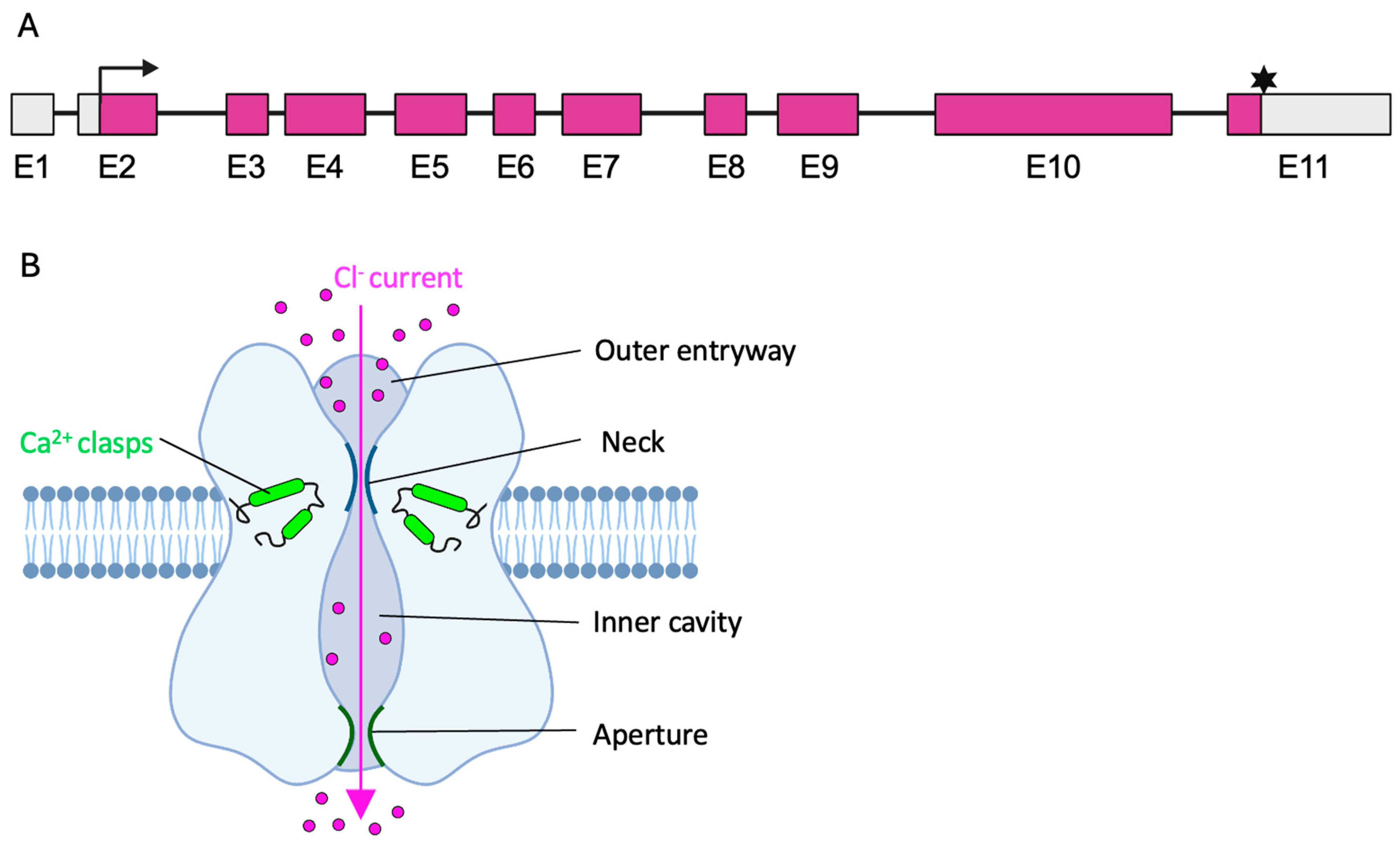
Figure 1. Schematic overview of the BEST1 gene and BEST1 protein. (A) The BEST1 gene consists of 11 exons (E). Translation of transcription variant 1 (NM_004183.3) starts in exon 2 (Black arrow) and terminates in exon 11 (black asterisk), resulting in a 585 amino acid protein. Translated sequence marked in purple. (B) Schematic presentation of the pentameric BEST1 channel. Assembly of the five subunits (blue) forms the ion pore with outer entryway, neck, inner cavity, and aperture. Opening of the gating apparatus is facilitated by the Ca2+ clasps and enables the Cl current through the ion pore.
Ijms 26 09421 g001
Figure 2. Overview of BEST1 variants found in the Leiden open-source variation database (LOVD). (A) Clinical classification of the identified BEST1 variants (n = 485). There were 366 (75%) variants labeled as Pathogenic/Likely pathogenic; 44 (9%) as conflicting* (Pathogenic/Likely pathogenic vs. benign/likely benign vs. uncertain significance); 3 (1%) as conflicting (Benign/Likely benign vs. VUS); 39 (8%) as variant with uncertain significance (VUS); 33 (7%) as Benign/Likely benign. (B) Schematic overview of pathogenic/likely pathogenic/conflicting* BEST1 variants in respect to their exon position and times reported. The ten most frequently reported variants are annotated with their respective DNA change.
Ijms 26 09421 g002
Figure 3. Fundus autofluorescence images (upper row) and optical coherence tomography (OCT) scans (bottom row) depicting the different stages of Best vitelliform macular dystrophy (AD) and the typical appearance of autosomal recessive bestrophinopathy (E). Green lines on the fundus images in the bottom row indicated the location of the corresponding cross-sectional OCT image shown beside them. Stages of Best vitelliform macular dystrophy include the vitelliform stage (A) presenting with well-defined macular lesions (pink arrow), the pseudohypopyon stage (B) where layering of lipofuscin creates the appearance of a pseudohypopyon (blue arrow), followed by the vitelliruptive stage (C) characterized by a “scrambled egg” appearance, and the atrophic stage (D) marked by RPE death and photoreceptor loss. Autosomal recessive bestrophinopathy (E) is characterized by multiple small vitelliform lesions and generalized RPE disruption seen as widespread hypoautofluorescent speckling. The images are obtained from Oxford Eye Hospital, Oxford University Hospital NHS Foundation Trust, Oxford.
Ijms 26 09421 g003
Figure 4. Schematic overview of the molecular disease mechanism and effect at protein level for a (A) wildtype gene, (B) gene with loss-of-function variant, (C) gene with dominant negative variant, and (D) gene with gain-of-function variant. Wildtype allele/protein indicated in green; pathogenic allele/protein indicated in purple. Green stars indicate the altered protein.
Ijms 26 09421 g004
Figure 5. Flowchart from the clinical diagnosis of bestrophinopathies to the choice of treatment strategy. BVMD, Best vitelliform macular dystrophy; AVMD, Adult vitelliform macular dystrophy; ARB, autosomal recessive bestrophinopathy; ADVIRC, autosomal dominant vitreoretinochoroidopathy; HDR, homology directed repair; BE, base editing; PE, prime editing; HITI, homology-independent targeted integration; SNPs, single nucleotide polymorphisms.
Ijms 26 09421 g005
Figure 6. Schematic overview of the CRISPR/Cas9 technology. The CRISPR/Cas9 complex mediates a double-strand break (DSB), which can be repaired via non-homologous end-joining (NHEJ) or homology-directed repair (HDR). (A) The NHEJ can facilitate gene silencing via the production of deletions (indicated as dotted lines) and insertions (indicated in blue) (indels) or precise gene correction via utilization of homology-independent targeted integration (HITI). The donor template (indicated in pink) is flanked by Cas9 cleavage sites (indicated with scissors). (B) Repair via the HDR pathway facilitates precise gene correction in the presence of a donor template with homology arms (HA).
Ijms 26 09421 g006
Table 1. Preclinical studies investigating gene therapy for betrophinopathies.
Gene Therapy StrategyDeliveryClinical ConditionInheritanceDisease ModelOutcomeRef
Gene augmentationBaculovirus vectorARBAutosomal recessiveiPSC-RPE cellsRescue of Ca2+-dependent Cl current (whole-cell patch clamp recording)[46]
Gene augmentationAAV2 vectorARB
(canine model)
Autosomal recessiveCanine BEST1 disease model Revision of subretinal detachment and microdetachment.
Correction of PR/RPE interface
[47]
Gene augmentationLentiviral vectorARBAutosomal recessiveiPSC-RPE cellsIncreased levels of BEST1 protein.
Restoration of BEST1 calcium-activated chloride channel activity.
Improvement of RPE function (Rhodopsin degradation)
[49]
Gene augmentationLentiviral vectorBest diseaseAutosomal dominantiPSC-RPE cellsIncreased levels of BEST1 protein.
Restoration of BEST1 calcium-activated chloride channel activity.
Improvement of RPE function (Rhodopsin degradation)
[49]
CRISPR/Cas9-mediated knock out of mutant alleleLentiviral vectorBest diseaseAutosomal dominantiPSC-RPE cellsIncreased levels of BEST1 protein.
Restoration of BEST1 calcium-activated chloride channel activity
[49]
Gene augmentationAAV2 vector/
Baculovirus vector
BVMDAutosomal dominant (loss-of-function)iPSC-RPERescue of Ca2+-dependent Cl current (whole-cell patch clamp recording)[50]
CRISPR/dCas9-mediated knock down of both alleles
+ gene augmentation
(i) Baculavirus vector expressing dCas9-
KRAB-MeCP2)
(ii) Baculovirus vector expressing wildtype BEST1
Autosomal dominant (gain-of-function)hPSC-RPE H1-iCas9 cells *Rescue of Ca2+ dependent Cl current (whole-cell patch clamp recording)[44]
* Human pluripotent stem cells (hPSC)-derived RPE (hPSC-RPE) from H1 background hPSC line, carrying an inducible Cas9 cassette. ARB, Autosomal recessive bestrophinopathy; iPSC, induced pluripotent stem cell; RPE, retinal pigment epithelium; AAV, adeno-associated virus; BVMD, Best vitelliform macular dystrophy.
Disclaimer/Publisher’s Note: The statements, opinions and data contained in all publications are solely those of the individual author(s) and contributor(s) and not of MDPI and/or the editor(s). MDPI and/or the editor(s) disclaim responsibility for any injury to people or property resulting from any ideas, methods, instructions or products referred to in the content.