Article

Effect of Chronic Social Defeat Stress on the Small-Intestinal Environment, Including the Gut Flora, Immune System, and Mucosal Barrier Integrity

1
Department of Gastroenterology, Hyogo Medical University School of Medicine, 1-1 Mukogawa, Nishinomiya 663-8501, Japan
2
Center for Clinical Research and Education, Hyogo Medical University, Nishinomiya 663-8501, Japan
3
Department of Pharmacology, Hyogo Medical University School of Medicine, Nishinomiya 663-8501, Japan
*
Author to whom correspondence should be addressed.
Int. J. Mol. Sci. 2025, 26(19), 9359; https://doi.org/10.3390/ijms26199359
Submission received: 5 August 2025 / Revised: 12 September 2025 / Accepted: 23 September 2025 / Published: 25 September 2025

Abstract

Psychological stress is deeply involved in the pathophysiology of gastrointestinal diseases. We investigated the effect of psychological stress on the small-intestinal environment, including gut flora, immune system, and mucosal integrity in mice subjected to chronic social defeat stress (CSDS). CSDS mice were established by exposing a C57BL/6N mouse to an ICR aggressor mouse. Stool samples were obtained to investigate its properties and the gut microbiome profile. Using small-intestinal tissues, the expression of cytokines, antimicrobial peptides, and tight junction proteins (TJPs) were examined by real-time RT-PCR and immunohistochemistry. Small-intestinal permeability was evaluated by transepithelial electrical resistance assay. For stool properties, mean Bristol scale score and fecal water content were significantly lower in the CSDS group. Pseudomonadota and Patescibacteria were significantly more abundant in the stools from CSDS mice. Among TJPs and antimicrobial peptides, the expression of Occludin, Claudin-4, and Regenerating gene IIIγ was significantly decreased in the small intestine epithelium of CSDS mice. The small-intestinal permeability was significantly increased in CSDS mice. Lipopolysaccharide immunoreactivity, the number of macrophages, and proinflammatory IL-1β expression were significantly increased in the small intestine of CSDS mice. These findings suggest that psychological stress is associated with mucosal barrier dysfunction and microinflammation in small-intestinal tissues.

1. Introduction

There is much evidence to suggest that psychological stress greatly affects not only gastrointestinal physiology but also the immune system [1,2,3]. Studies using several animal models have shown that psychological stress causes visceral hypersensitivity and/or dysmotility [4,5], and similarly, visceral hypersensitivity and dysmotility are considered to be central to functional gastrointestinal disorders in human patients with frequent mental disturbance [6,7]. Furthermore, it is known that patients with depression frequently suffer from constipation [8,9]. In patients with depression, recent studies have shown that the intestinal environment, including gut flora, immune system, and mucosal barrier function, is disrupted, affecting gastrointestinal physiology [10,11]. However, much remains unclear about the role of psychological stress in the pathophysiology of the gastrointestinal tract.
It is well known that the gut and brain show close physiological interactions, and that numerous molecules, including gut hormones, short-chain fatty acids, bile acids, antimicrobial peptides, and cytokines, play pivotal roles as mediators in the so-called brain–gut axis [12,13]. The profiles of these mediators are orchestrated by gut flora and the small-intestinal immune system, maintaining intestinal mucosal integrity [14] and protecting the host from pathogens and harmful antigens [15]. However, little is known about the effect of psychological stress on the small-intestinal immune system and its associated pathophysiology in terms of defecation.
Chronic psychological stress plays a pivotal role in the development of depression [16,17]. Since chronic social defeat stress (CSDS) effectively accelerates the development of depression [18,19], a mouse model exposed to CSDS is widely used to investigate the effect of psychological stress on not only the brain but also the gastrointestinal tract [20,21]. Interestingly, a few studies have shown that the alterations of gut flora and fecal metabolites in CSDS mice are consistent with those in patients with depression [22,23]. Therefore, in the present study, we comprehensively examined the effect of psychological stress on the defecation-associated intestinal environments, including the gut microbiome and intestinal immune system in a depression model using mice subjected to chronic social defeat stress (CSDS).

2. Results

2.1. Social Interaction Test in CSDS Mice

The social interaction test was performed to evaluate the effect of CSDS on the development of social avoidance. The time spent in the avoidance zone did not differ between the situations with and without the Institute of Cancer Research (ICR) aggressor mouse in the control group (Figure 1A). On the other hand, it was significantly longer in the CSDS group when the ICR mouse was present (60.3 ± 8.6%) than when it was absent (15.0 ± 1.4%). Furthermore, the social interaction ratio was significantly lower in the CSDS group (Figure 1B).

2.2. Stool Properties in Mice Subjected to CSDS

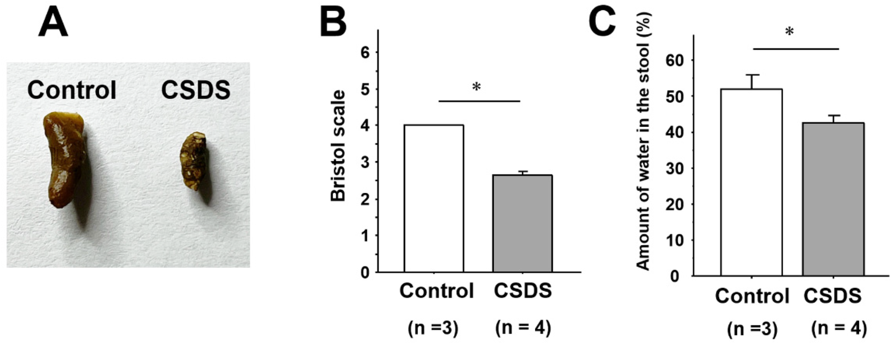
The appearance of the stools was scored according to the Bristol scale [24]. The feces of the control mice were all normal in shape and moisture content (Bristol scale score 4.0 ± 0.0) (Figure 2A,B). In the CSDS group, however, the Bristol scale score was significantly lower (2.6 ± 0.1), suggesting a reduction of stool moisture (Figure 2A,B). Indeed, stool water content in the stool was significantly lower in the CSDS group (42.6 ± 3.5%) than in the controls (56.9 ± 3.4%) (Figure 2C).

2.3. Effect of CSDS on the Small-Intestinal Gut Flora

We next investigated alterations of the gut flora by examining the stools of the experimental mice. The Chao-1 and Shannon diversity indices for α-diversity assessment did not differ between the control and the CSDS mice (Figure 3A). The assessment for β-diversity showed no significant difference between the control and the CSDS mice (Figure 3B). Furthermore, we investigated the phylum and genus profiles of the gut microbiome in these mice. At the phylum level, Patescibacteria and Pseudomonadota were significantly increased in the CSDS mice (Figure 3C). On the other hand, at the genus level, Lachnoclostridium was significantly decreased in the CSDS mice (Figure 3D).

2.4. Effect of CSDS on Tight Junction Proteins Expression, Morphology and Permeability in the Small-Intestinal Mucosa

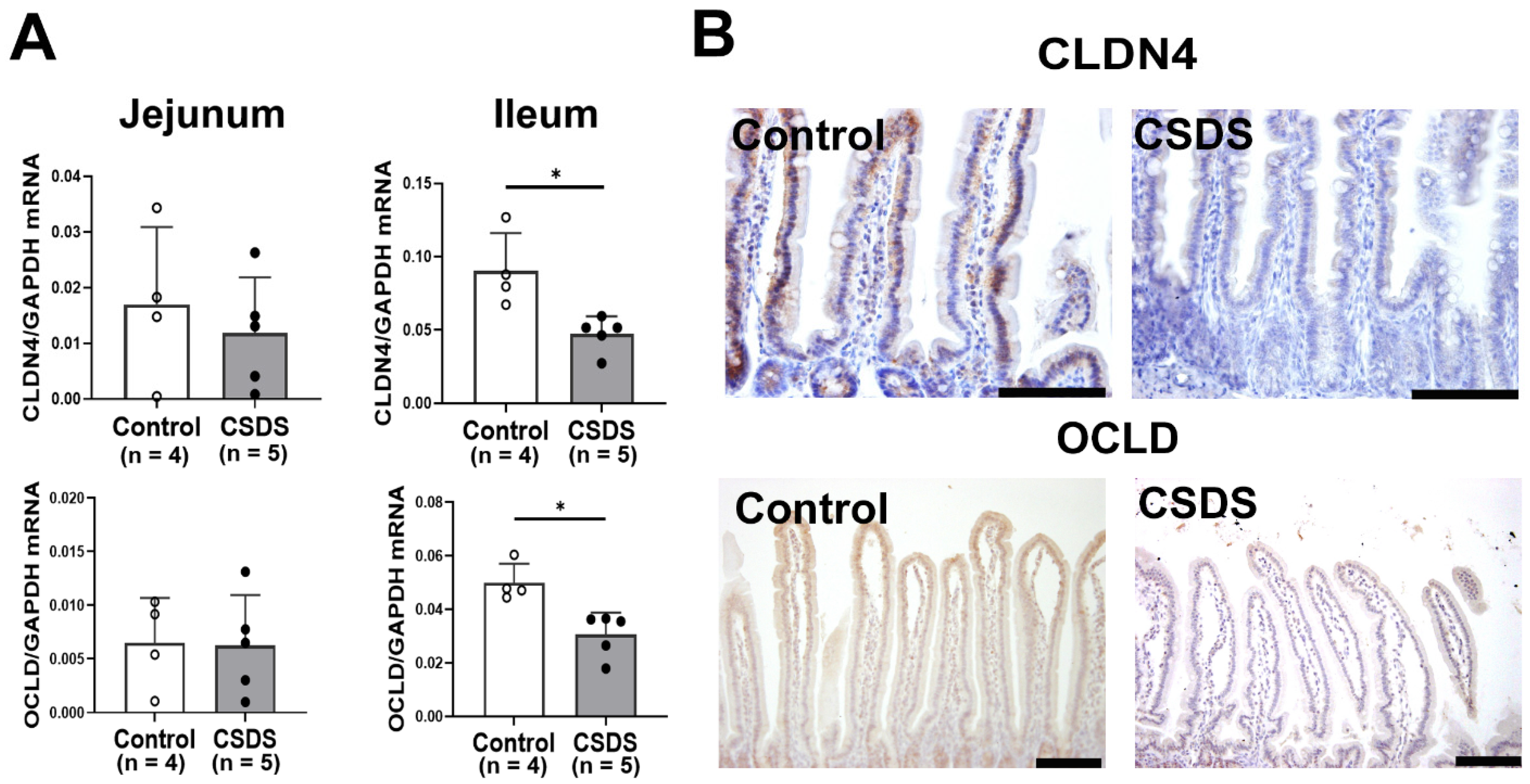
Furthermore, the expression of tight junction proteins (TJPs) in the small-intestinal tissues was examined in the experimental mice. The expression of Claudin-4 and Occludin mRNAs was significantly decreased in the ileum of the CSDS mice (Figure 4A). Consistent with these findings, immunohistochemistry showed that both Claudin-4 and Occludin expressions were also decreased in the small-intestinal epithelium of the CSDS mice compared with the controls (Figure 4B).
The transepithelial resistance values in the small-intestinal tissues were significantly lower in CSDS mice than in the controls (Figure 5). The height of villi was significantly lower in CSDS than in control mice, although the depth of crypt did not differ between the two groups statistically. However, the ratio of villi to crypt was subsequently smaller in CSDS than in control mice (Supplementary Figure S1).

2.5. Expression of Antimicrobial Peptides, Cytokines, LPS, and CD80 in the Small Intestine of CSDS Mice

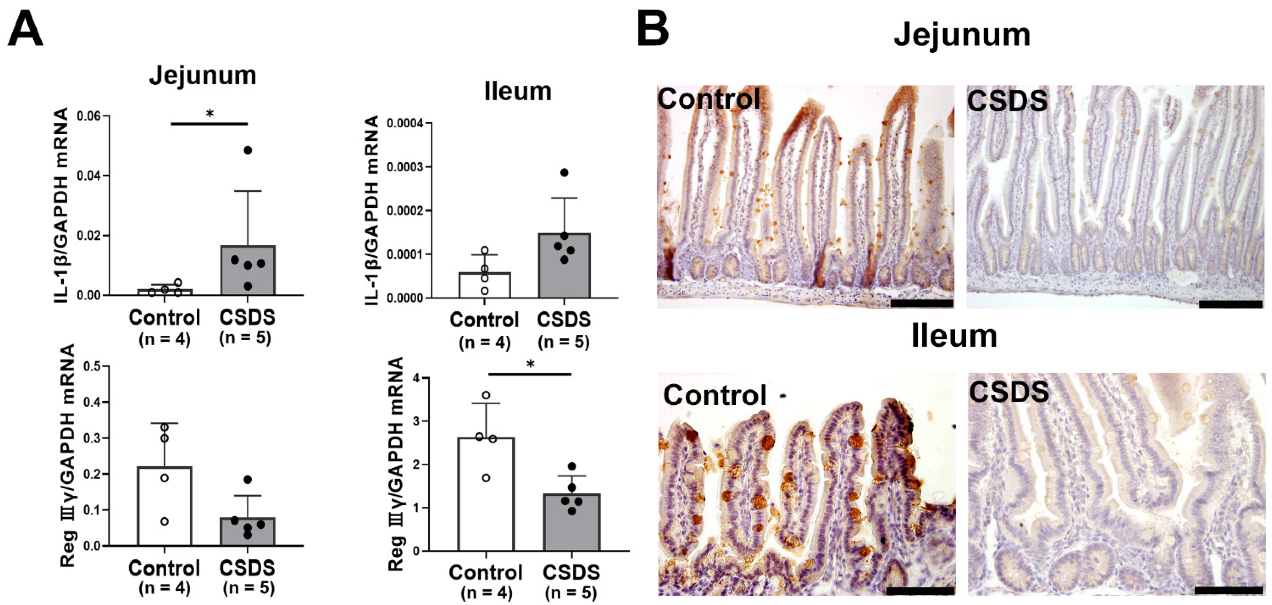
We further investigated the expression of antimicrobial peptides and cytokines in the small intestine of the experimental mice. The mRNA expression of Interleukin-1β (IL-1β) was significantly increased in the jejunum and tended to be higher in the ileum of the CSDS mice (Figure 6A). Among anti-microbial peptides, the mRNA expression of Reg IIIγ was significantly decreased in CSDS mice relative to the controls in the jejunum and ileum (Figure 6A). Indeed, Reg Ⅲγ was apparently decreased in the CSDS mice. (Figure 6B).
To examine changes in harmful antigens in the small-intestinal mucosa, we examined the immunoreactivity of LPS using immunohistochemistry. In both the jejunum and ileum, strong LPS immunoreactivity was evident in the lamina propria of the small intestine in CSDS mice relative to the controls (Figure 7A). The number of LPS-positive cells was also significantly increased in the jejunum and ileum of CSDS mice relative to the controls (Figure 7B).
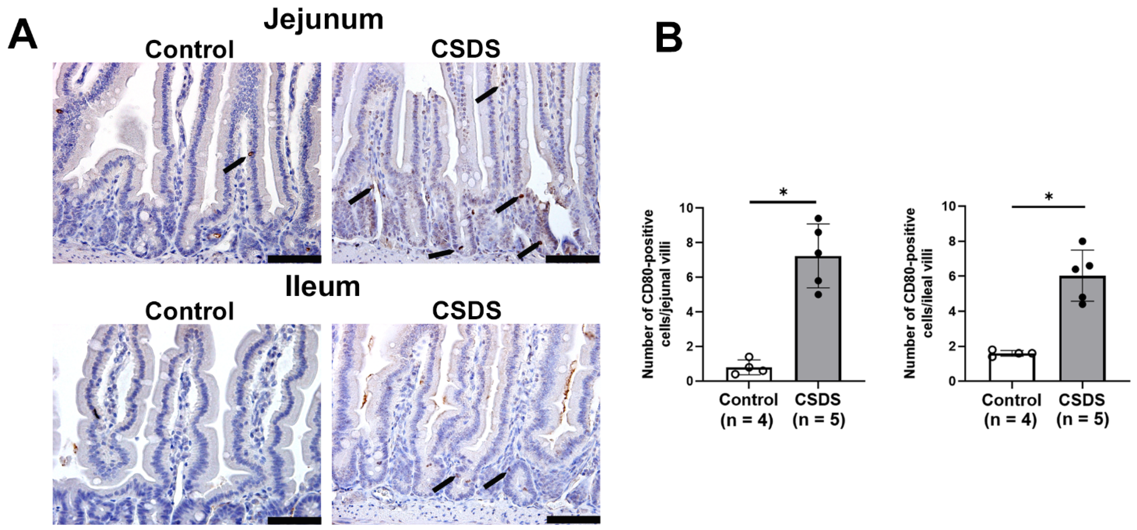
Moreover, we found that CD80-positive cells were increased in the lamina propria of the small intestine in CSDS mice relative to the controls (Figure 8A). As shown in Figure 8B, the number of CD80-positive cells was significantly increased in the jejunum and ileum of CSDS mice relative to the controls (Figure 8), suggesting that immune cells such as macrophages were increased in the latter.

3. Discussion

Accumulating evidence suggests that psychological stress alters the intestinal environment [25,26], although the underlying pathogenesis remains unclear. Multiple depression models (chronic unpredictable mild stress, chronic restraint stress, CSDS or learned helplessness, etc.) have been used to investigate the pathophysiology in patients with major depressive disorder [27]. However, major depressive disorder is considered to be a heterogeneous disease [28], and, therefore, it seems very difficult to establish a perfect animal model for major depressive disorder. In the present study, although we selected a CSDS model, this model cannot avoid physical stress, and its behavior phenotypes are various according to the differences in protocol [29,30]. Therefore, in order to assess the effect of psychological stress on the intestinal environment, we first isolated the only mice susceptible to CSDS using a social interaction test and used them for the subsequent experiment. Subsequently, we found a significant reduction of the Bristol scale for fecal humidity in CSDS mice and indeed confirmed a significant decrease of the stool water content in those mice. These findings are compatible with clinical evidence that patients with depression frequently suffer from constipation [8,31], suggesting that CSDS mice are a useful model for investigating the pathophysiology of depression-associated constipation.
The luminal environment of the intestinal tract plays a key role in the interaction between the gut and the brain [32]. There is now considerable evidence to suggest that an imbalance of the gut microbiome profile (dysbiosis) plays an important role in not only inflammatory diseases but also functional disorders of the gastrointestinal tract [33,34]. Moreover, dysbiosis has been associated with psychological stress [35,36], and in the present study, we were able to show that Patescibacteria and Pseudomonadota were significantly increased in the CSDS mice. Although the role of Patescibacteria in human diseases is not fully understood, it represents a diverse group of bacteria accounting for a significant proportion of microbial dark matter [37] and is frequently detected in various environments, including groundwater, freshwater, and the human oral cavity [38,39,40]. Interestingly, Pseudomonadota is also reported to be a predominant phylum in the oral cavity [41] and associated with gut dysbiosis and chronic intestinal inflammation [42,43]. It was noteworthy that the expression of proinflammatory IL-1β was consistently enhanced in the small-intestinal tissues of CSDS mice. It has often been debated whether dysbiosis is a pathophysiological cause or a consequence of specific diseases. Although various studies have reported dysbiosis in patients with constipation and/or depression, the candidate microbiota isolated varied considerably [44,45,46]. For instance, even in a similar CSDS model established by our group [47], the gut flora data obtained were quite different from those in the present study, suggesting that subtle differences in experimental conditions are likely to alter the gut flora. In this context, we propose that the dysbiosis observed in our CSDS model is a consequence rather than a cause.
The mucosal barrier of the intestine is crucial for protecting the host from invasion by harmful antigens or pathogens [48,49]. It has recently been highlighted that disruption of mucosal barrier function is an important first step in alteration of innate immunity, triggering microinflammation of the gastrointestinal mucosa that may be linked to altered pathophysiology in other organs [50,51]. Indeed, in the present study, we found that expression of tight junction proteins in the small intestine and mucosal permeability were enhanced in CSDS mice. These findings suggest that CSDS mice are likely to have disturbed small-intestinal mucosal barrier function. Furthermore, we found that antimicrobial Reg IIIγ was down-regulated in the small-intestinal mucosa, suggesting that host defense against pathogens may be inhibited in CSDS mice. Together, these changes in the environment of the small intestine might make it easier for harmful antigens/pathogens to invade. Compatible with these findings, the expression of LPS immunoreactivity was augmented in the small-intestinal mucosa, and the number of macrophages was also increased in the CSDS mice. Overall, psychological stress may disturb intestinal mucosal barrier function by inhibiting the expression of tight junction proteins and antimicrobial peptides, resulting in activation of proinflammatory immune cells against harmful antigens such as LPS.
Many physiological functions, including appetite, gastrointestinal motility, emotion, visceral sensation, and gut hormone secretion, are strictly controlled via the gut–brain axis. Regarding the link between gut environmental factors and the central nervous system, recent studies have suggested that gut microbiome-related metabolites (short-chain fatty acids, bile acids, vitamins, gut hormones, etc.) mediate gut to brain communication [45,52]. On the other hand, it remains unclear how psychological stress disturbs intestinal mucosal barrier function. In this context, Schneider et al. have recently proposed a mechanism whereby psychological stress induces inflammatory enteric glia, and that enteric neuron immaturity can lead to delayed intestinal transit time and monocyte-mediated inflammation in the gastrointestinal tract [53]. Although their stress model differs from ours, it is interesting that similar features, such as microinflammation and constipation, were observed in both. Since we did not examine the behavior of enteric neurons, we are yet unable to consider the extent to which the enteric nervous system (ENS) played a role in intestinal motility and inflammation in CSDS mice. However, in terms of increased intestinal mucosa permeability, it may be interesting to investigate how psychological stress affects the expression of tight junction proteins and antimicrobial peptides in the intestinal epithelial cells via ENS alteration.
In summary, we have shown that CSDS alters the flora and stool properties of mice. Furthermore, mice subjected to CSDS showed increased expression of tight junction proteins (Claudin-4 and Occludin) and microbial peptide Reg IIIγ in the small intestine and increased permeability of the intestinal mucosa. In addition, these mice showed an increase in the macrophage population and higher expression of proinflammatory IL-1β in the small-intestinal mucosa, in association with augmented LPS immunoreactivity. Although the underlying links between these changes remain unclear, we speculate that psychological stress led to mucosal barrier dysfunction, subsequently promoting the invasion of harmful antigens such as LPS, and resulting in microinflammation of the intestinal mucosa. As the ENS certainly plays a pivotal role in brain–gut communication [53], future studies will need to clarify the mechanism by which psychological stress acts on the ENS to disturb mucosal barrier function and intestinal motility.

4. Materials and Methods

4.1. Animal Model of Chronic Social Defeat Stress

C57BL/6N mice (male; 9 weeks old) and ICR mice (male; 9 weeks old) were purchased from Japan SLC. The mice were kept under specific pathogen-free conditions in a controlled clean room (temperature 23 ± 1 °C; humidity, 50 ± 5%) with a 12 h light/12 h dark regime and allowed free access to laboratory food (Oriental Yeast Co., Ltd., Tokyo, Japan) and water.
Since ICR mice are superior to C57BL/6N mice in term of power balance, ICR mouse attacks a C57BL/6N mouse when they were placed in the same cage. CSDS model is established using the repeated aggressive behavior from ICR mice to C57BL/6N mice. C57BL/6N intruder mice were subjected to chronic social defeat stress using an ICR aggressor mouse as described previously [47,54] with minor modification. Once a day, the C57BL/6N mouse was subjected to defeat stress by exposure to the ICR aggressor mouse for 10 min. As a control, C57BL/6N mice were placed in the cage without an ICR aggressor mouse. This procedure was repeated for 10 consecutive days. All animal experiments in this study were approved by the Animal Use and Care Committee of Hyogo Medical University.

4.2. Social Interaction Test

The social interaction test was performed as described previously [55], with minor modifications. In brief, C57BL/6N mice were placed in an open field chamber (44 cm × 44 cm) without an ICR aggressor mouse for 150 s. Thereafter, a mesh cage (10 cm × 6 cm) containing an ICR aggressor mouse was placed in the open field chamber, and the C57BL/6N mice were introduced for 150 s. The behaviors of the experimental mice were recorded for automated post-hoc analysis using the SMART video tracking system (Harvard Apparatus, Holliston, MA, USA). The time that the mice spent in the social interaction zone or the social avoidance zone was measured, respectively (Figure 9). The social interaction ratio (SIR) was calculated by dividing the time spent in the social interaction zone with the ICR mouse present by the time spent in the social interaction zone with the ICR mouse absent. In the present study, the mice were defined as susceptible to CSDS when the SIR was less than 50%, and only the mice susceptible to CSDS were used thereafter throughout the experiments.

4.3. Sampling of Stools and Intestinal Tissues

Fresh fecal samples from both the control and CSDS mice were collected after defecation on experimental day 11. The properties of the stools were scored according to the Bristol scale [24]. Fecal water content was assessed by the gravimetric drying method as previously described [56,57]. In brief, after weighing the wet stools, we dried them overnight at 65 °C and then determined their dry weight. The ratio of water in the stools was calculated as: (wet weight–dry weight)/wet weight. Then, under sevoflurane anesthesia, the mice were euthanized by cervical dislocation, and the small-intestinal tissues were removed. The small intestines obtained were cut open along the longitudinal axis, stored in nitrogen liquid for RNA extraction, or fixed in 10% buffered formalin solution for immunohistochemistry.

4.4. Analysis of the Gut Microbiome

In brief, bacterial DNA for sequencing was extracted from fresh stool samples using the phenol–chloroform method as described previously [58]. Analysis of the V3–V4 region of the microbial community 16S rDNA was amplified and sequenced using a 2 × 250-bp paired-end run on a MiSeq platform using MiSeq Reagent Kit v2 chemistry (Illumina, San Diego, CA, USA) as described previously [58]. Comparison of each taxon in the gut microbiota was conducted at both the genus and species levels. The Chao-1 and Shannon indices were calculated to assess the alpha diversity of the microbiota in the samples. Details of the methodology were described in the Section 4 of the reference paper [58].

4.5. Real-Time RT-PCR

Total RNA was extracted from small-intestinal tissues using Trizol reagent (Invitrogen, Carlsbad, CA, USA) in accordance with the manufacturer’s protocol and reverse-transcribed with an oligo-dT primer (Applied Biosystems, Branchburg, NJ, USA). Real-time RT-PCR was conducted using the 7900H Fast Real-time RT-PCR System (Applied Biosystems, Foster City, CA, USA) as reported previously [59]. Briefly, the sets of primers for mouse Claudin 4, Occludin, IL-1β, Regenerating gene IIIγ (Reg IIIγ), and GAPDH were prepared as shown in Supplementary Table S1. Real-time RT-PCR was performed with 200 ng of RNA equivalent cDNA, SYBR Green Master Mix (Applied Biosystems), and gene-specific primers at 500 nmol/l. The cycle conditions for real-time PCR were 50 °C for 15 s and 60 °C for 60 s. Relative mRNA levels of the target genes were normalized to the expression of GAPDH mRNA.

4.6. Histology and Immunohistochemistry

Immunohistochemical procedures were performed using an Envision kit (Dako, Kyoto, Japan) as described previously [47]. The primary antibodies used in this study were: anti-Occludin antibody (dilution 1:25, Invitrogen, Camarillo, CA, USA), anti-Reg IIIγ antibody (dilution 1:50; LSBio, Shirley, MA, USA), anti-lipopolysaccharide (LPS) antibody (dilution 1:50,000; Abcam, Cambridge, UK), and anti-CD80 antibody (dilution 1:1000; Abcam, Cambridge, UK). Briefly, the intestinal sections were incubated with primary antibodies for 60 min at room temperature, washed in PBS, and incubated with the corresponding secondary antibody for 30 min. The slides were visualized using 3,3′-diaminobenzidine tetrahydrochloride with 0.05% H2O2 for 3 min and then counterstained with Mayer’s hematoxylin. The number of LPS- or CD80-positive cells was evaluated as reported previously [60]. Briefly, four sections in each mouse were prepared for the small intestine. The positive cells were counted in at least five different visual fields in a 500 μm stretch of the entire length with well-oriented tissue sections, and the average was calculated as the number of positive cells per a villous in each mouse. The height of intestinal villi, the depth of intestinal crypt was evaluated using a microscope (DP72; Olympus, Tokyo, Japan) as previously reported [61].

4.7. Measurement of Transepithelial Electrical Resistance

Electrical resistance across the stratified epithelium was measured using a Millicell-ERS-2 instrument (Millipore, Bedford, MA, USA) with “chopstick” electrodes, as described previously [62]. Ex vivo permeability assays were performed as reported previously [63]. Briefly, the small-intestinal tissues were removed, opened longitudinally, placed apical side up on a 0.4 μm pore size membrane (Costar Incorporated, Corning, NY, USA), and transferred to a microsnapwell system. Electrical resistance was measured as described previously [62].

4.8. Statistical Analysis

Statistical analysis was performed using GraphPad Prism 8 (GraphPad, San Diego, CA, USA). Data are shown as mean ± SE. Significance of differences between two animal groups was analyzed by Mann-Whitney U-test. Differences were considered to be significant at p < 0.05.

Supplementary Materials

The following supporting information can be downloaded at: https://www.mdpi.com/article/10.3390/ijms26199359/s1.

Author Contributions

Conceptualization, S.Y., H.F., S.K., and S.S.; Methodology, T.S. (Tetsuya Shiraishi), K.K., M.W., Y.T.,M.I., T.S. (Toshiyuki Sato), M.K., Y.Y., T.T. (Tetsuya Takagawa), and T.T. (Toshihiko Tomita); Validation, S.Y., H.F., and S.K.; Formal analysis, S.Y., H.F., and S.K.; Investigation, S.Y., H.F., T.S. (Tetsuya Shiraishi), K.K., M.W., Y.T., M.I., T.S. (Toshiyuki Sato), M.K., Y.Y., T.T. (Tetsuya Takagawa), T.T. (Toshihiko Tomita), S.K., and S.S.; Resources, T.S. (Tetsuya Shiraishi), K.K., M.W., Y.T., M.I., T.S. (Toshiyuki Sato), M.K., Y.Y., T.T. (Tetsuya Takagawa), and T.T. (Toshihiko Tomita); Data curation, S.Y., H.F., S.K., and S.S.; Writing—original draft, S.Y., H.F., S.K., and S.S.; Writing—review and editing, S.Y., H.F., S.K., and S.S.; Supervision, T.T. (Toshihiko Tomita), S.K., and S.S.; Project administration, H.F., S.K., and S.S. All authors have read and agreed to the published version of the manuscript.

Funding

This work was supported in part by Grants-in-aid for Scientific Research 24K11118 from the Ministry of Education, Culture, Sports, Science, and Technology, Japan.

Institutional Review Board Statement

The animal study protocol was approved by the Institutional Review Board of Hyogo Medical University School of Medicine (protocol code 24-034A and date of approval 1 April 2024).

Informed Consent Statement

Not applicable.

Data Availability Statement

The datasets used and analyzed in this study are available from corresponding author.

Acknowledgments

We thank Mayumi Yamada (Hyogo Medical University School of Medicine) for technical assistance with the experiments.

Conflicts of Interest

The authors declare no conflicts of interest.

Abbreviations

The following abbreviations are used in this manuscript:
CD80Cluster of differentiation
CSDSChronic social defeat stress
ENSEnteric nervous system
GAPDHGlyceraldehyde-3-phosphate dehydrogenase
ICRInstitute of cancer research
ILInterleukin
LPSLipopolysaccharide
RegRegenerating gene
RT-PCRReverse transcription polymerase chain reaction
SEStandard error
TJPsTight junction proteins

References

  1. Ge, L.; Liu, S.; Li, S.; Yang, J.; Hu, G.; Xu, C.; Song, W. Psychological stress in inflammatory bowel disease: Psychoneuroimmunological insights into bidirectional gut–brain communications. Front. Immunol. 2022, 13, 1016578. [Google Scholar] [CrossRef]
  2. Glaser, R.; Kiecolt-Glaser, J.K. Stress-induced immune dysfunction: Implications for health. Nat. Rev. Immunol. 2005, 5, 243–251. [Google Scholar] [CrossRef]
  3. Leigh, S.J.; Uhlig, F.; Wilmes, L.; Sanchez-Diaz, P.; Gheorghe, C.E.; Goodson, M.S.; Kelly-Loughnane, N.; Hyland, N.P.; Cryan, J.F.; Clarke, G. The impact of acute and chronic stress on gastrointestinal physiology and function: A microbiota–gut–brain axis perspective. J. Physiol. 2023, 601, 4491–4538. [Google Scholar] [CrossRef]
  4. Bradesi, S.; Schwetz, I.; Ennes, H.S.; Lamy, C.M.R.; Ohning, G.; Fanselow, M.; Pothoulakis, C.; McRoberts, J.A.; Mayer, E.A. Repeated exposure to water avoidance stress in rats: A new model for sustained visceral hyperalgesia. Am. J. Physiol. Gastrointest. Liver Physiol. 2005, 289, G42–G53. [Google Scholar] [CrossRef]
  5. Larauche, M.; Gourcerol, G.; Million, M.; Adelson, D.W.; Taché, Y. Repeated psychological stress-induced alterations of visceral sensitivity and colonic motor functions in mice: Influence of surgery and postoperative single housing on visceromotor responses. Stress 2010, 13, 343–354. [Google Scholar] [CrossRef] [PubMed]
  6. Mönnikes, H.; Tebbe, J.J.; Hildebrandt, M.; Arck, P.; Osmanoglou, E.; Rose, M.; Klapp, B.; Wiedenmann, B.; Heymann-Mönnikes, I. Role of Stress in Functional Gastrointestinal Disorders Evidence for Stress-Induced Alterations in Gastrointestinal Motility and Sensitivity. Dig. Dis. 2001, 19, 201–211. [Google Scholar] [CrossRef] [PubMed]
  7. Drossman, D.A.; Hasler, W.L. Rome IV—Functional GI disorders: Disorders of gut–brain interaction. Gastroenterology 2016, 150, 1257–1261. [Google Scholar] [CrossRef]
  8. Cheng, C.; Chan, A.O.O.; Hui, W.M.; Lam, S.K. Coping strategies, illness perception, anxiety and depression of patients with idiopathic constipation: A population-based study. Aliment. Pharmacol. Ther. 2003, 18, 319–326. [Google Scholar] [CrossRef] [PubMed]
  9. Ballou, S.; Katon, J.; Singh, P.; Rangan, V.; Lee, H.N.; McMahon, C.; Iturrino, J.; Lembo, A.; Nee, J. Chronic diarrhea and constipation are more common in depressed individuals. Clin. Gastroenterol. Hepatol. 2019, 17, 2696–2703. [Google Scholar] [CrossRef]
  10. Nikolova, V.L.; Smith, M.R.B.; Hall, L.J.; Cleare, A.J.; Stone, J.M.; Young, A.H. Perturbations in gut microbiota composition in psychiatric disorders: A review and meta-analysis. JAMA Psychiatry 2021, 78, 1343–1354. [Google Scholar] [CrossRef]
  11. Ohlsson, L.; Gustafsson, A.; Lavant, E.; Suneson, K.; Brundin, L.; Westrin, Å.; Ljunggren, L.; Lindqvist, D. Leaky gut biomarkers in depression and suicidal behavior. Acta Psychiatr. Scand. 2019, 139, 185–193. [Google Scholar] [CrossRef]
  12. Fukui, H.; Xu, X.; Miwa, H. Role of Gut Microbiota-Gut Hormone Axis in the Pathophysiology of Functional Gastrointestinal Disorders. J. Neurogastroenterol. Motil. 2018, 24, 367–386. [Google Scholar] [CrossRef]
  13. Margolis, K.G.; Cryan, J.F.; Mayer, E.A. The microbiota–gut–brain axis: From motility to mood. Gastroenterology 2021, 160, 1486–1501. [Google Scholar] [CrossRef]
  14. Zheng, D.; Liwinski, T.; Elinav, E. Interaction between microbiota and immunity in health and disease. Cell Res. 2020, 30, 492–506. [Google Scholar] [CrossRef] [PubMed]
  15. Ullah, H.; Arbab, S.; Tian, Y.; Chen, Y.; Liu, C.; Li, Q.; Li, K. Crosstalk between gut microbiota and host immune system and its response to traumatic injury. Front. Immunol. 2004, 15, 1413485. [Google Scholar] [CrossRef] [PubMed]
  16. Cruz-Pereira, J.S.; Rea, K.; Nolan, Y.M.; O’Leary, O.F.; Dinan, T.G.; Cryan, J.F. Depression’s unholy trinity: Dysregulated stress, immunity, and the microbiome. Annu. Rev. Psychol. 2020, 71, 49–78. [Google Scholar] [CrossRef]
  17. Hammen, C. Stress and depression. Annu. Rev. Clin. Psychol. 2005, 1, 293–319. [Google Scholar] [CrossRef]
  18. Song, J.; Kim, Y.K. Animal models for the study of depressive disorder. CNS Neurosci. Ther. 2021, 27, 633–642. [Google Scholar] [CrossRef]
  19. Radley, J.J.; Herman, J.P. Preclinical models of chronic stress: Adaptation or pathology? Biol. Psychiatry 2023, 94, 194–202. [Google Scholar] [CrossRef] [PubMed]
  20. Suzuki, K.; Nakamura, K.; Shimizu, Y.; Yokoi, Y.; Ohira, S.; Hagiwara, M.; Wang, Y.; Song, Y.; Aizawa, T.; Ayabe, T. Decrease of α-defensin impairs intestinal metabolite homeostasis via dysbiosis in mouse chronic social defeat stress model. Sci. Rep. 2021, 11, 9915. [Google Scholar] [CrossRef]
  21. Yadav, S.K.; Ahmad, R.; Moshfegh, C.M.; Sankarasubramanian, J.; Joshi, V.; Elkhatib, S.K.; Chhonker, Y.S.; Murry, D.J.; Talmon, G.A.; Guda, C.; et al. Repeated social defeat stress induces an inflammatory gut milieu by altering the mucosal barrier integrity and gut microbiota homeostasis. Biol. Psychiatry Glob. Open Sci. 2023, 3, 824–836. [Google Scholar] [CrossRef]
  22. Bharwani, A.; Mian, M.F.; Foster, J.A.; Surette, M.G.; Bienenstock, J.; Forsythe, P. Structural & functional consequences of chronic psychosocial stress on the microbiome & host. Psychoneuroendocrinology 2016, 63, 217–227. [Google Scholar] [CrossRef] [PubMed]
  23. Liu, L.; Wang, H.; Chen, X.; Zhang, Y.; Zhang, H.; Xie, P. Gut microbiota and its metabolites in depression: From pathogenesis to treatment. EBioMedicine 2023, 90, 104527. [Google Scholar] [CrossRef] [PubMed]
  24. Lewis, S.J.; Heaton, K.W. Stool Form Scale as a Useful Guide to Intestinal Transit Time. Scand. J. Gastroenterol. 1997, 32, 920–924. [Google Scholar] [CrossRef]
  25. Chang, H.; Perkins, M.H.; Novaes, L.S.; Qian, F.; Zhang, T.; Neckel, P.H.; Scherer, S.; Ley, R.E.; Han, W.; de Araujo, I.E. Stress-sensitive neural circuits change the gut microbiome via duodenal glands. Cell 2024, 187, 5393–5412. [Google Scholar] [CrossRef] [PubMed]
  26. Vanuytsel, T.; van Wanrooy, S.; Vanheel, H.; Vanormelingen, C.; Verschueren, S.; Houben, E.; Rasoel, S.S.; Tóth, J.; Holvoet, L.; Farré, R.; et al. Psychological stress and corticotropin-releasing hormone increase intestinal permeability in humans by a mast cell-dependent mechanism. Gut 2014, 63, 1293–1299. [Google Scholar] [CrossRef]
  27. Liu, X.; Li, X.; Teng, T.; Jiang, Y.; Xiang, Y.; Fan, L.; Yu, Y.; Zhou, X.; Xie, P. Comparative analysis of gut microbiota and fecal metabolome features among multiple depressive animal models. J. Affect. Disord. 2022, 314, 103–111. [Google Scholar] [CrossRef]
  28. Spellman, T.; Liston, C. Toward circuit mechanisms of pathophysiology in depression. Am. J. Psychiatry 2020, 177, 381–390. [Google Scholar] [CrossRef] [PubMed]
  29. Nakatake, Y.; Furuie, H.; Yamada, M.; Kuniishi, H.; Ukezono, M.; Yoshizawa, K.; Yamada, M. The effects of emotional stress are not identical to those of physical stress in mouse model of social defeat stress. Neurosci. Res. 2020, 158, 56–63. [Google Scholar] [CrossRef]
  30. Díez-Solinska, A.; De Miguel, Z.; Azkona, G.; Vegas, O. Behavioral coping with chronic defeat stress in mice: A systematic review of current protocols. Neurobiol. Stress 2024, 33, 100689. [Google Scholar] [CrossRef]
  31. Wang, P.; Shen, X.; Wang, Y.; Jia, X. Association between constipation and major depression in adult Americans: Evidence from NHANES 2005–2010. Front. Psychiatry. 2003, 14, 1152435. [Google Scholar] [CrossRef] [PubMed]
  32. Wachsmuth, H.R.; Weninger, S.N.; Duca, F.A.; Wachsmuth, H.R.; Weninger, S.N.; Duca, F.A. Role of the gut–brain axis in energy and glucose metabolism. Exp. Mol. Med. 2022, 54, 377–392. [Google Scholar] [CrossRef] [PubMed]
  33. Cheng, X.; Ren, C.; Mei, X.; Jiang, Y.; Zhou, Y. Gut microbiota and irritable bowel syndrome: Status and prospect. Front. Med. 2024, 11, 1429133. [Google Scholar] [CrossRef]
  34. Wang, J.; Chen, W.D.; Wang, Y.D. The Relationship Between Gut Microbiota and Inflammatory Diseases: The Role of Macrophages. Front. Microbiol. 2020, 11, 1065. [Google Scholar] [CrossRef]
  35. Luna, R.A.; Foster, J.A. Gut brain axis: Diet microbiota interactions and implications for modulation of anxiety and depression. Curr. Opin. Biotechnol. 2015, 32, 35–41. [Google Scholar] [CrossRef]
  36. Leclercq, S.; Forsythe, P.; Bienenstock, J. Posttraumatic Stress Disorder: Does the Gut Microbiome Hold the Key? Can. J. Psychiatry. 2016, 61, 204–213. [Google Scholar]
  37. Wang, Y.; Gallagher, L.A.; Andrade, P.A.; Liu, A.; Humphreys, I.R.; Turkarslan, S.; Cutler, K.J.; Arrieta-Ortiz, M.L.; Li, Y.; Radey, M.C.; et al. Genetic manipulation of Patescibacteria provides mechanistic insights into microbial dark matter and the epibiotic lifestyle. Cell 2023, 186, 4803–4817. [Google Scholar] [CrossRef]
  38. Brown, C.T.; Hug, L.A.; Thomas, B.C.; Sharon, I.; Castelle, C.J.; Singh, A.; Wilkins, M.J.; Wrighton, K.C.; Williams, K.H.; Banfield, J.F. Unusual biology across a group comprising more than 15% of domain Bacteria. Nature 2015, 523, 208–211. [Google Scholar] [CrossRef] [PubMed]
  39. Luna, M.C.; Bulzu, P.A.; Andrei, A.S.; Okazaki, Y.; Nakano, S.; Haber, M.; Kavagutti, V.S.; Layoun, P.; Ghai, R.; Salcher, M.M. Ecogenomics sheds light on diverse lifestyle strategies in freshwater CPR. Microbiome 2022, 10, 84. [Google Scholar] [CrossRef] [PubMed]
  40. He, X.; McLean, J.S.; Edlund, A.; Yooseph, S.; Hall, A.P.; Liu, S.Y.; Dorrestein, P.C.; Esquenazi, E.; Hunter, R.C.; Cheng, G.; et al. Cultivation of a human-associated TM7 phylotype reveals a reduced genome and epibiotic parasitic lifestyle. Proc. Nat. Acad. Sci. USA 2014, 112, 244–249. [Google Scholar] [CrossRef]
  41. Wang, S.; Song, F.; Gu, H.; Wei, X.; Zhang, K.; Zhou, Y.; Luo, H. Comparative Evaluation of the Salivary and Buccal Mucosal Microbiota by 16S rRNA Sequencing for Forensic Investigations. Front. Microbiol. 2022, 13, 777882. [Google Scholar] [CrossRef]
  42. Khor, B.; Snow, M.; Herrman, E.; Ray, N.; Mansukhani, K.; Patel, K.A.; Said, A.N.N.; Maier, T.; Machida, C.A. Interconnections between the Oral and Gut Microbiomes: Reversal of Microbial Dysbiosis and the Balance between Systemic Health and Disease. Microorganisms 2021, 9, 496. [Google Scholar] [CrossRef]
  43. Kitamoto, S.; Kamada, N. Periodontal connection with intestinal inflammation: Microbiological and immunological mechanisms. Periodontol. 2000 2022, 89, 142–153. [Google Scholar] [CrossRef] [PubMed]
  44. Cheng, Y.; Zhu, Z.; Yang, Z.; Liu, X.; Qian, X.; Zhu, J.; Hu, X.; Jiang, P.; Cui, T.; Wang, Y.; et al. Alterations in fecal microbiota composition and cytokine expression profiles in adolescents with depression: A case-control study. Sci. Rep. 2025, 15, 12177. [Google Scholar] [CrossRef] [PubMed]
  45. Jia, M.; Fan, Y.; Ma, Q.; Yang, D.; Wang, Y.; He, X.; Zhao, B.; Zhan, X.; Qi, Z.; Ren, Y.; et al. Gut microbiota dysbiosis promotes cognitive impairment via bile acid metabolism in major depressive disorder. Transl. Psychiatry. 2024, 14, 503. [Google Scholar] [CrossRef]
  46. Radjabzadeh, D.; Bosch, J.A.; Uitterlinden, A.G.; Zwinderman, A.H.; Ikram, M.A.; van Meurs, J.B.J.; Luik, A.I.; Nieuwdorp, M.; Lok, A.; van Duijn, C.M.; et al. Gut microbiome-wide association study of depressive symptoms. Nat. Commun. 2022, 13, 7128. [Google Scholar] [CrossRef] [PubMed]
  47. Ebisutani, N.; Fukui, H.; Nishimura, H.; Nakanishi, T.; Morimoto, K.; Itou, S.; Nakamura, A.; Masutani, M.; Hori, M.; Tomita, T.; et al. Decreased Colonic Guanylin/Uroguanylin Expression and Dried Stool Property in Mice With Social Defeat Stress. Front. Physiol. 2020, 11, 599582. [Google Scholar] [CrossRef]
  48. Sheng, Y.H.; Hasnain, S.Z. Mucus and Mucins: The Underappreciated Host Defence System. Front. Cell Infect. Microbiol. 2022, 12, 856962. [Google Scholar]
  49. Sánchez, M.F.; Romero-Calvo, I.; Mascaraque, C.; Martínez-Augustin, O. Intestinal Inflammation and Mucosal Barrier Function. Inflamm. Bowel. Dis. 2014, 20, 2394–2404. [Google Scholar] [CrossRef]
  50. Oshima, T.; Miwa, H. Gastrointestinal mucosal barrier function and diseases. J. Gastroenterol. 2016, 51, 768–778. [Google Scholar] [CrossRef]
  51. An, J.; Liu, Y.; Wang, Y.; Fan, R.; Hu, X.; Zhang, F.; Yang, J.; Chen, J. The Role of Intestinal Mucosal Barrier in Autoimmune Disease: A Potential Target. Front. Immunol. 2022, 13, 871713. [Google Scholar] [CrossRef]
  52. Wouw, M.; Boehme, M.; Lyte, J.M.; Wiley, N.; Strain, C.; O’Sullivan, O.; Clarke, G.; Stanton, C.; Dinan, T.G.; Cryan, J.F. Short-chain fatty acids: Microbial metabolites that alleviate stress-induced brain-gut axis alterations. J. Physiol. 2018, 596, 4923–4944. [Google Scholar] [CrossRef]
  53. Schneider, K.M.; Blank, N.; Alvarez, Y.; Thum, K.; Lundgren, P.; Litichevskiy, L.; Sleeman, M.; Bahnsen, K.; Kim, J.; Kardo, S.; et al. The enteric nervous system relays psychological stress to intestinal inflammation. Cell 2023, 186, 2823–2838. [Google Scholar] [CrossRef]
  54. Kitaoka, S.; Tomohiro, A.; Ukeshima, S.; Liu, K.; Wake, H.; Kimura, S.H.; Yamamoto, Y.; Nishibori, M.; Furuyashiki, T. Repeated Social Defeat Stress Induces HMGB1 Nuclear Export in Prefrontal Neurons, Leading to Social Avoidance in Mice. Cells 2023, 12, 1789. [Google Scholar] [CrossRef] [PubMed]
  55. Tanaka, K.; Furuyashiki, T.; Kitaoka, S.; Senzai, Y.; Imoto, Y.; Segi-Nishida, E.; Deguchi, Y.; Breyer, R.M.; Breyer, M.D.; Narumiya, S. Prostaglandin E2-Mediated Attenuation of Mesocortical Dopaminergic Pathway Is Critical for Susceptibility to Repeated Social Defeat Stress in Mice. J. Neurosci. 2012, 32, 4319–4329. [Google Scholar] [CrossRef] [PubMed]
  56. Alemi, F.; Poole, D.P.; Chiu, J.; Schoonjans, K.; Cattaruzza, F.; Grider, J.R.; Bunnett, N.W.; Corvera, C.U. The receptor TGR5 mediates the prokinetic actions of intestinal bile acids and is required for normal defecation in mice. Gastroenterology 2013, 144, 145–154. [Google Scholar] [CrossRef]
  57. Li, Z.S.; Schmauss, C.; Cuenca, A.; Ratcliffe, E.; Gershon, M.D. Physiological modulation of intestinal motility by enteric dopaminergic neurons and the D2 receptor: Analysis of dopamine receptor expression, location, development, and function in wild-type and knock-out mice. J. Neurosci. 2006, 26, 2798–2807. [Google Scholar] [CrossRef]
  58. Nakanishi, T.; Fukui, H.; Wang, X.; Nishiumi, S.; Yokota, H.; Makizaki, Y.; Tanaka, Y.; Ohno, H.; Tomita, T.; Oshima, T.; et al. Effect of a high-fat diet on the small-intestinal environment and mucosal integrity in the gut-liver axis. Cells. 2021, 10, 3168. [Google Scholar] [CrossRef]
  59. Wang, X.; Fukui, H.; Ran, Y.; Xu, X.; Ebisutani, N.; Nakanishi, T.; Tanaka, Y.; Maeda, A.; Makizaki, Y.; Tomita, T.; et al. Probiotic Bifidobacterium bifidum G9-1 has a preventive effect on the acceleration of colonic permeability and M1 macrophage population in maternally separated rats. Biomedicines 2021, 9, 641. [Google Scholar] [CrossRef] [PubMed]
  60. Yang, M.; Fukui, H.; Eda, H.; Xu, X.; Kitayama, Y.; Hara, K.; Kodani, M.; Tomita, T.; Oshima, T.; Watari, J.; et al. Involvement of gut microbiota in association between GLP-1/GLP-1 receptor expression and gastrointestinal motility. Am. J. Physiol-Gastrointest Liver Physiol. 2017, 312, G367–G373. [Google Scholar] [CrossRef]
  61. Chen, Z.; Zhong, Y.; Chen, L.; Liu, W.; Lin, C.; Chen, Y.; Wang, X. HGF aggravated periodontitis-associated gut barrier and microbial dysfunction: Implications for oral-gut axis regulation. Biology 2025, 14, 496. [Google Scholar] [CrossRef] [PubMed]
  62. Kitayama, Y.; Fukui, H.; Hara, K.; Eda, H.; Kodani, M.; Yang, M.; Sun, C.; Yamagishi, H.; Tomita, T.; Oshima, T.; et al. Role of regenerating gene I in claudin expression and barrier function in the small intestine. Transl. Res. 2016, 173, 92–100. [Google Scholar] [CrossRef] [PubMed]
  63. Corridoni, D.; Pastorelli, L.; Mattioli, B.; Locovei, S.; Ishikawa, D.; Arseneau, K.O.; Chieppa, M.; Cominelli, F.; Pizarro, T.T. Probiotic Bacteria Regulate Intestinal Epithelial Permeability in Experimental Ileitis by a TNF-Dependent Mechanism. PLoS ONE 2012, 7, e42067. [Google Scholar] [CrossRef] [PubMed]
Figure 1. (A) Time spent in the avoidance zone in the absence or presence of the Institute of Cancer Research (ICR) aggressor mouse. (B) The social interaction ratio was calculated by dividing the time spent in the social interaction zone in the presence of the ICR aggressor mouse by the time spent in its absence. Significantly different between the two situations with and without the ICR aggressor mouse: * p < 0.05; ** p < 0.01 vs. control group. CSDS, chronic social defeat stress.
Ijms 26 09359 g001
Figure 2. Stool properties of mice with chronic social defeat stress. (A) Appearance of stools. (B) Stool form according to the Bristol scale. (C) Stool water content. * p < 0.05 vs. control group. CSDS, chronic social defeat stress.
Ijms 26 09359 g002
Figure 3. Effect of chronic social defeat stress on fecal microbiota. (A) Chao-1 and Shannon diversity indices indicate α-diversity of the gut microbiota. (B) The β-diversity of the gut microbiota. The relative abundance of small-intestinal bacteria at (C) the phylum and (D) the genus levels. * p < 0.05 vs. control group. CSDS, chronic social defeat stress.
Ijms 26 09359 g003
Figure 4. (A) Effect of chronic social defeat stress on mRNA expression of tight junction molecules in the small intestine. (B) Representative images of immunostaining for CLDN4 and OCLD in the small-intestinal mucosa. * p < 0.05 vs. control group. CLDN4, Claudin-4; OCLD, Occludin. CSDS, chronic social defeat stress. Bar = 100 μm.
Ijms 26 09359 g004
Figure 5. Effect of chronic social defeat stress on permeability of the small-intestinal mucosa. ** p < 0.01 vs. control group. CSDS, chronic social defeat stress.
Ijms 26 09359 g005
Figure 6. (A) Effect of chronic social defeat stress on mRNA expression of cytokines and antimicrobial peptide Reg Ⅲγ in mouse small-intestinal tissues. (B) Immunostaining for Reg Ⅲγ in the small-intestinal mucosa. * p < 0.05 vs. control group. CSDS, chronic social defeat stress. Bar = 100 μm.
Ijms 26 09359 g006
Figure 7. Effect of social defeat stress on LPS immunoreactivity in the small-intestinal tissue. (A) Immunostaining for LPS in the small-intestinal mucosa. (B) Graphs showing the number of LPS-positive cells in the small-intestinal mucosa. * p < 0.05 vs. control group. CSDS, chronic social defeat stress. Bar = 100 μm.
Ijms 26 09359 g007
Figure 8. Effect of social defeat stress on CD80 immunoreactivity in the small-intestinal tissue. (A) Immunostaining for CD80 in the small-intestinal mucosa. (B) Graphs showing the number of CD80-positive cells in the small-intestinal mucosa. * p < 0.05 vs. control group. CSDS, chronic social defeat stress. Arrows indicate CD80-positive cells. Bar = 100 μm.
Ijms 26 09359 g008
Figure 9. Social interaction test. C57BL/6N mice were placed in an open field chamber (44 cm × 44 cm) with or without an ICR aggressor mouse enclosed in a mesh cage (10 cm × 6 cm) for 150 s, respectively. The time that C57BL/6N mice spent in the avoidance zone and interaction zone was measured, respectively.
Ijms 26 09359 g009
Disclaimer/Publisher’s Note: The statements, opinions and data contained in all publications are solely those of the individual author(s) and contributor(s) and not of MDPI and/or the editor(s). MDPI and/or the editor(s) disclaim responsibility for any injury to people or property resulting from any ideas, methods, instructions or products referred to in the content.