Communication

The Localization of Cell Wall Components in the Whole-Mount Immunolabeled Nepenthes Digestive Glands

1
Department of Plant Cytology and Embryology, Institute of Botany, Faculty of Biology, Jagiellonian University, 9 Gronostajowa St., 30-387 Kraków, Poland
2
Bioimaging Laboratory, Faculty of Biology, University of Gdańsk, 59 Wita Stwosza St., 80-308 Gdańsk, Poland
3
Department of Vascular Surgery and Angiology, Medical University of Lublin, 16 Staszica St., 20-081 Lublin, Poland
4
Department of Botany, Physiology and Plant Protection, Faculty of Biotechnology and Horticulture, University of Agriculture in Kraków, 29 Listopada 54 Ave., 31-425 Kraków, Poland
5
Institute of Biology, Biotechnology and Environmental Protection, Faculty of Natural Sciences, University of Silesia in Katowice, 9 Bankowa St., 40-007 Katowice, Poland
*
Author to whom correspondence should be addressed.
Int. J. Mol. Sci. 2025, 26(18), 9174; https://doi.org/10.3390/ijms26189174
Submission received: 25 August 2025 / Revised: 15 September 2025 / Accepted: 18 September 2025 / Published: 19 September 2025
(This article belongs to the Collection Feature Papers in Molecular Plant Sciences)

Abstract

Glands from Nepenthes pitcher secrete various substances, including digestive enzymes, and absorb nutrients from digested prey. Due to the extreme specialization of these glands, they are an interesting model for studying secretory cells’ structure and activity. This study aimed to fill the gap in the literature concerning the immunocytochemistry of Nepenthes digestive glands in the major cell wall polysaccharides and glycoproteins. To do this, the localization of the cell wall components in the cell walls of glandular cells was performed using whole-mount immunolabeled glands of Nepenthes albomarginata. Also, we wanted to check to what extent the cuticles of glandular cells with discontinuities would be a barrier to the antibodies. The technique used allowed for the localization of de-esterified pectic homogalacturonans in the outer walls of gland cells. The remaining antibodies (which detect esterified pectins, hemicelluloses, and arabinogalactan protein) marked only debris or secretion residues on the gland or epidermal surfaces. Positive labeling with LM19 and CCRC-M38 antibodies suggests the presence of pectic homogalacturonan in the very superficial part of the glands’ cell walls, so they were easily accessible to antibodies.

1. Introduction

Carnivorous Nepenthes L. (tropical pitcher plants) are specialized carnivorous plants, which produce modified leaves in the form of pitchers to attract and trap animals, preventing their escape from it, and later digestion of and nutrient absorption from prey bodies [1,2,3,4]. Nepenthes species are highly diverse regarding pitchers; thus, 12 types have been distinguished [5].
The typical Nepenthes pitcher can be divided into structural and functional zones, which are related to attraction (lid and peristome with nectaries), retention (peristome, a wax zone), digestion and absorption (a zone with glands, on the bottom of the pitcher, Figure 1A–C). These zones differ in structure and have distinct macro- and micromorphological features (e.g., [1,6,7,8,9,10]). These zones may be strongly modified or completely reduced in some species (e.g., [6,11,12,13]); however, a digestion–absorption zone occurs in all examined species. The glands (Figure 1B,C and Figure 2A,B) from the digestion–absorption zone produce pitcher fluid which contains digestive enzymes (e.g., [14,15,16,17,18]). Enzyme presence was localized in digestive glands using cytochemical methods and substrate film technique [19,20,21,22]. However, according to Vassilyev and. Muravnik [23], also nectaries of the lid produce the digestive fluid into the pitcher in the growing still-closed pitchers of Nepenthes khasiana Hook.f. Vassilyev [24] proposed that the nectaries of the peristome in the closed pitchers of Nepenthes khasiana secrete polysacharide slime. So, at least in Nepenthes khasiana, the fluid in the pitcher is a mixture of secretions from different types of glands.
Juniper et al. [1] classified the digestive glands of Nepenthes as sessile glands, which lie on the epidermis. The digestive Nepenthes gland consists of several layers of glandular cells, a layer or layers of endodermal cells (see schematic Figure 18 in Owen et al. [25]), and basal cells that are in contact with vascular tissues [6,22,25,26].
Schnepf [27] described loose cuticles (made of cutin droplets) in the digestive glands of Nepenthes. According to Gorb et al. [7], in Nepenthes × ventrata Hort. ex Fleming digestive gland, there is a thin, up to 1 µm thick cuticle. Gorb and Gorb studied Nepenthes alata Blanco pitcher structure using cryo-SEM. They found that the upper cell layer of Nepenthes alata digestive glands bears extracellular coverage, which shows no evident layers, but has a coarse reticulate structure and consists of extremely inhomogeneous material [28]. According to Owen et al. [25], a thin cuticle layer occurs on the surface of this species’ outer cells of glands. These authors also observed that the outer gland cell wall was filled with cutinized deposits. Such a structure fits the cuticle model in Nepenthes glands proposed by Juniper et al. [1] based on the research of Russian researchers A.E. Vassilyev and Lyudmila E. Muravnik, who analyzed Nepenthes khasiana. Goebel has already shown that pitchers can take up organic compounds [6]. Still, it was Owen et al. [25] who showed experimentally how compounds are taken up by the symplastic and apoplastic pathways, respectively, using 6(5)carboxyfluorescein and lanthanum. Ivesic et al. [29] showed that the digestive glands of Nepenthes × ventrata may adsorb nutrients partly by endocytosis; thus, even whole protein molecules can be absorbed. Therefore, cuticle discontinuities of Nepenthes glands allow both secretion and absorption, and the cuticle of these glands is not a barrier. This study aimed to fill the gap in the literature concerning the immunocytochemistry of Nepenthes digestive glands in the major cell wall polysaccharides and glycoproteins. Cell wall components such as pectins (including pectic homogalacturonans), hemicelluloses, and arabinogalactans influence the structure of the wall and its properties. They also affect the ability to transport substances through the cell wall, which is particularly important for carnivorous plants, which must transport enzymes and then nutrients from their prey through the cell wall. However, the main aim was to check to what extent a cuticle with discontinuities would be a barrier to the antibodies in these Nepenthes digestive glands.

2. Results

2.1. Cuticle and Cuticular Discontinuities

Solutions of toluidine blue were used to test for permeable cuticles (occurrence of cuticular discontinuities). Digestive glands were intensively stained by toluidine blue, but epidermal cells in between did not take up the stain (Figure 1B).
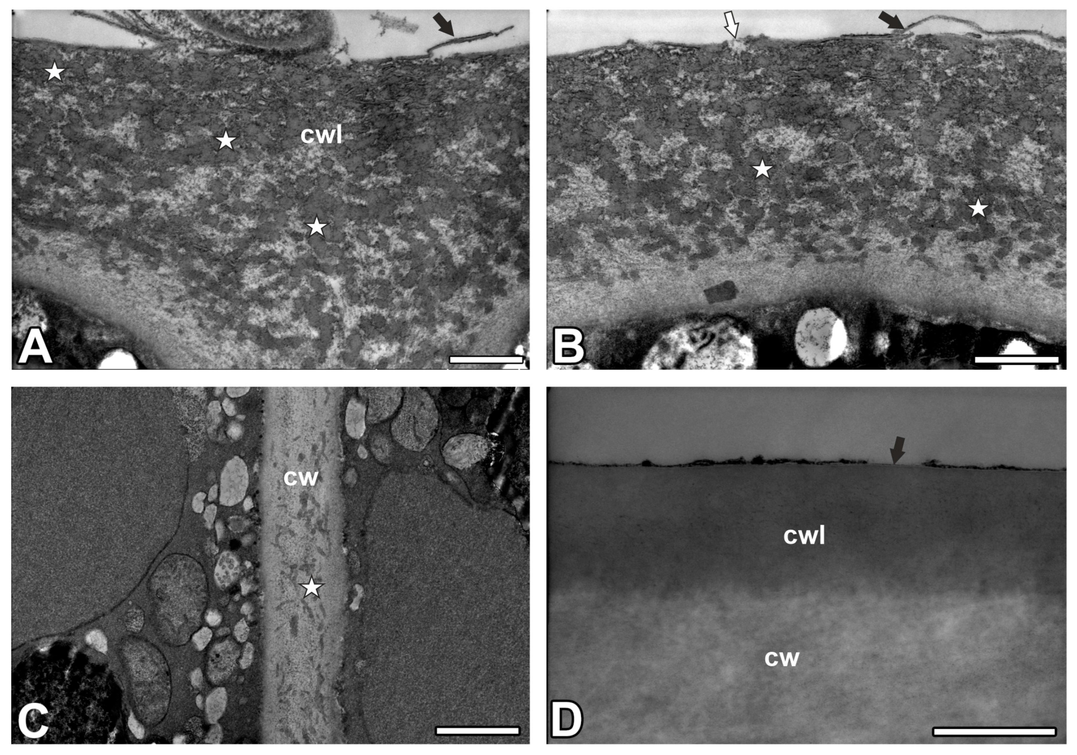
The digestive gland had a thin cuticularized layer, which was ruptured in some places. The cutinized wall layer was very thick, with a very dense network of cutin cystoliths (cuticular droplets) (Figure 3A,B). Between cystoliths, there was non-impregnated wall material. The cuticular gaps occurred (Figure 3A,B). The innermost layer of the cell wall did not have cutin cystoliths. However, cutin cystoliths occurred in radial cell walls (Figure 3C). These cell walls had strong autofluorescence under UV (Figure 2A). In the epidermal cells, there were no cuticle discontinuities (Figure 3D).

2.2. Pectic Homogalacturonan Distribution

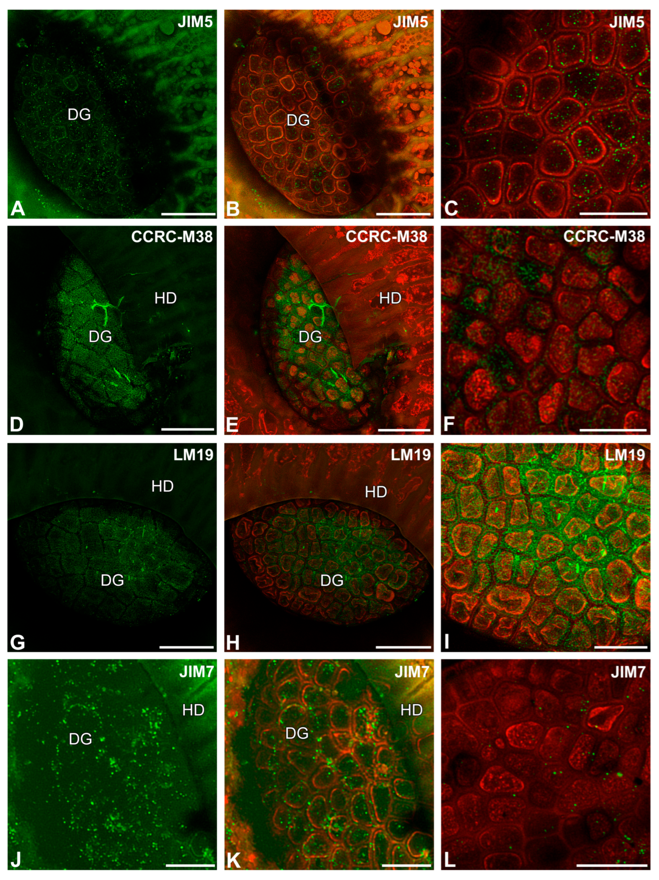
After immunolabeling with the JIM5 antibody (low methylesterified HGs), the signal was observed as dots on the surfaces of digestive gland cells and some epidermal cells (Figure 4A–C). The fluorescence signals detected by CCRC-M38 (a fully de-esterified HGs) (Figure 4D–F) and by LM19 (low methylesterified HG) were observed as a meshwork in the outer cell walls of digestive glands (Figure 4G–I). However, a fluorescence signal from highly esterified HGs (detected by JIM7) was observed as dots on the surfaces of digestive gland cells (Figure 4J–L).

2.3. Hemicellulose Distribution

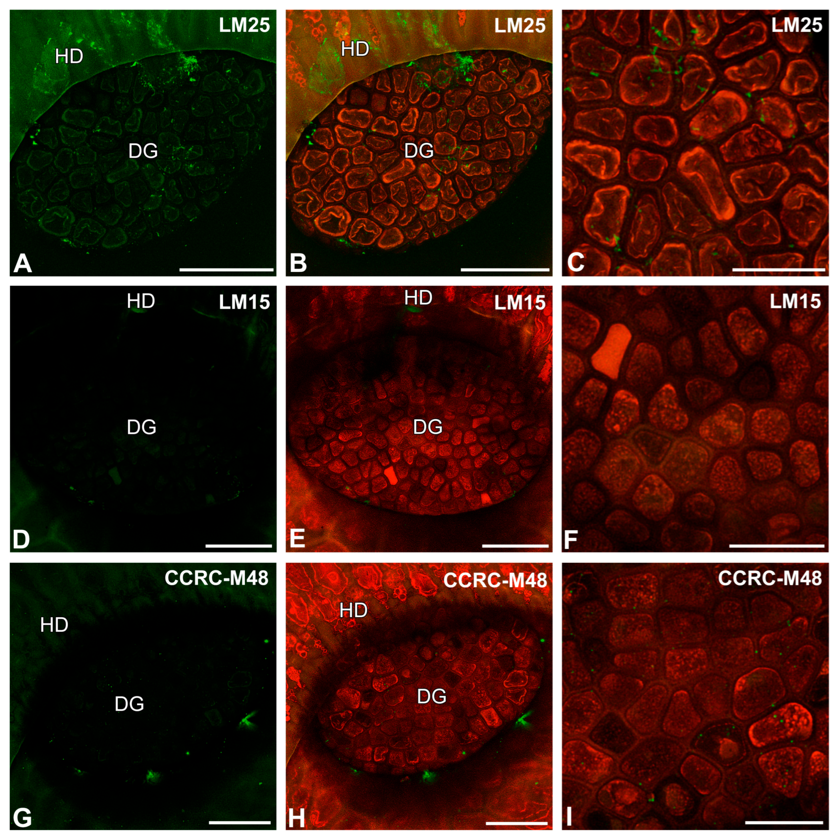
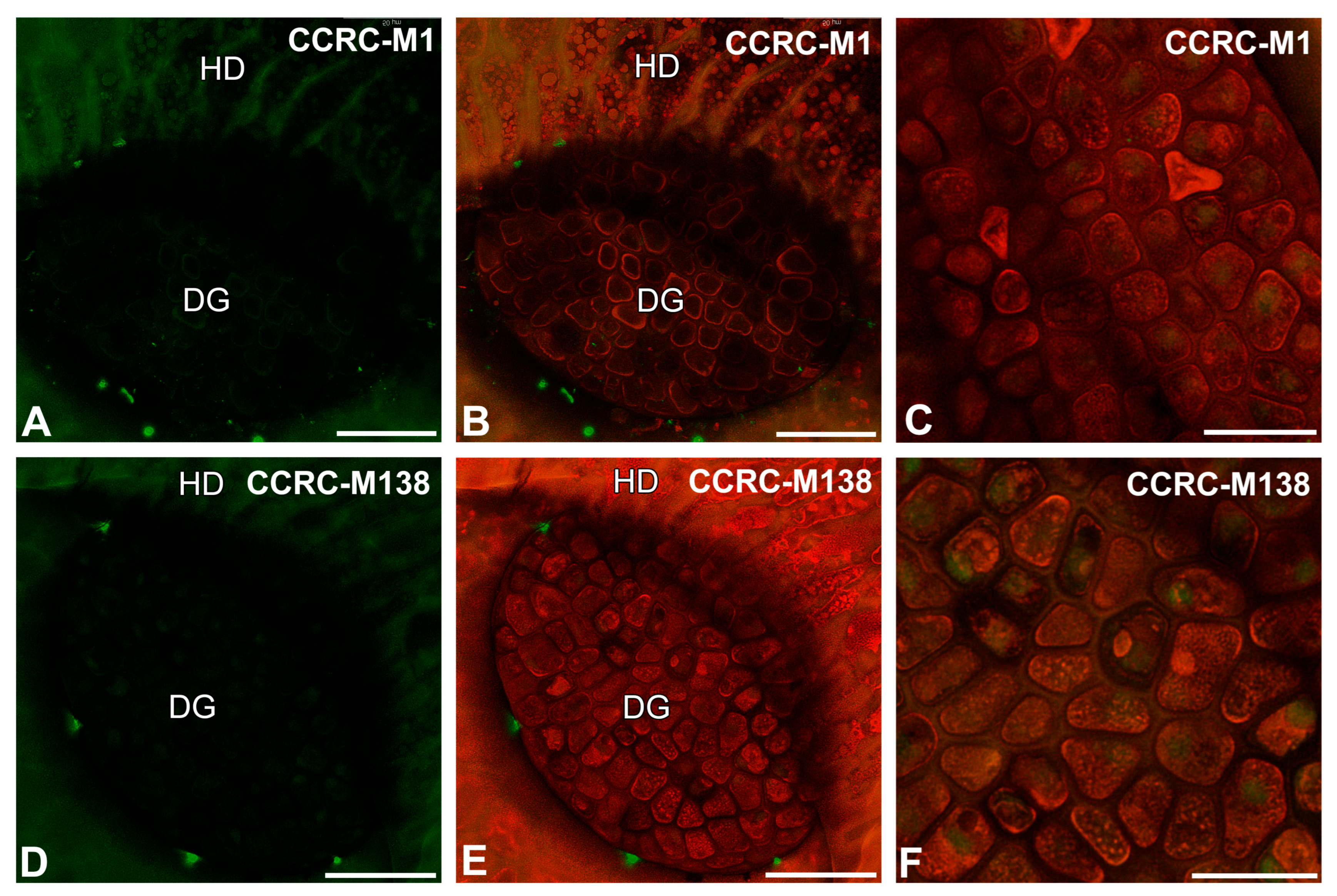
After immunolabeling with the LM25 antibody (which recognizes land plants galactoxyloglucan), the signal of this antibody was observed in secretion or debris (debris is understood as non-deified remains of organisms and secretions that are in a trap) on the digestive gland and epidermal cell surfaces (Figure 5A–C). After immunolabeling with the LM15 antibody (which reacts with xyloglucan), the signal of this antibody was not observed in cell walls (Figure 5D–F). The LM15 antibody yielded fluorescence signals in the debris on the cell surfaces (Figure 5D,E). After immunolabeling with the CCRC-M48 antibody (which reacts with xyloglucan and recognizes the XXLG, XLLG glycan group of Non-Fucosylated Xyloglucan-5), the signal was not observed in cell walls (Figure 5G–I) but occurred in the debris on the cell surfaces (Figure 5G–I). After immunolabeling with the CCRC-M1 antibody (which reacts with xyloglucan and recognizes the alpha-Fuc-(1,2)-beta-Gal glycan group of Fucosylated Xyloglucan), the signal was not observed in cell walls (Figure 6A–C). The CCRC-M1 antibody yielded fluorescence signals in the debris on the cell surfaces (Figure 6A,B). After immunolabeling with the CCRC-M138 antibody (which recognizes the glycan group of Xylan-6), the signal was not observed in the cell walls (Figure 6D–F). The CCRC-M138 antibody yielded fluorescence signals in the debris on the cell surfaces (Figure 6D,E).
When the sections were pre-treated with pectate lyase (during which pectins were removed), the fluorescence signals detected by LM25 and LM15 antibodies were observed as a meshwork in the outer cell walls of digestive glands and debris (Figure 7A–D).

2.4. The Arabinogalactan Protein (AGP) Distribution

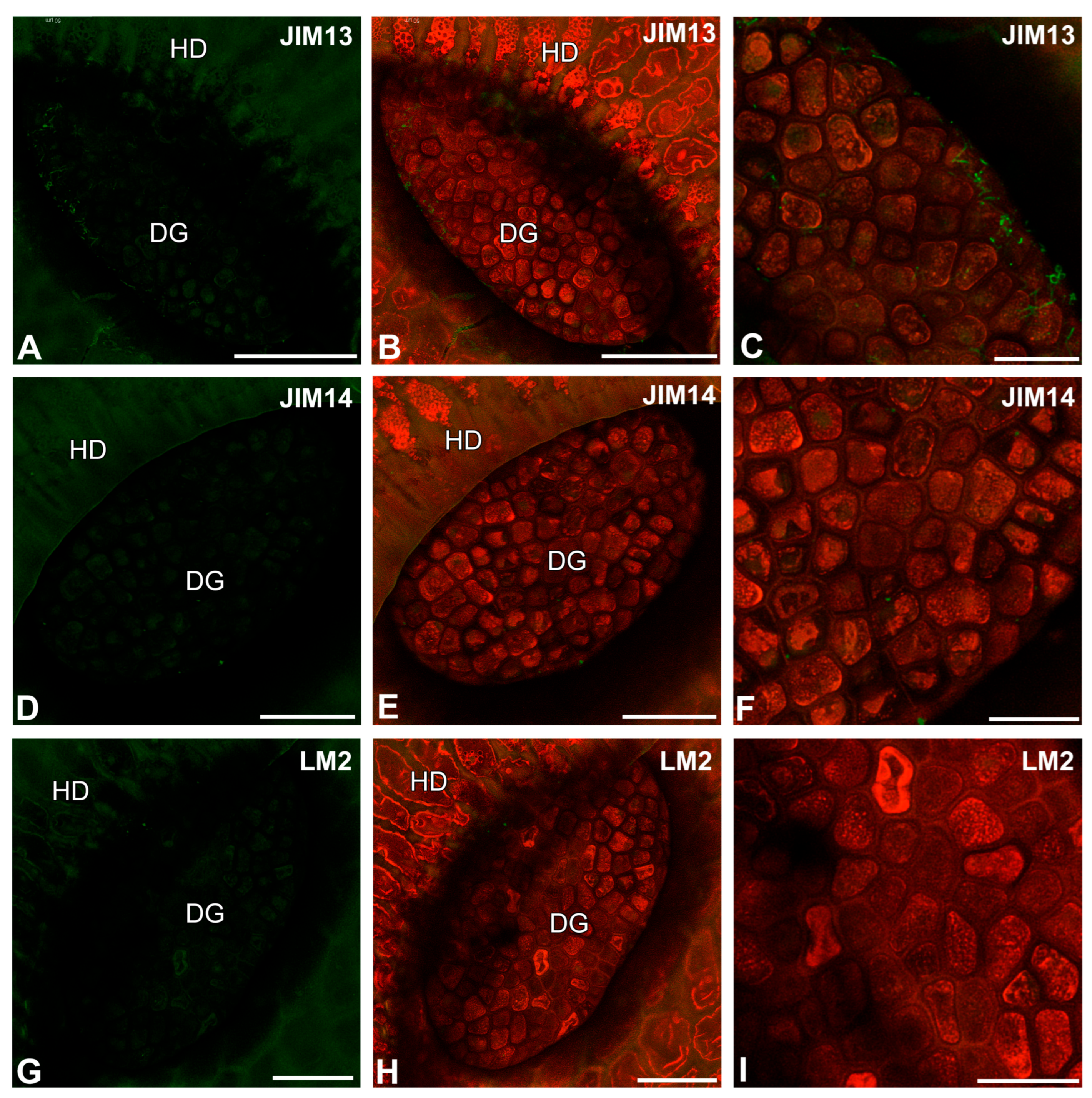
After immunolabeling with the JIM13 antibody, the signal of this antibody was observed in debris or bacteria on the digestive gland cell surfaces (Figure 8A–C). The immunolabeling results with the JIM14 antibody (Figure 8D,E) and the LM2 antibody (Figure 8G–I) were similar. The signal was rare and observed as dots on the cell surfaces.

3. Discussion

3.1. Cuticle Structure

To survive in nutrient-poor habitats, carnivorous plants have developed an astonishingly diverse digestive system [1,30]. Carnivorous plants have also developed various modifications of the cuticle in their glands to secrete mucus and enzymes and absorb nutrients from their prey [1]. Cuticle discontinuities may be developed differently and take the form of cuticular pores [31,32,33,34,35], cuticular gaps [36,37,38], and ‘ill-defined discontinuities’, all of which allow rapid movement across the leaf surface. In some species, both cuticular gaps and cuticular pores may occur: Roridula [39] and Utricularia (Joel and Juniper [36] interpretation of results of Finerean and Lee [40]). The specific, giant cuticular discontinuities occur in Byblis glands, forming cuticular holes [41]. The presence of cuticle discontinuities can be checked using vital dyes, as was demonstrated in the digestive glands of Nepenthes × coccinea, Nepenthes × ventrata by Adlassnig et al. [42], and here in the digestive glands of Nepenthes albomarginata.
We found that the cuticle of the digestive glands in Nepenthes albomarginata is similar in structure (presence of cutin cystoliths) to the cuticle of the glands in Nepenthes spp. [27], Nepenthes khasiana [1], and Nepenthes alata [25]. According to Juniper et al. 1989 [1], the cuticularized layer undergoes exfoliation during gland maturation, but we still observed it in Nepenthes albomarginata glands.
Only a few Nepenthes species have been studied in detail regarding cuticle structure. Nepenthes species are an extremely diverse group with different strategies for obtaining nutrient compounds and with various specializations in the morphology and function of pitchers [5,11,43,44,45]; therefore, in the future, it should be investigated whether glands from such different types of pitchers have a similar cuticle structure. It will be interesting to examine the structure of cell walls in glandular structures in other species that have pitcher traps: Sarracenia, Heliamphora, Darlingtonia, and Cephalotus [1,3].

3.2. Whole-Mount Immunolabeled Gland Technique

Whole-mount immunolabeled organ technique was used successfully in the analysis of plant organs lacking cuticles, such as pollen tubes [46,47] and root hairs [48,49,50]. Li et al. [46] showed arabinogalactan epitopes in the cell wall of pollen tubes of Nicotiana tabacum L. Chebli et al. [47] showed pectin epitopes in the cell wall of pollen tubes of Arabidopsis thaliana L. Larson et al. [49] showed that even live Arabidopsis thaliana root hairs may be labeled with cell wall polymer-specific antibodies. Marzec et al. [50] detected the LM2 epitope on the surface of primordia and root hair tubes in plants able to generate root hairs in Hordeum vulgare L. using whole-mount immunolabelled root sections, which were chemically fixed. As for carnivorous plants, the whole-mount immunolabeled organ technique was used twice. In case of Utricularia dichotoma subsp. novae-zelandiae (Hook.f) R.W.Jobson, the antibodies penetrated the cell wall only in the areas where the cuticle had an open structure; however, no labeling occurred in cell wall parts which were heavily impregnated with cutin [51,52]. Płachno and Kapusta [51], using whole-mount immunolabeled traps, found that the cell walls of the quadrifid arms were especially rich in low-methyl-esterified homogalacturonan. Also, arabinogalactan proteins were detected. Hemicelluloses were detected when the traps were pre-treated with pectate lyase (during which pectins were removed). This was caused by the fact that pectic homogalacturonans mask abundant sets of xyloglucan epitopes in plant cell walls [53]. The next examined species, Drosophyllum lusitanicum (L.) Link, is interesting because it relates to Nepenthes [54,55]. Płachno et al. [38], using whole-mount immunolabeled glands, studied the occurrence of wall components in this species. Despite the presence of cuticle gaps, antibody penetration was limited only to the cell wall surface (e.g., AGPs labeled with JIM8, pectic homogalacturonan labeled with LM19, galactoxyloglucan labeled with LM25, and xyloglucan labeled with LM15). It should be noted that some results were ambiguous, and it was sometimes difficult to distinguish whether the signal came from the peripheral part of the wall or from the secretion or the material on the cell surface.
Like Utricularia dichotoma and Drosophyllum lusitanicum glands, we found pectic low-methyl-esterified homogalacturonans in the outer cell walls of Nepenthes albomarginata. Positive labeling with LM19 and CCRC-M38 antibodies suggests the presence of pectic homogalacturonan in the very superficial part of the glands’ cell walls, so they were easily accessible to antibodies. Pectic homogalacturonans are known to be involved in plant cell wall porosity and hydration [56,57]. We suggest that the presence of these hydrophilic homogalacturonans on the gland surface may facilitate the processes of fluid secretion into the pitcher or absorption of compounds produced due to the digestion of prey. Recently pectic homogalacturonans and hemicelluloses were detected in cell walls of cells in Nepenthes external glands, which may function as may act as hydathodes or hydropotes [58].
We treat the technique used as a preliminary research stage on Nepenthes glands. Because the antibodies, which detect esterified pectins, hemicelluloses, and arabinogalactan protein marked only debris or secretion residues on the gland or epidermal surfaces, which may result either from a lack of epitopes in external parts of cell walls of glands or difficulties with antibody penetration. In the next stage, we plan to use cross-sections through the glands and also enzyme treatment, which will allow better access of antibodies to epitopes and provide a comprehensive picture of the occurrence of the major cell wall polysaccharides and glycoproteins.

4. Materials and Methods

4.1. Plant Material

Nepenthes albomarginata T.Lobb ex Lindl. pitchers were taken from the first author’s collection; they were grown in the humid terrarium.

4.2. Immunochemical Analysis

The bottom parts of pitchers (digestive zone) were cut for small fragments and fixed in 8% (w/v) formaldehyde (Sigma-Aldrich, Sigma-Aldrich Sp. zo.o. Poznań, Poland) mixed with 0.25% (v/v) glutaraldehyde (Sigma-Aldrich, Sigma-Aldrich Sp. zo.o. Poznań, Poland) in a PIPES buffer with addition of Tween (Sigma-Aldrich, Sigma-Aldrich Sp. zo.o. Poznań, Poland) or DMSO (Sigma-Aldrich, Sigma-Aldrich Sp. zo.o. Poznań, Poland).
The plant material was washed in a PBS buffer and later blocked with 1% bovine serum albumin (BSA, Sigma-Aldrich) in a PBS buffer and incubated with the following primary antibodies overnight at 4 °C: anti-homogalacturonans (HGs): JIM5 (low methylesterified HG), JIM7 (highly esterified HG), LM19 (low methylesterified HG), and CCRC-M38 (a fully de-esterified HG); and anti-hemicelluloses: LM25 (galactoxyloglucan), CCRC-M48 (xyloglucan), CCRC-M1 (xyloglucan), LM15 (xyloglucan), and CCRC-M138 (xylan) [59,60,61,62,63,64], Paul Knox, PhD, University of Leeds. Available online: https://www.kerafast.com/cat/799/paul-knox-phd (accessed on 13 November 2023), https://www.kerafast.com/(accessed on 13 November 2023), anti-arabinogalactan protein: JIM13, JIM14, and LM2 [65]. All of the primary antibodies were used in a 1:20 dilution. They were purchased from Plant Probes, UK (rat monoclonal antibodies: JIM5, JIM7, LM19, LM25, LM15, JIM13, JIM14, and LM2) and Agrisera, Sweden (mouse monoclonal antibodies: CCRC-M38, CCRC-M1, CCRC-M48, and CCRC-M138). Secondary antibodies: goat anti-rat secondary or anti-mouse antibody conjugated with FITC or Alexa Fluor 488, respectively, were purchased from Abcam (Cambridge, UK). The samples were then cover-slipped using a Mowiol mounting medium: a mixture of Mowiol ®4-88 (Sigma-Aldrich) and glycerol for fluorescence microscopy (Merck, Warsaw, Poland) with the addition of 2.5% DABCO (Carl Roth GmbH + Co. KG, Karlsruhe, Germany). They were viewed using a Leica STELLARIS 5 WLL confocal microscope with Lightning module deconvolution. Negative controls were created by omitting the primary antibody step, which caused no fluorescence signal in any of the control frames for any stained slides (Figure S1). To remove the HG from the cell walls, the material was pretreated with 0.1 M sodium carbonate pH = 11.4 for 2 h at room temperature. This was followed by digestion with a pectate lyase 1A (Nzytech) at 10 μg/mL for 2 h at room temperature in 50 mM N-cyclohexyl-3-aminopropane sulfonic acid (CAPS) with the addition of 2 mM of a CaCl2 buffer at pH 10, and then incubation with the antibodies, as described above.

4.3. Scanning Transmission Electron Microscopy

The glands were also examined using electron microscopy, as follows: Fragments of the traps were fixed in a mixture of 2.5% glutaraldehyde with 2.5% formaldehyde in a 0.05 M cacodylate buffer (Sigma-Aldrich, Sigma-Aldrich Sp. z o.o., Poznań, Poland; pH 7.2) for a few days, and later, the material was processed as in Płachno et al. [66]. The material was dehydrated with acetone and embedded in an Epoxy Embedding Medium Kit (Fluka) or Durcupan resin (Sigma-Aldrich Chemie GmbH, Taufkirchen, Germany). Ultrathin sections were cut on a Leica Ultracut UCT ultramicrotome. The sections were examined using a Hitachi UHR FE-SEM SU 8010 microscope housed at the University of Silesia in Katowice.

4.4. Scanning Electron Microscopy

For the scanning electron microscopy (SEM), the traps were cut and fixed in methanol, later transferred to ethanol, then transferred to acetone, and dried using supercritical CO2. The material was then sputter-coated with gold and examined using a Hitachi UHR FE-SEM SU 8010 microscope housed at the University of Silesia in Katowice.

4.5. Light Microscopy (LM)

The living glands were stained with 20 μM DiOC6 (3,3′-dihexyloxacarbocyanine iodide; Thermo Fisher, Rockland, MD, USA) dissolved in water [67]. The traps were examined using a Nikon Eclipse E400 light microscope. Autofluorescence of gland cell walls was examined using a Nikon Eclipse E400 light microscope (Tokyo, Japan) with a UV-2A filter (Ex. 330–380 nm, DM. 400 nm, Em. 420-α nm). A toluidine blue solution was applied to the pitcher to see if glands can take up aqueous solutions. After several hours, the pitcher was rinsed with water, and photos were taken using Emspira 3 Digital Microscope (Leica, PIK Instruments Sp. z o.o. Piaseczno, Poland).

5. Conclusions

The method used was not perfect (because not all antibodies gave positive results, which may result from a lack of epitopes or difficulties with antibody penetration). Still, it did show the presence of pectic homogalacturonans in the very superficial part of the cell walls of the glands. Our results contribute to the current knowledge regarding cell wall structure and cuticle structure in carnivorous plant glands.

Supplementary Materials

The following supporting information can be downloaded at: https://www.mdpi.com/article/10.3390/ijms26189174/s1.

Author Contributions

Conceptualization, B.J.P.; methodology, B.J.P., M.K. and M.F.; investigation, B.J.P. and M.K.; resources, B.J.P.; data curation, B.J.P.; writing—preparing the original draft, B.J.P.; writing—review and editing, B.J.P., P.S., M.F., P.Ś. and M.K.; visualization, B.J.P. and M.K.; supervision, B.J.P.; project administration, B.J.P.; funding acquisition, B.J.P., M.F., P.S. and P.Ś. All authors have read and agreed to the published version of the manuscript.

Funding

Research activity was financed from funds allocated to the European City of Science, Katowice, 2024. The research has been partially supported by the Ministry of Science and Higher Education of Poland, within the statutory activities of the Institute of Botany, Faculty of Biology, Jagiellonian University and Bioimaging Laboratory, Faculty of Biology, University of Gdańsk.

Institutional Review Board Statement

Not applicable.

Informed Consent Statement

Not applicable.

Data Availability Statement

The data presented in this study are available on request from the corresponding author.

Conflicts of Interest

The authors declare no conflicts of interest.

References

  1. Juniper, B.E.; Robbins, R.J.; Joel, D.M. The Carnivorous Plants; Academic Press: London, UK, 1989. [Google Scholar]
  2. Pavlovič, A.; Masarovičová, E.; Hudák, J. Carnivorous syndrome in Asian pitcher plants of the genus Nepenthes. Ann. Bot. 2007, 100, 527–536. [Google Scholar] [CrossRef]
  3. Król, E.; Płachno, B.J.; Adamec, L.; Stolarz, M.; Dziubińska, H.; Trębacz, K. Quite a few reasons for calling carnivores ‘the most wonderful plants in the world’. Ann. Bot. 2012, 109, 47–64. [Google Scholar] [CrossRef] [PubMed]
  4. Moran, J.A.; Clarke, C.M. The carnivorous syndrome in Nepenthes pitcher plants: Current state of knowledge and potential future directions. Plant Signal. Behav. 2010, 5, 644–648. [Google Scholar] [CrossRef]
  5. Cheek, M.; Jebb, M.; Murphy, B. A classification of functional pitcher types in Nepenthes (Nepenthaceae). bioRxiv 2019. [Google Scholar] [CrossRef]
  6. Lloyd, F.E. The Carnivorous Plants; Ronald Press: New York, NY, USA, 1942. [Google Scholar]
  7. Gorb, E.; Kastner, V.; Peressadko, A.; Arzt, E.; Gaume, L.; Rowe, N.; Gorb, S. Structure and properties of the glandular surface in the digestive zone of the pitcher in the carnivorous plant Nepenthes ventrata and its role in insect trapping and retention. J. Exp. Biol. 2004, 207, 2947–2963. [Google Scholar] [CrossRef]
  8. Gorb, E.V.; Gorb, S.N. Physicochemical properties of functional surfaces in pitchers of the carnivorous plant Nepenthes alata Blanco (Nepenthaceae). Plant Biol. 2006, 8, 841–848. [Google Scholar] [CrossRef]
  9. Bauer, U.; Jetter, R.; Poppinga, S. Non-motile traps. In Carnivorous Plants: Physiology, Ecology, and Evolution; Ellison, A.M., Adamec, L., Eds.; Oxford University Press: Oxford, UK, 2018. [Google Scholar]
  10. Lessware, O.C.; Mantell, J.M.; Bauer, U. Carnivorous Nepenthes pitcher plants combine common developmental processes to make a complex epidermal trapping surface. Ann. Bot. 2025, 135, 643–654. [Google Scholar] [CrossRef]
  11. Bauer, U.; Clemente, C.J.; Renner, T.; Federle, W. Form follows function: Morphological diversification and alternative trapping strategies in carnivorous Nepenthes pitcher plants. J. Evol. Biol. 2012, 25, 90–102. [Google Scholar] [CrossRef] [PubMed]
  12. Lenz, A.K.; Bauer, U. Pitcher geometry facilitates extrinsically powered ‘springboard trapping’ in carnivorous Nepenthes gracilis pitcher plants. Biol. Lett. 2022, 18, 20220106. [Google Scholar] [CrossRef]
  13. Chomicki, G.; Burin, G.; Busta, L.; Gozdzik, J.; Jetter, R.; Mortimer, B.; Bauer, U. Convergence in carnivorous pitcher plants reveals a mechanism for composite trait evolution. Science 2024, 383, 108–113. [Google Scholar] [CrossRef] [PubMed]
  14. Amagase, S. Digestive enzymes in insectivorous plants: III. Acid proteases in the genus Nepenthes and Drosera peltata. J. Bioch. 1972, 72, 73–81. [Google Scholar] [CrossRef] [PubMed]
  15. Tökés, Z.A.; Woon, W.C.; Chambers, S.M. Digestive enzymes secreted by the carnivorous plant Nepenthes macferlanei L. Planta 1974, 119, 39–46. [Google Scholar] [CrossRef] [PubMed]
  16. Hatano, N.; Hamada, T. Proteome analysis of pitcher fluid of the carnivorous plant Nepenthes alata. J. Prot. Res. 2008, 7, 809–816. [Google Scholar] [CrossRef]
  17. Buch, F.; Kaman, W.E.; Bikker, F.J.; Yilamujiang, A.; Mithöfer, A. Nepenthesin protease activity indicates digestive fluid dynamics in carnivorous Nepenthes plants. PLoS ONE 2015, 10, e0118853. [Google Scholar] [CrossRef] [PubMed]
  18. Saganová, M.; Bokor, B.; Stolárik, T.; Pavlovič, A. Regulation of enzyme activities in carnivorous pitcher plants of the genus Nepenthes. Planta 2018, 248, 451–464. [Google Scholar] [CrossRef]
  19. Heslop-Harrison, Y. Enzyme release in carnivorous plants. In Lysozymes in Biology and Pathology; Dingle, J.T., Dean, R.T., Eds.; North Holland Publishing Company: Amsterdam, The Netherlands, 1975; Volume 4, pp. 525–578. [Google Scholar]
  20. Parkes, D.M. Adaptive Mechanisms of Surface and Glands in Some Carnivorous Plants. Master’s Thesis, Monash University, Clayton, VIC, Australia, 1980. [Google Scholar]
  21. Płachno, B.J.; Adamec, L.; Lichtscheidl, I.K.; Peroutka, M.; Adlassnig, W.; Vrba, J. Fluorescence labelling of phosphatase activity in digestive glands of carnivorous plants. Plant Biol. 2006, 8, 813–820. [Google Scholar] [CrossRef]
  22. Thornhill, A.H.; Harper, I.S.; Hallam, N.D. The development of the digestive glands and enzymes in the pitchers of three Nepenthes species: N. alata, N. tobaica, and N. ventricosa (Nepenthaceae). Int. J. Plant. Sci. 2008, 169, 615–624. [Google Scholar] [CrossRef]
  23. Vassilyev, A.E.; Muravnik, L.E. The nectaries of the lid in closed pitchers of Nepenthes khasiana (Nepenthaceae) secrete a digestive fluid. Bot. Zhurnal 2007, 92, 1141–1144. [Google Scholar]
  24. Vassilyev, A.E. The nectaries of the peristome in the closed pitchers of Nepenthes khasiana (Nepenthaceae) secrete polysacharide slime. Bot. Zhurnal 2007, 92, 1554–1568. [Google Scholar]
  25. Owen, T.P.; Lennon, K.A.; Santo, M.J.; Anderson, A.Y. Pathways for nutrient transport in the pitchers of the carnivorous plant Nepenthes alata. Ann. Bot. 1999, 84, 459–466. [Google Scholar] [CrossRef]
  26. Fenner, C.A. Beitraege zur Kenntnis der Anatomie, Entwicklungsgeschichte und Biologie der Laubblaetter und Druesen einiger Insektivoren. Flora 1904, 93, 333–335. [Google Scholar]
  27. Schnepf, E. Die Morphologie der Sekretion in pflanzlichen Drüsen. Ber. Dtsch. Bot. Ges. 1965, 78, 478–483. [Google Scholar]
  28. Gorb, E.V.; Gorb, S.N. Functional Surfaces in the Pitcher of the Carnivorous Plant Nepenthes alata: A Cryo-Sem Approach. In Functional Surfaces in Biology; Gorb, S.N., Ed.; Springer: Dordrecht, The Netherlands, 2009. [Google Scholar]
  29. Ivesic, C.; Krammer, S.; Koller-Peroutka, M.; Laarouchi, A.; Gruber, D.; Lang, I.; Lichtscheidl, I.K.; Adlassnig, W. Quantification of Protein Uptake by Endocytosis in Carnivorous Nepenthales. Plants 2023, 12, 341. [Google Scholar] [CrossRef] [PubMed]
  30. Freund, M.; Graus, D.; Fleischmann, A.; Gilbert, K.J.; Lin, Q.; Renner, T.; Stigloher, C.; Albert, V.A.; Hedrich, R.; Fukushima, K. The digestive systems of carnivorous plants. Plant Physiol. 2022, 190, 44–59. [Google Scholar] [CrossRef] [PubMed]
  31. Williams, S.E.; Pickard, B.G. Secretion, absorption and cuticular structure in Drosera tentacles. Plant Physiol. 1969, 44, 5. [Google Scholar]
  32. Ragetli, H.W.; Weintraub, L.O.E. Characteristics of Drosera tentacles. I. Anatomical and cytological details. Can. J. Bot. 1972, 50, 159–168. [Google Scholar] [CrossRef]
  33. Williams, S.E.; Pickard, B.G. Connections and barriers between cells of Drosera tentacles in relation to their electrophysiology. Planta 1974, 116, 1–16. [Google Scholar] [CrossRef]
  34. Płachno, B.J.; Faber, J.; Jankun, A. Cuticular discontinuities in glandular hairs of Genlisea St.-Hil. in relation to their functions. Acta Bot. Gall. 2005, 152, 125–130. [Google Scholar] [CrossRef]
  35. Płachno, B.J.; Kozieradzka-Kiszkurno, M.; Świątek, P. Functional Ultrastructure of Genlisea (Lentibulariaceae) Digestive Hairs. Ann. Bot. 2007, 100, 195–203. [Google Scholar] [CrossRef] [PubMed]
  36. Joel, D.M.; Juniper, B.E. Cuticular gaps in carnivorous plant glands. In The Plant Cuticle; Cutler, D.F., Alvin, K.L., Price, C.E., Eds.; Academic Press: London, UK, 1982; pp. 121–130. [Google Scholar]
  37. Joel, D.M.; Rea, P.A.; Juniper, B.E. The cuticle of Dionaea muscipula Ellis (Venus’s Flytrap) in relation to stimulation, secretion and absorption. Protoplasma 1983, 114, 44–51. [Google Scholar] [CrossRef]
  38. Płachno, B.J.; Kapusta, M.; Stolarczyk, P.; Świątek, P. Do Cuticular Gaps Make It Possible to Study the Composition of the Cell Walls in the Glands of Drosophyllum lusitanicum? Int. J. Mol. Sci. 2024, 25, 1320. [Google Scholar] [CrossRef]
  39. Anderson, B. Adaptations to foliar absorption of faeces: A pathway in plant carnivory. Ann. Bot. 2005, 95, 757–761. [Google Scholar] [CrossRef] [PubMed][Green Version]
  40. Fineran, B.A.; Lee, M.S.L. Organization of quadrifid and bifid hairs in the trap of Utricularia monanthos. Protoplasma 1975, 84, 43–70. [Google Scholar] [CrossRef]
  41. Płachno, B.J.; Lancelle, S.; Świątek, P.; Hepler, P.K.; Weidinger, M.; Lichtscheidl, I. Cyto-architecture of Byblis glands and leaf cells based on freeze-substitution and conventional TEM. Ann. Bot. 2025, 135, 463–482. [Google Scholar] [CrossRef]
  42. Adlassnig, W.; Koller-Peroutka, M.; Bauer, S.; Koshkin, E.; Lendl, T.; Lichtscheidl, I.K. Endocytotic uptake of nutrients in carnivorous plants. Plant J. 2012, 71, 303–313. [Google Scholar] [CrossRef]
  43. Bauer, U.; Grafe, T.U.; Federle, W. Evidence for alternative trapping strategies in two forms of the pitcher plant, Nepenthes rafflesiana. J. Exp. Bot. 2011, 62, 3683–3692. [Google Scholar] [CrossRef]
  44. Bauer, U.; Di Giusto, B.; Skepper, J.; Grafe, T.U.; Federle, W. With a flick of the lid: A novel trapping mechanism in Nepenthes gracilis pitcher plants. PLoS ONE 2012, 7, e38951. [Google Scholar] [CrossRef] [PubMed]
  45. McPherson, S. Nepenthes: The Tropical Pitcher Plants; Redfern Natural History: Dorset, UK, 2023; Volume 1–3. [Google Scholar]
  46. Li, Y.Q.; Bruun, L.; Pierson, E.S.; Cresti, M. Periodic deposition of arabinogalactan epitopes in the cell wall of pollen tubes of Nicotiana tabacum L. Planta 1992, 188, 532–538. [Google Scholar] [CrossRef] [PubMed]
  47. Chebli, Y.; Kaneda, M.; Zerzour, R.; Geitmann, A. The cell wall of the Arabidopsis pollen tube--spatial distribution, recycling, and network formation of polysaccharides. Plant Physiol. 2012, 160, 1940–1955. [Google Scholar] [CrossRef] [PubMed]
  48. Willats, W.G.; McCartney, L.; Knox, J.P. In-situ analysis of pectic polysaccharides in seed mucilage and at the root surface of Arabidopsis thaliana. Planta 2001, 213, 37–44. [Google Scholar] [CrossRef]
  49. Larson, E.R.; Tierney, M.L.; Tinaz, B.; Domozych, D.S. Using monoclonal antibodies to label living root hairs: A novel tool for studying cell wall microarchitecture and dynamics in Arabidopsis. Plant Methods 2014, 10, 30. [Google Scholar] [CrossRef]
  50. Marzec, M.; Szarejko, I.; Melzer, M. Arabinogalactan proteins are involved in root hair development in barley. J. Exp. Bot. 2015, 5, 1245–1257. [Google Scholar] [CrossRef]
  51. Płachno, B.J.; Kapusta, M. The Localization of Cell Wall Components in the Quadrifids of Whole-Mount Immunolabeled Utricularia dichotoma Traps. Int. J. Mol. Sci. 2024, 25, 56. [Google Scholar] [CrossRef]
  52. Płachno, B.J.; Kapusta, M.; Stolarczyk, P.; Feldo, M.; Świątek, P. Do Arabinogalactan Proteins Occur in the Transfer Cells of Utricularia dichotoma? Int. J. Mol. Sci. 2024, 25, 6623. [Google Scholar] [CrossRef] [PubMed]
  53. Marcus, S.E.; Verhertbruggen, Y.; Hervé, C.; Ordaz-Ortiz, J.J.; Farkas, V.; Pedersen, H.L.; Willats, W.G.; Knox, J.P. Pectic homogalacturonan masks abundant sets of xyloglucan epitopes in plant cell walls. BMC Plant Biol. 2008, 8, 60. [Google Scholar] [CrossRef] [PubMed]
  54. Heubl, G.; Bringmann, G.; Meimberg, H. Molecular phylogeny and character evolution of carnivorous plant families in Caryophyllales—Revisited. Plant Biol. 2006, 8, 821–830. [Google Scholar] [CrossRef] [PubMed]
  55. Walker, J.F.; Yang, Y.; Moore, M.J.; Mikenas, J.; Timoneda, A.; Brockington, S.F.; Smith, S.A. Widespread paleopolyploidy, gene tree conflict, and recalcitrant relationships among the carnivorous Caryophyllales. Am. J. Bot. 2017, 104, 858–867. [Google Scholar] [CrossRef]
  56. Ridley, M.A.; O’Neill, D.; Mohnen, D. Pectins: Structure, biosynthesis, and oligogalac-turonide-related signaling. Phytochemistry 2001, 57, 929–967. [Google Scholar] [CrossRef]
  57. Peaucelle, A.; Braybrook, S.; Höfte, H. Cell wall mechanics and growth control in plants: The role of pectins revisited. Front. Plant Sci. 2012, 3, 121. [Google Scholar] [CrossRef]
  58. Płachno, B.J.; Kapusta, M.; Feldo, M.; Stolarczyk, P.; Małota, K.; Banaś, K. External Glands of Nepenthes Traps: Structure and Potential Function. Int. J. Mol. Sci. 2025, 26, 7788. [Google Scholar] [CrossRef]
  59. Liners, F.; Letesson, J.J.; Didembourg, C.; Van Cutsem, P. Monoclonal Antibodies against Pectin: Recognition of a Conformation Induced by Calcium. Plant Physiol. 1989, 91, 1419–1424. [Google Scholar] [CrossRef] [PubMed]
  60. Paul Knox, PhD, University of Leeds. Available online: https://www.kerafast.com/cat/799/paul-knox-phd (accessed on 13 November 2023).
  61. Knox, J.P.; Day, S.; Roberts, K. A set of cell surface glycoproteins forms an early marker of cell position, but not cell type, in the root apical meristem of Daucus carota L. Development 1989, 106, 47–56. [Google Scholar] [CrossRef]
  62. Verhertbruggen, Y.; Marcus, S.E.; Haeger, A.; Ordaz-Ortiz, J.J.; Knox, J.P. An extended set of monoclonal antibodies to pectic homogalacturonan. Carbohydr. Res. 2009, 28, 1858–1862. [Google Scholar] [CrossRef] [PubMed]
  63. Pattathil, S.; Avci, U.; Baldwin, D.; Swennes, A.G.; McGill, J.A.; Popper, Z.; Bootten, T.; Albert, A.; Davis, R.H.; Chennareddy, C.; et al. A comprehensive toolkit of plant cell wall gly-can-directed monoclonal antibodies. Plant Physiol. 2010, 153, 514–525. [Google Scholar] [CrossRef]
  64. McCartney, L.; Marcus, S.E.; Knox, J.P. Monoclonal antibodies to plant cell wall xylans and arabinoxylans. J. Histochem. Cytochem. 2005, 53, 543–546. [Google Scholar] [CrossRef]
  65. Available online: https://www.kerafast.com/ (accessed on 13 November 2023).
  66. Płachno, B.J.; Świątek, P.; Jobson, R.W.; Małota, K.; Brutkowski, W. Serial block face SEM visualization of unusual plant nuclear tubular extensions in a carnivorous plant (Utricularia, Lentibulariaceae). Ann. Bot. 2017, 120, 673–680. [Google Scholar] [CrossRef]
  67. Lichtscheidl, I.; Lancelle, S.; Weidinger, M.; Adlassnig, W.; Koller-Peroutka, M.; Bauer, S.; Krammer, S.; Hepler, P.K. Gland cell responses to feeding in Drosera capensis, a carnivorous plant. Protoplasma 2021, 258, 1291–1306. [Google Scholar] [CrossRef] [PubMed]
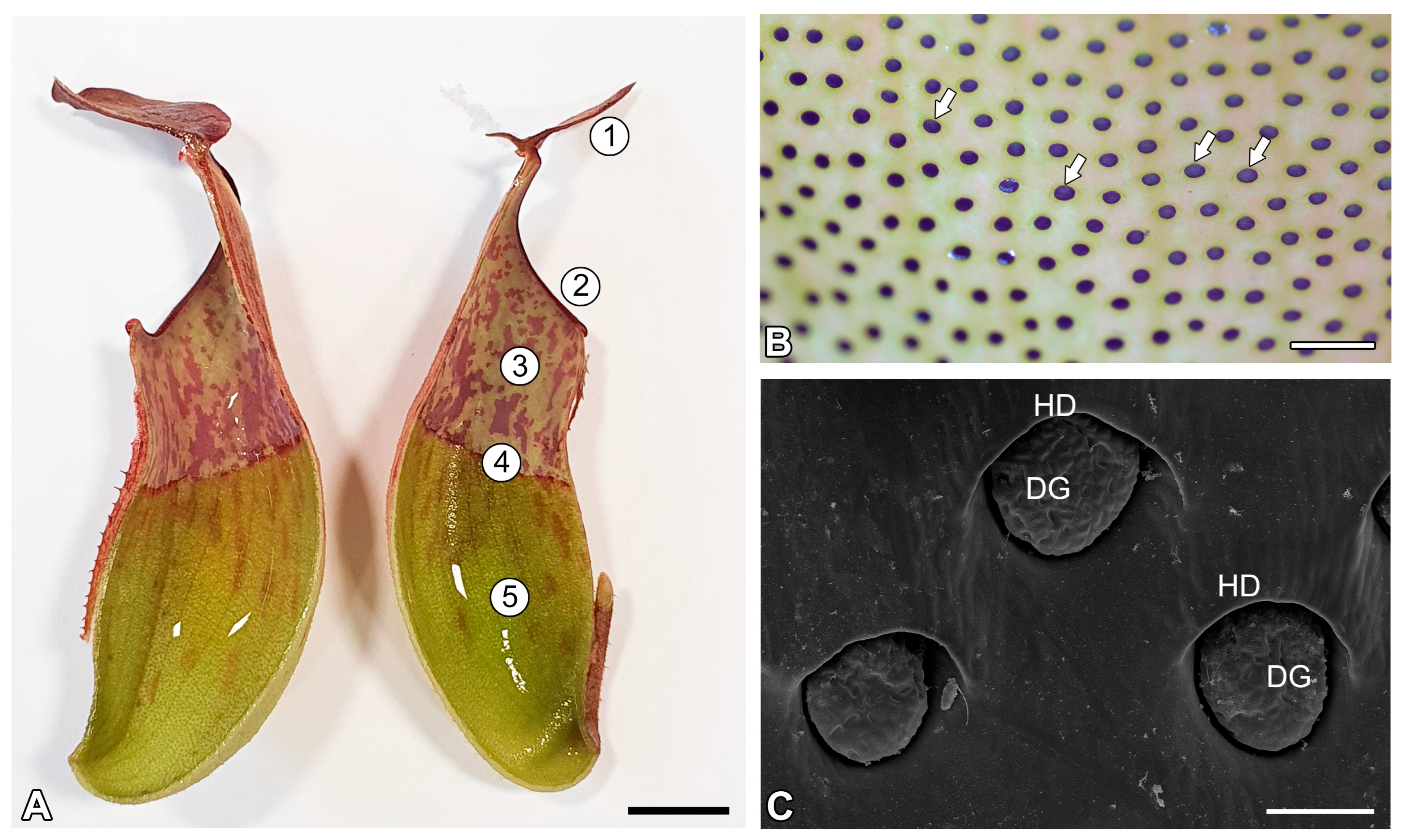
Figure 1. Pitcher zones and digestive glands, studied in Nepenthes albomarginata T.Lobb ex Lindl. (A) A dissected pitcher, with internal surfaces visible: 1. lid; 2. peristome; 3. slippery (wax) zone; 4. transitional zone; 5. digestive zone; bar = 1 cm. (B) Glands (arrows) from digestive zone, treated with toluidine blue; glands absorbed dye; bar = 1 mm. (C) Digestive gland morphology (scanning electron microscopy—SEM); digestive gland (DG); hood (HD); bar = 100 µm.
Ijms 26 09174 g001
Figure 2. Structure of the digestive glands of the Nepenthes albomarginata pitcher. (A) A digestive gland, visible glandular cells, and strong blue autofluorescence of the radial cell walls (arrow) of these cells; bar = 50 µm. (B) The same gland as in A, stained with DiOC6; see green fluorescence of protoplasts (star); cell wall (arrow); bar = 50 µm.
Ijms 26 09174 g002
Figure 3. Cuticle and cuticular discontinuities of the digestive glands and epidermal cells of the Nepenthes albomarginata. (A,B) Outer cell wall and cuticle of glandular cell of digestive gland; cutinized wall layer (cwl); cutin cystoliths (star); cuticular gaps (white arrow); cuticularized layer (black arrow); bar = 500 nm. (C) Radial cell walls (cw) of a glandular cell of the digestive gland; cutin cystoliths (star); bar = 1 µm. (D) Outer cell wall and cuticle of epidermal cell from digestive zone of pitcher; note the thick cutinized wall layer (cwl); cuticularized layer (black arrow); cell wall (cw); bar = 250 µm.
Ijms 26 09174 g003
Figure 4. Pectic homogalacturonan detected in the digestive gland of the Nepenthes albomarginata pitcher; digestive gland (DG), hood (HD) (intense green color—signal of antibody; red-brown color—autofluorescence). (A,B) Labeling of cells with JIM5 (low methylesterified HG), bar = 50 µm. (C) Labeling of cells with JIM5 antibody, glandular cells of digestive gland, bar = 25 µm. (D,E) Labeling of cells with CCRC-M38 antibody (a fully de-esterified HGs), note the positive signal in the form of a network, bar = 50 µm. (F) Labeling of cells with CCRC-M38 antibody, glandular cells of digestive gland, note the positive signal in the form of a network, bar = 25 µm. (G,H) Labeling of cells with LM19 antibody (low methylesterified HG), note the positive signal in the form of a network, bar = 50 µm. (I) Labeling of cells with LM19 antibody, glandular cells of digestive gland, note the positive signal in the form of a network, bar = 25 µm. (J,K) Labeling of cells with JIM7 antibody (high methylesterified HG), bar = 25 µm. (L) Labeling of cells with JIM7 antibody, glandular cells of digestive gland, bar = 25 µm.
Ijms 26 09174 g004
Figure 5. Hemicelluloses detected in the digestive gland of the Nepenthes albomarginata pitcher; digestive gland (DG), hood (HD) (intense green color—signal of antibody; red-brown color—autofluorescence). (A,B) Labeling of cells with LM25 antibody (xyloglu galactoxyloglucan), bar = 50 µm. (C) Labeling of cells with LM25 antibody, glandular cells of digestive gland, bar = 25 µm. (D,E) Labeling of cells with LM15 (xyloglucan), bar = 50 µm. (F) Labeling of cells with LM15 antibody, glandular cells of the digestive gland, bar = 25 µm. (G,H) Labeling of cells with CCRC-M48 antibody (xyloglucan), bar = 50 µm. (I) Labeling of cells with CCRC-M48 antibody glandular cells of the digestive gland, note the positive signal in the form of a network, bar = 25 µm.
Ijms 26 09174 g005
Figure 6. Hemicelluloses detected in the digestive gland of the Nepenthes albomarginata pitcher; digestive gland (DG), hood (HD) (intense green color—signal of antibody; red-brown color—autofluorescence). (A,B) Labeling of cells with CCRC-M1 antibody (xyloglucan), bar = 50 µm. (C) Labeling of cells with CCRC-M1 antibody, glandular cells of the digestive gland, bar = 25 µm. (D,E) Labeling of cells with CCRC-M138 antibody (xylan), note the positive signal in the form of a network, bar = 50 µm. (F) Labeling of cells with CCRC-M138 antibody, glandular cells of digestive gland, note the positive signal in the form of a network, bar = 25 µm.
Ijms 26 09174 g006
Figure 7. Hemicelluloses detected in the digestive gland of the Nepenthes albomarginata pitcher, after pre-treated with pectate lyase; digestive gland (DG), hood (HD) (intense green color—signal of antibody; red-brown color—autofluorescence). (A,B) Labeling of cells with LM25, bar = 50 µm. (C,D) Labeling of cells with LM15, bar = 50 µm.
Ijms 26 09174 g007
Figure 8. Arabinogalactan proteins detected in the digestive gland of the Nepenthes albomarginata pitcher; digestive gland (DG), hood (HD) (intense green color—signal of antibody; red-brown color—autofluorescence). (A,B) Labeling of cells with JIM13 antibody, bar = 50 µm. (C) Labeling of cells with JIM13 antibody, glandular cells of digestive gland, bar = 25 µm. (D,E) Labeling of cells with JIM14 antibody, note the positive signal in the form of a network, bar = 50 µm. (F) Labeling of cells with JIM14 antibody, glandular cells of digestive gland, bar = 25 µm. (G,H) Labeling of cells with LM2 antibody, bar = 50 µm. (I) Labeling of cells with LM2 antibody glandular cells of the digestive gland, bar = 25 µm.
Ijms 26 09174 g008
Disclaimer/Publisher’s Note: The statements, opinions and data contained in all publications are solely those of the individual author(s) and contributor(s) and not of MDPI and/or the editor(s). MDPI and/or the editor(s) disclaim responsibility for any injury to people or property resulting from any ideas, methods, instructions or products referred to in the content.