Article

Protective Effects of Methanolic Extract of Micromeria frivaldszkyana (Degen) Velen Against Acetaminophen-Induced Liver Toxicity in Male Wistar Rats

1
Department of Pharmacology, Toxicology, and Pharmacotherapy, Faculty of Pharmacy, Medical University of Plovdiv, Vasil Aprilov Str. 15A, 4002 Plovdiv, Bulgaria
2
Research Institute, Medical University of Plovdiv, 4002 Plovdiv, Bulgaria
3
Department of Medical Biochemistry, Faculty of Pharmacy, Medical University of Plovdiv, Vasil Aprilov Str. 15A, 4002 Plovdiv, Bulgaria
4
Department of Pharmacology and Clinical Pharmacology, Faculty of Medicine, Medical University of Plovdiv, Vasil Aprilov Str. 15A, 4002 Plovdiv, Bulgaria
5
Department of General and Clinical Pathology, Faculty of Medicine, Medical University of Plovdiv, 4000 Plovdiv, Bulgaria
6
Department of Botany and Biological Education, Faculty of Biology, University of Plovdiv “Paisii Hilendarski”, 24 Tsar Assen Str., 4000 Plovdiv, Bulgaria
7
Department of Bioorganic Chemistry, Faculty of Pharmacy, Medical University of Plovdiv, Vasil Aprilov Str. 15A, 4002 Plovdiv, Bulgaria
*
Author to whom correspondence should be addressed.
Int. J. Mol. Sci. 2025, 26(18), 9112; https://doi.org/10.3390/ijms26189112
Submission received: 8 August 2025 / Revised: 11 September 2025 / Accepted: 17 September 2025 / Published: 18 September 2025
(This article belongs to the Special Issue Advanced Research in Antioxidant Activity)

Abstract

Acetaminophen (APAP) overdose can result in potentially fatal acute liver failure, with free radical formation identified as a major mechanism of liver tissue damage. Micromeria frivaldszkyana (M. frivaldszkyana), a rare species endemic to Bulgaria, has demonstrated significant antioxidant activity. Male Wistar rats were treated orally for 7 days with saline; 250, 400, or 500 mg/kg of a water solution of dried methanolic extract of M. frivaldszkyana; 100 mg/kg rosmarinic acid (RA); or 125 mg/kg silymarin. Liver toxicity was induced by oral application of 2000 mg/kg APAP on the last day of treatment. Forty-eight hours later, blood and livers were collected for histological and biochemical analysis. The results revealed that treatment with 500 mg/kg of the dried methanolic extract significantly reduced the elevated levels of aspartate aminotransferase, alanine aminotransferase, malondialdehyde, 8-hydroxy-2′-deoxyguanosine, and tumor necrosis factor-alpha in APAP overdose. The present results clearly demonstrate, for the first time, that pre-treatment with methanolic extract of M. frivaldszkyana results in significant hepatoprotective effects in the APAP-induced rat model of liver injury. The mechanism of this effect may involve cell membrane protection, decreased lipid peroxidation and DNA damage, and attenuation of aseptic inflammation. These effects can be attributed to the main compounds identified in the extract (linarin, chlorogenic acid, rutin, eupatorin, apigenin, RA).

1. Introduction

Paracetamol (acetaminophen, N-acetyl-p-aminophenol, APAP) is a widely used over-the-counter drug available in various formulations. It is commonly used worldwide as an analgesic and antipyretic agent [1,2]. Although considered safe at therapeutic doses, APAP overdose can result in potentially fatal acute liver failure. According to recent reports, nearly 20% of poisoning-related deaths in the United States are attributed to APAP exposure [2].
At therapeutic doses, APAP is primarily metabolized through glucuronidation and sulfation pathways into non-toxic metabolites, which are excreted in the urine [3]. A small fraction undergoes oxidation via cytochrome P450 (CYP450) enzymes (CYP2E1, CYP1A2), resulting in the formation of N-acetyl-p-benzoquinone imine (NAPQI). NAPQI induces oxidative stress, mitochondrial dysfunction, and hepatocellular necrosis through covalent binding to liver proteins [2,4]. NAPQI can be neutralized by conjugation with glutathione (GSH), and the resulting complex is excreted renally. However, in overdose situations, excessive NAPQI production overwhelms GSH capacity, leading to liver damage [1,4].
APAP-induced toxicity is primarily mediated by the generation of free radicals and reactive oxygen species (ROS) [5]. ROS are normally neutralized by antioxidant enzymes such as superoxide dismutase (SOD), catalase (CAT), and glutathione peroxidase (GPx). Depletion of these enzymes is closely associated with tissue damage due to oxidative stress. Additionally, ROS trigger lipid peroxidation, leading to disruption of cell membranes [6]. The latter is indicated by a well-documented increase in the levels of malondialdehyde (MDA) [7]. APAP-driven oxidative stress is also associated with deoxyribonucleic acid (DNA) modifications, notably a marked increase in the levels of 8-hydroxy-2′-deoxyguanosine (8-OH-dG) [8]. Several comprehensive reviews have highlighted the role of inflammation and oxidative cell damage in NAPQI-mediated organ injury, along with the involvement of cytokines such as tumor necrosis factor-alpha (TNF-α) and interleukin-6 (IL-6) [9,10]. Furthermore, APAP hepatotoxicity is directly linked to increased serum levels of the hepatic transaminase enzymes—aspartate aminotransferase (AST) and alanine aminotransferase (ALT) [11]—as well as with hyperbilirubinemia [12].
In recent years, interest in plant-derived compounds with hepatoprotective properties has grown significantly. Furthermore, silymarin, a flavonoid complex extracted from Silybum marianum (milk thistle), has a well-documented history of over 50 years in the treatment of liver disorders, including viral hepatitis and alcohol-induced liver injury [13]. Hepatoprotective properties have also been reported for various plant-derived compounds [14], plant extracts [1,15], and even certain mushrooms, such as Pleurotus ostreatus [6].
Micromeria frivaldszkyana (M. frivaldszkyana), a rare plant species of the Lamiaceae family, is endemic to Bulgaria [16]. Scientific data on this species are scarce, and its pharmacological properties remain largely unexplored. Recent studies have described the phytochemical profile of its methanolic extract and assessed its safety in acute and subchronic application in rats [17,18]. The extract demonstrated significant anti-inflammatory activity in a rat paw edema model [17]. Furthermore, a comparative in vitro study involving methanolic extracts from M. frivaldszkyana, M. dalmatica, M. juliana, and M. cristata showed that M. frivaldszkyana exhibited the highest antioxidant activity in a 2,2′-diphenylpicrylhydrazyl (DPPH) radical scavenging assay [19]. The extract also demonstrated antibacterial activity, notably reducing the growth of Listeria monocytogenes ATCC 19111 at a minimum inhibitory concentration (MIC) of 10 mg/mL [16]. Other members of the Lamiaceae family, such as Stachys germanica (essential oil), have also been reported to exhibit antioxidant and anti-inflammatory activities, possibly due to the presence of constituents such as bornyl acetate and camphor [20].
In a previous study, ultra-performance liquid chromatography tandem mass spectrometry (UPLC-MS/MS) analysis of M. frivaldszkyana methanolic extract led to the identification of 192 compounds. Phenolic acids and flavonoids—primarily as flavonoid glycosides—were the predominant secondary metabolites. Among them, linarin and its derivatives, chlorogenic acid, rutin, eupatorin, and rosmarinic acid (RA) were present in the highest concentrations [17]. These compounds have demonstrated anti-inflammatory and antioxidant properties [21,22,23]. Furthermore, linarin, RA and chlorogenic acid were found effective against liver toxicity in vivo and in vitro [24,25,26].
Based on this evidence, we hypothesize that the methanolic extract of M. frivaldszkyana may attenuate liver damage induced by APAP overdose in rats.
The aim of this study is to evaluate the protective effect of orally administered water solution of dried methanolic extract of M. frivaldszkyana against APAP-induced liver injury in rats. Organ damage severity was assessed via histological analysis and quantification of serum and tissue levels of specific biomarkers.

2. Results

Effect of Methanolic Extract of M. frivaldszkyana in APAP-Induced Liver Toxicity

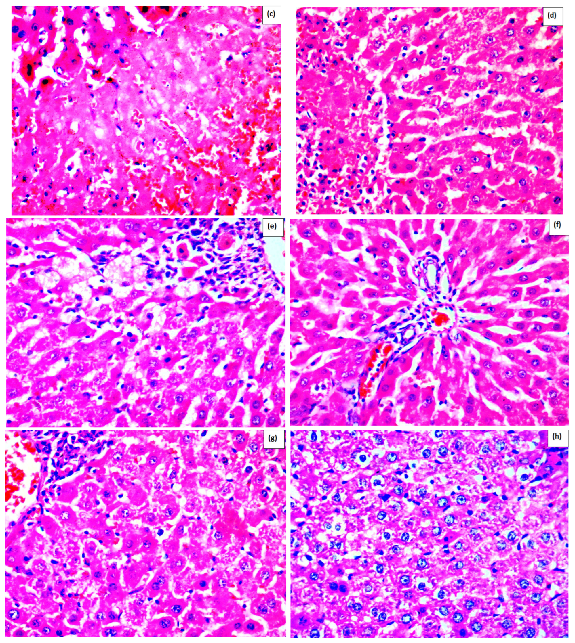
As shown in Figure 1, normal liver architecture was observed in the control rats (Figure 1a) and in the rats treated with 500 mg/kg methanolic extract without APAP administration (Figure 1b). The most significant pathological changes were detected in the rats that received an APAP overdose and saline (Figure 1c). In this group (APAP+S), hepatic cell necrosis, dilation of sinusoids, parenchymal hemorrhages, inflammatory and Kupffer cell infiltration, as well as disruption of normal liver architecture were observed. Treatment with the methanolic extract of M. frivaldszkyana (Figure 1d–f) reduced the severity of liver damage, however not to the extent observed in rats treated with silymarin (Figure 1h). A reduction in liver damage was also observed in rats treated with RA (Figure 1g); however, the decrease was more pronounced in the groups treated with silymarin and with 500 mg/kg methanolic extract (Figure 1f,h).
In the Control S group, the quantitative analysis showed no evidence of necrosis, sinusoidal dilation, or inflammatory infiltration. Similarly, in the ME500 group, no pathological alterations were detected; necrosis, sinusoidal dilation, and inflammatory infiltration were completely absent (0%). In contrast, the APAP+S group displayed the most severe pathological changes. Necrosis affected approximately 50% of hepatocytes, sinusoidal dilation was markedly increased (80%), and inflammatory infiltration was severe, accompanied by clear disruption of the normal hepatic architecture. In the 250 mg/kg methanolic extract + APAP group, necrosis was observed in 15% of hepatocytes, sinusoidal dilation was present in 30% of the liver tissue, and inflammatory infiltration was moderate. The 400 mg/kg methanolic extract + APAP group showed comparable findings, with necrosis limited to 15%, sinusoidal dilation reduced to 20%, and moderate inflammatory infiltration. The 500 mg/kg methanolic extract + APAP group exhibited results similar to those of the silymarin group. Necrosis was restricted to 8% of hepatocytes, sinusoidal dilation remained at 10%, and inflammatory infiltration was mild. In the RA+APAP group, necrosis was reduced to 18%, sinusoidal dilation reached 20%, and inflammatory infiltration was moderate. In the Silymarin+APAP group, only minimal pathological changes were observed. Necrosis was restricted to 7% of hepatocytes, sinusoidal dilation was limited to 10%, and inflammatory infiltration was mild.
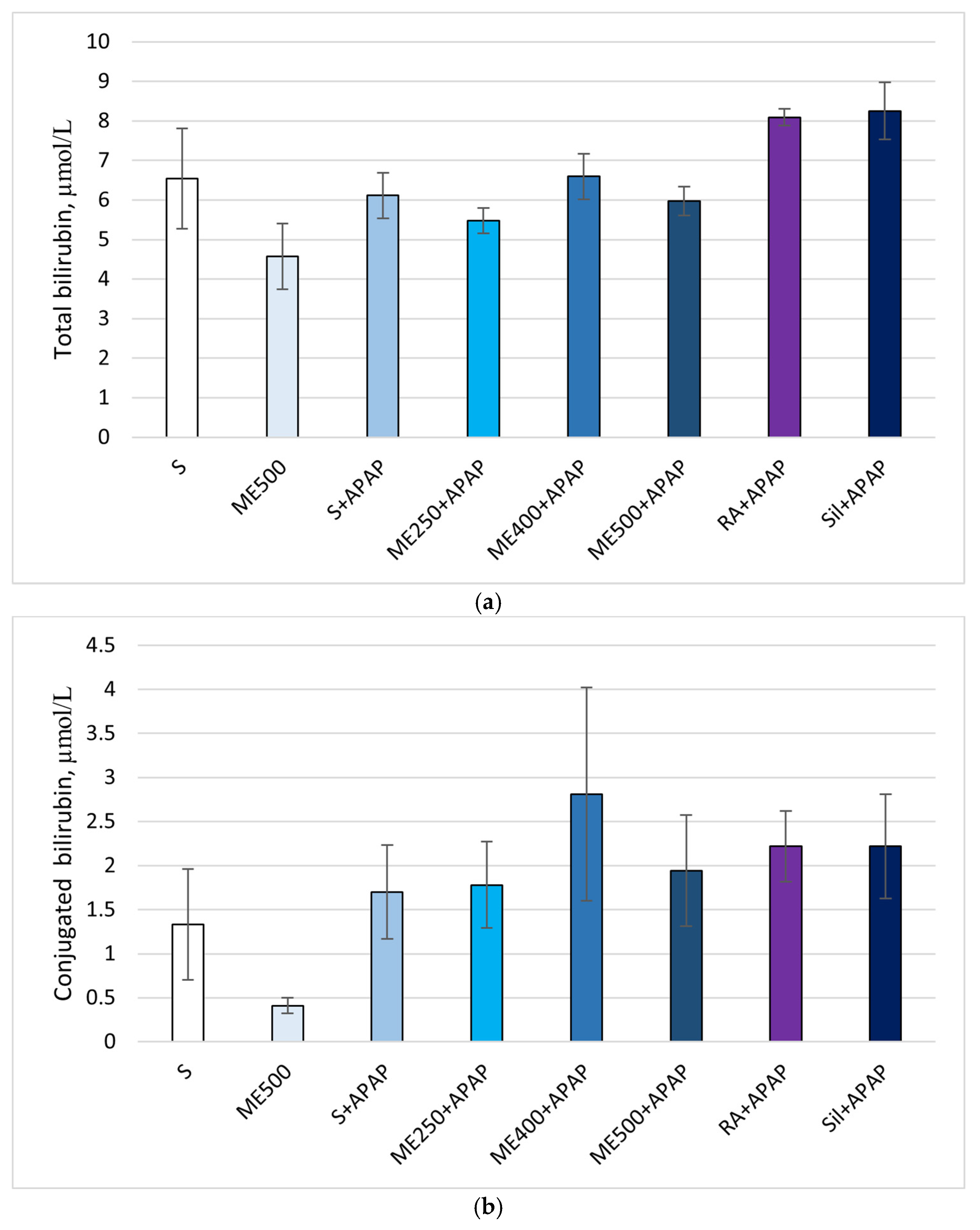
No statistically significant changes were observed in total and conjugated bilirubin levels (Figure 2a,b).
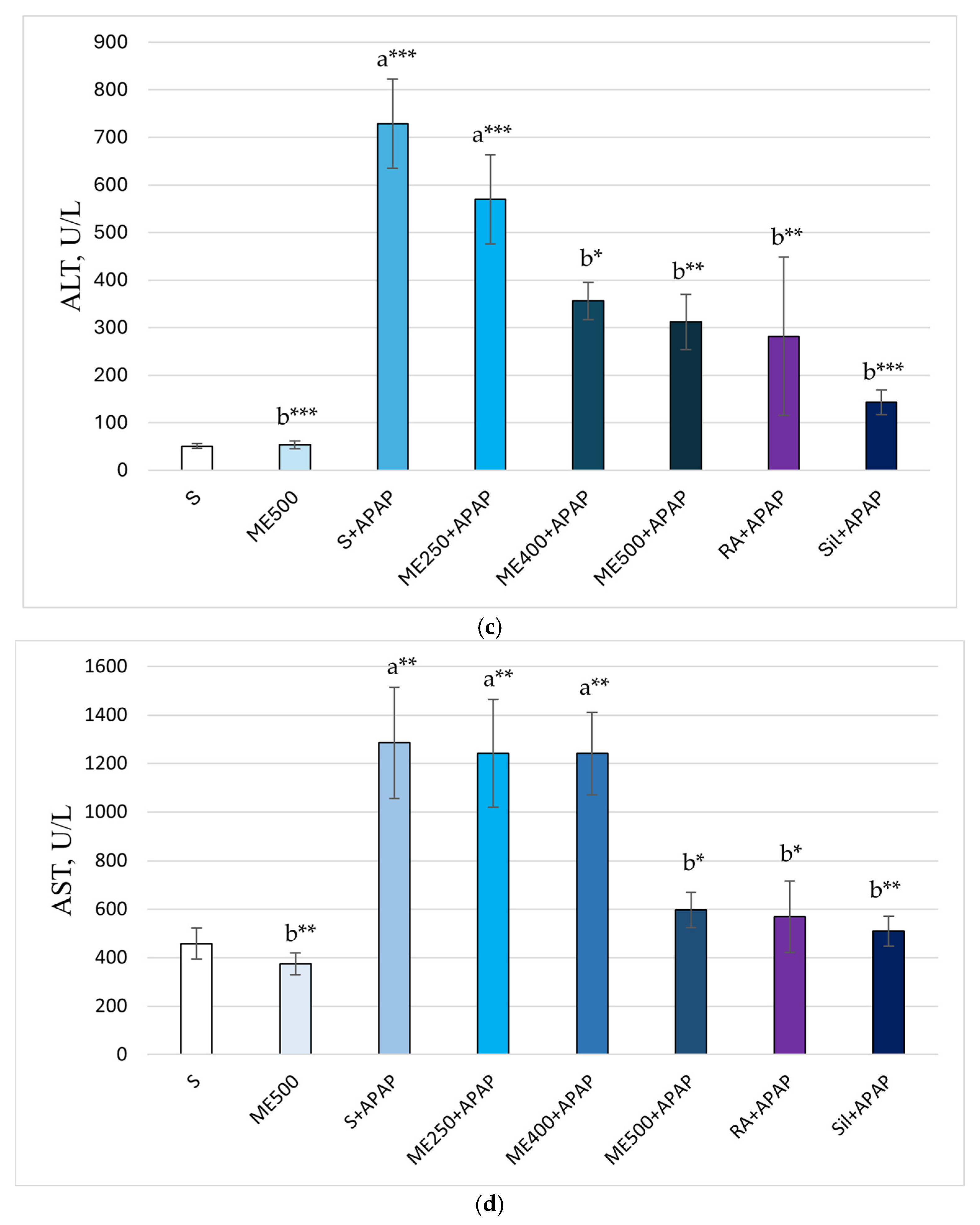
Figure 2c shows significantly higher ALT levels in the S+APAP and ME250+APAP groups compared to the control group (728.90 ± 93.94 vs. 50.63 ± 5.07, p < 0.001; 569.34 ± 93.82 vs. 50.63 ± 5.07; p ≤ 0.001). A significant decrease in ALT was observed in the ME500, ME400+APAP, ME500+APAP, RA+APAP, and Sil+APAP groups compared to the S+APAP group (53.61 ± 8.32 vs. 728.90 ± 93.94, p < 0.001; 356.34 ± 39.09 vs. 728.90 ± 93.94, p < 0.05; 311.88 ± 58.31 vs. 728.90 ± 93.94, p ≤ 0.01; 281.81 ± 165.88 vs. 728.90 ± 93.94, p < 0.01; 142.76 ± 25.59 vs. 728.90 ± 93.94, p < 0.001).
According to the analysis, a significant increase in serum AST levels was observed in the S+APAP, ME250+APAP, and ME400+APAP groups compared to the saline-treated control group (1284.94 ± 229.22 vs. 457.46 ± 63.08; 1241.21 ± 222.14 vs. 457.46 ± 63.08; 1240.38 ± 169.82 vs. 457.46 ± 63.08, p < 0.01). As shown in Figure 2d, serum AST levels were significantly decreased in the ME500, ME500+APAP, RA+APAP, and Sil+APAP groups compared to the S+APAP group (373.84 ± 44.16 vs. 1284.94 ± 229.22, p < 0.01; 596.25 ± 72.79 vs. 1284.94 ± 229.22 p < 0.05; 568.43 ± 148.36 vs. 1284.94 ± 229.22, p < 0.05; 509.03 ± 62.35 vs. 1284.94 ± 229.22, p < 0.01).
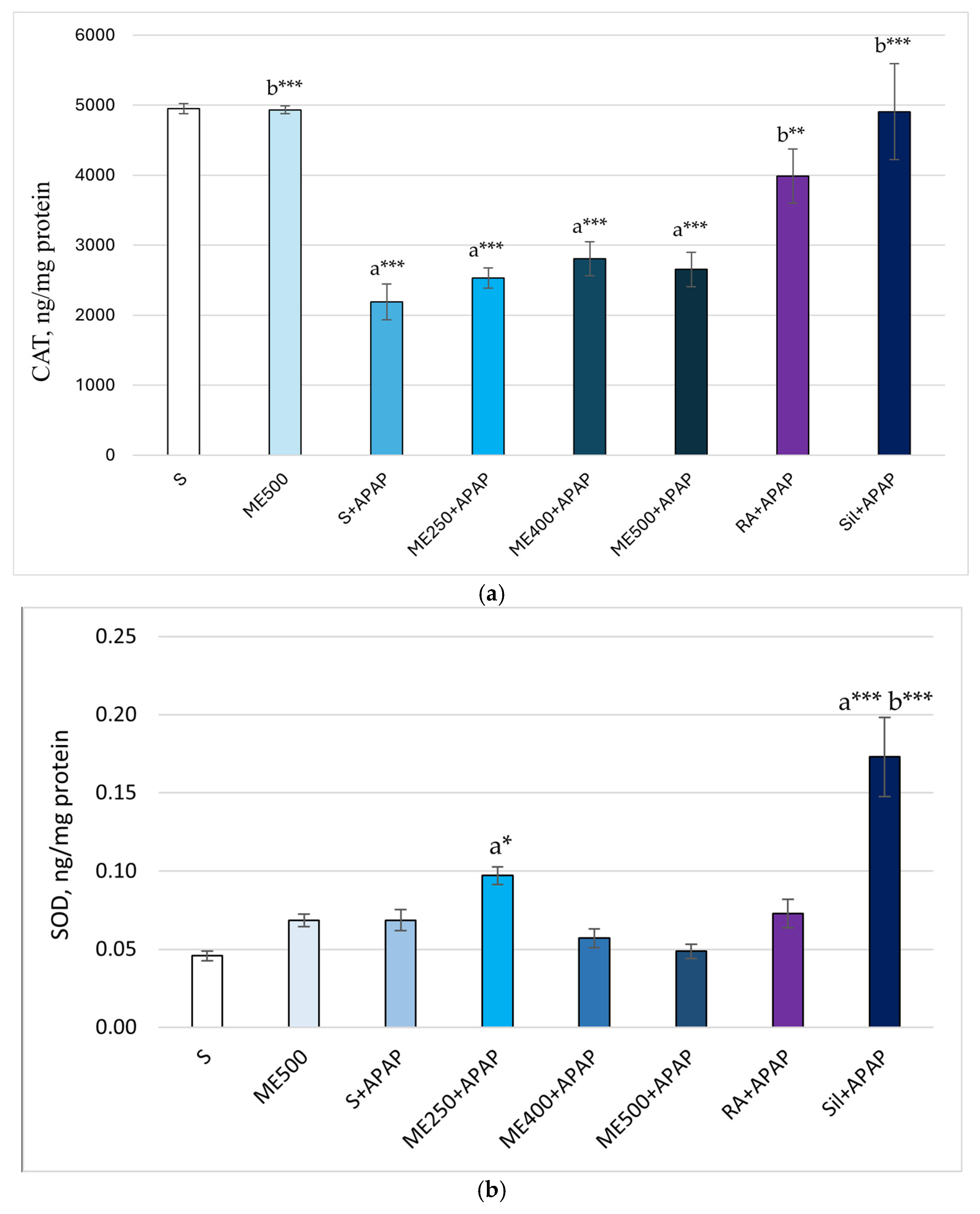
Figure 3a demonstrates significantly lower CAT levels in the S+APAP, ME250+APAP, ME400+APAP, ME500+APAP groups compared to the control group (2212.23 ± 254.01 vs. 4951.56 ± 71.55; 2529.66 ± 142.52 vs. 4951.56 ± 71.55, p < 0.001; 2807.60 ± 243.82 vs. 4951.56 ± 71.55, p ≤ 0.001; 2653.84 ± 245.31 vs. 4951.56 ± 71.55, p < 0.001). A significant increase in CAT enzyme levels was observed in the groups treated with ME500, RA and silymarin compared to the S+APAP group (4931. 99 ± 54.77 vs. 2212.23 ± 254.01, p < 0.001; 3986.52 ± 387.38 vs. 2212.23 ± 254.01, p < 0.01; 4907.14 ± 683.10 vs. 2212.23 ± 254.01, p < 0.001).
Elevated SOD levels were observed in ME250+APAP and Sil+APAP groups compared to the control group (0.10 ± 0.006 vs. 0.05 ± 0.003, p < 0.05; 0.17 ± 0.025 vs. 0.05 ± 0.003, p < 0.001). Additionally, the SOD levels were significantly higher in the Sil+APAP group in comparison to S+APAP (0.17 ± 0.025 vs. 0.07 ± 0.007, p < 0.001), as shown in Figure 3b.
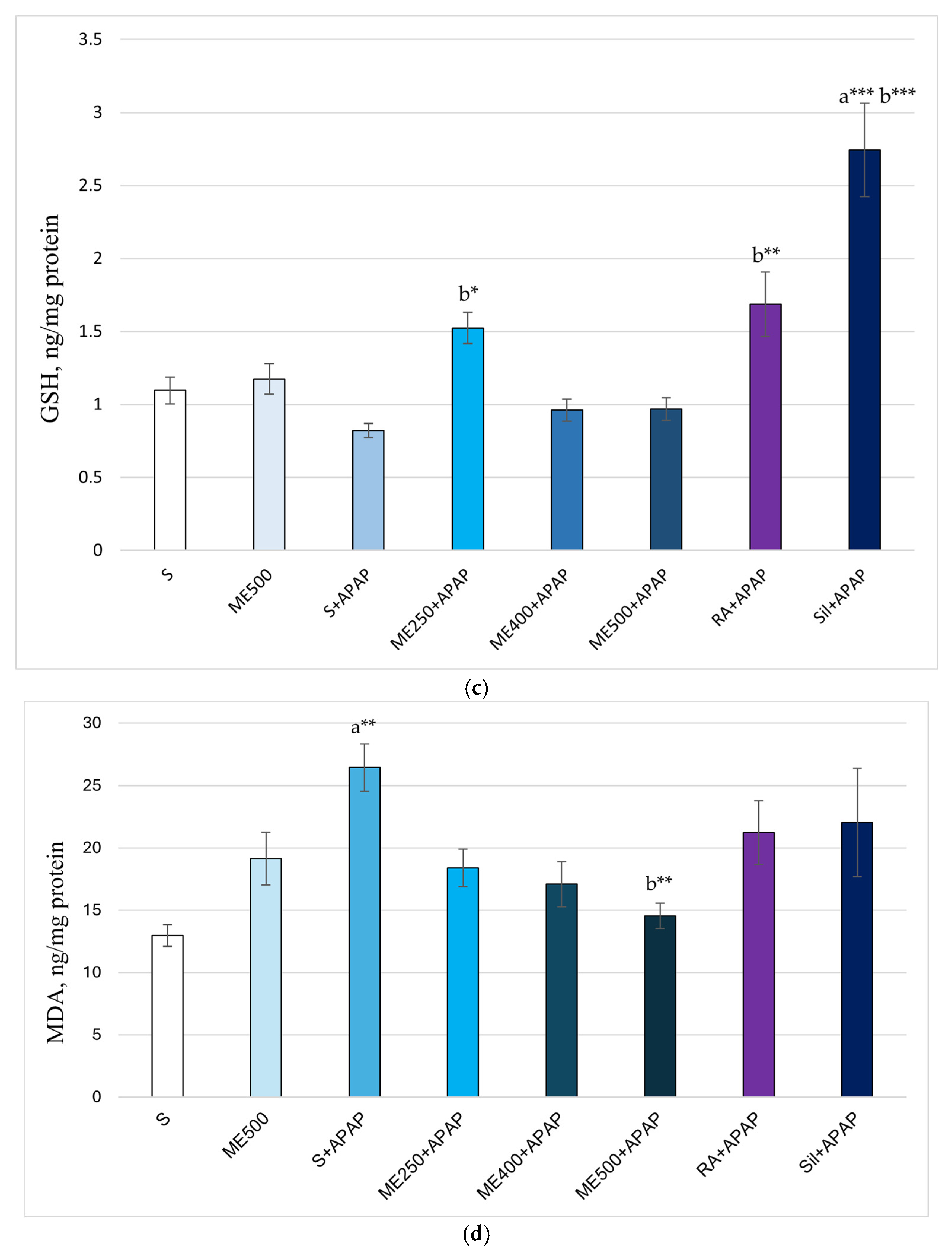
Figure 3c shows increased levels of reduced GSH in the Sil+APAP group compared to the controls (2.74 ± 0.32 vs. 1.10 ± 0.09, p < 0.001). Significantly higher GSH levels were also observed in groups ME250+APAP, RA+APAP, and Sil+APAP compared to S+APAP (1.52 ± 0.11 vs. 0.82 ± 0.05, p < 0.05; 1.68 ± 0.22 vs. 0.82 ± 0.05, p < 0.01; 2.74 ± 0.32 vs. 0.82 ± 0.05, p < 0.001).
As shown in Figure 3d, serum MDA levels were significantly increased in the S+APAP group compared to the controls (26.42 ± 1.89 vs. 12.97 ± 0.89, p < 0.01) and significantly decreased in the ME500+APAP group compared to group S+APAP (14.54 ± 1.02 vs. 26.42 ± 1.89, p ≤ 0.01).
Figure 3e illustrates a significant increase in 8-OH-dG levels in the S+APAP group compared to the control rats (1.79 ± 0.11 vs. 0.78 ± 0.06, p < 0.001). Significantly lower levels of this marker were found in groups ME250+APAP, ME400+APAP, ME500+APAP, RA+APAP, and Sil+APAP compared to group S+APAP (1.10 ± 0.10 vs. 1.79 ± 0.11, p < 0.01; 0.74 ± 0.05 vs. 1.79 ± 0.11, p < 0.001; 0.68 ± 0.03 vs. 1.79 ± 0.11, p < 0.001; 1.17 ± 0.12 vs. 1.79 ± 0.11, p ≤ 0.01; 1.24 ± 0.24 vs. 1.79 ± 0.11, p < 0.05.
The changes in the IL-6 levels are shown in Figure 3f. Significantly higher values were observed in the ME500+APAP and Sil+APAP groups in comparison to the controls (17.79 ± 2.16 vs. 9.99 ± 1.23, p < 0.05; 19.25 ± 2.05 vs. 9.99 ± 1.23, p < 0.01).
Figure 3g demonstrates a significant decrease in TNF-α levels in groups ME400+APAP and ME500+APAP compared to the S+APAP group (45.86 ± 3.21 vs. 126.33 ± 18.54, p < 0.001; 48.42 ± 3.16 vs. 126.33 ± 18.54, p < 0.001).

3. Discussion

The liver injury, which occurs in APAP overdose, is induced by elevated levels of NAPQI. This metabolite is produced through APAP oxidation by CYP450 enzymes and is usually neutralized by GSH. However, excessive production of NAPQI leads to depletion of GSH and liver injury through covalent binding of NAPQI to cellular proteins. Centrilobular hepatocytes are mainly affected by the mechanisms of oxidative stress, mitochondrial dysfunctions, and inflammation, ultimately resulting in cell necrosis [27]. The initial formation of ROS in the cytosol is followed by oxidative stress and the formation of protein adducts in the mitochondria. This induces mitochondrial membrane damage and subsequent hepatic cell necrosis [28]. However, the mechanism of APAP-induced liver injury is more complex, involving the processes of autophagy, sterile inflammation, liver repair, and regeneration [29].
The standard treatment of APAP overdose is based on N-acetylcysteine (NAC) application. NAC is a GSH precursor, which has been used as an antidote for 50 years. By increasing the levels of GSH, NAC reduces NAPQI toxicity and alleviates oxidative stress [30,31]. GSH is the main protective molecule against NAPQI-induced liver damage. In this study, elevated levels of GSH were observed after treatment with RA and silymarin, and similar results were reported by Dkhil et al. (2019) and Yang et al. (2013) [1,32].
The present study revealed a significant increase in the levels of AST and ALT in rats with APAP overdose, which indicates the primary injury of the hepatocyte membranes. Pre-treatment with RA, silymarin, and methanolic extract of M. frivaldszkyana reduced the severity of the injury. The optimal results were achieved by silymarin, RA, and the highest dose of the extract. The results are consistent with other authors, who reported decreased levels of hepatic enzymes after treatment with silymarin [1] and RA [7]. Moreover, the results are supported by the histopathological observation, as described in Figure 1. In contrast to Dkhil et al. (2019), we did not register any significant changes in the serum levels of total and conjugated bilirubin [1].
The essential role of oxidative stress in APAP-induced liver toxicity was discussed above and supported by the registered low levels of CAT in the APAP+S group. Silymarin and RA significantly increased this antioxidant enzyme, and these results align with Dkhil et al. (2019) and Yang et al. (2013) [1,32]. Interestingly, the extract of M. frivaldszkyana did not induce elevation of SOD or CAT levels in liver tissue. This observation suggests that the hepatoprotective effects of the extract are not mediated via upregulation of cell-protective antioxidant enzymes. However, the involvement of other antioxidant mechanisms cannot be excluded.
The other biomarker related to oxidative stress, MDA, was significantly reduced in rats, who received the highest dose of the extract. Although decreases were also noted at lower doses, they did not reach statistical significance. This suggests reduced lipid peroxidation after treatment with M. frivaldszkyana extract. Moreover, suppression of lipid peroxidation is of primary importance for maintaining the normal function of the hepatocyte membrane [15]. This mechanism may explain the observed effect of the extract on the AST and ALT levels. Overall, our results revealed the potential protective effect of the methanolic extract of M. frivaldszkyana on the hepatocyte membrane and its hepatoprotective properties in APAP overdose.
As mentioned before, plants from the Lamiaceae family have significant antioxidant activity [16]. Nikolova et al. (2017) performed a DPPH assay on methanolic extracts from various Micromeria species (M. juliana, M. cristata, M. frivaldszkyana, and M. dalmatica). The authors observed the highest antioxidant activity in M. frivaldszkyana and M. dalmatica [19]. More recent studies have further confirmed the strong antioxidant properties of M. frivaldszkyana extract. Mladenova et al. (2021) assessed this activity of M. frivaldszkyana extract using multiple complementary assays, including DPPH, 2,2-azinobis (3)-ethylbenzthiazoline-6-sulfonic acid (ABTS), ferric reducing antioxidant power (FRAP), cupric ion reducing antioxidant capacity (CUPRAC), and oxygen radical absorbance capacity (ORAC) [16]. Their findings indicated that the ORAC value of M. frivaldszkyana (3250.5 ± 208.1 μmol TE/g) is significantly higher than that of many other traditionally used Bulgarian medicinal plants [33].
Oxidation can also occur with DNA, resulting in the formation of 8-OH-dG. Elevated levels of this molecule are used as a marker of oxidative DNA damage. Wang et al. (2015) reported increased 8-OH-dG in rodents after prolonged treatment with APAP [34]. The results of the current study showed a similar increase after a single APAP overdose. A significant decrease was detected in all experimental groups, with the most pronounced effect observed in groups receiving 400 and 500 mg/kg of the extract. In general, the extract induced a significant dose-dependent decrease in this parameter. Based on this, we propose DNA protection as one of the mechanisms underlying the observed liver protection. Moreover, the DNA damage plays an important role in carcinogenesis, which implies further research into the anti-tumor properties of this extract [35].
Excessive ROS production initiates cellular signaling pathways, that upregulate the expression of pro-inflammatory cytokine genes [1]. The APAP-induced hepatocyte necrosis is followed by aseptic inflammation and elevated levels of pro-inflammatory cytokines (e.g., IL-6, IL-1β, TNF-α). Regarding the IL-6 levels, we found no statistical difference between the groups, and our results align with [36]. The decreased levels of TNF-α in the groups treated with 400 and 500 mg/kg methanolic extract of M. frivaldszkyana suggested the involvement of the anti-inflammatory properties of the extract in its hepatoprotective effect. Previously, the anti-inflammatory activity of the extract was reported by Stavrakeva et al. (2024) in a model of rat paw inflammation [17]. In contrast to other authors, who reported decreased levels of this cytokine in the same model of APAP-induced liver toxicity after treatment with silymarin, our results did not indicate such differences. This discrepancy may be due to differences in silymarin dosage [1]. The observed reduction in TNF-α levels may be attributed to the high linarin content in the methanolic extract. Ellapan et al. (2022) evaluated the effect of Ceiba pentandra extract on carbon tetrachloride (CCl4)-induced liver toxicity. The authors reported decreased TNF-α level in the rats, treated with the extract and explained this effect with the presence of linarin in the ethanolic extract [37]. Similarly, metabolomic analysis of M. frivaldszkyana extract identified linarin as the predominant compound [17]. In silico studies have demonstrated a high affinity of linarin for TNF-α converting enzyme. This cytokine is released by Kupffer cells following acute liver injury, which can be induced by hepatotoxic substances challenge [37].
Linarin (also known as acacetin 7-O-rutinoside) is a flavonol glycoside isolated from plant species from the Asteraceae and Lamiaceae families, and genera Mentha, Micromeria, and Satureja are described as particularly rich in linarin [38]. Among the numerous biological activities attributed to this compound, antioxidant, anti-inflammatory, and hepatoprotective properties are of particular interest [24,38]. Linarin has been found effective in CCl4-induced liver toxicity, leading to decreased MDA levels in vitro [24]. The oral administration of some bioactive compounds in vivo may result in low plasma levels due to degradation in the gastrointestinal tract or obstructed absorption by the mucus [39]. However, a pharmacokinetic study showed rapid absorption after oral administration in rats, with appropriate distribution of linarin and its metabolites in liver tissue [40]. This report allows us to hypothesize that the observed hepatoprotective effect may be partially related to the high content of linarin in the extract. However, one should keep in mind that all compounds may contribute to the observed effect, not only those present in the highest amounts [41].
Hepatoprotective properties have also been reported for chlorogenic acid, eupatorin, rutin, apigenin, naringenin, and RA, which are also abundant in the methanolic extract of M. frivaldszkyana. Chlorogenic acid (5-caffeoylquinic acid) is a phenolic compound found in significant quantities from coffee, apples, pears, berries, etc. The application of this compound alleviates hepatocellular injury and liver steatosis, reducing serum AST and ALT levels in mice with diet-induced nonalcoholic steatohepatitis [42]. Decreased serum levels of hepatic enzymes by chlorogenic acid were also reported by Hu et al. (2020) in a model of APAP-induced liver injury in mice [43]. In the same model, chlorogenic acid was shown to promote liver regeneration and repair [44]. Additionally, it has been reported to alleviate the hepatic injury in several models of hepatotoxicity by decreasing apoptosis, inflammation, and oxidative injury caused by methotrexate and heavy metals [45]. Increased levels of GSH, SOD, CAT, along with decreased MDA level were reported by Feng et al. (2016) in a model of d-galactose-induced liver toxicity in mice. The authors found a significant decrease in the pro-inflammatory cytokines TNF-α and IL-6 following chlorogenic acid treatment [46].
Eupatorin is another compound with a possible role in the observed effect of the extract. This flavone is a weak acid and undergoes biotransformation to 51 metabolites in rats. Eupatorin demonstrated anti-inflammatory activity in rodent models of paw edema [47].
Rutin (quercetin-3-O-rutinoside) showed hepatoprotective activity in a model of CCl4-induced liver toxicity in rats. Decreased mortality and lower serum levels of ALT and AST were observed in pre-treated animals [48]. The hepatoprotective properties of rutin may also be attributed to decreased TNF-α level in rats with deltamethrin-induced hepatotoxicity [49].
Goudarzi et al. (2021) reported restoration of AST, ALT, MDA, and TNF-α levels after apigenin administration in a rat model of methotrexate-induced hepatotoxicity. Treated animals also showed increased levels of SOD, CAT, and GSH compared to those treated only with methotrexate. The authors propose the antioxidant and anti-inflammatory activity of apigenin as a possible mechanism for the observed effects [50].
Similarly, Ahmed et al. (2019) registered reduced serum AST, ALT, and TNF-α levels in rats with APAP-induced liver toxicity after following treatment with naringin and naringenin [51].
Rosmarinic acid, an ester of caffeic acid and 3,4-dihydroxyphenyl lactic acid, is a common compound in many plants, and its biological effects are widely explored. Antibacterial, antioxidant, and anti-inflammatory properties are documented in vivo and in vitro [52]. This phenol carboxylic acid has shown hepatoprotective activity in a wide spectrum of experimental models, including liver injury induced by diabetes, CCl4, and tert-butyl hydroperoxide (t-BHP) [53]. In a rat model of APAP-induced hepatotoxicity, RA in a dose of 100 mg/kg significantly reduced the elevated levels of AST, ALT, MDA, while increasing hepatic GSH levels [7]. In this study, we detected the same changes in AST, ALT, and GSH. Although MDA levels decreased in the RA-treated group, the reduction did not reach statistical significance, which may be attributed to differences in the experimental protocol. As mentioned above, RA was reported as one of the compounds present at the highest concentrations in the methanolic extract of M. frivaldszkyana [17]. We included a group treated with RA to evaluate the similarity between the effects of RA and the extract. Table 1 compares the highest dose of the extract with the two positive controls (RA and silymarin). The changes in the main biomarkers of liver function (AST and ALT) induced by the extract and RA are identical, and the same applies to the levels of SOD. These results allow us to speculate that the abundance of RA in the methanolic extract may be responsible for some of the observed effects. However, in the extract, all components act synergistically and this hypothesis is further supported by the different results obtained for the two groups with respect to CAT, GSH, and MDA levels (Table 1).
Based on the current results, we can speculate that the mechanism of the observed hepatoprotective effect could be attributed to cell membrane protection, decreased lipid peroxidation and DNA damage, and attenuation of aseptic inflammation. However, the mechanism of APAP-induced liver toxicity is more complex and involves the formation of peroxynitrite and adducts of NAPQI to mitochondrial proteins, as well as an influence on signaling pathways with an essential role in cell survival and aseptic inflammation [1]. The present study sheds light on only a limited aspect of the mechanisms potentially modulated by the methanolic extract of M. frivaldszkyana. The contribution of additional pathways cannot be excluded and will be the subject of future investigations. Given the strong antioxidant properties of the extract, investigation of its effectiveness against other animal models of liver toxicity, as well as other organs, is also in the scope of our future research.

Limitations and Future Directions

The following limitations apply to this study: only male Wistar rats were used in the experiment; using a different sex, strain, or route of administration may yield different outcomes. The experiment was performed after 7 days of application of the extract, and longer pre-treatment may have different effects. The APAP toxicity was induced by a single overdose, and the outcome may differ in the case of chronic APAP administration. The present study is limited to exploring the effects of the methanolic extract of M. frivaldszkyana in APAP-induced overdose in rats. The specific pathways and target molecules remain to be elucidated and are beyond the scope of the current experiments. IL-6 and TNF-α levels were measured in the groups S, ME500, S+APAP, ME250+APAP, ME400+APAP, ME500+APAP, and Sil+APAP. The RA+APAP group was excluded due to insufficient biological material. In-depth mechanistic studies and extract standardization are necessary to ensure future translational potential.

4. Materials and Methods

The experiments were performed according to the following permissions: (1) permit № 352/30 May 2023 by the Bulgarian Food Safety Agency; (2) protocol № 6/05 October 2023 by the Ethics Committee of the Medical University, Plovdiv, Bulgaria; (3) permit № 996/09 August 2023 by the Ministry of Environment and Water for plant material. The study protocol was developed and performed in alignment with the ARRIVE guidelines, the EU Directive 2010/63/EU on animal experiments, and the relevant national and institutional regulations.

4.1. Chemicals and Reagents

Methanol (≥99.8%, cat. No. 179337), APAP (≥98.0%, batch No. MKCS3304), RA (≥96.0%, batch No. BCCJ6033), and silymarin (≥30.0%, batch No. BCCH4151) were obtained from Merck SA (Germany). Eosin Y (1% aqueous solution, cat. № 294/EOY-10-OT-2.5L), formaldehyde 4% (10% neutral buffer, cat. № 294/FNB4-10L), Histanol 100 (cat. № 294/H100-5L), Histanol 95 (cat. № 294/H95-5L), hematoxylin G3 (cat. № 294/HEMG3-OT-2.5L), acetone (cat. № 48/3413/5) and xylol (cat. № 348/3410/20) were obtained from BIOCARE Medical (Pacheco, CA, USA).

4.2. Plant Material and Preparation of the Methanolic Extract

The aerial parts of M. frivaldszkyana (approximately 800 g of fresh biomass) were harvested during the 2023–2024 vegetation season from the Bulgarka Nature Park, located in the Middle Stara Planina floristic region, in accordance with previously established protocols [17,18]. The herbarium of the Agricultural University in Plovdiv (SOA) houses the voucher specimen (accession № 062648). The plant material was dried for 10 days in a shaded area at a controlled ambient temperature of 22 ± 2 °C. Drying was deemed complete when leaf stalks fractured upon bending and the petioles crumbled easily under pressure. Following this, the dried material was milled to a fine powder with an average particle size below 400 μm using a GRINDOMIX GM200 laboratory mill (RETSCH GmbH, Haan, Germany).
To prepare the methanolic extract, 10 g of the powdered sample was macerated in 70% methanol (1:10 w/v) for 24 h at room temperature (22 ± 2 °C) with continuous stirring in a light-protected container. To improve extraction efficiency, the sample underwent ultrasonication in three 15-min cycles at 30 °C. The next step included centrifugation at 6000× g for 15 min, and filtration of the supernatant using Whatman No. 1 filter paper (Sigma-Aldrich, Burlington, MA, USA). The residual plant material was subjected to the same extraction process twice more. The solvent was removed from the combined extracts using a rotary evaporator (Heidolph, Schwabach, Germany) under reduced pressure at 50 °C [17,18]. The extraction yield was 54.8% of the initial dry plant material, corresponding to 5.48 g of dried extract.

4.3. Animals and Treatment

A total of 56 male Wistar rats (body weight 210–260 g) were used in the study. They were randomly assigned to eight groups, each comprising seven animals. The animals were maintained under standard laboratory conditions, including a temperature of 22 ± 1 °C, relative humidity of 45%, and a 12-h light/12-h dark cycle, with unrestricted access to food and water.
We followed the protocol for APAP-induced liver toxicity described by Ahmed et al. (2023) [4]. After a one-week acclimatization period, the animals were treated orally for 7 consecutive days according to the following group assignments:
Groups 1 (S, control) and 3 (S+APAP) received saline at a dose of 0.1 mL/100 g body weight (bw); group 2 (ME500) received 500 mg/kg of a water solution of the dried methanolic extract; groups 4, 5, and 6 (ME250+APAP, ME400+APAP, ME500+APAP) were treated with 250, 400, and 500 mg/kg water solution of the dried methanolic extract, respectively; group 7 (RA+APAP) received 100 mg/kg RA; group 8 (Sil+APAP) was treated with 125 mg/kg silymarin.
On the evening of day 6, all animals were fasted for 12 h with free access to water. On day 7, the groups S+APAP, ME250+APAP, ME400+APAP, ME500+APAP, RA+APAP, and Sil+APAP received 2000 mg/kg of APAP via gastric gavage. Three hours later, the respective treatment (extract or compound) was applied to each group. Animals were euthanized 48 h after APAP administration. Blood and liver samples were collected. Portions of each organ were fixed for histopathological analysis, while the remaining tissue was stored at −80 °C and later homogenized for biochemical evaluation.
The duration of pre-treatment prior to APAP administration was selected based on previous studies evaluating the protective effects of plant extracts and natural compounds in APAP-induced liver injury models [1,7,54]. The dose and duration of APAP administration were also based on a review of the literature. The methanolic extract doses were determined according to recommendations by Hanafy et al. (2016), which suggest using 1/10 and 1/20 of the estimated LD50 for pharmacological testing [55]. In our previous study, we demonstrated the safety of M. frivaldszkyana extract at single oral doses up to 5000 mg/kg [17]. The intermediate dose of 400 mg/kg was included to better characterize the extract’s dose-dependent effects.
The dose of RA was selected based on the study by Hasanein et al. (2017), which demonstrated its beneficial effect in the APAP-induced liver toxicity model [7]. Silymarin, a well-known hepatoprotective agent, was used as a positive control and its dose was also selected based on published data [56].

4.3.1. Histopathological Observation

The experimental protocol was based on the description of other authors [57,58,59] and was reported previously [18].
Immediately following collection, the organs were placed in a 10% neutral-buffered formalin solution to ensure proper fixation. The fixed samples were subsequently processed and embedded in paraffin blocks. The entire histological preparation included the following steps:
  • Tissues were immersed in formalin for 24 h, thoroughly rinsed, and then soaked in distilled water for 30 min to eliminate any residual fixative.
  • Dehydration was achieved through progressive immersion in 95% and then 100% ethanol (Histanol® 100%), each step lasting 4–5 h.
  • After dehydration, the tissues were treated with acetone for 20 min, followed by xylol for 30 min. They were then embedded in molten paraffin and formed into blocks. Thin slices, 5 μm in thickness, were obtained from these blocks and stained with hematoxylin and eosin for microscopic examination.
Histopathological analysis was performed independently by two pathologists who were blinded to the experimental groups. Morphological alterations in the tissues from treated groups were compared to controls. Leica DM500 (Leica Microsystems GmbH, Wetzlar, Germany) and Zeiss AXIO Scope A1 (Carl Zeiss Microscopy GmbH, Jena, Germany) microscopes were used for sample observation.
Liver samples were examined for pathological changes such as architectural disruption, portal and lobular inflammation, sinusoidal dilation and congestion, granuloma formation, necrosis, degeneration, and steatosis. Tissue pathology was classified according to the Roenigk scoring system (Table 2).

4.3.2. Preparation of Liver Homogenates and Evaluation of Tissue-Specific Toxicity Markers

On the day of the analysis, the frozen liver tissues were brought to room temperature and homogenized in a lysis 0.1 M PBS buffer with added Triton 100× (1:9 w/v), pH 7.4 [61] on a Polytron mechanical homogenizer (KINEMATICA, Malters, Switzerland). The obtained whole-liver homogenates were afterwords centrifuged at 10,000 rpm for 10 min at 4 °C using MPW-352R centrifuge (Warsaw, Poland) [62] and the resultant supernatant was used for the assessment of the following parameters: (1) oxidative cell damage and antioxidant protection markers—MDA, 8-OH-dG, CAT, SOD1, and GSH; (2) pro-inflammatory cytokines—IL-6 and TNF-α, all measured in the supernatant of whole-liver homogenate. All listed parameters were analyzed via an enzyme-linked immunosorbent assay (ELISA) method on an ELISA microplate reader (HumanReader, HUMAN, Wiesbaden, Germany) using commercial kits [ELISA Kit for CAT, Cloud-Clone Corp., Katy, TX, USA, Cat.No. SEC418Ra; MDA ELISA Kit, Cat.No. E-EL-0060; 8-OH-dG ELISA Kit, Cat.No. E-EL-0028; SOD1 (Soluble) ELISA Kit, Cat.No. E-EL-R1424; GSH ELISA Kit, Cat.No. E-EL-0026; Rat IL-6 ELISA Kit, Cat.No. E-EL-R0015; Rat TNF-α ELISA Kit, Cat.No. E-EL-R2856, Elabscience Biotechnology Inc., Houston, TX, USA]. All assays were carried out using ELISA kits from the same production lot in order to minimize batch-to-batch variability, and internal controls supplied by the manufacturer were included on each plate to ensure reproducibility.

4.3.3. Biochemical Markers in Serum

Whole blood was collected from the animals into VACUTEST KIMA tubes (Arzergrande, Italy) and left at room temperature until visible clot formation. The samples were then centrifuged in an MPW-352R unit (MPW Med. Instruments, Warsaw, Poland) at 3000 rpm for 10 min at 4 °C. The resulting serum was separated, aliquoted, and stored at −80 °C (ULT C200) until analysis. It was used for spectrophotometric assessment of the levels of the following markers for liver toxicity: AST (HUMAN Diagnostics GmbH, Wiesbaden, Germany, Ref. No. 12021), ALT (HUMAN Diagnostics GmbH, Wiesbaden, Germany, Ref. No. 12022), total bilirubin (HUMAN Diagnostics GmbH, Wiesbaden, Germany, Ref. No. 10746), and conjugated bilirubin. Biochemical measurements were performed on an Evolution 300 UV-Vis spectrophotometer (Thermo Fisher Scientific, Waltham, MA, USA) following the protocols supplied with the commercial kits.

4.4. Statistical Analysis

Statistical processing of the data was performed with SPSS software, version 17.0 (IBM, Armonk, NY, USA). Results are presented as mean ± SEM. Differences between group means were evaluated by one-way analysis of variance (ANOVA) and Tukey’s post hoc test. Statistical significance was defined as p ≤ 0.05.

5. Conclusions

The present results clearly demonstrate, for the first time, that pre-treatment with methanolic extract of M. frivaldszkyana for 7 days results in significant hepatoprotective effects in an APAP-induced rat model of liver injury. The mechanism of this effect could be attributed to cell membrane protection, decreased lipid peroxidation and DNA damage, and attenuation of aseptic inflammation. These effects can be attributed to the main compounds identified in the extract (linarin, chlorogenic acid, rutin, eupatorin, apigenin, RA).

Author Contributions

Conceptualization, E.A. and A.B.; methodology, M.C. and I.D.; validation, R.M. and P.S.; investigation, E.A., V.K., K.S., I.D., M.C., I.K. and I.B.; resources, D.D.; writing—original draft preparation, E.A., K.S., M.C., I.D. and M.K.; writing—review and editing, I.K.; visualization, I.B.; supervision, E.A. and I.K.; project administration, E.A.; funding acquisition, E.A. All authors have read and agreed to the published version of the manuscript.

Funding

This research was funded by the Medical University of Plovdiv through the Interuniversity project number NO-03/2023. The APC was funded by the Medical University of Plovdiv.

Institutional Review Board Statement

The experimental protocol was approved by the Bulgarian Food Safety Agency (permit number: 352/30 May 2023) and the Ethics Committee of the Medical University, Plovdiv, Bulgaria (protocol number: 6/05 October 2023). The ARRIVE guidelines, the EU Directive 2010/63/EU for animal experiments, and the relevant national and institutional rules and regulations were taken into account during this study. The plant material was collected after obtaining a permit from the Ministry of Environment and Water (permit number 996/09.08.2023).

Data Availability Statement

The data presented in this study are available on request from the corresponding author.

Acknowledgments

The authors would like to thank T. Mladenova and K. Todorov for obtaining the plant gathering permit and providing the raw plant material for the preparation of the extract. We would like to express our gratitude to the program “Research, Innovation and Digitalisation for Smart Transformation” 2021–2027, funded by the European Union, Project BG16RFPR002-1.014-0007 Center for Competence “PERIMED-2” for supporting this article.

Conflicts of Interest

The authors declare no conflicts of interest.

Abbreviations

The following abbreviations are used in this manuscript:
8-OH-dG8-hydroxy-2′-deoxyguanosine
ABTS2,2-azinobis (3)-ethylbenzthiazoline-6-sulfonic acid
ALTAlanine aminotransferase
APAPAcetaminophen (paracetamol)
ASTAspartate aminotransferase
CATCatalase
CCL4Carbon tetrachloride
CUPRACCupric ion reducing antioxidant capacity
CYPCytochrome P450
DNADeoxyribonucleic acid
DPPH2,2′-diphenylpicrylhydrazyl
ELISAEnzyme-linked immunosorbent assay
FRAPFerric reducing antioxidant power
GPxGlutathione peroxidase
GSHGlutathione
IL-6Interleukin-6
MDAMalondialdehyde
MICMinimum inhibitory concentration
NACN-acetylcysteine
NAPQIN-acetyl-p-benzoquinone imine
ORACOxygen radical absorbance capacity
RARosmarinic acid
ROSReactive oxygen species
SODSuperoxide dismutase
t-BHPTert-butyl hydroperoxide
TNF-αTumor necrosis factor-alpha
UPLC-MS/MSUltra-performance liquid chromatography tandem mass spectrometry

References

  1. Dkhil, M.A.; Abdel Moneim, A.E.; Hafez, T.A.; Mubaraki, M.A.; Mohamed, W.F.; Thagfan, F.A.; Al-Quraishy, S. Myristica fragrans Kernels prevent paracetamol-induced hepatotoxicity by inducing anti-apoptotic genes and Nrf2/HO-1 pathway. Int. J. Mol. Sci. 2019, 20, 993. [Google Scholar] [CrossRef]
  2. Mullins, M.E. Epidemiology of acetaminophen toxicity. In Acetaminophen Toxicity; Academic Press: Cambridge, MA, USA, 2025; pp. 455–465. [Google Scholar]
  3. Stavrakeva, K.; Popova, M.; Esad, M.; Apostolova, E.; Kokova, V.; Bacelova, M.; Alakidi, A.; Bivolarska, A. Drug-induced liver toxicity. Acta Medica Bulg. 2024, 51, 77–85. [Google Scholar] [CrossRef]
  4. Ahmed, H.M.; Shehata, H.H.; El-Saeed, G.S.M.; Abou Gabal, H.H.; El-Daly, S.M. Paracetamol overdose induces acute liver injury accompanied by oxidative stress and inflammation. Egypt. J. Chem. 2023, 66, 399–408. [Google Scholar] [CrossRef]
  5. Ashafaq, M.; Hussain, S.; Alshahrani, S.; Madkhali, O.; Siddiqui, R.; Khuwaja, G.; Alam, M.I.; Islam, F. Role of cinnamon oil against acetaminophen overdose induced neurological aberrations through brain stress and cytokine upregulation in rat brain. Drug Chem. Toxicol. 2022, 45, 633–640. [Google Scholar] [CrossRef] [PubMed]
  6. Naguib, Y.M.; Azmy, R.M.; Samaka, R.M.; Salem, M.F. Pleurotus ostreatus opposes mitochondrial dysfunction and oxidative stress in acetaminophen-induced hepato-renal injury. BMC Complement. Altern. Med. 2014, 14, 494. [Google Scholar] [CrossRef] [PubMed]
  7. Hasanein, P.; Sharifi, M. Effects of rosmarinic acid on acetaminophen-induced hepatotoxicity in male Wistar rats. Pharm. Biol. 2017, 55, 1809–1816. [Google Scholar] [CrossRef]
  8. Ishii, Y.; Okamura, T.; Inoue, T.; Tasaki, M.; Umemura, T.; Nishikawa, A. Dietary catechol causes increased oxidative DNA damage in the livers of mice treated with acetaminophen. Toxicology 2009, 263, 93–99. [Google Scholar] [CrossRef]
  9. Krenkel, O.; Mossanen, J.C.; Tacke, F. Immune mechanisms in acetaminophen-induced acute liver failure. Hepatobiliary Surg. Nutr. 2014, 3, 331–343. [Google Scholar] [CrossRef]
  10. Woolbright, B.L.; Jaeschke, H. Role of the inflammasome in acetaminophen-induced liver injury and acute liver failure. J. Hepatol. 2017, 66, 836–848. [Google Scholar] [CrossRef] [PubMed]
  11. Ahmad, S.T.; Arjumand, W.; Nafees, S.; Seth, A.; Ali, N.; Rashid, S.; Sultana, S. Hesperidin alleviates acetaminophen induced toxicity in Wistar rats by abrogation of oxidative stress, apoptosis and inflammation. Toxicol. Lett. 2012, 208, 149–161. [Google Scholar] [CrossRef]
  12. Adetuyi, B.O.; Adebisi, O.A.; Adetuyi, O.A.; Ogunlana, O.O.; Toloyai, P.E.; Egbuna, C.; Uche, C.Z.; Khan, J.; Adumanya, O.C.U.; Patrick-Iwuanyanwu, K.C. Ficus exasperata attenuates acetaminophen-induced hepatic damage via NF-κB signaling mechanism in experimental rat model. BioMed Res. Int. 2022, 2022, 6032511. [Google Scholar] [CrossRef]
  13. Onaolapo, O.J.; Adekola, M.A.; Azeez, T.O.; Salami, K.; Onaolapo, A.Y. l-Methionine and silymarin: A comparison of prophylactic protective capabilities in acetaminophen-induced injuries of the liver, kidney and cerebral cortex. Biomed. Pharmacother. 2017, 85, 323–333. [Google Scholar] [CrossRef]
  14. Ivanova, S.; Dzhakova, Z.; Staynova, R.; Ivanov, K. Salvia verticillata (L.)—Biological activity, chemical profile, and future perspectives. Pharmaceuticals 2024, 17, 859. [Google Scholar] [CrossRef]
  15. Fotio, A.L.; Nguepi, M.S.D.; Tonfack, L.B.; Temdie, R.J.G.; Nguelefack, T.B. Acetaminophen induces liver injury and depletes glutathione in mice brain: Prevention by Moringa oleifera extract. S. Afr. J. Bot. 2020, 129, 317–323. [Google Scholar] [CrossRef]
  16. Mladenova, T.; Stoyanov, P.; Denev, P.; Dimitrova, S.; Katsarova, M.; Teneva, D.; Todorov, K.; Bivolarska, A. Phytochemical composition, antioxidant and antimicrobial activity of the Balkan endemic Micromeria frivaldszkyana (Degen) Velen. (Lamiaceae). Plants 2021, 10, 710. [Google Scholar] [CrossRef]
  17. Stavrakeva, K.; Metodieva, K.; Benina, M.; Bivolarska, A.; Dimov, I.; Choneva, M.; Kokova, V.; Alseekh, S.; Ivanova, V.; Vatov, E.; et al. Metabolic composition of methanolic extract of the Balkan endemic species Micromeria frivaldszkyana (Degen) Velen and its anti-inflammatory effect on male Wistar rats. Int. J. Mol. Sci. 2024, 25, 5396. [Google Scholar] [CrossRef]
  18. Apostolova, E.; Stavrakeva, K.; Kokova, V.; Dimov, I.; Choneva, M.; Delev, D.; Kostadinov, I.; Bivolarski, I.; Koleva, M.; Mladenova, T.; et al. Subchronic toxicity and effect of the methanolic extract of Micromeria frivaldszkyana (Degen) Velen on cognition in male Wistar rats. Plants 2025, 14, 1837. [Google Scholar] [CrossRef] [PubMed]
  19. Nikolova, M.; Aneva, I.; Zhelev, P.; Dimitrova, M. Flavonoid compounds and antioxidant activity of Bulgarian species of Micromeria. Annu. L’université Sofia “Kliment Ohridski” Fac. Biol. 2017, 102, 7–13. [Google Scholar]
  20. Ivanova, S.; Pashova, S.; Dyankov, S.; Georgieva, Y.; Ivanov, K.; Benbassat, N.; Koleva, N.; Bozhkova, M.; Karcheva-Bahchevanska, D. Chemical composition and future perspectives of essential oil obtained from a wild population of Stachys germanica L. distributed in the Balkan Mountains in Bulgaria. Int. J. Anal Chem. 2023, 2023, 4275213. [Google Scholar] [CrossRef]
  21. Feng, X.; Wang, X.; Liu, Y.; Di, X. Linarin inhibits the acetylcholinesterase activity in-vitro and ex-vivo. Ira. J. Phar. Res. 2015, 14, 949–954. [Google Scholar]
  22. Naveed, M.; Hejazi, V.; Abbas, M.; Kamboh, A.A.; Khan, G.J.; Shumzaid, M.; Ahmad, F.; Babazadeh, D.; Xia, F.F.; Modarresi-Ghazani, F.; et al. Chlorogenic acid (CGA): A pharmacological review and call for further research. Biomed. Pharmacother. 2018, 97, 67–74. [Google Scholar] [CrossRef]
  23. Choi, S.S.; Park, H.R.; Lee, K.A. A comparative study of rutin and rutin glycoside: Antioxidant activity, anti-inflammatory effect, effect on platelet aggregation and blood coagulation. Antioxidants 2021, 10, 1696. [Google Scholar] [CrossRef] [PubMed]
  24. Li, L.; Lan, Y.; Wang, F.; Gao, T. Linarin protects against CCl4-induced acute liver injury via activating autophagy and inhibiting the inflammatory response: Involving the TLR4/MAPK/Nrf2 pathway. Drug Des. Dev. Ther. 2023, 17, 3589–3604. [Google Scholar] [CrossRef]
  25. Boonyarikpunchai, W.; Sukrong, S.; Towiwat, P. Antinociceptive and anti-inflammatory effects of rosmarinic acid isolated from Thunbergia laurifolia Lindl. Pharmacol. Biochem. Behav. 2014, 124, 67–73. [Google Scholar] [CrossRef]
  26. Wei, M.; Zheng, Z.; Shi, L.; Jin, Y.; Ji, L. Natural polyphenol chlorogenic acid protects against acetaminophen-induced hepatotoxicity by activating ERK/Nrf2 antioxidative pathway. Toxicol. Sci. 2018, 162, 99–112. [Google Scholar] [CrossRef]
  27. Chowdhury, A.; Nabila, J.; Adelusi Temitope, I.; Wang, S. Current etiological comprehension and therapeutic targets of acetaminophen-induced hepatotoxicity. Pharmacol. Res. 2020, 161, 105102. [Google Scholar] [CrossRef]
  28. Jaeschke, H.; Murray, F.J.; Monnot, A.D.; Jacobson-Kram, D.; Cohen, S.M.; Hardisty, J.F.; Atillasoy, E.; Hermanowski-Vosatka, A.; Kuffner, E.; Wikoff, D.; et al. Assessment of the biochemical pathways for acetaminophen toxicity: Implications for its carcinogenic hazard potential. Regul. Toxicol. Pharmacol. 2021, 120, 104859. [Google Scholar] [CrossRef]
  29. Yan, M.; Huo, Y.; Yin, S.; Hu, H. Mechanisms of acetaminophen-induced liver injury and its implications for therapeutic interventions. Redox Biol. 2018, 17, 274–283. [Google Scholar] [CrossRef]
  30. Kennon-McGill, S.; McGill, M.R. Extrahepatic toxicity of acetaminophen: Critical evaluation of the evidence and proposed mechanisms. J. Clin. Transl. Res. 2017, 3, 297–310. [Google Scholar] [CrossRef] [PubMed]
  31. Licata, A.; Minissale, M.G.; Stankevičiūtė, S.; Sanabria-Cabrera, J.; Lucena, M.I.; Andrade, R.J.; Almasio, P.L. N-Acetylcysteine for Preventing Acetaminophen-Induced Liver Injury: A Comprehensive Review. Front. Pharmacol. 2022, 13, 828565. [Google Scholar] [CrossRef] [PubMed]
  32. Yang, S.Y.; Hong, C.O.; Lee, G.P.; Kim, C.T.; Lee, K.W. The hepatoprotection of caffeic acid and rosmarinic acid, major compounds of Perilla frutescens, against t-BHP-induced oxidative liver damage. Food Chem. Toxicol. 2013, 55, 92–99. [Google Scholar] [CrossRef]
  33. Kratchanova, M.; Denev, P.; Ciz, M.; Lojek, A.; Mihailov, A. Evaluation of antioxidant activity of medicinal plants containing polyphenol compounds. Comparison of two extraction systems. Acta Biochim. Pol. 2010, 57, 229–234. [Google Scholar] [CrossRef] [PubMed]
  34. Wang, Y.; Li, D.; Cheng, N.; Gao, H.; Xue, X.; Cao, W.; Sun, L. Antioxidant and hepatoprotective activity of vitex honey against paracetamol induced liver damage in mice. Food Funct. 2015, 6, 2339–2349. [Google Scholar] [CrossRef] [PubMed]
  35. Valavanidis, A.; Vlachogianni, T.; Fiotakis, C. 8-hydroxy-2′ -deoxyguanosine (8-OHdG): A critical biomarker of oxidative stress and carcinogenesis. J. Environ. Sci. Health Part C Environ. Carcinog. Ecotoxicol. Rev. 2009, 27, 120–139. [Google Scholar] [CrossRef] [PubMed]
  36. Hussan, F.; Mansor, A.S.; Hassan, S.N.; Tengku Nor Effendy Kamaruddin, T.N.; Budin, S.B.; Othman, F. Anti-Inflammatory Property of Plantago major Leaf Extract Reduces the Inflammatory Reaction in Experimental Acetaminophen-Induced Liver Injury. Evid.-Based Complement. Altern. Med. 2015, 2015, 347861. [Google Scholar] [CrossRef]
  37. Ellappan, T.; Ramar, M.; Manikrishnan, R.; Melepuram, S.G.; Balaji, P.; Sekar, V.K.; Chidambaram, K. Protective effect of Ceiba pentandra (L) Gaertn on CCl4-induced oxidative stress and liver damage in rats. Pharmacol. Res.-Mod. Chin. Med. 2022, 5, 100196. [Google Scholar] [CrossRef]
  38. Mottaghipisheh, J.; Taghrir, H.; Boveiri Dehsheikh, A.; Zomorodian, K.; Irajie, C.; Mahmoodi Sourestani, M.; Iraji, A. Linarin, a glycosylated flavonoid, with potential therapeutic attributes: A comprehensive review. Pharmaceuticals 2021, 14, 1104. [Google Scholar] [CrossRef]
  39. Gvozdeva, Y.; Staynova, R. pH-dependent drug delivery systems for ulcerative colitis treatment. Pharmaceutics 2025, 17, 226. [Google Scholar] [CrossRef]
  40. Li, Y.; Guang, C.; Zhao, N.; Feng, X.; Qiu, F. LC–MS/MS method for simultaneous determination of Linarin and its metabolites in rat plasma and liver tissue samples: Application to pharmacokinetic and liver tissue distribution study after oral administration of Linarin. Molecules 2019, 24, 3342. [Google Scholar] [CrossRef]
  41. Ivanova, S.; Gvozdeva, Y.; Staynova, R.; Grekova-Kafalova, D.; Nalbantova, V.; Benbassat, N.; Koleva, N.; Ivanov, K. Essential oils—A review of the natural evolution of applications and some future perspectives. Pharmacia 2025, 72, 1–12. [Google Scholar] [CrossRef]
  42. Miao, H.; Ouyang, H.; Guo, Q.; Wei, M.; Lu, B.; Kai, G.; Ji, L. Chlorogenic acid alleviated liver fibrosis in methionine and choline deficient diet-induced nonalcoholic steatohepatitis in mice and its mechanism. J. Nutr. Biochem. 2022, 106, 109020. [Google Scholar] [CrossRef]
  43. Hu, F.; Guo, Q.; Wei, M.; Huang, Z.; Shi, L.; Sheng, Y.; Ji, L. Chlorogenic acid alleviates acetaminophen-induced liver injury in mice via regulating Nrf2-mediated HSP60-initiated liver inflammation. Eur. J. Pharmacol. 2020, 883, 173286. [Google Scholar] [CrossRef]
  44. Wei, M.; Gu, X.; Li, H.; Zheng, Z.; Qiu, Z.; Sheng, Y.; Lu, B.; Wang, Z.; Ji, L. EGR1 is crucial for the chlorogenic acid-provided promotion on liver regeneration and repair after APAP-induced liver injury. Cell Biol. Toxicol. 2023, 39, 2685–2707. [Google Scholar] [CrossRef] [PubMed]
  45. Xue, H.; Wei, M.; Ji, L. Chlorogenic acids: A pharmacological systematic review on their hepatoprotective effects. Phytomedicine Int. J. Phytother. Phytopharm. 2023, 118, 154961. [Google Scholar] [CrossRef]
  46. Feng, Y.; Yu, Y.H.; Wang, S.T.; Ren, J.; Camer, D.; Hua, Y.Z.; Zhang, Q.; Huang, J.; Xue, D.L.; Zhang, X.F.; et al. Chlorogenic acid protects D-galactose-induced liver and kidney injury via antioxidation and anti-inflammation effects in mice. Pharm. Biol. 2016, 54, 1027–1034. [Google Scholar] [CrossRef]
  47. Chriscensia, E.; Arham, A.A.; Wibowo, E.C.; Gracius, L.; Nathanael, J.; Hartrianti, P. Eupatorin from Orthosiphon aristatus: A review of the botanical origin, pharmacological effects and isolation methods. Curr. Bioact. Compd. 2023, 19, 45–60. [Google Scholar] [CrossRef]
  48. Janbaz, K.H.; Saeed, S.A.; Gilani, A.H. Protective effect of rutin on paracetamol- and CCl4-induced hepatotoxicity in rodents. Fitoterapia 2002, 73, 557–563. [Google Scholar] [CrossRef] [PubMed]
  49. Muvhulawa, N.; Dludla, P.V.; Ziqubu, K.; Mthembu, S.X.H.; Mthiyane, F.; Nkambule, B.B.; Mazibuko-Mbeje, S.E. Rutin ameliorates inflammation and improves metabolic function: A comprehensive analysis of scientific literature. Pharmacol. Res. 2022, 178, 106163. [Google Scholar] [CrossRef] [PubMed]
  50. Goudarzi, M.; Kalantar, M.; Sadeghi, E.; Karamallah, M.H.; Kalantar, H. Protective effects of apigenin on altered lipid peroxidation, inflammation, and antioxidant factors in methotrexate-induced hepatotoxicity. Naunyn-Schmiedeberg’s Arch. Pharmacol. 2021, 394, 523–531. [Google Scholar] [CrossRef]
  51. Ahmed, O.M.; Fahim, H.I.; Ahmed, H.Y.; Al-Muzafar, H.M.; Ahmed, R.R.; Amin, K.A.; El-Nahass, E.S.; Abdelazeem, W.H. The preventive effects and the mechanisms of action of navel orange peel hydroethanolic extract, naringin, and naringenin in N-Acetyl-p-aminophenol-induced liver injury in Wistar rats. Oxidative Med. Cell. Longev. 2019, 2019, 2745352. [Google Scholar] [CrossRef]
  52. Luo, C.; Zou, L.; Sun, H.; Peng, J.; Gao, C.; Bao, L.; Ji, R.; Jin, Y.; Sun, S. A review of the anti-inflammatory effects of rosmarinic acid on inflammatory diseases. Front. Pharmacol. 2020, 11, 153. [Google Scholar] [CrossRef]
  53. Elufioye, T.O.; Habtemariam, S. Hepatoprotective effects of rosmarinic acid: Insight into its mechanisms of action. Biomed. Pharmacother. 2019, 112, 108600. [Google Scholar] [CrossRef] [PubMed]
  54. Palani, S.; Raja, S.; Karthi, S.; Archana, S.; Kumar, B.S. In vivo analysis of nephro- and hepatoprotective effects and antioxidant activity of Madhuca longifolia against acetaminophen-induced toxicity and oxidative stress. J. Pharm. Res. 2010, 3, 9–16. [Google Scholar]
  55. Hanafy, A.; Aldawsari, H.M.; Badr, J.M.; Ibrahim, A.K.; Abdel-Hady, S.E.-S. Evaluation of hepatoprotective activity of Adansonia digitata extract on acetaminophen-induced hepatotoxicity in rats. Evid. Based Complement. Altern. Med. 2016, 2016, 4579149. [Google Scholar] [CrossRef] [PubMed]
  56. Ahmed, A.M.A.; Rahman, M.A.; Hossen, M.A.; Reza, A.S.M.A.; Islam, M.S.; Rashid, M.M.; Rafi, M.K.J.; Siddiqui, M.T.A.; Al-Noman, A.; Uddin, M.N. Epiphytic Acampe ochracea orchid relieves paracetamol-induced hepatotoxicity by inhibiting oxidative stress and upregulating antioxidant genes in in vivo and virtual screening. Biomed. Pharmacother. 2021, 143, 112215. [Google Scholar] [CrossRef]
  57. Yuan, G.; Dai, S.; Yin, Z.; Lu, H.; Jia, R.; Xu, J.; Song, X.; Li, L.; Shu, Y.; Zhao, X. Toxicological assessment of combined lead and cadmium: Acute and sub-chronic toxicity study in rats. Food Chem. Toxicol. 2014, 65, 260–268. [Google Scholar] [CrossRef]
  58. Patel, C.; Dadhaniya, P.; Hingorani, L.; Soni, M.G. Safety assessment of pomegranate fruit extract: Acute and subchronic toxicity studies. Food Chem. Toxicol. 2008, 46, 2728–2735. [Google Scholar] [CrossRef]
  59. Yuet Ping, K.; Darah, I.; Chen, Y.; Sreeramanan, S.; Sasidharan, S. Acute and subchronic toxicity study of Euphorbia hirta L. methanol extract in rats. BioMed Res. Int. 2013, 2013, 182064. [Google Scholar] [CrossRef]
  60. Berends, M.; van Oijen, M.; Snoek, J.; van de Kerkhof, P.; Drenth, J.; Han van Krieken, J.; de Jong, E. Reliability of the Roenigk classification of liver damage after methotrexate treatment for psoriasis: A clinicopathologic study of 160 liver biopsy specimens. Arch Dermatol. 2007, 143, 1515–1519. [Google Scholar] [CrossRef]
  61. Dimitrov, I.V.; Kamenov, V.I.; Boyadjiev, N.P.; Georgieva, K.N.; Bivolarska, A.V.; Draganova-Filipova, M.N.; Angelova-Hristova, P.A.; Delchev, S.; Daskalova, E.; Gerginska, F.; et al. Impact of a high-fat diet on the development of chronic inflammation in heart of Wistar rats. Folia Medica 2019, 61, 404–410. [Google Scholar] [CrossRef]
  62. Asiwe, J.N.; Daubry, T.M.E.; Okon, I.A.; Akpotu, A.E.; Adagbada, L.C.; Eruotor, H.; Agbugba, L.C.; Buduburisi, B.R. Ginkgo biloba supplement reverses lead (II) acetate–induced haematological imbalances, hepatic and renal dysfunctions in male Wistar rat. Biol. Trace Elem. Res. 2022, 200, 5134–5144. [Google Scholar] [CrossRef] [PubMed]
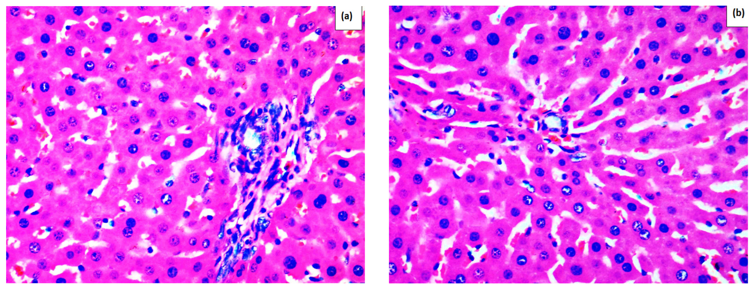
Figure 1. Histological examination of liver tissue. (a) group S—control rats, which received saline at 0.1 mL/100 g bw without APAP application; (b) group ME500—treated with 500 mg/kg of a water solution of the dried methanolic extract without APAP application; (c) group 3 (S+APAP)—saline at a dose of 0.1 mL/100 g and APAP application; (d) group 4 (ME250+APAP) rats treated with 250 mg/kg water solution of the dried methanolic extract and APAP application; (e) group 5 (ME400+APAP) rats treated with 400 mg/kg water solution of the dried methanolic extract and APAP application; (f) group 6 (ME500+APAP) rats treated with 500 mg/kg water solution of the dried methanolic extract and APAP application; (g) group 7 (RA+APAP) received 100 mg/kg RA and APAP application; (h) group 8 (Sil+APAP) was treated with 125 mg/kg silymarin and APAP application.
Ijms 26 09112 g001aIjms 26 09112 g001b
Figure 2. Biochemical markers in serum. (a) total bilirubin; (b) conjugated bilirubin; (c) ALT; (d) AST. Data are presented as mean ± SEM. The symbol a ** indicates p ≤ 0.01 vs. control group; a *** indicates p ≤ 0.001 vs. control group; b * indicates p < 0.05 vs. APAP + saline-treated rats; b ** indicates p < 0.01 vs. APAP + saline-treated rats; b *** indicates p < 0.01 vs. APAP + saline-treated rats, according to one-way ANOVA followed by Tukey’s post hoc test.
Ijms 26 09112 g002aIjms 26 09112 g002b
Figure 3. Biochemical markers in liver tissue. (a) CAT; (b) SOD; (c) GSH; (d) MDA; (e) 8-OH-dG; (f) IL-6; (g) TNF-α. Data are presented as mean ± SEM. The symbol a * indicates p < 0.05 vs. control group; a ** indicates p ≤ 0.01 vs. control group; a *** indicates p ≤ 0.001 vs. control group; b * indicates p < 0.05 vs. APAP+saline-treated rats; b ** indicates p < 0.01 vs. APAP+saline-treated rats; b *** indicates p < 0.001 vs. APAP+saline-treated rats (One-way ANOVA followed by Tukey’s post hoc test).
Ijms 26 09112 g003aIjms 26 09112 g003bIjms 26 09112 g003cIjms 26 09112 g003d
Table 1. A comparison of the effects of ME500, RA, and silymarin application on biochemical markers in rats with a model of APAP-induced liver toxicity 1.
MarkerME500+APAPRA+APAPSil+APAP
Total bilirubin000
Conjugated bilirubin000
ALT---
AST---
CAT0++
SOD00+
GSH0++
MDA-00
8-OH-dG000
IL-60n/a0
TNF-α-n/a0
1 “+” indicates increased levels, “-“ indicates decreased levels, and “0” indicates no statistical changes in comparison to the group treated only with APAP (group S+APAP); n/a—no available data.
Table 2. Roenigk classification (adopted from Berends et al. (2007) [60]).
Grade 1Accumulation of FatsNuclear
Pleomorphism
FibrosisNecrosis/Inflammation
1Mild or noneMild or noneNoneWith or without mild portal inflammation
2Moderate or severeModerate or severeNoneModerate or severe portal inflammation
3aWith or withoutWith or withoutMild (fibrosis extending into acini)With or without
3bWith or withoutWith or withoutModerate or severeWith or without
4With or withoutWith or withoutCirrhosisWith or without
1 Grade 1—mild toxicity; Grade 2—moderate toxicity; Grade 3a—severe toxicity; Grade 3b or 4—indication for discontinuation of the treatment.
Disclaimer/Publisher’s Note: The statements, opinions and data contained in all publications are solely those of the individual author(s) and contributor(s) and not of MDPI and/or the editor(s). MDPI and/or the editor(s) disclaim responsibility for any injury to people or property resulting from any ideas, methods, instructions or products referred to in the content.