Article

Proteomic Analysis Uncovers Enhanced Inflammatory Phenotype and Distinct Metabolic Changes in IDH1 Mutant Glioma Cells

by
Sigrid Ravn Berg
1,2,†,
Alessandro Brambilla
1,2,*,†,
Lars Hagen
1,3,
Animesh Sharma
1,3,
Cathrine Broberg Vågbø
1,3,
Nina Beate Liabakk
1,2,
Miroslava Kissova
1,2,
Miquel Arano Barenys
1,
Magnar Bjørås
1,2,4,5,
Sverre Helge Torp
1,6 and
Geir Slupphaug
1,2,3,*
1
Department of Clinical and Molecular Medicine, Norwegian University of Science and Technology, 7491 Trondheim, Norway
2
Clinic of Laboratory Medicine, St. Olavs Hospital, 7491 Trondheim, Norway
3
The Proteomics and Modomics Core Facility, PROMEC, at NTNU and the Central Norway Regional Health Authority, 7491 Trondheim, Norway
4
Department of Microbiology, Oslo University Hospital and University of Oslo, 0372 Oslo, Norway
5
Norway Centre of Embryology (CRESCO), University of Oslo, 0313 Oslo, Norway
6
Department of Pathology, St. Olavs Hospital, Trondheim University Hospital, 7006 Trondheim, Norway
*
Authors to whom correspondence should be addressed.
These authors contributed equally to this work.
Int. J. Mol. Sci. 2025, 26(18), 9075; https://doi.org/10.3390/ijms26189075
Submission received: 25 June 2025 / Revised: 21 August 2025 / Accepted: 3 September 2025 / Published: 18 September 2025
(This article belongs to the Special Issue Novel Molecular Pathways in Oncology, 3rd Edition)

Abstract

Isocitrate dehydrogenase 1 (IDH1) mutations are key drivers of glioma biology, influencing tumor aggressiveness and treatment response. To elucidate their molecular impact, we performed proteome analysis on patient-derived (PD) and U87MG glioma cell models with either mutant or wild-type IDH1. We quantified over 6000 protein groups per model, identifying 1594 differentially expressed proteins in PD-AS (IDH1MUT) vs. PD-GB (IDH1WT) and 904 in U87MUT vs. U87WT. Both IDH1MUT models exhibited enhanced MHC antigen presentation and interferon signaling, indicative of an altered immune microenvironment. However, metabolic alterations were model-dependent: PD-AS cells shifted toward glycolysis and purine salvage, while U87MUT cells retained oxidative phosphorylation, potentially due to D2-hydroxyglutarate (2OHG)-mediated HIF1A stabilization. We also observed a predominance of downregulated DNA repair proteins in IDH1MUT models, particularly those involved in homologous recombination. In contrast, RB1 and ASMTL were strongly upregulated in both IDH1MUT models, implicating them in DNA repair and cellular stress responses. We also found distinct expression patterns of proteins regulating histone methylation in IDH1MUT cells, favoring increased methylation of H3K4, H3K9, and H3K36. A key driver of this may be the upregulation of SETD2 in PD-AS, an H3K4 and H3K36 trimethyltransferase linked to the recruitment of HIF1A as well as DNA mismatch repair proteins. This study uncovers candidate biomarkers and pathways relevant to glioma progression and therapeutic targeting, but also underscores the complexity of predicting glioma pathogenesis and treatment responses based on IDH1 mutation status. While proteome profiling provides valuable insights, a comprehensive understanding of IDH1MUT gliomas will likely require integrative multi-omics approaches, including DNA/RNA methylation profiling, histone and protein post-translational modification analyses, and targeted DNA damage and repair assays.

1. Introduction

Glioblastoma (GB) is the most prevalent primary malignant brain tumor in adults, accounting for approximately 50% of all gliomas [1]. According to the 2021 WHO classification, tumors formerly known as adult-type diffuse gliomas are now categorized into three types: astrocytoma, isocitrate dehydrogenase (IDH) mutant; oligodendroglioma, IDH mutant and 1p/19q codeleted; and glioblastoma, IDH wild-type [2]. IDH-mutant astrocytomas usually receive radiotherapy in conjunction with adjuvant temozolomide (TMZ), whereas glioblastomas receive radiotherapy and either concurrent or adjuvant TMZ [3]. Despite these treatments, glioblastomas have a median survival of only 12–15 months. Although IDH-mutant gliomas, often classified as low-grade gliomas (LGGs), are associated with a better prognosis, the majority ultimately recur. IDH missense mutations primarily affect arginines in the isocitrate-binding pocket. Most commonly, R132 in cytosolic IDH1 or the analogous R172 in mitochondrial IDH2 are affected. Change of either arginine to a less polar residue, like histidine, glycine, cysteine, or lysine, reduces its affinity for isocitrate while increasing its preference for α-ketoglutarate (2OG) and NADPH [4]. Wild-type IDH1 exists in the cytoplasm as homodimers that produce 2OG by using NADP+ as a cofactor. Since IDH1 mutations are monoallelic, this results in a mix of mutant and wild-type dimers. Such dimers irreversibly convert 2OG to the D2-enantiomer of hydroxyglutarate (2OHG in the following) and use NADPH as cofactor. In these tumor cells, 2OHG can rise to millimolar concentrations, while 2OG is strongly reduced. 2OHG competitively inhibits 2OG-dependent enzymes such as histone demethylases and TET- and ALKBH-family enzymes, resulting in RNA, DNA, and histone hypermethylation, which are epigenetic regulatory mechanisms associated with tumorigenesis. 2OHG is thus classified as an oncometabolite (5). Interestingly, astrocytomas harboring the IDH1 R132H mutation (~90%) display lower genomic methylation compared to non-R132H mutations, which signifies a poorer prognosis [5].
While numerous studies have sought to distinguish IDH-mutant from IDH-wild-type tumors using genomic and transcriptomic analyses, these approaches may not fully capture the alterations occurring at the proteomic level. This discrepancy is partly due to the well-documented poor correlation between mRNA and protein expression [6], which is particularly relevant for brain tumors [7,8,9]. To this end, we have subjected patient-derived tumor cells from an R132C IDH1 mutant astrocytoma (grade III) patient (PD-AS) and an IDH1 wild-type (grade IV) glioblastoma patient (PD-GB) to label-free quantitative proteome profiling. Furthermore, to identify potentially differentially expressed proteins (DEPs) directly associated with mutant IDH1, we have compared the DEPs from the patient cell lines with those found in isogenic wild-type U87MG cells (U87WT) compared to U87MG cells expressing monoallelic IDH1 R132H (U87MUT). Among the biological processes most similarly affected by the IDH1 mutation in our cell models was upregulated interferon signaling, and antigen processing and presentation via MHC complexes, implying that the IDH1 mutation status may be directly relevant for therapeutic strategies involving immunotherapy [10,11].

2. Results and Discussion

2.1. Common DEPs in IDH1 Mutant Versus IDH1 Wild-Type Glioma Cells Are Associated with Tumor Aggressiveness and Response to Treatment

Proteins extracted from PD-GB, PD-AS, U87WT, and U87MUT cells were subjected to label-free quantitative (LFQ) LC-MS/MS proteome analysis. In total, we quantified 6334 and 6359 protein groups in PD-AS and PD-GB, respectively, of which 6148 protein groups were common to both (Supplementary Table S1). In U87MUT and U87WT, we quantified 5793 and 5789 protein groups, respectively, of which 5699 protein groups were common to both (Supplementary Table S2).
Among the most strongly upregulated proteins in PD-AS compared to PD-GB were SQOR, MYO1B, TYMP, IFIT2, GGT5, MGLL, and OAS2 (Figure 1A, left panel). Several of these have documented relevance in glioma biology. MYO1B has been shown to promote glioblastoma angiogenesis [12], while TYMP (thymidine phosphorylase) supports glioma stem cell proliferation and self-renewal [13]. The >100-fold higher expression of interferon-induced IFIT2 in PD-AS relative to PD-GD may be linked to the presence of PTEN, which was only detected in PD-AS (Supplementary Table S1). IFIT2 expression has been correlated with PTEN status at the transcript level, and PTEN loss was associated with reduced glioma immunogenicity, including decreased CD8 infiltration and lower interferon-inducible gene expression [14]. Consistently, OAS2, another interferon-stimulated gene, was also among the most upregulated in PD-AS (Figure 1A). Conversely, CD70 was ~90-fold lower in PD-AS compared to PD-GB. Given that CD70 promotes immune suppression by inducing CD8+ T-cell death and its expression correlates with poor survival in glioma [15], this downregulation may reflect a more immunologically permissive phenotype in PD-AS.
The U87WT vs. U87MUT comparison revealed fewer outlier proteins overall (Figure 1A, right panel), consistent with the isogenic nature of this model. Notably, ABCC2, a transporter linked to multidrug resistance, was upregulated ~28-fold in U87MUT, and LCP1 (plastin-2) was increased ~6-fold. LCP1 knockdown has been reported to reduce glioma cell viability and invasiveness [16]. In contrast, the intermediate filament protein nestin (NES), a widely used glioma stem cell marker associated with increased malignancy and invasiveness [17], was ~78-fold lower in U87MUT compared to U87WT. Interestingly, a recent IHC-based study found NES expressions to correlate positively with IDH1 R132H mutation status in gliomas [18]. The molecular mechanisms underlying this apparently contrasting finding remain to be further investigated. The divergence between our proteomic results and these tissue-level findings suggests that the regulation of NES in IDH1-mutant gliomas may be context-dependent, influenced by model system and microenvironment, and merits further investigation.
To identify proteins and pathways potentially involved in tumor aggressiveness and treatment responses, we selected differentially expressed proteins (DEPs) for further analysis using a statistical threshold of p < 0.05 and an absolute log2 LFQ difference > |0.585|, corresponding to a 1.5-fold change in expression (up- or downregulated). In addition, proteins that were consistently quantified in only one of the cell lines but not the other were also classified as DEPs. These proteins were assigned differential expression ratios of either 0.01 or 100, as presented in Supplementary Tables S1 and S2. Although statistical significance could not be calculated for these proteins, they may still represent biologically relevant processes in the cells.
The filtering identified 1594 DEPs in PD-AS vs. PD-GB (777 upregulated, 817 downregulated) and 904 DEPS in U87MUT vs. U87WT (316 upregulated, 588 downregulated) (Figure 1B). The greater number of DEPs in the patient-derived comparison likely reflects differences in genetic background: the U87 system is isogenic, where variation is attributable mainly to the introduced IDH1 mutation, whereas the PD models originate from distinct patients with broader genomic divergence. This background also influences the directionality of changes. In U87MUT, downregulated DEPs were strongly overrepresented, consistent with the well-established DNA hypermethylation phenotype of IDH1 mutations being more apparent in an otherwise identical background. This was corroborated by ShinyGO analysis of the GC vs. AT content of the genes encoding the DEPs compared to the genes encoding all quantified proteins in the dataset. This revealed a significant shift (p = 0.002) towards higher GC content in genes encoding downregulated DEPs in U87MUT vs. U87WT (Figure 1C). This would conform to a higher probability of silencing methylations at CpG islands in U87MUT than in U87WT due to inhibited activity of 2OG-dependent TET demethylases. We found a similar shift towards higher GC content in genes encoding upregulated DEPs in PD-AS vs. PD-GB (Figure 1B). This cannot be explained by DNA hypermethylation. However, RNA methylation status is also regulated by 2OG-dependent demethylases. Of these, ALKBH5 and FTO are known erasers of m6A, the most abundant modification in mRNA [19,20], and they have also been assigned prognostic value in gliomas [21]. Their inhibition by 2OHG would lead to elevated levels of m6A in mRNA and may alter mRNA stability and translation efficiency depending on which “reader” proteins bind to the modifications [22,23,24]. Increased degradation of hypermethylated, adenine-rich transcripts could thus contribute to the shift in GC-content in the upregulated DEPs in PD-AS vs. PD-GB.
Among proteins commonly assigned prognostic/therapeutic value in glioma, several were differentially expressed in PD-AS vs. PD-GB (Figure 1D). Of these, PTEN was robustly detected in all PD-AS replicates but in neither of the PD-GB replicates. It is a well-established tumor suppressor that acts as a critical negative regulator of the PI3K signaling pathway, which is essential for controlling cell proliferation and survival. PTEN mutation (chromosome 10 loss) occurs frequently in glioblastoma and contributes to an immunosuppressive environment and resistance to immune checkpoint inhibitors [25].
CDKN2A (tumor suppressor ARF) was also strongly upregulated in PD-AS vs. PD-GB. Loss of CDKN2A is associated with shorter survival in both IDH mutant and wild-type gliomas [26]. EGFR was fourfold upregulated in PD-AS vs. PD-GB. Amplification or gain-of-function mutations in EGFR occur in about half of IDHWT glioblastomas [27]. Whereas EGFR inhibitors have proven successful in e.g., lung cancer treatment, results have been disappointing in the treatment of gliomas. MGMT was threefold higher in PD-AS vs. PD-GB. It is the primary enzyme for the direct reversal of O6-methylguanosine, the most cytotoxic DNA lesion induced by TMZ, and MGMT promoter methylation status is an important prognostic biomarker and potential predictor of response to TMZ [28]. The uncertainty regarding predicting TMZ response is primarily due to discordance between analyses of promoter methylation status and MGMT expression, underscoring the importance of quantifying this biomarker at the protein level. TERT was below the detection levels in both PD-AS and PD-GB, indicating the absence of activating mutations in the TERT promoter. Finally, ATRX was robustly detected in both samples. However, given the plethora of ATRX mutations reported in gliomas [29], we cannot exclude the presence of functionally inactivating mutations.
We identified 84 DEPs that were common in our two IDH1 MUT/WT cell models, 25 of which were upregulated, and 59 of which were downregulated. Among the common downregulated DEPs in the IDH1 mutant cells, nearly half have previously been assigned potential value as prognostic/predictive biomarkers in gliomas, and of these, most predict a negative prognosis when highly expressed (Figure 1E, blue box, Supplementary Table S3). Interestingly, 12 of the common upregulated proteins in the IDH mutant cells have previously been associated with a negative prognosis, whereas only one, GNG2, has been associated with a positive prognosis. RB1 and PML were robustly detected in both PD-AS and U87MUT but were below the level of detection in their IDH1 WT counterparts. RB1 is a tumor suppressor protein that, in its unphosphorylated form, binds E2F and halts the cell cycle at the G1/S phase border. Sequential phosphorylation by cyclinD-CDK4/6 and cyclin E-CDK2 promotes detachment of RB1 from E2F and allows S-phase entry. We did not assess the phosphorylation stoichiometry of RB1; however, cyclin D (CCND) and cyclin E (CCNE) were below the detection level in both cell models. Moreover, both CDK6 and CDK2 were significantly downregulated in PD-AS vs. PD-GB, whereas the cyclinD-CDK4/6 inhibitor CDKN2A was strongly upregulated. These alterations suggest enhanced RB1-induced arrest at the G1/S border in the PD-AS cells. Cell cycle analysis revealed, however, no G1/S-arrest, but rather accumulation of cells in G2 (Figure 1F, upper right panel). Additionally, a significant population of haploid or near-haploid cells was observed. This population does not appear to consist of apoptotic cell fragments, as mass spectrometry analysis showed no increase in activating CASP3 cleavages at Asp28 or Asp175 in PD-AS relative to PD-GB (Supplementary Figure S1). The underlying mechanism for this phenomenon remains unclear, but haploid cell clones have also been observed in other malignancies, including brain tumors [30,31]. Potential explanations include dysregulation of the G2/M or intra-M checkpoints, or downregulation of proteins associated with centrosome function or spindle assembly. Notably, whereas most proteins involved in the latter processes were expressed at similar levels in PD-AS and PD-GB, the outer kinetochore complex subunit KNL1 constituted an exception to this. It was robustly detected in all PD-GB replicates but was non-detectable in PD-AS. KNL1 is a large, predominantly unstructured protein that plays a key role in kinetochore-microtubule attachment and in early mitosis signaling [32,33], and defects in KNL1 are linked to primary microcephaly [34]. Although KNL1 is upregulated in a wide range of cancers, including gliomas [35], there is currently little evidence of a causal link between dysregulated KNL1 and tumorigenesis. An alternative explanation for the accumulation of apparently haploid cells in PD-AS could be upregulation of meiotic proteins. Genes that contribute to this belong to a class denoted cancer/testis genes, which are expressed in some tumors, including brain tumors [36]. We aim to study this further by single-cell transcriptome analysis of the PD-AS cells.
Finally, we compared the differentially expressed proteins (DEPs) between our IDH1MUT/WT models and a recently published 36-gene panel (UAB36), in which increased mRNA expression correlates with resistance to over 20 cancer drugs across 777 cancer cell lines [37]. This gene set shows particularly strong associations with resistance to chemotherapeutics commonly used in glioblastoma treatment, including TMZ and carmustine. As illustrated in Figure 1G, most proteins encoded by these resistance-associated mRNAs were upregulated in PD-AS relative to PD-GB, whereas most of these proteins were downregulated in U87MUT compared to U87WT cells. Notably, the two common downregulated proteins in our two IDH mutant cell models, KRT8 and ABCC3, were recently assigned oncogenic roles in gliomas, and the low expression of these proteins predicted enhanced response to chemotherapy [38,39].

2.2. IDH Mutant Glioma Cells Display Elevated MHC Antigen Presentation and Interferon Signaling

Further mapping of the DEPs on GO biological processes using the PANTHER over-representation test revealed significant upregulation of processes such as antigen processing and assembly with MHC class I complex, purine ribonucleoside salvage, IL-27-mediated signaling, and canonical glycolysis as top differential biological processes in PD-AS vs. PD-GB (Figure 2A, left panel). Notably, in U87MUT vs. U87WT, antigen presentation of endogenous peptides via MHC class II emerged as the most enriched pathway (Figure 2A, right panel). This shared emphasis on antigen presentation suggests a potential link between increased antigen presentation and the IDH1 mutation status. IPA analysis further supported these findings, identifying MHC class I and II antigen presentation as among the most upregulated pathways in PD-AS vs. PD-GB (Figure 2B, upper panel). Specifically, core components of the MHC I complex, HLA-A, HLA-B, and beta-2-microglobulin (B2M) were >100-, 68-, and 5.8-fold upregulated, respectively. This aligns with previous studies demonstrating reduced MHC I expression in migrating glioma cells, which helps these cells evade immune surveillance [40]. Furthermore, PD-AS exhibited a shift toward immunoproteasomes, which enhance MHC I antigen presentation by replacing canonical proteasomal β subunits (PSMB5/6/7) with interferon-γ-induced subunits (PSMB8/9/10). Consistent with this, PSMB8, PSMB9, and PSMB10 were upregulated by 4.1-, 9.2-, and 4.1-fold, respectively, in PD-AS vs. PD-GB, while the canonical subunits PSMB5 and PSMB6 were downregulated by 2.3- and 2.9-fold (Supplementary Table S1). In U87MUT vs. U87WT, MHC-related upregulation was less pronounced compared to PD-AS vs. PD-GB. However, significant upregulation was observed in MHC II-related components, including HLA-DQA1, HLA-DRA, and HLA-DRB1. Notably, GO cellular component analysis of upregulated DEPs in U87MUT vs. U87WT highlighted significant enrichment of lysosomal proteins (p = 2.9 × 10⁻⁵), consistent with the lysosomal processing of internalized proteins for MHC II presentation.
The PANTHER analysis identified IL27 signaling as the second most upregulated pathway in PD-AS vs. PD-GB. IL-27 has previously been shown to mediate potent antitumor activity, with reduced IL-27 levels linked to increased glioma susceptibility [41]. Although IL-27 itself was not detected in our LFQ analyses, key downstream effectors within this pathway were markedly upregulated in PD-AS, including OAS1 (27-fold), OAS2 (>100-fold), MX1 (>100-fold), and STAT1 (6.2-fold). It should be noted, however, that several of these proteins are also upregulated by interferon and other cytokines, as corroborated by IPA analysis, which highlighted interferon α/β signaling and overall interferon responses as significantly enriched pathways in PD-AS (Figure 2B). These findings suggest a diminished inflammatory response in PD-GB compared to PD-AS. Supporting this, 13 of 15 canonical interferon-stimulated proteins quantified in our study were significantly upregulated in PD-AS, with some showing over 100-fold increases (e.g., IFIT1, IFIT2). Several of these proteins, such as IFI35 [42] and MX2 [43], have been proposed as therapeutic targets in glioma. Low IFIT2 expression, previously linked to PTEN loss, aligns with the lack of PTEN expression in glioblastoma tumors, which are also characterized by reduced CD8+ T-cell infiltration and impaired cGAS-STING signaling [14]. Consistently, STING1 was robustly expressed in PTEN-proficient PD-AS cells but below the detection limit in PTEN-deficient PD-GB cells (Supplementary Table S1). It should also be noted that IPA reported interferon gamma signaling as the most significantly common upregulated pathway in both PD-AS and U87MUT (Figure 2C). Interestingly, recent studies have shown that elevated interferon signaling, including upregulation of STAT1, correlates with poor survival in glioblastoma patients and may represent a potential therapeutic target [44,45]. The significantly higher expression of STAT1 (6-fold, p = 8.5 × 10⁻⁵) and other interferon-stimulated genes in PD-AS cells compared to PD-GB highlights the relevance of further investigating these pathways, particularly in certain lower-grade gliomas.
Both PANTHER and IPA reported processes related to cytoplasmic translation among the most upregulated processes in U87MUT vs. U87WT (Figure 2A,B). It is tempting to speculate that this may, at least in part, be related to the strong (>100-fold) upregulation of the lysine methyltransferase SMYD5. Two independent reports very recently described lysine trimethylation of the mature ribosomal subunit RPL40. This protein is expressed from UBA52 as a ubiquitin-ribosomal fusion protein that is subsequently cleaved into ubiquitin and mature RPL40. SMYD5-mediated trimethylation of K22 in RPL40 leads to a robust overall increase in translational output. Inhibiting the SMYD5-RPL40me3 axis reprograms protein translation to attenuate oncogenic gene expression signatures and renders tumor cells hypersensitive to mTOR inhibitors [46,47]. To what degree RPL40me3 is a reversible modification remains elusive. In histone H3, however, K9me3 embedded in an RKS sequence is demethylated by 2OG-dependent PHF8, which is strikingly similar to the RKC sequence at K22me3 in RPL40.
Paradoxically, IPA reported collagen biosynthesis and modifying enzymes among the top upregulated pathways in PD-AS vs. PD-GB, whereas this pathway was among the most downregulated in U87MUT vs. U87WT (Figure 2B). In PD-AS, upregulation was especially evident for collagen II, IV, and VI, as well as the 2OG-dependent prolyl- and lysyl hydroxylases critical for their maturation. In contrast, nearly all these proteins were significantly reduced in U87MUT to U87WT (Supplementary Table S1). Although 2OHG is expected to inhibit collagen hydroxylases, potentially causing misfolded collagen accumulation and ER stress, we detected no broad ER stress response in either IDH-mutant model. The sole exception was significantly higher SERPINH1 (HSP47) levels in both cell types. Indeed, in PD-AS, SERPINH1 ranked second among all proteins by MS intensity (Supplementary Table S1). Furthermore, the collagen prolyl-4-hydroxylase (P4H) subunit P4HA1 was sixfold upregulated in PD-AS, whereas the subunit P4HB was the fourth most abundant among the PD-AS proteins. This suggests that a high abundance of collagen P4H may partially overcome 2-OHG’s inhibitory effects, maintaining sufficient collagen hydroxylation. Additionally, SERPINH1, a specialized collagen-binding chaperone, not only facilitates procollagen folding and ER-to-Golgi transport independent of collagen P4H activity, but may also stabilize under-hydroxylated collagen, preventing aggregation [48].

2.3. IDH1 Mutations Are Associated with Distinct Metabolic Shifts Depending on Genetic Background

Purine salvage was among the top upregulated pathways in PD-AS vs. PD-GB, with significant upregulation of S-methyl-5-thioadenosine phosphorylase (MTAP, 3.3-fold), adenosine kinase (ADK, 3.2-fold), purine nucleoside phosphorylase (PNP, 3.2-fold), adenine phosphoribosyltransferase (APRT, 2.1-fold), and phosphopentomutase (PGM2, 1.8-fold). Notably, loss of MTAP expression occurs in about 45% of GBs, and is accompanied by increased stemness and more aggressive tumors [49]. MTAP is a key enzyme in the purine and methionine salvage pathways. It catalyzes the breakdown of methylthioadenosine, a byproduct of polyamine biosynthesis, into adenine and methionine, thereby salvaging these metabolites for other biochemical reactions. Loss of MTAP results in epigenetic dysregulation in glioma cells, and inhibition of de novo purine biosynthesis was recently shown to specifically deplete therapy-resistant GB stem-cell-like cells [49]. Neither of the above purine salvage enzymes was upregulated in U87MUT vs. U87WT, suggesting that the altered purine metabolism is not an obligatory consequence of IDH1 mutation. Preclinical studies recently suggested that targeting inosine-5′-monophosphate dehydrogenase (IMPDH), a key enzyme in the de novo purine pathway, can improve the efficacy of radiation and TMZ in GB. A follow-up phase 0/1 clinical study very recently demonstrated that the IMPDH inhibitor Mycophenolate mofetil (MMF), a commonly used oral immunosuppressant, was reasonably well tolerated, and a randomized phase 2/3 trial through the Alliance for Clinical Trials in Oncology is currently being planned [50]. Our findings suggest that quantification of proteins in salvage purine biosynthesis could aid the stratification of glioma patients in such trials.
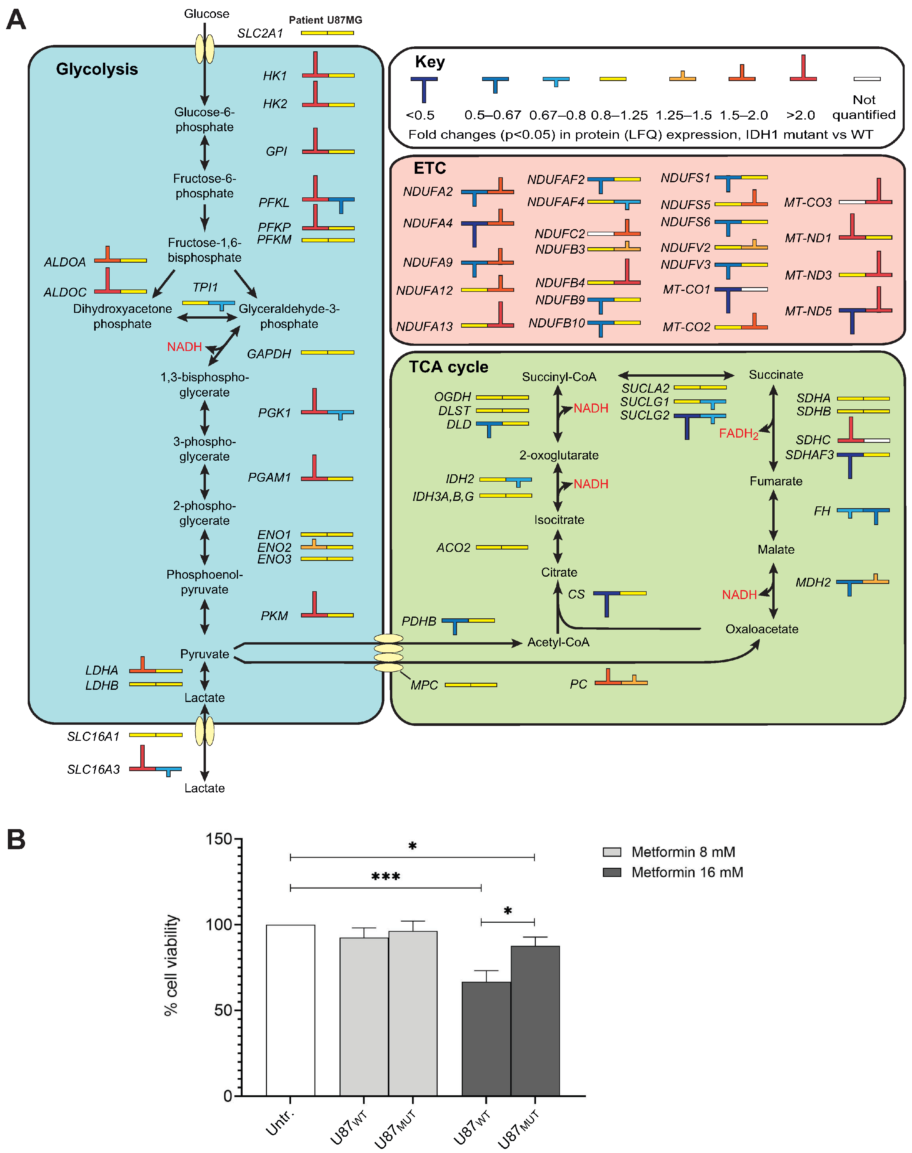
PANTHER analysis also identified canonical glycolysis as one of the top upregulated pathways in PD-AS compared to PD-GB. This finding was corroborated by IPA analysis, which highlighted upregulated glycolysis (p = 8.6E-4, z-score 2.6) alongside downregulated TCA cycle activity (p = 1.2E-4, z-score −1.9) and oxidative phosphorylation (p = 5.2 × 10⁻⁵, z-score −2.5) in PD-AS relative to PD-GB. Notably, ten glycolytic enzymes were upregulated, whereas TCA cycle enzymes and proteins in the electron transport chain (ETC) were overall downregulated (Figure 3A). This metabolic reprogramming, characterized by a preference for glycolysis even in the presence of oxygen, is commonly referred to as the Warburg effect and is frequently associated with gliomas, as reviewed in [51]. Mutations in either IDH1 or IDH2 can contribute to this phenomenon via several mechanisms. First, in IDH1 mutant gliomas, cytosolic 2OHG may be transported into mitochondria, where it disrupts TCA cycle function and ATP production via oxidative phosphorylation [52]. Second, the balance between glycolysis and the TCA cycle is regulated by the transcription factor HIF1, which is key to cellular oxygen sensing. It is a heterodimer consisting of an unstable (HIF1A) and a stable (ARNT) subunit. At normoxia, HIF1A becomes hydroxylated by oxygen- and 2OG-dependent prolyl hydroxylases EGLN1,2 and 3, which mediates its proteasomal degradation. Under hypoxic conditions, hydroxylation is attenuated, leading to increased levels of HIF1A and a HIF1-mediated transcriptional response, including upregulation of several glycolytic enzymes and suppressed flow of carbon into the TCA cycle via activation of PDK1 [53]. Inhibition of the EGLNs by 2OHG would stabilize HIF1A even under normoxic conditions, leading to a “pseudohypoxic” state [54]. Third, 2OHG also attenuates the activity of the HIF1A-inhibitor HIF1AN, which hydroxylates HIF1A at Asn803 and blocks binding to its transcriptional coactivators [53]. Fourth, the high levels of collagen P4H and the abundance of substrate collagens in PD-AS likely drain 2OG and oxygen available for EGLN1–3, since collagen P4H has markedly higher affinity for both co-substrates than the HIF prolyl hydroxylases [55], and thereby contributes to HIF1 stabilization. Finally, factors not directly related to the IDH1 status may contribute to the glycolytic shift in PD-AS compared to PD-GB. One such candidate is EGFR, which was fourfold higher expressed in PD-AS vs. PD-GB, whereas it was not significantly different in U87MUT vs. U87WT. EGFR signaling upregulates glycolytic enzymes and downregulates OXPHOS, and inhibitors targeting EGFR have been shown to reverse this process [56].
Interestingly, we observed no shift from TCA to glycolysis in U87MUT cells compared to U87WT cells, suggesting that induction of a pseudohypoxic state is not an obligatory consequence of mutated IDH1. Potentially, this could be mediated by differential expression of factors involved in HIF1 regulation and transcriptional output in the PD and U87 cell models. Notably, however, TCA activity does not solely depend on the levels of the involved proteins. Accumulating evidence suggests that the functions of several TCA cycle enzymes are regulated by binding to long non-coding RNAs (lncRNAs) [57]. One such example is the lncRNA GAS5, which binds MDH2 and thereby inhibits the metabolic enzyme tandem association of FH, MDH2, and CS [58]. Very recent research strongly indicates that the level of GAS5 is regulated by m6A modification in gliomas. Firstly, the levels of the m6A erasers FTO and ALKBH5 are very significantly negatively correlated with GAS5. Secondly, GAS5 expression is significantly higher in IDH mutant- than in IDH WT gliomas (p = 2.9 ×10⁻¹⁸) [59]. It is thus tempting to speculate that 2OHG-mediated inhibition of FTO and ALKBH5 downregulates TCA flux in U87MUT via upregulation of GAS5.
Mitochondrial electron transport (NADH to ubiquinone) was among the most upregulated biological processes in U87MUT (Figure 2A), driven by a notable increase in several Complex I components (Figure 3A). Potentially contributing to this could be that ETC proteins are upregulated by HIF1 under normoxia [60]. In U87MUT, the accumulation of 2OHG would inhibit HIF1 hydroxylation and inactivation, resulting in constitutive overexpression of HIF1-regulated genes. Enhanced ETC activity in U87MUT was further supported by the fourfold upregulation of the mitochondrial glutathione transporter SLC25A40. Since glutathione is exclusively synthesized in the cytosol, its import into mitochondria via SLC25A39/40 is critical for the proper functioning of ETC proteins, particularly those containing iron-sulfur clusters [61], which are abundant in Complex I.
The upregulation of Complex I, an NADH-accepting component of the ETC, despite no apparent changes in glycolytic or TCA cycle activity, suggests alternative sources of NADH in U87MUT cells. We hypothesize that fatty acid oxidation may serve as a major contributor to NADH production in this context. Supporting this, CPT1A, which catalyzes the rate-limiting step in β-oxidation by transferring the acyl group of long-chain fatty acid-CoA conjugates onto carnitine, was upregulated 1.6-fold (p = 0.02) in U87MUT compared to U87WT. Previously, knockdown of CPT1A in an IDH1WT background reduced tumor growth and increased survival in a glioma mouse xenograft model [62]. Interestingly, metformin, which is widely used to treat type 2 diabetes, is a reversible inhibitor of the ETC Complex I [63]. It is also one of the few drugs that can cross the blood–brain barrier [64]. A recent Phase I trial of combined metformin and radiation treatment for high-grade gliomas demonstrated promising clinical efficacy [65]. However, there was no information on the IDH status of the tumors. Likewise, a recruiting Phase II trial (NCT04945148) will include metformin in a standard radiation/TMZ treatment of newly diagnosed GB (IDHWT). Our results suggest that future research and clinical trials encompassing drugs that target fatty acid oxidation and the ETC should also encompass patients with IDH1MUT gliomas.
To investigate whether inhibition of Complex I could be exploited as a specific vulnerability in gliomas with increased ETC and normal glycolytic flux, we treated U87MUT and U87WT with metformin, which has been suggested as a potentially selective adjuvant in the treatment of IDH-mutant gliomas [66]. Somewhat unexpectedly, the U87WT cells were significantly more sensitive to 16 mM metformin than U87MUT (Figure 3B). The most logical explanation for this would be that the U87MUT cells harbor enhanced levels of functional Complex I and thus tolerate higher levels of the inhibitor. It will be interesting in the future to study how metformin affects these cells when co-administered with other metabolic inhibitors, e.g., glutaminase inhibitors.
Collectively, our findings indicate that the metabolic alterations associated with IDH1 mutations in glioma cells strongly depend on the overall genetic background of the cells. Potentially, the U87MUT cells have elevated compensatory mechanisms to buffer cytosolic 2OG, compared to PD-AS. Such mechanisms could include elevated mitochondrial 2OG generation by enhanced TCA cycle turnover or by decreased OGDH expression [67], increased 2OG transport from mitochondria to the cytoplasm, or increased glutaminolysis. In support of this, TCA enzymes were more negatively affected by the IDH1 mutation in PD-AS than in U87MUT (Figure 3A). Moreover, U87MUT had higher levels of the mitochondrial 2-oxoglutarate/malate carrier SLC24A11 and glutamate dehydrogenase (GLUD1) than PD-AS, based on normalized MS intensity values. Finally, the nature of the IDH1 mutant likely contributes to higher 2OHG in PD-AS than in U87MUT, since the R132H mutant (U87MUT) has a lower 2OHG production capacity than R132C (PD-AS) [68].

2.4. DNA Repair Processes Are Differentially Affected in IDH-Mutant Glioma Cells

DNA repair is essential for maintaining genomic stability in cells, and mapping repair activity can help predict tumor responses to DNA-damaging chemotherapeutic agents and radiation [69]. The impact of IDH mutations on DNA repair has largely been attributed to 2OHG-mediated inhibition of DNA and histone demethylation via TET enzymes and KDMs. Many DNA repair proteins are regulated by promoter methylation, which can influence treatment strategies. Additionally, DNA hypermethylation may contribute to a mutator phenotype, as the spontaneous deamination of 5-methylcytosine (5-meC) occurs at a significantly higher rate than that of unmethylated cytosine, leading to C > T mutations if not properly repaired [70]. However, IDH1 R132H mutations have also been proposed to act as tumor suppressors in gliomas by epigenetically upregulating mRNAs encoding DNA damage response (DDR) proteins [71].
To explore whether this regulation extends to protein expression, we analyzed our LFQ data for proteins implicated in DNA repair and genome maintenance [72,73,74]. Among the 98 DNA repair proteins that were differentially expressed in at least one dataset (Figure 4, left panel), 30 were significantly upregulated, while 51 were significantly downregulated in PD-AS vs. PD-GB. Similarly, 11 proteins were upregulated and 23 were downregulated in U87MUT vs. U87WT (Figure 4, right panel). Notably, both IDH-mutant cell models exhibited a predominance of downregulated DNA repair-associated DEPs compared to their wild-type counterparts. This highlights the poor correlation between mRNA and protein levels, particularly in tumor cells [6]. The smaller number of DNA repair DEPs in U87MUT compared to the isogenic U87WT suggests that the differential expression of DNA repair proteins in PD-AS vs. PD-GB is primarily driven by other factors than the IDH1 mutant. This aligns with the small overlap in total DEPs observed between the two IDHMUT/WT models (Figure 1E).
Among the proteins involved in DNA repair and genome maintenance, RB1 and ASMTL were the only two commonly upregulated (>100-fold) proteins across both IDHMUT models compared to their IDHWT counterparts. Beyond its well-established role in G1/S checkpoint regulation, RB1 also plays a role in the repair of DNA double-strand breaks (DSBs) that is independent of cell cycle regulation, and loss of RB1 sensitizes prostate cancer cells to ionizing radiation [75]. Paradoxically, loss of RB1 expression has been reported as a negative prognostic factor in several neoplasms, including gliomas [76,77,78]. This suggests that increased mutation rates due to the loss of RB1-mediated G1/S checkpoint control may outweigh the reduced DNA repair capacity in determining tumor fitness, especially during treatment with genotoxic agents. ASMTL is a member of the Maf (multicopy-associated filamentation) family of evolutionarily conserved “house-cleaning” nucleotide pyrophosphatases. These enzymes hydrolyze a variety of canonical and non-canonical nucleoside triphosphates into their corresponding monophosphates and are thought to play critical roles in both cell cycle arrest and the prevention of modified nucleotide incorporation into cellular nucleic acids. Human ASMTL exhibits its highest enzymatic activity against canonical nucleotides such as UTP, CTP, dTTP, and dCTP, as well as non-canonical nucleotides, including pseudo-UTP, 5-methyl-UTP, 5-methyl-CTP, N4-methyl-dCTP, 8-oxo-GTP, and N1-methyl-GTP [79]. The extent to which the altered metabolic state of IDH1-mutated glioma cells contributes to the markedly enhanced expression of ASMTL remains unclear. However, it is plausible that elevated levels of 8-oxo-GTP, which accumulate due to the increased oxidative stress characteristic of IDH1-mutated malignancies, could act as a contributing factor. Notably, 8-oxo-GTP can serve as a substrate for RNA polymerase II during transcription, thereby impairing translation and causing erroneous protein synthesis [80,81].
The human NUDT1 (MTH1) enzyme is also capable of hydrolyzing 8-oxo-GTP to 8-oxo-GMP, albeit with an efficiency approximately 50-fold lower than its activity in hydrolyzing 8-oxo-dGTP [81]. Consequently, NUDT1 is primarily regarded as a key enzyme for clearing oxidized deoxyribonucleotides from the nucleotide pool. Since ASMTL and NUDT1 exhibited similar expression levels in both IDHMUT/WT models, ASMTL is likely the primary enzyme responsible for clearing 8-oxo-GTP in these glioma cells. Potentially, ASMTL-mediated clearance of 8-oxo-GTP may also contribute to the resistance to oxidative stress induced by chemotherapy, including TMZ [82]. A growing body of evidence, including work from our own laboratory, suggests that chemotherapy-induced damage to RNA may play a more prominent role in cytotoxicity than previously anticipated [83,84,85,86].
Interestingly, of the eight DNA repair proteins commonly downregulated in our two IDH1MUT cell models (Figure 4, right panel), seven have been implicated in the repair of DSBs, primarily via homologous recombination (HR). The replication-dependent histone chaperone ASF1A promotes HR repair of DSBs at stalled or collapsed replication forks [87]. CHD4 is part of the histone deacetylase NuRD complex and promotes HR repair via upregulation of RAD51 in glioma cells [88], while HTATSF1 recruits TOPBP1 to PARylated RPA to facilitate RPA/RAD51-dependent HR [89]. POGZ recruits heterochromatin proteins to maintain BRCA1/BARD1 at DSBs and promote HR [90]. Two of the downregulated proteins, SMCHD1 and SMARCD1, are parts of chromatin remodeling complexes that promote DSB repair via NHEJ [90,91].
Finally, it should be noted that one of the interferon-induced proteins that was highly upregulated in PD-AS vs. PD-GB, SLFN11 (96-fold, p = 0.0001), acts as a negative regulator of HR [92], and that high expression of SLFN11 shows an extremely significant positive correlation with the response to TOP1/2 inhibitors, alkylating agents and DNA synthesis inhibitors in in cancer cell lines of different origin [93]. Nevertheless, the Human Protein Atlas (www.proteinatlas.org, accessed on 20 February 2025) reports high expression of SLFN11 mRNA as a poor prognostic factor in glioblastoma (p < 0.001). The underlying reasons for this apparent discrepancy remain, however, to be elucidated.

2.5. Potential Contribution of Altered Histone Lysine Methylation to DNA Damage Responses in the IDH-Mutant Cell Models

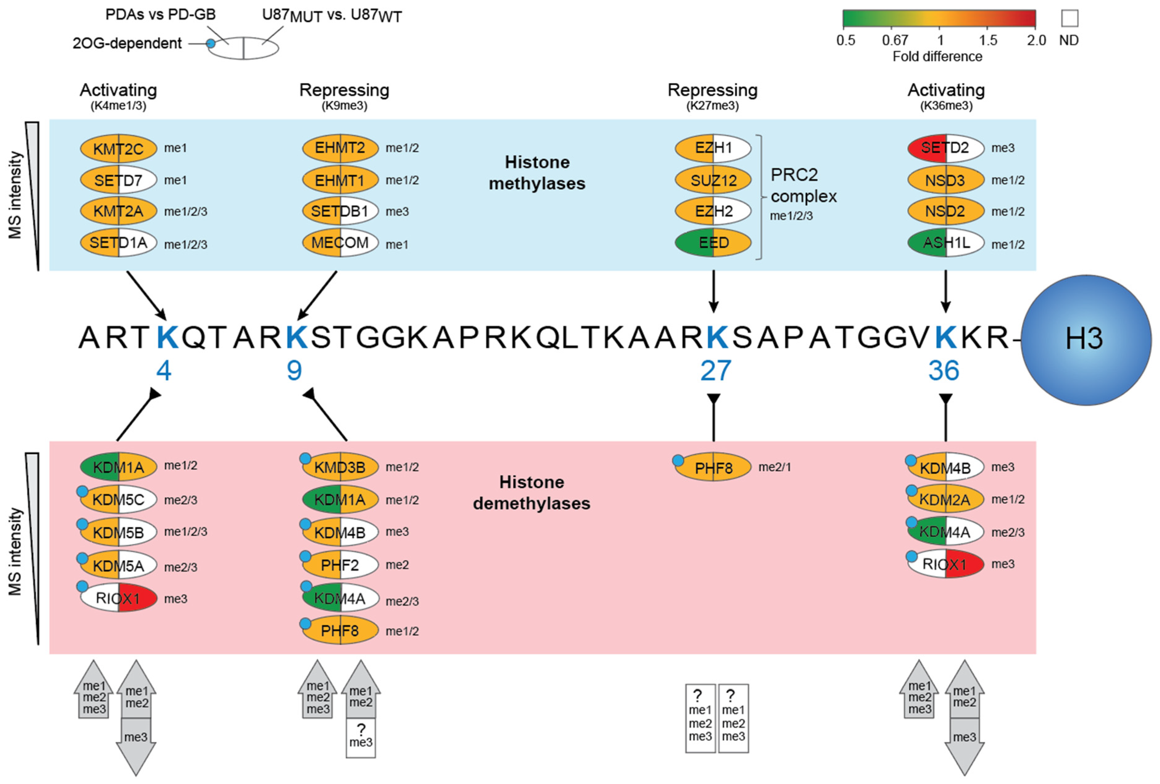
The efficiency of DNA damage responses is determined not only by the levels and activities of DNA repair proteins but also by their accessibility to DNA lesions within chromatin. This accessibility is dynamically regulated by post-translational histone modifications, including methylation. Additionally, histone methylation directly or indirectly influences the transcription of many DNA repair genes. Particularly in the context of DSB repair, histone methylations dictate the choice between HR or NHEJ pathways [94]. Histone methylation is controlled by histone lysine methyltransferases (KMTs) and histone lysine demethylases (KDMs) and can either activate or repress gene expression, depending on the specific residues modified and the number of methyl groups added. Dysregulated histone methylation has been implicated in various cancers, including gliomas [95]. Among the most well-characterized histone methylation marks are trimethylation at lysine residues K4, K9, K27, and K36 on histone H3. Of these, H3K4me3 and H3K36me3 are generally associated with active gene expression, whereas H3K9me3 and H3K27me3 typically mediate transcriptional repression. We quantified 16 KMTs and 15 KDMs across our cell models, including ten 2OG-dependent demethylases (Figure 5, Supplementary Table S4).
Based on the expression levels of involved proteins, distinct and complex alterations of histone lysine methylation in the IDH1 mutants emerge compared to their IDH1 WT counterparts. Our findings suggest that the IDH1-mutant PD-AS model exhibits increased mono- di- and trimethylation at H3K4, H3K9, and H3K36 compared to the IDH1 wild-type PD-GB model. In particular, H3K36me3 is likely to be highly enriched in PD-AS vs. PD-GB due to the marked upregulation (>100-fold) of SETD2, the sole human methyltransferase responsible for catalyzing this modification. This effect is further reinforced by 2OG-mediated inhibition of the H3K36me3 demethylases KDM4A/B and the concomitant four-fold downregulation of KDM4A. Depletion of SETD2 has been shown to severely block HR repair [96]. Thus, although many HR proteins were downregulated in PD-AS vs. PD-GB, the marked upregulation of SETD2 may compensate for this and allow efficient repair of HR in actively transcribed genes. Conversely, since SETD2 was below the detection limit in U87, this should further disfavor HR. Notably, however, it appears that inactive genes depleted in H3K36me3 may instead be targeted to NHEJ repair [97].
SETD2-induced H3K36me3 also plays a crucial role in recruiting the DNA mismatch repair (MMR) machinery to chromatin via interaction with MutSα (MSH2/MSH6) [98]. This may be especially important in the context of mutated IDH, since this would enhance genomic 5-meC due to TET inhibition, leading to enhanced levels of 5-meC deamination-induced G:T mismatches, which are repaired by MMR [99]. Since SETD2 remained below the detection level in U87MUT, we speculate that the marked induction in PD-AS is not directly mediated by the IDH1 mutation. Nevertheless, it may be of strong predictive value regarding the treatment response to TMZ. Intact MMR promotes TMZ cytotoxicity, especially in MGMT-deficient gliomas, since TMZ-induced O6-meG mispairs with thymine. During MMR, O6-meG persists while thymine is removed, leading to thymine reinsertion and cytotoxic futile repair cycles. This leads to gradually increased DNA resection, induction of SSBs and DSBs, and apoptosis [100]. Notably, both MSH2 and MSH3 were significantly reduced in PD-AS (Figure 4). Nevertheless, MMR may still be enhanced compared to PD-GB due to increased targeting to the mismatches via H3K36me3.
We hypothesize that the opposite regulation of trimethylation at H3K36 also holds true for H3K4. Firstly, SETD1A, a key H3K4me3 methyltransferase, remained below the detection level in U87MUT vs. U87WT. In addition, the >100-fold upregulation of RIOX1, which also catalyzes me3 demethylation at this position, should further contribute to H3K4me3 depletion. Since HIF1-α prefers H3K4me3-decorated binding sites [101], primarily associated with promoters and transcriptional start sites [102], this might contribute to the different metabolic alterations observed in the two IDHMUT cell models. Notably, the proposed inverse level of H3K4me3 and H3K36me3 in our two IDHMUT models is also corroborated by the ratio between up- and downregulated DEPs (Figure 1B), in which there is a marked shift towards downregulated DEPs in U87MUT vs. U87WT.

3. Materials and Methods

3.1. Cell Culture

Isogenic IDH1 wild-type (U-87MG HTB-14) and heterozygous mutant (R132H) (U-87MG HTB-14IG) were purchased from ATCC (Manassas, VA, USA). The cells were grown in 5% CO2 at 37 °C, in DMEM high glucose medium (Sigma-Aldrich, St. Louis, MO, USA) supplemented with 10% fetal bovine serum (FBS, Sigma-Aldrich, St. Louis, MO, USA), 100 µg/mL penicillin-streptomycin (P/S, Sigma-Aldrich, St. Louis, MO, USA), and 2 mM L-glutamine (Sigma-Aldrich, St. Louis, MO, USA). The patient-derived cells (PD-AS, PD-GB) were cultured on Geltrex-coated plates (Gibco, Waltham, MA, USA) in 5% CO2 at 37 °C, in Knockout DMEM/F12 (Gibco, Waltham, MA, USA) medium supplemented with 1×Stempro Neural Supplement (Gibco, Waltham, MA, USA), 2 mM L-glutamine (Sigma-Aldrich, St. Louis, MO, USA), 100 µg/mL P/S (Sigma-Aldrich, St. Louis, MO, USA), and 20 ng/mL of both Fibroblast Growth Factor 2 (FGF2, Sigma-Aldrich, St. Louis, MO, USA) and Epidermal Growth Factor (EGF, Sigma-Aldrich, St. Louis, MO, USA). ROCK Inhibitor (Sigma-Aldrich, St. Louis, MO, USA) was added to the culture medium for the first 24 h following passaging to ensure a single-cell suspension.

3.2. Cell Cycle Analyses

Cells were fixed in ice-cold methanol at a concentration of 104–105 cells/mL. After fixation, cells were washed with PBS and incubated with 200 µL RNase A solution (100 µg/mL in PBS) at 37 °C for 30 min. DNA was stained by adding 200 µL of propidium iodide (PI, 50 μg/mL, Sigma, St. Louis, MO, USA), followed by incubation at 37 °C for 30 min. Flow cytometry was performed using a BD FACS Canto flow cytometer (BD Biosciences, San Jose, CA, USA), with PI fluorescence excited at 488 nm (blue laser) and detected in the phycoerythrin (PE) channel (578 nm). Data were analyzed using FlowJo v10 software.

3.3. Viability Assay

A total of 5000 U87 cells per well were seeded in 96-well plates. After 24h incubation at 37 °C, cells were treated with Metformin or DMSO vehicle and incubated further for 72 h. To determine cell viability, a PrestoBlue assay was conducted. The PrestoBlueTM HS Cell Viability Reagent (Thermo Fischer, Waltham, MA, USA, P50201) was prepared in culture medium according to the manufacturer’s instructions. After 30 min incubation at 37 °C, fluorescence intensity was measured using a BMG-Fluostar Omega plate reader (BMG Labtech, Ortenberg, Germany). Each treatment was performed in triplicate and in two independent experiments. The data were normalized to the vehicle-treated control (DMSO) (Sigma-Aldrich, St. Louis, MO, USA) and plotted using GraphPad Prism 10.

3.4. LC-MS/MS Analyses

Cells were cultured in 10 cm plates and incubated at 37 °C until a confluency of approximately 50%. Cells were harvested by trypsination and resuspended in 100 µL 1% sodium deoxycholate, 100 mM Tris-HCl, pH 8.5, 10 mM Tris (2-carboxyethyl)phosphine (TCEP), 40 mM chloroacetamide (CAA), and heated at 90 °C for 45 min. After sonication for 10 cycles (30 s ON/30 s OFF) with a Bioruptor® Pico sonicator (Diagenode, Liège, Belgium), debris was pelleted at 16,000× g for 10 min. 50 µg protein from each sample was added to 100 µL 0.1 M ammonium bicarbonate, 0.5 µg trypsin, and digested at 37 °C overnight. Peptides were desalted using C18 spin columns, dried in a speedvac centrifuge, and resuspended in 0.1% formic acid (FA) prior to MS analysis. LC-MS/MS was performed on a timsTOF Pro (Bruker Daltonics, Bremen, Germany) connected to a nanoElute (Bruker Daltonics, Bremen, Germany) HPLC. Five hundred ng peptides from each sample were injected and separated over a Bruker15 (75 µm × 15 cm) column with running buffers A (0.1% FA) and B (0.1% FA in acetonitrile) with a gradient from 2% B to 40% B over 100 min. The timsTof was operated in DDA PASEF mode with 10 PASEF scans per acquisition cycle and accumulation and ramp times of 100 ms each. The ‘target value’ was set to 20,000, and dynamic exclusion was activated and set to 0.4 min. The quadrupole isolation width was set to 2 Th for m/z < 700 and 3 Th for m/z > 800.

3.5. Bioinformatic Analyses

Raw data were inspected using Open Workflow [103] to determine optimal search parameters for MaxQuant v2.4.3.0 [104]. In addition to default settings, deamidation of asparagine and glutamine was included as a dynamic post-translational modification. Protein identification was performed using Andromeda, the built-in search engine of MaxQuant, against the human proteome, including isoforms, downloaded from UniProt (April 2023), along with MaxQuant’s internal contaminant database. To minimize missing values, the match-between-runs (MBR) function was enabled. MBR identifies peptide signals present in one sample but absent in another by leveraging high mass accuracy and a restricted retention time window of 1 min, within an overall 20 min sliding window. If a matching signal is detected, it is labeled as ‘By matching’ instead of ‘By MS/MS’ in the output. Protein and peptide group identifications were filtered to a 1% false discovery rate (FDR), and only high-confidence unique peptides were used for final protein group identification. LFQ [105] was performed using at least one unique peptide, followed by log2 transformation of LFQ values. The median value of biological replicates was used to represent the log2-LFQ value for each protein group. Statistical analyses were conducted using two-sided Student’s t-tests in R: (https://www.r-project.org/, accessed on 22 November 2024). p-values were computed, ranked, and adjusted for multiple testing using the Benjamini–Hochberg correction [106] to obtain an FDR-adjusted p-value (q-value). For proteins present in all control replicates but absent in all experimental samples, the LFQ ratio was set to −100 with an FDR of 0.0. Conversely, proteins detected in all experimental replicates but absent in controls were assigned an LFQ ratio of 100, with both p- and q-values set to 0.0. Biological pathway analyses were performed using the PANTHER overrepresentation test (Release 20221013, GO Ontology database, released 1 July 2022) [107,108], ShinyGO (http://bioinformatics.sdstate.edu/go/, accessed on 11 November 2024) [109], and Ingenuity® Pathway Analysis (IPA) (Ingenuity Systems, www.ingenuity.com, Redwood City, CA, USA) [110].

4. Conclusions

Our findings underscore the complexity of predicting glioma pathogenesis and treatment responses based solely on IDH1 mutation status. While processes such as MHC antigen presentation and interferon signaling were consistently upregulated in our IDH1-mutant models, other pathways, particularly those involving metabolic reprogramming, varied significantly. This variability suggests that the phenotypic consequences of IDH1 mutations are not determined by the mutation in isolation but are instead shaped by the broader genomic and proteomic context, including the differences in expression of key glioma oncogenes and tumor suppressors that may modulate the impact of the 2OHG oncometabolite. These observations highlight the critical importance of investigating IDH1 mutations in experimental systems that represent a spectrum of genetic backgrounds to capture the range of potential biological outcomes and treatment vulnerabilities. Although proteomic profiling offers valuable insights, a comprehensive understanding of IDH1-mutant gliomas to aid treatment decisions will require integrative multi-omics approaches, including DNA/RNA methylation profiling, histone and protein post-translational modifications, and targeted DNA damage and repair assays.

Supplementary Materials

The following supporting information can be downloaded at https://www.mdpi.com/article/10.3390/ijms26189075/s1. References [79,111,112,113,114,115,116,117,118,119,120,121,122,123,124,125,126,127,128,129,130,131,132,133,134,135,136,137,138,139,140,141,142,143,144,145,146,147,148,149,150,151,152,153,154,155,156,157,158,159,160,161,162,163,164,165,166,167,168,169,170,171,172,173,174,175,176,177,178,179,180,181,182,183,184,185] are cited in the Supplementary Materials.

Author Contributions

Conceptualization, G.S., S.R.B. and M.B.; Methodology, S.R.B., A.B., L.H., C.B.V., N.B.L., M.K. and M.A.B.; Software, A.S. and G.S.; Validation, A.B. and M.A.B.; Formal Analysis, S.R.B., A.B., L.H., C.B.V., N.B.L., M.K. and M.A.B.; Resources, M.B. and G.S.; Data Curation, A.S. and G.S.; Writing—Original Draft Preparation, S.R.B. and G.S.; Writing—Review and Editing, G.S.; Visualization, S.R.B., N.B.L. and G.S.; Supervision, C.B.V. and S.H.T. Project Administration, G.S.; Funding Acquisition, G.S. and M.B. All authors have read and agreed to the published version of the manuscript.

Funding

This research was funded by grants from The Liaison Committee between the Central Norway regional Health Authority and Norwegian University of Science and Technology (#3253-02-04-22), and the Cancer Fund at St. Olavs Hospital, Trondheim. PROMEC is funded by NTNU and the Liaison Committee for Education, Research and Innovation in Central Norway. PROMEC is a member of the National Network of Advanced Proteomics Infrastructure (NAPI), which is funded by the RCN INFRASTRUKTUR-program (#295910). Data storage and handling are supported under the PRIDE and Norstore/Notur projects NN9036K/NS9036K, respectively.

Institutional Review Board Statement

Not applicable.

Informed Consent Statement

Not applicable.

Data Availability Statement

The mass spectrometry proteomics data have been deposited to the ProteomeXchange Consortium via the PRIDE [186] partner repository with the dataset identifier PXD065327.

Conflicts of Interest

The authors declare no conflicts of interest. The funders had no role in the design of the study; in the collection, analyses, or interpretation of data; in the writing of the manuscript; or in the decision to publish the results.

Abbreviations

The following abbreviations are used in this manuscript:
2OGα-ketoglutarate, 2-oxoglutarate
2OHGD-2-hydroxyglutarate
DEPDifferentially expressed protein
ETCElectron transport chain
GOGene ontology
HLAHuman leukocyte antigen
IDHIsocitrate dehydrogenase
KMTLysine methyltransferase
KDMLysine demethylase
LC-MS/MSLiquid chromatography with tandem mass spectrometry
LFQLabel-free quantitative
MHCMajor histocompatibility complex
PD-ASPatient-derived astrocytoma, IDH1 mutant
PD-GBPatient-derived glioblastoma, IDH wild-type
TMZTemozolomide

References

  1. Messaoudi, K.; Clavreul, A.; Lagarce, F. Toward an effective strategy in glioblastoma treatment. Part I: Resistance mechanisms and strategies to overcome resistance of glioblastoma to temozolomide. Drug Discov. Today 2015, 20, 899–905. [Google Scholar] [CrossRef] [PubMed]
  2. Berger, T.R.; Wen, P.Y.; Lang-Orsini, M.; Chukwueke, U.N. World Health Organization 2021 Classification of Central Nervous System Tumors and Implications for Therapy for Adult-Type Gliomas: A Review. JAMA Oncol. 2022, 8, 1493–1501. [Google Scholar] [CrossRef] [PubMed]
  3. Mohile, N.A.; Messersmith, H.; Gatson, N.T.; Hottinger, A.F.; Lassman, A.; Morton, J.; Ney, D.; Nghiemphu, P.L.; Olar, A.; Olson, J.; et al. Therapy for Diffuse Astrocytic and Oligodendroglial Tumors in Adults: ASCO-SNO Guideline. J. Clin. Oncol. 2022, 40, 403–426. [Google Scholar] [CrossRef]
  4. Dang, L.; White, D.W.; Gross, S.; Bennett, B.D.; Bittinger, M.A.; Driggers, E.M.; Fantin, V.R.; Jang, H.G.; Jin, S.; Keenan, M.C.; et al. Cancer-associated IDH1 mutations produce 2-hydroxyglutarate. Nature 2009, 462, 739–744. [Google Scholar] [CrossRef] [PubMed]
  5. Tesileanu, C.M.S.; Vallentgoed, W.R.; Sanson, M.; Taal, W.; Clement, P.M.; Wick, W.; Brandes, A.A.; Baurain, J.F.; Chinot, O.L.; Wheeler, H.; et al. Non-IDH1-R132H IDH1/2 mutations are associated with increased DNA methylation and improved survival in astrocytomas, compared to IDH1-R132H mutations. Acta Neuropathol. 2021, 141, 945–957. [Google Scholar] [CrossRef]
  6. Liu, Y.; Beyer, A.; Aebersold, R. On the Dependency of Cellular Protein Levels on mRNA Abundance. Cell 2016, 165, 535–550. [Google Scholar] [CrossRef]
  7. Curry, R.N.; Glasgow, S.M. The Role of Neurodevelopmental Pathways in Brain Tumors. Front. Cell Dev. Biol. 2021, 9, 659055. [Google Scholar] [CrossRef]
  8. Kolodziejczak-Guglas, I.; Simoes, R.L.S.; de Souza Santos, E.; Demicco, E.G.; Lazcano Segura, R.N.; Ma, W.; Wang, P.; Geffen, Y.; Storrs, E.; Petralia, F.; et al. Proteomic-based stemness score measures oncogenic dedifferentiation and enables the identification of druggable targets. Cell Genom. 2025, 5, 100851. [Google Scholar] [CrossRef]
  9. Statoulla, E.; Chalkiadaki, K.; Karozis, D.; Gkogkas, C.G. Regulation of mRNA translation in stem cells; links to brain disorders. Cell Signal 2021, 88, 110166. [Google Scholar] [CrossRef]
  10. Cristescu, R.; Mogg, R.; Ayers, M.; Albright, A.; Murphy, E.; Yearley, J.; Sher, X.; Liu, X.Q.; Lu, H.; Nebozhyn, M.; et al. Pan-tumor genomic biomarkers for PD-1 checkpoint blockade-based immunotherapy. Science 2018, 362, eaar3593, Erratum in Science 2019, 363, eaax1384. https://doi.org/10.1126/science.aax1384. [Google Scholar] [CrossRef]
  11. Axelrod, M.L.; Cook, R.S.; Johnson, D.B.; Balko, J.M. Biological Consequences of MHC-II Expression by Tumor Cells in Cancer. Clin. Cancer Res. 2019, 25, 2392–2402. [Google Scholar] [CrossRef]
  12. Lv, W.; Yang, F.; Ge, Z.; Xin, L.; Zhang, L.; Zhai, Y.; Liu, X.; Guo, Q.; Mao, X.; Luo, P.; et al. Aberrant overexpression of myosin 1b in glioblastoma promotes angiogenesis via VEGF-myc-myosin 1b-Piezo1 axis. J. Biol. Chem. 2024, 300, 107807. [Google Scholar] [CrossRef]
  13. Zhang, J.; Sun, R.; Lyu, Y.; Liu, C.; Liu, Y.; Feng, Y.; Fu, M.; Wong, P.J.C.; Du, Z.; Qiu, T.; et al. Proteomic profiling of gliomas unveils immune and metabolism-driven subtypes with implications for anti-nucleotide metabolism therapy. Nat. Commun. 2024, 15, 10005. [Google Scholar] [CrossRef] [PubMed]
  14. Dogan, E.; Yildirim, Z.; Akalin, T.; Ozgiray, E.; Akinturk, N.; Aktan, C.; Solmaz, A.E.; Biceroglu, H.; Caliskan, K.E.; Ertan, Y.; et al. Investigating the effects of PTEN mutations on cGAS-STING pathway in glioblastoma tumours. J. Neuro-Oncol. 2024, 166, 283–292. [Google Scholar] [CrossRef] [PubMed]
  15. Jin, L.; Ge, H.; Long, Y.; Yang, C.; Chang, Y.E.; Mu, L.; Sayour, E.J.; De Leon, G.; Wang, Q.J.; Yang, J.C.; et al. CD70, a novel target of CAR T-cell therapy for gliomas. Neuro-Oncology 2018, 20, 55–65. [Google Scholar] [CrossRef] [PubMed]
  16. Tang, Y.; Zhang, X.; Tang, X.; Yuan, Y.; Wang, W. Lactylation-related gene signatures identify glioma molecular subtypes with prognostic, immunological, and therapeutic implications. Front. Oncol. 2025, 15, 1613423. [Google Scholar] [CrossRef]
  17. Biserova, K.; Jakovlevs, A.; Uljanovs, R.; Strumfa, I. Cancer Stem Cells: Significance in Origin, Pathogenesis and Treatment of Glioblastoma. Cells 2021, 10, 621. [Google Scholar] [CrossRef]
  18. Selvaraj, S.; Srinivas, B.H.; Verma, S.K.; Ms, G. Significance of Nestin and CD133 as cancer stem cell markers in diffuse glioma and association with p53 expression and IDH status. Int. J. Clin. Exp. Pathol. 2024, 17, 208–218. [Google Scholar] [CrossRef]
  19. Jia, G.; Fu, Y.; Zhao, X.; Dai, Q.; Zheng, G.; Yang, Y.; Yi, C.; Lindahl, T.; Pan, T.; Yang, Y.G.; et al. N6-methyladenosine in nuclear RNA is a major substrate of the obesity-associated FTO. Nat. Chem. Biol. 2011, 7, 885–887. [Google Scholar] [CrossRef]
  20. Zheng, G.; Dahl, J.A.; Niu, Y.; Fedorcsak, P.; Huang, C.M.; Li, C.J.; Vagbo, C.B.; Shi, Y.; Wang, W.L.; Song, S.H.; et al. ALKBH5 is a mammalian RNA demethylase that impacts RNA metabolism and mouse fertility. Mol. Cell 2013, 49, 18–29. [Google Scholar] [CrossRef]
  21. Liu, P.; Yan, X.; Ma, C.; Gu, J.; Tian, F.; Qu, J. Prognostic value of m6A regulators and the nomogram construction in glioma patients. Medicine 2022, 101, e30643. [Google Scholar] [CrossRef] [PubMed]
  22. Chen, Z.; Zhong, X.; Xia, M.; Zhong, J. The roles and mechanisms of the m6A reader protein YTHDF1 in tumor biology and human diseases. Mol. Ther. Nucleic Acids 2021, 26, 1270–1279. [Google Scholar] [CrossRef] [PubMed]
  23. Elcheva, I.A.; Gowda, C.P.; Bogush, D.; Gornostaeva, S.; Fakhardo, A.; Sheth, N.; Kokolus, K.M.; Sharma, A.; Dovat, S.; Uzun, Y.; et al. IGF2BP family of RNA-binding proteins regulate innate and adaptive immune responses in cancer cells and tumor microenvironment. Front. Immunol. 2023, 14, 1224516. [Google Scholar] [CrossRef]
  24. Du, H.; Zhao, Y.; He, J.; Zhang, Y.; Xi, H.; Liu, M.; Ma, J.; Wu, L. YTHDF2 destabilizes m(6)A-containing RNA through direct recruitment of the CCR4-NOT deadenylase complex. Nat. Commun. 2016, 7, 12626. [Google Scholar] [CrossRef]
  25. Yu, J.; Lai, M.; Zhou, Z.; Zhou, J.; Hu, Q.; Li, J.; Li, H.; Chen, L.; Wen, L.; Zhou, M.; et al. The PTEN-associated immune prognostic signature reveals the landscape of the tumor microenvironment in glioblastoma. J. Neuroimmunol. 2023, 376, 578034. [Google Scholar] [CrossRef] [PubMed]
  26. Lu, V.M.; O’Connor, K.P.; Shah, A.H.; Eichberg, D.G.; Luther, E.M.; Komotar, R.J.; Ivan, M.E. The prognostic significance of CDKN2A homozygous deletion in IDH-mutant lower-grade glioma and glioblastoma: A systematic review of the contemporary literature. J. Neuro-Oncol. 2020, 148, 221–229. [Google Scholar] [CrossRef]
  27. The Cancer Genome Atlas Research Network. Comprehensive genomic characterization defines human glioblastoma genes and core pathways. Nature 2008, 455, 1061–1068. [Google Scholar] [CrossRef]
  28. Butler, M.; Pongor, L.; Su, Y.T.; Xi, L.; Raffeld, M.; Quezado, M.; Trepel, J.; Aldape, K.; Pommier, Y.; Wu, J. MGMT Status as a Clinical Biomarker in Glioblastoma. Trends Cancer 2020, 6, 380–391. [Google Scholar] [CrossRef]
  29. Haase, S.; Garcia-Fabiani, M.B.; Carney, S.; Altshuler, D.; Nunez, F.J.; Mendez, F.M.; Nunez, F.; Lowenstein, P.R.; Castro, M.G. Mutant ATRX: Uncovering a new therapeutic target for glioma. Expert. Opin. Ther. Targets 2018, 22, 599–613. [Google Scholar] [CrossRef]
  30. Leeb, M.; Wutz, A. Haploid genomes illustrate epigenetic constraints and gene dosage effects in mammals. Epigenet. Chromatin 2013, 6, 41. [Google Scholar] [CrossRef][Green Version]
  31. Jentus, M.M.; Bakker, L.; Verstegen, M.; Pelsma, I.; van Wezel, T.; Ruano, D.; Kapiteijn, E.; Crobach, S.; Biermasz, N.; Morreau, H. Chromosomal alteration patterns in PitNETs: Massive losses in aggressive tumors. Endocr. Relat. Cancer 2025, 32, e240070. [Google Scholar] [CrossRef]
  32. Ghongane, P.; Kapanidou, M.; Asghar, A.; Elowe, S.; Bolanos-Garcia, V.M. The dynamic protein Knl1—A kinetochore rendezvous. J. Cell Sci. 2014, 127, 3415–3423. [Google Scholar] [CrossRef] [PubMed]
  33. Polley, S.; Raisch, T.; Ghetti, S.; Korner, M.; Terbeck, M.; Grater, F.; Raunser, S.; Aponte-Santamaria, C.; Vetter, I.R.; Musacchio, A. Structure of the human KMN complex and implications for regulation of its assembly. Nat. Struct. Mol. Biol. 2024, 31, 861–873. [Google Scholar] [CrossRef]
  34. Fellows, B.J.; Tolezano, G.C.; Pires, S.F.; Ruegg, M.S.G.; Knapp, K.M.; Krepischi, A.C.V.; Bicknell, L.S. A novel KNL1 intronic splicing variant likely destabilizes the KMN complex, causing primary microcephaly. Am. J. Med. Genet. A 2024, 194, e63468. [Google Scholar] [CrossRef] [PubMed]
  35. Ding, Y.; Wang, K.; Zhao, S.; Li, Y.; Qiu, W.; Zhu, C.; Wang, Y.; Dong, C.; Liu, J.; Lu, Y.; et al. Role of Kinetochore Scaffold 1 (KNL1) in Tumorigenesis and Tumor Immune Microenvironment in Pan-Cancer: Bioinformatics Analyses and Validation of Expression. Int. J. Gen. Med. 2023, 16, 4883–4906. [Google Scholar] [CrossRef]
  36. Sahin, U.; Koslowski, M.; Tureci, O.; Eberle, T.; Zwick, C.; Romeike, B.; Moringlane, J.R.; Schwechheimer, K.; Feiden, W.; Pfreundschuh, M. Expression of cancer testis genes in human brain tumors. Clin. Cancer Res. 2000, 6, 3916–3922. [Google Scholar] [PubMed]
  37. Sahu, D.; Shi, J.; Segura Rueda, I.A.; Chatrath, A.; Dutta, A. Development of a polygenic score predicting drug resistance and patient outcome in breast cancer. npj Precis. Oncol. 2024, 8, 219. [Google Scholar] [CrossRef]
  38. Yu, W.; Ma, Y.; Hou, W.; Wang, F.; Cheng, W.; Qiu, F.; Wu, P.; Zhang, G. Identification of Immune-Related lncRNA Prognostic Signature and Molecular Subtypes for Glioblastoma. Front. Immunol. 2021, 12, 706936. [Google Scholar] [CrossRef]
  39. Shen, L.; Shen, H.; Wang, T.; Chen, G.; Yu, Z.; Liu, F. Analysis of ABCC3 in glioma progression: Implications for prognosis, immunotherapy, and drug resistance. Discov. Oncol. 2025, 16, 179. [Google Scholar] [CrossRef]
  40. Zagzag, D.; Salnikow, K.; Chiriboga, L.; Yee, H.; Lan, L.; Ali, M.A.; Garcia, R.; Demaria, S.; Newcomb, E.W. Downregulation of major histocompatibility complex antigens in invading glioma cells: Stealth invasion of the brain. Lab. Investig. 2005, 85, 328–341. [Google Scholar] [CrossRef]
  41. Zhao, B.; Meng, L.Q.; Huang, H.N.; Pan, Y.; Xu, Q.Q. A novel functional polymorphism, 16974 A/C, in the interleukin-12-3′ untranslated region is associated with risk of glioma. DNA Cell Biol. 2009, 28, 335–341. [Google Scholar] [CrossRef]
  42. Li, D.; Wang, X.; Chen, K.; Shan, D.; Cui, G.; Yuan, W.; Lin, Q.; Gimple, R.C.; Dixit, D.; Lu, C.; et al. IFI35 regulates non-canonical NF-kappaB signaling to maintain glioblastoma stem cells and recruit tumor-associated macrophages. Cell Death Differ. 2024, 31, 738–752. [Google Scholar] [CrossRef] [PubMed]
  43. Wang, H.; Guan, Q.; Nan, Y.; Ma, Q.; Zhong, Y. Overexpression of human MX2 gene suppresses cell proliferation, migration, and invasion via ERK/P38/NF-kappaB pathway in glioblastoma cells. J. Cell. Biochem. 2019, 120, 18762–18770. [Google Scholar] [CrossRef] [PubMed]
  44. Zamora-Salas, S.X.; Macias-Silva, M.; Tecalco-Cruz, A.C. Upregulation of the canonical signaling pathway of interferon-gamma is associated with glioblastoma progression. Mol. Biol. Rep. 2024, 51, 64. [Google Scholar] [CrossRef] [PubMed]
  45. Khan, S.; Mahalingam, R.; Sen, S.; Martinez-Ledesma, E.; Khan, A.; Gandy, K.; Lang, F.F.; Sulman, E.P.; Alfaro-Munoz, K.D.; Majd, N.K.; et al. Intrinsic Interferon Signaling Regulates the Cell Death and Mesenchymal Phenotype of Glioblastoma Stem Cells. Cancers 2021, 13, 5284. [Google Scholar] [CrossRef]
  46. Miao, B.; Ge, L.; He, C.; Wang, X.; Wu, J.; Li, X.; Chen, K.; Wan, J.; Xing, S.; Ren, L.; et al. SMYD5 is a ribosomal methyltransferase that catalyzes RPL40 lysine methylation to enhance translation output and promote hepatocellular carcinoma. Cell Res. 2024, 34, 648–660. [Google Scholar] [CrossRef]
  47. Park, J.; Wu, J.; Szkop, K.J.; Jeong, J.; Jovanovic, P.; Husmann, D.; Flores, N.M.; Francis, J.W.; Chen, Y.C.; Benitez, A.M.; et al. SMYD5 methylation of rpL40 links ribosomal output to gastric cancer. Nature 2024, 632, 656–663. [Google Scholar] [CrossRef]
  48. Tasab, M.; Batten, M.R.; Bulleid, N.J. Hsp47: A molecular chaperone that interacts with and stabilizes correctly-folded procollagen. EMBO J. 2000, 19, 2204–2211. [Google Scholar] [CrossRef]
  49. Hansen, L.J.; Sun, R.; Yang, R.; Singh, S.X.; Chen, L.H.; Pirozzi, C.J.; Moure, C.J.; Hemphill, C.; Carpenter, A.B.; Healy, P.; et al. MTAP Loss Promotes Stemness in Glioblastoma and Confers Unique Susceptibility to Purine Starvation. Cancer Res. 2019, 79, 3383–3394. [Google Scholar] [CrossRef]
  50. Umemura, Y.; Clarke, N.; Al-Holou, W.; Elaimy, A.; Scott, A.; Leung, D.; Kim, M.; Ferris, S.; Thomas, J.; Heth, J.; et al. AB036. Targeting glioblastoma de-novo purine metabolism to overcome chemoradiation resistance: An interim result of phase 0/1 clinical trial in newly diagnosed and recurrent glioblastoma. Chin. Clin. Oncol. 2024, 13, AB036. [Google Scholar] [CrossRef]
  51. Cortes Ballen, A.I.; Amosu, M.; Ravinder, S.; Chan, J.; Derin, E.; Slika, H.; Tyler, B. Metabolic Reprogramming in Glioblastoma Multiforme: A Review of Pathways and Therapeutic Targets. Cells 2024, 13, 1574. [Google Scholar] [CrossRef] [PubMed]
  52. Fu, X.; Chin, R.M.; Vergnes, L.; Hwang, H.; Deng, G.; Xing, Y.; Pai, M.Y.; Li, S.; Ta, L.; Fazlollahi, F.; et al. 2-Hydroxyglutarate Inhibits ATP Synthase and mTOR Signaling. Cell Metab. 2015, 22, 508–515. [Google Scholar] [CrossRef] [PubMed]
  53. Kaelin, W.G., Jr.; Ratcliffe, P.J. Oxygen sensing by metazoans: The central role of the HIF hydroxylase pathway. Mol. Cell 2008, 30, 393–402. [Google Scholar] [CrossRef] [PubMed]
  54. Hayashi, Y.; Yokota, A.; Harada, H.; Huang, G. Hypoxia/pseudohypoxia-mediated activation of hypoxia-inducible factor-1alpha in cancer. Cancer Sci. 2019, 110, 1510–1517. [Google Scholar] [CrossRef]
  55. Hirsila, M.; Koivunen, P.; Gunzler, V.; Kivirikko, K.I.; Myllyharju, J. Characterization of the human prolyl 4-hydroxylases that modify the hypoxia-inducible factor. J. Biol. Chem. 2003, 278, 30772–30780. [Google Scholar] [CrossRef]
  56. De Rosa, V.; Iommelli, F.; Monti, M.; Fonti, R.; Votta, G.; Stoppelli, M.P.; Del Vecchio, S. Reversal of Warburg Effect and Reactivation of Oxidative Phosphorylation by Differential Inhibition of EGFR Signaling Pathways in Non-Small Cell Lung Cancer. Clin. Cancer Res. 2015, 21, 5110–5120. [Google Scholar] [CrossRef]
  57. Shen, T.; Wang, H.; Tang, B.; Zhu, G.; Wang, X. The impact of RNA binding proteins and the associated long non-coding RNAs in the TCA cycle on cancer pathogenesis. RNA Biol. 2023, 20, 223–234. [Google Scholar] [CrossRef]
  58. Sang, L.; Ju, H.Q.; Yang, Z.; Ge, Q.; Zhang, Z.; Liu, F.; Yang, L.; Gong, H.; Shi, C.; Qu, L.; et al. Mitochondrial long non-coding RNA GAS5 tunes TCA metabolism in response to nutrient stress. Nat. Metab. 2021, 3, 90–106. [Google Scholar] [CrossRef]
  59. Liao, Y.; Du, L.; Qiu, E.; Zeng, Y. Characterization of growth arrest-specific transcript 5 and growth arrest-specific transcript 5-related m6A gene signature in glioma: An observational study. Medicine 2024, 103, e39414. [Google Scholar] [CrossRef]
  60. Huang, X.; Zhao, L.; Peng, R. Hypoxia-Inducible Factor 1 and Mitochondria: An Intimate Connection. Biomolecules 2022, 13, 50. [Google Scholar] [CrossRef]
  61. Wang, Y.; Yen, F.S.; Zhu, X.G.; Timson, R.C.; Weber, R.; Xing, C.; Liu, Y.; Allwein, B.; Luo, H.; Yeh, H.W.; et al. SLC25A39 is necessary for mitochondrial glutathione import in mammalian cells. Nature 2021, 599, 136–140. [Google Scholar] [CrossRef]
  62. Sperry, J.; Condro, M.C.; Guo, L.; Braas, D.; Vanderveer-Harris, N.; Kim, K.K.O.; Pope, W.B.; Divakaruni, A.S.; Lai, A.; Christofk, H.; et al. Glioblastoma Utilizes Fatty Acids and Ketone Bodies for Growth Allowing Progression during Ketogenic Diet Therapy. iScience 2020, 23, 101453. [Google Scholar] [CrossRef] [PubMed]
  63. Foretz, M.; Guigas, B.; Viollet, B. Metformin: Update on mechanisms of action and repurposing potential. Nat. Rev. Endocrinol. 2023, 19, 460–476. [Google Scholar] [CrossRef] [PubMed]
  64. Sharma, S.; Zhang, Y.; Akter, K.A.; Nozohouri, S.; Archie, S.R.; Patel, D.; Villalba, H.; Abbruscato, T. Permeability of Metformin across an In Vitro Blood-Brain Barrier Model during Normoxia and Oxygen-Glucose Deprivation Conditions: Role of Organic Cation Transporters (Octs). Pharmaceutics 2023, 15, 1357. [Google Scholar] [CrossRef] [PubMed]
  65. Porper, K.; Shpatz, Y.; Plotkin, L.; Pechthold, R.G.; Talianski, A.; Champ, C.E.; Furman, O.; Shimoni-Sebag, A.; Symon, Z.; Amit, U.; et al. A Phase I clinical trial of dose-escalated metabolic therapy combined with concomitant radiation therapy in high-grade glioma. J. Neuro-Oncol. 2021, 153, 487–496. [Google Scholar] [CrossRef]
  66. Kitagawa, Y.; Kobayashi, A.; Cahill, D.P.; Wakimoto, H.; Tanaka, S. Molecular biology and novel therapeutics for IDH mutant gliomas: The new era of IDH inhibitors. Biochim. Biophys. Acta Rev. Cancer 2024, 1879, 189102. [Google Scholar] [CrossRef]
  67. Zhao, Y.; Yang, Y.; Yang, R.; Sun, C.; Gao, X.; Gu, X.; Yuan, Y.; Nie, Y.; Xu, S.; Han, R.; et al. IDH1 mutation inhibits differentiation of astrocytes and glioma cells with low oxoglutarate dehydrogenase expression by disturbing alpha-ketoglutarate-related metabolism and epigenetic modification. Life Metab. 2024, 3, loae002. [Google Scholar] [CrossRef]
  68. Avellaneda Matteo, D.; Grunseth, A.J.; Gonzalez, E.R.; Anselmo, S.L.; Kennedy, M.A.; Moman, P.; Scott, D.A.; Hoang, A.; Sohl, C.D. Molecular mechanisms of isocitrate dehydrogenase 1 (IDH1) mutations identified in tumors: The role of size and hydrophobicity at residue 132 on catalytic efficiency. J. Biol. Chem. 2017, 292, 7971–7983. [Google Scholar] [CrossRef]
  69. Erasimus, H.; Gobin, M.; Niclou, S.; Van Dyck, E. DNA repair mechanisms and their clinical impact in glioblastoma. Mutat. Res. Rev. Mutat. Res. 2016, 769, 19–35. [Google Scholar] [CrossRef]
  70. Duncan, B.K.; Miller, J.H. Mutagenic deamination of cytosine residues in DNA. Nature 1980, 287, 560–561. [Google Scholar] [CrossRef]
  71. Nunez, F.J.; Mendez, F.M.; Kadiyala, P.; Alghamri, M.S.; Savelieff, M.G.; Garcia-Fabiani, M.B.; Haase, S.; Koschmann, C.; Calinescu, A.A.; Kamran, N.; et al. IDH1-R132H acts as a tumor suppressor in glioma via epigenetic up-regulation of the DNA damage response. Sci. Transl. Med. 2019, 11, eaaq1427. [Google Scholar] [CrossRef] [PubMed]
  72. Mjelle, R.; Hegre, S.A.; Aas, P.A.; Slupphaug, G.; Drablos, F.; Saetrom, P.; Krokan, H.E. Cell cycle regulation of human DNA repair and chromatin remodeling genes. DNA Repair 2015, 30, 53–67. [Google Scholar] [CrossRef] [PubMed]
  73. Wood, R.; Lowery, M. Human DNA Repair Genes. Update of the Table Cited in Wood RD, Mitchell M, & Lindahl T Mutation Research, 2005, in Science, 2001, in the Reference Book DNA Repair and Mutagenesis, 2nd Edition, 2006, and in Nature Reviews Cancer, 2011. Available online: https://www.mdanderson.org/documents/Labs/Wood-Laboratory/human-dna-repair-genes.html#chr (accessed on 14 February 2023).
  74. Gene Ontology, C.; Aleksander, S.A.; Balhoff, J.; Carbon, S.; Cherry, J.M.; Drabkin, H.J.; Ebert, D.; Feuermann, M.; Gaudet, P.; Harris, N.L.; et al. The Gene Ontology knowledgebase in 2023. Genetics 2023, 224, iyad031. [Google Scholar] [CrossRef] [PubMed]
  75. Thangavel, C.; Boopathi, E.; Ciment, S.; Liu, Y.; O’Neill, R.; Sharma, A.; McMahon, S.B.; Mellert, H.; Addya, S.; Ertel, A.; et al. The retinoblastoma tumor suppressor modulates DNA repair and radioresponsiveness. Clin. Cancer Res. 2014, 20, 5468–5482, Erratum in Clin. Cancer Res. 2024, 30, 5695. https://doi.org/10.1158/1078-0432.CCR-24-3735. [Google Scholar] [CrossRef]
  76. Nakamura, M.; Konishi, N.; Hiasa, Y.; Tsunoda, S.; Fukushima, Y.; Tsuzuki, T.; Takemura, K.; Aoki, H.; Kobitsu, K.; Sakaki, T. Immunohistochemical detection of CDKN2, retinoblastoma and p53 gene products in primary astrocytic tumors. Int. J. Oncol. 1996, 8, 889–893. [Google Scholar] [CrossRef]
  77. Xu, H.J.; Quinlan, D.C.; Davidson, A.G.; Hu, S.X.; Summers, C.L.; Li, J.; Benedict, W.F. Altered retinoblastoma protein expression and prognosis in early-stage non-small-cell lung carcinoma. J. Natl. Cancer Inst. 1994, 86, 695–699. [Google Scholar] [CrossRef]
  78. Kornblau, S.M.; Andreeff, M.; Hu, S.X.; Xu, H.J.; Patel, S.; Theriault, A.; Koller, C.; Kantarjian, H.; Estey, E.; Deisseroth, A.B.; et al. Low and maximally phosphorylated levels of the retinoblastoma protein confer poor prognosis in newly diagnosed acute myelogenous leukemia: A prospective study. Clin. Cancer Res. 1998, 4, 1955–1963. [Google Scholar]
  79. Tchigvintsev, A.; Tchigvintsev, D.; Flick, R.; Popovic, A.; Dong, A.; Xu, X.; Brown, G.; Lu, W.; Wu, H.; Cui, H.; et al. Biochemical and structural studies of conserved Maf proteins revealed nucleotide pyrophosphatases with a preference for modified nucleotides. Chem. Biol. 2013, 20, 1386–1398. [Google Scholar] [CrossRef][Green Version]
  80. Dai, D.P.; Gan, W.; Hayakawa, H.; Zhu, J.L.; Zhang, X.Q.; Hu, G.X.; Xu, T.; Jiang, Z.L.; Zhang, L.Q.; Hu, X.D.; et al. Transcriptional mutagenesis mediated by 8-oxoG induces translational errors in mammalian cells. Proc. Natl. Acad. Sci. USA 2018, 115, 4218–4222. [Google Scholar] [CrossRef]
  81. Hayakawa, H.; Hofer, A.; Thelander, L.; Kitajima, S.; Cai, Y.; Oshiro, S.; Yakushiji, H.; Nakabeppu, Y.; Kuwano, M.; Sekiguchi, M. Metabolic fate of oxidized guanine ribonucleotides in mammalian cells. Biochemistry 1999, 38, 3610–3614. [Google Scholar] [CrossRef]
  82. Resende, F.F.B.; Titze-de-Almeida, S.S.; Titze-de-Almeida, R. Function of neuronal nitric oxide synthase enzyme in temozolomide-induced damage of astrocytic tumor cells. Oncol. Lett. 2018, 15, 4891–4899. [Google Scholar] [CrossRef] [PubMed]
  83. Narendrula, R.; Mispel-Beyer, K.; Guo, B.; Parissenti, A.M.; Pritzker, L.B.; Pritzker, K.; Masilamani, T.; Wang, X.; Lanner, C. RNA disruption is associated with response to multiple classes of chemotherapy drugs in tumor cell lines. BMC Cancer 2016, 16, 146. [Google Scholar] [CrossRef]
  84. Pettersen, H.S.; Visnes, T.; Vagbo, C.B.; Svaasand, E.K.; Doseth, B.; Slupphaug, G.; Kavli, B.; Krokan, H.E. UNG-initiated base excision repair is the major repair route for 5-fluorouracil in DNA, but 5-fluorouracil cytotoxicity depends mainly on RNA incorporation. Nucleic Acids Res. 2011, 39, 8430–8444. [Google Scholar] [CrossRef] [PubMed]
  85. Vagbo, C.B.; Slupphaug, G. RNA in DNA repair. DNA Repair. 2020, 95, 102927. [Google Scholar] [CrossRef] [PubMed]
  86. Simms, N.; Knight, J.R.P. RNA damage: The forgotten target of clinical compounds. Front. RNA Res. 2023, 1, 1248236. [Google Scholar] [CrossRef]
  87. Huang, T.H.; Fowler, F.; Chen, C.C.; Shen, Z.J.; Sleckman, B.; Tyler, J.K. The Histone Chaperones ASF1 and CAF-1 Promote MMS22L-TONSL-Mediated Rad51 Loading onto ssDNA during Homologous Recombination in Human Cells. Mol. Cell 2018, 69, 879–892.e5. [Google Scholar] [CrossRef]
  88. McKenzie, L.D.; LeClair, J.W.; Miller, K.N.; Strong, A.D.; Chan, H.L.; Oates, E.L.; Ligon, K.L.; Brennan, C.W.; Chheda, M.G. CHD4 regulates the DNA damage response and RAD51 expression in glioblastoma. Sci. Rep. 2019, 9, 4444. [Google Scholar] [CrossRef]
  89. Zhao, J.; Tian, S.; Guo, Q.; Bao, K.; Yu, G.; Wang, X.; Shen, X.; Zhang, J.; Chen, J.; Yang, Y.; et al. A PARylation-phosphorylation cascade promotes TOPBP1 loading and RPA-RAD51 exchange in homologous recombination. Mol. Cell 2022, 82, 2571–2587. [Google Scholar] [CrossRef]
  90. Heath, J.; Cheyou, E.S.; Findlay, S.; Luo, V.M.; Carpio, E.P.; Lee, J.; Djerir, B.; Chen, X.; Morin, T.; Lebeau, B.; et al. POGZ promotes homology-directed DNA repair in an HP1-dependent manner. EMBO Rep. 2022, 23, e51041. [Google Scholar] [CrossRef]
  91. Vancevska, A.; Ahmed, W.; Pfeiffer, V.; Feretzaki, M.; Boulton, S.J.; Lingner, J. SMCHD1 promotes ATM-dependent DNA damage signaling and repair of uncapped telomeres. EMBO J. 2020, 39, e102668. [Google Scholar] [CrossRef]
  92. Mu, Y.; Lou, J.; Srivastava, M.; Zhao, B.; Feng, X.H.; Liu, T.; Chen, J.; Huang, J. SLFN11 inhibits checkpoint maintenance and homologous recombination repair. EMBO Rep. 2016, 17, 94–109. [Google Scholar] [CrossRef]
  93. Zoppoli, G.; Regairaz, M.; Leo, E.; Reinhold, W.C.; Varma, S.; Ballestrero, A.; Doroshow, J.H.; Pommier, Y. Putative DNA/RNA helicase Schlafen-11 (SLFN11) sensitizes cancer cells to DNA-damaging agents. Proc. Natl. Acad. Sci. USA 2012, 109, 15030–15035. [Google Scholar] [CrossRef]
  94. Wei, S.; Li, C.; Yin, Z.; Wen, J.; Meng, H.; Xue, L.; Wang, J. Histone methylation in DNA repair and clinical practice: New findings during the past 5-years. J. Cancer 2018, 9, 2072–2081. [Google Scholar] [CrossRef] [PubMed]
  95. McCornack, C.; Woodiwiss, T.; Hardi, A.; Yano, H.; Kim, A.H. The function of histone methylation and acetylation regulators in GBM pathophysiology. Front. Oncol. 2023, 13, 1144184. [Google Scholar] [CrossRef] [PubMed]
  96. Ernst, J.; Kheradpour, P.; Mikkelsen, T.S.; Shoresh, N.; Ward, L.D.; Epstein, C.B.; Zhang, X.; Wang, L.; Issner, R.; Coyne, M.; et al. Mapping and analysis of chromatin state dynamics in nine human cell types. Nature 2011, 473, 43–49. [Google Scholar] [CrossRef] [PubMed]
  97. Aymard, F.; Bugler, B.; Schmidt, C.K.; Guillou, E.; Caron, P.; Briois, S.; Iacovoni, J.S.; Daburon, V.; Miller, K.M.; Jackson, S.P.; et al. Transcriptionally active chromatin recruits homologous recombination at DNA double-strand breaks. Nat. Struct. Mol. Biol. 2014, 21, 366–374. [Google Scholar] [CrossRef]
  98. Li, F.; Mao, G.; Tong, D.; Huang, J.; Gu, L.; Yang, W.; Li, G.M. The histone mark H3K36me3 regulates human DNA mismatch repair through its interaction with MutSalpha. Cell 2013, 153, 590–600. [Google Scholar] [CrossRef]
  99. Fang, H.; Zhu, X.; Yang, H.; Oh, J.; Barbour, J.A.; Wong, J.W.H. Deficiency of replication-independent DNA mismatch repair drives a 5-methylcytosine deamination mutational signature in cancer. Sci. Adv. 2021, 7, eabg4398. [Google Scholar] [CrossRef]
  100. Zhang, J.; Stevens, M.F.; Bradshaw, T.D. Temozolomide: Mechanisms of action, repair and resistance. Curr. Mol. Pharmacol. 2012, 5, 102–114. [Google Scholar] [CrossRef]
  101. Smythies, J.A.; Sun, M.; Masson, N.; Salama, R.; Simpson, P.D.; Murray, E.; Neumann, V.; Cockman, M.E.; Choudhry, H.; Ratcliffe, P.J.; et al. Inherent DNA-binding specificities of the HIF-1alpha and HIF-2alpha transcription factors in chromatin. EMBO Rep. 2019, 20, e46401. [Google Scholar] [CrossRef]
  102. Barski, A.; Cuddapah, S.; Cui, K.; Roh, T.Y.; Schones, D.E.; Wang, Z.; Wei, G.; Chepelev, I.; Zhao, K. High-resolution profiling of histone methylations in the human genome. Cell 2007, 129, 823–837. [Google Scholar] [CrossRef] [PubMed]
  103. Geiszler, D.J.; Kong, A.T.; Avtonomov, D.M.; Yu, F.; Leprevost, F.D.V.; Nesvizhskii, A.I. PTM-Shepherd: Analysis and Summarization of Post-Translational and Chemical Modifications From Open Search Results. Mol. Cell. Proteom. 2021, 20, 100018. [Google Scholar] [CrossRef] [PubMed]
  104. Cox, J.; Mann, M. MaxQuant enables high peptide identification rates, individualized p.p.b.-range mass accuracies and proteome-wide protein quantification. Nat. Biotechnol. 2008, 26, 1367–1372. [Google Scholar] [CrossRef]
  105. Cox, J.; Hein, M.Y.; Luber, C.A.; Paron, I.; Nagaraj, N.; Mann, M. Accurate proteome-wide label-free quantification by delayed normalization and maximal peptide ratio extraction, termed MaxLFQ. Mol. Cell. Proteom. 2014, 13, 2513–2526. [Google Scholar] [CrossRef]
  106. Benjamini, Y.; Hochberg, Y. Controlling the False Discovery Rate—A Practical and Powerful Approach to Multiple Testing. J. R. Stat. Soc. Ser. B 1995, 57, 289–300. [Google Scholar] [CrossRef]
  107. Thomas, P.D.; Ebert, D.; Muruganujan, A.; Mushayahama, T.; Albou, L.P.; Mi, H. PANTHER: Making genome-scale phylogenetics accessible to all. Protein Sci. 2022, 31, 8–22. [Google Scholar] [CrossRef]
  108. Mi, H.; Muruganujan, A.; Huang, X.; Ebert, D.; Mills, C.; Guo, X.; Thomas, P.D. Protocol Update for large-scale genome and gene function analysis with the PANTHER classification system (v.14.0). Nat. Protoc. 2019, 14, 703–721. [Google Scholar] [CrossRef]
  109. Ge, S.X.; Jung, D.; Yao, R. ShinyGO: A graphical gene-set enrichment tool for animals and plants. Bioinformatics 2020, 36, 2628–2629. [Google Scholar] [CrossRef]
  110. Kramer, A.; Green, J.; Pollard, J., Jr.; Tugendreich, S. Causal analysis approaches in Ingenuity Pathway Analysis. Bioinformatics 2014, 30, 523–530. [Google Scholar] [CrossRef]
  111. Aldaz, P.; Martin-Martin, N.; Saenz-Antonanzas, A.; Carrasco-Garcia, E.; Alvarez-Satta, M.; Elua-Pinin, A.; Pollard, S.M.; Lawrie, C.H.; Moreno-Valladares, M.; Sampron, N.; et al. High SOX9 Maintains Glioma Stem Cell Activity through a Regulatory Loop Involving STAT3 and PML. Int. J. Mol. Sci. 2022, 23, 4511. [Google Scholar] [CrossRef]
  112. Nisole, S.; Maroui, M.A.; Mascle, X.H.; Aubry, M.; Chelbi-Alix, M.K. Differential Roles of PML Isoforms. Front. Oncol. 2013, 3, 125. [Google Scholar] [CrossRef] [PubMed]
  113. Dono, A.; Ramesh, A.V.; Wang, E.; Shah, M.; Tandon, N.; Ballester, L.Y.; Esquenazi, Y. The role of RB1 alteration and 4q12 amplification in IDH-WT glioblastoma. Neurooncol Adv. 2021, 3, vdab050. [Google Scholar] [CrossRef] [PubMed]
  114. Zhou, X.; Wang, R.; Li, X.; Yu, L.; Hua, D.; Sun, C.; Shi, C.; Luo, W.; Rao, C.; Jiang, Z.; et al. Splicing factor SRSF1 promotes gliomagenesis via oncogenic splice-switching of MYO1B. J. Clin. Invest. 2019, 129, 676–693. [Google Scholar] [CrossRef] [PubMed]
  115. Nayak, R.; Mallick, B. LncRNA-associated competing endogenous RNA network analysis uncovered key lncRNAs involved in temozolomide resistance and tumor recurrence of glioblastoma. J. Mol. Recognit. 2023, 36, e3060. [Google Scholar] [CrossRef]
  116. Edmonds, N.L.; Flores, S.E.; Mahmutovic, A.; Young, S.J.; Mauldin, I.S.; Slingluff, C.L., Jr. CD103 and periplakin are potential biomarkers for response of metastatic melanoma to pembrolizumab. Melanoma Res. 2022, 32, 440–450. [Google Scholar] [CrossRef]
  117. Liu, Y.; Jiang, Y.; Qiu, P.; Ma, T.; Bai, Y.; Bu, J.; Hu, Y.; Jin, M.; Zhu, T.; Gu, X. RGS10 deficiency facilitates distant metastasis by inducing epithelial-mesenchymal transition in breast cancer. Elife 2024, 13. [Google Scholar] [CrossRef]
  118. Connolly, N.P.; Galisteo, R.; Xu, S.; Bar, E.E.; Peng, S.; Tran, N.L.; Ames, H.M.; Kim, A.J.; Woodworth, G.F.; Winkles, J.A. Elevated fibroblast growth factor-inducible 14 expression transforms proneural-like gliomas into more aggressive and lethal brain cancer. Glia 2021, 69, 2199–2214. [Google Scholar] [CrossRef]
  119. Paradowski, M.; Bilinska, M.; Bar, J. Characteristics of the expression of KAI1/CD82 and PDGFRbeta and their impact on glioma progression. Folia Neuropathol. 2016, 54, 241–248. [Google Scholar] [CrossRef]
  120. Leng, X.; Ma, J.; Liu, Y.; Shen, S.; Yu, H.; Zheng, J.; Liu, X.; Liu, L.; Chen, J.; Zhao, L.; et al. Mechanism of piR-DQ590027/MIR17HG regulating the permeability of glioma conditioned normal BBB. J. Exp. Clin. Cancer Res. 2018, 37, 246. [Google Scholar] [CrossRef]
  121. Zhang, R.; Tremblay, T.L.; McDermid, A.; Thibault, P.; Stanimirovic, D. Identification of differentially expressed proteins in human glioblastoma cell lines and tumors. Glia 2003, 42, 194–208. [Google Scholar] [CrossRef]
  122. Gyorffy, B. Integrated analysis of public datasets for the discovery and validation of survival-associated genes in solid tumors. Innovation 2024, 5, 100625. [Google Scholar] [CrossRef]
  123. Wang, Y.; Li, Y.; Sun, Q.; Yuan, F.; Xu, Y.; Tong, S.; Li, Y.; Yi, S.; Yan, T.; Chen, Q.; et al. BCAS3 accelerates glioblastoma tumorigenesis by restraining the P53/GADD45alpha signaling pathway. Exp. Cell Res. 2022, 417, 113231. [Google Scholar] [CrossRef] [PubMed]
  124. Xu, W.; Wang, Y.; Zhang, N.; Lin, X.; Zhu, D.; Shen, C.; Wang, X.; Li, H.; Xue, J.; Yu, Q.; et al. The Antipsychotic Drug Penfluridol Inhibits N-Linked Glycoprotein Processing and Enhances T-cell-Mediated Tumor Immunity. Mol. Cancer Ther. 2024, 23, 648–661. [Google Scholar] [CrossRef] [PubMed]
  125. Zhang, F.; Suarez, G.; Sha, J.; Sierra, J.C.; Peterson, J.W.; Chopra, A.K. Phospholipase A2-activating protein (PLAA) enhances cisplatin-induced apoptosis in HeLa cells. Cell Signal 2009, 21, 1085–1099. [Google Scholar] [CrossRef] [PubMed]
  126. Shen, Z.; Gu, L.; Liu, Y.; Wang, L.; Zhu, J.; Tang, S.; Wei, X.; Wang, J.; Zhang, S.; Wang, X.; et al. PLAA suppresses ovarian cancer metastasis via METTL3-mediated m(6)A modification of TRPC3 mRNA. Oncogene 2022, 41, 4145–4158. [Google Scholar] [CrossRef]
  127. Sauers, M.E.; Abraham, R.T.; Alvin, J.D.; Zemaitis, M.A. Factors influencing dimethylphenobarbital N-demethylation by isolated hepatocytes from untreated and phenobarbital-treated rats. Drug Metab. Dispos. 1980, 8, 208–211. [Google Scholar] [CrossRef]
  128. Rosi, A.; Ricci-Vitiani, L.; Biffoni, M.; Grande, S.; Luciani, A.M.; Palma, A.; Runci, D.; Cappellari, M.; De Maria, R.; Guidoni, L.; et al. (1) H NMR spectroscopy of glioblastoma stem-like cells identifies alpha-aminoadipate as a marker of tumor aggressiveness. NMR Biomed. 2015, 28, 317–326. [Google Scholar] [CrossRef]
  129. Sun, G.; Liu, W. The neutrophil extracellular traps-related gene signature predicts the prognosis of glioblastoma multiforme. Folia Neuropathol. 2024, 62, 59–75. [Google Scholar] [CrossRef]
  130. Li, J.; Cao, H.; Yang, J.; Wang, B. CircCDK1 blocking IGF2BP2-mediated m6A modification of CPPED1 promotes laryngeal squamous cell carcinoma metastasis via the PI3K/AKT signal pathway. Gene 2023, 884, 147686. [Google Scholar] [CrossRef]
  131. Hudson, A.L.; Cho, A.; Colvin, E.K.; Hayes, S.A.; Wheeler, H.R.; Howell, V.M. CA9, CYFIP2 and LGALS3BP-A Novel Biomarker Panel to Aid Prognostication in Glioma. Cancers 2024, 16, 1069. [Google Scholar] [CrossRef]
  132. Wang, Z.; Chen, G.; Yuan, D.; Wu, P.; Guo, J.; Lu, Y.; Wang, Z. Caveolin-1 promotes glioma proliferation and metastasis by enhancing EMT via mediating PAI-1 activation and its correlation with immune infiltrates. Heliyon 2024, 10, e24464. [Google Scholar] [CrossRef]
  133. Se, Y.B.; Kim, S.H.; Kim, J.Y.; Kim, J.E.; Dho, Y.S.; Kim, J.W.; Kim, Y.H.; Woo, H.G.; Kim, S.H.; Kang, S.H.; et al. Underexpression of HOXA11 Is Associated with Treatment Resistance and Poor Prognosis in Glioblastoma. Cancer Res. Treat. 2017, 49, 387–398. [Google Scholar] [CrossRef]
  134. Yu, J.; Deng, X.; Lin, X.; Xie, L.; Guo, S.; Lin, X.; Lin, D. DST regulates cisplatin resistance in colorectal cancer via PI3K/Akt pathway. J. Pharm. Pharmacol. 2024, 77, 698–713. [Google Scholar] [CrossRef] [PubMed]
  135. Deng, C.; Zhang, L.; Ma, X.; Cai, S.; Jia, Y.; Zhao, L. RFTN1 facilitates gastric cancer progression by modulating AKT/p38 signaling pathways. Pathol. Res. Pract. 2022, 234, 153902. [Google Scholar] [CrossRef] [PubMed]
  136. Wang, X.S.; Yu, X.J.; Wei, K.; Wang, S.X.; Liu, Q.K.; Wang, Y.G.; Li, H.; Huang, C. Mesenchymal stem cells shuttling miR-503 via extracellular vesicles enhance glioma immune escape. Oncoimmunology 2022, 11, 1965317. [Google Scholar] [CrossRef] [PubMed]
  137. Liang, Y.; Voshart, D.; Paridaen, J.; Oosterhof, N.; Liang, D.; Thiruvalluvan, A.; Zuhorn, I.S.; den Dunnen, W.F.A.; Zhang, G.; Lin, H.; et al. CD146 increases stemness and aggressiveness in glioblastoma and activates YAP signaling. Cell Mol. Life Sci. 2022, 79, 398. [Google Scholar] [CrossRef]
  138. Mansuer, M.; Zhou, L.; Wang, C.; Gao, L.; Jiang, Y. Erianin induces ferroptosis in GSCs via REST/LRSAM1 mediated SLC40A1 ubiquitination to overcome TMZ resistance. Cell Death Dis. 2024, 15, 522. [Google Scholar] [CrossRef]
  139. Shang, Z.; Lai, Y.; Cheng, H. DPP2/7 is a Potential Predictor of Prognosis and Target in Immunotherapy in Colorectal Cancer: An Integrative Multi-omics Analysis. Comb. Chem. High. Throughput Screen. 2024, 27, 1642–1660. [Google Scholar] [CrossRef]
  140. Jiang, X.; Tan, J.; Wen, Y.; Liu, W.; Wu, S.; Wang, L.; Wangou, S.; Liu, D.; Du, C.; Zhu, B.; et al. MSI2-TGF-beta/TGF-beta R1/SMAD3 positive feedback regulation in glioblastoma. Cancer Chemother. Pharmacol. 2019, 84, 415–425. [Google Scholar] [CrossRef]
  141. Mohd Amin, A.; Panneerselvan, N.; Md Noor, S.; Mohtaruddin, N.; Sathar, J.; Norbaya, W.S.; Osman, R.; Kee, L.H.; Mohd Yaakub, W.H.; Cheong, S.K.; et al. ENPP4 and HOXA3 as potential leukaemia stem cell markers in acute myeloid leukaemia. Malays. J. Pathol. 2023, 45, 65–76. [Google Scholar]
  142. Kong, F.; Yang, S.; Shi, R.; Peng, Y. The Up-Regulated Expression of Mitochondrial Membrane Molecule RHOT1 in Gastric Cancer Predicts the Prognosis of Patients and Promotes the Malignant Biological Behavior of Cells. Mol. Biotechnol. 2024, 67, 1095–1108. [Google Scholar] [CrossRef]
  143. Motaln, H.; Gruden, K.; Hren, M.; Schichor, C.; Primon, M.; Rotter, A.; Lah, T.T. Human mesenchymal stem cells exploit the immune response mediating chemokines to impact the phenotype of glioblastoma. Cell Transplant. 2012, 21, 1529–1545. [Google Scholar] [CrossRef]
  144. Ruiz-Lopez, E.; Jovcevska, I.; Gonzalez-Gomez, R.; Tejero, H.; Al-Shahrour, F.; Muyldermans, S.; Schuhmacher, A.J. Nanobodies targeting ABCC3 for immunotargeted applications in glioblastoma. Sci. Rep. 2022, 12, 22581. [Google Scholar] [CrossRef]
  145. An, S.; Song, I.H.; Woo, C.G. Diagnostic Value of Nestin Expression in Adult Gliomas. Int. J. Surg. Pathol. 2023, 31, 1014–1020. [Google Scholar] [CrossRef] [PubMed]
  146. Budhiraja, S.; Baisiwala, S.; Cho, S.; Chojak, R.; Kazi, H.A.; Stepniak, A.; Perrault, E.N.; Chen, L.; Park, C.H.; Dmello, C.; et al. THOC1 complexes with SIN3A to regulate R-loops and promote glioblastoma progression. bioRxiv 2024. [Google Scholar] [CrossRef]
  147. Tang, M.; Chen, Z.; Wang, C.; Feng, X.; Lee, N.; Huang, M.; Zhang, H.; Li, S.; Xiong, Y.; Chen, J. Histone chaperone ASF1 acts with RIF1 to promote DNA end joining in BRCA1-deficient cells. J. Biol. Chem. 2022, 298, 101979. [Google Scholar] [CrossRef] [PubMed]
  148. Rieckher, M.; Gallrein, C.; Alquezar-Artieda, N.; Bourached-Silva, N.; Vaddavalli, P.L.; Mares, D.; Backhaus, M.; Blindauer, T.; Greger, K.; Wiesner, E.; et al. Distinct DNA repair mechanisms prevent formaldehyde toxicity during development, reproduction and aging. Nucleic Acids Res. 2024, 52, 8271–8285. [Google Scholar] [CrossRef] [PubMed]
  149. Restall, I.J.; Cseh, O.; Richards, L.M.; Pugh, T.J.; Luchman, H.A.; Weiss, S. Brain Tumor Stem Cell Dependence on Glutaminase Reveals a Metabolic Vulnerability through the Amino Acid Deprivation Response Pathway. Cancer Res. 2020, 80, 5478–5490. [Google Scholar] [CrossRef]
  150. Shi, Z.; Ge, X.; Li, M.; Yin, J.; Wang, X.; Zhang, J.; Chen, D.; Li, X.; Wang, X.; Ji, J.; et al. Argininosuccinate lyase drives activation of mutant TERT promoter in glioblastomas. Mol. Cell 2022, 82, 3919–3931.e3917. [Google Scholar] [CrossRef]
  151. Kolar, E.A.; Shi, X.; Clay, E.M.; Liu, Y.; Xia, S.; Zhang, C.; Le, A.; Watkins, P.A. Depleting glioblastoma cells of very long-chain acyl-CoA synthetase 3 (ACSVL3) produces metabolic alterations in non-lipid pathways. bioRxiv 2023. [Google Scholar] [CrossRef]
  152. Chen, S.Y.; Wu, J.; Chen, Y.; Wang, Y.E.; Setayeshpour, Y.; Federico, C.; Mestre, A.A.; Lin, C.C.; Chi, J.T. NINJ1 regulates ferroptosis via xCT antiporter interaction and CoA modulation. Cell Death Dis. 2024, 15, 755. [Google Scholar] [CrossRef]
  153. Xie, Y.; Tan, Y.; Yang, C.; Zhang, X.; Xu, C.; Qiao, X.; Xu, J.; Tian, S.; Fang, C.; Kang, C. Omics-based integrated analysis identified ATRX as a biomarker associated with glioma diagnosis and prognosis. Cancer Biol. Med. 2019, 16, 784–796. [Google Scholar] [CrossRef]
  154. Zhao, X.C.; Wang, G.Z.; Wen, Z.S.; Zhou, Y.C.; Hu, Q.; Zhang, B.; Qu, L.W.; Gao, S.H.; Liu, J.; Ma, L.; et al. Systematic identification of CDC34 that functions to stabilize EGFR and promote lung carcinogenesis. EBioMedicine 2020, 53, 102689. [Google Scholar] [CrossRef]
  155. Song, T.; Hu, Z.; Liu, J.; Huang, W. FLOT2 upregulation promotes growth and invasion by interacting and stabilizing EphA2 in gliomas. Biochem. Biophys. Res. Commun. 2021, 548, 67–73. [Google Scholar] [CrossRef]
  156. Xie, Y.; Ding, W.; Xiang, Y.; Wang, X.; Yang, J. Calponin 3 Acts as a Potential Diagnostic and Prognostic Marker and Promotes Glioma Cell Proliferation, Migration, and Invasion. World Neurosurg. 2022, 165, e721–e731. [Google Scholar] [CrossRef] [PubMed]
  157. Zhu, Y.; Wang, H.; Fei, M.; Tang, T.; Niu, W.; Zhang, L. Smarcd1 Inhibits the Malignant Phenotypes of Human Glioblastoma Cells via Crosstalk with Notch1. Mol. Neurobiol. 2021, 58, 1438–1452. [Google Scholar] [CrossRef] [PubMed]
  158. Dings, M.P.G.; van der Zalm, A.P.; Bootsma, S.; van Maanen, T.F.J.; Waasdorp, C.; van den Ende, T.; Liu, D.; Bailey, P.; Koster, J.; Zwijnenburg, D.A.; et al. Estrogen-related receptor alpha drives mitochondrial biogenesis and resistance to neoadjuvant chemoradiation in esophageal cancer. Cell Rep. Med. 2022, 3, 100802. [Google Scholar] [CrossRef] [PubMed]
  159. Kalailingam, P.; Tan, H.B.; Pan, J.Y.; Tan, S.H.; Thanabalu, T. Overexpression of CDC42SE1 in A431 Cells Reduced Cell Proliferation by Inhibiting the Akt Pathway. Cells 2019, 8, 117. [Google Scholar] [CrossRef]
  160. Heath, J.; Mirabelli, C.; Annis, M.G.; Sabourin, V.; Hebert, S.; Findlay, S.; Kim, H.; Witcher, M.; Kleinman, C.L.; Siegel, P.M.; et al. The Neurodevelopmental Protein POGZ Suppresses Metastasis in Triple Negative Breast Cancer by Attenuating TGFbeta Signaling. Cancer Res. 2024, 84, 3743–3760. [Google Scholar] [CrossRef]
  161. El Husseini, K.; Marguet, F.; Lamy, A.; Magne, N.; Fontanilles, M. Major response to temozolomide as first-line treatment for newly-diagnosed DDR2-mutated glioblastoma: A case report. Rev. Neurol. 2020, 176, 402–404. [Google Scholar] [CrossRef]
  162. Gao, X.; Ge, J.; Gao, X.; Mei, N.A.; Su, Y.; Shan, S.; Qian, W.; Guan, J.; Zhang, Z.; Wang, L. IQGAP3 promotes the progression of glioma as an immune and prognostic marker. Oncol. Res. 2024, 32, 659–678. [Google Scholar] [CrossRef]
  163. Zhang, W.; Liu, Z.; Liu, B.; Jiang, M.; Yan, S.; Han, X.; Shen, H.; Na, M.; Wang, Y.; Ren, Z.; et al. GNG5 is a novel oncogene associated with cell migration, proliferation, and poor prognosis in glioma. Cancer Cell Int. 2021, 21, 297. [Google Scholar] [CrossRef]
  164. Liang, S.; Zhu, L.; Yang, F.; Dong, H. Transcription factor YY1-activated GNG5 facilitates glioblastoma cell growth, invasion, stemness and glycolysis through Wnt/beta-catenin pathway. Sci. Rep. 2024, 14, 25234. [Google Scholar] [CrossRef]
  165. Han, Y.; Zhang, Y.Y.; Pan, Y.Q.; Zheng, X.J.; Liao, K.; Mo, H.Y.; Sheng, H.; Wu, Q.N.; Liu, Z.X.; Zeng, Z.L.; et al. IL-1beta-associated NNT acetylation orchestrates iron-sulfur cluster maintenance and cancer immunotherapy resistance. Mol. Cell 2023, 83, 1887–1902.e8. [Google Scholar] [CrossRef] [PubMed]
  166. Mao, Y.; Shen, G.; Su, Z.; Du, J.; Xu, F.; Yu, Y. RAD21 inhibited transcription of tumor suppressor MIR4697HG and led to glioma tumorigenesis. Biomed. Pharmacother. 2020, 123, 109759. [Google Scholar] [CrossRef]
  167. Teng, L.; Nakada, M.; Zhao, S.G.; Endo, Y.; Furuyama, N.; Nambu, E.; Pyko, I.V.; Hayashi, Y.; Hamada, J.I. Silencing of ferrochelatase enhances 5-aminolevulinic acid-based fluorescence and photodynamic therapy efficacy. Br. J. Cancer 2011, 104, 798–807. [Google Scholar] [CrossRef] [PubMed]
  168. Nie, X.H.; Qiu, S.; Xing, Y.; Xu, J.; Lu, B.; Zhao, S.F.; Li, Y.T.; Su, Z.Z. Paeoniflorin Regulates NEDD4L/STAT3 Pathway to Induce Ferroptosis in Human Glioma Cells. J. Oncol. 2022, 2022, 6093216. [Google Scholar] [CrossRef]
  169. Wang, H.; Zhao, B.; Bian, E.; Zong, G.; He, J.; Wang, Y.; Ma, C.; Wan, J. Ubiquitination Destabilizes Protein Sphingosine Kinase 2 to Regulate Glioma Malignancy. Front. Cell Neurosci. 2021, 15, 660354. [Google Scholar] [CrossRef]
  170. Geng, X.; Geng, Z.; Li, H.; Zhang, Y.; Li, J.; Chang, H. Over-expression of TFB2M facilitates cell growth and metastasis via activating ROS-Akt-NF-kappaB signalling in hepatocellular carcinoma. Liver Int. 2020, 40, 1756–1769. [Google Scholar] [CrossRef]
  171. Fok, P.T.; Huang, K.C.; Holland, P.C.; Nalbantoglu, J. The Coxsackie and adenovirus receptor binds microtubules and plays a role in cell migration. J. Biol. Chem. 2007, 282, 7512–7521. [Google Scholar] [CrossRef]
  172. Yang, M.; Chen, X.; Zhang, J.; Xiong, E.; Wang, Q.; Fang, W.; Li, L.; Fei, F.; Gong, A. ME2 Promotes Proneural-Mesenchymal Transition and Lipogenesis in Glioblastoma. Front. Oncol. 2021, 11, 715593. [Google Scholar] [CrossRef]
  173. Tsai, C.K.; Huang, L.C.; Tsai, W.C.; Huang, S.M.; Lee, J.T.; Hueng, D.Y. Overexpression of PLOD3 promotes tumor progression and poor prognosis in gliomas. Oncotarget 2018, 9, 15705–15720. [Google Scholar] [CrossRef]
  174. Hu, Z.; Li, M.; Chen, Y.; Chen, L.; Han, Y.; Chen, C.; Lu, X.; You, N.; Lou, Y.; Huang, Y.; et al. Disruption of PABPN1 phase separation by SNRPD2 drives colorectal cancer cell proliferation and migration through promoting alternative polyadenylation of CTNNBIP1. Sci. China Life Sci. 2024, 67, 1212–1225. [Google Scholar] [CrossRef]
  175. Ni, P.; Xu, W.; Zhang, Y.; Chen, Q.; Li, A.; Wang, S.; Xu, S.; Zhou, J. TXNL1 induces apoptosis in cisplatin resistant human gastric cancer cell lines. Curr. Cancer Drug Targets 2015, 14, 850–859. [Google Scholar] [CrossRef]
  176. Xue, S.; Cai, Y.; Liu, J.; Ji, K.; Yi, P.; Long, H.; Zhang, X.; Li, P.; Song, Y. Dysregulation of phosphoenolpyruvate carboxykinase in cancers: A comprehensive analysis. Cell Signal 2024, 120, 111198. [Google Scholar] [CrossRef]
  177. Fnu, S.; Williamson, E.A.; De Haro, L.P.; Brenneman, M.; Wray, J.; Shaheen, M.; Radhakrishnan, K.; Lee, S.H.; Nickoloff, J.A.; Hromas, R. Methylation of histone H3 lysine 36 enhances DNA repair by nonhomologous end-joining. Proc. Natl. Acad. Sci. USA 2011, 108, 540–545. [Google Scholar] [CrossRef] [PubMed]
  178. Li, T.; Fu, X.; Wang, J.; Shang, W.; Wang, X.; Zhang, L.; Li, J. Mechanism of NURP1 in temozolomide resistance in hypoxia-treated glioma cells via the KDM3A/TFEB axis. Oncol. Res. 2023, 31, 345–359. [Google Scholar] [CrossRef]
  179. Yoo, J.; Kim, G.W.; Jeon, Y.H.; Lee, S.W.; Kwon, S.H. Epigenetic roles of KDM3B and KDM3C in tumorigenesis and their therapeutic implications. Cell Death Dis. 2024, 15, 451. [Google Scholar] [CrossRef] [PubMed]
  180. Wang, Z.; Cai, H.; Li, Z.; Sun, W.; Zhao, E.; Cui, H. Histone demethylase KDM4B accelerates the progression of glioblastoma via the epigenetic regulation of MYC stability. Clin. Epigenet. 2023, 15, 192. [Google Scholar] [CrossRef] [PubMed]
  181. Mallm, J.P.; Windisch, P.; Biran, A.; Gal, Z.; Schumacher, S.; Glass, R.; Herold-Mende, C.; Meshorer, E.; Barbus, M.; Rippe, K. Glioblastoma initiating cells are sensitive to histone demethylase inhibition due to epigenetic deregulation. Int. J. Cancer 2020, 146, 1281–1292. [Google Scholar] [CrossRef]
  182. Long, C.; Song, Y.; Pan, Y.; Wu, C. Identification of molecular subtypes and a risk model based on inflammation-related genes in patients with low grade glioma. Heliyon 2023, 9, e22429. [Google Scholar] [CrossRef] [PubMed]
  183. Laukka, T.; Myllykoski, M.; Looper, R.E.; Koivunen, P. Cancer-associated 2-oxoglutarate analogues modify histone methylation by inhibiting histone lysine demethylases. J. Mol. Biol. 2018, 430, 3081–3092. [Google Scholar] [CrossRef]
  184. Shao, P.; Liu, Q.; Qi, H.H. KDM7 Demethylases: Regulation, Function and Therapeutic Targeting. Adv. Exp. Med. Biol. 2023, 1433, 167–184. [Google Scholar] [CrossRef]
  185. Xiao, Y.; Li, J.; Liao, X.; He, Y.; He, T.; Yang, C.; Jiang, L.; Jeon, S.M.; Lee, J.H.; Chen, Y.; et al. RIOX1-demethylated cGAS regulates ionizing radiation-elicited DNA repair. Bone Res. 2022, 10, 19. [Google Scholar] [CrossRef]
  186. Perez-Riverol, Y.; Bandla, C.; Kundu, D.J.; Kamatchinathan, S.; Bai, J.; Hewapathirana, S.; John, N.S.; Prakash, A.; Walzer, M.; Wang, S.; et al. The PRIDE database at 20 years: 2025 update. Nucleic Acids Res. 2025, 53, D543–D553. [Google Scholar] [CrossRef]
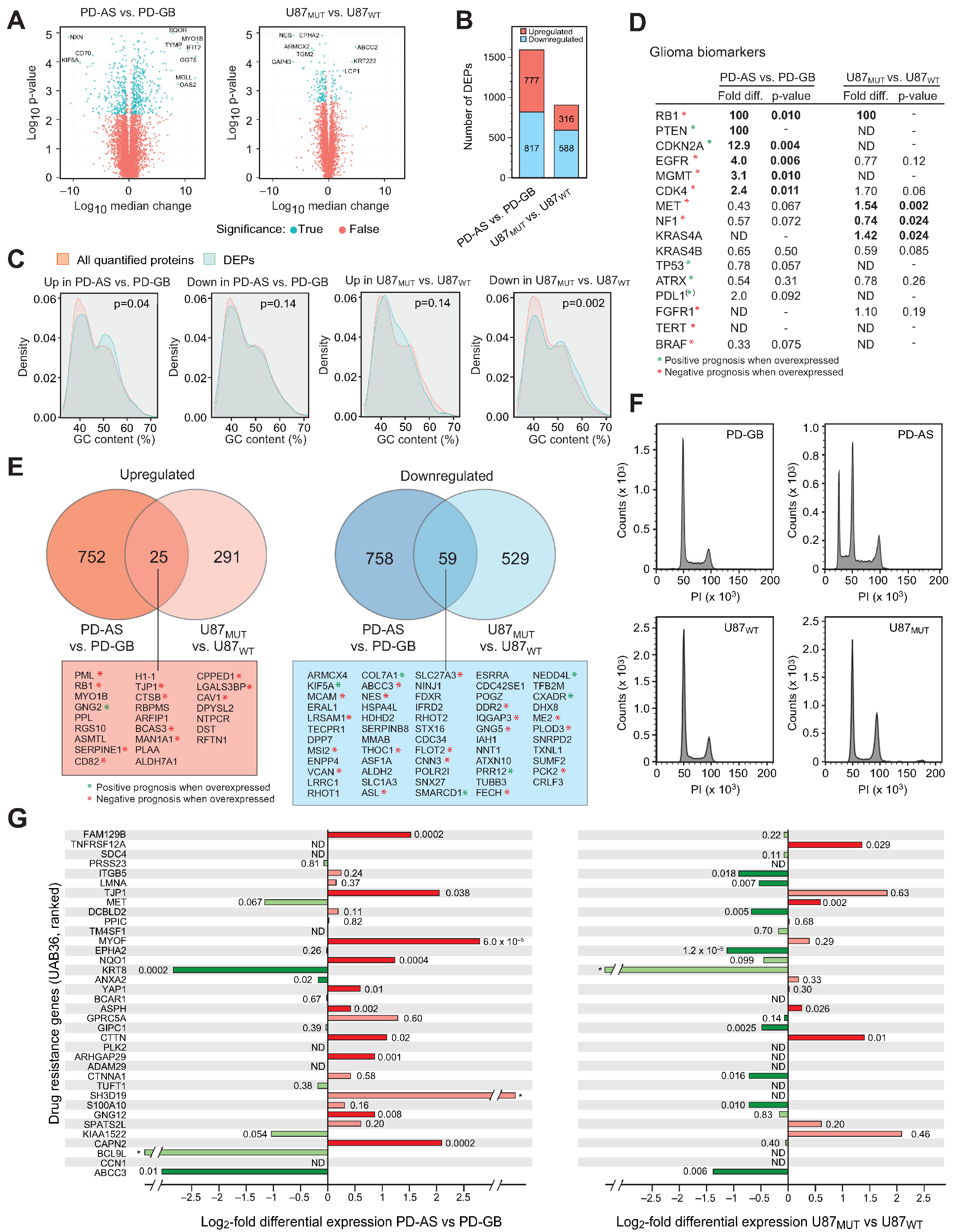
Figure 1. Differentially expressed proteins in the IDH1 mutant PD-AS and U87MUT cells compared to their IDH1 wild-type counterparts. (A) Volcano plots of DEPs in PD-AS vs. PD-GB (left panel) and U87MUT vs. U87WT (right panel). (B) The number of up- and downregulated DEPs in PD-AS vs. PD-GB and in U87MUT vs. U87WT. (C) ShinyGO analysis of the GC-content of the genes encoding DEPs compared to the genes encoding all quantified proteins in the dataset. (D) Expression of proteins commonly used as prognostic and/or therapeutic biomarkers in glioma. ND: Not detected. Green stars represent positive prognosis when overexpressed, red stars represent negative prognosis when overexpressed. (E) Venn diagrams illustrating common up- and downregulated DEPs in the two model systems. Common DEPs are listed in the bottom boxes, ranging from most upregulated (red box) to most downregulated (blue box). Green stars represent positive prognosis when overexpressed, red stars represent negative prognosis when overexpressed. (F) Cell cycle analysis of freely cycling PD-GB and PD-AS cells (upper panels) and U87WT and U87MUT cells (bottom panels). (G) The majority of the DEPs encoded by the drug resistance genes in the UAB36 panel were upregulated in the IDH1 mutant PD-AS cells compared to IDH wild-type PD-GB, whereas most of the UAB36 genes were downregulated in U87MUT vs. U87WT. *; detected only in one of the cell pairs, thus reported as non-significant.
Ijms 26 09075 g001
Figure 2. Analysis of differentially regulated biological processes in PD-AS vs. PD-GB and in U87MUT vs. U87WT. (A) Most affected GO biological processes reported by the PANTHER over-representation test. (B) Most affected biological processes reported by IPA. (C) Most significantly, common upregulated (left panel) and downregulated (right panel) processes in the cell models.
Ijms 26 09075 g002
Figure 3. Opposite metabolic shifts and drug resistance profiles observed in PD-AS vs. PD-GB compared to U87MUT vs. U87WT. (A) Whereas the IDH1-mutant PD-AS displayed a marked upregulation of glycolytic enzymes and downregulation of ETC proteins compared to PD-GB, the U87MUT cells had downregulated or unchanged glycolytic enzymes and overall upregulated ETC proteins compared to U87WT. (B) The Complex I inhibitor metformin was significantly more cytotoxic to U87WT than to U87MUT. Statistical significance was calculated using one-way ANOVA followed by Turkey post hoc test ( * p = 0.01–0.05; *** p = 0.0001–0.001).
Ijms 26 09075 g003
Figure 4. Most DEPs associated with DNA repair and genome maintenance are downregulated in IDH1 mutant glioma cells (PD-AS and U87MUT) compared to IDH1 wild-type glioma cells (PD-GB and U87WT). Graphic representation of DNA repair proteins significantly differentially expressed (> + / − 1.5-fold, p  <  0.05) in at least one of the IDH1 mutants (PD-AS or U87MUT) is shown in the left panel. Asterisks denote which of the two IDH1 mutants has the highest baseline expression of each protein. The common ten DEPs found in both cell models are shown in the right panel.
Ijms 26 09075 g004
Figure 5. The IDH1-mutant glioma cells harbor distinct expression of proteins regulating histone lysine methylation. Histone lysine methyltransferases (KMTs) detected in our datasets are shown in the blue box (ranked after MS intensities in PD-AS) and pointing to their respective lysine targets in Histone H3. Likewise, histone lysine demethylases (KDMs) are shown in the pink box, below their lysine targets. A filled blue circle indicates that the KDM is 2OG-dependent and thus should be inhibited in the IDH1-mutant cells. The color bar at the top right indicates fold change in expression in the IDH-mutant cells vs. their IDH wild-type counterparts.
Ijms 26 09075 g005
Disclaimer/Publisher’s Note: The statements, opinions and data contained in all publications are solely those of the individual author(s) and contributor(s) and not of MDPI and/or the editor(s). MDPI and/or the editor(s) disclaim responsibility for any injury to people or property resulting from any ideas, methods, instructions or products referred to in the content.