Article

Ecotoxicological Impacts of Perfluorooctane Sulfonate on the Freshwater Snail Lanistes carinatus: Oxidative Stress, Neurotoxicity, and Histopathological Alterations

by
Mohamed Hamed
1,*,
Mohammed Abdel-Wahab
2,
Rashad E. M. Said
2 and
Alaa El-Din H. Sayed
3,4
1
Department of Comparative Biomedical Sciences, School of Veterinary Medicine, Louisiana State University, Skip Bertman Drive, Baton Rouge, LA 70803, USA
2
Department of Zoology, Faculty of Science, Al-Azhar University (Assiut Branch), Assiut 71524, Egypt
3
Department of Zoology, Faculty of Science, Assiut University, Assiut 71516, Egypt
4
Molecular Biology Research & Studies Institute, Assiut University, Assiut 71516, Egypt
*
Author to whom correspondence should be addressed.
Int. J. Mol. Sci. 2025, 26(18), 8898; https://doi.org/10.3390/ijms26188898
Submission received: 19 August 2025 / Revised: 5 September 2025 / Accepted: 10 September 2025 / Published: 12 September 2025
(This article belongs to the Special Issue Toxicological Impacts of Emerging Contaminants on Aquatic Organisms)

Abstract

Perfluorooctane sulfonate (PFOS), which is known for its environmental persistence and bioaccumulation, poses substantial impacts to aquatic ecosystems. This study assesses the toxic effects of PFOS in the freshwater snail Lanistes carinatus using biomarkers for antioxidant activity, neurotoxicity, and tissue damage. Snails exposed to PFOS (1, 3, 10 mg/L for 14 days) displayed lipid peroxidation (LPO) levels that increased by 16.3–67.5%, and malondialdehyde (MDA) levels that rose by 10.0–58.4%, indicating oxidative damage. Enzyme activities for glutathione S-transferase (GST), glutathione peroxidase (GPx), and catalase (CAT) increased, ranging from 10.0 to 58.3%, 10.0 to 58.4%, and 10.0 to 58.4%, respectively, whereas levels of reduced glutathione (GSH) dropped by 15.0–41.5% and Superoxide dismutase (SOD) decreased by 15.0–41.4%. The activity of acetylcholinesterase (AchE) was reduced by a range of 15.0–40.0%, suggesting neurotoxic effects. Histopathological changes in the digestive gland were also noted. Further research on the effects of PFOS on mollusks is required, and investigation into sex-specific toxicity is needed. This shed light on L. carinatus as a sentinel species, providing helpful information for the monitoring and regulation of PFOS in aquatic environments.

1. Introduction

Per- and polyfluoroalkyl substances (PFASs) are synthetic molecules used in consumer and industrial environments around the world. The variety of PFAS found in the environment is on the increase. About 9000 chemical substances listed by the USEPA in 2021 increased to over 12,000 by 2023 [1]. The persistence of PFAS in the environment and their ability to accumulate in living organisms make them widespread global pollutants linked to ecological and human health hazards [2,3,4].
A vast range of products and human needs including semiconductors, surfactants, cosmetics, food packing, etc., extensively use PFAS [5,6,7]. The widespread presence of these compounds is due to the unique carbon–fluorine (C-F) bonds they contain, which impart exceptional resistance to oil, heat, and water [8,9]. The same properties drive the persistence and bioaccumulation potential of PFAS in different biotopes and organisms including humans [10,11,12]. As a result, its widespread use and environmental stability have led to chronic adverse impacts on ecosystems and public health [9,13,14,15].
Regulatory attention continues to be focused primarily on specific PFAS, such as perfluorooctanoic acid (PFOA) and perfluorooctane sulfonate (PFOS), even though there are thousands of documented compounds. Globally, PFOA/PFOS are extensively studied because they persist, can travel over long distances, are found in different environmental samples, living organisms, and humans, and have been shown to disrupt immune, metabolic, and endocrine systems [16,17,18,19]. Notably, PFOS contaminates surface waters [20,21,22], wastewater [23,24], and humans at a concentration of mg/L in affected communities [25,26]. The regulatory–environmental disconnect has driven an increase in ecological research [27,28,29,30].
Human exposure to PFOS was associated with hepatic damage, thyroid problems, reproductive inhibition, developmental toxicity, and elevated cholesterol [31,32,33,34], and long-term exposure has been linked to a potential increased risk of cancer [35]. PFOS causes harm to wildlife’s organs, reproductive systems, and metabolism [36,37]. Invertebrates, which are vital to ecosystem health because they play a crucial role in cycling nutrients and food chains, are experiencing population declines caused by pollution resulting from human activities [38,39]. The vulnerability of PFOS highlights the need to study its ecotoxicity, as invertebrate biomass loss directly threatens the multifunctionality of ecosystems [40].
PFOS tends to accumulate in aquatic organisms through respiration and consumption, often present at low concentrations (0.2–100 ng/L) across various environmental media [41]. Oxidative stress is driven by this amplification, which disrupts antioxidant defenses and detoxification pathways [42,43], primarily through impaired nuclear receptor signaling, fatty acid β-oxidation, and mitochondrial permeability [44]. Mechanistically, PFOS disrupts the normal functioning of detoxification, lipid metabolism, and xenobiotic processing pathways [45,46]. In Daphnia carinata, PFOS causes acute effects, with a 48 h LC50 of 8.8 mg/L, which is more toxic than PFOA (78.2 mg/L) [47].
Snails and other sentinel organisms can accumulate persistent pollutants and metals, making them useful for monitoring the effects of contaminants on development [48,49]. Their heightened sensitivity to pollutants helps in early detection of environmental stress [50]. L. carinatus, a widely distributed freshwater snail in Egypt, mostly inhabiting still water environments, serves as an example for monitoring aquatic pollution [51,52]. The selection of this animal was based on local abundance, ease of collection, and laboratory hardiness. It is essential to comprehend invertebrate toxicity, as its negative effects have a ripple effect throughout food chains. Through examining their responses to contaminants, more comprehensive ecological results can be predicted. Oxidative stress defenses in cells rely on the coordinated action of crucial biomarkers, such as lipid peroxidation (LPO) and malondialdehyde (MDA), as well as enzymes like glutathione transferase (GST), catalase (CAT), glutathione peroxidase (GPx), reduced glutathione (GSH), and superoxide dismutase (SOD).
These biomarkers play a crucial role in reducing oxidative impact and are well-employed indicators of contamination-induced stress [53,54]. Exposure to PFOS has a substantial impact on antioxidant activity [55], which can be monitored through fluctuations in SOD, CAT, and GPx levels. Prolonged exposure to PFOS in earthworms leads to increased levels of malondialdehyde (MDA), a byproduct of lipid peroxidation that indicates the accumulation of ROS [56]. A typical ROS surge in this context results in a biphasic antioxidant response, characterized by elevated records of SOD, POD, CAT, and GPx, which then decrease. In line with this pattern, PFAS causes oxidative stress across aquatic species [57,58], as seen in increased SOD/CAT/GR activities in Oreochromis niloticus hepatocytes [59] and higher MDA in Pimephales promelas [60].
A large body of research has confirmed the toxicity of PFAS in aquatic organisms, particularly fish, resulting in cellular and molecular disruptions [58,61]. Invertebrate responses to pollutants have a major role in determining the viability of aquatic ecosystems. Despite this, there is a lack of PFOS toxicity data for key invertebrates, particularly snails such as L. carinatus, making an urgent evaluation necessary. Thus, the main goal of the current research was to assess PFOS (1, 3, and 10 mg/L) toxicity in the freshwater snail L. carinatus, following 14 days of exposure. Multiple oxidative stress markers (LPO, MDA, GST, CAT, GPx, GSH, and SOD), acetylcholinesterase activity as a neurotoxicity indicator, along with histopathological examinations in the digestive gland, were all assessed.

2. Results

2.1. Antioxidant Defense Enzymes

Exposure to PFOS caused significant alterations in antioxidant defense enzymes in snails (Figure 1) compared with non-exposed groups. GST activity increased by 10.0% at 1 mg/L, 31.9% at 3 mg/L, and 58.3% at 10 mg/L, compared with the control group (p < 0.0001). GPx levels increased by 10.0% at 1 mg/L, 31.9% at 3 mg/L, and 58.4% at 10 mg/L, when compared with the control (p < 0.0001). CAT activity also increased significantly, with a 10.0% increase at 1 mg/L, 32.0% at 3 mg/L, and 58.4% at 10 mg/L, relative to the control (p < 0.0001). GSH levels declined significantly compared with the control (p < 0.0001), showing a 15.0% decrease at 1 mg/L, 29.2% at 3 mg/L, and 41.5% at 10 mg/L, reflecting antioxidant depletion. SOD activity also decreased, with reductions of 15.0% at 1 mg/L, 29.4% at 3 mg/L, and 41.4% at 10 mg/L, in comparison with the control (p < 0.0001).

2.2. Oxidative Stress Markers

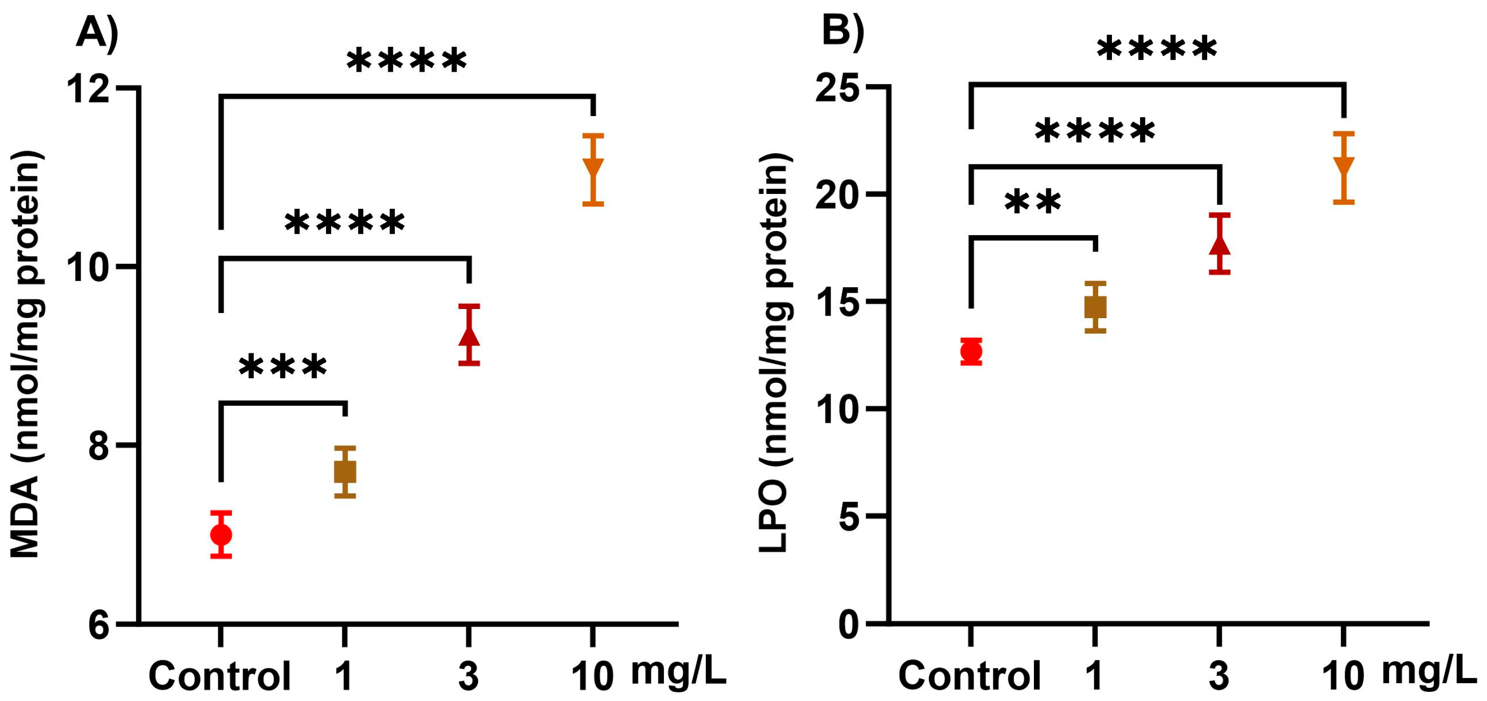
Exposure to PFOS induced significant, dose-dependent (1, 3, and 10 mg/L) alterations in oxidative stress markers in snails (Figure 2). LOP increased progressively with PFOS exposure, showing a 16.3% increase at 1 mg/L, 39.6% at 3 mg/L, and 67.5% at 10 mg/L, compared with the control (p < 0.0001). MDA, a marker of lipid peroxidation, was elevated by 10.0% at 1 mg/L, 32.0% at 3 mg/L, and 58.4% at 10 mg/L, relative to the control group (p < 0.0001).

2.3. Neurotoxicity Markers

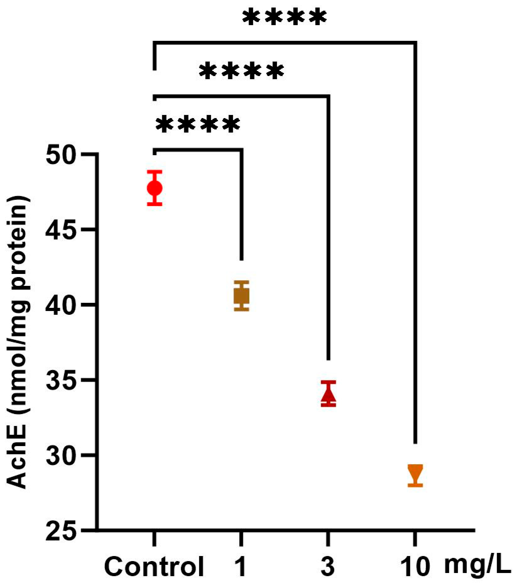
Exposure to increasing concentrations of PFOS (1, 3, and 10 mg/L) induced significant, dose-dependent alterations in the neurotoxicity marker in snails (Figure 3). AchE activity significantly decreased relative to the control by 15.0% at 1 mg/L, 28.6% at 3 mg/L, and 40.0% at 10 mg/L (p < 0.0001), indicating potential neurotoxic effects.

2.4. Histology of the Digestive Gland

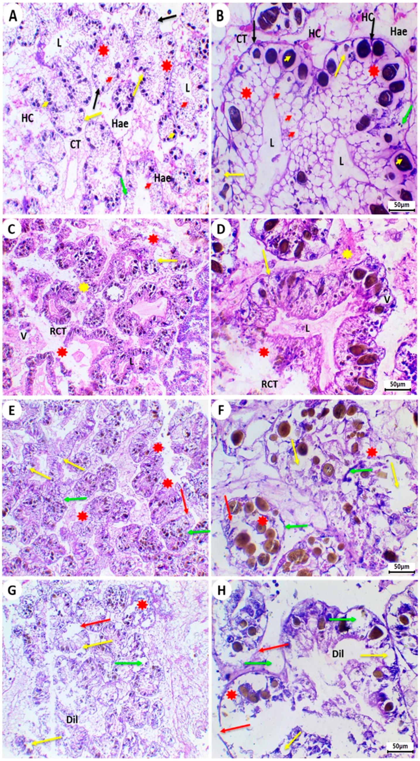
Typically, the control group’s L. carinatus digestive glands are composed of tubules, each of which has a central lumen surrounded by a single layer of columnar epithelial cells that have undergone differentiation into digestive and secretory cells in addition to calcium cells. Digestive and secretory cells are located on the basement membrane and encircle a lumen in the middle of the tubule. The intertubular connective tissue binds the tubules together along with haemocoele fluid. There were many large-sized corpuscles in the basal third of the epithelial cells in the acidophilic cytoplasm of pyramidal cells (Figure 4A,B).
In the L. carinatus exposed to 1 mg/L PFOS, a range of deformities have been spotted. These damages include fused tubules, vacuolation, ruptured connective tissues, necrotic basement membranes, and degenerated digestive tubules with a clogged lumen (Figure 4C,D).
Following exposure to 3 mg/L PFOS, increased histopathological alterations have been observed in the digestive glands. These changes include ruptured villi, disconnected basement membranes of the digestive tubules, high degeneration and shrinking of some tubules, and cells that lost their normal morphologies and became vacuolated (Figure 4E,F).
Higher concentrations of PFOS (10 mg/L) caused the digestive glands of the Lanistes carinatus to exhibit severe degeneration in the digestive tubules, including the digestive cells, execratory cells, calcium cells, and villi, as well as the rupture of the digestive tubules’ basement membranes, dilatation of the tubular lumen, various vacuolations, and swelling cells (Figure 4G,H).

3. Discussion

During this study, we assessed the outcomes of PFOS exposure in the freshwater snail L. carinatus using a multi-marker approach. Snails were tested individually following a two-week exposure to 1.3 and 10 mg/L PFOS for biochemical markers, oxidative stress, and tissue damage. L. carinatus snails have become prominent sentinel models for ecotoxicological research during the last decades. Snails also play a significant role in tracking pollutants within aquatic environments [51]. Unfortunately, very little information has been published on the impacts of PFOS for these aquatic invertebrates in Egypt. When exposed to polluted water, contaminated food, or sediment, snails can absorb toxic substances through their skin. Their ability makes them useful as bioindicators of environmental pollution, as they can accumulate toxins in their tissues [62]. Accordingly, the present assessment focused on short-term exposure by examining the impacts of PFOS on antioxidant defenses such as (LPO, MDA, GST, CAT, GPx, GSH, SOD, and AchE). These indicators were selected for their ability to compensate for oxidative impacts of diverse environmental pollutants in previous investigations. However, oxidative stress can activate these defenses and thereby alter compensating mechanisms [63]. Several biological markers are widely employed in toxicity studies in freshwater snails [64].
Biomarkers are commonly employed in invertebrate toxicology to assess contaminant effects [65]. Specifically, measuring the impairment of cellular defenses offers valuable insights into how oxidative stress, lipid peroxidation (LPO), and apoptosis contribute to toxicity [66]. Malondialdehyde (MDA), a key product of LPO, serves as a widely recognized marker for oxidative damage; elevated MDA levels indicate the presence of reactive oxygen species (ROS) [67]. Antioxidants such as CAT and SOD counteract ROS to preserve cellular redox balance. Consequently, alterations in ROS levels, MDA concentrations, and inhabited antioxidant enzymes (e.g., CAT, SOD) are sensitive signals for inferring the harmful impacts of pollutant exposure [48,68].
PFASs are well-established inducers of oxidative stress [58]. Numerous studies confirm that PFAS exposure triggers oxidative stress in aquatic organisms, disrupting antioxidant enzyme activities (including CAT and SOD) and detoxification pathways [57]. The selection of 1 µg/L as the lowest test concentration was informed by documented environmental PFOS levels in surface waters (0.04–2709 ng/L), which vary based on factors like industrial proximity and wastewater discharge [69,70]. Higher concentrations were included to elucidate dose-dependent effects. Recent research, particularly on marine mussels, has demonstrated the impact of various PFASs on these oxidative stress biomarkers [71]. While snails naturally regulate reactive oxygen species (ROS) at low, stable concentrations under physiological conditions, chemical contaminants disrupt this balance, leading to the overproduction ROS, such as superoxide anions (O2), hydroxyl radicals (OH), and H2O2. Inefficient removal of these ROSs overwhelms cellular defenses, resulting in macromolecular damage and lipid peroxidation [72]. We found that PFOS, in a dose-dependent manner, increases MDA content in L. carinatus: 10.0% at 1 mg/L, 32.0% at 3 mg/L, and 58.4% at 10 mg/L. This elevation suggests PFOS triggers oxidative stress in this species. Similarly, freshwater ray-finned fish Pimephales promelas exposed to PFOS showed substantial elevations in MDA content [60]. Increased MDA levels in snails exposed to contaminants strongly suggest that animals were harmed by excessive ROS [73,74].
PFOS can pose oxidative impacts and various histopathological problems, including changes in animal behavior, depending on the species and exposure levels [75,76]. LPO levels reflect the severity of intracellular damage and are commonly used to assess oxidative stress. Snail MDA content was utilized to evaluate LPO [77]. LPO remarkably increased progressively by exposure to PFOS showing a 16.3% increase at 1 mg/L, 39.6% at 3 mg/L, and 67.5% at 10 mg/L.
Two powerful antioxidant enzymes, SOD and CAT, form part of the antioxidant system, which resists the oxidative stress triggered by exogenous agents. SOD is considered the primary defense mechanism against reactive oxygen species. Under the catalysis of SOD, the O2 is first disproportionated into H2O2, and subsequently converted to H2O and O2 by CAT [78,79]. Our results indicate that following a 14-day period of exposure, the activity of SOD in L. carinatus treated with PFOS declined substantially compared with the control samples. Decreased enzyme activity may suggest that the antioxidant capacity has been exceeded by the concentration of hydroperoxide products from lipid peroxidation, detectable in the LPO levels of these tissues. After seven days of receiving a mixture of perfluorinated compounds [80], SOD activity in the snail Perna viridis exhibited higher records at lower concentrations (0–100 µg/L), while dropping at the highest levels (100–10,000 µg/L). Also, in Unio ravoisieri, the mean values of SOD were higher in groups treated with higher PFOS levels (2–6 mg/L) but declined in those exposed to 10 mg/L [57] for one week. SOD activity in the amphipod (Gammarus insensitives) increased and then decreased during four days at PFOS doses of 1 mg/L, 1.6 mg/L, and 3.1 mg/L [81]. Previous experiments have shown that the crab Eriocheir sinensis subjected to 10 mg/L PFOS for 21 days had decreased SOD activity [82] and over the course of seven days in D. magna exposed to PFOS or PFNA doses ranging from 0.008 to 5 mg/L [83].
In L. carinatus, CAT activity rose dramatically after 14 days of PFOS exposure compared with controls. Similar increases were noted in the freshwater mussel Unio ravoisieri subjected to 2–10 mg/L PFOS [57], and in Mytilus galloprovincialis treated with 1, 10, and 100 µg/L PFOA [84]. Furthermore, P. viridis displayed elevated CAT levels after 7 days of exposure to perfluorinated compounds (≤100 µg/L) [80]. O. niloticus similarly showed a significant increase in CAT activity following a 24 h treatment with 1, 5, 15, 30 mg/L PFOS or PFOA [59]. However, studies found decreased CAT activity in medaka fish (Oryzias latipes) liver following a 7-day exposure to 10, 50, or 100 mg/L PFOA [85], and in D. magna water fleas exposed to 0.04 mg/L PFOS or PFNA [83]. The findings show that species-specific variability and sensitivity in the perfluorinated compound-induced response in CAT activity are likely due to variations in their antioxidant potency. For hydroperoxide detoxification, GPx is the most significant peroxidase [86], where it converts H2O2 and organic hydroperoxides into water and alcohol.
In the present study, GPx levels increased in L. carinatus snails by 10.0% at 1 mg/L, 31.9% at 3 mg/L, and 58.4% at 10 mg/L, after 14 days of PFOS exposure compared with controls. Similarly, exposure to PFOA induced GPx activity in bivalves. Mytilus edulis exhibited a significant GPx increase, primarily at 200 μg/L, after 7 days [87]. In the clam Ruditapes philippinarum, a drastic rise in both GPx and peroxidase activity occurred following a 21-day exposure to PFOA (0.2, 2, 20 μg/L) [88]. Consistent with our findings, zebrafish (Danio rerio) exhibited elevated GPx levels after 96 h of exposure to 0.4–1.6 mg/L PFAS [89]. Conversely, studies reported suppressed GPx activity in the planarian Dugesia japonica following a 14-day exposure to 0.5–20 mg/L PFOA [90], and in the clam Scrobicularia plana exposed to 1 mg/L PFOS [91]. Interactions between PFAS and co-pollutants like microplastics also impact mollusk antioxidant responses. For instance, O’Donovan, Mestre [91] reported that exposure to microplastic-adsorbed PFOS altered the antioxidant system in Scrobicularia plana, increasing GPx activity in gills after 7 days [91]. This elevation is a common component of the oxidative stress response triggered by PFOS. The most well-known function of GST is its ability to catalyze the conversion of reduced glutathione (GSH) to xenobiotic compounds for detoxification [92].
By altering the availability of glutathione, this improves antioxidant defenses by making dangerous substances less reactive and easier to remove [93]. In L. carinatus, PFOS exposure significantly increased GST activity after 14 days. Similarly, elevated GST activity was observed in various species exposed to PFAS, including M. galloprovincialis mussels (digestive gland, PFOS/PFOA) [84], Dugesia japonica planarians (PFOA) [90], and Melanotaenia fluviatilis rainbowfish gills and liver (PFOA) [94]. Notably, transgenerational exposure in D. magna offspring also induced significantly higher GST levels from the second to fourth generations, indicating increased oxidative stress and adverse effects [95,96]. Conversely, GST inhibition occurred in O. niloticus hepatocytes (PFOS/PFOA) [59]. This species-, tissue-, and generation-specific variability underscores the complex role of GST in aquatic organism detoxification and antioxidant defense. GST induction, particularly at high contaminant concentrations as seen in mussels [97], suggests an adaptive detoxification response aimed at mitigating accumulation through enhanced biotransformation and excretion.
It is evident from the current study that MDA levels and GSH content are inversely related. Since GSH functions as a reducing agent and free radical scavenger, it is one of the most crucial components defending against oxidative attacks by ROS, including lipid peroxidation [98]. Unlike the variable responses of GST, CAT, and GPx, GSH depletion showed a strong positive linear correlation with PFOS exposure concentration. This dose-dependent decline establishes GSH as an excellent biomarker for quantifying oxidative stress in PFOS-exposed snails.
In addition to affecting critical antioxidant systems, such as glutathione and catalase, PFAS exposure also disrupts essential neurochemical processes. An essential enzyme for biological nerve conduction between cholinergic synapses is AchE [99]. Exposure to PFAS raises AchE levels and overexcites nerve cells, which impacts AchE activity. Furthermore, it alters the expression of genes involved in the development of nerve cells, which leads to fewer nerve cells and less branching, ultimately impairing nerve cell functioning [96]. In similar study on the planarian Dugesia japonica, AchE activity was altered following exposure to PFOS [90]. This result may be explained by the significantly accumulating acetylcholine in the fish tissues described by Ahammad Sahib, Sailatha [100], which is caused by AchE inhibition during pesticide stress, which decreases with exposure duration. Consistently, the current data matches those found in the published literature. Liang, Zhou [28] resulted that long-term exposure to PFOS D. magna inhibited the amount of cholinesterase (ChE). AchE activity significantly increased in D. magna exposed to PFOS; however, it was found that the organism downregulated AchE during non-exposure and elevated it during exposure [95].
Histopathology serves as a promising tool in ecotoxicology for evaluating tissue-level toxicity. The digestive gland of mollusks is primarily responsible for xenobiotic detoxification, metabolic regulation, immune defense, and homeostasis [101], making it a key focus in toxicology studies [102,103]. Our analysis showed substantial damage in snail digestive glands caused by PFOS. The control specimens displayed typical histological architecture without any pathological changes (Figure 4A,B). In contrast, PFOS exposure caused concentration-dependent damage including lesions to the digestive tubule components, such as digestive, excretory, and calcium cells, as well as villi, along with basement membrane ruptures, tubular lumen dilations, and cellular vacuolation and swelling (Figure 4C–H). The degeneration of these tubules is consistent with known reactions to foreign substances and may compromise digestive function [104,105]. Previous research has shown degeneration of digestive tubules after exposure to toxic substances [106]. These pathologies also correlate with molluscan inflammatory responses, which are validated biomarkers of toxicant exposure [107,108].

4. Materials and Methods

4.1. Chemicals

Perfluorooctanesulfonic acid potassium salt (PFOS; CAS No. 2795-39-3; molecular weight 538.22 g/mol; purity ≥ 98%; Product No. 77282) was sourced from Sigma-Aldrich (St. Louis, MO, USA). To make a stock solution, PFOS was dissolved in dimethyl sulfoxide (DMSO; final concentration in exposure water 0.01%) and stored at 4 °C in the dark to prevent degradation. Working solutions of the desired concentrations were freshly prepared daily by diluting the stock with dechlorinated tap water. All glassware and containers used in the preparation were rinsed thoroughly with methanol and ultrapure water to minimize contamination.

4.2. Experimental Animals

L. carinatus snails were collected from the Nile River at Assiut, Egypt, then transported to the laboratory in Nile water. To adapt, snails were kept in tanks with filtered, dechlorinated tap water for two weeks after collecting. In a laboratory environment, the snails were housed in water with a pH of 7.2 and a 12 h light–dark period. Following acclimation to the laboratory conditions, L. carinatus snails of similar size (7 ± 0.5 gm) were divided into glass tanks of four groups (three replicates for each/15 individual for each aquarium): one control group and three (1, 3 and 10 mg/L) PFOS concentration groups. Snails were fed lettuce during the 14-day experiment, and the aquarium’s water was replaced with a corresponding PFOS-continuing volume.
The selected concentrations of PFOS (1, 3, and 10 mg/L) were based on documented levels in contaminated aquatic ecosystems and previous ecotoxicological studies that successfully demonstrated dose-dependent oxidative, biochemical, and histopathological alterations in aquatic invertebrates and fish models [61,109,110]. Although the highest concentration (10 mg/L) exceeds typical environmental levels, such concentrations are commonly employed experimentally to simulate acute exposure scenarios, define worst-case thresholds, and uncover mechanistic pathways of toxicity. In highly contaminated freshwater systems near industrial and wastewater discharges, PFOS concentrations ranging from the µg/L scale to mg/L levels have been reported [61,109,110,111], with potential for bioaccumulation and trophic transfer leading to elevated internal burdens. Thus, the lowest concentration tested (1 mg/L) reflects a realistic sublethal exposure level in polluted waters, while higher doses enable the assessment of concentration-dependent responses in Lanistes carinatus. This concentration range therefore provides a balanced evaluation of both environmentally relevant and mechanistically informative exposure levels.
After 14 days, six snails were selected randomly from each treatment including control. For histopathological investigations, snails were separated from shells and dissected. Hemolymph was extracted for biochemical analysis using a fine needle and sterile 10 mL. Following collection, samples were centrifuged at 4000 rpm for 10 min to separate the cell-free supernatant. All hemolymph cell-free supernatant was stored in sterile Eppendorf tubes at −20 °C for later use. Six biological replicates per group were analyzed.

4.3. Antioxidant Enzymes

CAT, GPX, SOD, and GST were assessed using standard methods. CAT activity was determined following the protocol by Aebi [112] in accordance with the manufacturer’s instructions [112]. GPX level was recorded using a commercial assay kit according to the provided protocol. SOD was measured using the clinical assay adopted from Sun, Oberley [113]. GST activity was quantified using a microplate method containing 190 μL of sodium phosphate buffer, 200 mM of reduced glutathione, and 1-chloro-2,4-dinitrobenzene, as outlined by Valenzuela-Jiménez, Durruty-Lagunes [114].

4.4. Oxidative Stress

Lipid peroxidation (LPO) levels were determined using the PeroxiDetect© kit (Sigma-Aldrich, St. Louis, MO, USA) following the method of Fox, Blow [115]. Malondialdehyde (MDA) content, a marker of LPO, was measured at a wavelength of 535 nm using a UV–VIS spectrophotometer based on the thiobarbituric acid reaction described by Ohkawa, Ohishi [116] and modified by Hamed, Soliman [117]

4.5. Neurotoxicity Biomarker

Ellman method was followed to determine the activity of AchE, using the Fish AchE ELISA Kit (cat. no. MBS035436, BioSource, San Diego, CA, USA) [118,119].

4.6. Histopathology

From each group, six samples of L. carinatus digestive glands were taken for histological analysis, and they were fixed in Davidson’s fixative for an entire day [120], and then transferred to 70% ethanol. Tissues were then slowly treated with ethyl alcohol before being imbedded in paraffin wax. H&E staining was used after paraffin blocks were sliced into 5 μm sections [121]. The slides were examined under a microscope after staining.

4.7. Statistical Analysis

All output data are presented as mean ± SD, after statistical processing by the GraphPad Prism software version 9. Following ANOVA, Fisher’s least significant difference (LSD) post hoc test was applied to test and compare the differences between groups at a p-value of 0.05. Significant differences between control and treated groups are marked by asterisks, where * p < 0.05, ** p < 0.01, *** p < 0.001, and **** p < 0.0001.

5. Conclusions

As far as we know, this work presents a unique comprehensive assessment of PFOS-related ecological risks in L. carinatus. Our integrated analysis of neurotoxicity, lipid peroxidation, and antioxidant responses revealed that PFOS exposure in L. carinatus induces oxidative stress, characterized by higher MDA levels, depleted GSH content, dysregulated antioxidant enzymes (CAT, GPx, GST), and hindered AchE activity. Histopathological alterations in the digestive gland were also observed. These findings provide critical insights into environmental policy, supporting enhanced PFOS regulation and monitoring in aquatic ecosystems especially in high-aquatic food-consumption regions. Aquatic PFOS levels must be strictly limited to lessen ecological and human health issues. Furthermore, preventive actions, remediation plans, and health protection programs are required to accomplish thorough pollution management. This study enhances our comprehension of the biochemical impacts of PFOS on aquatic species, thereby supporting a more comprehensive approach to tackling the difficulties presented by long-lasting environmental pollutants. Future studies should investigate underlying mechanisms across additional perfluorinated compounds and species to inform comprehensive environmental protection strategies.

Author Contributions

M.H.: Conceptualization, investigation, experimental design, methodology, data collection, formal analysis, validation, writing—original draft, and writing–review and editing. M.A.-W.: Investigation, formal analysis, validation, data collection, writing—original draft, and writing—review and editing. R.E.M.S.: Investigation, formal analysis, data collection, validation, writing—original draft, and writing–review and editing. A.E.-D.H.S.: Conceptualization, investigation, project administration, formal analysis, validation, supervision, and writing—review and editing. All authors have read and agreed to the published version of the manuscript.

Funding

This research received no external funding.

Institutional Review Board Statement

All experimental procedures were approved by the Faculty of Science, Al-Azhar University (Assiut Branch), under the ethical approval code AZHAR25/2025, approval date: 2 March 2025.

Informed Consent Statement

Not applicable.

Data Availability Statement

Data will be available upon request from the corresponding author.

Conflicts of Interest

The authors declare no conflicts of interest.

Abbreviations

PFOSPerfluorooctane sulfonate
PFASPer- and polyfluoroalkyl substances
PFOAPerfluorooctanoic acid
C-FCarbon–fluorine (bond)
LC50Lethal Concentration for 50% of test organisms
LPOLipid peroxidation
MDAMalondialdehyde
GSTGlutathione S-transferase
GPxGlutathione peroxidase
CATCatalase
GSHReduced glutathione
SODSuperoxide dismutase
AchE (or AChE)Acetylcholinesterase
ROSReactive oxygen species
PODPeroxidase
GRGlutathione reductase
ChECholinesterase
DMSODimethyl sulfoxide
H&EHematoxylin and eosin (stain)
SDStandard deviation
ANOVAAnalysis of variance
LSDLeast significant difference (test)
L. carinatusLanistes carinatus (snail species, used in short form)
µg/L, mg/L, ng/LMicrograms/milligrams/nanograms per liter

References

  1. Schymanski, E.L.; Zhang, J.; Thiessen, P.A.; Chirsir, P.; Kondic, T.; Bolton, E.E. Per-and polyfluoroalkyl substances (PFAS) in PubChem: 7 million and growing. Environ. Sci. Technol. 2023, 57, 16918–16928. [Google Scholar] [CrossRef]
  2. Giesy, J.P.; Kannan, K. Global distribution of perfluorooctane sulfonate in wildlife. Environ. Sci. Technol. 2001, 35, 1339–1342. [Google Scholar] [CrossRef]
  3. Silva, A.V.; Ringblom, J.; Lindh, C.; Scott, K.; Jakobsson, K.; Öberg, M. A probabilistic approach to evaluate the risk of decreased total triiodothyronine hormone levels following chronic exposure to PFOS and PFHxS via contaminated drinking water. Environ. Health Perspect. 2020, 128, 076001, Erratum in Environ Health Perspect. 2020, 128, 89001. [Google Scholar] [CrossRef] [PubMed] [PubMed Central]
  4. Zhao, Z.; Li, J.; Zhang, X.; Wang, L.; Wang, J.; Lin, T. Perfluoroalkyl and polyfluoroalkyl substances (PFASs) in groundwater: Current understandings and challenges to overcome. Environ. Sci. Pollut. Res. 2022, 29, 49513–49533. [Google Scholar] [CrossRef]
  5. Oliaei, F.; Kriens, D.; Weber, R.; Watson, A. PFOS and PFC releases and associated pollution from a PFC production plant in Minnesota (USA). Environ. Sci. Pollut. Res. 2013, 20, 1977–1992. [Google Scholar] [CrossRef]
  6. Sunderland, E.M.; Hu, X.C.; Dassuncao, C.; Tokranov, A.K.; Wagner, C.C.; Allen, J.G. A review of the pathways of human exposure to poly-and perfluoroalkyl substances (PFASs) and present understanding of health effects. J. Expo. Sci. Environ. Epidemiol. 2019, 29, 131–147. [Google Scholar] [CrossRef]
  7. Aminot, Y.; Sayfritz, S.J.; Thomas, K.V.; Godinho, L.; Botteon, E.; Ferrari, F.; Boti, V.; Albanis, T.; Köck-Schulmeyer, M.; Diaz-Cruz, M.S. Environmental risks associated with contaminants of legacy and emerging concern at European aquaculture areas. Environ. Pollut. 2019, 252, 1301–1310. [Google Scholar] [CrossRef] [PubMed]
  8. Wang, Z.; DeWitt, J.C.; Higgins, C.P.; Cousins, I.T. A never-ending story of per-and polyfluoroalkyl substances (PFASs)? Environ. Sci. Technol. 2017, 51, 2508–2518, Correction in Environ. Sci. Technol. 2018, 52, 3325. [Google Scholar] [CrossRef] [PubMed]
  9. Fenton, S.E.; Ducatman, A.; Boobis, A.; DeWitt, J.C.; Lau, C.; Ng, C.; Smith, J.S.; Roberts, S.M. Per-and polyfluoroalkyl substance toxicity and human health review: Current state of knowledge and strategies for informing future research. Environ. Toxicol. Chem. 2021, 40, 606–630. [Google Scholar] [CrossRef] [PubMed]
  10. Koponen, J.; Airaksinen, R.; Hallikainen, A.; Vuorinen, P.J.; Mannio, J.; Kiviranta, H. Perfluoroalkyl acids in various edible Baltic, freshwater, and farmed fish in Finland. Chemosphere 2015, 129, 186–191. [Google Scholar] [CrossRef]
  11. Kodavanti, P.R.S.; Loganathan, B.G. Organohalogen pollutants and human health. In The International Encyclopedia of Public Health; Quah, S.R., Cockerham, W.C., Eds.; Elsevier: Amsterdam, The Netherlands, 2017; Volume 5, pp. 359–366. [Google Scholar]
  12. Rodil Rodríguez, M.d.R.; Villaverde de Sáa, M.E.; Cobas, J.; Quintana Álvarez, J.B.; Cela Torrijos, R.; Carro, N. Legacy and emerging pollutants in marine bivalves from the Galician coast (NW Spain). Environ. Int. 2019, 129, 364–375. [Google Scholar] [CrossRef] [PubMed]
  13. Falk, S.; Stahl, T.; Fliedner, A.; Rüdel, H.; Tarricone, K.; Brunn, H.; Koschorreck, J. Levels, accumulation patterns and retrospective trends of perfluoroalkyl acids (PFAAs) in terrestrial ecosystems over the last three decades. Environ. Pollut. 2019, 246, 921–931. [Google Scholar] [CrossRef] [PubMed]
  14. Gao, K.; Miao, X.; Fu, J.; Chen, Y.; Li, H.; Pan, W.; Fu, J.; Zhang, Q.; Zhang, A.; Jiang, G. Occurrence and trophic transfer of per-and polyfluoroalkyl substances in an Antarctic ecosystem. Environ. Pollut. 2020, 257, 113383. [Google Scholar] [CrossRef]
  15. Burkhard, L.P. Evaluation of published bioconcentration factor (BCF) and bioaccumulation factor (BAF) data for per-and polyfluoroalkyl substances across aquatic species. Environ. Toxicol. Chem. 2021, 40, 1530–1543, Correction in Environ. Toxicol. Chem. 2021, 40, 2935–2940. [Google Scholar] [CrossRef]
  16. Kurwadkar, S.; Dane, J.; Kanel, S.R.; Nadagouda, M.N.; Cawdrey, R.W.; Ambade, B.; Struckhoff, G.C.; Wilkin, R. Per-and polyfluoroalkyl substances in water and wastewater: A critical review of their global occurrence and distribution. Sci. Total Environ. 2022, 809, 151003. [Google Scholar] [CrossRef]
  17. Tansel, B. Geographical characteristics that promote persistence and accumulation of PFAS in coastal waters and open seas: Current and emerging hot spots. Environ. Chall. 2024, 14, 100861. [Google Scholar] [CrossRef]
  18. Hamed, M.; Vats, A.; Lim, I.E.; Sapkota, B.; Abdelmoneim, A. Effects of developmental exposure to individual and combined PFAS on development and behavioral stress responses in larval zebrafish. Environ. Pollut. 2024, 349, 123912. [Google Scholar] [CrossRef]
  19. Soares, L.O.S.; de Araujo, G.F.; Gomes, T.B.; Júnior, S.F.S.; Cuprys, A.K.; Soares, R.M.; Saggioro, E.M. Antioxidant system alterations and oxidative stress caused by polyfluoroalkyl substances (PFAS) in exposed biota: A review. Sci. Total Environ. 2025, 977, 179395. [Google Scholar] [CrossRef]
  20. Zhang, J.; Xue, X.; Huang, C.; Chen, M.; Zhang, L.; Ren, M.; Hu, G.; Liu, S. Survey of perfluorooctanoic acid and perfluorooctane sulfonate in surface water from Tongsha Reservoir of Pearl River Delta, South China. Environ. Chem. 2017, 36, 2600–2608. [Google Scholar]
  21. Ololade, I.A.; Oladoja, N.A.; Ololade, O.O.; Oloye, F.F.; Adeola, A.O.; Alabi, A.B.; Ademila, O.; Adanigbo, P.; Owolabi, M.B. Geographical distribution of perfluorooctanesulfonate and perfluorooctanoate in selected rivers from Nigeria. J. Environ. Chem. Eng. 2018, 6, 4061–4069. [Google Scholar] [CrossRef]
  22. Picard, J.-C.; Munoz, G.; Duy, S.V.; Sauvé, S. Longitudinal and vertical variations of waterborne emerging contaminants in the St. Lawrence Estuary and Gulf during winter conditions. Sci. Total Environ. 2021, 777, 146073. [Google Scholar] [CrossRef]
  23. Loos, R.; Carvalho, R.; António, D.C.; Comero, S.; Locoro, G.; Tavazzi, S.; Paracchini, B.; Ghiani, M.; Lettieri, T.; Blaha, L. EU-wide monitoring survey on emerging polar organic contaminants in wastewater treatment plant effluents. Water Res. 2013, 47, 6475–6487. [Google Scholar] [CrossRef]
  24. Arvaniti, O.S.; Stasinakis, A.S. Review on the occurrence, fate and removal of perfluorinated compounds during wastewater treatment. Sci. Total Environ. 2015, 524, 81–92. [Google Scholar] [CrossRef]
  25. Olsen, G.W. PFAS biomonitoring in higher exposed populations. In Toxicological Effects of Perfluoroalkyl and Polyfluoroalkyl Substances; Springer: Berlin/Heidelberg, Germany, 2015; pp. 77–125. [Google Scholar]
  26. Poothong, S.; Papadopoulou, E.; Padilla-Sánchez, J.A.; Thomsen, C.; Haug, L.S. Multiple pathways of human exposure to poly-and perfluoroalkyl substances (PFASs): From external exposure to human blood. Environ. Int. 2020, 134, 105244. [Google Scholar] [CrossRef]
  27. Mao, R.; Lu, Y.; Zhang, M.; Wang, C.; Sun, B.; Shi, Y.; Song, S.; Wang, P.; Yuan, J.; Zhao, J. Distribution of legacy and novel per-and polyfluoroalkyl substances in surface and groundwater affected by irrigation in an arid region. Sci. Total Environ. 2023, 858, 159693. [Google Scholar] [CrossRef]
  28. Liang, X.; Zhou, J.; Yang, X.; Jiao, W.; Wang, T.; Zhu, L. Disclosing the bioaccumulation and biomagnification behaviors of emerging per/polyfluoroalkyl substances in aquatic food web based on field investigation and model simulation. J. Hazard. Mater. 2023, 445, 130566. [Google Scholar] [CrossRef]
  29. Wang, Q.; Ruan, Y.; Jin, L.; Tao, L.S.; Lai, H.; Li, G.; Yeung, L.W.; Leung, K.M.; Lam, P.K. Legacy and emerging per-and polyfluoroalkyl substances in a subtropical marine food web: Suspect screening, isomer profile, and identification of analytical interference. Environ. Sci. Technol. 2023, 57, 8355–8364. [Google Scholar] [CrossRef] [PubMed]
  30. Bartley, M.C.; Tremblay, T.; De Silva, A.O.; Kamula, C.M.; Ciastek, S.; Kuzyk, Z.Z.A. Sedimentary records of contaminant inputs in Frobisher Bay, Nunavut. Environ. Sci. Ecotechnol. 2024, 18, 100313. [Google Scholar] [CrossRef]
  31. Zhang, Y.-T.; Bao, H.; Zhang, L.; Wen, S.; Tan, W.; Zeeshan, M.; Sun, M.-K.; Chu, C.; Gui, Z.-H.; Lin, L.-Z.; et al. Health risk assessment of perfluorooctane sulfonate and perfluorooctanoic acid exposure in China based on epidemiological data. Hyg. Environ. Health Adv. 2023, 7, 100066. [Google Scholar] [CrossRef]
  32. Gou, X.; Tian, M.; Yan, L.; Xia, P.; Ji, H.; Tan, H.; Shi, W.; Yu, H.; Zhang, X. A novel molecular pathway of lipid accumulation in human hepatocytes caused by PFOA and PFOS. Environ. Int. 2024, 191, 108962. [Google Scholar] [CrossRef]
  33. Xie, L.-N.; Wang, X.-C.; Su, L.-Q.; Ji, S.-S.; Gu, W.; Barrett, H.; Dong, X.-J.; Zhu, H.-J.; Hou, S.-S.; Li, Z.-H. The association between per-/polyfluoroalkyl substances in serum and thyroid function parameters: A cross-sectional study on teenagers living near a Chinese fluorochemical industrial plant. Sci. Total Environ. 2024, 920, 170985. [Google Scholar] [CrossRef] [PubMed]
  34. Tang, L.; Hao, G.; Zhou, D.; Fan, Y.; Wei, Z.; Li, D.; Shen, Y.; Fang, H.; Lin, F.; Zhao, M. Hepatotoxicity in Carp (Carassius auratus) Exposed to Perfluorooctane Sulfonate (PFOS): Integrative Histopathology and Transcriptomics Analysis. Animals 2025, 15, 610. [Google Scholar] [CrossRef]
  35. van Gerwen, M.; Colicino, E.; Guan, H.; Dolios, G.; Nadkarni, G.N.; Vermeulen, R.C.; Wolff, M.S.; Arora, M.; Genden, E.M.; Petrick, L.M. Per-and polyfluoroalkyl substances (PFAS) exposure and thyroid cancer risk. EBioMedicine 2023, 97, 104831. [Google Scholar] [CrossRef]
  36. Pickard, H.M.; Ruyle, B.J.; Thackray, C.P.; Chovancova, A.; Dassuncao, C.; Becanova, J.; Vojta, S.; Lohmann, R.; Sunderland, E.M. PFAS and precursor bioaccumulation in freshwater recreational fish: Implications for fish advisories. Environ. Sci. Technol. 2022, 56, 15573–15583. [Google Scholar] [CrossRef] [PubMed]
  37. Wang, Q.; Gu, X.; Mo, L.; Wan, N.; Wu, L.; Liu, S.; Zhang, M.; Li, M.; Liu, X.; Liu, Y. Per-and polyfluoroalkyl substances induce lipid metabolic impairment in fish: Integration on field investigation and laboratory study. Environ. Int. 2024, 187, 108687. [Google Scholar] [CrossRef]
  38. Hamed, M.; Said, R.E.; Shaalan, W.M.; Elbaghdady, H.A.M.; Sayed, A.E.-D.H. Immunological, neurological, and intestinal changes in red swamp crayfish (Procambarus clarkii) exposed to the combined toxicity of Pyrogallol and microplastics. Mar. Pollut. Bull. 2025, 213, 117641. [Google Scholar] [CrossRef]
  39. Said, R.E.; Hamed, M.; Shaalan, W.M.; Elbaghdady, H.A.M.; Sayed, A.E.-D.H. Exploring the Coexposure Effects of Pyrogallol and Microplastic on the Red Swamp Crayfish Procambarus clarkii. Aquac. Res. 2025, 2025, 6084150. [Google Scholar] [CrossRef]
  40. Ahrens, L.; Siebert, U.; Ebinghaus, R. Total body burden and tissue distribution of polyfluorinated compounds in harbor seals (Phoca vitulina) from the German Bight. Mar. Pollut. Bull. 2009, 58, 520–525. [Google Scholar] [CrossRef]
  41. Guo, C.; Hu, S.; Cheng, P.; Cheng, K.; Yang, Y.; Chen, G.; Wang, Q.; Wang, Y.; Liu, T. Speciation and biogeochemical behavior of perfluoroalkyl acids in soils and their environmental implications: A review. Eco-Environ. Health 2024, 3, 505–515. [Google Scholar] [CrossRef]
  42. Zhang, S.-Q.; Li, P.; Zhao, X.-L.; He, S.-W.; Xing, S.-Y.; Cao, Z.-H.; Zhang, H.-Q.; Li, Z.-H. Hepatotoxicity in carp (Cyprinus carpio) exposed to environmental levels of norfloxacin (NOR): Some latest evidences from transcriptomics analysis, biochemical parameters and histopathological changes. Chemosphere 2021, 283, 131210. [Google Scholar] [CrossRef] [PubMed]
  43. Belek, N.; Erkmen, B.; Dinçel, A.S.; Gunal, A.C. Does persistent organic pollutant PFOS (perfluorooctane sulfonate) negative impacts on the aquatic invertebrate organism, Astacus leptodactylus [Eschscholtz, 1823]. Ecotoxicology 2022, 31, 1217–1230. [Google Scholar] [CrossRef]
  44. Pertea, M.; Pertea, G.M.; Antonescu, C.M.; Chang, T.-C.; Mendell, J.T.; Salzberg, S.L. StringTie enables improved reconstruction of a transcriptome from RNA-seq reads. Nat. Biotechnol. 2015, 33, 290–295. [Google Scholar] [CrossRef]
  45. Wang, W.; Yang, S.; Wang, C.; Shi, L.; Guo, H.; Chan, S. Gill transcriptomes reveal involvement of cytoskeleton remodeling and immune defense in ammonia stress response in the banana shrimp Fenneropenaeus merguiensis. Fish Shellfish Immunol. 2017, 71, 319–328. [Google Scholar] [CrossRef]
  46. Li, B.; Dewey, C.N. RSEM: Accurate transcript quantification from RNA-Seq data with or without a reference genome. BMC Bioinform. 2011, 12, 323. [Google Scholar] [CrossRef]
  47. Sun, T.; Ji, C.; Li, F.; Wu, H. Time is ripe for targeting per-and polyfluoroalkyl substances-induced hormesis: Global aquatic hotspots and implications for ecological risk assessment. Environ. Sci. Technol. 2024, 58, 9314–9327. [Google Scholar] [CrossRef]
  48. Bakr, Z.; Abdel-Wahab, M.; Thabet, A.A.; Hamed, M.; Abd El-Aal, M.; Saad, E.; Faheem, M.; Sayed, A.E.-D.H. Toxicity of silver, copper oxide, and polyethylene nanoparticles on the earthworm Allolobophora caliginosa using multiple biomarkers. Appl. Soil Ecol. 2023, 181, 104681. [Google Scholar] [CrossRef]
  49. Cossi, P.F.; Herbert, L.T.; Yusseppone, M.S.; Pérez, A.F.; Kristoff, G. Environmental concentrations of azinphos-methyl cause different toxic effects without affecting the main target (cholinesterases) in the freshwater gastropod Biomphalaria straminea. Ecotoxicol. Environ. Saf. 2018, 162, 287–295. [Google Scholar] [CrossRef]
  50. Beyer, J.; Green, N.W.; Brooks, S.; Allan, I.J.; Ruus, A.; Gomes, T.; Bråte, I.L.N.; Schøyen, M. Blue mussels (Mytilus edulis spp.) as sentinel organisms in coastal pollution monitoring: A review. Mar. Environ. Res. 2017, 130, 338–365. [Google Scholar] [CrossRef] [PubMed]
  51. Baroudi, F.; Al Alam, J.; Fajloun, Z.; Millet, M. Snail as sentinel organism for monitoring the environmental pollution; a review. Ecol. Indic. 2020, 113, 106240. [Google Scholar] [CrossRef]
  52. El-Gawad, S. The mollusk gastropod Lanistes carinatus (Olivier, 1804) as abiomonitor for some trace metals in the Nile River. Int. J. Zool. Res. 2009, 5, 115–125. [Google Scholar] [CrossRef]
  53. Hamed, M.; Soliman, H.A.; Said, R.E.; Martyniuk, C.J.; Osman, A.G.; Sayed, A.E.-D.H. Oxidative stress, antioxidant defense responses, and histopathology: Biomarkers for monitoring exposure to pyrogallol in Clarias gariepinus. J. Environ. Manag. 2024, 351, 119845. [Google Scholar] [CrossRef] [PubMed]
  54. Hamed, M.; Soliman, H.A.M.; Eid, Z.; Al Naggar, Y.; Sayed, A.E.-D.H. Dietary Feeding Lycopene, Citric Acid, and Chlorella Alleviated the Neurotoxicity of Polyethylene Microplastics in African Catfish (Clarias gariepinus). Front. Environ. Sci. 2022, 10. [Google Scholar] [CrossRef]
  55. Liang, R.; Shao, X.; Shi, Y.; Jiang, L.; Han, G. Antioxidant defenses and metabolic responses of blue mussels (Mytilus edulis) exposed to various concentrations of erythromycin. Sci. Total Environ. 2020, 698, 134221. [Google Scholar] [CrossRef]
  56. Xu, D.; Li, C.; Wen, Y.; Liu, W. Antioxidant defense system responses and DNA damage of earthworms exposed to perfluorooctane sulfonate (PFOS). Environ. Pollut. 2013, 174, 121–127. [Google Scholar] [CrossRef]
  57. Amraoui, I.; Khalloufi, N.; Touaylia, S. Effects to perfluorooctane sulfonate (PFOS) on the mollusk Unio ravoisieri under laboratory exposure. Chem. Ecol. 2018, 34, 324–339. [Google Scholar] [CrossRef]
  58. Lee, J.W.; Choi, K.; Park, K.; Seong, C.; Do Yu, S.; Kim, P. Adverse effects of perfluoroalkyl acids on fish and other aquatic organisms: A review. Sci. Total Environ. 2020, 707, 135334. [Google Scholar] [CrossRef]
  59. Liu, C.; Yu, K.; Shi, X.; Wang, J.; Lam, P.K.; Wu, R.S.; Zhou, B. Induction of oxidative stress and apoptosis by PFOS and PFOA in primary cultured hepatocytes of freshwater tilapia (Oreochromis niloticus). Aquat. Toxicol. 2007, 82, 135–143. [Google Scholar] [CrossRef]
  60. Yang, S.; Liu, S.; Ren, Z.; Jiao, X.; Qin, S. Induction of oxidative stress and related transcriptional effects of perfluorononanoic acid using an in vivo assessment. Comp. Biochem. Physiol. Part C Toxicol. Pharmacol. 2014, 160, 60–65. [Google Scholar] [CrossRef]
  61. Ma, T.; Ye, C.; Wang, T.; Li, X.; Luo, Y. Toxicity of per-and polyfluoroalkyl substances to aquatic invertebrates, planktons, and microorganisms. Int. J. Environ. Res. Public Health 2022, 19, 16729. [Google Scholar] [CrossRef] [PubMed]
  62. De Vaufleury, A.G.; Pihan, F. Growing snails used as sentinels to evaluate terrestrial environment contamination by trace elements. Chemosphere 2000, 40, 275–284. [Google Scholar] [CrossRef]
  63. Ismail, R.F.; Hamed, M.; Sayed, A.E.-D.H. Lycopene supplementation: Effects on oxidative stress, sex hormones, gonads and thyroid tissue in tilapia Oreochromis niloticus during Harness® exposure. Front. Physiol. 2023, 14, 1237159. [Google Scholar] [CrossRef]
  64. Khalil, A.M. Toxicological effects and oxidative stress responses in freshwater snail, Lanistes carinatus, following exposure to chlorpyrifos. Ecotoxicol. Environ. Saf. 2015, 116, 137–142. [Google Scholar] [CrossRef] [PubMed]
  65. El-SiKaily, A.; Shabaka, S. Biomarkers in aquatic systems: Advancements, applications and future directions. Egypt. J. Aquat. Res. 2024, 50, 169–182. [Google Scholar] [CrossRef]
  66. Su, L.-J.; Zhang, J.-H.; Gomez, H.; Murugan, R.; Hong, X.; Xu, D.; Jiang, F.; Peng, Z.-Y. Reactive oxygen species-induced lipid peroxidation in apoptosis, autophagy, and ferroptosis. Oxidative Med. Cell. Longev. 2019, 2019, 5080843. [Google Scholar] [CrossRef]
  67. Gaweł, S.; Wardas, M.; Niedworok, E.; Wardas, P. Malondialdehyde (MDA) as a lipid peroxidation marker. Wiadomosci lekarskie. 2004, 57, 453–455. [Google Scholar]
  68. Mohamed, I.A.; Hamed, M.; Abdel-Tawab, H.S.; Mansour, S.; Soliman, H.A.; Lee, J.-S.; Sayed, A.E.-D.H. Multi-biomarkers approach to assess the toxicity of novel insecticide (Voliam flexi®) on Clarias gariepinus: From behavior to immunotoxicity. Fish Shellfish Immunol. 2022, 125, 54–64. [Google Scholar] [CrossRef]
  69. Wee, S.Y.; Aris, A.Z. Revisiting the “forever chemicals”, PFOA and PFOS exposure in drinking water. NPJ Clean Water 2023, 6, 57. [Google Scholar] [CrossRef]
  70. Teng, J.; Tang, S.; Ou, S. Determination of perfluorooctanesulfonate and perfluorooctanoate in water samples by SPE-HPLC/electrospray ion trap mass spectrometry. Microchem. J. 2009, 93, 55–59. [Google Scholar] [CrossRef]
  71. Geng, Q.; Zou, L.; Guo, M.; Peng, J.; Li, F.; Bi, Y.; Jiang, S.; Qin, H.; Tan, Z. Insights into the combined toxicity and mechanisms of BDE-47 and PFOA in marine blue mussel: An integrated study at the physiochemical and molecular levels. Aquat. Toxicol. 2024, 273, 106999. [Google Scholar] [CrossRef]
  72. Zhang, W.; Liu, K.; Li, J.; Chen, L.; Lin, K. Uptake and depuration kinetics of lead (Pb) and biomarker responses in the earthworm Eisenia fetida after simultaneous exposure to decabromodiphenyl ether (BDE209). Ecotoxicol. Environ. Saf. 2015, 113, 45–51. [Google Scholar] [CrossRef]
  73. Rijnders, J.; Bervoets, L.; Prinsen, E.; Eens, M.; Beemster, G.T.; AbdElgawad, H.; Groffen, T. Perfluoroalkylated acids (PFAAs) accumulate in field-exposed snails (Cepaea sp.) and affect their oxidative status. Sci. Total Environ. 2021, 790, 148059. [Google Scholar] [CrossRef]
  74. Ilhan, S.; Somuncu, S.; Atmaca, H. Effects of acute exposure to azoxystrobin on embryos and juveniles of the freshwater snail Lymnaea stagnalis. Comp. Biochem. Physiol. Part C Toxicol. Pharmacol. 2025, 295, 110209. [Google Scholar] [CrossRef] [PubMed]
  75. Ankley, G.T.; Kuehl, D.W.; Kahl, M.D.; Jensen, K.M.; Linnum, A.; Leino, R.L.; Villeneuve, D.A. Reproductive and developmental toxicity and bioconcentration of perfluorooctanesulfonate in a partial life-cycle test with the fathead minnow (Pimephales promelas). Environ. Toxicol. Chem. 2005, 24, 2316–2324. [Google Scholar] [CrossRef] [PubMed]
  76. Chen, T.; Zhang, L.; Yue, J.-Q.; Lv, Z.-Q.; Xia, W.; Wan, Y.-J.; Li, Y.-Y.; Xu, S.-Q. Prenatal PFOS exposure induces oxidative stress and apoptosis in the lung of rat off-spring. Reprod. Toxicol. 2012, 33, 538–545. [Google Scholar] [CrossRef]
  77. Siwela, A.H.; Nyathi, C.; Naik, Y.S. A comparison of metal levels and antioxidant enzymes in freshwater snails, Lymnaea natalensis, exposed to sediment and water collected from Wright Dam and Lower Mguza Dam, Bulawayo, Zimbabwe. Ecotoxicol. Environ. Saf. 2010, 73, 1728–1732. [Google Scholar] [CrossRef] [PubMed]
  78. Zhang, J.; Wang, X.; Vikash, V.; Ye, Q.; Wu, D.; Liu, Y.; Dong, W. ROS and ROS-mediated cellular signaling. Oxidative Med. Cell. Longev. 2016, 2016, 4350965. [Google Scholar] [CrossRef]
  79. Sayed, A.E.-D.H.; Said, R.E.M.; El-Aal, M.A.; Saad, E.; Kamel, W.A.; Hamed, M. Black sand nanoparticles and heat stress impacts the neurological and oxidative stress indices and splenic-renal histology of Clarias gariepinus. Sci. Rep. 2024, 14, 21993. [Google Scholar] [CrossRef]
  80. Liu, C.; Gin, K.Y.; Chang, V.W. Multi-biomarker responses in green mussels exposed to PFCs: Effects at molecular, cellular, and physiological levels. Environ. Sci. Pollut. Res. 2014, 21, 2785–2794. [Google Scholar] [CrossRef]
  81. Touaylia, S.; Khazri, A.; Ali, M.; Bejaoui, M. Effects of emerging persistent organic pollutant perfluorooctane sulfonate (PFOS) on the Crustacean Gammarus insensibilis. Hum. Ecol. Risk Assess. Int. J. 2019, 25, 2133–2141. [Google Scholar] [CrossRef]
  82. Zhang, F.; Wei, J.; Li, Q.; Jiang, R.; Yu, N.; Qin, J.; Chen, L. Effects of perfluorooctane sulfonate on the immune responses and expression of immune-related genes in Chinese mitten-handed crab Eriocheir sinensis. Comp. Biochem. Physiol. Part C Toxicol. Pharmacol. 2015, 172, 13–18. [Google Scholar] [CrossRef]
  83. Lu, G.-h.; Liu, J.-C.; Sun, L.-S.; Yuan, L.-J. Toxicity of perfluorononanoic acid and perfluorooctane sulfonate to Daphnia magna. Water Sci. Eng. 2015, 8, 40–48. [Google Scholar] [CrossRef]
  84. Copeto, S.; Ganço, S.; Ferreira, I.J.; Sanchez, D.; Nunes, M.J.; Motta, C.; Silva, M.; Diniz, M. The Impact of Perfluorooctanoic Acid (PFOA) on the Mussel Mytilus galloprovincialis: A Multi-Biomarker Evaluation. In Proceedings of the Oceans, Halifax, NS, Canada, 23–26 September 2024; pp. 857–873. [Google Scholar]
  85. Yang, J.-H. Perfluorooctanoic acid induces peroxisomal fatty acid oxidation and cytokine expression in the liver of male Japanese medaka (Oryzias latipes). Chemosphere 2010, 81, 548–552. [Google Scholar] [CrossRef]
  86. Orbea, A.; Dariush Fahimi, H.; Cajaraville, M.P. Immunolocalization of four antioxidant enzymes in digestive glands of mollusks and crustaceans and fish liver. Histochem. Cell Biol. 2000, 114, 393–404. [Google Scholar] [CrossRef]
  87. Li, F.; Yu, Y.; Guo, M.; Lin, Y.; Jiang, Y.; Qu, M.; Sun, X.; Li, Z.; Zhai, Y.; Tan, Z. Integrated analysis of physiological, transcriptomics and metabolomics provides insights into detoxication disruption of PFOA exposure in Mytilus edulis. Ecotoxicol. Environ. Saf. 2021, 214, 112081. [Google Scholar] [CrossRef]
  88. Li, F.; Liu, Z.; Yao, L.; Jiang, Y.; Qu, M.; Yu, Y.; Gong, X.; Tan, Z.; Li, Z. Immunotoxicity of perfluorooctanoic acid to the marine bivalve species Ruditapes philippinarum. Environ. Toxicol. Chem. 2022, 41, 426–436. [Google Scholar] [CrossRef]
  89. Du, J.; Cai, J.; Wang, S.; You, H. Oxidative stress and apotosis to zebrafish (Danio rerio) embryos exposed to perfluorooctane sulfonate (PFOS) and ZnO nanoparticles. Int. J. Occup. Med. Environ. Health 2017, 30, 213–229. [Google Scholar] [CrossRef] [PubMed]
  90. Yuan, Z.; Miao, Z.; Gong, X.; Zhao, B.; Zhang, Y.; Ma, H.; Zhang, J.; Zhao, B. Changes on lipid peroxidation, enzymatic activities and gene expression in planarian (Dugesia japonica) following exposure to perfluorooctanoic acid. Ecotoxicol. Environ. Saf. 2017, 145, 564–568. [Google Scholar] [CrossRef] [PubMed]
  91. O’Donovan, S.; Mestre, N.C.; Abel, S.; Fonseca, T.G.; Carteny, C.C.; Cormier, B.; Keiter, S.H.; Bebianno, M.J. Ecotoxicological effects of chemical contaminants adsorbed to microplastics in the clam Scrobicularia plana. Front. Mar. Sci. 2018, 5, 143. [Google Scholar] [CrossRef]
  92. Jifa, W.; Yu, Z.; Xiuxian, S.; You, W. Response of integrated biomarkers of fish (Lateolabrax japonicus) exposed to benzo [a] pyrene and sodium dodecylbenzene sulfonate. Ecotoxicol. Environ. Saf. 2006, 65, 230–236. [Google Scholar] [CrossRef]
  93. Vidal-Liñán, L.; Bellas, J.; Fumega, J.; Beiras, R. Bioaccumulation of BDE-47 and effects on molecular biomarkers acetylcholinesterase, glutathione-S-transferase and glutathione peroxidase in Mytilus galloprovincialis mussels. Ecotoxicology 2015, 24, 292–300. [Google Scholar] [CrossRef]
  94. Miranda, A.F.; Trestrail, C.; Lekamge, S.; Nugegoda, D. Effects of perfluorooctanoic acid (PFOA) on the thyroid status, vitellogenin, and oxidant–antioxidant balance in the Murray River rainbowfish. Ecotoxicology 2020, 29, 163–174. [Google Scholar] [CrossRef]
  95. Jeong, T.-Y.; Yuk, M.-S.; Jeon, J.; Kim, S.D. Multigenerational effect of perfluorooctane sulfonate (PFOS) on the individual fitness and population growth of Daphnia magna. Sci. Total Environ. 2016, 569–570, 1553–1560. [Google Scholar] [CrossRef]
  96. Zhang, J.; Naveed, H.; Chen, K.; Chen, L. Toxicity of Per-and Polyfluoroalkyl Substances and Their Substitutes to Terrestrial and Aquatic Invertebrates—A Review. Toxics 2025, 13, 47. [Google Scholar] [CrossRef]
  97. Gülsever, G.; Parlak, H. Effects of perfluorooctane sulfonate compounds on the biochemical activities in mussels (Mytilus galloprovincialis). Ege J. Fish. Aquat. Sci. 2018, 35, 417–422. [Google Scholar] [CrossRef]
  98. Verma, R.S.; Mehta, A.; Srivastava, N. In vivo chlorpyrifos induced oxidative stress: Attenuation by antioxidant vitamins. Pestic. Biochem. Physiol. 2007, 88, 191–196. [Google Scholar] [CrossRef]
  99. Yu, F.; Wang, Z.; Ju, B.; Wang, Y.; Wang, J.; Bai, D. Apoptotic effect of organophosphorus insecticide chlorpyrifos on mouse retina in vivo via oxidative stress and protection of combination of vitamins C and E. Exp. Toxicol. Pathol. 2008, 59, 415–423. [Google Scholar] [CrossRef]
  100. Ahammad Sahib, I.K.; Sailatha, D.; Ramana Rao, K.V. Impact of malathion on acetylcholinesterase in the tissues of the fishTilapia mossambica (Peters)—A time course study. J. Biosci. 1980, 2, 37–41. [Google Scholar] [CrossRef]
  101. Moore, M.N.; Allen, J.I. A computational model of the digestive gland epithelial cell of marine mussels and its simulated responses to oil-derived aromatic hydrocarbons. Mar. Environ. Res. 2002, 54, 579–584. [Google Scholar] [CrossRef] [PubMed]
  102. Bignell, J.; Dodge, M.; Feist, S.; Lyons, B.; Martin, P.; Taylor, N.; Stone, D.; Travalent, L.; Stentiford, G. Mussel histopathology: Effects of season, disease and species. Aquat. Biol. 2008, 2, 1–15. [Google Scholar] [CrossRef]
  103. Marigómez, I.; Garmendia, L.; Soto, M.; Orbea, A.; Izagirre, U.; Cajaraville, M.P. Marine ecosystem health status assessment through integrative biomarker indices: A comparative study after the Prestige oil spill “Mussel Watch”. Ecotoxicology 2013, 22, 486–505. [Google Scholar] [CrossRef] [PubMed]
  104. Bignell, J.; Stentiford, G.; Taylor, N.; Lyons, B. Histopathology of mussels (Mytilus sp.) from the Tamar estuary, UK. Mar. Environ. Res. 2011, 72, 25–32. [Google Scholar] [CrossRef]
  105. Kuzukiran, O.; Yurdakok-Dikmen, B.; Erkmen, B.; Gunal, A.C.; Arslan, P.; Pacal, E.; Totan, F.E.; Filazi, A.; Yildirim, Z.; Erkoç, F. Sublethal responses of the indicator Unio species (mussel) to selected phthalate esters. Biologia 2022, 77, 851–864. [Google Scholar] [CrossRef]
  106. Lowe, D.; Clarke, K. Contaminant-induced changes in the structure of the digestive epithelium of Mytilus edulis. Aquat. Toxicol. 1989, 15, 345–358. [Google Scholar] [CrossRef]
  107. Carella, F.; Feist, S.; Bignell, J.; De Vico, G. Comparative pathology in bivalves: Aetiological agents and disease processes. J. Invertebr. Pathol. 2015, 131, 107–120. [Google Scholar] [CrossRef]
  108. Carella, F.; Villari, G.; Maio, N.; De Vico, G. Disease and disorders of freshwater unionid mussels: A brief overview of recent studies. Front. Physiol. 2016, 7, 489. [Google Scholar] [CrossRef]
  109. Wee, S.Y.; Aris, A.Z. Environmental impacts, exposure pathways, and health effects of PFOA and PFOS. Ecotoxicol. Environ. Saf. 2023, 267, 115663. [Google Scholar] [CrossRef]
  110. Hong, M.-S.; Lee, J.-S.; Lee, M.-C.; Lee, J.-S. Ecotoxicological effects of per-and polyfluoroalkyl substances in aquatic organisms: A review. Mar. Pollut. Bull. 2025, 214, 117678. [Google Scholar] [CrossRef] [PubMed]
  111. Li, M.H. Toxicity of perfluorooctane sulfonate and perfluorooctanoic acid to plants and aquatic invertebrates. Environ. Toxicol. Int. J. 2009, 24, 95–101. [Google Scholar] [CrossRef] [PubMed]
  112. Aebi, H. Catalase in vitro. Methods Enzymol. 1984, 105, 121–126. [Google Scholar] [PubMed]
  113. Sun, Y.; Oberley, L.W.; Li, Y. A simple method for clinical assay of superoxide dismutase. Clin. Chem. 1988, 34, 497–500. [Google Scholar] [CrossRef]
  114. Valenzuela-Jiménez, M.; Durruty-Lagunes, C.; Cuzon, G.; Pacheco, E.; Arévalo, M.; Aguilera-Rivera, D.; Wasielesky, W.; Rodríguez-Fuentes, G.; Barreto, A.; Gaxiola, G. Effect of water salinity on the oxidative system of juveniles of the North Atlantic white shrimp Litopenaeus setiferus reared in biofloc technology. J. World Aquac. Soc. 2022, 53, 258–270. [Google Scholar] [CrossRef]
  115. Fox, C.J.; Blow, P.; Brown, J.H.; Watson, I. The effect of various processing methods on the physical and biochemical properties of shrimp head meals and their utilization by juvenile Penaeus monodon Fab. Aquaculture 1994, 122, 209–226. [Google Scholar] [CrossRef]
  116. Ohkawa, H.; Ohishi, N.; Yagi, K. Assay for lipid peroxides in animal tissues by thiobarbituric acid reaction. Anal. Biochem. 1979, 95, 351–358. [Google Scholar] [CrossRef]
  117. Hamed, M.; Soliman, H.A.; Osman, A.G.; Sayed, A.E.-D.H. Antioxidants and molecular damage in Nile Tilapia (Oreochromis niloticus) after exposure to microplastics. Environ. Sci. Pollut. Res. 2020, 27, 14581–14588. [Google Scholar] [CrossRef]
  118. Ellman, G.L.; Courtney, K.D.; Andres, V.; Featherstone, R.M. A new and rapid colorimetric determination of acetylcholinesterase activity. Biochem. Pharmacol. 1961, 7, 88–95. [Google Scholar] [CrossRef] [PubMed]
  119. Hamed, M.; Martyniuk, C.J.; Soliman, H.A.; Osman, A.G.; Said, R.E. Neurotoxic and cardiotoxic effects of pyrogallol on catfish (Clarias gariepinus). Environ. Toxicol. Pharmacol. 2024, 109, 104481. [Google Scholar] [CrossRef] [PubMed]
  120. Bell, T.A.; Lightner, D.V. A Handbook of Normal Penaeid Shrimp Histology; World Aquaculture Society: Baton Rouge, LA, USA, 1988. [Google Scholar]
  121. Fischer, A.H.; Jacobson, K.A.; Rose, J.; Zeller, R. Hematoxylin and eosin staining of tissue and cell sections. Cold Spring Harb. Protoc. 2008, 2008, pdb.prot4986. [Google Scholar] [CrossRef]
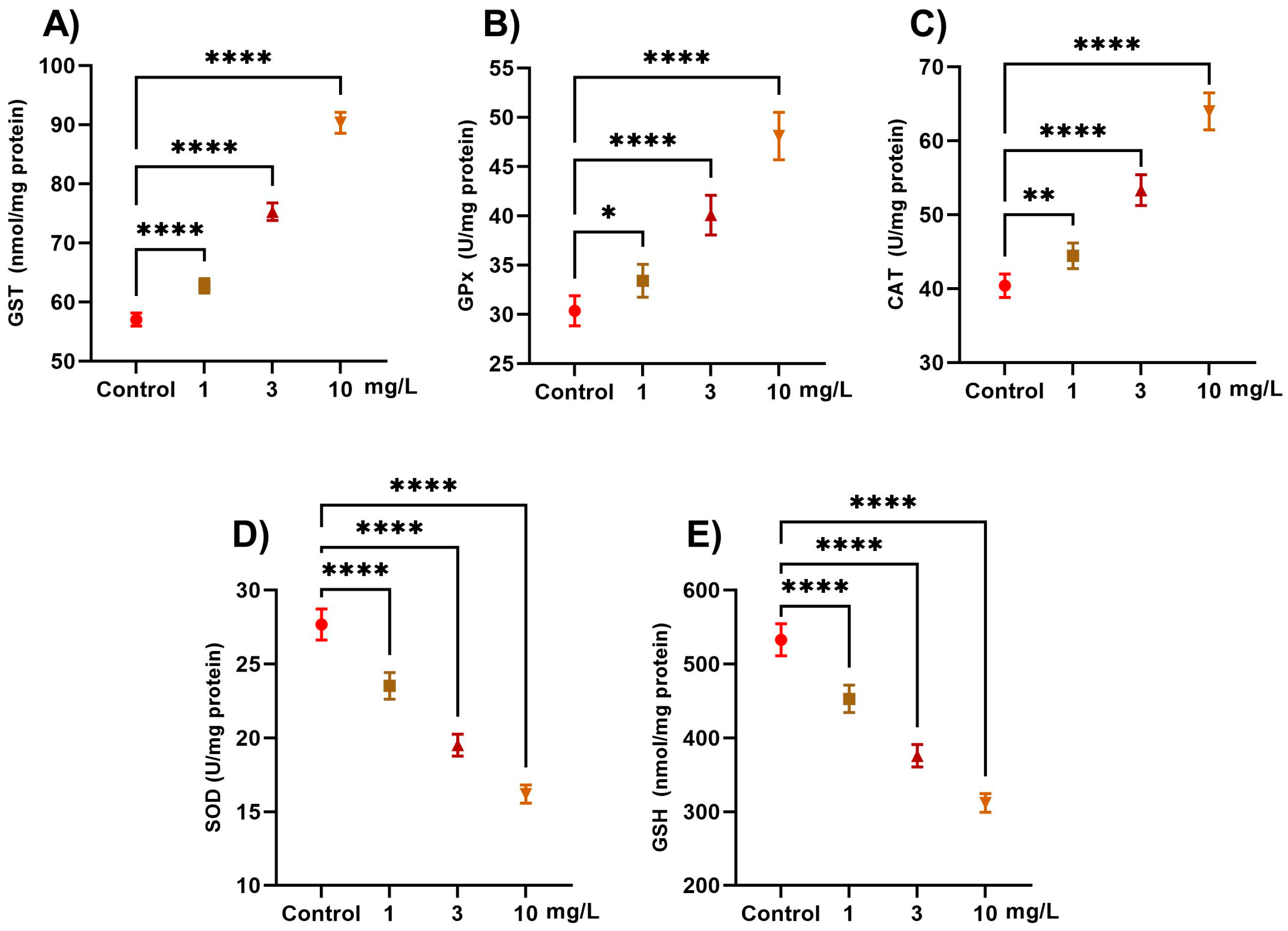
Figure 1. Effect of PFOS exposure on antioxidant enzymes and glutathione levels in L. carinatus. Snails were exposed to PFOS at concentrations of 1, 3, and 10 mg/L. (A) Glutathione S-transferase (GST, nmol/mg protein), (B) glutathione peroxidase (GPX, U/mg protein), (C) catalase (CAT, U/mg protein), (D) superoxide dismutase (SOD, U/mg protein), and (E) reduced glutathione (GSH, nmol/mg protein). Data are presented as the mean ± SD of six biological replicates. Statistical significance compared with the control: p < 0.05 (*), p < 0.01 (**), and p < 0.0001 (****).
Ijms 26 08898 g001
Figure 2. Effect of PFOS exposure on oxidative stress markers in L. carinatus snails were exposed to PFOS at concentrations of 1, 3, and 10 mg/L. (A) Malondialdehyde (MDA, nmol/mg protein) and (B) lipid peroxides (LPO, nmol/mg protein). Data are presented as mean ± SD of six biological replicates. Statistical significance compared with the control: p < 0.01 (**), p < 0.001 (***), and p < 0.0001 (****).
Ijms 26 08898 g002
Figure 3. Effect of PFOS exposure on acetylcholinesterase activity (AchE) in L. carinatus. Snails were exposed to PFOS at concentrations of 1, 3, and 10 mg/L. Data are presented as mean ± SD of six biological replicates. Statistical significance compared with the control: p < 0.0001 (****).
Ijms 26 08898 g003
Figure 4. H & E-stained digestive glands of L. carinatus. (A,B) Digestive glands of the control Lanistes carinatus group showing normal structures with digestive tubules and secretory and digestive cells. Digestive tubules (red stars) with epithelial layers resting on a basement membrane (black arrows) and containing a narrow tubular lumen (L). Digestive tubules are connected to each other by normal connective tissues (CTs) between them and haemocoele (Hae) with hemocytes (HCs). Different types of cells are abundant, including digestive cells (red arrowheads), calcium cells (green arrows), and execratory cells (yellow arrows); in addition, dark stained corpuscles (yellow arrowheads) are clearly visible. (A = 10X, B = 40X). (C,D) Digestive glands of the Lanistes carinatus exposed to 1 mg/L PFOS showing degenerated digestive tubules (red stars), with congested lumens (L), ruptured connective tissues (RCTs), vacuolation (V), necrotic basement membranes (yellow arrows), and fused tubules (yellow stars). (H&E, C = 10X, D = 40X). (E,F) Digestive glands of the Lanistes carinatus exposed to 3 mg/L PFOS showing high degeneration (stars) and shrinkage of some tubules and cells that lost their regular shapes and became vacuolated (yellow arrows), ruptured villi (red arrows), and detached basement membranes of the digestive tubules (green arrows). (E = 10X, F = 40X). (G,H) Digestive glands of the Lanistes carinatus are exposed to 10 mg/L PFOS showing sever degeneration (stars) in digestive tubules including digestive cells, execratory cells, calcium cells and villi. In addition, rupture of the basement membranes of the digestive tubules (red arrows), and dilation (Dil) in tubular lumens (L) were observed. Also, different vacuolations (yellow arrows) and swelling cells (green arrows) were noted. (G = 10X, H = 40X).
Ijms 26 08898 g004
Disclaimer/Publisher’s Note: The statements, opinions and data contained in all publications are solely those of the individual author(s) and contributor(s) and not of MDPI and/or the editor(s). MDPI and/or the editor(s) disclaim responsibility for any injury to people or property resulting from any ideas, methods, instructions or products referred to in the content.