Article

Integrated Multi-Omics Analysis Reveals the Role of Resveratrol in Regulating the Intestinal Function of Megalobrama amblycephala via m6A Methylation

1
Wuxi Fisheries College, Nanjing Agricultural University, Wuxi 214081, China
2
Key Laboratory of Freshwater Fisheries and Germplasm Resources Utilization, Ministry of Agriculture and Rural Affairs, Freshwater Fisheries Research Center, Chinese Academy of Fishery Sciences, Wuxi 214081, China
*
Authors to whom correspondence should be addressed.
Int. J. Mol. Sci. 2025, 26(17), 8587; https://doi.org/10.3390/ijms26178587
Submission received: 18 June 2025 / Revised: 19 August 2025 / Accepted: 29 August 2025 / Published: 3 September 2025
(This article belongs to the Section Molecular Genetics and Genomics)

Abstract

Resveratrol (RES), a natural polyphenol with lipid metabolism-regulating properties, also demonstrates remarkable efficacy in strengthening intestinal barrier integrity. In order to elucidate the mechanism by which RES ameliorates intestinal damage and lipid metabolism disturbances in Megalobrama amblycephala under a high-fat (HF) diet, a conventional diet (CON), an HF diet (HF), or an HF diet supplemented with 0.6, 3, or 6 g/kg RES (HF + 0.06%, 0.3%, or 0.6% RES) was fed to fish. After 8 weeks, RES supplementation in the HF diet significantly improved the growth performance and alleviated hepatic lipid deposition. Microbiota profiling revealed RES improved intestinal barrier function by reducing α-diversity, Actinobacteria and Bosea abundances, and enriching Firmicutes abundance. RES also maintained the integrity of the intestinal physical barrier and inhibited the inflammatory response. MeRIP-seq analysis indicated that RES modulated intestinal mRNA m6A methylation by upregulating methyltransferase-like 3 (mettl3) and downregulating fat mass and obesity-associated gene (fto) and Alk B homolog 5 (alkbh5). Combined RNA-seq and MeRIP-seq data revealed that RES alleviated endoplasmic reticulum stress (ERS) by upregulating the m6A methylation and gene level of heat shock protein 70 (hsp70). Correlation analyses identified significant associations between intestinal microbiota composition and ERS, tight junction, and inflammation. In summary, RES ameliorates lipid dysregulation via a synergistic mechanism involving intestinal microbiota, m6A modification, ERS, barrier function, and inflammatory response.

Graphical Abstract

1. Introduction

As essential nutrients for fish growth, lipids play a pivotal role in exerting a protein-sparing effect within feed formulations. Researches have indicated that appropriate modulation of dietary lipid levels can substantially improve protein efficiency and deposition [1]. However, this nutritional strategy is constrained by a critical dose-dependent threshold. Prolonged high-fat (HF) diet intake readily leads to excessive lipid accumulation, contributing to metabolic dysfunction and increased mortality, ultimately resulting in economic losses in aquaculture [2]. Studies have shown that fish exhibit a considerably lower tolerance to excess lipids in the diet than mammals [3]. Chronic feeding of aquatic animals with HF diets not only compromises intestinal barrier integrity by downregulating tight junction proteins and increasing intestinal permeability, but also triggers microbiota dysbiosis, excessive secretion of inflammatory cytokines, and endotoxin translocation. These effects exacerbate systemic metabolic abnormalities through the intestine–liver axis network [4,5]. The functional integrity of the intestine is fundamental for maintaining fish health as the first line of defense against pathogens and a vital regulator of internal homeostasis [6]. Accordingly, the identification of safe and effective feed additives to counteract HF diet-induced intestinal damage and metabolic dysregulation has emerged as a key focus in aquatic nutrition research.
Resveratrol (RES), a naturally occurring plant-derived polyphenol possesses high medicinal value. It possesses diverse functions such as regulating lipid metabolism, resisting oxidative stress, and suppressing inflammatory responses [7,8,9]. These broad bioactivities also enable RES to exert significant therapeutic effects in modulating intestinal functions and enhancing intestinal barrier integrity. In red tilapia (Oreochromis niloticus), RES can repair HF diet-induced intestinal mucosal damage by increasing the goblet cell density and mucus secretion [10]. Studies on koi carp (Cyprinus carpio) demonstrate that RES enhances disease resistance by remodeling the composition of intestinal microbiota, such as raising the abundance of Lactobacillus [11]. Notably, recent research has shown that RES acts as a regulator of N6-methyladenosine (m6A) methyltransferase-like 3 (METTL3), influencing RNA epigenetic modification patterns to modulate the stability and translational efficiency of peroxisome proliferator-activated receptor alpha (ppar-α) in mice [12]. The above research indicates that these biological activities of RES are closely associated with the regulation of the mRNA m6A methylation level.
In eukaryotes, m6A methylation represents the most prevalent form of RNA modification. It precisely regulates the post-transcriptional gene expression via the synergistic role of methyltransferases, demethylases, and m6A-binding proteins, thus modulating multiple biological functions [13]. The study indicates that dietary RES supplementation enhances intestinal functionality and antioxidant capacity by reducing m6A methylation levels of heme oxygenase-1 (ho-1) and tight junction protein mRNAs, improving piglets’ growth performance [14]. Moreover, RES alleviates hepatic steatosis in obese mice by inhibiting the activity of the demethylase fat mass and obesity-associated (FTO) protein, thus upregulating the expression of hepatic genes related to lipid metabolism [12]. Recent evidence suggests that m6A modifications may serve as a key epigenetic target through which RES modulates intestinal homeostasis and lipid metabolism. However, the biological impact of RES-mediated m6A modifications on intestinal function in fish remains undefined, and a significant gap persists in understanding its regulatory role within the ‘nutrition–epigenetics–intestinal function’ axis.
Megalobrama amblyocephala, also known as Wuchang bream, belongs to Cypriniformes, Cyprinidae, and Megalobrama. As a major freshwater species of high economic value, M. amblyocephala occupies a prominent position in Chinese aquaculture. However, the emergence of metabolic syndrome associated with prolonged HF diet administration under intensive farming conditions has become increasingly evident, posing a significant barrier to industrial development [15,16]. Therefore, this study aimed to elucidate the epigenetic regulatory function of RES in reducing growth retardation and intestinal damage, while enhancing lipid metabolism via the intestine–liver axis in HF diet-fed juveniles. The results provide a theoretical basis for incorporating RES into aquafeed and reveal a novel mechanism by which m6A methylation regulates in nutrient metabolism, thus offering innovative approaches for precision nutrition and sustainable aquaculture practices.

2. Results

2.1. Effects of RES on the Growth Performance and Lipid Metabolism of Juveniles Fed with an HF Diet

After the feeding trial (8 weeks), juvenile M. amblycephala growth performance indices are shown in Figure 1A–C. Compared with the CON group, the HF group showed significant reductions in weight gain rate (WGR) and specific growth rate (SGR) (Independent t-test, p < 0.05). The HF + 0.06%, 0.3%, and 0.6% RES groups showed significant improvements in WGR and SGR relative to the HF group (Duncan’s test, p < 0.05). Compared with the HF group, the feed conversion ratio (FCR) in the HF + 0.06% RES group was dramatically decreased (Duncan’s test, p < 0.05). Figure 1D–F demonstrates that plasma alanine aminotransferase (ALT) and total cholesterol (TC) concentrations in the HF group were significantly greater than the CON group (Independent t-test, p < 0.01). Compared with the HF group, plasma ALT, TC, and low-density lipoprotein (LDL) levels in the three RES addition groups were significantly decreased (Duncan’s test, p < 0.05). Quantitative analysis using ImageJ software (v1.8.0.112) confirmed that the red area of hepatic lipid droplet was considerably larger in the HF group than in the CON group (Figure 1G,H, Independent t-test, p < 0.01). Each RES-treated group showed a significant reduction in lipid accumulation compared with the HF group (Figure 1G,H, Duncan’s test, p < 0.05). Among the RES doses, 0.06% RES yielded significant positive effects. Therefore, gene expression was analyzed between HF and HF + 0.06% RES (HF-RES) groups. Hepatic gene levels of ppar-α, peroxisome proliferator-activated receptor beta (ppar-β), and cholesterol 7-alpha hydroxylase (cyp7a1) were significantly upregulated, whereas lipoprotein lipase (lpl) expression was downregulated in the HF-RES group compared with the HF group (p < 0.01; Figure 1I–L).

2.2. Intestinal Microbiota Analysis

The hind intestinal contents from the HF group and the HF + 0.06% RES group (HF-RES group) were selected for intestinal microbiota profiling. Operational taxonomic units (OTUs) were clustered at a similarity threshold of ≥97% using the RDP classifier Bayesian algorithm. A total of 1221 OTUs were identified across the eight samples, classified into 25 phyla, 60 classes, 152 orders, 269 families, 480 genera, and 631 species. Venn diagram analysis (Figure 2A) revealed 649 OTUs shared between the HF and HF-RES groups, with 292 OTUs unique to the HF group and 280 unique to the HF-RES group after the 8-week dietary intervention. Principal component analysis (PCA) (Figure 2B) of intestinal microbiota showed a significant separation between the HF and HF-RES groups, with the first two primary components responsible for 62.91% (PCA1) and 16.03% (PCA2) of variation between groups, respectively. Alpha diversity analysis revealed significantly reduced Shannon and Simpson indices in the HF-RES group compared with the HF group (p < 0.05), while Chao1 and ACE indices showed no significant differences (p > 0.05) (Figure 2C).
At the phylum level (Figure 2D), the HF-RES group displayed increased relative abundances of Proteobacteria and Firmicutes, whereas Actinobacteriota and Bacteroidota were more abundant in the HF group. At the genus level (Figure 2E), higher relative abundances of Variovorax and Bradyrhizobium were observed in the HF-RES group, while Phyllobacterium, Mycobacterium, and Chitinophaga showed higher enrichment levels in the HF group.
Based on LEfSe (Linear Discriminant Analysis Effect Size) analysis (Figure 2F), the HF-RES group showed a higher number of differentially abundant microbial taxa than the HF group. As shown in Figure 2G, taxa with linear discriminant analysis (LDA) scores > 2 in the HF-RES group included Gammaproteobacteria, Burkholderiales, Mitochondria, and Rickettsiales, while Propionibacteriales, Caldilineaceae, Chloroflexia, and Bosea were predominant in the HF group.

2.3. Role of RES in Maintaining Intestinal Barrier Health in Juvenile M. amblycephala Fed with an HF Diet

Intestinal tissues from the HF group and the HF + 0.06% RES group (HF-RES group) were selected to investigate the protective effect of RES against the intestinal injuries and on barrier health of HF diet-fed juveniles. The Hematoxylin and Eosin (H&E) staining results of the intestinal tissues (Figure 3A–C) revealed that the HF-RES group exhibited a clear increase in intestinal muscle thickness and villus length compared with the HF group (p < 0.01). Analysis of antioxidant indicators showed that 0.06% RES supplementation in the HF diet markedly reduced the levels of lipid peroxide (LPO) and malondialdehyde (MDA) in the intestinal tissues (Figure 3H,I; p < 0.01). As illustrated in Figure 3D–F,J–L, compared with the HF group, the HF-RES group displayed upregulated expression levels of junctional adhesion molecule 2 (jam2) (p < 0.01), zonula occludens 1 (zo1), and claudin41 (p < 0.05), and downregulated expression levels of toll-like receptor 4 (tlr4) (p < 0.05), nuclear factor kappa-B (nf-κb) and interleukin-1beta (il-1β) (p < 0.01).

2.4. MeRIP-seq of the Intestinal Tissues

Intestinal tissues from the HF group and the HF + 0.06% RES group (HF-RES group) were selected for MeRIP-seq. The sequencing results of the IP and Input libraries showed that more than 81.97% of the clean data mapped to the M. amblycephala reference genome, with preeminent concordance among replicates (Q30 > 93.75%, Q20 > 97.72%) (Tables S1 and S2). A total of 18,314 and 16,029 m6A peaks were identified in the HF and HF-RES groups, respectively, with 4978 peaks shared between both groups (Figure 4A). As presented in Figure 4B, m6A peaks in both groups were predominantly enriched in coding sequences (CDS), terminal regions of the 5′ untranslated regions (5′ UTRs), and initiation regions of the 3′ untranslated regions (3′ UTRs). According to Figure 4C, the HF group showed m6A peak distributions of 35.7% in CDS regions, 28.7% in termination regions, 28% in 3′ UTRs, 4.2% in promoter regions, and 3.1% in 5′ UTRs. In comparison, the HF-RES group showed distributions of 39%, 25%, 25.2%, 4.6%, and 5.8%, respectively. Figure 4D displays the conserved m6A motifs identified in both groups, with shared methylation sites indicated by lines, though variations in frequency were observed.

2.5. Differential m6A Peaks Identification and Functional Analysis in HF and HF-RES Groups

All 20 m6A peaks demonstrating the most significant methylation alterations are listed in Table 1. Among these, elevated methylation levels were identified in NYN domain and retroviral integrase containing (nynrin), histone H3 family member (histone H3.v1), neuraminidase 4 (neu4), carcinoembryonic antigen-related cell adhesion molecule 1 (ceacam1), OTU deubiquitinase 7A (otud7a), interferon regulatory factor 4 (irf4), zinc finger protein 184 (znf184), slit guidance ligand 2A (slitla), solute carrier family 2 member 12 (slc2a12), zinc finger protein Y-linked 1 (zfy1), bone morphogenetic protein-like A (bmpla), cysteine and tyrosine-rich 1 (cyyr1), mitochondrial transcription rescue factor 1 (mtres1), LOC125253164, nuclear factor erythroid 2-like 1B (nfe2l1b), and zinc finger protein 692 (znf692). Decreased methylation levels were observed in tripartite motif-containing protein 39 (trim39), tetratricopeptide repeat domain 38 (ttc38), LOC125260582, and ribonuclease inhibitor (ri).
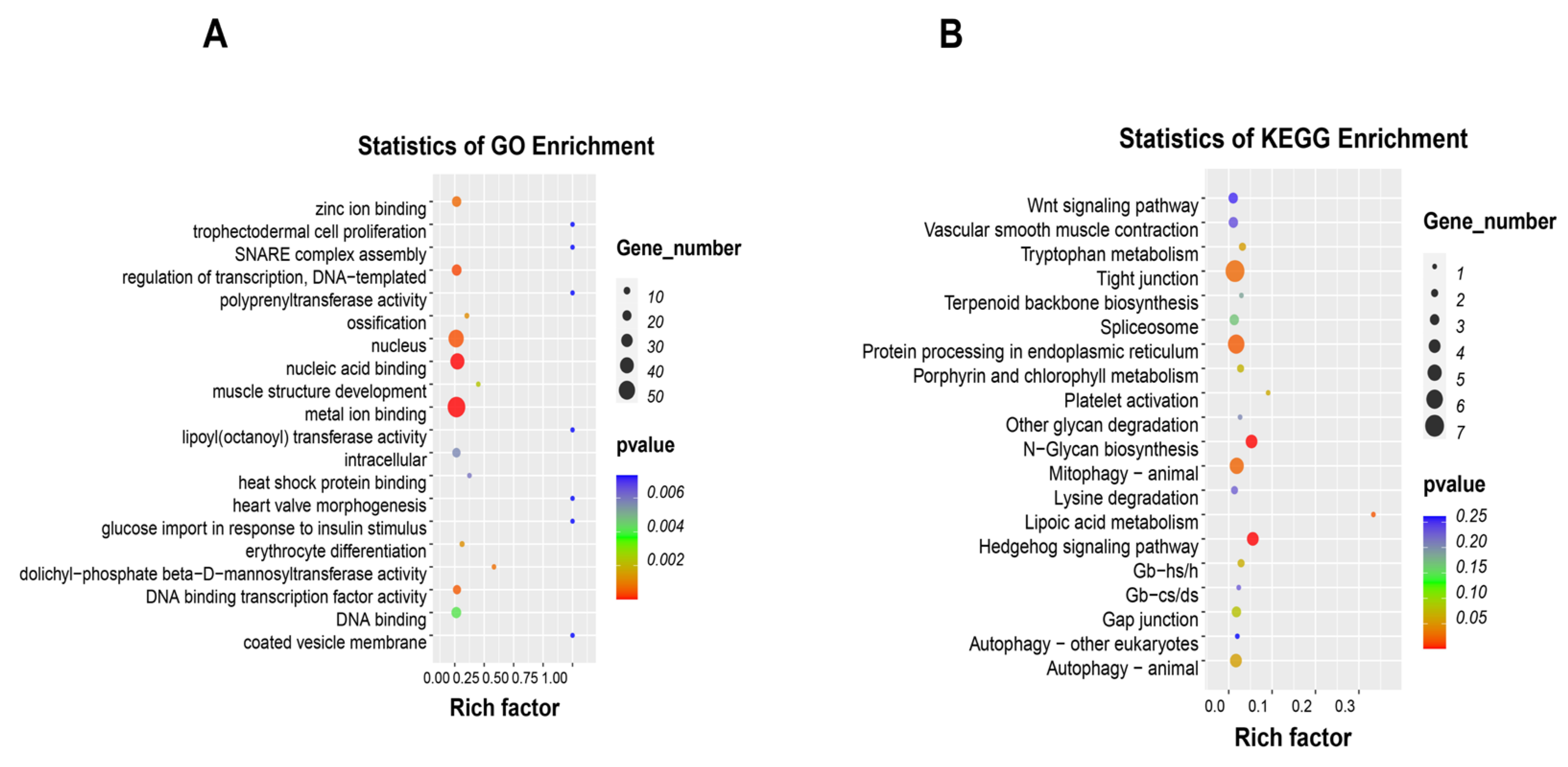
Gene Ontology (GO) functional categorization showed categories such as metal ion binding, nucleic acid binding, nucleus, glucose import in response to insulin stimulus, and heat shock protein binding (Figure 5A). Kyoto Encyclopedia of Genes and Genomes (KEGG) analysis further linked these differential peaks to signaling pathways such as Wnt, tight junctions, protein processing in the endoplasmic reticulum, mitophagy–animal, and autophagy–animal (Figure 5B).

2.6. Association Analysis Between DEGs and Differential m6A Peaks

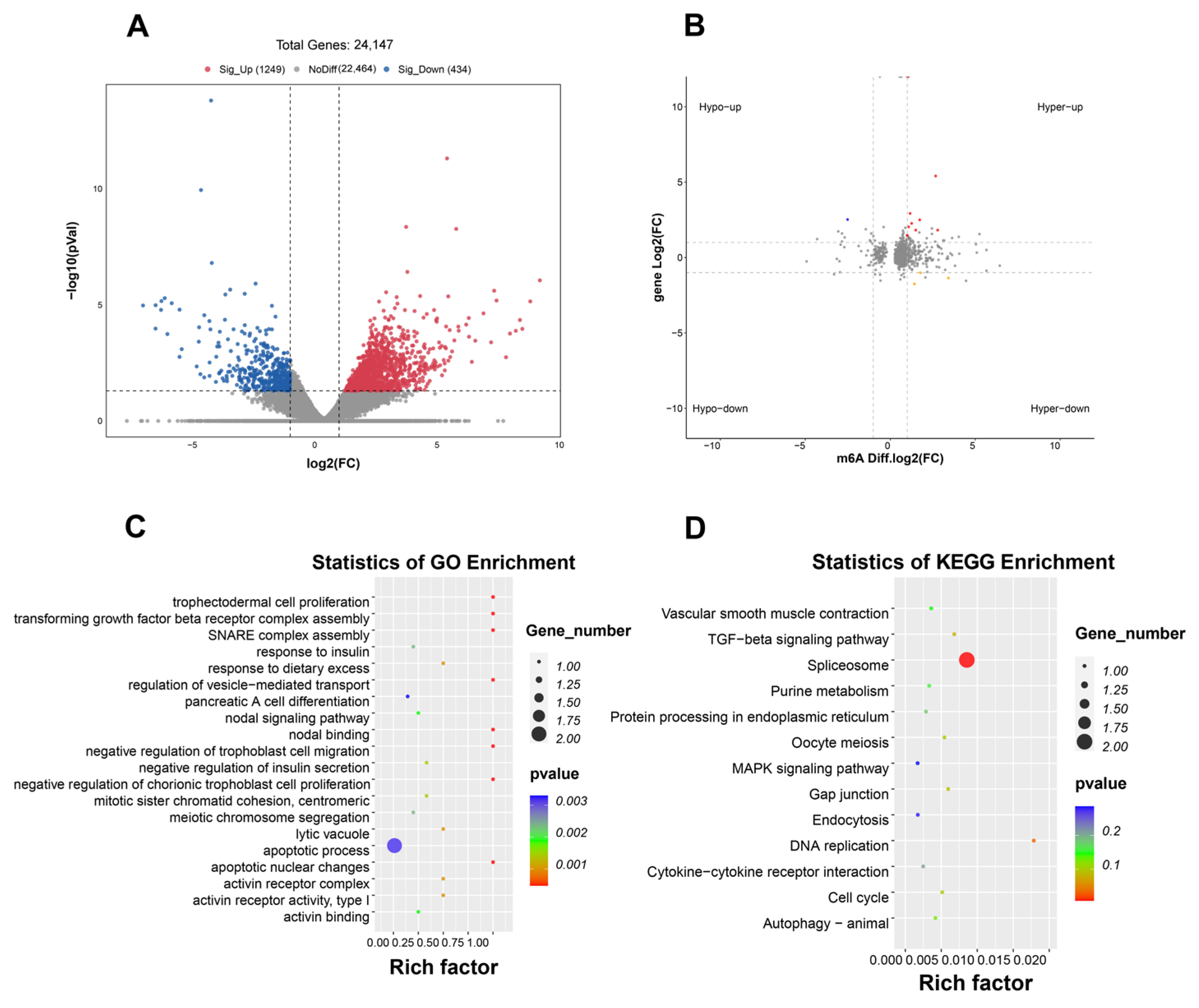
RNA-seq results revealed 1683 differentially expressed genes (DEGs) (|log2(fold change) | ≥ 1 and p < 0.05) between the HF and HF-RES groups (HF + 0.06% RES group), including 1249 upregulated and 434 downregulated genes (Figure 6A). Integrated analysis of RNA-seq and MeRIP-seq identified 12 genes presenting differential mRNA expressions and m6A methylation levels. These genes were divided into four expression patterns (Figure 6B): nine genes including hsp70, UV radiation resistance-associated gene (uvrag), transmembrane protein 119B (tmem119b), protein phosphatase one regulatory subunit 13B, a (ppp1r13ba), FK506 binding protein 5 (fkbp5), ribosome production factor 2 homolog (rpf2), minichromosome maintenance complex component 3 (mcm3), structural maintenance of chromosomes 4 (smc4), and guanylate cyclase one soluble subunit beta 2 (gucy1b2) showed increased m6A methylation and upregulated mRNA expression (Hyper-up); mitochondrial transcription rescue factor 1 (mtres1) and tumor necrosis factor (ligand) superfamily member 10 (tnfsf10) showed elevated m6A methylation but reduced mRNA expression (Hyper-down); shugoshin-like 1 (sgo1) displayed decreased m6A methylation alongside increased mRNA expression (Hypo-up). However, no genes were observed with downregulated m6A methylation and reduced mRNA expression (Hypo-down). GO analysis revealed that the 12 genes were primarily enriched in biological processes including apoptotic nuclear changes, apoptotic process, response to dietary excess, negative regulation of insulin secretion, and nodal binding (Figure 6C). KEGG pathway analysis indicated associations with signaling pathways, such as TGF-beta, MAPK, protein processing in the endoplasmic reticulum, gap junction, and autophagy-animal (Figure 6D).

2.7. Expression Analysis of Intestinal m6A Methylase and Endoplasmic Reticulum Stress-Related Genes

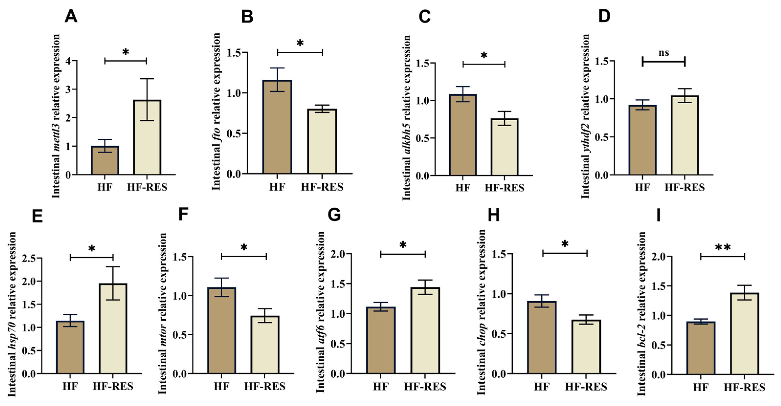
Intestinal tissues from the HF and the HF + 0.06% RES groups (HF-RES group) were selected to analyze the gene levels related to m6A methylation and endoplasmic reticulum stress (ERS). Compared with the HF group (Figure 7A–D), mettl3 expression was significantly elevated, whereas fto and Alk B homolog 5 (alkbh5) levels were reduced in the HF-RES group (p < 0.05), with no significant difference detected in YTH N6-methyladenosine RNA binding protein 2 (ythdf2) expression (p > 0.05). As depicted in Figure 7E–I, HF-RES group showed significantly increased expression of B-cell lymphoma-2 (bcl-2) (p < 0.01), heat shock protein 70 (hsp70), and activating transcription factor 6 (atf6) (p < 0.05), while C/EBP-homologous protein (chop) and mechanistic target of mammalian target of rapamycin (mtor) transcript levels were considerably reduced (p < 0.05), indicating modulation of ERS-related gene responses.

2.8. Intestinal Microbiota, m6A Methylation, Intestinal Barrier, and Lipid Metabolism Correlation Analysis

Intestinal mettl3 expression level positively correlated with the hepatic ppar-β expression level and negatively associated with the lpl expression level (p < 0.05, Figure 8A). Intestinal fto showed inverse correlations with hepatic ppar-β and cyp7a1 (p < 0.05). Intestinal hsp70 negatively associated with plasma ALT level (p < 0.05). Intestinal atf6 was positively associated with hepatic ppar-α expression (p < 0.05). Intestinal bcl-2 expression demonstrated strong positive correlations with hepatic ppar-β and cyp7a1 expression levels (p < 0.01).
Intestinal microbiota, which played important roles in the HF and the HF-RES groups (HF + 0.06% RES group), were prioritized for correlation analysis to decipher their interplay with genes related to m6A methylation, intestinal barrier, and inflammation (Figure 8B). Bosea positively correlated with mtor and tlr4 expressions (p < 0.05). Actinobacteria were positively associated with zo1 (p < 0.05). Mettl3 had positive correlations with hsp70, jam2 and zo1 (p < 0.05). Fto expression showed a positive regulatory relationship with tlr4, nf-κb, and il-1β, while negatively correlated with bcl-2 (p < 0.05). Alkbh5 showed positive connections with chop, nf-κb, and il-1β, but a negative correlation with atf6 (p < 0.05). Hsp70 depicted a regulatory relationship with zo1 (p < 0.05). Atf6 revealed negative correlations with il-1β and nf-κb (p < 0.05). Mtor showed a positive regulatory relationship with nf-κb (p < 0.05). Chop also demonstrated a regulatory relationship with tlr4 (p < 0.05). Bcl-2 expression was positively related to jam2 and zo1 but negatively associated with il-1β and nf-κb (p < 0.05). Tlr4 demonstrated negative correlations with jam2 and zo1 (p < 0.05).

3. Discussion

In aquaculture, lipids are routinely used as alternative energy sources to partially replace dietary protein, reduce feed costs, and enhance protein utilization efficiency [17]. However, an excessive lipid concentration in the feed may induce adverse physiological responses in fish, including inhibited growth, disrupted lipid metabolism, oxidative stress, and compromised immune function [18]. Soybean oil is rich in polyunsaturated fatty acids (PUFAs), predominantly ω-6 PUFAs [19]. Excessive intake of ω-6 PUFAs can disrupt the ω-6/ω-3 balance, leading to abnormal lipid metabolism and inflammatory responses [20]. Studies on fish and mammals show that soybean oil has been successfully used to establish high-fat-induced injury models [21,22,23]. Similarly, in this experiment, we constructed a high-fat injury model for M. amblycephala by adding excessive soybean oil to the diet in this experiment. Previous studies have demonstrated that supplementation of RES in an HF diet significantly alleviates lipid-induced growth suppression in common carp, as reflected by elevated WGR and improved FCR [22]. These findings are consistent with the current results, wherein dietary RES supplementation in an HF diet enhanced WGR and SGR, reduced FCR, and alleviated growth suppression in HF diet-fed juvenile M. amblycephala.
Plasma ALT, TC, and LDL contents are crucial indicators for assessing lipid metabolism. The liver, functioning as the central regulator of lipid metabolism, facilitates lipid digestion and absorption through bile acid secretion and controls fatty acid oxidation [24]. CYP7A1 acts as a key enzyme in bile acid biosynthesis, whereas PPARs and LPL are key regulators of lipid oxidation and hydrolysis. In the present study, RES supplementation significantly attenuated HF diet-induced increases in plasma ALT, TC, and LDL levels, evidently upregulated hepatic expressions of cyp7a1, ppar-α, and ppar-β, and suppressed lpl gene expression. Based on Nile Red staining, RES reduced hepatic lipid deposition induced by the HF diet in juveniles. Similar results were observed in red tilapia, where dietary RES significantly decreased serum ALT, TC, and LDL levels, downregulated hepatic lpl expression, and upregulated ppar-α expression [25]. These findings indicate that the regulatory effects of RES on lipid metabolism in fish may be attributed to the high hepatic lipid catabolism. Furthermore, among the tested RES concentrations, 0.06% RES in the HF diet produced a significant therapeutic effect. Thus, a comparative analysis between the HF and HF + 0.06% RES groups (designated as HF-RES group) was undertaken to explore specific mechanisms underlying RES-mediated regulation of intestinal health and lipid metabolism.
The intestinal barrier is a multi-layered defense system that balances the intestinal internal and external environments. Its dynamic functional equilibrium relies on microbial community stability, mucus layer integrity, and immune regulation. In the intestinal biological barrier, RES treatment has been widely reported to enhance the α-diversity of gut microbiota in mice, ameliorating dysbiosis associated with HF dietary intake [26]. However, the present findings demonstrated a divergent outcome, wherein RES supplementation under HF dietary conditions resulted in a reduction in α-diversity of the intestinal microbiota in juvenile M. amblycephala, as indicated by decreases in Shannon and Simpson diversity indices. Physiologically, fish possess shorter intestines and simpler microbial compositions compared to mammals, which may lead to differences in the absorption and metabolism of RES, consequently resulting in distinct regulatory effects on the intestinal microbiota [27]. Previous studies in diabetic mouse models have suggested that RES facilitates the production of intestinal short-chain fatty acids (SCFAs), which may indirectly regulate microbial community structure by inhibiting the proliferation and activity of pathogenic bacterial species [28]. Therefore, it is hypothesized that the observed decrease in intestinal microbiota α-diversity may be attributed to the inhibition of symbiotic pathogenic bacterial survival. Firmicutes, as a core phylum within the intestinal microbiota, are pivotal in shaping microbial composition and substantially affect host physiological health. Studies have shown that SCFAs produced by Firmicutes can inhibit pathogen growth, suppress pro-inflammatory factors (like tnf-α and il-6) expressions, enhance intestinal epithelial cells’ tight junctions, stabilize the intestinal environment, and alleviate inflammatory bowel diseases [29]. Research on rats has shown that an HF diet increases Actinobacteria abundance in intestinal microbiota and significant lipid metabolism dysfunction [30]. Moreover, the pathogenic bacterial genus Bosea has been identified as a characteristic microbiota in the mice's intestines under an HF diet [31]. The present study revealed that RES supplementation in an HF diet significantly elevated the relative abundance of Firmicutes, decreased Actinobacteria, and altered the dominant status of Bosea within the intestinal microbiota of juveniles M. amblycephala under HF dietary conditions. These findings indicate that RES positively regulates intestinal microbial homeostasis and contributes to preserving intestinal function.
In the case of physical barriers, intestinal crypt depth and villus length are important indicators for assessing intestinal structural integrity. It is reported that RES supplementation in the HF diet significantly increased the intestinal villus length in rats, thus ameliorating HF diet-induced intestinal structural damage [32]. In the current study, RES addition significantly elevated intestinal villus length and muscle thickness of juvenile M. amblycephala, demonstrating its capacity to repair HF diet-induced intestinal structural damage. Moreover, epithelial tight junctions represent a basic structural component of the intestinal physical barrier, with junctional proteins such as Claudin, JAM, and Occludin serving crucial roles in maintaining intercellular cohesion and barrier functionality [33]. Dietary intake of HF regimens has been associated with elevated levels of intestinal lipopolysaccharide (LPS), which impairs the transcriptional expression of genes encoding tight junction-associated proteins [34]. Luo et al. [35] reported that RES administration effectively reversed LPS-induced downregulation of zo-1, claudin-1, and occludin genes expression in HT-29 cells, thus maintaining tight junction integrity and preventing epithelial barrier disruption. The current findings align with these observations; expression levels of zo1, jam2, and claudin41 in juveniles M. amblycephala intestinal tissues in the HF-RES group (HF + 0.06% RES) exceeded those in the HF group, further confirming the RES role in enhancing intestinal barrier function under the HF diet.
It is reported that HF diets induced oxidative damage in intestinal tissues by generating reactive oxygen species (ROS) through fatty acid oxidation, depleting non-enzymatic antioxidants like reduced glutathione (GSH), and increasing LPO and MDA, thus disrupting tight junctions, enhancing intestinal permeability, and impairing barrier function in mice. However, RES treatment significantly increased intestinal GSH levels, reduced LPO and MDA concentrations, and ameliorated the mice’s oxidative stress [36]. Consistent with previous findings, RES supplementation counteracted the diet-induced reduction in GSH levels and the elevation of LPO and MDA concentrations, thus enhancing the antioxidant potential of the intestinal tract in juveniles M. amblycephala. Intestinal barrier dysfunction can trigger inflammation and metabolic disorders. At the molecular level, RES has been demonstrated to attenuate HF diet-induced inflammation by regulating multiple intracellular signaling pathways. Among these, inhibition of the TLR4/NF-κB signaling cascade (due to the lower secretion of pro-inflammatory mediators, i.e., IL-1β) is widely recognized as a main mechanism underlying its anti-inflammatory effects [22]. Similarly, the current findings found that in comparison with the HF group, supplementing RES significantly downregulated tlr4, nf-κb, and il-1β gene levels of juveniles M. amblycephala intestinal tracts.
A recent study has shown that RES, functioning as a methyl donor, positively affects lipid metabolic disturbances in HF diet-fed mice, primarily through the modulation of mRNA m6A modification levels [12]. These findings offer a valuable framework for elucidating the molecular mechanisms by which RES may alleviate lipid metabolism abnormalities in juvenile M. amblycephala by regulating intestinal function. Moreover, m6A methylation, recognized as the most prevalent epigenetic modification of RNA, is known to regulate various biological processes. The present study employed MeRIP-seq to identify 18,314 and 16,029 m6A peaks in the HF and HF-RES groups, respectively. These methylation peaks were predominantly localized within the 5′UTRs, CDS, and 3′UTRs, consistent with the m6A distribution patterns previously reported by Dominissini et al. [37]. It has been observed that m6A modifications frequently occur at consensus “RRACH” motifs, reflecting sequence specificity [37]. In the current analysis, similar motif patterns were detected in both HF and HF-RES groups, suggesting a potential role of m6A modification in mediating the regulatory effects of RES on intestinal physiology.
Several studies indicate that RES can achieve comprehensive regulation of intestinal function through multiple signaling pathways, including NF-κB, MAPK, and TGF-beta, from anti-inflammatory, anti-oxidation, barrier function enhancement, and other ways [38]. Combined examination of RNA-seq and MeRIP-seq datasets revealed that genes showing differential expressions at both mRNA and m6A methylation levels were primarily enriched in signaling pathways (TGF-beta, protein processing in endoplasmic reticulum, autophagy–animal, MAPK, and gap junction). Furthermore, the presented data highlighted significant enrichment of endoplasmic reticulum stress (ERS)-related pathways within the identified m6A methylation peaks, suggesting that m6A modifications may play a pivotal role in RES-mediated regulation of intestinal function, potentially through mechanisms intricately related to ERS modulation. The endoplasmic reticulum is the site of protein folding and maturation in the eukaryotic cell. Studies have shown that prolonged HF diet consumption can cause ERS in the intestinal tissues of Micropterus salmoides. ERS can mediate protein homeostasis imbalance by activating UPR and serve as an upstream regulatory hub connecting inflammatory responses and intestinal barrier damage [39]. RES has been observed to alleviate ERS via multiple mechanisms, including modulating UPR, balancing autophagy and apoptosis, and regulating calcium homeostasis through PERK/ATF4/CHOP [40,41].
Sequencing analysis revealed that RES supplementation in the context of an HF diet significantly increased both m6A methylation levels and the transcriptional expression of hsp70. As a key member of the heat shock protein family, HSP70 plays a pivotal role in regulating the unfolded protein response (UPR), thus preserving endoplasmic reticulum (ER) homeostasis. This is achieved through its chaperone activity by interacting with ERS sensors, such as ATF6, or by reducing mTOR-dependent suppression of autophagy [42]. Under conditions where ER stress exceeds cellular adaptive capacity, HSP70 stabilizes Bcl-2 family proteins by inhibiting CHOP-driven apoptotic signaling, ultimately attenuating ERS-induced apoptotic pathways [43]. Studies indicate that under acute heat shock stress, METTL3-mediated m6A modification recruits nuclear-localized YTHDF2 to protect HSP70 mRNA from erasure by the demethylase FTO, thereby significantly enhancing HSP70 protein synthesis efficiency and alleviating heat-induced damage [44]. In the present study, the expression of hsp70 was quantified using qRT-PCR, along with the evaluation of genes involved in m6A RNA methylation (mettl3, fto, alkbh5, and ythdf2) and ER stress-related markers (mtor, atf6, chop, and bcl-2). The analysis indicated that adding RES to an HF diet significantly upregulated the mettl3, hsp70, atf6, and bcl-2 expressions, and downregulated fto, alkbh5, mtor, and chop. These findings suggested that RES may enhance the m6A methylation level of hsp70 by modulating key methylation-related enzymes, including mettl3, fto, and alkbh5, thus promoting hsp70 gene expression. Thereby alleviating HF diet-induced ERS and modulating intestinal function of juvenile M. amblycephala by regulating the UPR (upregulating atf6) or inhibiting apoptosis (downregulating mtor and chop).
Studies have shown that ERS-induced disruption to intestinal barrier function allows intestinal LPS and free fatty acids to enter the liver via portal vein circulation, thus inhibiting fatty acid β-oxidation and bile acid synthesis, exacerbating steatosis and contributing to the intestine–liver axis lipid metabolism disorders [45,46]. In this study, intergroup Pearson correlation analysis revealed a negative association between the gene level of intestinal hsp70 and plasma ALT level, a positive association between intestinal atf6 gene level and hepatic ppar-α gene expression, and positive correlations between intestinal bcl-2 and hepatic ppar-β and cyp7a1 expressions. Compared with the HF group, the HF-RES group depicted upregulated expressions of atf6, bcl-2, ppar-α, ppar-β, and cyp7a1, indicating that RES can promote hepatic fatty acid β-oxidation and bile acid synthesis by improving HF diet-induced ERS via m6A methylation, thus alleviating lipid metabolism disorders in M. amblycephala.
Mantel test correlation analysis revealed that intestinal microbiota is directly associated with ERS, tight junctions in intestinal epithelial cells, and inflammatory responses. Studies report that intestinal microbiota have the potential to regulate tight junction protein expression through metabolites such as SCFAs, and then maintain intestinal integrity. Microbial dysbiosis can induce ERS, disrupt tight junction structures, increase intestinal permeability, and exacerbate inflammatory responses [47,48]. The experimental results showed that the relative abundance of the opportunistic bacterium Bosea was positively associated with the expression of ERS-related gene mtor and the pro-inflammatory gene tlr4. RES supplementation significantly decreased the intestinal abundance of Bosea and the expression levels of mtor and tlr4 in M. amblycephala. These observations suggested that the intestinal protective effects of RES may involve a favorable regulatory loop, characterized by restoring microbial homeostasis, attenuating ERS, preserving intestinal barrier integrity, and suppressing inflammatory signaling pathways.

4. Materials and Methods

4.1. Experimental Diets

Resveratrol (RES; HPLC ≥ 98%) was purchased from Beyotime Biotechnology Co., Ltd. (Shanghai, China). As per the methodology outlined by Yan [49], soybean oil was used as the dietary lipid source, while fishmeal, soybean meal, rapeseed meal, and cottonseed meal were the main protein sources for the preparation of five experimental diets. The diets included a conventional diet (CON group), an HF diet (HF group), and an HF diet enriched with 0.6 g/kg RES (HF + 0.06% RES group), 3 g/kg RES (HF + 0.3% RES group), or 6 g/kg RES (HF + 0.6% RES group). The composition of each diet is detailed in Table 2. All ingredients were finely ground, passed through a 60-mesh sieve, and weighed precisely according to the prescribed formulation. All components were homogenized, followed by the addition of water and soybean oil. The resulting mixture was extruded into 2 mm pellets using an F-26 (II) granulator (South China University of Technology, Guangzhou, China). After air-drying, the pellets were stored at −20 °C until further use.

4.2. Fish and Feeding Trial

An indoor system with recirculating water system with and temperature control was used for the aquaculture trial. Juveniles M. amblycephala were provided by M. amblycephala National Breeding Farm (Wuhan, China). A total of 225 4-month-old healthy fish (8 ± 0.5 g) were randomly divided into 15 glass tanks and divided into five dietary groups: CON, HF, HF + 0.06% RES, HF + 0.3% RES, and HF + 0.6% RES. Each group comprised three replicate tanks with a 15 fish per 350 L tank. For 8 weeks, the juveniles were fed to apparent satiety three times daily (07:00, 12:00, and 17:00). The feeding trial was conducted under natural light conditions, with water temperature maintained at 26–28 °C, ammonia nitrogen ≤ 0.05 mg/L, dissolved oxygen ≥ 6 mg/L, and pH 7.0–7.5.

4.3. Sample Collection

After feeding for 8 weeks, fish were fasted for 24 h and anesthetized using MS-222 (100 mg/L; Sigma, Saint Louis, MO, USA). The number and weight of fish in each tank were recorded to determine growth performance indices. Three fish per tank were randomly selected for blood collection via the caudal vein. Plasma was obtained by centrifugation at 4000 rpm for 10 min at 4 °C, and the supernatant was stored at −20 °C for later biochemical analysis. Liver, hind intestine, and hind intestinal contents were quickly harvested in duplicate, flash-frozen in liquid nitrogen, and stored at −80 °C. One set was allocated for hepatic and intestinal gene expression analysis, while the other was used for 16S rRNA sequencing of hind intestinal contents and MeRIP-seq of hind intestinal tissue. An additional three fish per tank were sampled to collect liver and hind intestine tissues; a portion of the tissue was stored at −20 °C for antioxidant enzyme activity analysis, and the remaining was fixed in 4% paraformaldehyde (PFA) for histopathological analysis using Nile Red and H&E staining.

4.4. Parameter Measurement

4.4.1. Growth Performance Analysis

Specific growth rate (SGR, %/d) = 100 × (lnWt − lnW0)/t;
Weight gain rate (WGR, %) = 100 × (WtW0)/W0;
Feed conversion ratio (FCR) = F/(WtW0).
where
Wt = final total weight of fish (g);
W0 = initial total weight of fish (g);
t = feeding duration (d);
F = total feed intake (air-dry basis, g).

4.4.2. Biochemical Index Analysis

Plasma LDL, TC, and ALT levels were measured using a BS-400 Q2080 fully automated biochemical analyzer (Mindray, Shenzhen, China), employing commercial kits. The concentrations of GSH and MDA, and LPO activities in hind intestinal tissues were quantified using a microplate reader (Thermo Fisher, Cleveland, OH, USA). All examination kits were provided by the Nanjing Jiancheng Bioengineering Institute (Nanjing, China).

4.5. Histological and Fluorescence Staining

Hepatic tissues fixed in PFA were processed into frozen sections for Nile Red staining. Following the method of Shen et al. [50], sections were air-dried at room temperature, encircled using a hydrophobic barrier pen, and stained with Nile Red solution in the dark. After rinsing and drying, DAPI was used in the dark for nuclear staining. After final washing, sections were sealed using a fade-resistant mounting medium and examined under a fluorescence microscope. The red lipid deposition area (%) in hepatic tissue was quantified using ImageJ software (v1.8.0.112).
Based on Kamyab-Hesary et al. [51] PFA-fixed intestinal tissues were trimmed and embedded in paraffin after dehydration and waxing steps. Paraffin blocks were sectioned, deparaffinized with xylene, and rehydrated through a graded ethanol series. Staining was performed using Hematoxylin (nuclear) and Eosin (cytoplasmic) dyes. Slides were mounted and examined microscopically to assess histological morphology, followed by image acquisition.

4.6. Microbiota Analysis in Intestinal Contents

Four hind intestinal content samples were prepared for both the HF group and the HF + 0.06%RES group. Three of these were composed of an equal-quantity mixture of intestinal contents from three fish per replicate, respectively, while the remaining sample was a pooled mixture formed by combining equal volumes of these three mixtures. This pooled sample integrated intestinal contents from all nine fish within the treatment group. As described by Qian et al. [52], genomic DNA was extracted and evaluated for quality and concentration via 1% agarose gel electrophoresis. PCR was amplified with specific primers targeting the V4-V5 hypervariable region of the bacterial 16S rRNA gene. Following sequencing standards, PCR products were purified and pooled in defined ratios. Library sequencing was performed on the Illumina platform (PE250) (Illumina, San Diego, CA, USA). Optimized data were clustered into OTUs at 97% sequence identity, and the taxonomic assignment was executed using the RDP Classifier with a Bayesian algorithm. Microbial community composition was statistically analyzed [53].

4.7. MeRIP-seq

4.7.1. RNA Extraction, Library Preparation, and Sequencing

According to previously reported procedures [54], the isolation of total RNA from juvenile M. amblycephala hind intestinal tissue samples of the HF and HF + 0.06%RES group was performed using TRIzol reagent (Invitrogen, Carlsbad, CA, USA). A ND-1000 spectrophotometer (NanoDrop Technologies, Wilmington, DE, USA) was used to assess the quality of the extracted RNA. Oligo (dT) magnetic beads were used to selectively capture Polyadenylated RNA via two rounds of purification. After being fragmented, a portion of the RNA was pre-mixed with magnetic beads and an m6A-specific antibody for immunoprecipitation (IP), while the remaining portion served as the input sample and underwent direct library construction. Before the second strand synthesis, the IP product was reverse-transcribed into cDNA. cDNA fragments were size-selected and purified using magnetic beads. Sequencing libraries were constructed via PCR amplification and sequenced on Illumina NovaseqTM 6000 (PE150) (Illumina, San Diego, CA, USA).

4.7.2. Bioinformatics Analysis

After quality control, clean reads from all IP and input samples were aligned to the M. amblycephala reference genome using HISAT2 (http://daehwankimlab.github.io/hisat2, accessed on 2 September 2024) [55]. The R package (version 3.3) exomePeak was used for differential peak analysis of BAM files (https://www.bioconductor.org/packages/3.3/bioc/html/exomePeak.html, accessed on 13 September 2024). Peak annotation was conducted using ANNOVAR (Gencode v41 Basic collection) (http://www.openbioinformatics.org/annovar/, accessed on 17 September 2024) and motif enrichment analysis was performed using HOMER (http://homer.ucsd.edu/homer/motif, accessed on 21 September 2024) [56]. Gene assembly and quantification were conducted via StringTie (https://ccb.jhu.edu/software/stringtie, accessed on 8 October 2024) and gene expression levels were computed using the FPKM method. DEGs were identified using the R package edgeR, with selection criteria set at |log2(fold change)| ≥ 1 and p < 0.05 [57].

4.8. Real-Time PCR Analysis

After the procedure of the previous research [52], the RNAiso Plus Kit (Takara, Daliang, China) was used to isolate total RNA from the hind intestines of M. amblycephala. RNA concentration and quality were assessed by measuring OD 260/280 ratios (1.8–2.1). Target gene expression was quantified using cDNA, which was synthesized by the extracted RNA, as the template via qRT-PCR. The reference gene β-actin was used to calculate relative gene expression through the 2−ΔΔCT method. The sequences of primers are provided in Table 3, and primers were composed in Sangon Biotech (Shanghai, China).

4.9. Statistical Analysis

Statistical analyses were performed using SPSS 25.0 software. The experimental data were examined to determine if they followed a normal distribution via the Shapiro–Wilk test, and confirmed to meet the requirements of variance homogeneity via Levene’s test. Statistical comparisons were conducted using one-way ANOVA followed by Duncan’s test or an Independent t-test. Data are presented as mean ± standard error mean (SEM). Statistical significance was attributed to the intergroup differences when p < 0.05.

5. Conclusions

In conclusion, this study elucidates the regulatory mechanisms by which RES modulates intestinal function and mitigates lipid metabolism disorders in high-fat diet-fed juveniles M. amblycephala, as revealed through integrated multi-omics analyses. Dietary RES supplementation contributed to maintaining the homeostasis of the intestine–liver axis and reducing hepatic lipid accumulation by stabilizing intestinal microbiota composition, modulating m6A methylation levels, alleviating endoplasmic reticulum stress, restoring intestinal barrier integrity, and suppressing inflammatory signaling pathways. These findings not only provide a theoretical basis for the application of RES as a functional nutraceutical in aquaculture, and highlight the pivotal role of epigenetic regulation in maintaining intestinal health and metabolic balance in aquatic species.

Supplementary Materials

The following supporting information can be downloaded at: https://www.mdpi.com/article/10.3390/ijms26178587/s1.

Author Contributions

Conceptualization, L.M. and X.G.; methodology, Z.G. and Q.M.; software, Z.G. and Q.M.; validation, Q.M.; investigation, L.Q., W.J. and S.L.; resources, Y.L.; data curation, Z.G.; writing—original draft preparation, Z.G.; writing—review and editing, L.M.; visualization, Z.G.; supervision, L.M. and X.G.; project administration, L.M.; funding acquisition, L.M. All authors have read and agreed to the published version of the manuscript.

Funding

This research was funded by the National Key Research and Development Plan Program of China (2022YFD2400600/2022YFD2400603), the China Agriculture Research System (CARS-45), the National Natural Science Foundation of China (32172990), and the Science and Technology Innovation Team (2023TD63).

Institutional Review Board Statement

The experimental procedures in our study strictly complied with the guidelines (LAECFFRC 2023-04-19) for the Care and Use of Laboratory Animals (Approval Date: 19 April 2023), and were supervised by Freshwater Fisheries Research Center, Chinese Academy of Fishery Sciences.

Informed Consent Statement

Not applicable.

Data Availability Statement

The authors confirm that the data supporting the findings of this study are available within the manuscript and tables.

Conflicts of Interest

The authors declare no conflicts of interest.

References

  1. Fu, S.; Xie, X.; Zhang, W.; Cao, Z. The nuntrition of Silurus meridionalis: III. protein-sparing effect og dietary lipid. Acta Hydrobiol. Sin. 2001, 25, 70–75. [Google Scholar] [CrossRef]
  2. Lu, K.; Xu, W.; Li, J.; Li, X.; Huang, G.; Liu, W. Alterations of liver histology and blood biochemistry in blunt snout bream Megalobrama amblycephala fed high-fat diets. Fish. Sci. 2013, 79, 661–671. [Google Scholar] [CrossRef]
  3. Wei, M.; Song, L.; Yuan, X.; Li, H.; Ji, H.; Sun, J. Dietary supplementation with a PPARγ agonist promotes adipocyte hyperplasia and improves high-fat diet tolerance and utilization in grass carp (Ctenopharyngodon idellus). Aquaculture 2023, 578, 740081. [Google Scholar] [CrossRef]
  4. Wei, M.; Yuan, X.; Song, L.; Li, H.; Ji, H.; Sun, J. Comparative proteomic analysis of pathological characterization of adipose tissue remodeling induced by high-fat diet and high-carbohydrate diet in grass carp (Ctenopharyngodon idellus). Aquaculture 2024, 590, 741079. [Google Scholar] [CrossRef]
  5. Yu, C.; Zhang, J.; Qin, Q.; Liu, J.; Xu, J.; Xu, W. Berberine improved intestinal barrier function by modulating the intestinal microbiota in blunt snout bream (Megalobrama amblycephala) under dietary high-fat and high-carbohydrate stress. Fish Shellfish Immunol. 2020, 102, 336–349. [Google Scholar] [CrossRef]
  6. Tang, X.; Liu, H.; Yang, S.; Li, Z.; Zhong, J.; Fang, R. Epidermal growth factor and intestinal barrier function. Mediat. Inflamm. 2016, 2016, 1927348. [Google Scholar] [CrossRef]
  7. González-Garzón, A.C.; Ramón-Ugalde, J.P.; Ambríz-García, D.A.; Vazquez-Avendaño, J.R.; Hernández-Pichardo, J.E.; Rodríguez-Suastegui, J.L.; Cortez-Romero, C.; Navarro-Maldonado, M.D.C. Resveratrol reduces ROS by increasing GSH in vitrified sheep embryos. Animals 2023, 13, 3602. [Google Scholar] [CrossRef]
  8. Zhang, Y.; Chen, D.; Tian, R.; Yan, X.; Zhou, Y. Resveratrol alleviates amyloid β-induced neuronal apoptosis, inflammation, and oxidative and endoplasmic reticulum stress by circ_0050263/miR-361-3p/PDE4A axis during Alzheimer’s disease. Chem. Biol. Drug Des. 2023, 102, 1121–1132. [Google Scholar] [CrossRef]
  9. Posey, K.L. Curcumin and resveratrol: Nutraceuticals with so much potential for pseudoachondroplasia and other ER-stress conditions. Biomolecules 2024, 14, 154. [Google Scholar] [CrossRef]
  10. Shi, Y.; Yang, X.; Zheng, Y.; Qiu, L.; Hu, G.; Bing, X.; Chen, J. The effect of resveratrol on intestinal histology and inflammatory factor content in red tilapia. Chin. J. Anim. Nutr. 2022, 34, 589–599. [Google Scholar] [CrossRef]
  11. Zhang, R.; Kang, X.; Liu, L.; Wang, X.; Li, H.; Zhu, J.; Cao, Y.; Zhu, H. Gut microbiota modulation by plant polyphenols in koi carp (Cyprinus carpio L.). Front. Microbiol. 2022, 13, 977292. [Google Scholar] [CrossRef]
  12. Wu, J.; Li, Y.; Yu, J.; Gan, Z.; Wei, W.; Wang, C.; Zhang, L.; Wang, T.; Zhong, X. Resveratrol attenuates high-fat diet induced hepatic lipid homeostasis disorder and decreases m6A RNA methylation. Front. Pharmacol. 2020, 11, 568006. [Google Scholar] [CrossRef] [PubMed]
  13. Zhang, L.; Chen, S.; Ma, J.; Liu, Z.; Liu, H. REW-ISA V2: A biclustering method fusing homologous information for analyzing and mining epi-transcriptome data. Front. Genet. 2021, 12, 654820. [Google Scholar] [CrossRef]
  14. Gan, Z.; Wei, W.; Wu, J.; Zhao, Y.; Zhang, L.; Wang, T.; Zhong, X. Resveratrol and curcumin improve intestinal mucosal integrity and decrease m6A RNA methylation in the intestine of weaning piglets. ACS Omega 2019, 4, 17438–17446. [Google Scholar] [CrossRef] [PubMed]
  15. Zhou, Z.; Ren, Z.; Zeng, H.; Yao, B. Apparent digestibility of various feedstuffs for bluntnose black bream Megalobrama amblycephala Yih. Aquac. Nutr. 2008, 14, 153–165. [Google Scholar] [CrossRef]
  16. Sun, D.; Huang, F. Analysis of the current situation and development strategies of the aquaculture of Megalobrama amblyocephala in Gehu. J. Aquac. 2019, 40, 47–49. [Google Scholar] [CrossRef]
  17. Jin, M.; Pan, T.; Cheng, X.; Zhu, T.T.; Sun, P.; Zhou, F.; Ding, X.; Zhou, Q. Effects of supplemental dietary l-carnitine and bile acids on growth performance, antioxidant and immune ability, histopathological changes and inflammatory response in juvenile black seabream (Acanthopagrus schlegelii) fed high-fat diet. Aquaculture 2019, 504, 199–209. [Google Scholar] [CrossRef]
  18. Xie, J.; Liao, S.; Wang, R.; He, X.; Fang, H.; Zhuang, Z.; Wei, D.; Tian, L.; Liu, Y.; Niu, J. Molecular cloning, functional characterization and expression analysis of p65 subunit of golden pompano (Trachinotus ovatus) and response to high fat diet and LPS administration. Aquaculture 2020, 514, 734508. [Google Scholar] [CrossRef]
  19. Deol, P.; Ruegger, P.; Logan, G.D.; Shawki, A.; Li, J.; Mitchell, J.D.; Yu, J.; Piamthai, V.; Radi, S.H.; Hasnain, S.; et al. Diet high in linoleic acid dysregulates the intestinal endocannabinoid system and increases susceptibility to colitis in Mice. Gut Microbes 2023, 15, 229945. [Google Scholar] [CrossRef]
  20. de Roos, B.; Mavrommatis, Y.; Brouwer, I.A. Long-chain n-3 polyunsaturated fatty acids: New insights into mechanisms relating to inflammation and coronary heart disease. Br. J. Pharmacol. 2009, 158, 413–428. [Google Scholar] [CrossRef]
  21. Yu, Y.; Zhang, N.; Jiang, H.; Xue, W.; Guo, X.; Xu, X.; Fang, W.; Wang, H.; Hua, E. Resveratrol treatment improves plasma and blood glucose concentration and lipid metabolism in high-fat-fed C57BL/6J mice. Eur. Food Res. Technol. 2016, 592, 741170. [Google Scholar] [CrossRef]
  22. Wu, D.; Li, J.; Fan, Z.; Wang, L.; Zheng, X. Resveratrol ameliorates oxidative stress, inflammatory response and lipid metabolism in common carp (Cyprinus carpio) fed with high-fat diet. Front. Immunol. 2022, 13, 965954. [Google Scholar] [CrossRef]
  23. Zhang, D.; Yan, Y.; Tian, H.; Jiang, G.; Li, X.; Liu, W. Resveratrol supplementation improves lipid and glucose metabolism in high-fat diet-fed blunt snout bream. Fish Physiol. Biochem. 2017, 44, 163–173. [Google Scholar] [CrossRef]
  24. Tong, L.; Chen, Z.; Li, Y.; Wang, X.; Yang, C.; Li, Y.; Zhu, Y.; Lu, Y.; Liu, Q.; Xu, N.; et al. Transketolase promotes MAFLD by limiting inosine-induced mitochondrial activity. Cell Metab. 2024, 36, 1013–1029. [Google Scholar] [CrossRef] [PubMed]
  25. Zheng, Y.; Shi, Y.; Yang, X.; Gao, J.; Nie, Z.; Xu, G. Effects of resveratrol on lipid metabolism in liver of red tilapia Oreochromis niloticus. Comp. Biochem. Physiol. C Toxicol. Pharmacol. 2022, 261, 109408. [Google Scholar] [CrossRef] [PubMed]
  26. Chen, K.; Zhao, H.; Shu, L.; Xing, H.; Wang, C.; Lu, C.; Song, G. Effect of resveratrol on intestinal tight junction proteins and the gut microbiome in high-fat diet-fed insulin resistant mice. Int. J. Food Sci. Nutr. 2020, 71, 965–978. [Google Scholar] [CrossRef] [PubMed]
  27. Nayak, S.K. Role of gastrointestinal microbiota in fish. Aquac. Res. 2010, 41, 1553–1573. [Google Scholar] [CrossRef]
  28. Yan, H.; Zhang, Y.; Lin, X.; Huang, J.; Zhang, F.; Chen, C.; Ren, H.; Zheng, S.; Yang, J.; Hui, S. Resveratrol improves diabetic kidney disease by modulating the gut microbiota-short chain fatty acids axis in db/db mice. Int. J. Food Sci. Nutr. 2024, 75, 264–276. [Google Scholar] [CrossRef]
  29. Sun, Y.; Zhang, S.; Nie, Q.; He, H.; Tan, H.; Geng, F.; Ji, H.; Hu, J.; Nie, S. Gut firmicutes: Relationship with dietary fiber and role in host homeostasis. Crit. Rev. Food Sci. Nutr. 2022, 63, 12073–12088. [Google Scholar] [CrossRef]
  30. Li, X.; Sun, F.; Zhang, Y.; Guo, Y.; Men, Q. Analysis of gut microbiota in rats with non-alcoholic fatty liver disease based on 16S rRNA gene sequencing technology. Chin. J. Hepatol. 2023, 15, 39–46. [Google Scholar] [CrossRef]
  31. Qiao, B.; Liu, J.; Deng, N.; Cai, Y.; Bian, Y.; Wu, Y.; Tan, Z. Gut content microbiota dysbiosis and dysregulated lipid metabolism in diarrhea caused by high-fat diet in a fatigued state. Food Funct. 2023, 14, 3880. [Google Scholar] [CrossRef]
  32. Ding, Y.; Zhao, Y.; Wang, L.; Han, J.; Song, Z.; Hu, G.; Li, G.; Fan, Z. The effects of resveratrol on growth, lipid metabolism, and antioxidant capacity of rats fed with high-fat diet. Acta Vet. Zootech. Sin. 2017, 48, 2216–2224. [Google Scholar] [CrossRef]
  33. Zeisel, M.B.; Dhawan, P.; Baumert, T.F. Tight junction proteins in gastrointestinal and liver disease. Gut 2018, 68, 316906. [Google Scholar] [CrossRef] [PubMed]
  34. Malesza, I.J.; Malesza, M.; Walkowiak, J.; Mussin, N.; Walkowiak, D.; Aringazina, R.; Bartkowiak-Wieczorek, J.; Mądry, E. High-fat, western-style diet, systemic inflammation, and gut microbiota: A narrative review. Cells 2021, 10, 3164. [Google Scholar] [CrossRef] [PubMed]
  35. Luo, Y.; Yu, X.; Zhao, P.; Huang, J.; Huang, X. Effects of resveratrol on tight junction proteins and the Notch1 pathway in an HT-29 cell model of inflammation induced by lipopolysaccharide. Inflammation 2022, 45, 2449–2464. [Google Scholar] [CrossRef] [PubMed]
  36. Wang, P.; Gao, J.; Ke, W.; Wang, J.; Li, D.; Liu, R.; Jia, Y.; Wang, X.; Chen, X.; Chen, F.; et al. Resveratrol reduces obesity in high-fat diet-fed mice via modulating the composition and metabolic function of the gut microbiota. Free Radic. Biol. Med. 2020, 156, 83–98. [Google Scholar] [CrossRef]
  37. Dominissini, D.; Moshitch-Moshkovitz, S.; Schwartz, S.; Salmon-Divon, M.; Ungar, L.; Osenberg, S.; Cesarkas, K.; Jacob-Hirsch, J.; Amariglio, N.; Kupiec, M.; et al. Topology of the human and mouse m6A RNA methylomes revealed by m6A-seq. Nature 2012, 485, 201–206. [Google Scholar] [CrossRef]
  38. Huang, J.; Yin, H.; Zhang, Y.; Zhang, C.; Qiao, H.; Wang, J.; Liu, L. Regulation of resveratrol on intestinal mucosal barrier function based on TGF-β signaling pathway. Feed Res. 2019, 42, 112–114. [Google Scholar] [CrossRef]
  39. Zheng, H.; Wang, B.; Li, Q.; Zhao, T.; Xu, P.; Song, Y.; Luo, Z. Dietary lecithin attenuates adverse effects of high fat diet on growth performance, lipid metabolism, endoplasmic reticulum stress and antioxidant capacity in the intestine of largemouth bass (Micropterus salmoides). Aquaculture 2024, 595, 741688. [Google Scholar] [CrossRef]
  40. Zhu, H.; Li, X.; Qiao, M.; Sun, X.; Li, G. Resveratrol alleviates inflammation and ER stress through SIRT1/NRF2 to delay ovarian aging in a short-lived fish. J. Gerontol. A Biol. Sci. Med. Sci. 2023, 78, 596–602. [Google Scholar] [CrossRef]
  41. Wang, B.; Ge, S.; Xiong, W.; Xue, Z. Effects of resveratrol pretreatment on endoplasmic reticulum stress and cognitive function after surgery in aged mice. BMC Anesthesiol. 2018, 18, 141. [Google Scholar] [CrossRef]
  42. Buck, T.M.; Wright, C.M.; Brodsky, J.L. The activities and function of molecular chaperones in the endoplasmic reticulum. Semin. Cell Dev. Biol. 2007, 18, 751–761. [Google Scholar] [CrossRef]
  43. Huang, L.; Wang, Y.; Bai, J.; Yang, Y.; Wang, F.; Feng, Y.; Zhang, R.; Li, F.; Zhang, P.; Lv, N.; et al. Blockade of HSP70 by VER-155008 synergistically enhances bortezomib-induced cytotoxicity in multiple myeloma. Cell Stress Chaperones 2020, 25, 357–367. [Google Scholar] [CrossRef]
  44. Chen, B.; Yuan, C.; Guo, T.; Liu, J.; Yang, B.; Lu, Z. Molecular Mechanism of m6A Methylation Modification Genes METTL3 and FTO in Regulating Heat Stress in Sheep. Int. J. Mol. Sci. 2023, 24, 11926. [Google Scholar] [CrossRef]
  45. Zeng, W.; Wang, Y. Endoplasmic reticulum stress and liver injury in non-alcoholic fatty liver disease. Chin. Hepatol. 2023, 28, 633–636. [Google Scholar] [CrossRef]
  46. Guo, Y.; Gao, Y.; Hu, G. Research progress on the relationship between endoplasmic reticulum stress and hepatic lipid metabolism. J. Toxicol. 2015, 29, 231–234. [Google Scholar] [CrossRef]
  47. Ke, X.; You, K.; Pichaud, M.; Haiser, H.J.; Graham, D.B.; Vlamakis, H.; Porter, J.A.; Xavier, R.J. Gut bacterial metabolites modulate endoplasmic reticulum stress. Genome Biol. 2021, 22, 292. [Google Scholar] [CrossRef]
  48. Duan, J.; Matute, J.D.; Unger, L.W.; Hanley, T.; Schnell, A.; Lin, X.; Krupka, N.; Griebel, P.; Lambden, C.; Sit, B.; et al. Endoplasmic reticulum stress in the intestinal epithelium initiates purine metabolite synthesis and promotes Th17 cell differentiation in the gut. Immunity 2023, 56, 1115–1131. [Google Scholar] [CrossRef] [PubMed]
  49. Yan, Y. Effects of Resveratrol and Quercetin on Lipid Metabolism, Growth Performance, and Antioxidant Capacity of Megalobrama amblycephala Fed High-Fat Diet. Master’s Thesis, Nanjing Agricultural University, Nanjing, China, 2017. [Google Scholar]
  50. Shen, S.; Liao, Q.; Zhang, T.; Pan, R.; Lin, L. Myricanol modulates skeletal muscle-adipose tissue crosstalk to alleviate high-fat diet-induced obesity and insulin resistance. Br. J. Pharmacol. 2019, 176, 3983–4001. [Google Scholar] [CrossRef]
  51. Kamyab-Hesary, K.; Ghanadan, A.; Balighi, K.; Mousavinia, S.F.; Nasimi, M. Immunohistochemical Staining in the Assessment of Melanoma Tumor Thickness. Pathol. Oncol. Res. 2019, 26, 885–891. [Google Scholar] [CrossRef] [PubMed]
  52. Qian, L.; Lu, S.; Jiang, W.; Mu, Q.; Lin, Y.; Gu, Z.; Wu, Y.; Ge, X.; Miao, L. Lactobacillus plantarum Alters Gut Microbiota and Metabolites Composition to Improve High Starch Metabolism in Megalobrama amblycephala. Animals 2025, 15, 583. [Google Scholar] [CrossRef] [PubMed]
  53. Amato, K.R.; Yeoman, C.J.; Kent, A.; Righini, N.; Carbonero, F.; Estrada, A.; Gaskins, H.R.; Stumpf, R.M.; Yildirim, S.; Torralba, M.; et al. Habitat degradation impacts black howler monkey (Alouatta pigra) gastrointestinal microbiomes. ISME J. 2013, 7, 1344–1353. [Google Scholar] [CrossRef]
  54. Jiang, W.; Lin, Y.; Qian, L.; Lu, S.; Gu, Z.; Ge, X.; Miao, L. m6A Methylation Mediated Autophagy and Nucleotide-Binding Oligomerization Domain-like Receptors Signaling Pathway Provides New Insight into the Mitigation of Oxidative Damage by Mulberry Leaf Polysaccharides. Int. J. Mol. Sci. 2025, 26, 4345. [Google Scholar] [CrossRef]
  55. Kim, D.; Langmead, B.; Salzberg, S.L. HISAT: A fast spliced aligner with low memory requirements. Nat. Methods 2015, 12, 357–360. [Google Scholar] [CrossRef]
  56. Wang, K.; Li, M.; Hakonarson, H. ANNOVAR: Functional annotation of genetic variants from high-throughput sequencing data. Nucleic Acids Res. 2010, 38, e164. [Google Scholar] [CrossRef] [PubMed]
  57. Robinson, M.D.; McCarthy, D.J.; Smyth, G.K. edgeR: A Bioconductor package for differential expression analysis of digital gene expression data. Bioinformatics 2009, 26, 139–140. [Google Scholar] [CrossRef] [PubMed]
Figure 1. Effects of RES on the growth performance and lipid metabolism of juveniles fed with an HF diet. (AC) Growth performance indicators: weight gain rate (WGR), specific growth rate (SGR), and feed conversion ratio (FCR). (DF) Plasma biochemical parameters: alanine aminotransferase (ALT), total cholesterol (TC), and low-density lipoprotein (LDL). (G,H) Nile Red staining of hepatic tissues; red fluorescence (white arrows) indicates lipid droplets, and blue fluorescence marks nuclei. (IL) Hepatic mRNA levels of peroxisome proliferator-activated receptor alpha (ppar-α), peroxisome proliferator-activated receptor beta (ppar-β), lipoprotein lipase (lpl), and cholesterol 7-alpha hydroxylase (cyp7a1). HF-RES group stands for HF + 0.06% RES group. Data are expressed as mean ± standard error mean (SEM). Significant differences in Independent t-test are denoted by asterisks, * p < 0.05, ** p < 0.01, and ns indicates no significant difference. While differences in Duncan’s test are labeled with distinct lowercase letters (p < 0.05).
Ijms 26 08587 g001aIjms 26 08587 g001b
Figure 2. Intestinal microbiota analysis of juveniles M. amblycephala. HF-RES group stands for HF + 0.06% RES group. (A) Venn diagram of OTUs in the intestinal microbiota of juvenile M. amblycephala. (B) PCA of intestinal microbiota in juvenile M. amblycephala. (C) Alpha diversity indices (Shannon, Simpson, Chao1, and ACE) of intestinal microbiota; values above the error bars indicate p-values. (D) Differential microbiota at the phylum classification. (E) Differential microbiota at genus classification. (F) Evolutionary branching diagram. (G) Histogram of LDA scores distribution (LDA > 2). LEfSe was performed using the OmicStudio tools at https://www.omicstudio.cn/tool/ accessed on 20 May 2025.
Ijms 26 08587 g002
Figure 3. RES role in maintaining intestinal barrier health in juveniles M. amblycephala fed with an HF diet. HF-RES group stands for HF + 0.06% RES group. (AC) H&E staining of the intestinal tissues, (a) Villus length, (b) muscular layer thickness. (DF) Gene expression levels of junctional adhesion molecule 2 (jam2), zonula occludens 1 (zo1), and claudin41. (GI) Glutathione (GSH), lipid peroxide (LPO), and malondialdehyde (MDA) levels as indicators of intestinal antioxidant status. (JL) Expression levels of inflammatory markers: toll-like receptor 4 (tlr4), nuclear factor kappa-B (nf-κb), and interleukin-1beta (il-1β). Data are presented as mean ± Standard Error Mean (SEM). Significant differences determined by Independent t-test are indicated by asterisks: * p < 0.05, ** p < 0.01, and ns indicates no significant difference.
Ijms 26 08587 g003
Figure 4. MeRIP-seq analysis of the intestinal tissues. HF-RES group stands for HF + 0.06% RES group. (A) Venn diagram of mRNA m6A peaks in HF and HF-RES groups. (B) Distribution of m6A peaks across mRNA CDS, 5′ UTR, and 3′ UTR regions in HF and HF-RES groups. (C) Pie charts illustrating the regional distribution of m6A peaks in the HF and HF-RES group. (D) Sequence motifs enriched in m6A-modified region in both groups.
Ijms 26 08587 g004
Figure 5. Analysis of GO and KEGG enrichment for differential m6A peaks. (A) GO enrichment scatter plot. (B) KEGG enrichment scatter plot.
Ijms 26 08587 g005
Figure 6. Relationship between DEGs and differential m6A peaks. HF-RES group stands for HF + 0.06% RES group. (A) DEGs Volcano plot between HF and HF-RES group. (B) Four-quadrant plot illustrating the association between DEGs and differential m6A peaks. Red represents significant upregulation, blue represents significant downregulation, and gray represents insignificant differences. (C) GO enrichment scatter plot for DEGs under an integrated analysis of MeRIP-seq and RNA-seq. (D) KEGG enrichment scatter plot for DEGs under an integrated analysis of MeRIP-seq and RNA-seq.
Ijms 26 08587 g006
Figure 7. Expression analysis of intestinal m6A methylation and ERS-related genes. HF-RES group stands for HF + 0.06% RES group. (AI) Intestinal gene expression levels of mettl3, fto, alkbh5, YTH N6-methyladenosine RNA binding protein 2 (ythdf2), hsp70, mammalian target of rapamycin (mtor), activating transcription factor 6 (atf6), C/EBP-homologous protein (chop) and B-cell lymphoma-2 (bcl-2). Data are presented as mean ± standard error mean (SEM). Significant differences determined by Independent t-test are indicated by asterisks: * p < 0.05, ** p < 0.01, and ns indicates no significant difference.
Ijms 26 08587 g007
Figure 8. Intestinal microbiota, m6A methylation, intestinal barrier, and lipid metabolism correlation analysis. (A) Pearson correlation heatmap. Red and blue indicate positive and negative correlations, respectively. Color intensity and square size reflect the strength of associations. * p < 0.05, ** p < 0.01. (B) Combined Pearson correlation heatmap and Mantel test-based network analysis. The network diagram illustrates significant correlations with solid lines (p < 0.05); line thickness corresponds to the absolute value of the correlation coefficient.
Ijms 26 08587 g008
Table 1. All 20 m6A peaks with the most significant methylation differences.
GeneGeneIDDiffModLog2FC 1p-ValueChromsomeStartEndPeak-LengthRegion
nynrinLOC1252436396.440.00NC_063056.17,889,8867,890,0972123′UTR
histone H3. v1LOC1252504195.660.00NC_063060.141,658,95041,662,65837093′UTR
neu4LOC1252521185.660.00NC_063061.111,575,76611,576,660895exonic
ceacam1LOC1252634885.250.03NC_063045.19,519,2509,519,431182exonic
otud7aotud7a5.090.04NC_063058.19,780,5899,780,7391513′UTR
irf4LOC1252535565.040.00NC_063061.17,605,4087,612,56271553′UTR
trim39LOC125263520−4.920.00NC_063045.110,315,58810,315,953366exonic
ttc38LOC125253910−4.300.00NC_063062.122,714,72122,715,5668465′UTR
znf184LOC1252486354.060.01NC_063059.140,038,02640,049,7333543′UTR
slit1aslit1a4.030.00NC_063053.137,082,92737,183,8866913′UTR
slc2a12slc2a123.810.02NC_063067.115,936,23715,936,508272exonic
zfy1LOC1252663233.640.02NC_063047.13,706,8883,707,277390exonic
bmp1abmp1a3.640.03NC_063055.135,350,24835,350,4281813′UTR
cyyr1cyyr13.540.01NC_063050.156,166,84356,167,0502085′UTR
mtres1mtres13.420.03NC_063053.120,679,50220,682,29327925′UTR
LOC125253164LOC1252531643.390.01NC_063061.132,536,18132,536,362182exonic
nfe2l1bnfe2l1b3.380.02NC_063063.120,348,63620,348,876241exonic
znf692znf6923.320.03NC_063055.113,164,87813,165,057180exonic
LOC125260582LOC125260582−3.300.00NC_063067.14,134,9644,135,136173exonic
riLOC125250829−3.140.02NC_063060.147,937,45047,937,6241753′UTR
1 The log2-transformed fold change in differential methylation peaks between the HF-RES and HF groups.
Table 2. Basic feed formulation and nutrient composition for all experiment groups (%, dry basis).
IngredientCONHFHF + 0.06%RESHF + 0.3%RESHF + 0.6%RES
Fishmeal 16.006.006.006.006.00
Soybean meal (46%) 130.0030.0030.0030.0030.00
Rapeseed meal 112.0012.0012.0012.0012.00
Cottonseed meal 110.0010.0010.0010.0010.00
Cottonseed protein concentrate14.004.004.004.004.00
Wheat flour 118.0010.0010.0010.0010.00
Rice bran 16.007.007.007.007.00
Soybean oil 24.5011.5011.5011.5011.50
Calcium dihydrogen phosphate 12.002.002.002.002.00
Mineral premix 10.500.500.500.500.50
Vitamin premix 10.500.500.500.500.50
Vitamin C 11.001.001.001.001.00
Choline chloride 10.500.500.500.500.50
Microcrystalline cellulose 15.005.004.944.704.40
RES000.060.300.60
Total100.00100.00100100100
Nutrition composition (air dry basis)
Crude protein, %32.7531.7931.9432.0331.68
Crude fat, %7.7014.7614.4614.3514.58
Gross energy (KJ/g)16.2315.0114.7315.1614.91
1 Provided by Beijing Dabeinong Technology Group Co., Ltd. (Shanghai, China). 2 Provided by Fulinmen Commodity Soybean Oil, purchased from the local RT-mart supermarket (Wuxi, China).
Table 3. Sequences of primers used for qRT-PCR.
GenePrimer Sequence (5′-3′)Product Length (bps)Accession No.
tlr4F: GAATGCTGGACAAGGACAGGA102XM_048204248.1
R: GTGATAGGAAGACTGCTGGGA
nf-κbF: AGTCCGATCCATCCGCACTA85XM_048176853.1
R: ACTGGAGCCGGTCATTTCAG
il-1βF: ACCAGCACGACCTTGCAGTG174XM_048181166.1
R: CTGGGATGCATTCGGTTTGA
jam2F: CCTCCGTGGTGTTACACAGA105XM_048193479.1
R: AGCACATTGAGGGTGACGAT
zo1F: CCTCTGGTGATGTGTGGTCC75XM_048184358.1
R: AGACGCACAATGAGGTAGGC
claudin41F: TTGTGATTGGGATCCTGGGC84XM_048167602.1
R: TGGTTTTGGAGCTCTCGTCC
ppar-αF: GTGCCAATACTGTCGCTTTCAG104XM_048158021.1
R: CCGCCTTTAACCTCAGCTTCT
ppar-βF: CATCCTCACGGGCAAGAC150XM_048209548.1
R: TGGCAGCGGTAGAAGACA
lplF: TCTGATGGGATCTGGCAC85XM_048164066.1
R: GTTTCTGGATTTGGGTCG
cyp7a1F: TTTCCGTCAGACGCTTCAGG123XM_048186424.1
R: CCCTTCTTCAAGCCAGTCGT
hsp70F: CCAGGTGTACGAGGGAGAGA113XM_048209656.1
R: AGGTCACTTCAATCTGCGGG
atf6F: CGATCAGGATGGAGAGTGGGATA108.30XM_048207041.1
R: AGGGCTACTCCACAATGGGT
chopF: ATGTGGTGCAGAGTTGGAGG108.20XM_048198700.1
R: CACATCCAGAAACTCGGGCT
bcl-2F: CGTCTACCTGGACAACCACA106.50XM_048179299.1
R: GCGTTTCTGTGCAATGAGTG
mtorF: ACGGTCTCTACTCTGCCAGT106.30XM_048206869.1
R: ACCAGGGGGCATAAAACTCG
mettl3F: GTTTGCAGTGGTGATGGCTG103.20XM_048185876.1
R: TTTCGTGTGCCTGGAGACAG
ftoF: GATTCTGCAGCTGGTGGACT103.50XM_048184627.1
R: GTCTGTCTGTGCTGCTGTCT
alkbh5F: GATCGATGAGGTGGTTGCCA109.20XM_048197746.1
R: TACGTGTAGCCCTCTCCGAA
ythdf2F: GGACAAGTGGAAGGGACGTT100.40XM_048191909.1
R: TCCAGTGGAACCTCCTGAGT
β-actinF: TCGTCCACCGCAAATGCTTCTA152AY170122.2
R: CCGTCACCTTCACCGTTCCAGT
Disclaimer/Publisher’s Note: The statements, opinions and data contained in all publications are solely those of the individual author(s) and contributor(s) and not of MDPI and/or the editor(s). MDPI and/or the editor(s) disclaim responsibility for any injury to people or property resulting from any ideas, methods, instructions or products referred to in the content.