Review

Latest Nanoparticles to Modulate Hypoxic Microenvironment in Photodynamic Therapy of Cervical Cancer: A Review of In Vivo Studies

by
Dorota Bartusik-Aebisher
1,
Mohammad A. Saad
2,
Agnieszka Przygórzewska
3,
Paweł Woźnicki
4 and
David Aebisher
5,*
1
Department of Biochemistry and General Chemistry, Collegium Medicum, Faculty of Medicine, University of Rzeszów, 35-310 Rzeszów, Poland
2
Wellman Center for Photomedicine, Massachusetts General Hospital and Harvard Medical School, Boston, MA 02114, USA
3
English Division Students Science Club, Collegium Medicum, Faculty of Medicine, University of Rzeszów, 35-310 Rzeszów, Poland
4
Doctoral School, Collegium Medicum of the University of Rzeszów, 35-310 Rzeszów, Poland
5
Department of Photomedicine and Physical Chemistry, Collegium Medicum, Faculty of Medicine, University of Rzeszów, 35-310 Rzeszów, Poland
*
Author to whom correspondence should be addressed.
Int. J. Mol. Sci. 2025, 26(17), 8503; https://doi.org/10.3390/ijms26178503
Submission received: 3 August 2025 / Revised: 27 August 2025 / Accepted: 31 August 2025 / Published: 1 September 2025
(This article belongs to the Section Molecular Nanoscience)

Abstract

Photodynamic therapy (PDT) is a promising, minimally invasive treatment for cervical cancer, but its efficacy is significantly limited by hypoxia—oxygen deficiency in the tumour microenvironment. The aim of this study was to present strategies to counteract hypoxia in PDT using the latest nanotechnologies. Based on a review of the literature available in PubMed/MEDLINE, Scopus, and Web of Science databases, covering the period from January 2024 to March 2025, nine original in vivo studies were identified that investigated the use of nanoparticle-based strategies to overcome hypoxia and enhance the efficacy of PDT in cervical cancer. A variety of approaches to improve tumour oxygenation are described, including the catalytic decomposition of hydrogen peroxide (H2O2) with manganese oxide (MnO2), the use of bimetallic nanozymes (e.g., Au2Pt), and FeOOH structures and oxygen storage and control systems (e.g., endoperoxides). Strategies to reduce oxygen consumption by cancer cells, such as nitric oxide (NO) release or inhibition of mitochondrial oxidative phosphorylation, are also discussed. The review shows that appropriately designed nanoparticles can effectively counteract hypoxia, enhancing the efficacy of PDT by intensifying reactive oxygen species (ROS) generation and modulating HIF-1α factor expression. The strategies presented here have the potential to significantly improve the efficacy of photodynamic therapy in the treatment of cervical cancer, especially under conditions of limited oxygen availability.

1. Introduction

Cervical cancer represents the fourth most common malignancy in women worldwide, second only to breast cancer, colorectal cancer, and lung cancer. Globally, there were approximately 570,000 new cases and 311,000 deaths from this cancer in 2018. The age-standardised incidence rate at the time was 13.1 per 100,000 women [1,2]. Cervical cancer is also the most commonly diagnosed malignancy in pregnancy, with an incidence estimated at 0.1–12 cases per 10,000 pregnancies. The median age at diagnosis is now 55 years and has decreased by about 15 years over the past two decades. Approximately 25 per cent of cases are in women under 35 years of age [3]. The pathogenesis of cervical cancer is multifactorial. Well-documented risk factors include smoking, early initiation of sexual intercourse, sexual multiparity, long-term use of oral contraceptives, low socioeconomic status, and immunosuppression related to HIV infection or iatrogenic immunosuppression. However, a key role in the development of this cancer is played by chronic infection with oncogenic human papilloma virus (HPV) types [4,5,6,7]. Approximately 80% of all cases of cervical cancer are squamous cell carcinoma. In recent decades, however, there has been an increase in the proportion of cervical adenocarcinoma, which was previously much rarer. Other less common histological subtypes include adeno-epithelial, serous papillary, and neuroendocrine carcinoma [8,9].
The primary factor determining the choice of therapeutic strategy is the clinical stage of the tumour, usually assessed according to the classification of the International Federation of Gynaecology and Obstetrics (FIGO). Also of importance are the histological type, the patient’s age, the presence of comorbidities, and, if possible, the preference for fertility preservation [10,11,12]. In the early, micro-invasive stages (IA1-IA2), the recommended treatment is conisation or trachelectomy in women planning to become mothers, while in other cases, simple hysterectomy is the recommended treatment. In the absence of consent to surgical treatment, neoadjuvant radiotherapy with further follow-up is an option. In stages IB1-IIA1, the treatment of choice is radical hysterectomy with pelvic lymphadenectomy, usually supplemented with chemoradiotherapy. In advanced stages (IIB-IVA), combined chemoradiotherapy remains the standard treatment. Surgical treatment in these cases can only be considered in individually qualified cases where radical resection can be achieved. Otherwise, surgery may lead to an unfavourable delay of systemic therapy and be associated with significant surgical risks in patients in poorer general condition. The 5-year survival rate for advanced/metastatic cervical cancer is low, ~15–30% which highlights the need for alternative therapies [13,14,15,16,17,18].
Photodynamic therapy (PDT) is a minimally invasive, selective method of treating cancer using the interaction of three key elements: light at a specific wavelength, a photosensitiser (PS), and molecular oxygen (O2) [19,20,21,22]. The mechanism of action of PDT is through excitation of a PS by exposure to light, which leads to its transition from the ground state to the triplet state. In this state, the PS can participate in two types of photochemical reactions: type I, involving the transfer of electrons or protons and the generation of radicals, and type II, in which energy is transferred directly to the oxygen molecule in the ground state (3O2), leading to the formation of reactive singlet oxygen (1O2) [23,24,25]. Of these mechanisms, the predominant pathway for the generation of cytotoxic reactive oxygen species in an anticancer context is the type II reaction. The reactive oxygen species generated induce cell death by apoptosis, necrosis, or autophagy, damage tumour vascular structures, and can activate the host immune response [26,27]. Although PDT offers significant advantages, including precise local action, low systemic toxicity, and no cumulative side effects, its clinical efficacy remains limited by its dependence on the availability of molecular oxygen, particularly in solid tumours [28,29].
One of the most serious limitations of PDT is the presence of hypoxia within the tumour, a state of oxygen deficiency that results from inefficient and chaotic vascularisation and a rapid rate of tumour cell proliferation [30,31]. The oxygen partial pressure in hypoxic tissues usually falls below 2.5 mm Hg, whereas in healthy tissues these values exceed 40 mm Hg, allowing metabolic processes to function properly [32,33]. Tumour hypoxia promotes tumour progression, invasiveness, angiogenesis, and increases the risk of resistance to treatment [34,35,36,37,38,39].
High oxygen consumption during PDT treatment can lead to a paradoxical effect, the further aggravation of local hypoxia [40]. As a result of the reduction in oxygen supply and its intensive consumption during photodynamic reactions, the amount of reactive oxygen species generated decreases over successive therapy cycles, significantly reducing their effectiveness [41,42,43,44,45]. At the same time, hypoxia promotes cancer cell adaptation, leading to changes in gene expression, proteomics, and cell phenotype that promote cell survival under stress conditions [46,47]. This is particularly the case for decreased susceptibility of cells to apoptosis, increased resistance to anticancer agents, and increased development of tumourigenic features [48]. At the molecular level, the main regulator of the cellular response to hypoxia is hypoxia-inducible factor 1 (HIF-1). HIF-1α, stabilised under conditions of low oxygen partial pressure, activates a number of genes responsible for metabolic adaptation, angiogenesis, and drug resistance. Of particular importance is the MDR1 gene, which encodes P-glycoprotein, an ABC family transport protein responsible for actively removing cytostatic drugs from the cell, leading to a reduction in their intracellular concentration [49,50]. In addition, HIF-1 induces a number of autophagy-related proteins, such as BNIP3, Beclin-1, ATG5, ATG7, and ATG9A, thereby promoting tumour cell survival under conditions of oxidative stress and nutrient deprivation [51,52,53,54,55]. A key role in this process is also played by inhibition of the activity of the mTORC1 complex, a central regulator of cell growth, which, under conditions of hypoxia and nutrient limitation, initiates the autophagy pathway [56,57]. Worsening hypoxia during PDT can also contribute to the closure of local blood vessels, which further restricts oxygen supply to the tumour and reduces the effectiveness of the therapy [51]. In practice, this means that after several episodes of PDT, the amount of reactive oxygen species generated decreases dramatically, and the cancer cells are given time to adapt and develop resistance [58,59].
Hypoxia is, therefore, one of the main limitations to the effectiveness of PDT. In recent years, there has been increased interest in the use of nanotechnologies to modulate the tumour microenvironment, including improved oxygenation of tumour tissues [26]. Nanoparticles (NPs), defined as structures with at least one dimension between 1 and 100 nm [60,61,62], provide a promising platform for the design of advanced drug delivery systems. Among the strategies under development, nanoparticles designed to actively counteract hypoxia by catalytically generating oxygen in situ by controlled release of stored singlet oxygen and by reducing oxygen consumption by cancer cells are of particular interest. These systems not only enable local tumour reoxidation but often integrate different mechanisms of action, offering high selectivity, the possibility of precise drug release, and synergistic enhancement of the effect of photodynamic therapy.
Our review summarises research on the utility of nanoparticles in overcoming hypoxia during cervical cancer targeted PDT, published between 2024 and March 2025. An article search was conducted in the PubMed/MEDLINE, Scopus, and Web of Science databases on 17 March 2025, using the phrase “PDT AND nanoparticles”. A total of 2063 articles were identified. The inclusion and exclusion criteria of the retrieved articles for this review are presented in Table 1. Finally, nine original scientific articles describing nanoparticles in photodynamic therapy for cervical cancer performed in vivo to overcome hypoxia were eligible for the study.

2. H2O2 Decomposition

One promising strategy for increasing oxygen availability in areas of tumour hypoxia is the use of targeted compounds capable of converting endogenous hydrogen peroxide (H2O2) into O2 directly inside the tumour. Tumour cells exhibit significantly higher concentrations of H2O2 than normal cells, creating a selective advantage for this approach [63]. Furthermore, a decrease in H2O2 levels in cancer cells can lead to inhibition of proliferative pathways [64], including those regulated by HIF-1α and NF-κB [65,66], which may further enhance the anti-tumour effect. Three approaches have been used in nanoparticles dedicated to PDT of cervical cancer: the use of manganese (II) oxide (MnO2), Au2Pt nanozyme, and FeOOH.

2.1. Manganese (II) Oxide

MnO2 can effectively degrade intracellular H2O2 in the acidic tumour microenvironment to produce oxygen and alleviate hypoxia during PDT [67]. A MnO2-based strategy to combat hypoxia was applied in the studies reported by Yang et al. [68], who developed a DMPIM nanoparticle composed of mesoporous silica loaded with MnO2, the photosensitiser indocyanine green, and doxorubicin, all coated with a layer of polydopamine. It was confirmed that the MnO2 layer in the DMPIM nanoparticles reacts with H2O2, generating O2 and consequently increasing ROS production under irradiation relative to the nanoparticles without MnO2, demonstrating the key role of MnO2 in enhancing the photodynamic effect. Incorporation of MnO2 into the nanoparticles contributed to a significant reduction in HeLa cell survival in vitro compared to nanoparticles lacking this component, and showed the highest efficacy in inhibiting tumour growth in a mouse model of U14 tumours, achieving 92.91% inhibition versus 88.25% in the group without MnO2. Figure 1 shows the mechanism of the MnO2 reaction and the structure and mode of action of DMPIM nanoparticles [68]. Cai et al. [69] developed a DPIMGC nanoparticle composed of a core containing doxorubicin surrounded by polydopamine, a layer of indocyanine green, a shell of MnO2, and an outer layer of celecoxib-conjugated gelatin. Manganese dioxide-functionalised nanoparticles (MnO2-NPs) showed significantly increased generation of reactive oxygen species compared to non-functionalised nanoparticles, which translated into higher cytotoxicity against the HeLa cell line in vitro. In a preclinical cervical cancer model, developed by the implantation of U14 cells in the axilla of mice, MnO2-NPs led to the strongest inhibition of tumour growth, achieving an inhibition rate of 91.47%. Furthermore, tumour tissues from MnO2-NPs-treated animals showed a marked reduction in HIF-1α factor expression, suggesting an effective reduction of hypoxia in the tumour microenvironment. In comparison, the group receiving MnO2-free NPs showed a lower level of tumour growth inhibition (77.06% inhibition) and HIF-1α levels similar to those observed in the control group [69]. Wang et al. [70] designed the M-HMnO2@ICG nanoplatform, which consists of a hollow mesoporous MnO2 core that acts as both carrier and catalyst due to its large specific surface area. The poly (allylamine hydrochloride) coating allows efficient loading of indocyanine green, while being surrounded by a cell membrane derived from the HeLa line ensures selective binding to cancer cells of the same phenotype and avoidance of phagocytosis. The HMnO2 core plays a key role as a multifunctional catalytic component, showing synergistic effects in cancer therapy. Firstly, it catalyses the decomposition of hydrogen peroxide to molecular oxygen, helping to improve oxygenation in the tumour microenvironment. Secondly, it increases the efficiency of the conversion of near-infrared (NIR) light into heat, which intensifies the photothermal effect. Thirdly, by releasing Mn2+ ions, it initiates the Fenton reaction, generating hydroxyl radicals, enabling chemodynamic therapy. Importantly, HMnO2 degrades selectively in the acidic and reducing tumour microenvironment, leading to the controlled release of both ICG and Mn2+ ions. In vivo studies conducted on a mouse model of HeLa tumours showed a significant reduction in HIF-1α factor expression in groups treated with M-HMnO2@ICG in combination with NIR irradiation, indicating an effective reoxidation of the tumour environment induced by the nanoparticles. Furthermore, the M-HMnO2@ICG + NIR treatment resulted in 88% inhibition of tumour growth in a HeLa mouse model, confirming its high anti-tumour efficacy [70].

2.2. Metallic Nanozymes

Metallic nanozymes are another type of artificial catalase [71]. The catalytic activity of single noble metals is often limited by the tumour microenvironment. Bimetallic nanozymes using the synergistic effect of two atoms are the answer to this problem. They feature both superior catalytic and multi-enzymatic properties compared to single metal nanozymes [72]. Bimetallic Au2Pt nanozymes were used by Liu et al. [73]. These nanozymes exhibit properties similar to both catalase and peroxidase [74]. The LRZAPH (LuAG:Tb/Ce-RB@ZIF-8-Au2Pt-HA) nanoplatform designed by Liu et al. consists of a core of scintillating LuAG nanoparticles doped with terbium and cerium ions and a Rose Bengal photosensitiser. The core is surrounded by a pH-sensitive ZIF-8 coating, which protects the structure and allows selective release of the components in the acidic tumour microenvironment. Au2Pt nanozymes are embedded in the coating. The entire nanoplatform is coated with a layer of hyaluronic acid. Studies have shown that Au2Pt exhibits catalase activity over a wide pH range from weakly acidic to basic, with maximum efficiency at pH ≈ 8, while under low pH conditions, peroxidase-like reactions leading to the generation of -OH radicals dominate, at the expense of catalase activity [73]. This is crucial because hyaluronic acid-functionalised nanoparticles accumulate in endosomes and lysosomes [75], where the pH is acidic [76]. Compared to the control group (LH; LuAG:Tb/Ce-RB), in in vitro studies on HeLa cells, LRZAPH exhibited significantly higher generation of reactive oxygen species, greater cytotoxicity, and more effective long-term inhibition of colony formation (colony survival rate: LRZAPH ~20% vs. LH ~40%). In a mouse model of HeLa tumours, LRZAPH therapy after X-ray irradiation led to 93% inhibition of tumour growth. It should be noted, however, that the control nanoparticles were devoid not only of Au2Pt, but also ZIF-8 and HA groups [73]. Therefore, the observed differences may be due to both Au2Pt activity and pH-dependent component release and targeted delivery via hyaluronate, and it is not possible to determine the exact role of Au2Pt in the efficacy of LRZAPH.

2.3. FeOOH

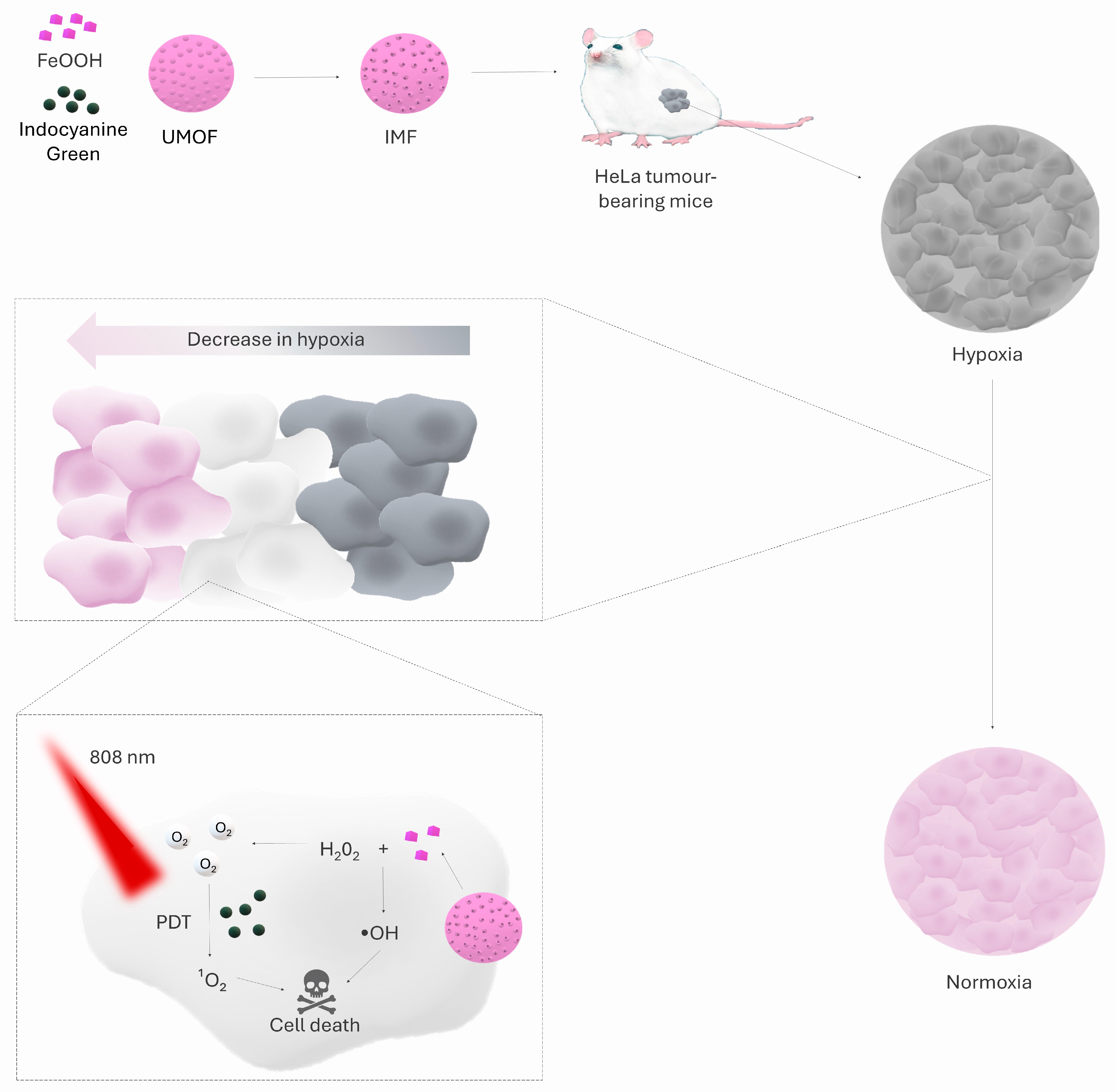
Coordinated iron ions exhibit artificial enzymatic activity [77] and can function in a similar manner to catalase. This property was exploited by Gao et al. [78] by synthesising an IMF nanoparticle (ICG ⊂ UMOF@FeOOH). Its structural core is a modified UiO-66-(COOH)2 metal-organic framework (UMOF), developed from a hexameric Zr6O4(OH)4(CO2)12 cluster linked by a 1,2,4,5-benzenetetetracarboxylic ligand (H4btec). A thin amorphous FeOOH film was deposited on the UMOF surface, acting as catalase-like active centres, due to the strong coordination of Fe3+ ions with -COOH groups. Indocyanine green was loaded into the UMOF pore. In vitro studies on HeLa cells showed that the IMF nanoparticle generated molecular oxygen under both normoxia (21% O2) and hypoxia (2% O2) conditions, translating into a significant reduction in hypoxia-induced factor HIF-1α levels. Further analyses revealed that immobilisation of the amorphous FeOOH shell on the UMOF support (UMOF@FeOOH) increased the degradation efficiency of H2O2 compared to free FeOOH, indicating an enhancement of catalase-like activity due to the coordination of Fe3+ ions with the carboxyl groups of UMOF. In vivo studies in a mouse model of subcutaneous HeLa xenografts confirmed the key role of FeOOH in the IMF system. Animals treated with photodynamic therapy using IMF and 808 nm laser irradiation showed a clear inhibition of tumour growth within the first two days after treatment, followed by a systematic reduction in tumour volume in the absence of further interventions. In the control group receiving IM nanoparticles (UMOF + ICG, devoid of FeOOH) and an identical irradiation regimen, no inhibition of tumour growth was observed, and tumour volume continued to increase. Moreover, two out of three mice in the IMF + laser group experienced complete tumour regression, providing promising evidence that overcoming hypoxia of the tumour microenvironment with FeOOH catalase-like activity can lead not only to inhibition of tumour growth but even to its complete eradication in a mouse model. Figure 2 shows the structure of IMF nanoparticles and their therapeutic mechanism [78].

3. Oxygen Generator

Endoperoxides

Endoperoxides are compounds in which two oxygen atoms form a peroxide bridge (-O-O-) within a cyclic structure. They occur both as natural products and as synthetic compounds [79,80,81]. Cleavage of the -O-O- bridge leads to the release of molecular O2, which can modulate tissue hypoxia and improve cell survival in anoxic models [79]. Furthermore, some endoperoxides also exhibit anticancer properties through the production of reactive oxygen species [82,83]. Oxygen source endoperoxides were used in the work by Zhou et al. [84]. They developed M1-EPO-NPs as a triangular metallacycle with three aza-BODIPY units conjugated by acetylides to Pt(II) complexes. Two 1,4-dimethylanthracene groups were attached to each aza-BODIPY, storing singlet oxygen as endoperoxides and releasing it under heat. The whole was surrounded by DSPE-PEG2000 phospholipids. Compared to nanoparticles without endoperoxide groups, M1-EPO-NPs generated singlet oxygen under both normoxic (21% O2) and hypoxic (5% O2) conditions after laser irradiation, whereas for nanoparticles without endoperoxides, 1O2 production was only observed under normoxia. The increased efficiency of reactive oxygen species generation translated into stronger cytotoxicity against HeLa cells in vitro, with apoptosis rates of 71.8% in 21% O2 and 56.5% in 5% O2 for M1-EPO-NPs, compared to 62.6% (normoxia) and 44.3% (hypoxia) for endoperoxide-free nanoparticles. In a mouse model of HeLa tumours, M1-EPO-NPs nanoparticles led to complete tumour regression, whereas analogous formulations without endoperoxides failed to induce complete tumour eradication [84].

4. Inhibition of Mitochondrial Respiration

Mitochondrial respiration is the main process of oxygen consumption by living cells [85]. Although under hypoxia, tumour cells adaptively increase anaerobic glycolysis, the mitochondria still account for much of the ATP production by oxidative phosphorylation (OXPHOS) and consume most of the available O2 [86]. In view of this, strategies to pharmacologically inhibit the respiratory chain may temporarily reduce mitochondrial oxygen consumption, allowing oxygen to accumulate and be used more efficiently during PDT [87,88]. Two approaches have been used for this purpose in cervical cancer PDT: nitric oxide and atovaquone.

4.1. Nitrogen Monoxide (II)

Low levels of NO drive oncogenic pathways, immunosuppression, metastasis, and angiogenesis, while higher levels lead to apoptosis and reduced hypoxia, and sensitise tumour cells to conventional therapies [89]. The main NO derivatives, such as nitrogen dioxide and peroxynitrite, cause cell death by inducing protein and lipid peroxidation and/or DNA damage [90]. Importantly, excess NO can also affect mitochondrial respiration as it binds to cytochrome c oxidase, a critical component of the mitochondrial electron transport chain [91,92]. For this reason, NO generation may be an attractive form of increasing oxygen availability for PDT. A strategy based on the use of NO as a means of inhibiting mitochondrial respiration was used by Wang et al. [93] and Lin et al. [94] However, the source of NO differs in their work. To generate NO, Wang et al. used Roussin’s Black Salt [93]. Roussin’s Black Salt is a metal–nitrosine complex that releases NO upon illumination. Approximately 3.7 molecules of NO are released from one molecule of Roussin’s Black Salt. The relatively high cytotoxicity of Roussin’s Black Salt in the dark, however, precludes its use as a systemic anticancer agent in vivo, unless applied topically [95]. Designed by Wang et al. [93], the UM-RZ nanoparticle is composed of a core–shell–shell multilayer UCNP nanocrystal (NaErF4:Tm@NaYF4@NaYbF4:Tm@NaYF4), which converts near-infrared light into visible signals, asymmetrically deposited on a half-core shell of mesoporous silica, the photosensitiser zinc phthalocyanine, and Roussin’s Black Salt, described earlier. The mechanism of action of the UM-RZ nanoparticle relies on sequential activation. First, 980 nm light induces NO release, and then 808 nm light activates zinc phthalocyanine. In vitro studies conducted on HeLa cells demonstrated the high efficacy of using a nanoparticle containing Roussin’s Black Salt. Using only 808 nm radiation (ZnPc activation and ROS generation), a typical PDT-induced decrease in oxygen levels to ≈20% was observed, while earlier exposure of samples to 980 nm light (UM-RZ activation and NO release) resulted in inhibition of mitochondrial respiration, which maintained oxygen levels at a significantly higher level of ≈70%. In addition, NO generation from UM-RZ promoted active movement of nanoparticles and accelerated their internalisation by cells. Importantly, this approach also proved effective under hypoxia; while the use of the ‘808→980 nm’ sequence or simultaneous irradiation did not significantly improve the therapeutic effect, the ‘980→808 nm’ strategy (NO release first, followed by ROS generation) led to significant cytotoxicity under hypoxia (survival rate ≈ 15%), comparable to the effect observed in normoxia (≈9%), demonstrating an effective breakthrough of the hypoxia barrier. Results from in vivo studies on a mouse model of HeLa tumours confirmed that sequential activation of UM-RZ with 980 nm light, followed by 808 nm light, significantly increased the efficacy of PDT, leading to more than 86% inhibition of tumour growth [93].
For NO generation, Lin et al. used L-arginine [94]. L-arginine is a naturally occurring amino acid that is biocompatible, inexpensive, and produces a large number of NO molecules when catalysed by reactive oxygen species in PDT [96,97]. The UCN@mSiO2@ZnPc@L-Arg nanoparticle constructed by Lin et al. [94] consists of an upconverting NaYF4 core doped with Yb3+, Er3+, and Gd3+ ions, surrounded by two shells: NaLuF4:Y3+ and NaYF4. The whole was coated with mesoporous silicon oxide, allowing the loading of zinc phthalocyanine and L-arginine. In in vitro studies using HeLa cells, it was shown that the presence of L-arginine enables NO generation and modifies the kinetics of reactive oxygen species, as some oxygen radicals are consumed for the production of NO and nitro-oxygen derivatives. At the same time, it was confirmed that the level of intracellular ROS generation remains sufficient to induce a therapeutic effect. In the ONOO-specific fluorescence probe assay, only a weak signal was noted after irradiation with 980 nm light, suggesting the formation of small amounts of ONOO-. Furthermore, the presence of L-arginine was found to enhance the cytotoxic effects of PDT both in vitro and in vivo in a mouse model of HeLa tumours, compared to nanoparticles lacking L-arginine. However, it should be noted that the authors did not assess the effect of generated NO on mitochondrial respiration or oxygen bioavailability, which prevents a clear interpretation of the role of L-arginine as a strategy to counteract hypoxia [94].

4.2. Atovaquone

Atovaquone is a clinically approved antimalarial drug [98], which inhibits cytochrome bc1 function in the mitochondrial respiratory chain. By blocking oxidative phosphorylation [99], the main ATP production pathway in cancer cells [99], it alleviates tumour hypoxia and promotes the effectiveness of PDT [100,101]. Atovaquone was used in a nanoparticle by Gao et al. [102]. TNPs/IA is based on spherical nanostructures formed from mPEG-PLGA and PLGA-b-PEG copolymers modified with a triphenylphosphonium group. Two active compounds were encapsulated: IR780, a third-generation photosensitiser activated by NIR light (808 nm), and atovaquone. In vitro studies on HeLa cell lines showed that TNPs/IA effectively inhibited mitochondrial oxygen consumption. In TNPs/IA-treated cells, ATP levels decreased to only 37.8% of the initial value, providing convincing evidence for blocking the respiratory chain in mitochondria. Furthermore, under hypoxia, TNPs/IA generated the highest concentrations of reactive oxygen species relative to the control groups, as a result of the simultaneous inhibition of OXPHOS and the increased availability and precise localisation of IR780 in the mitochondria. The control groups lacking atovaquone (free IR780, NPs/I) and the group containing atovaquone, but without mitochondriotropic ligand (NPs/IA), showed significantly weaker effects both in vitro and in vivo on the mouse HeLa tumour model. This confirms that the synergistic combination of the OXPHOS inhibitor atovaquone and mitochondrial targeting is key to increasing oxygen availability in tumour cells and maximising the efficacy of PDT [102]. Table 2 shows summary of nanoparticles described in the article.

5. Conclusions

PDT is currently one of the promising minimally invasive treatments for cancer, including cervical cancer, due to its high selectivity of action and low systemic toxicity. However, its efficacy is often limited by hypoxia, commonly observed in solid tumours. To counter this phenomenon, over the past year, nanomedicine-based innovative strategies have been developed to increase oxygenation of the tumour microenvironment. Notable among these are catalytic oxygen generation from hydrogen peroxide, oxygen storage and controlled release, and inhibition of mitochondrial oxygen consumption in tumour cells. Preclinical studies have unequivocally demonstrated that all of these approaches result in an increase in oxygen concentration within the tumour and, consequently, significantly potentiate the efficacy of PDT. Importantly, the different approaches may have their own additional advantages, serving other functions, as well. MnO2-based nanomaterials, which act as a mesoporous carrier core, in addition to reversing hypoxia, increase the conversion of NIR radiation into heat, enhancing the photothermal effect, and release Mn2+ ions, initiating the Fenton reaction and generating hydroxyl radicals. Au2Pt hybrid systems exhibit peroxidase-like activity in the acidic environment of lysosomes, catalysing the formation of hydroxyl radicals (-OH) and thus further enhancing the efficacy of the therapy. The release of NO by Roussin’s Black Salt promotes the active transport of nanoparticles and accelerates their internalisation by cells, while NO generated from L-arginine initiates the formation of highly reactive ONOO- radicals. Moreover, the integration of these solutions in multifunctional platforms combining photodynamic, photothermal, and chemodynamic mechanisms allows not only to overcome the multidimensional barriers of cellular resistance but also to precisely target subsequent therapeutic agents while minimising damage to healthy tissues. Despite promising results from in vitro and in vivo studies, further rigorous safety, biocompatibility, and efficacy testing in preclinical models remains crucial to fully confirm the suitability of these nanosystems in clinical practice. In light of the accumulated evidence, nanocarriers that modulate oxygen levels in the tumour microenvironment provide a solid foundation for future, more selective and individualised photodynamic treatment strategies for cervical cancer, with the prospect of extending their application to other solid tumours.

6. Recommendations

Based on our analysis, we recommend that further work on nanomaterials for hypoxia modulation in photodynamic cancer therapy should focus primarily on the development of catalytic platforms that degrade H2O2 (e.g., MnO2, FeOOH) and on oxygen storage and controlled release systems (endoperoxides), which have shown the most consistent improvement in oxygenation and proven therapeutic benefit in preclinical studies. In parallel, we advocate the design of multifunctional systems combining local oxygen production with mechanisms that limit oxygen consumption by tumour cells (e.g., encapsulation of OXPHOS inhibitors or controlled release of NO), as data show synergy of these approaches in enhancing ROS generation and PDT efficacy. To facilitate comparability of results and expedite the identification of candidates for clinical translation, we recommend a minimum reporting standard including pO2 pre- and post-therapy measurements, quantification of HIF-1α expression, quantification of ROS, measures of efficacy (e.g., % inhibition or tumor regression), and complete biodistribution and toxicology data. In addition, we recommend direct head-to-head comparisons of the most promising constructs in the same models and protocols, and expanded pharmacokinetic studies and long-term safety assessments (metal ion fate, material degradation, immunotoxicity). Finally, it is advisable to study the optimal sequence and timing of activation of multifunctional components (e.g., staged release of NO or inhibition of OXPHOS prior to activation of the photosensitiser), since the sequence of events adopted has a significant impact on the oxygenation of the microenvironment and the therapeutic outcome.

Author Contributions

Conceptualization, D.B.-A., M.A.S., A.P., P.W. and D.A.; methodology, D.B.-A., M.A.S., A.P., P.W. and D.A.; formal analysis, D.B.-A., M.A.S., A.P., P.W. and D.A.; investigation, D.B.-A., M.A.S., A.P., P.W. and D.A.; resources, D.B.-A., M.A.S., A.P., P.W. and D.A.; data curation, D.B.-A., M.A.S., A.P., P.W. and D.A.; writing—original draft preparation, D.B.-A., M.A.S., A.P., P.W. and D.A.; writing—review and editing, D.B.-A., M.A.S., A.P., P.W. and D.A.; visualization, D.B.-A., M.A.S., A.P., P.W. and D.A.; supervision, D.A.; funding acquisition, D.A. All authors have read and agreed to the published version of the manuscript.

Funding

This research received no external funding.

Conflicts of Interest

The authors declare no conflicts of interest.

References

  1. Arbyn, M.; Weiderpass, E.; Bruni, L.; de Sanjosé, S.; Saraiya, M.; Ferlay, J.; Bray, F. Estimates of Incidence and Mortality of Cervical Cancer in 2018: A Worldwide Analysis. Lancet Glob. Health 2020, 8, e191–e203, Correction in Lancet Glob. Health 2022, 10, e41. [Google Scholar] [CrossRef]
  2. Cervical Cancer Overview: Prevalence and Differences—ClinicalKey. Available online: https://www.Clinicalkey.com/#!/Content/playContent/1-S2.0-S0027968420300432?scrollTo=%23bib1 (accessed on 25 June 2025).
  3. Leitlinienprogramm Onkologie (Deutsche Krebsgesellschaft, Deutsche Krebshilfe, AWMF): S3-Leitlinie Diagnostik, Therapie Und Nachsorge Der Patientin Mit Zervixkarzinom, Langversion, 2.2. 2022; AWMF-Registernummer: 032/033OL. Available online: https://www.Leitlinienprogramm-Onkologie.de (accessed on 17 August 2022).
  4. Ibeanu, O.A. Molecular Pathogenesis of Cervical Cancer. Cancer Biol. Ther. 2011, 11, 295–306. [Google Scholar] [CrossRef]
  5. Schubert, M.; Bauerschlag, D.O.; Muallem, M.Z.; Maass, N.; Alkatout, I. Challenges in the Diagnosis and Individualized Treatment of Cervical Cancer. Medicina 2023, 59, 925. [Google Scholar] [CrossRef]
  6. Yadav, G.; Srinivasan, G.; Jain, A. Cervical Cancer: Novel Treatment Strategies Offer Renewed Optimism. Pathol.-Res. Pract. 2024, 254, 155136. [Google Scholar] [CrossRef]
  7. Kashyap, N.; Krishnan, N.; Kaur, S.; Ghai, S. Risk Factors of Cervical Cancer: A Case-Control Study. Asia Pac. J. Oncol. Nurs. 2019, 6, 308–314. [Google Scholar] [CrossRef]
  8. McCluggage, W.G.; Singh, N.; Gilks, C.B. Key Changes to the World Health Organization (WHO) Classification of Female Genital Tumours Introduced in the 5th Edition (2020). Histopathology 2022, 80, 762–778. [Google Scholar] [CrossRef]
  9. Raspollini, M.R.; Lax, S.F.; McCluggage, W.G. The Central Role of the Pathologist in the Management of Patients with Cervical Cancer: ESGO/ESTRO/ESP Guidelines. Virchows Arch. 2018, 473, 45–54. [Google Scholar] [CrossRef] [PubMed]
  10. Meng, Y.; Chu, T.; Lin, S.; Wu, P.; Zhi, W.; Peng, T.; Ding, W.; Luo, D.; Wu, P. Clinicopathological Characteristics and Prognosis of Cervical Cancer with Different Histological Types: A Population-Based Cohort Study. Gynecol. Oncol. 2021, 163, 545–551. [Google Scholar] [CrossRef] [PubMed]
  11. Cao, L.; Wen, H.; Feng, Z.; Han, X.; Wu, X. Distinctive Clinicopathologic Characteristics and Prognosis for Different Histologic Subtypes of Early Cervical Cancer. Int. J. Gynecol. Cancer 2019, 29, 1244–1251. [Google Scholar] [CrossRef] [PubMed]
  12. NCCN Guidelines for Cervical Cancer n.d. Available online: https://www.Nccn.org/Professionals/Physician_gls/Pdf/Cervical.Pdf (accessed on 18 June 2023).
  13. Kokka, F.; Bryant, A.; Brockbank, E.; Jeyarajah, A. Surgical Treatment of Stage IA2 Cervical Cancer. Cochrane Database Syst. Rev. 2014, 2014, CD010870. [Google Scholar] [CrossRef]
  14. Dicu-Andreescu, I.-G.; Marincaș, A.-M.; Ungureanu, V.-G.; Ionescu, S.-O.; Prunoiu, V.-M.; Brătucu, E.; Simion, L. Current Therapeutic Approaches in Cervical Cancer Based on the Stage of the Disease: Is There Room for Improvement? Medicina 2023, 59, 1229. [Google Scholar] [CrossRef] [PubMed]
  15. Usami, T.; Takahashi, A.; Matoda, M.; Okamoto, S.; Kondo, E.; Kanao, H.; Umayahara, K.; Takeshima, N. Review of Treatment and Prognosis of Stage IVB Cervical Carcinoma. Int. J. Gynecol. Cancer 2016, 26, 1239–1245. [Google Scholar] [CrossRef] [PubMed]
  16. Shrivastava, S.; Mahantshetty, U.; Engineer, R.; Chopra, S.; Hawaldar, R.; Hande, V.; Kerkar, R.A.; Maheshwari, A.; Shylasree, T.S.; Ghosh, J.; et al. Cisplatin Chemoradiotherapy vs Radiotherapy in FIGO Stage IIIB Squamous Cell Carcinoma of the Uterine Cervix: A Randomized Clinical Trial. JAMA Oncol. 2018, 4, 506–513. [Google Scholar] [CrossRef] [PubMed]
  17. Yamashita, H.; Okuma, K.; Kawana, K.; Nakagawa, S.; Oda, K.; Yano, T.; Kobayashi, S.; Wakui, R.; Ohtomo, K.; Nakagawa, K. Comparison between Conventional Surgery plus Postoperative Adjuvant Radiotherapy and Concurrent Chemoradiation for FIGO Stage IIB Cervical Carcinoma: A Retrospective Study. Am. J. Clin. Oncol. 2010, 33, 583–586. [Google Scholar] [CrossRef]
  18. Poddar, P.; Maheshwari, A. Surgery for Cervical Cancer: Consensus & Controversies. Indian. J. Med. Res. 2021, 154, 284–292. [Google Scholar] [CrossRef]
  19. dos Santos, A.F.; de Almeida, D.R.Q.; Terra, L.F.; Baptista, M.S.; Labriola, L. Photodynamic therapy in cancer treatment—An update review. J. Cancer Metastasis Treat. 2019, 5, 25. [Google Scholar] [CrossRef]
  20. Jiang, W.; Liang, M.; Lei, Q.; Li, G.; Wu, S. The current status of photodynamic therapy in cancer treatment. Cancers 2023, 15, 585. [Google Scholar] [CrossRef]
  21. Aebisher, D.; Przygórzewska, A.; Myśliwiec, A.; Dynarowicz, K.; Krupka-Olek, M.; Bożek, A.; Kawczyk-Krupka, A.; Bartusik-Aebisher, D. Current photodynamic therapy for glioma treatment: An update. Biomedicines 2024, 12, 375. [Google Scholar] [CrossRef]
  22. Tavakkoli Yaraki, M.; Liu, B.; Tan, Y.N. Emerging strategies in enhancing singlet oxygen generation of nano-photosensitizers toward advanced phototherapy. Nano-Micro Lett. 2022, 14, 123. [Google Scholar] [CrossRef]
  23. Correia, J.H.; Rodrigues, J.A.; Pimenta, S.; Dong, T.; Yang, Z. Photodynamic Therapy Review: Principles, Photosensitizers, Applications, and Future Directions. Pharmaceutics 2021, 13, 1332. [Google Scholar] [CrossRef]
  24. Mansoori, B.; Mohammadi, A.; Amin Doustvandi, M.; Mohammadnejad, F.; Kamari, F.; Gjerstorff, M.F.; Baradaran, B.; Hamblin, M.R. Photodynamic Therapy for Cancer: Role of Natural Products. Photodiagnosis Photodyn. Ther. 2019, 26, 395–404. [Google Scholar] [CrossRef]
  25. Austin, E.; Wang, J.Y.; Ozog, D.M.; Zeitouni, N.; Lim, H.W.; Jagdeo, J. Photodynamic therapy: Overview and mechanism of action. J. Am. Acad. Dermatol. 2025; in press. [Google Scholar] [CrossRef]
  26. De Silva, P.; Saad, M.A.; Thomsen, H.C.; Bano, S.; Ashraf, S.; Hasan, T. Photodynamic therapy, priming and optical imaging: Potential co-conspirators in treatment design and optimization—A Thomas Dougherty Award for Excellence in PDT paper. J. Porphyr. Phthalocyanines 2020, 24, 1320–1360. [Google Scholar] [CrossRef]
  27. Du, Y.; Han, J.; Jin, F.; Du, Y. Recent Strategies to Address Hypoxic Tumor Environments in Photodynamic Therapy. Pharmaceutics 2022, 14, 1763. [Google Scholar] [CrossRef]
  28. Li, M.; Xu, Y.; Peng, X.; Kim, J.S. From low to no O2-dependent hypoxia photodynamic therapy (hPDT): A new perspective. Acc. Chem. Res. 2022, 55, 3253–3264. [Google Scholar] [CrossRef]
  29. Pucelik, B.; Sułek, A.; Barzowska, A.; Dąbrowski, J.M. Recent Advances in Strategies for Overcoming Hypoxia in Photodynamic Therapy of Cancer. Cancer Lett. 2020, 492, 116–135. [Google Scholar] [CrossRef]
  30. Hong, L.; Li, J.; Luo, Y.; Guo, T.; Zhang, C.; Ou, S.; Long, Y.; Hu, Z. Recent Advances in Strategies for Addressing Hypoxia in Tumor Photodynamic Therapy. Biomolecules 2022, 12, 81. [Google Scholar] [CrossRef] [PubMed]
  31. Hong, L.; Pliss, A.M.; Zhan, Y.; Zheng, W.; Xia, J.; Liu, L.; Qu, J.; Prasad, P.N. Perfluoropolyether Nanoemulsion Encapsulating Chlorin E6 for Sonodynamic and Photodynamic Therapy of Hypoxic Tumor. Nanomaterials 2020, 10, 2058. [Google Scholar] [CrossRef] [PubMed]
  32. Sun, Y.; Zhao, D.; Wang, G.; Wang, Y.; Cao, L.; Sun, J.; Jiang, Q.; He, Z. Recent Progress of Hypoxia-Modulated Multifunctional Nanomedicines to Enhance Photodynamic Therapy: Opportunities, Challenges, and Future Development. Acta Pharm. Sin. B 2020, 10, 1382–1396. [Google Scholar] [CrossRef] [PubMed]
  33. Moulder, J.E.; Rockwell, S. Tumor Hypoxia: Its Impact on Cancer Therapy. Cancer Metastasis Rev. 1987, 5, 313–341. [Google Scholar] [CrossRef]
  34. Jahanban-Esfahlan, R.; de la Guardia, M.; Ahmadi, D.; Yousefi, B. Modulating Tumor Hypoxia by Nanomedicine for Effective Cancer Therapy. J. Cell Physiol. 2018, 233, 2019–2031. [Google Scholar] [CrossRef]
  35. Feng, L.; Cheng, L.; Dong, Z.; Tao, D.; Barnhart, T.E.; Cai, W.; Chen, M.; Liu, Z. Theranostic Liposomes with Hypoxia-Activated Prodrug to Effectively Destruct Hypoxic Tumors Post-Photodynamic Therapy. ACS Nano 2017, 11, 927–937. [Google Scholar] [CrossRef]
  36. Du, J.; Shi, T.; Long, S.; Chen, P.; Sun, W.; Fan, J.; Peng, X. Enhanced Photodynamic Therapy for Overcoming Tumor Hypoxia: From Microenvironment Regulation to Photosensitizer Innovation. Coord. Chem. Rev. 2021, 427, 213604. [Google Scholar] [CrossRef]
  37. Li, X.; Chen, L.; Huang, M.; Zeng, S.; Zheng, J.; Peng, S.; Wang, Y.; Cheng, H.; Li, S. Innovative Strategies for Photodynamic Therapy against Hypoxic Tumor. Asian J. Pharm. Sci. 2023, 18, 100775. [Google Scholar] [CrossRef]
  38. Hsu, Y.-L.; Hung, J.-Y.; Chang, W.-A.; Lin, Y.-S.; Pan, Y.-C.; Tsai, P.-H.; Wu, C.-Y.; Kuo, P.-L. Hypoxic Lung Cancer-Secreted Exosomal miR-23a Increased Angiogenesis and Vascular Permeability by Targeting Prolyl Hydroxylase and Tight Junction Protein ZO-1. Oncogene 2017, 36, 4929–4942. [Google Scholar] [CrossRef]
  39. Gilkes, D.M.; Semenza, G.L.; Wirtz, D. Hypoxia and the Extracellular Matrix: Drivers of Tumour Metastasis. Nat. Rev. Cancer 2014, 14, 430–439. [Google Scholar] [CrossRef] [PubMed]
  40. Wang, W.; Cheng, Y.; Yu, P.; Wang, H.; Zhang, Y.; Xu, H.; Ye, Q.; Yuan, A.; Hu, Y.; Wu, J. Perfluorocarbon Regulates the Intratumoural Environment to Enhance Hypoxia-Based Agent Efficacy. Nat. Commun. 2019, 10, 1580. [Google Scholar] [CrossRef] [PubMed]
  41. Yao, C.; Wang, W.; Wang, P.; Zhao, M.; Li, X.; Zhang, F. Near-Infrared Upconversion Mesoporous Cerium Oxide Hollow Biophotocatalyst for Concurrent pH-/H2 O2 -Responsive O2 -Evolving Synergetic Cancer Therapy. Adv. Mater. 2018, 30, 1704833. [Google Scholar] [CrossRef] [PubMed]
  42. Cheng, Y.; Cheng, H.; Jiang, C.; Qiu, X.; Wang, K.; Huan, W.; Yuan, A.; Wu, J.; Hu, Y. Perfluorocarbon Nanoparticles Enhance Reactive Oxygen Levels and Tumour Growth Inhibition in Photodynamic Therapy. Nat. Commun. 2015, 6, 8785. [Google Scholar] [CrossRef]
  43. Song, X.; Feng, L.; Liang, C.; Gao, M.; Song, G.; Liu, Z. Liposomes Co-Loaded with Metformin and Chlorin E6 Modulate Tumor Hypoxia during Enhanced Photodynamic Therapy. Nano Res. 2017, 10, 1200–1212. [Google Scholar] [CrossRef]
  44. Liu, J.; Liang, H.; Li, M.; Luo, Z.; Zhang, J.; Guo, X.; Cai, K. Tumor Acidity Activating Multifunctional Nanoplatform for NIR-Mediated Multiple Enhanced Photodynamic and Photothermal Tumor Therapy. Biomaterials 2018, 157, 107–124. [Google Scholar] [CrossRef]
  45. Zhang, M.; Cui, Z.; Song, R.; Lv, B.; Tang, Z.; Meng, X.; Chen, X.; Zheng, X.; Zhang, J.; Yao, Z.; et al. SnWO4-Based Nanohybrids with Full Energy Transfer for Largely Enhanced Photodynamic Therapy and Radiotherapy. Biomaterials 2018, 155, 135–144. [Google Scholar] [CrossRef]
  46. Zhou, T.-J.; Xing, L.; Fan, Y.-T.; Cui, P.-F.; Jiang, H.-L. Light Triggered Oxygen-Affording Engines for Repeated Hypoxia-Resistant Photodynamic Therapy. J. Control. Release 2019, 307, 44–54. [Google Scholar] [CrossRef]
  47. Zhou, R.; Zeng, X.; Zhao, H.; Chen, Q.; Wu, P. Combating the Hypoxia Limit of Photodynamic Therapy through Reversing the Survival-Related Pathways of Cancer Cells. Coord. Chem. Rev. 2022, 452, 214306. [Google Scholar] [CrossRef]
  48. Yan, W.; Lang, T.; Zhu, R.; Zhu, X.; Li, Y.; Wu, T.; Yin, Q.; Li, Y. Anti-Hypoxia Nanosized Drug Delivery Systems Improving Cancer Therapy. Nano Today 2022, 42, 101376. [Google Scholar] [CrossRef]
  49. Chen, J.; Ding, Z.; Peng, Y.; Pan, F.; Li, J.; Zou, L.; Zhang, Y.; Liang, H. HIF-1α Inhibition Reverses Multidrug Resistance in Colon Cancer Cells via Downregulation of MDR1/P-Glycoprotein. PLoS ONE 2014, 9, e98882. [Google Scholar] [CrossRef] [PubMed]
  50. Dean, M.; Annilo, T. Evolution of the ATP-Binding Cassette (ABC) Transporter Superfamily in Vertebrates. Annu. Rev. Genom. Hum. Genet. 2005, 6, 123–142. [Google Scholar] [CrossRef]
  51. Cerrada, I.; Ruiz-Saurí, A.; Carrero, R.; Trigueros, C.; Dorronsoro, A.; Sanchez-Puelles, J.M.; Diez-Juan, A.; Montero, J.A.; Sepúlveda, P. Hypoxia-Inducible Factor 1 Alpha Contributes to Cardiac Healing in Mesenchymal Stem Cells-Mediated Cardiac Repair. Stem Cells Dev. 2013, 22, 501–511. [Google Scholar] [CrossRef]
  52. Gui, L.; Liu, B.; Lv, G. Hypoxia Induces Autophagy in Cardiomyocytes via a Hypoxia-Inducible Factor 1-Dependent Mechanism. Exp. Ther. Med. 2016, 11, 2233–2239. [Google Scholar] [CrossRef]
  53. Zhou, J.; Li, C.; Yao, W.; Alsiddig, M.C.; Huo, L.; Liu, H.; Miao, Y.-L. Hypoxia-Inducible Factor-1α-Dependent Autophagy Plays a Role in Glycolysis Switch in Mouse Granulosa Cells. Biol. Reprod. 2018, 99, 308–318. [Google Scholar] [CrossRef]
  54. Abdul Rahim, S.A.; Dirkse, A.; Oudin, A.; Schuster, A.; Bohler, J.; Barthelemy, V.; Muller, A.; Vallar, L.; Janji, B.; Golebiewska, A.; et al. Regulation of Hypoxia-Induced Autophagy in Glioblastoma Involves ATG9A. Br. J. Cancer 2017, 117, 813–825. [Google Scholar] [CrossRef] [PubMed]
  55. Jing, X.; Yang, F.; Shao, C.; Wei, K.; Xie, M.; Shen, H.; Shu, Y. Role of Hypoxia in Cancer Therapy by Regulating the Tumor Microenvironment. Mol. Cancer 2019, 18, 157. [Google Scholar] [CrossRef] [PubMed]
  56. Kim, D.-H.; Sarbassov, D.D.; Ali, S.M.; King, J.E.; Latek, R.R.; Erdjument-Bromage, H.; Tempst, P.; Sabatini, D.M. mTOR Interacts with Raptor to Form a Nutrient-Sensitive Complex That Signals to the Cell Growth Machinery. Cell 2002, 110, 163–175. [Google Scholar] [CrossRef] [PubMed]
  57. Jung, C.H.; Ro, S.-H.; Cao, J.; Otto, N.M.; Kim, D.-H. mTOR Regulation of Autophagy. FEBS Lett. 2010, 584, 1287–1295. [Google Scholar] [CrossRef]
  58. Rosin, F.C.P.; Teixeira, M.G.; Pelissari, C.; Corrêa, L. Resistance of Oral Cancer Cells to 5-ALA-Mediated Photodynamic Therapy. J. Cell Biochem. 2018, 119, 3554–3562. [Google Scholar] [CrossRef]
  59. Li, Y.; Zhou, R.; Xiao, D.; Shi, S.; Peng, S.; Wu, S.; Wu, P.; Lin, Y. Polypeptide Uploaded Efficient Nanophotosensitizers to Overcome Photodynamic Resistance for Enhanced Anticancer Therapy. Chem. Eng. J. 2021, 403, 126344. [Google Scholar] [CrossRef]
  60. Najahi-Missaoui, W.; Arnold, R.D.; Cummings, B.S. Safe Nanoparticles: Are We There Yet? Int. J. Mol. Sci. 2020, 22, 385. [Google Scholar] [CrossRef]
  61. Auffan, M.; Rose, J.; Bottero, J.-Y.; Lowry, G.V.; Jolivet, J.-P.; Wiesner, M.R. Towards a Definition of Inorganic Nanoparticles from an Environmental, Health and Safety Perspective. Nat. Nanotechnol. 2009, 4, 634–641. [Google Scholar] [CrossRef]
  62. Gwinn, M.R.; Vallyathan, V. Nanoparticles: Health Effects—Pros and Cons. Environ. Health Perspect. 2006, 114, 1818–1825. [Google Scholar] [CrossRef]
  63. Szatrowski, T.P.; Nathan, C.F. Production of Large Amounts of Hydrogen Peroxide by Human Tumor Cells1. Cancer Res. 1991, 51, 794–798. [Google Scholar]
  64. Liu, C.-P.; Wu, T.-H.; Liu, C.-Y.; Chen, K.-C.; Chen, Y.-X.; Chen, G.-S.; Lin, S.-Y. Self-Supplying O2 through the Catalase-Like Activity of Gold Nanoclusters for Photodynamic Therapy against Hypoxic Cancer Cells. Small 2017, 13, 1700278. [Google Scholar] [CrossRef] [PubMed]
  65. Kobayashi, Y.; Nishikawa, M.; Hyoudou, K.; Yamashita, F.; Hashida, M. Hydrogen Peroxide-mediated Nuclear Factor κB Activation in Both Liver and Tumor Cells during Initial Stages of Hepatic Metastasis. Cancer Sci. 2008, 99, 1546–1552. [Google Scholar] [CrossRef]
  66. Bonello, S.; Zähringer, C.; BelAiba, R.S.; Djordjevic, T.; Hess, J.; Michiels, C.; Kietzmann, T.; Görlach, A. Reactive Oxygen Species Activate the HIF-1alpha Promoter via a Functional NFkappaB Site. Arterioscler. Thromb. Vasc. Biol. 2007, 27, 755–761. [Google Scholar] [CrossRef] [PubMed]
  67. Ma, Z.; Jia, X.; Bai, J.; Ruan, Y.; Wang, C.; Li, J.; Zhang, M.; Jiang, X. MnO2 Gatekeeper: An Intelligent and O2-Evolving Shell for Preventing Premature Release of High Cargo Payload Core, Overcoming Tumor Hypoxia, and Acidic H2O2-Sensitive MRI. Adv. Funct. Mater. 2017, 27, 1604258. [Google Scholar] [CrossRef]
  68. Yang, Y.; Zhang, X.; Bai, Z.; Cui, Z.; Liang, W.; Zhang, J.; Li, K.; Shi, M.; Liu, Z.; Wang, J.; et al. Progressive Enhanced Photodynamic Therapy and Enhanced Chemotherapy Fighting against Malignant Tumors with Sequential Drug Release. Biomed. Mater. 2024, 19, 045004. [Google Scholar] [CrossRef]
  69. Cai, J.; Yang, Y.; Zhang, J.; Bai, Z.; Zhang, X.; Li, K.; Shi, M.; Liu, Z.; Gao, L.; Wang, J.; et al. Multilayer Nanodrug Delivery System with Spatiotemporal Drug Release Improves Tumor Microenvironment for Synergistic Anticancer Therapy. Biofabrication 2024, 16, 025012. [Google Scholar] [CrossRef]
  70. Wang, Y.; Xu, Y.; Song, J.; Liu, X.; Liu, S.; Yang, N.; Wang, L.; Liu, Y.; Zhao, Y.; Zhou, W.; et al. Tumor Cell-Targeting and Tumor Microenvironment–Responsive Nanoplatforms for the Multimodal Imaging-Guided Photodynamic/Photothermal/Chemodynamic Treatment of Cervical Cancer. Int. J. Nanomed. 2024, 19, 5837–5858. [Google Scholar] [CrossRef]
  71. Fan, J.; Yin, J.-J.; Ning, B.; Wu, X.; Hu, Y.; Ferrari, M.; Anderson, G.J.; Wei, J.; Zhao, Y.; Nie, G. Direct Evidence for Catalase and Peroxidase Activities of Ferritin–Platinum Nanoparticles. Biomaterials 2011, 32, 1611–1618. [Google Scholar] [CrossRef]
  72. Park, J.; Kim, D.; Byun, S.W.; Shin, H.; Ju, Y.; Min, H.; Kim, Y.J.; Heo, I.; Hazlett, M.J.; Kim, M.; et al. Impact of Pd:Pt Ratio of Pd/Pt Bimetallic Catalyst on CH4 Oxidation. Appl. Catal. B Environ. 2022, 316, 121623. [Google Scholar] [CrossRef]
  73. Liu, B.; Liu, Z.; Li, J. X-Ray Triggered Scintillator Versatile Nanocatalytic Platform for Synergistic Photodynamic/Chemodynamic Therapy. Talanta 2025, 281, 126886. [Google Scholar] [CrossRef]
  74. Cheng, Y.; Xia, Y.-D.; Sun, Y.-Q.; Wang, Y.; Yin, X.-B. “Three-in-One” Nanozyme Composite for Augmented Cascade Catalytic Tumor Therapy. Adv. Mater. 2024, 36, 2308033. [Google Scholar] [CrossRef]
  75. Qhattal, H.S.S.; Liu, X. Characterization of CD44-Mediated Cancer Cell Uptake and Intracellular Distribution of Hyaluronan-Grafted Liposomes. Mol. Pharm. 2011, 8, 1233–1246. [Google Scholar] [CrossRef]
  76. Huotari, J.; Helenius, A. Endosome Maturation. EMBO J. 2011, 30, 3481–3500. [Google Scholar] [CrossRef] [PubMed]
  77. Feng, D.; Gu, Z.-Y.; Li, J.-R.; Jiang, H.-L.; Wei, Z.; Zhou, H.-C. Zirconium-Metalloporphyrin PCN-222: Mesoporous Metal–Organic Frameworks with Ultrahigh Stability as Biomimetic Catalysts. Angew. Chem. Int. Ed. 2012, 51, 10307–10310. [Google Scholar] [CrossRef] [PubMed]
  78. Gao, G.; Wang, Y.; Jiang, Y.; Luo, S.; Li, M.; Cao, Y.; Ma, Y.; Tang, B. A Novel Multi-Carboxyl Functionalized MOF Platform for Effective Photodynamic Therapy with Hypoxia Modulation Based on Prominent Self-Oxygen Generation. Inorg. Chem. Front. 2024, 11, 1186–1197. [Google Scholar] [CrossRef]
  79. Benz, S.; Nötzli, S.; Siegel, J.S.; Eberli, D.; Jessen, H.J. Controlled Oxygen Release from Pyridone Endoperoxides Promotes Cell Survival under Anoxic Conditions. J. Med. Chem. 2013, 56, 10171–10182. [Google Scholar] [CrossRef]
  80. Dembitsky, V.M.; Ermolenko, E.; Savidov, N.; Gloriozova, T.A.; Poroikov, V.V. Antiprotozoal and Antitumor Activity of Natural Polycyclic Endoperoxides: Origin, Structures and Biological Activity. Molecules 2021, 26, 686. [Google Scholar] [CrossRef]
  81. Bu, M.; Yang, B.B.; Hu, L. Natural Endoperoxides as Drug Lead Compounds. Curr. Med. Chem. 2016, 23, 383–405. [Google Scholar] [CrossRef]
  82. Zhu, P.; Tong, B.M.; Wang, R.; Chen, J.P.; Foo, S.; Chong, H.C.; Wang, X.L.; Ang, G.Y.; Chiba, S.; Tan, N.S. Nox4-Dependent ROS Modulation by Amino Endoperoxides to Induce Apoptosis in Cancer Cells. Cell Death Dis. 2013, 4, e552. [Google Scholar] [CrossRef]
  83. Berdelle, N.; Nikolova, T.; Quiros, S.; Efferth, T.; Kaina, B. Artesunate Induces Oxidative DNA Damage, Sustained DNA Double-Strand Breaks, and the ATM/ATR Damage Response in Cancer Cells. Mol. Cancer Ther. 2011, 10, 2224–2233. [Google Scholar] [CrossRef]
  84. Zhou, R.; Lv, W.; Li, B.; Yu, B.; Zhang, S.; Zhou, Y.; Liu, S.; Zhao, Q. Reversibly Sensitizing-Storing-Releasing 1O2 within a Single Platinum(II)-Acetylide-Based Metallacycle Molecule via Laser Power Modulation. Sci. China Chem. 2024, 67, 604–611. [Google Scholar] [CrossRef]
  85. Bettink, M.A.W.; Arbous, M.S.; Raat, N.J.H.; Mik, E.G. Mind the Mitochondria! J. Emerg. Crit. Care Med. 2019, 3, 45. [Google Scholar] [CrossRef]
  86. Zheng, J. Energy Metabolism of Cancer: Glycolysis versus Oxidative Phosphorylation (Review). Oncol. Lett. 2012, 4, 1151–1157. [Google Scholar] [CrossRef]
  87. Zhao, Y.; Lv, B.; Xue, G.; Sun, Y.; Cao, J. Smart Nanosystem-Mediated Inhibition of Mitochondrial Respiration for Enhanced Phototherapy-Induced Antitumor Immunity. Int. J. Nanomed. 2023, 18, 3443–3457. [Google Scholar] [CrossRef] [PubMed]
  88. Cen, Y.; Chen, X.; Liu, Y.; Yu, B.; Yan, M.; Yang, N.; Kong, R.; Li, S.; Ti, H.; Cheng, H. Drug Induced Mitochondria Dysfunction to Enhance Photodynamic Therapy of Hypoxic Tumors. J. Control. Release 2023, 358, 654–666. [Google Scholar] [CrossRef] [PubMed]
  89. Miranda, K.M.; Ridnour, L.A.; McGinity, C.L.; Bhattacharyya, D.; Wink, D.A. Nitric Oxide and Cancer: When to Give and When to Take Away? Inorg. Chem. 2021, 60, 15941–15947. [Google Scholar] [CrossRef] [PubMed]
  90. Kamm, A.; Przychodzen, P.; Kuban-Jankowska, A.; Jacewicz, D.; Dabrowska, A.M.; Nussberger, S.; Wozniak, M.; Gorska-Ponikowska, M. Nitric Oxide and Its Derivatives in the Cancer Battlefield. Nitric Oxide 2019, 93, 102–114. [Google Scholar] [CrossRef] [PubMed]
  91. Watson, S.A.; McStay, G.P. Functions of Cytochrome c Oxidase Assembly Factors. Int. J. Mol. Sci. 2020, 21, 7254. [Google Scholar] [CrossRef]
  92. Fry, N.L.; Mascharak, P.K. Photoactive Ruthenium Nitrosyls as NO Donors: How to Sensitize Them toward Visible Light. Acc. Chem. Res. 2011, 44, 289–298. [Google Scholar] [CrossRef]
  93. Wang, J.; Zhang, Y.; Chen, H.; Wu, Y.; Liu, J.; Che, H.; Zhang, Y.; Zhu, X. Motor-Cargo Structured Nanotractors for Augmented NIR Phototherapy via Gas-Boosted Tumor Penetration and Respiration-Impaired Mitochondrial Dysfunction. Adv. Healthc. Mater. 2024, 13, e2402063. [Google Scholar] [CrossRef]
  94. Lin, Z.; Zhu, T.; Zhong, X. NIR-Triggered NO Production Combined with Photodynamic Therapy for Tumor Treatment. Photodiagnosis Photodyn. Ther. 2024, 49, 104241. [Google Scholar] [CrossRef]
  95. Janczyk, A.; Wolnicka-Glubisz, A.; Chmura, A.; Elas, M.; Matuszak, Z.; Stochel, G.; Urbanska, K. NO-Dependent Phototoxicity of Roussin’s Black Salt against Cancer Cells. Nitric Oxide 2004, 10, 42–50. [Google Scholar] [CrossRef] [PubMed]
  96. Wan, S.-S.; Zeng, J.-Y.; Cheng, H.; Zhang, X.-Z. ROS-Induced NO Generation for Gas Therapy and Sensitizing Photodynamic Therapy of Tumor. Biomaterials 2018, 185, 51–62. [Google Scholar] [CrossRef] [PubMed]
  97. Cao, Y.; Liu, M.; Cheng, J.; Yin, J.; Huang, C.; Cui, H.; Zhang, X.; Zhao, G. Acidity-Triggered Tumor-Targeted Nanosystem for Synergistic Therapy via a Cascade of ROS Generation and NO Release. ACS Appl. Mater. Interfaces 2020, 12, 28975–28984. [Google Scholar] [CrossRef]
  98. Nixon, G.L.; Moss, D.M.; Shone, A.E.; Lalloo, D.G.; Fisher, N.; O’neill, P.M.; Ward, S.A.; Biagini, G.A. Antimalarial Pharmacology and Therapeutics of Atovaquone. J. Antimicrob. Chemother. 2013, 68, 977–985. [Google Scholar] [CrossRef] [PubMed]
  99. Ashton, T.M.; Fokas, E.; Kunz-Schughart, L.A.; Folkes, L.K.; Anbalagan, S.; Huether, M.; Kelly, C.J.; Pirovano, G.; Buffa, F.M.; Hammond, E.M.; et al. The Anti-Malarial Atovaquone Increases Radiosensitivity by Alleviating Tumour Hypoxia. Nat. Commun. 2016, 7, 12308. [Google Scholar] [CrossRef]
  100. Li, M.; Shao, Y.; Kim, J.H.; Pu, Z.; Zhao, X.; Huang, H.; Xiong, T.; Kang, Y.; Li, G.; Shao, K.; et al. Unimolecular Photodynamic O2-Economizer To Overcome Hypoxia Resistance in Phototherapeutics. J. Am. Chem. Soc. 2020, 142, 5380–5388. [Google Scholar] [CrossRef]
  101. Larue, L.; Myrzakhmetov, B.; Ben-Mihoub, A.; Moussaron, A.; Thomas, N.; Arnoux, P.; Baros, F.; Vanderesse, R.; Acherar, S.; Frochot, C. Fighting Hypoxia to Improve PDT. Pharmaceuticals 2019, 12, 163. [Google Scholar] [CrossRef]
  102. Gao, Y.; Li, Y.; Pan, Z.; Xu, C.; Zhang, X.; Li, M.; Wang, W.; Jia, F.; Wu, Y. OXPHOS-Targeted Nanoparticles for Boosting Photodynamic Therapy against Hypoxia Tumor. Int. J. Pharm. 2024, 654, 123943. [Google Scholar] [CrossRef]
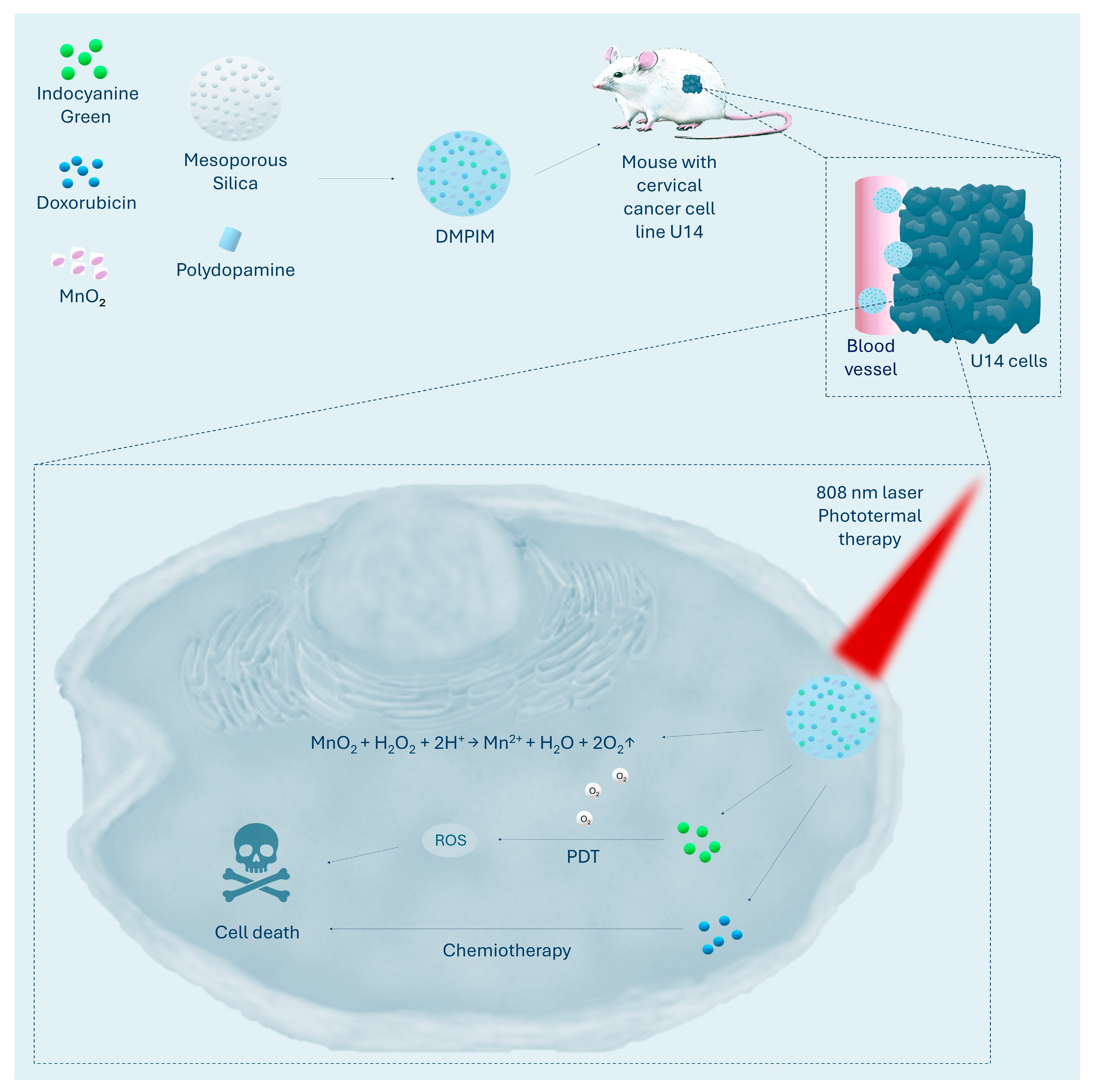
Figure 1. The figure shows DMPIM nanoparticles composed of mesoporous silica, doxorubicin, polydopamine, indocyanine green, and MnO2. After tumour accumulation, MnO2 generates oxygen to enhance photodynamic therapy, 808 nm laser irradiation activates indocyanine to produce reactive oxygen species, polydopamine and indocyanine induce photothermal effects, and doxorubicin release provides complementary chemotherapy. O2↑ indicates an increase in oxygen levels [68].
Ijms 26 08503 g001
Figure 2. The figure shows IMF nanoparticles, composed of UMOF, FeOOH, and ICG. In hypoxic tumours, FeOOH converts H2O2 into O2 and cytotoxic •OH, which, together with ICG-activated singlet oxygen under 808 nm light, drive a synergistic photodynamic therapy effect. Arrow shows direction from higher to lower intensivity of hypoxia in tumor [78].
Ijms 26 08503 g002
Table 1. Criteria for inclusion and exclusion of retrieved articles for overgrowth.
Inclusion Criteria
Articles describing photodynamic therapy
Articles describing cancer therapy
Articles describing nanoparticles
Articles published in 2024 and by March 2025
Exclusion Criteria
Articles describing nanoparticles without a strategy for overcoming hypoxia
Articles describing cancers other than cervical cancer
Articles other than original research papers
Articles in which the results of therapy were described only in vitro
Articles in a language other than English and Polish
Table 2. Summary of nanoparticles described in the article.
NanoparticleConstructionHypoxia Reversal MethodEffectiveness of Hypoxia ReversalTherapeutic EfficacyReference
DMPIMMesoporous silica + MnO2 + green indocyanine + doxorubicin + polydopamineDecomposition of H2O2 to O2 via MnO2Increase in ROS production relative to NP without MnO292.91% inhibition of tumour growth in a mouse model of U14[68]
DPIMGCDoxorubicin + polidopamine + green indocyanine + MnO2 + gelatin with celecoxibDecomposition of H2O2 to O2 via MnO2Increased ROS production relative to NP without MnO2, decreased HIF-1α in musim model U1491.47% inhibition of tumour growth in a mouse model of U14[67]
M-HMnO2@ICGMnO2 core + poly(allylamine hydrochloride) + green indocyanine + HeLa cell membraneDistribution of H2O2 to O2 via MnO2Downregulation of HIF-1α in a mouse model of HeLa88% inhibition of tumour growth in a mouse model of HeLa [70]
LRZAPHLuAG:Tb/Ce + Rose Bengal + ZIF-8 + Au2Pt + hyaluronic acidCatalase/peroxidase activity Au2PtIncrease in ROS production relative to NP without Au2Pt 193% inhibition of tumour growth in a mouse model of HeLa[73]
IMF (ICG ⊂ UMOF@FeOOH)UMOF + FeOOH + ICG coatingDecomposition of H2O2 to O2 via FeOOHDownregulation of HIF-1α in normoxia and hypoxiaComplete regression of 2/3 of tumours in a mouse model of HeLa[78]
M1-EPO-NPsMetallacycle + aza-BODIPY + endoperoxides + DSPE-PEG2000Release of O2 from endoperoxidesGeneration of singlet oxygen in both normoxia and hypoxia by M1-EPO-NPs relative to NPs without endoperoxides, in which 1O2 production occurred exclusively in normoxiaComplete tumour regression in a mouse model of HeLa[84]
UM-RZUCNP + mesoporous silica + ZnPc + Roussin’s Black SaltNO-mediated inhibition of mitochondrial respirationActivation of ZnPc with 808 nm light led to a decrease in oxygen levels to ≈20%, while pre-exposure to 980 nm (UM-RZ activation and NO release) maintained oxygen levels at ≈70%.86% tumour inhibition in a mouse model of HeLa[93]
UCN@mSiO2@ZnPc@L-ArgUCNP + mesoporous silica + ZnPc + L-arginineNO-mediated inhibition of mitochondrial respirationNo clear assessment of hypoxia78.5% inhibition of tumour growth in a mouse model of HeLa[94]
TNPs/IAmPEG-PLGA + PLGA-b-PEG + IR780 + atovaquone + TPP+OXPHOS inhibition (atovaquone)Increased ROS production under hypoxia relative to NP without atovaquoneHighest in vivo efficacy in a mouse model of HeLa relative to control groups[102]
1 Au2Pt exhibits catalase activity over a wide pH range from weakly acidic to basic, with maximum efficiency at pH ≈ 8, while under low pH conditions, peroxidase-like reactions leading to the generation of -OH radicals dominate, at the expense of catalase activity [73].
Disclaimer/Publisher’s Note: The statements, opinions and data contained in all publications are solely those of the individual author(s) and contributor(s) and not of MDPI and/or the editor(s). MDPI and/or the editor(s) disclaim responsibility for any injury to people or property resulting from any ideas, methods, instructions or products referred to in the content.