Review

ZnO-Based Nanoparticles for Targeted Cancer Chemotherapy and the Role of Tumor Microenvironment: A Systematic Review

by
Vasilis-Spyridon Tseriotis
1,2,*,
Dimitrios Ampazis
3,4,
Sofia Karachrysafi
5,6,
Theodora Papamitsou
5,6,
Georgios Petrakis
7,
Dimitrios Kouvelas
2,
Paraskevas Mavropoulos
2,
Konstantinos Lallas
8,
Aleksandar Sič
9,
Vasileios Fouskas
10,
Konstantinos Stergiou
5,6,
Pavlos Pavlidis
2,11 and
Marianthi Arnaoutoglou
12
1
Department of Neurology, Agios Pavlos General Hospital of Thessaloniki, Leoforos Ethnikis Antistaseos 161, Kalamaria, 55134 Thessaloniki, Greece
2
Laboratory of Clinical Pharmacology, Aristotle University Campus, Aristotle University of Thessaloniki, 54124 Thessaloniki, Greece
3
Respiratory Department, Cavan & Monaghan Hospital, HSE/RCSI, H12Y7W1 Cavan, Ireland
4
RCSI University of Medicine & Health Sciences, 123 St. Stephen’s Green, Dublin D02 YN77, Ireland
5
Research Team “Histologistas”, Interinstitutional Postgraduate Program “Health and Environmental Factors”, Department of Medicine, Faculty of Health Sciences, Aristotle University of Thessaloniki, 54124 Thessaloniki, Greece
6
Laboratory of Histology-Embryology, Department of Medicine, Faculty of Health Sciences, Aristotle University of Thessaloniki, 54124 Thessaloniki, Greece
7
Pathology Department, University General Hospital of Thessaloniki AHEPA, Medical School, Aristotle University of Thessaloniki, 54636 Thessaloniki, Greece
8
Department of Medical Oncology, School of Medicine, Aristotle University of Thessaloniki, 54124 Thessaloniki, Greece
9
Faculty of Medicine, University of Belgrade, 11000 Begrade, Serbia
10
International Hellenic University, 14th km Thessaloniki, Nea Moudania, 57001 Thessaloniki, Greece
11
ENT-Clinic, “George Papanikolaou” General Hospital of Thessaloniki, Leoforos Papanikolaou, Exochi, 57010 Thessaloniki, Greece
12
Department of Clinical Neurophysiology, AHEPA University Hospital of Thessaloniki, Aristotle University of Thessaloniki, Kiriakidi 1, 54636 Thessaloniki, Greece
*
Author to whom correspondence should be addressed.
Int. J. Mol. Sci. 2025, 26(17), 8417; https://doi.org/10.3390/ijms26178417
Submission received: 30 July 2025 / Revised: 25 August 2025 / Accepted: 26 August 2025 / Published: 29 August 2025

Abstract

Cancer, a leading global cause of death responsible for nearly 10 million deaths annually, demands innovative therapeutic strategies. Intrinsic cytotoxicity and biocompatibility of zinc oxide nanoparticles (ZnO-NPs) have rendered them promising nanoplatforms in oncology. We herein systematically review their applications for targeted cancer chemotherapy, with a focus on physicochemical properties, drug delivery mechanisms, and interactions with the tumor microenvironment (TME). We searched PubMed, SCOPUS, and Web of Science from inception through December 2024 for peer-reviewed preclinical studies on cancer models. Results were qualitatively synthesized. Quality was assessed with the SYRCLE risk of bias tool. Among 20 eligible studies, ZnO-NPs were frequently functionalized with ligands to enhance tumor targeting and minimize systemic toxicity. Chemotherapeutic agents (doxorubicin, 5-fluorouracil, docetaxel, cisplatin, gemcitabine, and tirapazamine) were loaded into ZnO-based carriers, with improved anticancer efficacy compared to free drug formulations, particularly in multidrug-resistant cell lines and in vivo murine xenografts. The mildly acidic TME was exploited for pH-responsive drug release, premature leakage reduction, and improvement of intratumoral accumulation. Enhanced therapeutic outcomes were attributed to reactive oxygen species generation, zinc ion-mediated cytotoxicity, mitochondrial dysfunction, and efflux pump inhibition. Deep tumor penetration, apoptosis induction, and tumor growth suppression were also reported, with minimal toxicity to healthy tissues. ZnO-NPs might constitute a versatile and promising strategy for targeted cancer chemotherapy, offering synergistic anticancer effects and improved safety profiles. Future studies emphasizing long-term toxicity, immune responses, and scalable production could lead to clinical translation of ZnO-based nanomedicine in oncology.

1. Introduction

Cancer remains one of the leading causes of death worldwide, with complex biological mechanisms contributing to the increasing incidence [1]. Despite conventional treatment options, such as chemotherapy, radiation, and immunotherapy, response is often limited by poor drug bioavailability, systemic toxicity, and resistance mechanisms [2]. The tumor microenvironment (TME), a highly heterogeneous and dynamic ecosystem comprising cancer, immune and stromal cells, blood vessels, and extracellular matrix (ECM) components, plays a crucial role in the progression of tumors [3]. The involvement of the TME in metastasis, immune evasion, and resistance to therapy makes it a potential target for optimized drug delivery that could lead to successful cancer treatment and improved outcomes in patients with cancer [4].
Advancements in nanomedicine, a fast-developing field that bridges nanotechnology and medicine, have recently been studied in the context of oncology, aiming to improve efficacy and reduce adverse effects with the use of nanoparticles (NPs) at tumor sites [5]. Among various inorganic nanomaterials, zinc oxide- (ZnO) based nanoparticles (ZnO-NPs) possess unique physicochemical properties, such as small particle size, large surface area, biocompatibility, and ease of functionalization [6]. Their ability to enhance the localized delivery of chemotherapeutic agents and improve treatment efficacy by modulating the TME makes ZnO-NPs particularly attractive for cancer therapy [7]. Moreover, thanks to their stability under physiological conditions, they can be engineered to optimize drug release in a controlled and stimuli-responsive way [8].
ZnO-NPs can modify the TME through various mechanisms, enhancing their potential for targeted cancer chemotherapy. An important characteristic is the ability to take advantage of the acidic conditions found in the TME. In an environment with low pH, ZnO-NPs undergo biodegradation, and the released Zn2+ ions can modify cellular metabolic processes, inhibit tumor growth, and modulate the immune response [9]. This pH-adaptive quality enables controlled drug release and reinforces the enhanced permeability and retention (EPR) effect, improving the retention of drugs at tumor sites. ZnO-NPs’ potential to improve drug delivery and reduce resistance to treatment is also supported through interactions with the ECM, immune cells, and the vasculature [5,10]. The functionalization of ZnO-NPs with ligands, such as folic acid or antibodies, allows therapeutic agents to be selectively delivered to tumor cells, minimizing adverse effects and improving the precision of conventional cancer therapies [11].
In this systematic review, we explore the multifaceted role of ZnO-based nanoparticles in targeted cancer chemotherapy, with a particular focus on their interactions with the TME. We examine the unique properties of ZnO-NPs that enable them to effectively modulate the TME and improve drug delivery, while also discussing their potential for overcoming some of the most pressing challenges in cancer therapy, such as drug resistance and systemic toxicity. By understanding the complex relationship between ZnO-NPs and the TME, we aim to provide insights into the future directions of ZnO-based nanomedicine in cancer treatment.

2. Methods

We reported our study following the recommendations of the PRISMA statement [12].

2.1. Search Strategy and Information Sources

We searched three major databases (PubMed/MEDLINE, Scopus, and Web of Science) from inception through 21 December 2024. Free keywords and controlled vocabulary were used, with synonyms for “tumor microenvironment”, “zinc”, and “nanoparticles”. An example of our full search strategy is shown in Supplementary Table S1. Techniques such as forward and backward citation searching and manual review of gray literature sources were also employed, including ProQuest and Google Scholar.

2.2. Eligibility Criteria

In our review, we intended to include both basic research studies performed in vivo or in vitro. Included studies should also clearly describe the nanoparticle used and explain its synthesis, either as part of their original report or by referencing previously peer-reviewed publications. Moreover, studies should describe laboratory methods used for the characterization of the nanoparticle. In terms of cancer studied, cancer type, and the preclinical models (in vitro or in vivo) should be clearly described. Lastly, the reports should also include a study of the mechanism of action and the most important experimental findings after applications in terms of anticancer performance. Lastly, all eligible studies should be peer-reviewed and provide their full report in English.
We excluded all studies that did not fulfill the aforementioned eligibility criteria. We also excluded studies that did not model human cancer types. For reports identified through search on gray literature sources, e.g., congress abstracts, dissertations, and preprints, these were excluded if they were not peer-reviewed by committees before their publication.

2.3. Data Management

We employed the Systematic Review Accelerator for deduplication [13] and we used the application Rayyan for article screening [14]. Eligible studies were imported to the reference manager “Mendeley” (Desktop version 1.19.8).

2.4. Study Selection

Article screening was done in two phases: abstracts and titles were screened by two independent reviewers (VST and SK), blinded to each other’s decisions. Conflicts were solved with the involvement of a third investigator (VF). Subsequently, full articles were also screened in a blinded manner by the same investigators, and conflicts were solved as previously described.

2.5. Data Extraction and Quality Assessment

Two independent reviewers extracted the following characteristics (VST, PM): first author/publication year, nanoparticle, synthesis method, characterization, cancer type studied, animal model used (in vitro and in vivo), route of administration in the case of experiments in vivo, mechanism of action, and anticancer performance. We performed quality assessment on Review Manager (RevMan) 5.4 using the SYRCLE risk of bias tool for animal studies [15]. Throughout quality assessment, the two reviewers worked independently. Disagreements during data extraction or quality assessment were resolved by a third reviewer (SK).

2.6. Qualitative Synthesis

The main results of our review were summarized in tabular form. We narratively performed a qualitative synthesis of eligible studies.

3. Results

Among 682 search results, 182 duplicates were removed and 514 were kept. After abstract and full-text screening, 20 studies investigating ZnO-based nanoparticles for cancer therapy were included [16,17,18,19,20,21,22,23,24,25,26,27,28,29,30,31,32,33,34,35]. Eligible studies were published between 2016 and 2024, using formulations that shared core components based on ZnO nanostructures and were functionalized for drug delivery, tumor targeting, or physicochemical modulation. A PRISMA flow diagram is shown in Figure 1, while Table 1 summarizes all included studies.

3.1. NP Composition and Synthesis Methods

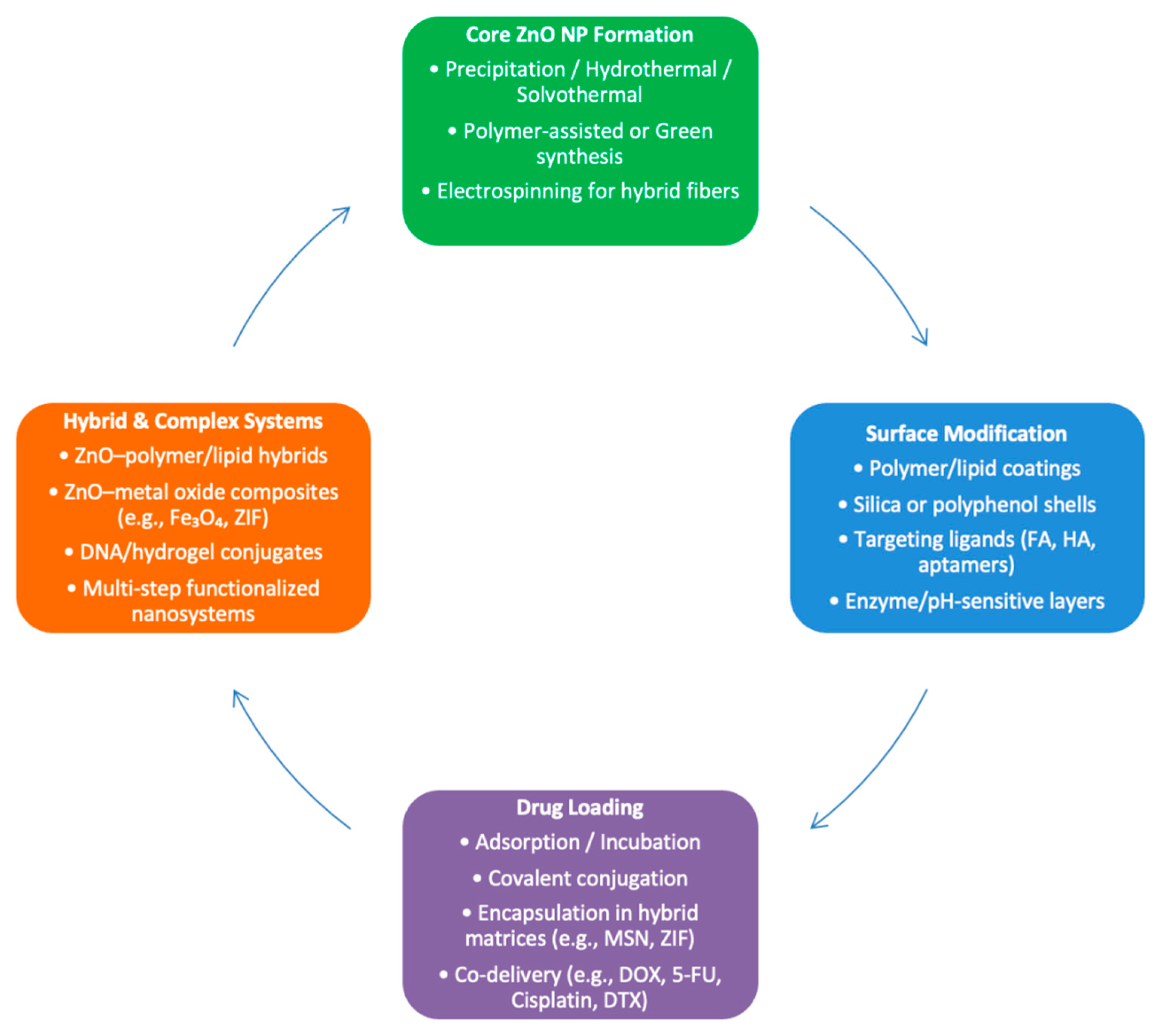
A wide range of ZnO-based nanoplatforms was employed, often functionalized with targeting ligands, polymers, or combined with additional therapeutic agents to improve delivery and efficacy (Figure 2). Most platforms relied on the intrinsic pH sensitivity and oxidative potential of ZnO, while synergistic effects were achieved through integration of various chemotherapeutic agents, such as doxorubicin (DOX), 5-fluorouracil (5-FU), docetaxel (DTX), and cisplatin, and/or biofunctional layers.
Nanoparticle synthesis methods varied in complexity, from simple physical adsorption strategies like the incubation of ZnO nanoparticles with DOX under stirring, followed by centrifugation and freeze-drying [18,31] to co-precipitation or hydrothermal synthesis for ZnO core formation, followed by drug loading and surface modification [28,29]. More elaborate constructions incorporated layer-by-layer functionalization, such as the sequential modification in ZnO-DOX/R8@HA involving 3-aminopropyltriethoxysilane (APTES)-mediated amination, thioketal crosslinking, and hyaluronic acid shell coating [17]. Similarly, the 5-FU@ZIF-90@ZnO nanocomposite was prepared by encapsulating 5-FU into ZIF-90, followed by ZnO shell coating via triethanolamine-mediated precipitation [32].
Several studies adopted hybrid or core–shell architectures to enhance stability, targeting, and multifunctionality. Liu et al. employed solvothermal methods, followed by calcination and surface conjugation with polyethyleneimine and folic acid [25]. Zhou et al. developed dual-responsive NPs by layering anionic lipids and enzyme-sensitive polyethylene glycol (PEG) chains onto ZnO cores, employing emulsion-sonication and solvent evaporation [34].
Other studies integrated polymers or mesoporous structures which utilized thermosensitive copolymers during ZnO hybridization [22] or relied on amination reactions to attach ZnO quantum dots onto carboxylated mesoporous silica [24]. Functional DNA nanoscaffolds were also explored; for instance, Zhi et al. assembled positively charged ZnO within aptamer-DNAzyme frameworks to form ZnO-based bio-barcodes, enabling multi-level control of release and targeting [33].

3.2. Physicochemical Characterization of ZnO-Based NPs

Characterization methods were consistently employed to confirm nanoparticle morphology, size, surface charge, chemical composition, and drug loading efficiency. Transmission electron microscopy (TEM) was the most commonly used imaging modality, providing high-resolution visualization of NP morphology and confirming nanoscale dimensions and surface structure [17,18,35]. High-resolution variants (HRTEM) were used by some studies to achieve detailed structural insights [22].
Dynamic light scattering (DLS) was performed to assess hydrodynamic diameter and polydispersity, while zeta potential measurements were used to evaluate surface charge and colloidal stability in physiological environments [25,31]. X-ray diffraction (XRD) confirmed the crystalline structure of ZnO, validating ZnO-based nanohybrid formation [18,19,22,27,28,29,32,35]. Fourier transform infrared spectroscopy (FT-IR) was widely used to verify chemical bonding and functional group incorporation, especially for surface modifications and drug conjugation [17,18,22,23,24,26,32,34].
Additional techniques included UV–Vis and fluorescence spectroscopy for drug loading and release kinetics [16,18,21], scanning electron microscopy (SEM) for surface morphology, and energy-dispersive X-ray spectroscopy (EDS) for elemental mapping [16,33]. Some studies employed more advanced tools, such as superconducting quantum interference device (SQUID) magnetometry to assess magnetic behavior [25], thermogravimetric analysis (TGA) for thermal stability [20], and proton nuclear magnetic resonance (1 H NMR) spectroscopy for polymer characterization [35].
Drug quantification and release profiles were often measured via high-performance liquid chromatography (HPLC) [20] or liquid chromatography–tandem mass spectrometry (LC–MS/MS) to ensure precise analysis of chemotherapeutic content [35]. In specialized constructs, additional tools such as inductively coupled plasma mass spectrometry (ICP-MS) and isothermal nitrogen adsorption (BET analysis) were used to evaluate elemental content and surface area, respectively [25].

3.3. Cancer Models and Experimental Systems

Most studies incorporated both 2D monolayer cultures, with the most commonly studied cancer types in vitro, including breast cancer (MDA-MB-231, 4T1), lung adenocarcinoma (A549, A549/DDP), hepatocellular carcinoma (HepG2, SMMC-7721), prostate cancer (PC3, PC-3M), and colon cancer (HCT116). Squamous cell carcinoma (SCC-7), lymphoma (2D/3D cultures), cervical cancer (HeLa), and pancreatic cancer (BxPC3, AsPC-1) were also studied [16,17,18,19]. Importantly, drug-resistant variants were used to explore efficacy in multidrug-resistant (MDR) phenotypes. Examples include A549/DDP cells (cisplatin-resistant) [33], MES-SA/Dx5 (doxorubicin-resistant uterine sarcoma), and NCI/ADR-RES (doxorubicin-resistant ovarian cancer) [31]. Some studies also employed normal cell lines (e.g., COS7, L929 fibroblasts) as negative controls to assess cancer selectivity [17,34].
Twelve studies used in vivo models to assess antitumor efficacy, primarily xenograft models in BALB/c nude mice, generated by subcutaneous injection of cancer cell lines. For instance, SCC-7, 4T1, HCT116, and MDA-MB-231 tumors were established in immunocompromised mice to evaluate tumor accumulation and suppression following NP administration [17,18,35]. While most studies used intravenous tail vein administration, some opted for intratumoral injection [19]. In addition, Gomaa et al. utilized both in vitro EAC tumor cells and an in vivo EAC murine model with intraperitoneal injection, expanding the diversity of tumor systems and delivery routes assessed [21].
Several studies lacked in vivo validation and focused solely on cell-based assays, including those targeting pancreatic cancer [16], lymphoma [26], or when specific mechanistic endpoints were prioritized over whole-animal outcomes [24,27].
Three-dimensional (3D) tumor spheroids were employed in some studies to bridge the gap between 2D cultures and animal models. For instance, 3D multicellular tumor spheroids were used to assess nanoparticle penetration, cytotoxicity, and drug retention in simulated tumor environments, providing more accurate predictions of in vivo efficacy compared to standard 2D assays [31].

3.4. Mechanisms of Anticancer Action and pH-Responsiveness

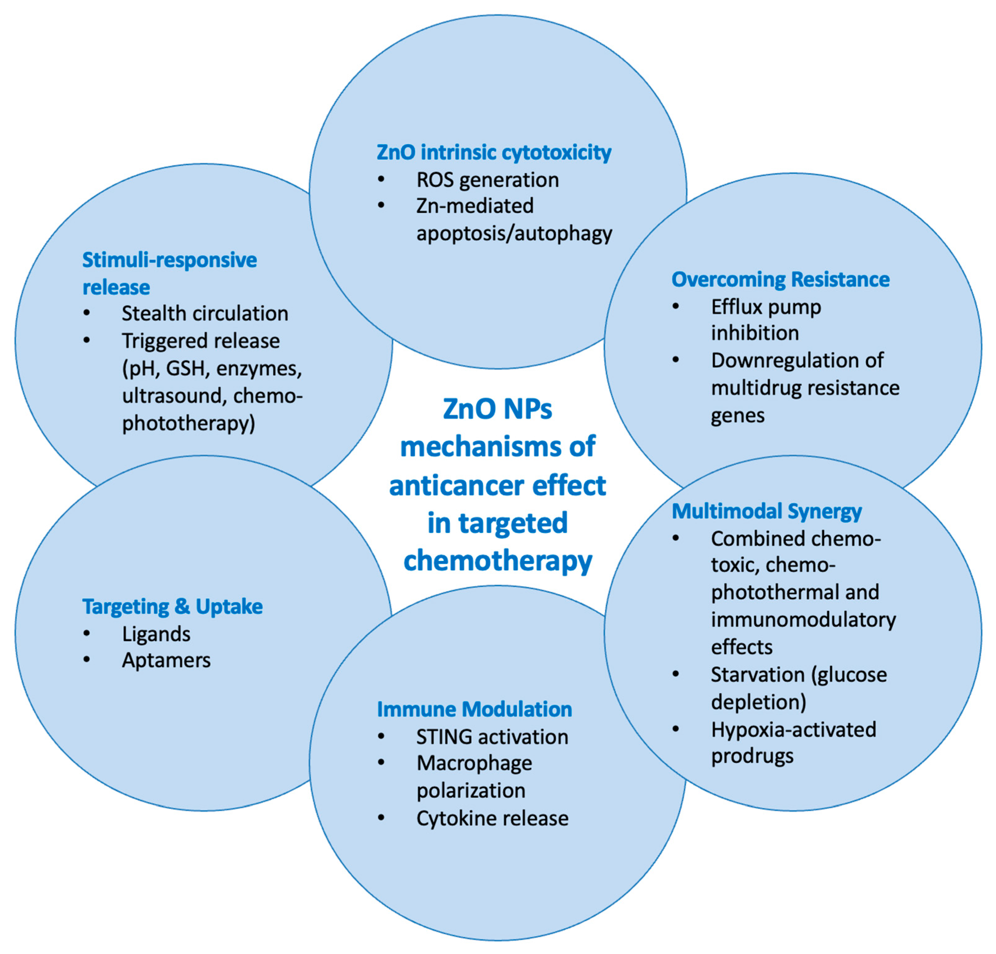
Most NPs operated through multiple synergistic pathways, combining traditional chemotherapy with the intrinsic cytotoxicity of ZnO. Zn2+-induced cytotoxicity, ROS generation, controlled pH-triggered release with reduced premature leakage, and targeted (tumor-specific) delivery through the use of various ligands were among the most consistently described mechanisms. Additionally, enhanced drug accumulation and reduced drug efflux in MDR cells, chemo-immunotherapy and starvation therapy via glucose depletion, as well as carbon-coated ZnO cores with photothermal effects and enhanced cytotoxicity through localized heating were also reported.
A key design and TME-tailored feature across nearly all studies was pH-responsive drug release, typically activated at mildly acidic pH values between 4.5 and 6.5. This characteristic enabled the selective release of chemotherapeutic agents in the TME while minimizing systemic toxicity. Optimal drug release was reported at pH 5.0–5.5 for most studies, mimicking endosomal or lysosomal conditions. For example, ZnO degradation at pH 5.5 was shown to enhance the release of DOX or 5-FU, triggering apoptosis via intracellular Zn2+ and reactive oxygen species (ROS) production [18,32]. In contrast, a system with charge-reversal behavior activated cellular uptake and drug release at pH 6.5, simulating the mildly acidic extracellular tumor milieu [34].
In addition to pH sensitivity, enzyme-or redox-responsiveness was also integrated. Examples include the incorporation of a matrix metalloproteinase 2-(MMP2) cleavable PEG shell facilitating enzyme-triggered detachment and enhancing uptake and release at pH 5.0 [35], as well as glutathione-sensitive linkages to achieve dual pH- and GSH-responsive delivery, ensuring intracellular drug release within cancer cells [24].

3.5. Anticancer Performance

Eligible studies consistently demonstrated superior anticancer performance of ZnO-based nanoparticles compared to free chemotherapeutic agents or single-modality controls across both in vitro and in vivo experiments, with enhanced cytotoxicity, tumor penetration, and therapeutic synergy.
When evaluating cytotoxic effects in vitro using human cancer cell lines, ZnO-based formulations consistently outperformed free drugs by promoting greater intracellular accumulation, ROS generation, and apoptosis. For instance, ZnO-DOXNPs significantly increased cytotoxicity in doxorubicin-resistant MDA-MB-231 cells, also inducing mitochondrial membrane depolarization and bypassing efflux pumps [35]. Similarly, PZnO@DOX NPs triggered STING pathway activation, leading to immunogenic cell death in 4T1 cells [30].
Several studies employed 3D tumor spheroid models to better mimic the in vivo TME. ZnO-LD NPs displayed deep tumor penetration and significant ROS-mediated apoptosis in 3D cultures of PC-3M and 4T1 cells [23], while ZnO/DPPG/PEG-pp-PE/DOX nanoparticles enhanced spheroid penetration following MMP2 activation [35]. ZnO@BBCs were further shown to enhance intracellular platinum accumulation and overcome drug resistance in A549/DDP cells by downregulating multidrug resistance 1 (MDR1) gene expression [33].
Among studies reporting on in vivo experiments in murine xenograft models, the majority observed marked tumor growth inhibition following nanoparticle administration. Significant tumor suppression was reported in SCC-7 [17], HepG2 [22], and HCT116 [18] xenografts treated with ZnO-based formulations. Enhanced survival and tumor shrinkage were noted in 4T1-bearing mice treated with MTGZ@PPD, which combined starvation therapy (via glucose oxidase) and hypoxia-activated chemotherapy [34]. Synergistic antitumor effects were evident in mice receiving ZnO/DOX NPs with STING pathway activation [30], with increased apoptosis and reduced tumor burden compared to controls. Importantly, minimal systemic toxicity was consistently reported, with stable body weights and no significant organ histopathology [33,34].
A key feature was the selective toxicity toward cancer cells, with reduced effects on normal cells. For example, ZnO-Lipo-pep-Gem NPs demonstrated stronger inhibition of non-metastatic BxPC3 cells than metastatic AsPC-1 cells, with lower toxicity to healthy cells [16]. ZnO-cisplatin formulations reduced IC50 values and enhanced apoptosis in PC3 cells, particularly when pretreated with NO donors [28].
Moreover, ZnO nanocarriers were instrumental in overcoming multidrug resistance, as shown in studies targeting P-gp overexpression or using nucleolin-targeting aptamers for enhanced intracellular delivery [31,33].
Studies integrating multiple modalities (e.g., chemotherapy, immunotherapy, starvation therapy, or photothermal therapy) consistently achieved superior efficacy. Synergistic chemo-photothermal treatment using Fe3O4@C/ZnO-DOX-FA under laser irradiation achieved enhanced tumor suppression [25], whereas combining glucose depletion mediated by glucose oxidase (GOx) with tirapazamine- (TPZ) induced DNA damage achieved starvation–chemo synergy [34]. ZnO-NPs/DOX/FA NPs displayed dose-dependent cytotoxicity in vitro, with the highest IC50 values indicating controlled release and reduced off-target effects. Results of the in vivo study included suppression of EAC tumor burden, enhanced apoptosis (Annexin V/PI), reduced IL-6 and TNF-α cytokines, and preserved liver/kidney function, confirming synergistic antitumor and immunomodulatory effects with a favorable biosafety profile [21].
None of the included studies systematically evaluated nanoparticle clearance or pharmacokinetics. While some reported prolonged circulation, preserved liver and kidney function, and negligible systemic toxicity, detailed data on biodistribution, metabolism, and excretion were not provided. Mechanisms of anticancer action are graphically summarized in Figure 3.
NPs: nanoparticles, STING: Stimulator of Interferon Genes, ROS: reactive oxygen species; GSH: glutathione

3.6. Quality Assessment

As shown in Figure 4A,B, an unclear risk of bias across several domains was found for most studies due to limited methodological reporting. Random sequence generation, allocation concealment, and blinding were rarely described. In contrast, outcome reporting and attrition bias were generally rated as low risk. Overall, these findings highlight the need for improved transparency and standardization in preclinical nanoparticle research.

4. Discussion

The findings of this review highlight the versatile and potent role of ZnO-based NPs in enhancing targeted cancer chemotherapy. Across eligible studies, ZnO nanoplatforms significantly improved therapeutic outcomes compared to free drugs, validating their capability to modulate the TME for better drug delivery and efficacy. ZnO-NPs are intrinsically multifunctional, as they can directly induce cancer cell death via multiple mechanisms, like ROS generation and the formation of ionic zinc species (Zn2+) during ZnO dissolution, triggering mitochondrial damage and apoptosis [10], but also serve as biocompatible, biodegradable drug carriers that selectively target tumor cells [36]. Consistent improvements in therapeutic efficacy can be rationalized by the dual action of ZnO-NPs, which both enhance intracellular delivery of chemotherapeutics and contribute intrinsic cytotoxic effects through Zn2+ release, ROS generation, and mitochondrial dysfunction. Together, these findings highlight that the enhanced anticancer performance of ZnO-NPs is not merely additive but arises from a synergistic interplay between nanoparticle physicochemical properties, TME-responsive behavior, and the loaded chemotherapeutic agents.
We herein highlight pH-responsive behavior as a central advantage of ZnO-NPs. ZnO dissolves in the mild acidity of the TME (pH around 6.5 in interstitium and around 5.0 in endo/lysosomal compartments), leading to accelerated drug release specifically in tumor tissues. Controlled, on-demand payload release within tumors with simultaneous minimization of premature drug leakage in blood circulation boosts intratumoral drug concentration but also augments the therapy with ZnO’s inherent cytotoxic effects [9,37]. Importantly, dissolution itself primarily yields Zn2+, which disrupts cellular homeostasis. ROS are not a direct product of dissolution but arise secondarily through Zn2+-induced mitochondrial dysfunction, redox imbalance, and surface defect-mediated catalytic reactions of ZnO nanoparticles in biological environments. The excess Zn2+ ions, together with ROS, can damage cancer cells synergistically with loaded chemotherapeutics, especially in the acidic, often hypoxic core of tumors. Notably, the rapid proliferation of tumor cells makes them more susceptible to such stress, compared to healthy cells that are relatively spared, underscoring the selective toxicity and TME-specificity of ZnO-NPs [5,38,39].
Another key feature is the ability of ZnO nanocarriers to overcome multidrug resistance mechanisms in cancer, outperforming free drugs by increasing intracellular drug accumulation and promoting cell death in chemo-refractory tumors. This aligns with recent reports that ZnO-NPs can intercept critical chemoresistance pathways by inhibiting efflux pumps (e.g., P-glycoprotein/ABC transporters) and reactivating pro-apoptotic signaling [8]. By downregulating the activity of drug-efflux transporters while increasing DNA damage and caspase activation, ZnO-NPs restore chemosensitivity in formerly resistant cells [8,40]. Importantly, overcoming drug resistance is combined with ZnO-induced stress without adding significant systemic toxicity. In fact, some evidence suggests that at appropriate low doses, ZnO-NPs may mitigate the side effects of conventional chemotherapy [41]. The ZnO matrix can act as a reservoir that releases the drug locally in the tumor, reducing the exposure of healthy tissues and thereby diminishing off-target damage [42].
Furthermore, the favorable safety profile of ZnO-NPs is emphasized among the main findings of eligible studies. Negligible systemic toxicity was noted with several ZnO-based formulations in animal models. Treated mice demonstrated stable body weights and no major organ damage relative to controls. A representative example is a folic acid-targeted ZnO-doxorubicin nanocomposite, which not only intensifies tumor apoptosis but also blunts systemic inflammation in mice [21]. Treated tumor-bearing mice exhibited lowered IL-6 and TNF-α levels and near-normal liver and kidney function, whereas free doxorubicin caused significant inflammation and organ stress. This dual benefit of enhanced tumor kill with reduced side effects is a recurring theme and a major impetus for further development of ZnO nanotherapies [8].
With zinc being an essential trace element present in all body tissues generally recognized as safe by the FDA in other contexts [43], the intrinsic biocompatibility of ZnO supports the idea that ZnO-based NPs could be safe for potential clinical trials, especially if they are designed to avoid any “unpredictable” interactions of bare ZnO in vivo [7]. However, challenges and limitations before ZnO-NPs can be applied in clinical practice include potential unpredictable toxic effects or immune reactions. While ZnO-NPs are comparatively less toxic than many other metal oxides [10], at the nano level, they can interact with biological systems in complex ways. Unwanted oxidative stress or inflammation is a major concern if ZnO-based dissolution is not properly controlled. Our review shows that many of the current data regard only the evaluation of acute toxicity. Data about long-term effects like chronic ZnO or zinc ions accumulation in organs, are still sparse. Studies examining in vivo immunological responses to ZnO-NPs are also limited. Extensive experimentation in preclinical models is needed to cover these aspects through rigorous safety evaluations (toxicology, immunogenicity, pharmacokinetics) that better simulate human tumors and the human immune system [5,38].
NP design optimizations will be crucial in surmounting future research challenges and limitations. Surface functionalization and doping strategies are employed to improve the stability and safety of ZnO-NPs [7,38,40]. Trace elements (e.g., rare-earth or transition metals) help create more stable and even multifunctional NPs that enhance the biostability and biocompatibility of ZnO-NPs, while endowing them with additional features like improved imaging contrast (for theranostic use) and magnetic or luminescent properties [7,44]. Our review demonstrates that organic or inorganic coatings, from polymers and lipids to silica shells, have been used not only to stabilize ZnO-NPs and prevent aggregation, but also to impart tumor-targeting ligands and stimuli-responsive elements. Layer-by-layer constructions such as pH-sensitive or enzyme-cleavable shells can ensure NP activity only upon reaching the tumor site [40,44]. “Safe-by-design” engineering of ZnO-NPs should be a top priority encompassing the use of biocompatible coatings, environmentally benign (“green”) synthesis routes for nanoparticle production, and thorough characterization of physicochemical properties to predict and control biological interactions [9]. Encouraging eco-friendly production methods that avoid harsh chemicals and yield intrinsically biocompatible particles includes the use of plant extracts and microbes [45]. From a mechanistic perspective, more studies are needed to unravel how exactly ZnO-NPs interact with various components of the TME, such as the influence of released Zn2+ ions on cancer cell metabolism. There is emerging evidence that ZnO-NPs can stimulate an immunogenic form of tumor cell death (via pathways like cGAS-STING), suggesting a potential role in cancer immunotherapy when used in combination with other agents, beyond chemotherapeutics [5,46]. In theory, ZnO-NPs could enhance radiosensitization through ROS generation and immune responses by modulating cytokine profiles and exposing danger signals from dying cancer cells, which could eventually tackle tumor recurrence and metastasis. In terms of theragnostics and personalized oncology, owing to their semiconducting nature, ZnO nanostructures (especially quantum dots or doped nanoparticles) have intrinsic photoluminescence that can be utilized for simultaneous cancer imaging and therapy [7,44,47].
From bench to bedside, practical pipeline considerations include scalable and good manufacturing practice conditions, as well as reproducibility [40,42]. In this respect, ZnO-NPs are relatively easy to synthesize in large quantities and at low cost, especially compared to more complex nanocarriers [9]. Consistent characterization (size, shape, charge, dissolution rate) and rigorous quality control will be indispensable for regulatory purposes. An important gap review is the absence of systematic data on clearance and pharmacokinetics of ZnO-based nanoparticles. Although studies consistently reported minimal systemic toxicity, preserved organ function, and in some cases prolonged circulation, no investigations provided quantitative insights into long-term biodistribution, metabolism, or excretion. Given that zinc is an essential trace element but may accumulate in tissues when delivered in nanoparticulate form, future research should prioritize rigorous pharmacokinetic and clearance studies to ensure safe clinical translation. Similarly, regulatory agencies will require extensive toxicological data; thus, systematic studies on long-term toxicity, genotoxicity, and ecotoxicology of ZnO-NPs, under an interdisciplinary lens, must accompany their development.
Compared with silica (SiO2) and titanium dioxide (TiO2) nanocarriers, ZnO-NPs offer unique multifunctionality. SiO2 is biocompatible and easily functionalized but inert, while TiO2 is inexpensive and photoactive yet requires light activation and shows slow biodegradation. ZnO-NPs combine pH-responsive dissolution, intrinsic cytotoxicity, and biodegradability into zinc ions, though at the cost of less predictable long-term stability and potential oxidative stress. In parallel, hydrogel-based nanocomposites are gaining attention as platforms for localized and sustained drug delivery in cancer. While most current reports involve other nanomaterials such as iron oxide or polymeric carriers, the principle of embedding nanoparticles into hydrogels highlights a promising route that could, in the future, be applied to ZnO-based systems as well [48]. This underscores how the field is converging toward multifunctional, biocompatible scaffolds that combine structural support with targeted therapy.

5. Conclusions

ZnO-based nanoparticles have emerged as a multifaceted platform capable of reshaping cancer therapy. By leveraging TME-specific triggers and multi-modal therapeutic mechanisms, they offer solutions to longstanding challenges like drug resistance and systemic toxicity. The evidence assembled in this review strongly supports the further development of ZnO nanocarriers as targeted chemotherapy adjuvants and beyond. With continued innovation in nanoparticle design and a concerted push toward thorough in vivo validation, ZnO nanomedicine stands poised to become a valuable component of the future oncology toolkit. Future steps will involve refining these nanosystems for maximum efficacy and safety, scaling up production, and diligently evaluating performance in clinically relevant models. Upon achieving these milestones, ZnO nanoparticles could indeed revolutionize cancer therapy by providing more effective, targeted, and patient-friendly treatment options.

Supplementary Materials

The following supporting information can be downloaded at: https://www.mdpi.com/article/10.3390/ijms26178417/s1.

Author Contributions

V.-S.T.: conceptualization, data curation, formal analysis, investigation, methodology, project administration, resources, validation, writing—original draft, writing—review and editing; D.A.: data curation, formal analysis, validation, writing—original draft, writing—review and editing; S.K.: conceptualization, data curation, investigation, validation, writing—original draft; T.P.: conceptualization, formal analysis, validation; G.P.: conceptualization, formal analysis, investigation, resources; D.K.: conceptualization, investigation, resources; P.M.: formal analysis, methodology, validation; K.L.: investigation, software, visualization; A.S.: data curation, visualization; V.F.: project administration, software; K.S.: formal analysis, investigation, methodology; P.P.: investigation, methodology, writing—original draft; M.A.: conceptualization, supervision, writing—original draft, writing—review and editing. All authors have read and agreed to the published version of the manuscript.

Funding

This research received no external funding.

Institutional Review Board Statement

Not applicable.

Informed Consent Statement

Not applicable.

Data Availability Statement

Our search strategy is provided as Supplementary Material.

Conflicts of Interest

V.-S.T. has received travel grants from ECTRIMS and the European Academy of Neurology, as well as coverage of congress registration fees from Inovis, Genesis Pharma and Novartis; D.A. declares no conflicts of interest; S.K. declares no conflicts of interest; T.P. declares no conflicts of interest; G.P. declares no conflicts of interest; D.K. declares no conflicts of interest; P.M. declares no conflicts of interest; K.L. declares no conflicts of interest; A.S. declares no conflicts of interest; V.F. declares no conflicts of interest; K.S. declares no conflicts of interest; P.P. declares no conflicts of interest; M.A. declares no conflicts of interest.

References

  1. Wu, Z.; Xia, F.; Lin, R. Global burden of cancer and associated risk factors in 204 countries and territories, 1980–2021: A systematic analysis for the GBD 2021. J. Hematol. Oncol. 2024, 17, 119. [Google Scholar] [CrossRef]
  2. Bi, J.H.; Tuo, J.Y.; Xiao, Y.X.; Tang, D.D.; Zhou, X.H.; Jiang, Y.F.; Ji, X.W.; Tan, Y.T.; Yuan, H.Y.; Xiang, Y.B. Observed and relative survival trends of lung cancer: A systematic review of population-based cancer registration data. Thorac. Cancer 2024, 15, 142–151. [Google Scholar] [CrossRef] [PubMed]
  3. Mo, J.; Zhang, H.; Swarbrick, A. TiME for a change: The tumor microenvironment as the missing piece in cancer therapeutics. PLoS Biol. 2025, 23, e3003276. [Google Scholar] [CrossRef]
  4. El-Tanani, M.; Rabbani, S.A.; Babiker, R.; Rangraze, I.; Kapre, S.; Palakurthi, S.S.; Alnuqaydan, A.M.; Rizzo, M.; El-Tanani, Y.; Tambuwala, M.M. Unraveling the tumor microenvironment: Insights into cancer metastasis and therapeutic strategies. Cancer Lett. 2024, 591, 216894. [Google Scholar] [CrossRef]
  5. Guo, Y.; Morshedi, M. Cutting-edge nanotechnology: Unveiling the role of zinc oxide nanoparticles in combating deadly gastrointestinal tumors. Front. Bioeng. Biotechnol. 2025, 13, 1547757. [Google Scholar] [CrossRef]
  6. DeLong, R.K.; Cheng, Y.H.; Pearson, P.; Lin, Z.; Coffee, C.; Mathew, E.N.; Hoffman, A.; Raelene, M.W.; Higginbotham, M.L. Translating Nanomedicine to Comparative Oncology-the Case for Combining Zinc Oxide Nanomaterials with Nucleic Acid Therapeutic and Protein Delivery for Treating Metastatic Cancer. J. Pharmacol. Exp. Ther. 2019, 370, 671–681. [Google Scholar] [CrossRef]
  7. Carofiglio, M.; Barui, S.; Cauda, V.; Laurenti, M. Doped Zinc Oxide Nanoparticles: Synthesis, Characterization and Potential Use in Nanomedicine. Appl. Sci. 2020, 10, 5194. [Google Scholar] [CrossRef]
  8. Vaghari-Tabari, M.; Jafari-Gharabaghlou, D.; Mohammadi, M.; Hashemzadeh, M.S. Zinc Oxide Nanoparticles and Cancer Chemotherapy: Helpful Tools for Enhancing Chemo-sensitivity and Reducing Side Effects? Biol. Trace Elem. Res. 2024, 202, 1878–1900. [Google Scholar] [CrossRef] [PubMed]
  9. Lebaka, V.R.; Ravi, P.; Reddy, M.C.; Thummala, C.; Mandal, T.K. Zinc Oxide Nanoparticles in Modern Science and Technology: Multifunctional Roles in Healthcare, Environmental Remediation, and Industry. Nanomaterials 2025, 15, 754. [Google Scholar] [CrossRef] [PubMed]
  10. Jiang, J.; Pi, J.; Cai, J. The Advancing of Zinc Oxide Nanoparticles for Biomedical Applications. Bioinorg. Chem. Appl. 2018, 2018, 1062562. [Google Scholar] [CrossRef]
  11. Fu, Z.; Xiang, J. Aptamer-Functionalized Nanoparticles in Targeted Delivery and Cancer Therapy. Int. J. Mol. Sci. 2020, 21, 9123. [Google Scholar] [CrossRef] [PubMed]
  12. Page, M.J.; McKenzie, J.E.; Bossuyt, P.M.; Boutron, I.; Hoffmann, T.C.; Mulrow, C.D.; Shamseer, L.; Tetzlaff, J.M.; Akl, E.A.; Brennan, S.E.; et al. The PRISMA 2020 statement: An updated guideline for reporting systematic reviews. BMJ 2021, 372, n71. [Google Scholar] [CrossRef]
  13. Clark, J.; Glasziou, P.; Del Mar, C.; Bannach-Brown, A.; Stehlik, P.; Scott, A.M. A full systematic review was completed in 2 weeks using automation tools: A case study. J. Clin. Epidemiol. 2020, 121, 81–90. [Google Scholar] [CrossRef]
  14. Ouzzani, M.; Hammady, H.; Fedorowicz, Z.; Elmagarmid, A. Rayyan—A web and mobile app for systematic reviews. Syst. Rev. 2016, 5, 210. [Google Scholar] [CrossRef]
  15. Hooijmans, C.R.; Rovers, M.M.; De Vries, R.B.M.; Leenaars, M.; Ritskes-Hoitinga, M.; Langendam, M.W. SYRCLE’s risk of bias tool for animal studies. BMC Med. Res. Methodol. 2014, 14, 43. [Google Scholar] [CrossRef] [PubMed]
  16. Barui, S.; Percivalle, N.M.; Conte, M.; Dumontel, B.; Racca, L.; Carofiglio, M.; Cauda, V. Development of doped ZnO-based biomimicking and tumor-targeted nanotheranostics to improve pancreatic cancer treatment. Cancer Nanotechnol. 2022, 13, 37. [Google Scholar] [CrossRef]
  17. Chen, W.H.; Luo, G.F.; Qiu, W.X.; Lei, Q.; Hong, S.; Wang, S.B.; Zheng, D.W.; Zhu, C.H.; Zeng, X.; Feng, J.; et al. Programmed Nanococktail for Intracellular Cascade Reaction Regulating Self-Synergistic Tumor Targeting Therapy. Small 2016, 12, 733–744. [Google Scholar] [CrossRef]
  18. Cui, T.; Yan, Z.; Qin, H.; Sun, Y.; Ren, J.; Qu, X. A Sequential Target-Responsive Nanocarrier with Enhanced Tumor Penetration and Neighboring Effect In Vivo. Small 2019, 15, 1903323. [Google Scholar] [CrossRef] [PubMed]
  19. Delong, R.K.; Comer, J.; Mathew, E.N.; Jaberi-Douraki, M. Comparative Molecular Immunological Activity of Physiological Metal Oxide Nanoparticle and its Anticancer Peptide and RNA Complexes. Nanomaterials 2019, 9, 1670. [Google Scholar] [CrossRef]
  20. Ekambaram, R.; Saravanan, S.; Babu, V.P.S.; Dharmalingam, S. Fabrication and evaluation of Docetaxel doped ZnO nanoparticles incorporated PCL nanofibers for its hemocompatibility, cytotoxicity and apoptotic effects against A549. Materialia 2022, 21, 101278. [Google Scholar] [CrossRef]
  21. Gomaa, S.; Nassef, M.; Tabl, G.; Zaki, S.; Abdel-Ghany, A. Doxorubicin and folic acid-loaded zinc oxide nanoparticles-based combined anti-tumor and anti-inflammatory approach for enhanced anti-cancer therapy. BMC Cancer 2024, 24, 34. [Google Scholar] [CrossRef]
  22. He, L.; Sun, X.; Nan, X.; Wang, T.; Bai, P. Thermal and pH responsive ZnO-based nanoparticles for efficient drug delivery. AIP Adv. 2019, 9, 125026. [Google Scholar] [CrossRef]
  23. Li, Z.; Zhang, S.; Liu, M.; Zhong, T.; Li, H.; Wang, J.; Zhao, H.; Tian, Y.; Wang, H.; Wang, J.; et al. Antitumor Activity of the Zinc Oxide Nanoparticles Coated with Low-Molecular-Weight Heparin and Doxorubicin Complex In Vitro and In Vivo. Mol. Pharm. 2022, 19, 4179–4190. [Google Scholar] [CrossRef]
  24. Li, J.; Chen, H.; Rui, Q.; Liu, P.; Cai, W.; Kong, Y.; Zuo, X. Biodegradable Controlled Drug Delivery Platform Based on Carboxylated Mesoporous Silica Nanoparticles—Zinc Oxide Quantum Dots. Ind. Eng. Chem. Res. 2024, 63, 13459–13468. [Google Scholar] [CrossRef]
  25. Liu, X.; Wang, C.; Wang, X.; Tian, C.; Shen, Y.; Zhu, M. A dual-targeting Fe3O4@C/ZnO-DOX-FA nanoplatform with pH-responsive drug release and synergetic chemo-photothermal antitumor in vitro and in vivo. Mater. Sci. Eng. C Mater. Biol. Appl. 2021, 118, 111455. [Google Scholar] [CrossRef] [PubMed]
  26. Mendoza-Martinez, N.L.; Cadena-Galeana, A.D.; Villanueva-Sanchez, F.G.; Perez-Cornejo, N.; Avelar-Juarez, K.M.; Ramos-Baena, J.D.; Cruz-Monroy, E.A.; Vazquez-Zuniga, U.; Garcia-Contreras, R. Efficacy of Antineoplastic Nanocarriers on 3D Oral Cancer Spheroids. Vivo 2023, 37, 1658–1665. [Google Scholar] [CrossRef] [PubMed]
  27. Pavan, S.R.; Prabhu, A. Acid Degradable Zinc Oxide Nanoparticle Loaded Etoposide Nanoformulation for Targeting Lung Adenocarcinoma via Drug Metabolism and Crosstalk Between Angiogenesis and Akt Pathways. Bionanoscience 2024, 14, 4406–4417. [Google Scholar] [CrossRef]
  28. Pieretti, J.C.; Freire, B.M.; Armentano, G.M.; Melo Santana, B.D.; Batista, B.L.; Carneiro-Ramos, M.S.; Seabra, A.B. Chronic exposure to nitric oxide sensitizes prostate cancer cells and improved ZnO/CisPt NPs cytotoxicity and selectivity. Int. J. Pharm. 2023, 640, 122998. [Google Scholar] [CrossRef]
  29. Saravanan, K.; Manickam, R.; Govindasamy, C.; El-Newehy, A.S.; Hussein-Al-Ali, S.H.; Senthilkumar, S. Fabrication of verrucarin-a-loaded zinc oxide nanocomposite for inducing apoptosis in triple-negative breast cancer cells. Process Biochem. 2024, 145, 13–21. [Google Scholar] [CrossRef]
  30. Tian, Y.; Tian, H.; Li, B.; Feng, C.; Dai, Y. An Ultrasound-Triggered STING Pathway Nanoagonist for Enhanced Chemotherapy-Induced Immunogenic Cell Death. Small 2024, 20, 2309850. [Google Scholar] [CrossRef]
  31. Wang, J.; Lee, J.S.; Kim, D.; Zhu, L. Exploration of Zinc Oxide Nanoparticles as a Multitarget and Multifunctional Anticancer Nanomedicine. ACS Appl. Mater. Interfaces 2017, 9, 39971–39984. [Google Scholar] [CrossRef]
  32. Xiao, X.; Liang, S.; Zhao, Y.; Huang, D.; Xing, B.; Cheng, Z.; Lin, J. Core–shell structured 5-FU@ZIF-90@ZnO as a biodegradable nanoplatform for synergistic cancer therapy. Nanoscale 2020, 12, 3846–3854. [Google Scholar] [CrossRef]
  33. Zhi, S.; Zhang, X.; Zhang, J.; Wang, X.Y.; Bi, S. Functional Nucleic Acids-Engineered Bio-Barcode Nanoplatforms for Targeted Synergistic Therapy of Multidrug-Resistant Cancer. ACS Nano 2023, 17, 13533–13544. [Google Scholar] [CrossRef]
  34. Zhou, Y.; Gao, X.; Lu, Y.; Zhang, R.; Lv, K.; Gong, J.; Feng, J.; Zhang, H. A pH-Responsive Charge-Convertible Drug Delivery Nanocarrier for Precise Starvation and Chemo Synergistic Oncotherapy. Chempluschem 2023, 88, e202200394. [Google Scholar] [CrossRef] [PubMed]
  35. Zhou, Q.; Zhang, L.; Li, Y.; Wang, J.; He, X.; Zhang, J.; Qiao, Y.; Wu, H.; Zhu, L. Targeting and sensitizing MDR cancer by an MMP2 and pH dual-responsive ZnO-based nanomedicine. Cancer Nanotechnol. 2023, 14, 56. [Google Scholar] [CrossRef]
  36. Anjum, S.; Hashim, M.; Malik, S.A.; Khan, M.; Lorenzo, J.M.; Abbasi, B.H.; Hano, C. Recent Advances in Zinc Oxide Nanoparticles (ZnO NPs) for Cancer Diagnosis, Target Drug Delivery, and Treatment. Cancers 2021, 13, 4570. [Google Scholar] [CrossRef]
  37. Amiryaghoubi, N.; Fathi, M.; Barar, J.; Omidian, H.; Omidi, Y. Advanced nanoscale drug delivery systems for bone cancer therapy. Biochim. Biophys. Acta Mol. Basis Dis. 2023, 1869, 166739. [Google Scholar] [CrossRef]
  38. Kapoor, D.; Maheshwari, N.; Soni, N.; Singhai, N.J.; Sharma, M.C.; Prajapati, B.; Yele, S.; Maheshwari, R. Metallic nanoparticles in cancer: Types, green synthesis, applications, tumor microenvironment and toxicity considerations. J. Drug Deliv. Sci. Technol. 2024, 92, 105307. [Google Scholar] [CrossRef]
  39. Bahreyni, A.; Mohamud, Y.; Luo, H. Emerging nanomedicines for effective breast cancer immunotherapy. J. Nanobiotechnol. 2020, 18, 180. [Google Scholar] [CrossRef] [PubMed]
  40. Kumar, S.; Shukla, M.K.; Sharma, A.K.; Jayaprakash, G.K.; Tonk, R.K.; Chellappan, D.K.; Singh, S.K.; Dua, K.; Ahmed, F.; Bhattacharyya, S.; et al. Metal-based nanomaterials and nanocomposites as promising frontier in cancer chemotherapy. MedComm 2023, 4, e253. [Google Scholar] [CrossRef]
  41. Al-Shehaby, N.; Elshoky, H.A.; Zidan, M.; Salaheldin, T.A.; Gaber, M.H.; Ali, M.A.; El-Sayed, N.M. In vitro localization of modified zinc oxide nanoparticles showing selective anticancer effects against colorectal carcinoma using biophysical techniques. Sci. Rep. 2025, 15, 16811. [Google Scholar] [CrossRef]
  42. Shi, Z.; Li, Q.; Mei, L. pH-Sensitive nanoscale materials as robust drug delivery systems for cancer therapy. Chin. Chem. Lett. 2020, 31, 1345–1356. [Google Scholar] [CrossRef]
  43. Plum, L.M.; Rink, L.; Hajo, H. The essential toxin: Impact of zinc on human health. Int. J. Environ. Res. Public Health 2010, 7, 1342–1365. [Google Scholar] [CrossRef]
  44. Tan, Y.Y.; Yap, P.K.; Lim, G.L.X.; Mehta, M.; Chan, Y.; Ng, S.W.; Kapoor, D.N.; Negi, P.; Anand, K.; Singh, S.K.; et al. Perspectives and advancements in the design of nanomaterials for targeted cancer theranostics. Chem. Biol. Interact. 2020, 329, 109221. [Google Scholar] [CrossRef] [PubMed]
  45. El-Saadony, M.T.; Fang, G.; Yan, S.; Alkafaas, S.S.; El Nasharty, M.A.; Khedr, S.A.; Hussien, A.M.; Ghosh, S.; Dladla, M.; Elkafas, S.S.; et al. Green Synthesis of Zinc Oxide Nanoparticles: Preparation, Characterization, and Biomedical Applications—A Review. Int. J. Nanomed. 2024, 19, 12889–12937. [Google Scholar] [CrossRef]
  46. Xiao, M.; Tang, Q.; Zeng, S.; Yang, Q.; Yang, X.; Tong, X.; Zhu, G.; Lei, L.; Li, S. Emerging biomaterials for tumor immunotherapy. Biomater. Res. 2023, 27, 47. [Google Scholar] [CrossRef] [PubMed]
  47. Figueiredo, A.Q.; Rodrigues, C.F.; Fernandes, N.; de Melo-Diogo, D.; Correia, I.J.; Moreira, A.F. Metal-Polymer Nanoconjugates Application in Cancer Imaging and Therapy. Nanomaterials 2022, 12, 3166. [Google Scholar] [CrossRef]
  48. Shammeri, A.; Abu-Huwaij, R.; Hamed, R. Development and characterization of magnetic hydrogels loaded with greenly synthesized iron-oxide nanoparticles conjugated with cisplatin. Pharm. Dev. Technol. 2024, 29, 383–392. [Google Scholar] [CrossRef]
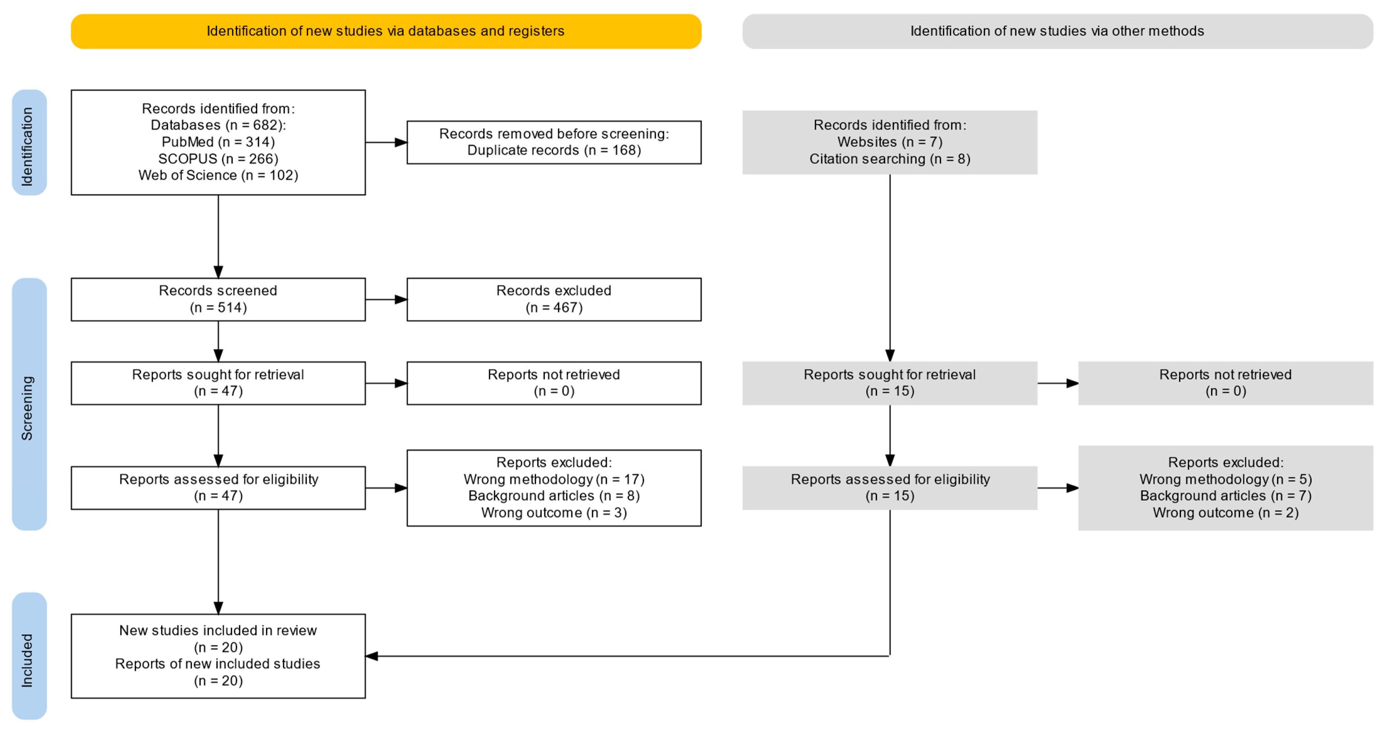
Figure 1. PRISMA flow chart.
Ijms 26 08417 g001
Figure 2. Simplified approach of nanoparticle composition and synthesis methods. ZnO: zinc oxide; NP: nanoparticle; FA: folic acid; HA: hyaluronic acid; MSN: Mesoporous silica nanoparticle; ZIF: zeolitic imidazolate framework-90; DOX: doxorubicin; 5-FU: 5-Fluorouracil; DTX: docetaxel.
Ijms 26 08417 g002
Figure 3. Summary of anticancer action mechanisms of ZnO nanoparticles for achieving targeted chemotherapy.
Ijms 26 08417 g003
Figure 4. Risk of bias and applicability concerns with the use of the SYRCLE risk of bias tool for the quality assessment of animal studies. (A) Authors’ judgements regarding specific domains in eligible studies. Green and red indicate “low” and “high” risk of bias, respectively, while blank cells represent “unclear” risk of bias. (B) Overall summary of applicability concerns, with each domain shown as a percentage across included studies. The blank part in each bar represents an “unclear” applicability concern.
Ijms 26 08417 g004
Table 1. Summary of methodological characteristics, mechanisms, and anticancer performance in eligible preclinical studies.
StudyNanoparticleSynthesis MethodPhysicochemical
Characterization Methods
Cancer Type StudiedModel (In Vitro/In Vivo) and Route of AdministrationPh-Responsive Value *MechanismImportant Findings/Anticancer Performance
Chen et al., 2016 [17]ZnO-DOX/R8@HASequential functionalization protocol (forming ZnO-DOX/R8@HA):
  • APTES modification
  • Thioketal reaction
  • R8 conjugation
  • DOX conjugation
  • HA shell coating
HRTEM, XPS, EDX, FT-IR spectroscopy, DLSSquamous cell carcinoma In vitro: SCC-7 cell line with CD44 receptor overexpressed on the cell membrane and COS7 cell line with negative CD44 receptor expression

In vivo: SCC-7 tumor-bearing mice (IV tail injection)
5Stealthy circulation
Targeted delivery
Controlled release
Synergistic therapeutic effects
  • Selective targeting of CD44-overexpressing tumor cells responded to HAase in the tumor microenvironment (TME).
  • Cell-penetrating peptide facilitated enhanced cellular uptake.
  • Degradation of ZnO released toxic Zn2+ ions and generated ROS.
  • This process triggered controlled drug release and synergistic tumor cell destruction (ROS + chemotherapy).
  • Enabled on-demand drug activation within the tumor site.
Wang et al., 2017 [31]ZnO-DOXDOX loading by ZnO-NPs in aqueous DOX solution (5 mg/mL) under stirring for 24 h in the dark, followed by centrifugation, washing, and freeze-drying.TEM, DLS, Zeta potential measurement, UV–Vis spectroscopy, fluorescence microscopy, flow cytometryBreast cancer, cervical cancer, ovarian cancer, and uterine sarcomaIn vitro: MDA-MB-231 (Dox-sensitive); HeLa (Dox-sensitive); NCI/ADR-RES; MES-SA/Dx5

In vivo: NA
5pH-responsive drug release; Enhanced cellular uptake via endocytosis; ROS generation and Zn2+-mediated apoptosis; CD44 downregulation and CSC sensitization; Immunomodulation via M1-like macrophage polarization; Synergistic therapeutic effects
  • Enhanced intracellular accumulation and cytotoxicity in multidrug-resistant cancer cells.
  • Superior penetration and antitumor activity in 3D tumor spheroids vs. free DOX.
  • Downregulation of CD44 expression with impaired adhesion, migration, and spheroid formation.
  • Macrophage-conditioned medium further potentiated tumor cell apoptosis.
  • Synergistic anticancer effects, overcoming drug resistance and activating innate immune responses.
Cui et al., 2019 [18]ZnO-DOXDOX loading onto commercially obtained ZnO-NPs by mixing ZnO-NPs with DOX solution and incubating at 37 °C for 24 h in the dark; the mixture was then centrifuged, washed, and dried under vacuum.TEM, DLS, Zeta potential measurement, UV–Vis spectroscopy, FT-IR spectroscopy, XRD, fluorescence microscopyColon cancerIn vitro: Human colon cancer cell line HCT116

In vivo: HCT116 tumor-bearing BALB/c nude mice (IV tail injection)
5.5pH-responsive drug release; ROS generation and oxidative damage; Zn2+-mediated apoptosis and autophagy induction; Secretion of proinflammatory cytokines (TNF-α, IL-6, IFN-γ); Synergistic effects
  • Significant inhibition of HCT116 cell proliferation with induction of apoptosis and autophagy via ROS overproduction.
  • Triggered release of proinflammatory cytokines (TNF-α, IL-6, IFN-γ), amplifying tumor immunogenicity.
  • Superior antitumor efficacy in vivo vs. free DOX, with reduced tumor volume and increased apoptosis in xenograft models.
DeLong et al., 2019 [19]ZnO-LL37/carboplatinSuspension of ZnO-NPs in serum-free culture medium, followed by incubation with LL-37 peptide (10 μg/mL) to allow surface adsorption. For combination treatment, carboplatin was added separately to the ZnO–LL37 complex prior to administration.TEM, DLS, Zeta potential measurement, UV–Vis spectroscopy, XRDLung cancerIn vitro: Human non-small cell lung cancer cell line A549 (Intratumoral injection)
In vivo: A549 xenograft tumors in immunocompromised mice
NAROS generation and Zn2+-mediated cytotoxicity; Immune stimulation via LL-37–induced proinflammatory cytokines; Immune infiltration in the TME; Synergistic chemosensitization
  • Local immune cell recruitment and proinflammatory cytokine production in the TME.
  • Significant reduction of tumor burden in vivo compared to monotherapies.
  • Enhanced cytotoxicity and immune activation in both 2D and 3D A549 lung cancer models.
  • Synergistic antitumor effects by coupling immune stimulation with chemotherapy sensitization, supporting its potential as a localized immuno-chemotherapeutic strategy.
He et al., 2019 [22]ZnO@PMNESynthesis of ZnO@PMNE NPs via
  • Polymerization of Zn(MAA)2 in triethylene glycol (TEG) at 72 °C
  • Addition of LiOH·H2O to induce ZnO hybrid NP formation
  • Purification by dialysis, DOX loading by incubating ZnO@PMNE NPs (10 mg) in DOX solution (0.5 mg/mL) for 12 h in the dark.
HRTEM, FT-IR spectroscopy, UV–Vis spectroscopy, photoluminescence spectroscopy, XRD, DLS, confocal fluorescence microscopyHepatocellular carcinomaIn vitro: HepG2 cells

In vivo: HepG2 tumor-bearing nude mice (xenograft model—IV tail injection)
5.4pH-responsive drug release; ROS generation and Zn2+-mediated apoptosis; Thermo- and pH-sensitive polymer shell enabling stimuli-responsive delivery; Enhanced cellular uptake; Synergistic anticancer effects
  • Significant enhancement of cytotoxicity compared to free DOX and ZnO alone.
  • Superior cellular uptake and intracellular DOX accumulation under acidic tumor-like conditions.
  • Effective ROS-mediated apoptosis in HepG2 cells.
  • Substantial tumor growth inhibition in vivo, demonstrating synergistic efficacy and good biosafety.
Xiao et al., 2019 [32]5-FU@ZIF-90@ZnO (FZZ)Solution preparation for porous ZIF-90 NPs under stirring, encapsulation of 5-FU, coating with ZnO through the addition of TEA and zinc acetate into the ZIF-90 solution.TEM, EDS, XRD, FT-IR, DLS, Zeta potential measurementHuman cervical cancer; Hepatocellular carcinomaIn vitro: HeLa cells

In vivo: H22 xenograft tumor model (Balb/c mice—IV tail injection)
5.5ZnO core–shell structure; Controlled drug release; Synergistic effect (zinc ions and 5-FU)
  • Selective targeting of tumor cells within the acidic TME.
  • ZnO shell as a gatekeeper, reducing premature leakage and enabling pH-triggered drug release.
  • ZnO degradation under acidic conditions with Zn2+ release, inducing apoptosis and reducing drug resistance via survivin, XIAP, and HIF-1α downregulation.
  • Synergistic tumor cell destruction through combined 5-FU and Zn2+-mediated apoptosis, ensuring on-demand activation and enhanced efficacy.
Liu et al., 2021 [25]Fe3O4@C/ZnO-DOX-FASolvothermal synthesis of Fe3O4 NPs, MAA functionalization, and sequential ZIF-8 shell growth. Fe3O4@C/ZnO obtained via calcination, followed by PEI modification and FA conjugation through crosslinking. DOX loading achieved via ultrasonication.SEM, TEM, FT-IR, UV–Vis/XRD/Raman spectroscopy, BET analysis, EDS, IR thermal imaging, SQUID magnetometry, fluorescence microscopy, MTT assayHuman cervical cancer; HepatomaIn vitro: HeLa cell line for photothermal, chemo-photothermal assays, and cellular uptake studies

In vivo: H22 hepatoma tumor-bearing female mice (IV tail injection)
5Magnetic targeting; Cancer cell-specific targeting via FA conjugation; pH-responsive drug release; Synergistic chemo-photothermal therapy
  • Enhanced tumor accumulation through dual targeting: magnetic (Fe3O4 core) and cancer cell-specific (FA).
  • Controlled DOX release via pH-responsive ZnO degradation.
  • Carbon shell with photothermal properties under 638 nm laser irradiation, enabling synergistic chemo-photothermal therapy.
Li et al., 2022 [23]ZnO-LD NPs (ZnO-NPs coated with LMHP-DOX complex)Dispersing ZnO-NPs in DMSO,
LMHP-DOX complex formation, ZnO-NPs incubation with LMHP-DOX, dropwise addition, dialysis for the formation of ZnO-LD NPs.
TEM, DLS, XRD, FT-IR spectroscopy, UV–Vis spectroscopy, fluorescence spectroscopyProstate cancer, breast cancerIn vitro: PC-3M, PC-3 (high and low metastatic human prostate cancer), and 4T1 (mammary breast cancer) cell lines (IV tail injection)

In vivo: PC-3M tumor-bearing BALB/c nude male mice mice,
4T1 tumor-bearing BALB/c female mice
4.5pH-sensitive drug release, ROS generation to induce apoptosis; Enhanced penetration into tumor tissues for treatment and imaging with second-order nonlinearity techniques
  • Enhanced cellular uptake of ZnO-LD NPs in cancer cells.
  • ROS production leading to apoptosis via caspase activation.
  • Superior antitumor activity in vitro.
  • Significant tumor growth inhibition in vivo, with enhanced apoptosis and tumor suppression vs. controls.
  • Deep tumor penetration and effective imaging, supporting potential for theranostic (therapy + diagnosis) applications.
Barui et al., 2022 [16]EV-Lipo-pep-NCs-Gem
(NCs constists of Gd-doped ZnO NCs)
  • ZnO-NPs synthesized by mixing zinc nitrate with PEG and ammonia, stirred, heated to 70 °C, washed, and dried
  • Electrospinning of 10 wt% PCL in chloroform:DMF (3:1), with 1 wt% ZnO and 5 wt% DTX; stirred for 4 h, loaded into syringe, electrospun at 18 kV, 0.5 mL/h, 12 cm distance, 28 °C, 62% RH to form nanofibrous scaffolds (PCL, PCL + ZnO, PCL + ZnO + DTX).
FESEM, X-ray detector for energy-dispersive
spectroscopy (EDS) analysis, DC magnetometer, DLS, Zeta potential measurements, NP tracking analysis (NTA), TEM, fluorescence microscopy analyses
Pancreatic cancer (ATCC)In vitro: BxPC3 (non metastatic cell line) and AsPC-1(metastatic cell line)

In vivo: NA
-ZnO NCs: cytotoxicity mechanisms
preferably affecting cancer cells (mainly based on reactive oxygen species, ROS, generation and Zn2+ ions release), reach the tumor area in a selective way, sparing healthy cells, and thus minimizing toxicity toward them, enhancing the response to chemotherapy (Gemcitabine)
  • Higher efficacy of drug-loaded nanoconstruct vs. free gemcitabine.
  • Cell viability reduced from ~60% (Gemcitabine alone) to 30–40% (EV-Lipo-pep-NCs-Gem).
  • Greater reduction in metabolic activity observed in BxPC-3 cells compared to the metastatic cell line.
Ekambaram et al., 2022 [20]PCL + ZnO + DTX nanofibers
  • Dissolvement of zinc nitrate hexahydrate in distilled water.
  • Mixing of PEG flakes with distilled water.
  • Addition of liquid ammonia and mixing with the previously prepared solution.
  • Rapid heating, stirring, cooling to room temperature and washing.

ZnO-NPs + docetaxel (DTX) + PCL were weighed and stirred in magnetic stirrer. Then Electrospinning was carried out.
SEM, EDAX, FT-IR, XRD, TGA, DSC, UV–Vis spectroscopy, HPLC
Human lung cancer
In vitro: Human lung cancer A549 cell line
In vivo: NA
5.5Electrospun nanofiber matrix for sustained release; ZnO-mediated cytotoxicity; DTX-induced apoptosis; pH-responsive release in acidic tumor microenvironment; Synergistic chemo-nanotoxic effect
  • Enhanced cytotoxicity against A549 lung cancer cells with PCL + ZnO + DTX nanofibers vs. PCL or PCL + ZnO.
  • ZnO facilitated ROS generation and improved DTX penetration, promoting apoptosis.
  • Sustained, pH-responsive drug release (pH 5.5), achieving ~90% DTX release over 600 h.
  • Cell cycle arrest and apoptosis: 48.84% in treated A549 cells vs. 2.41% in controls.
  • Biocompatibility and hemocompatibility preserved (hemolysis <5%).

Improved mechanical and release properties, supporting use in post-operative cancer therapy.
Zhou et al., 2022 [34]MTGZ@PPD (MSN-TPZ-GOx@ZnO@PAH-PEG-DMMA)
  • MSNs prepared with TEOS/CTAB; amination and carboxylation steps followed by TPZ loading and GOx conjugation (MTG);
  • ZnO QDs synthesized and linked to MTG (MTGZ); final PPD coating under alkaline pH (MTGZ@PPD)
XRD, TEM, UV–Vis spectroscopy, FT-IR spectroscopy, TGA, ICP-AES4T1 cell line mimics human breast cancerIn vitro: 4T1 (mouse breast cancer) cells, L929 (normal fibroblast) cells

In vivo: 4T1 cells tumor-bearing BALB/c female mice (IV tail injection)
6.5Stealth circulation via charge-convertible PPD shell; pH-responsive charge reversal promotes tumor cell uptake; ZnO QDs decompose under acidic TME, triggering controlled release; Glucose depletion via GOx and hypoxia-activated TPZ enable starvation–chemo synergistic therapy
  • pH-triggered charge reversal of MTGZ@PPD enhanced cellular uptake in the acidic TME.
  • ZnO quantum dots decomposed under low pH, releasing GOx and TPZ in a controlled manner.
  • GOx-induced glucose depletion caused tumor starvation.
  • Hypoxia-activated TPZ enabled targeted chemotherapy.
  • Superior cytotoxicity in vitro and maximal tumor inhibition in vivo without systemic toxicity, confirming effective starvation–chemo synergistic therapy.
Pieretti et al., 2023 [28]ZnO/Cisplatin NPs
  • Hydrothermal synthesis of ZnO-NPs by adding sodium hydroxide to zinc acetate
  • Heating at 170 °C for 10 h
  • Washing, drying, and powdering
  • ZnO-NPs incubated with cisplatin (1:1 w/w) in water at room temperature for 24 h
  • Centrifuged, freeze-dried, and stored
  • Drug loading efficiency assessed using ICP-MS.
TEM, XRD, FT-IR, SEM/EDS, DLS, Zeta potential measurement, ICP-MS, MTT assay, fluorescence microscopy
Prostate cancerIn vitro: PC3 cell line

In vivo: NA
5.5pH-triggered release; NP-mediated cytotoxicity; NO-enhanced chemosensitivity; Sustained platinum release; ZnO intrinsic toxicity (oxidative stress induction); improved selectivity
  • Enhanced cytotoxicity of ZnO/cisplatin NPs vs. free cisplatin or ZnO alone.
  • pH-responsive cisplatin release (45% at pH 5.5), enabling targeted action in acidic TME.
  • NO preconditioning increased chemosensitivity, lowering IC50 and promoting apoptosis.
  • Combination treatment (ZnO/cisplatin + NO) showed highest tumor selectivity and synergistic cytotoxicity in prostate cancer cells.
Mendoza-Martinez et al., 2023 [26]5-FU + ZnO-NPs; Cis + ZnO-NPs
  • Conjugation of ZnO-NPs with 5-FU and cisplatin in a 1:1 ratio to form nanocarriers
  • Solution preparation: stock solutions of 5-FU (1 mol) and cisplatin (1 mol) were prepared
  • NPs were mixed with these chemotherapeutic agents (concentrations of 1 mg/mL).
TEM; DLS; XRD; FT-IR; Zeta potential measurementLymphoid cancerIn vitro: 2D Monolayer Cell Culture; 3D Spheroid Culture

In vivo: NA
NANanocarrier Drug Delivery (ZnO as a carrier for 5-FU and Cis);
ROS generation and apoptosis induction; endocytosis-mediated uptake; Synergistic therapeutic effects between ZnO and chemotherapeutic agents
  • Cytotoxicity demonstrated in both 2D and 3D cancer models, highlighting ZnO-NPs’ therapeutic potential.
  • Enhanced anticancer efficacy when ZnO-NPs were conjugated with 5-FU or cisplatin.
  • ROS generation contributed to apoptosis and amplified anticancer effects.
  • Targeted drug delivery capabilities of ZnO-NPs as nanocarriers.
Li et al., 2024 [24]ZnO−BMSN−MTX
  • Carboxylation of MSN to form COOH−BMSN
  • Encapsulation of MTX into the mesopores of COOH−BMSN
  • Synthesis of NH2-functionalized ZnO QDs
  • Amidation reaction to form ZnO−BMSN−MTX by attaching ZnO−NH2 onto COOH−BMSN and encapsulating MTX.
FT-IR spectroscopy, XRD, TEM, DLS, nitrogen adsorption/desorption isothermsHepatocellular carcinomaIn vitro: SMMC-7721 tumor cells (human hepatocellular carcinoma cell line)

In vivo: NA
5pH-responsive drug release; GSH-responsive drug release; synergistic therapeutic effects (MTX and Zn2+)
  • Enhanced cytotoxicity against SMMC-7721 hepatocellular carcinoma cells.
  • Dual pH- and GSH-responsive drug release, enabling localized delivery in the TME.
  • Synergistic therapeutic effects from MTX and Zn2+ ions via apoptosis induction.
  • Good biocompatibility with low toxicity in normal cells.
Pavan et al., 2024 [27]ZnO-ETP
  • Formation of ZnO-NPs (mixing of zinc and magnesium acetate in ethanol, stirring, and precipitating with n-hexane)
  • Amine functionalization
  • Etoposide loading to amine-functionalized ZnO-NPs
  • Drug Encapsulation to remove unbound etoposide (by dialysis) for the final ZnO-ETP nanoformulation.
SEM, DLS, XRD, FT-IR, UV–Vis spectroscopy, Zeta potential measurement, MTT assayLung adenocarcinomaIn vitro: A549 lung adenocarcinoma cell line

In vivo: NA
5pH-responsive drug release; Inhibition of Cytochrome P450 (CYP3A4), reducingmetabolism and excretion of etoposide; Induction of apoptosis in cancer cells by activating the caspase-7 and caspase-9 pathways; Anti-angiogenesis
  • ZnO-ETP showed superior anticancer effects compared to pure etoposide, even at lower concentrations;
  • pH-responsive drug release(pH 5.0);
  • Increased activation of caspase-7 and caspase-9, leading to cancer cell death;
  • Significant reduction of A549 cell migration, preventing metastasis; Downregulated angiogenesis markers like VEGF, reducing tumor blood supply;
  • Reduced side effects, showing good compatibility with normal cells.
Saravanan et al., 2024 [29]Ver-A@ZnNCsCo-precipitation method for synthesis of ZnO-NPs (zinc acetate and lithium hydroxide as precursors in 100% ethanol)
Chitosan Coating (dropwise addition of chitosan solution in 0.1% acetic acid)
Verrucarin-A Loading with dropwise addition of an ethanolic solution of Verrucarin-A and removal of unbound drug.
TEM, XRD, FT-IR, UV–Vis spectroscopy, DLS, Zeta potential measurement, ICP-MS, MTT assay
Breast cancerIn vitro: MDA-MB-231 TNBC cell line

In vivo: NA
5.5Targeted delivery; pH-responsive drug release; Induction of apoptosis; Synergistic therapeutic effect through ROS generation and inhibition of cancer cell proliferation
  • Selective targeting of TNBC cells with enhanced uptake in acidic TME (pH 5.5).
  • ZnO core degradation at pH 5.5, releasing Verrucarin-A inside tumors.
  • Apoptosis induction via caspase activation and mitochondrial dysfunction.
  • Significant inhibition of MDA-MB-231 cell growth.
  • ROS generation contributes to cancer cell destruction.
Tian et al., 2024 [30]PZnO@DOX
  • Formation of ZnO-NPs (mixing zinc acetate and sodium hydroxide in ethanol)
  • Polyphenol coating
  • DOX loading: purification of PZnO@DOX NPs by centrifugation
TEM, DLS, XRD, FT-IR, UV–Vis spectroscopy, Zeta potential measurement, MTT assayBreast cancerIn vitro: 4T1 breast cancer cell line

In vivo: 4T1 tumor-bearing BALB/c mice (IV tail injection)
5Targeted delivery; Ultrasound-triggered DOX release; STING pathway activation; pH-responsive drug release; Synergistic therapeutic effects
  • PZnO@DOX NPs exhibited enhanced tumor targeting with significant intratumoral drug accumulation and ultrasound-triggered DOX release, improving local drug delivery.
  • NPs induced apoptosis in 4T1 tumor cells via STING pathway activation and subsequent immune stimulation.
  • NPs triggered immunogenic cell death, enhancing anticancer immunity.
  • Combined chemotherapy and STING activation significantly suppressed tumors in vitro and in vivo, leading to tumor growth inhibition and improved survival in Balb/c mice.
Zhi et al., 2023 [33]Cisplatin-loaded ZnO@BBCs
  • DSCP prodrug synthesized from cisplatin via oxidation (H2O2) and succinic anhydride addition
  • AS1411 aptamers and DNAzymes covalently linked via amide-coupling to form BBC nanoscaffolds
  • Positively charged ZnO-NPs electrostatically encapsulated into DNA network in one-pot self-assembly, forming ZnO@BBCs with varying aptamer/DNAzyme ratios.
TEM, DLS, Zeta potential measurement, FT-IR spectroscopy, 1 H NMR spectroscopy, MALDI-TOF MS, HAADF-STEM with EDX mapping, PXRD, EDS, XPS, native PAGE, MTT assayMDR lung adenocarcinomaIn vitro: A549/DDP cells of MDR human lung adenocarcinoma

In vivo:xenograft-
tumor models were established by subcutaneously injecting A549/DDP cells into nude BALB/c mice (IV injection)
4.5−5.5pH-responsive drug release (acidic tumor conditions); ROS generation and Zn2+-mediated cytotoxicity; Circumvention of multidrug resistance by enhancing intracellular drug accumulation and reducing drug efflux
  • Tumor-specific uptake of ZnO@2BBC via nucleolin receptor–mediated endocytosis using multivalent AS1411 aptamers.
  • Lysosomal accumulation with successful nanoparticle internalization and ZnO degradation.
  • Increased platinum accumulation in tumor cells.
  • Egr-1 and MDR1 mRNA downregulation with enhanced intracellular LPO generation.
  • Egr-1 silencing promoted drug retention and increased apoptosis.
  • Demonstrated tumor specificity and biocompatibility.
  • Greatest tumor growth inhibition in vivo, with pronounced apoptosis and no significant toxicity (stable body weight, no histological damage in major organs).
Zhou et al., 2023 [35]ZnO/DPPG/PEG-pp-PE/DOX
  • ZnO-NPs loaded with DOX by incubation
  • DPPG coating in chloroform
  • PEG-pp-PE addition
  • Emulsion formed via sonication and solvent evaporation
  • DOX loaded post-assembly
  • Free drug removed by ultrafiltration
DLS, Zeta potential measurement, TEM, LC–MS/MS, 1 H NMR spectroscopyBreast cancer
In vitro: MDA-MB-231 triple-negative breast cancer cell line
In vivo: MDA-MB-231 xenograft tumor model in BALB/c nude mice (IV tail injection)
5Stealthy circulation; Tumor-targeted delivery via MMP2-cleavable PEG; pH- and enzyme-responsive drug release; Efflux pump evasion; Synergistic cytotoxicity via Zn2+-induced mitochondrial dysfunction and DOX-induced apoptosis
  • ZnO/DOX NPs enhanced cellular uptake and overcame drug efflux in MDR cancer cells.
  • Dual-responsive ZnO/DPPG/PEG-pp-PE/DOX NPs showed increased cytotoxicity and deep penetration in 3D tumor spheroids after MMP2 activation.
  • Synergistic cytotoxicity mediated by mitochondrial membrane depolarization and ROS production.
  • Prolonged blood circulation via PEG-pp-PE modification, with tumor accumulation through the EPR effect and reduced liver/kidney off-target uptake.
  • Significant tumor growth inhibition and apoptosis in MDR xenograft mice treated with ZnO/DPPG/PEG-pp-PE/DOX.
  • No significant systemic toxicity observed.
Gomaa et al., 2024 [21]ZnO-NPs/DOX/FA
  • ZnO-NPs synthesis: hydrolysis, condensation of zinc acetate dihydrate with potassium hydroxide in alcoholic medium at low temperature.
  • Purification.
  • DOX loading: ZnO-NPs incubated in aqueous solution, then centrifuged and freeze-dried.
  • FA functionalization (rZnO/FA): by suspension in PBS and sonication.
  • Final nanocomposites: DOX added to rZnO/FA, stirred, centrifuged, and characterized.
UV–Vis spectroscopy, TEM, MTT assayEACIn vitro: EAC cells

In vivo: EAC tumor-bearing Swiss albino mice (female, 6–8 weeks old) injected intraperitoneally with 2.5 × 10⁵ EAC cells per mouse (IP injection)
NAFA-mediated targeting for enhanced uptake by tumor cells; Controlled cytotoxicity through DOX release and Zn2+-induced ROS generation; Synergistic antitumor effect via combined chemotoxic and immunomodulatory mechanisms
  • Dose-dependent cytotoxicity in vitro, with ZnO-NPs/DOX/FA showing the highest IC50 (38.8 µg/mL), indicating controlled release and reduced off-target toxicity.
  • In vivo, ZnO-NPs/DOX/FA significantly reduced EAC tumor burden.
  • Enhanced apoptosis confirmed via Annexin V/PI staining.
  • Suppressed inflammatory cytokines (IL-6, TNF-α).
  • Preserved liver and kidney function, demonstrating good biosafety.
ZnO: zinc oxide; DOX: doxorubicin; HA: hyaluronic acid; APTES: 3-aminopropyltriethoxysilane; HRTEM: high-resolution transmission electron m\icroscopy; FT-IR spectroscopy: Fourier transform-infrared spectroscopy; DLS: dynamic light scattering; SCC-7: squamous cell carcinoma cell line; COS7: normal kidney fibroblast cell line; IV: intravenous; TME: tumor microenvironment: HAase: hyaluronidase; ZIF: zeolitic imidazolate framework-90; 5-FU: 5-Fluorouracil; TEM: transmission electron microscopy; EDS: energy-dispersive X-ray spectroscopy; XRD: X-ray diffraction analysis; TEA: triethanolamine; FA: folic acid; SEM: scanning electron microscope; UV–Vis: ultraviolet–visible; BET: Brunauer–Emmett–Teller; IR: infrared; SQUID: superconducting quantum interference device; MTT: 3-(4,5-Dimethylthiazol-2-yl)-2,5-diphenyltetrazolium bromide; NPs: nanoparticles; LMHP-DOX complex: low-molecular-weight heparin and doxorubicin complex; DMSO: dimethyl sulfoxide; BMSN: biodegradable mesoporous silica nanoparticles; NO: nitric oxide; IC50: half maximal inhibitory concentration; MTX: methotrexate; QDs: quantum dots; Cis: cisplatin; NA: not applicable; GSH: glutathione; ROS: reactive oxygen species; Ver-A: Verrucarin-A; NCs: nanocomposite; PC3: prostate cancer cells; ICP-AES: inductivelycoupled plasma atomic emission spectrometry; ICP-MS: inductively coupled plasma mass spectrometry; CLSM: confocal laser scanning microscopy; TNBC: triple-negative breast cancer; PZnO: PEG-polyphenol ZnO; STING: Stimulator of Interferon Genes; HeLa: human cervical cancer cell line derived from adenocarcinoma; DTX: docetaxel; MDA-MB-231: human breast cancer cell line derived from triple-negative, highly invasive ductal carcinoma; NCI/ADR-RES: multidrug-resistant human ovarian cancer cell line, originally derived from the OVCAR-8 line; MES-SA/Dx5: multidrug-resistant human uterine sarcoma cell line derived from the parental MES-SA line; HCT116: human colorectal carcinoma cell line; LL37: A human cathelicidin antimicrobial peptide composed of 37 amino acids, with the sequence starting with two leucines (“LL”); PMNE: hydrophilic copolymer poly(methacrylate-co-N-isopropylacrylamide-co-polyethylene glycol methyl acrylate); HepG2: human hepatocellular carcinoma cell line G2; BBC: bio-barcode;MDR: multidrug-resistant;1 H NMR spectroscopy: proton nuclear magnetic resonance spectroscopy;MALDI-TOFMS: matrix-assisted laser-desorption ioniza-tionandtime-of-flightmass; HAADF-STEM: high-angle annular dark-field scanningtransmission electron microscopy; EDX: energy-dispersive X-ray; XPS: X-rayphotoelectron spectroscopy; PAGE: polyacrylamide gel electrophoresis; DSCP: inactive precursor ofcisplatin; LPO: lipid peroxidation; MSN: Mesoporous silica nanoparticle; TPZ: tirapazamine; GOx: glucose oxidase;TEOS: tetraethyl orthosilicate;CTAB: Cetyltrimethylammonium bromide; PPD: refers to PAH-PEG-DMMA (Polyallylamine hydrochloride-Polyethylene glycol-Dimethyl maleic anhydride); TGA: thermogravimetric analysis; PCL: polycaprolactone; DMF: Dimethylformamide; TPZ: Tirapazamine; HPLC: high-performance liquid chromatography; Gd: Gadolinium; NCs: nanocrystals; Lipo-pep: liposomes functionalized with the targeting peptide CKAAKN; Gem: Gemcitabine; PEG-pp-PE: enzyme-sensitive amphiphilic polymer; DPPG: anionic phospholipid;LC–MS/MS: liquid chromatography–tandem mass apectrometry; 1H NMR Spectroscopy: proton nuclear magnetic resonance spectroscopy; MMP2: matrix metalloproteinase 2; EPR: enhanced permeability and retention effect; EAC: Ehrlich ascites carcinoma; IP: intraperitoneal; IL-6: interleukin-6; TNF-α: tumor necrosis factor-α. * The value corresponding to the highest reported percentage of chemotherapeutic drug release was extracted.
Disclaimer/Publisher’s Note: The statements, opinions and data contained in all publications are solely those of the individual author(s) and contributor(s) and not of MDPI and/or the editor(s). MDPI and/or the editor(s) disclaim responsibility for any injury to people or property resulting from any ideas, methods, instructions or products referred to in the content.