Article

Double Pathogenic or Likely Pathogenic Variants in Cancer Predisposition Genes in Hungarian Cancer Patients

1
Department of Molecular Genetics and the National Tumor Biology Laboratory, National Institute of Oncology, 1122 Budapest, Hungary
2
HUN-REN-OOI-TTK-HCEMM Oncogenomics Research Group, 1052 Budapest, Hungary
3
Department of Laboratory Medicine, Semmelweis University, 1089 Budapest, Hungary
4
Department of Oncology Biobank and the National Tumor Biology Laboratory, National Institute of Oncology, 1122 Budapest, Hungary
*
Authors to whom correspondence should be addressed.
These authors contributed equally to this work.
Int. J. Mol. Sci. 2025, 26(17), 8390; https://doi.org/10.3390/ijms26178390
Submission received: 7 August 2025 / Revised: 24 August 2025 / Accepted: 26 August 2025 / Published: 29 August 2025
(This article belongs to the Special Issue Role of Mutations and Polymorphisms in Various Diseases: 2nd Edition)

Abstract

Identification of two or more pathogenic/likely pathogenic (P/LP) variants in cancer susceptibility genes carried by the same patient have important consequences for patient management. We have limited information about the effect of double heterozygosity (DH) in cancer susceptibility genes. The prevalence of DH among Hungarian cancer patients referred to oncogenetic counselling, and comparison of their phenotypes to single variant carriers were performed. In total, 2050 patients were analysed by multigene panel sequencing. Variants of 48 established cancer predisposition genes by ACMG guidelines were evaluated. In overall, P/LP variants were found in 19.8% of cases. DH was observed in 16 cases, amount to 0.8% of all patients, and to 4.0% of positive cases. Appearance of multiple primary tumours was not associated with DH compared to non-P/LP and single P/LP carriers (p = 0.71 and p = 0.54, respectively). Within a cohort of patients referred with suspected HBOC syndrome, earlier tumour formation was observed when DH cases were compared to non-P/LP carriers (p = 0.01), but difference between single and DH carriers was not statistically significant (p = 0.19; Bonferroni corrected alpha = 0.017). Our observations provide information about the incidence of DH status among Hungarian hereditary cancer patients and suggest that DH did not increase the risk of cancer compared to individuals with single P/LP mutation.

1. Introduction

Hereditary cancer syndromes account for approximately 5–10% of all cancers [1,2]. These syndromes are often characterised by familial clustering of cancer-related diseases, early age of onset, or the presence of multiple primary tumours, which suggest underlying germline mutations in cancer susceptibility genes. Alterations in these genes predispose individuals to specific malignancies. BRCA1 and BRCA2 are among the most studied cancer susceptibility genes, significantly increasing the risk of breast, ovarian, pancreatic and prostate cancers. This condition is collectively known as hereditary breast and ovarian cancer (HBOC) syndrome. Similarly, mutations in mismatch repair (MMR) genes, including MLH1, MSH2, MSH6, and PMS2, associate with Lynch syndrome, predisposing individuals to colorectal and endometrial cancers as well as other malignancies including gastric, small bowel, biliary, urothelial, ovarian, and brain cancers [1,2,3].
Identifying pathogenic alterations in cancer susceptibility genes is critical for guiding patient management as genetic diagnoses influence surveillance strategies, preventive measures, surgical decision-making, and therapeutic approaches, such as the use of PARP inhibitors in BRCA1/2 mutation carriers [1,3,4]. The advent of next-generation sequencing (NGS) has been revolutionised genetic diagnostics by enabling the simultaneous analysis of hundreds of genes, or even entire genomes. This has led to an increased detection of pathogenic/likely pathogenic (P/LP) variants, including patients carrying two or more P/LP variants in cancer predisposition genes [5,6].
Cases harbouring multiple germline P/LP variants in cancer susceptibility genes are commonly referred to as double heterozygosity (DH). Other terms, such as multilocus inherited neoplasia alleles syndrome (MINAS) [6,7] and transheterozygosity [8] are also used to describe this phenomenon. Although DH is rare, its frequency depends on the studied population and generally ranges between 0.2% and 1.3% [5,6,7,9,10,11,12,13,14,15,16,17]. Prior to the NGS era, only a few DH cases and gene combinations were reported, with BRCA1-BRCA2 DH cases being the most extensively studied. Despite the growing number of identified DH cases, unique gene combinations remain rare, and the phenotypic impact of one or both variants often remains uncertain.
To date, no comprehensive study has assessed the incidence or clinical significance of DH in Hungarian cancer patients. This study aims to evaluate the phenotypes of Hungarian DH carriers and compare their clinical characteristics to those of single P/LP variant carriers.

2. Results

2.1. Cohort Characteristics and Detection Rate

The majority of consecutive patients (n = 2050) referred to our Comprehensive Cancer Centre had a suspicion of HBOC syndrome, which accounted for 73.0% (n = 1496), followed by patients with Lynch syndrome-related tumours or polyposis, which accounted for 13.1% (n = 268). Further, 7.1% (n = 146) of the patients had endocrine-system originated cancer, and 4.1% (n = 84) of cases had other tumour types (e.g., melanoma, renal, bladder, liver cancer, etc.). The remaining 2.7% (n = 56) of the cohort comprised individuals who were not affected by a malignant disorder, but for whom the rationale for genetic testing was based on the family history. Demographic and clinical parameters of patient’s subgroups are summarised in Table 1. Females predominated in the group of suspected HBOC cases, accounting for 95.5% (n = 1428) and in the group of unaffected patients, accounting for 100% (n = 56; 100%) of cases. Men represented 30.2% (n = 81) of the Lynch/polyposis group, 41.1% (n = 60) of and 46.4% (n = 39) of patients with endocrine and other tumours, respectively. The mean age of disease onset differed between these groups. In the HBOC and Lynch/polyposis suspect cohorts, the mean age of onset was 49.5 and 51.9 years, respectively, whereas patients with endocrine manifestations and other tumours developed symptoms at an earlier age, 41.2 and 41.9 years, respectively.
Pathogenic or likely pathogenic (P/LP) variants were identified in 19.8% (n = 405) of all cases, with the highest prevalence observed in patients with suspected Lynch/polyposis syndrome (27.2%). The detection rate was 18.5% in patients with suspected HBOC syndrome, 21.2% in those with endocrine tumours, 21.4% in patients with other tumour types, and 10.7% in unaffected individuals.
P/LP variants were predominantly found in genes expected from the phenotype (Figure 1). Namely, 91.3% of suspected HBOC cases, 84.9% of patients with suspected Lynch/polyposis syndrome and 67.7% of patients with endocrine manifestations have variants in genes corresponding to the patient’s phenotype. The relatively lower rate in the endocrine group may be due to a variable and mixed group of patients. The P/LP variant ratio was the highest in the group of Lynch/polyposis suspects and the lowest in the unaffected cases.

2.2. Double Heterozygosity

Sixteen (0.8% of all patients) individuals were identified carrying two or more P/LP variants among 2050 patients suspected of hereditary cancer. Of these, 13 patients had suspected HBOC syndrome (0.9% within suspected HBOC cases): 11 presented with breast cancer, one with both breast and endometrial cancer, and one with prostate cancer. One-one case was identified in the Lynch/polyposis, other, and unaffected patient cohorts, giving frequencies of 0.4%, 1.2%, and 1.8%, respectively (Table 1). The clinicopathological characteristics and P/LP variants of these double-hit cases are detailed in Table 2A,B.
In nine cases, two P/LP variants were found in genes associated with HBOC. Five cases exhibited combinations of HBOC and endocrine or Lynch syndrome genes (e.g., SDHAF2, SDHA, MSH6). One patient carried P/LP variants in MSH2 (a Lynch syndrome gene) and SDHB (associated with paraganglioma and pheochromocytoma). One patient carried three P/LP variants, two in genes associated with HBOC and one associated with HBOC endocrine genes (Table 2A,B).
Two patients carried both variants in high-penetrance genes, while 11 had one high- and one/or two moderate/low-penetrance variant(s), and three had both variants in moderate/low-penetrance genes (Figure 2A,B). Genes were classified as high, moderate and low risk genes based on their penetrance data, focusing on the highest risk regardless of the type of tumour, using former published data and data from National Comprehensive Cancer Network (NCCN) guidelines [18,19,20,21,22].
Of the 10 cases with double P/LP variants in HBOC genes, eight had breast cancer, one had prostate cancer, and one had a fumarate hydratase (FH)-deficient leiomyoma with a family history of breast cancer. In three cases, BRCA1/2 variants co-occurred with MSH6 variants. In four cases, SDHA and SDHAF2 variants were identified together with HBOC genes. One of these had three P/LP variants (BRCA1, ATM and SDHA) (case #12; Table 2B). Another case (case #2) had MSH2 and SDHB variants, which predispose to hereditary pheochromocytoma-paraganglioma syndrome. Of the 16 DH cases, two (case #4 and #15) involved P/LP variants within the same gene (CHEK2). The cis-trans positioning was not determined, but familial data suggested a trans configuration for one of them (Supplementary Figure S1).
In cases carrying P/LP variants in two different syndrome genes, the phenotype typically reflected the effect of one variant. In one case, neither of the found P/LP variants explained the phenotype: a patient with FH-deficient leiomyoma had P/LP variants in CDH1 and ATM (case #16). An unaffected patient carrying a BRCA1 and MSH6 variants had a family history of breast cancer (case #5). Case #10 with P/LP variants in CHEK2 and SDHAF2 variants had a mixed phenotype with endometrial and breast cancer.
Two of the DH cases exhibited second or third solid malignancies. The frequency of multiple solid tumours among DH cases (12.5%) was not elevated compared to non-carriers (11.7%; p = 0.71) and single-variant carriers (21.9%; p = 0.54) analysed by Fisher’s exact test. No significant difference was found when restricted to HBOC-suspected cases (p = 0.66 and p = 0.74 compared to non- and single-variant carriers, respectively).
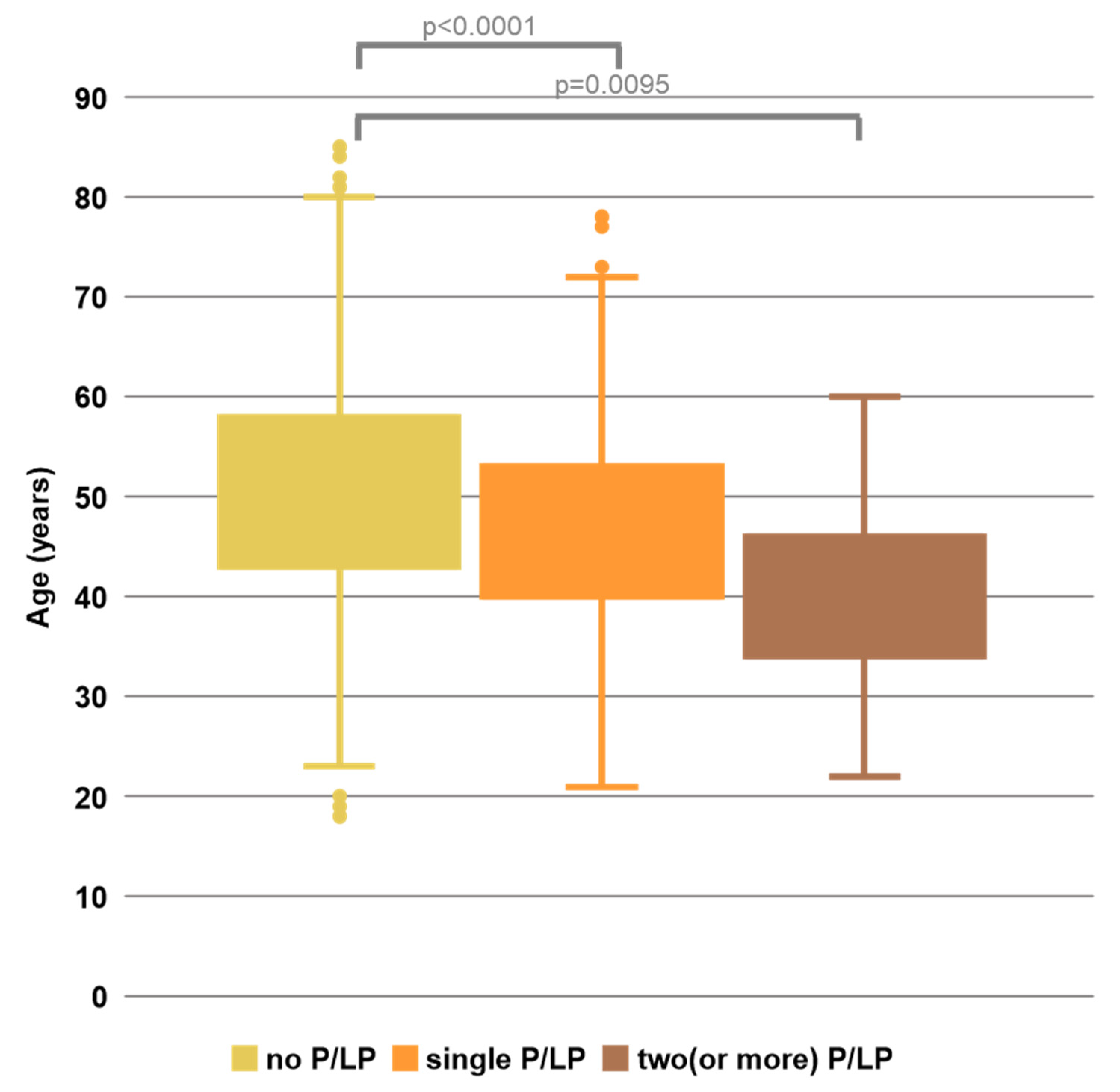
To evaluate whether DH status influences the age of tumour onset, HBOC-suspected patients were analysed. The mean ages at diagnosis were 50.2, 46.8, and 40.7 years for non-carriers, single-variant carriers, and DH cases, respectively. While both single-variant and DH cases had significantly lower ages than non-carriers, the difference between single-variant and DH cases was not statistically significant analysed by the Kruskal–Wallis analysis of variance with Dunn’s post hoc test (p = 0.19; Bonferroni-corrected α = 0.017). However, DH cases tended to have a lower mean age (40.7 years vs. 46.8 years; Figure 3).

3. Discussion

The introduction of NGS has revolutionised genetic analysis, enabling its integration into clinical practice. NGS provides a cost-effective method for generating vast amounts of genetic data, including information about genes not directly related to the patient’s phenotype. Consequently, the detection of complex genotypes with multiple P/LP variants is increasing. However, predicting the phenotypic effects of harbouring two or more P/LP variants in cancer-predisposing genes remains a challenge [5,6].
The BRCA1-BRCA2 combination has been the most extensively studied. Some reports indicated that double-hit carriers develop cancer at an earlier age compared to single P/LP carriers [11,13,23]. However, analyses based on the Consortium of Investigators of Modifiers of BRCA1/2 (CIMBA dataset) have suggested that DH carriers exhibit a phenotype similar to BRCA1 carriers alone [8]. Other studies found no significant difference in disease severity between DH and single-variant carriers, particularly in breast cancer patients [14,24]. Former studies performed systematic reviews to describe the genetic architecture and phenotypic consequences of DH/MINAS cases. In a former study, 82 cases were collected with co-occurring P/LP variants in 17 hereditary cancer genes [7]. The most frequently reported combinations were BRCA1/BRCA2, BRCA2/TP53, BRCA1/MLH1, and APC/MLH1, reflecting the relevance of the most commonly screened genes for major hereditary cancer syndromes, as well as the presence of founder variants in certain populations. A later update review [6] analysed 385 patients with two or more P/LP variants. At least one P/LP variant in the BRCA1 and/or BRCA2 genes was identified in most cases (74.6%). The ratio of multiple primary tumours was 28.1% in this cohort of patients, and 15% of patients showed an atypical tumour phenotype (tumours not related to any of the disrupted genes). The authors concluded that the clinical phenotype was rather an independent effect of two (or more) P/LP variants; however, some cases with unusual tumour phenotypes raise the possibility of complex interaction [6]. In a retrospective study, several DH cases were identified, showing a younger age of onset in ATM/CHEK2 and double CHEK2 carriers [25]. A more recent review investigated correlations between the DH status and clinical characteristics (n = 413). Based on their results, DH carriers tend to have more malignancies and an early onset of the first cancer [26].
The controversial conclusions of studies dealing with multiple pathogenic variants among cancer patients may be due to the different genetic architecture of DH cases in the studied cohorts, different founder mutations in some populations, the slightly different gene panels used and the different assignment of PVs. For example, MUTYH heterozygous variants are often referred to as pathogenic variants despite their autosomal recessive inheritance [15].
This is the first study assessing the prevalence and phenotypic features of DH cases in a Hungarian cohort of patients suspected of hereditary cancer predisposition. Previous studies using large gene panels reported DH frequencies of 0.2–1.3% in all patients and 1.8–6.2% of P/LP variants. In our cohort, DH cases comprised 0.8% of all patients and 4.1% of P/LP carriers, aligning with earlier findings [5,12,13,14,15,16,17]. Of the P/LP positive cases, DH carriers were more prevalent among HBOC cases (0.9%) compared to Lynch/polyposis cases (0.4%).
Overall, in our cohort, P/LP variants were most frequently detected in patients with suspected Lynch/polyposis syndrome (27.2%). The prevalence was 18.5% in patients with suspected HBOC syndrome, and slightly above 20% in those with endocrine tumours (21.2%) and other tumour types (21.4%). As expected, the lowest P/LP detection rate was observed in the group of unaffected individuals (10.7%). The detection rates of P/LP variants in patients with suspected HBOC and Lynch/polyposis syndromes are consistent with our previous findings [27,28]. Other studies have also reported similar frequencies of P/LP variants using multigene panel testing, although rates vary depending on cancer type and the panel applied [20,29,30].
Our analysis found no evidence that DH contributes to more primary solid tumour development or significantly influences the clinical phenotype, in agreement with other studies [14]. Similarly, our results showed no earlier tumour onset among DH carriers compared to single-variant carriers, aligning with findings by other researchers [10,16,31]. While our DH carriers had a lower mean age at diagnosis, the difference was not statistically significant, likely due to the small sample size.
Some studies suggested a higher rate of multiple primary tumours among DH carriers (28%) [6] and found that DH status was associated with multiple primary cancers [26]. In contrast, our data showed that the rate of multiple independent (non-syndromic) malignancies in our DH cohort was 12.5%. This might be related to the patient group studied. Our study included consecutive cancer patients referred for genetic counselling.
Interestingly, despite BRCA1 and BRCA2 accounting for most pathogenic variants in our study, only one case had DH of BRCA1-BRCA2. Moreover, high-penetrance cancer susceptibility gene combinations were rare (2/16 cases: BRCA1-BRCA2 and BRCA2-CDH1). Seven of the 16 DH cases involved secondary findings in genes, such as MSH6 or SDHx genes, not associated with the phenotype and without associated tumour types observed during follow-up. The phenotypes of DH cases in our cohort seem to support that the effect of two P/LP variants on cancer risk is probably independent rather than synergistic, as suggested by former studies [5,6].
In conclusion, DH carriers did not show an increased risk of earlier tumour onset or multiple primary tumours compared to patients with only one P/LP variant in tumour susceptibility genes. DH remains rare among Hungarian cancer patients, with approximately 1 in 25 P/LP carriers harbouring an additional variant. While rare, DH has significant implications for offspring and family screening, with a 75% chance of inheriting one or two P/LP variants, compared to 50% for single carriers. Our findings suggest that DH patients do not require stricter surveillance than recommended for their most severe P/LP variant. However, specific gene combinations may warrant tighter protocols, as suggested by some studies [25,32,33]. Limitations of our study include the small sample size and lack of long-term follow-up data. In our workflow, self-reported family histories were collected during pre-test counselling, which may contain inaccuracies and represent a limitation of our study [34]. However, in our practice, sending a questionnaire in advance regarding medical and family history improved accuracy. Our previous experience showed that families of P/LP variant carriers present with significantly more syndrome-specific tumours compared to probands without a genetic predisposition [27]. Therefore, we consider our approach to adequately reflect the clinical situation. Further systematic studies are needed to clarify the clinical relevance of DH and its impact on cancer predisposition and management.

4. Materials and Methods

4.1. Patients

Two thousand fifty consecutive, unrelated patients referred to the Department of Molecular Genetics of the National Institute of Oncology (Budapest, Hungary) for possible hereditary tumour syndrome between March 2021 and August 2023 were included in the study. In accordance with Hungarian legislation, all patients were provided with clinical genetic (pre-test) counselling, during which all aspects of the molecular genetic tests were discussed (https://kollegium.okfo.gov.hu/iranyelvek (accessed on 25 August 2025)). During genetic counselling, all patients provided written informed consent to undergo genetic testing and participate in related research. Relevant medical history and family data were collected. The indication for genetic testing was assessed according to international guidelines, including the NCCN Guidelines Version 2.2024: Genetic/Familial High-Risk Assessment: Breast, Ovarian, and Pancreatic, the NCCN Guidelines Version 1.2024: Genetic/Familial High-Risk Assessment: Colorectal, Endometrial, and Gastric, as well as established clinical guidelines for endocrine tumours [35]. Following the completion of molecular genetic testing, the results were presented to the patients during post-test counselling. Being a comprehensive centre, patients with potential hereditary predisposition for several types of cancer, including breast, ovarian and colorectal cancer, as well as endocrine or other rare tumours, have been evaluated. The study was approved by the Scientific and Research Committee of the Medical Research Council of the Ministry of Health, Hungary (ETT-TUKEB 53720-4/2019/EÜIG, ETT-TUKEB 4457/2012/EKU).

4.2. Genetic Analysis

Peripheral blood samples were collected from patients, and genomic DNA was isolated using Gentra Puregene Blood Kit (Cat No.: 158389, Qiagen, Hilden, Germany) according to the manufacturer’s instructions. For genetic testing, the TruSight Hereditary Cancer Panel (#20029551, Illumina, San Diego, CA, USA) was employed, which contains 113 genes that are predisposing to various cancers. Library construction and next-generation sequencing were performed on an Illumina MiSeq or NextSeq 550Dx platforms, according to the manufacturer’s instructions. Data were analysed by DRAGEN Enrichment pipeline (Version 4.0.3, Illumina, San Diego, CA, USA). Sequencing reads were aligned to the human GRCh38 genome reference. Variants were filtered as follows: 3% threshold for population allele frequency (data imported from gnomAD v2.1.1), the region of interest covered the exonic target regions, which were extended to overreach exon/intron boundaries by 30 base pairs. DRAGEN pipeline was used for small nucleotide variant, copy number variant (CNV) and structural variant (SV) calling. Variants with a total read depth was above 10, and variant allele frequency was above 20% were further interpreted. For CNVs and SVs calls, highly recurrent variants (>1% in our examined cohort) were omitted.
In addition to the DRAGEN pipeline, we also used the VarSeq CNV algorithm (v2.6.2 Golden Helix, Bozeman, Montana, USA) for CNV calling. Calls were retained when p < 0.05 and the Z-score  ≤  −3 or Z-score  ≥  3, and normalised read depth < 0.7 or >1.2 for heterozygous deletions and duplications, respectively. Variants were annotated by BaseSpace Variant Interpreter (Illumina, San Diego, CA, USA) and VarSeq (Golden Helix, Bozeman, MT, USA).
For variant classification, the American College of Medical Genetics and Genomics (ACMG) [36] guideline was used, supplemented by the information deposited in NBCI ClinVar (https://www.ncbi.nlm.nih.gov/clinvar/ (accessed between 1 September 2021–12 January 2025)), Franklin (https://franklin.genoox.com/clinical-db/home (accessed between 1 September 2021–12 January 2025)) and Varsome (https://varsome.com/) databases. Of the 113 genes of the applied panel, only a set of 48 selected genes was evaluated based on the NCCN (2024) and ACMG [36] guidelines for HBOC (NCCN Guidelines Version 2.2024, Genetic/Familial High-Risk Assessment: Breast, Ovarian, and Pancreatic) and colorectal, endometrial, and gastric cancers (NCCN Guidelines Version 1.2024, Genetic/Familial High-Risk Assessment: Colorectal, Endometrial, and Gastric), and also well-established genes predispose to endocrine and other tumour syndromes (Supplementary Table S1) [1,35,37,38]. Regarding genes with recessive inheritance, e.g., MUTYH and MSH3, only biallelic P/LP variants were considered as one hit. For EPCAM variants, only deletions of the 3’ end were accepted as pathogenic. Low penetrance CHEK2 variants (p.I157T, p.T476M and p.S428F) were excluded from our analysis, due to their inconsistent interpretation and lack of clinical consequence [39]. Due to similar reasons, APC p.I1307K was not considered as a P/LP variant [40,41].
All P/LP variants were validated by the “gold standard” Sanger sequencing for single-nucleotide variants (SNVs) and for small insertions/deletions, and by multiplex ligation-dependent probe amplification (MLPA; SALSA MLPA probe mixes for appropriate genes, probe sets are listed in Supplementary Table S1, MRC-Holland, Amsterdam, The Netherlands) for large-scale CNV changes. The Illumina Hereditary Cancer Panel we used is specifically designed to avoid interference from pseudogenes. Target enrichment is achieved by hybridisation-based capture with unique probes that selectively bind to the canonical genes, thereby minimising pseudogene amplification. DRAGEN (Illumina) pipeline applies stringent alignment algorithms to uniquely map reads to the canonical locus. In addition, all pathogenic and likely pathogenic variants were validated by Sanger sequencing. During primer design, care was taken to avoid products derived from pseudogenes, providing locus-specific confirmation.

4.3. Statistical Analyses

To evaluate whether DH has any disadvantages in the development of primary solid tumours, we examined whether there is an earlier age of disease onset or a trend to form multiple primary solid tumours in the probands. The occurrence of multiple primary solid tumours was compared between cases of DH and those with a single or non-P/LP variant carriers by Fisher’s exact test. A comparison of the age of disease onset between DH cases and single or non-P/LP cases was conducted exclusively within the patient cohort referred for HBOC suspicion to ensure the homogeneity of the samples, given that the mean age of different disease groups varies significantly. The number of DH cases in the other groups was insufficient to perform statistical analysis. Statistical analysis was performed using GraphPad Prism (v.8.0.1) software. A Kruskal–Wallis test with Dunn’s post hoc test was employed to ascertain whether there were any significant differences in the age of disease onset between the following groups: no variant carriers, single variant carriers and two (or more) variant carriers among HBOC-suspected patients. Bonferroni correction was applied for multiple comparisons, and p-values < 0.017 were accepted as statistically significant.

Supplementary Materials

The following supporting information can be downloaded at: https://www.mdpi.com/article/10.3390/ijms26178390/s1.

Author Contributions

Conceptualization, T.P., H.B. and A.P.; Methodology, J.P., T.P. and A.B.; Formal Analysis, T.P. and H.B.; Investigation, H.B., T.P., J.P., A.B., P.N. and V.K.G.; Resources, A.P.; Data Curation, H.B. and A.P.; Writing—Original Draft Preparation, T.P. and H.B.; Writing—Review and Editing, H.B. and A.P.; Visualisation, T.P. and J.P.; Supervision, H.B. and A.P.; Funding Acquisition, A.P. and H.B. All authors have read and agreed to the published version of the manuscript.

Funding

Hungarian Scientific Research Grant of the National Research, Development and Innovation (NRDI) Office Fund of the Ministry of Culture and Innovation under the National Laboratories Program (National Tumour Biology Laboratory (2022-2.1.1-NL-2022-00010)) grant agreements (A.P. and H.B.) and the Hungarian Thematic Excellence Program (TKP2021-EGA/TKP2021-NVA/TKP2021-NKTA, MOLORKIV (A.P.).

Institutional Review Board Statement

The study was conducted in accordance with the Declaration of Helsinki and approved by the Scientific and Research Committee of the Medical Research Council of the Ministry of Health, Hungary (ETT-TUKEB 53720-4/2019/EÜIG), approved on 18 December 2019.

Informed Consent Statement

During genetic counselling, written informed consent was obtained from all subjects involved in the study.

Data Availability Statement

All data in the study are included in the article, further inquiries can be directed to the corresponding author.

Conflicts of Interest

The authors declare no conflicts of interest. The funders had no role in the design of the study; in the collection, analyses, or interpretation of data; in the writing of the manuscript; or in the decision to publish the results.

References

  1. Garutti, M.; Foffano, L.; Mazzeo, R.; Michelotti, A.; Da Ros, L.; Viel, A.; Miolo, G.; Zambelli, A.; Puglisi, F. Hereditary Cancer Syndromes: A Comprehensive Review with a Visual Tool. Genes 2023, 14, 1025. [Google Scholar] [CrossRef] [PubMed]
  2. Nagy, R.; Sweet, K.; Eng, C. Highly Penetrant Hereditary Cancer Syndromes. Oncogene 2004, 23, 6445–6470. [Google Scholar] [CrossRef] [PubMed]
  3. Imyanitov, E.N.; Kuligina, E.S.; Sokolenko, A.P.; Suspitsin, E.N.; Yanus, G.A.; Iyevleva, A.G.; Ivantsov, A.O.; Aleksakhina, S.N. Hereditary Cancer Syndromes. World J. Clin. Oncol. 2023, 14, 40–68. [Google Scholar] [CrossRef]
  4. Ragupathi, A.; Singh, M.; Perez, A.M.; Zhang, D. Targeting the BRCA1/2 Deficient Cancer with PARP Inhibitors: Clinical Outcomes and Mechanistic Insights. Front. Cell Dev. Biol. 2023, 11, 1133472. [Google Scholar] [CrossRef]
  5. Infante, M.; Arranz-Ledo, M.; Lastra, E.; Abella, L.E.; Ferreira, R.; Orozco, M.; Hernández, L.; Martínez, N.; Durán, M. Increased Co-Occurrence of Pathogenic Variants in Hereditary Breast and Ovarian Cancer and Lynch Syndromes: A Consequence of Multigene Panel Genetic Testing? Int. J. Mol. Sci. 2022, 23, 11499. [Google Scholar] [CrossRef]
  6. McGuigan, A.; Whitworth, J.; Andreou, A.; Hearn, T.; Genomics England Research Consortium; Ambrose, J.C.; Arumugam, P.; Bevers, R.; Bleda, M.; Boardman-Pretty, F.; et al. Multilocus Inherited Neoplasia Allele Syndrome (MINAS): An Update. Eur. J. Hum. Genet. 2022, 30, 265–270. [Google Scholar] [CrossRef]
  7. Whitworth, J.; Skytte, A.-B.; Sunde, L.; Lim, D.H.; Arends, M.J.; Happerfield, L.; Frayling, I.M.; Van Minkelen, R.; Woodward, E.R.; Tischkowitz, M.D.; et al. Multilocus Inherited Neoplasia Alleles Syndrome: A Case Series and Review. JAMA Oncol. 2016, 2, 373–379. [Google Scholar] [CrossRef]
  8. Rebbeck, T.R.; Friebel, T.M.; Mitra, N.; Wan, F.; Chen, S.; Andrulis, I.L.; Apostolou, P.; Arnold, N.; Arun, B.K.; Barrowdale, D.; et al. Inheritance of Deleterious Mutations at Both BRCA1 and BRCA2 in an International Sample of 32,295 Women. Breast Cancer Res. 2016, 18, 112. [Google Scholar] [CrossRef]
  9. Le Page, C.; Rahimi, K.; Rodrigues, M.; Heinzelmann-Schwarz, V.; Recio, N.; Tommasi, S.; Bataillon, G.; Portelance, L.; Golmard, L.; Meunier, L.; et al. Clinicopathological Features of Women with Epithelial Ovarian Cancer and Double Heterozygosity for BRCA1 and BRCA2: A Systematic Review and Case Report Analysis. Gynecol. Oncol. 2020, 156, 377–386. [Google Scholar] [CrossRef]
  10. Laish, I.; Friedman, E.; Levi-Reznick, G.; Kedar, I.; Katz, L.; Levi, Z.; Halpern, N.; Parnasa, S.; Abu-Shatya, A.; Half, E.; et al. Double Heterozygotes of BRCA1/BRCA2 and Mismatch Repair Gene Pathogenic Variants: Case Series and Clinical Implications. Breast Cancer Res. Treat. 2021, 188, 685–694. [Google Scholar] [CrossRef] [PubMed]
  11. Bang, Y.J.; Kwon, W.K.; Nam, S.J.; Kim, S.W.; Chae, B.-J.; Lee, S.K.; Ryu, J.M.; Kim, J.-W.; Yu, J.; Lee, J.E. Clinicopathological Characterization of Double Heterozygosity for BRCA1 and BRCA2 Variants in Korean Breast Cancer Patients. Cancer Res. Treat. 2022, 54, 827–833. [Google Scholar] [CrossRef] [PubMed]
  12. Van Der Merwe, N.C.; Buccimazza, I.; Rossouw, B.; Araujo, M.; Ntaita, K.S.; Schoeman, M.; Vorster, K.; Napo, K.; Kotze, M.J.; Oosthuizen, J. Clinical Relevance of Double Heterozygosity Revealed by Next-Generation Sequencing of Homologous Recombination Repair Pathway Genes in South African Breast Cancer Patients. Breast Cancer Res. Treat. 2024, 207, 331–342. [Google Scholar] [CrossRef]
  13. Laitman, Y.; Niskakoski, A.; Bernstein-Molho, R.; Koskinen, L.; Rabina, D.; Koskenvuo, J.; Friedman, E. Phenotypes of Carriers of Two Mutated Alleles in Major Cancer Susceptibility Genes. Breast Cancer Res. Treat. 2024, 208, 589–595. [Google Scholar] [CrossRef] [PubMed]
  14. Megid, T.B.C.; Barros-Filho, M.C.; Pisani, J.P.; Achatz, M.I. Double Heterozygous Pathogenic Variants Prevalence in a Cohort of Patients with Hereditary Breast Cancer. Front. Oncol. 2022, 12, 873395. [Google Scholar] [CrossRef]
  15. Agaoglu, N.B.; Doganay, L. Concurrent Pathogenic Variations in Patients with Hereditary Cancer Syndromes. Eur. J. Med. Genet. 2021, 64, 104366. [Google Scholar] [CrossRef]
  16. Stradella, A.; Del Valle, J.; Rofes, P.; Feliubadaló, L.; Grau Garces, È.; Velasco, À.; González, S.; Vargas, G.; Izquierdo, Á.; Campos, O.; et al. Does Multilocus Inherited Neoplasia Alleles Syndrome Have Severe Clinical Expression? J. Med. Genet. 2019, 56, 521–525. [Google Scholar] [CrossRef] [PubMed]
  17. Kwong, A.; Ho, C.Y.S.; Au, C.-H.; Ma, E.S.K. Double Heterozygosity for Germline Mutations in Chinese Breast Cancer Patients. Cancers 2024, 16, 2547. [Google Scholar] [CrossRef]
  18. Abe, A.; Imoto, I.; Ueki, A.; Nomura, H.; Kanao, H. Moderate-Risk Genes for Hereditary Ovarian Cancers Involved in the Homologous Recombination Repair Pathway. IJMS 2022, 23, 11790. [Google Scholar] [CrossRef]
  19. Daly, M.B.; Pal, T.; Berry, M.P.; Buys, S.S.; Dickson, P.; Domchek, S.M.; Elkhanany, A.; Friedman, S.; Goggins, M.; Hutton, M.L.; et al. Genetic/Familial High-Risk Assessment: Breast, Ovarian, and Pancreatic, Version 2.2021, NCCN Clinical Practice Guidelines in Oncology. J. Natl. Compr. Cancer Netw. 2021, 19, 77–102. [Google Scholar] [CrossRef]
  20. LaDuca, H.; Polley, E.C.; Yussuf, A.; Hoang, L.; Gutierrez, S.; Hart, S.N.; Yadav, S.; Hu, C.; Na, J.; Goldgar, D.E.; et al. A Clinical Guide to Hereditary Cancer Panel Testing: Evaluation of Gene-Specific Cancer Associations and Sensitivity of Genetic Testing Criteria in a Cohort of 165,000 High-Risk Patients. Genet. Med. 2020, 22, 407–415. [Google Scholar] [CrossRef]
  21. Rijken, J.A.; Niemeijer, N.D.; Jonker, M.A.; Eijkelenkamp, K.; Jansen, J.C.; Van Berkel, A.; Timmers, H.J.L.M.; Kunst, H.P.M.; Bisschop, P.H.L.T.; Kerstens, M.N.; et al. The Penetrance of Paraganglioma and Pheochromocytoma in SDHB Germline Mutation Carriers. Clin. Genet. 2018, 93, 60–66. [Google Scholar] [CrossRef]
  22. Maniam, P.; Zhou, K.; Lonergan, M.; Berg, J.N.; Goudie, D.R.; Newey, P.J. Pathogenicity and Penetrance of Germline SDHA Variants in Pheochromocytoma and Paraganglioma (PPGL). J. Endocr. Soc. 2018, 2, 806–816. [Google Scholar] [CrossRef] [PubMed]
  23. Lavie, O.; Narod, S.; Lejbkowicz, F.; Dishon, S.; Goldberg, Y.; Gemer, O.; Rennert, G. Double Heterozygosity in the BRCA1 and BRCA2 Genes in the Jewish Population. Ann. Oncol. 2011, 22, 964–966. [Google Scholar] [CrossRef] [PubMed]
  24. Leegte, B. Phenotypic Expression of Double Heterozygosity for BRCA1 and BRCA2 Germline Mutations. J. Med. Genet. 2005, 42, e20. [Google Scholar] [CrossRef]
  25. Agaoglu, N.B.; Bychkovsky, B.L.; Horton, C.; Lo, M.-T.; Polfus, L.; Carraway, C.; Hemyari, P.; Young, C.; Richardson, M.E.; Scheib, R.; et al. Cancer Burden in Individuals with Single versus Double Pathogenic Variants in Cancer Susceptibility Genes. Genet. Med. Open 2024, 2, 101829. [Google Scholar] [CrossRef]
  26. Yuen, J.; Zhou, S.; Caeser, R.; Venkatramani, M.; Bte Ishak, D.N.; Li, S.-T.; Zhang, Z.; Chiang, J.; Chan, S.H.; Ngeow, J. Multi-Locus Inherited Neoplasia Alleles Syndromes in Cancer: Implications for Clinical Practice. Eur. J. Hum. Genet. 2025, 33, 289–296. [Google Scholar] [CrossRef]
  27. Nagy, P.; Papp, J.; Grolmusz, V.K.; Bozsik, A.; Pócza, T.; Oláh, E.; Patócs, A.; Butz, H. Comprehensive Clinical Genetics, Molecular and Pathological Evaluation Efficiently Assist Diagnostics and Therapy Selection in Breast Cancer Patients with Hereditary Genetic Background. Int. J. Mol. Sci. 2024, 25, 12546. [Google Scholar] [CrossRef]
  28. Horti-Oravecz, K.; Bozsik, A.; Pócza, T.; Vereczkey, I.; Strausz, T.; Tóth, E.; Sedlackova, T.; Rusnakova, D.; Szemes, T.; Likó, I.; et al. Whole Genome Sequencing Completes the Molecular Genetic Testing Workflow of Patients with Lynch Syndrome. npj Genom. Med. 2025, 10, 5. [Google Scholar] [CrossRef] [PubMed]
  29. Bilyalov, A.; Nikolaev, S.; Shigapova, L.; Khatkov, I.; Danishevich, A.; Zhukova, L.; Smolin, S.; Titova, M.; Lisica, T.; Bodunova, N.; et al. Application of Multigene Panels Testing for Hereditary Cancer Syndromes. Biology 2022, 11, 1461. [Google Scholar] [CrossRef]
  30. Syed, H.; Sommovilla, J.; Burke, C.A.; McGee, S.; Macaron, C.; Heald, B.; Lyu, R.; Schmit, S.L.; Nair, K.; Kamath, S.; et al. Referral, Uptake, and Outcome of Genetic Counseling and Testing in Patients With Early-Onset Colorectal Cancer. J. Natl. Compr. Cancer Netw. 2023, 21, 1156–1163.e5. [Google Scholar] [CrossRef]
  31. Aguilar, D.; Garza-Rodríguez, M.L.; Pérez-Ibave, D.C.; Muñiz-Garza, C.E.; Treviño, V.; Villarreal-Garza, C.M.; Vidal-Gutiérrez, O.; Burciaga-Flores, C.H. Landscape of Multilocus Inherited Neoplasia Allele Syndrome in Mexican Population. JCO Glob. Oncol. 2025, 11, e2400065. [Google Scholar] [CrossRef]
  32. Heidemann, S.; Fischer, C.; Engel, C.; Fischer, B.; Harder, L.; Schlegelberger, B.; Niederacher, D.; Goecke, T.O.; Doelken, S.C.; Dikow, N.; et al. Double Heterozygosity for Mutations in BRCA1 and BRCA2 in German Breast Cancer Patients: Implications on Test Strategies and Clinical Management. Breast Cancer Res. Treat. 2012, 134, 1229–1239. [Google Scholar] [CrossRef]
  33. Rainville, I.; Hatcher, S.; Rosenthal, E.; Larson, K.; Bernhisel, R.; Meek, S.; Gorringe, H.; Mundt, E.; Manley, S. High Risk of Breast Cancer in Women with Biallelic Pathogenic Variants in CHEK2. Breast Cancer Res. Treat. 2020, 180, 503–509. [Google Scholar] [CrossRef] [PubMed]
  34. Ozanne, E.M.; O’Connell, A.; Bouzan, C.; Bosinoff, P.; Rourke, T.; Dowd, D.; Drohan, B.; Millham, F.; Griffin, P.; Halpern, E.F.; et al. Bias in the Reporting of Family History: Implications for Clinical Care. J. Genet. Couns. 2012, 21, 547–556. [Google Scholar] [CrossRef] [PubMed]
  35. Brock, P.; Geurts, J.L.; Van Galen, P.; Blouch, E.; Welch, J.; Kunz, A.; Desrosiers, L.; Gauerke, J.; Hyde, S. Hereditary Endocrine Tumours: Current State-of-the-art and Research Opportunities: Challenges and Opportunities in Genetic Counseling for Hereditary Endocrine Neoplasia Syndromes. Endocr. Relat. Cancer 2020, 27, T65–T75. [Google Scholar] [CrossRef]
  36. Richards, S.; Aziz, N.; Bale, S.; Bick, D.; Das, S.; Gastier-Foster, J.; Grody, W.W.; Hegde, M.; Lyon, E.; Spector, E.; et al. Standards and Guidelines for the Interpretation of Sequence Variants: A Joint Consensus Recommendation of the American College of Medical Genetics and Genomics and the Association for Molecular Pathology. Genet. Med. 2015, 17, 405–424. [Google Scholar] [CrossRef] [PubMed]
  37. Oleinikov, K.; Grozinsky-Glasberg, S. Hereditary Syndromes Associated with Neuroendocrine Tumors. Curr. Opin. Endocr. Metab. Res. 2021, 18, 230–235. [Google Scholar] [CrossRef]
  38. Patócs, A.; Nagy, P.; Papp, J.; Bozsik, A.; Antal, B.; Grolmusz, V.K.; Pócza, T.; Butz, H. Cost-Effectiveness of Genetic Testing of Endocrine Tumor Patients Using a Comprehensive Hereditary Cancer Gene Panel. J. Clin. Endocrinol. Metab. 2024, 109, 3220–3233. [Google Scholar] [CrossRef]
  39. Bychkovsky, B.L.; Agaoglu, N.B.; Horton, C.; Zhou, J.; Yussuf, A.; Hemyari, P.; Richardson, M.E.; Young, C.; LaDuca, H.; McGuinness, D.L.; et al. Differences in Cancer Phenotypes Among Frequent CHEK2 Variants and Implications for Clinical Care—Checking CHEK2. JAMA Oncol. 2022, 8, 1598–1606. [Google Scholar] [CrossRef]
  40. Rozen, P.; Naiman, T.; Strul, H.; Taussky, P.; Karminsky, N.; Shomrat, R.; Samuel, Z.; Yaron, Y.; Orr-Urtreger, A. Clinical and Screening Implications of the I1307K Adenomatous Polyposis Coli Gene Variant in Israeli Ashkenazi Jews with Familial Colorectal Neoplasia. Evidence for a Founder Effect. Cancer 2002, 94, 2561–2568. [Google Scholar] [CrossRef]
  41. Ukaegbu, C.; Levi, Z.; Fehlmann, T.D.; Uno, H.; Chittenden, A.; Inra, J.A.; Grover, S.; Kastrinos, F.; Syngal, S.; Yurgelun, M.B. Characterizing Germline APC and MUTYH Variants in Ashkenazi Jews Compared to Other Individuals. Fam. Cancer 2021, 20, 111–116. [Google Scholar] [CrossRef] [PubMed]
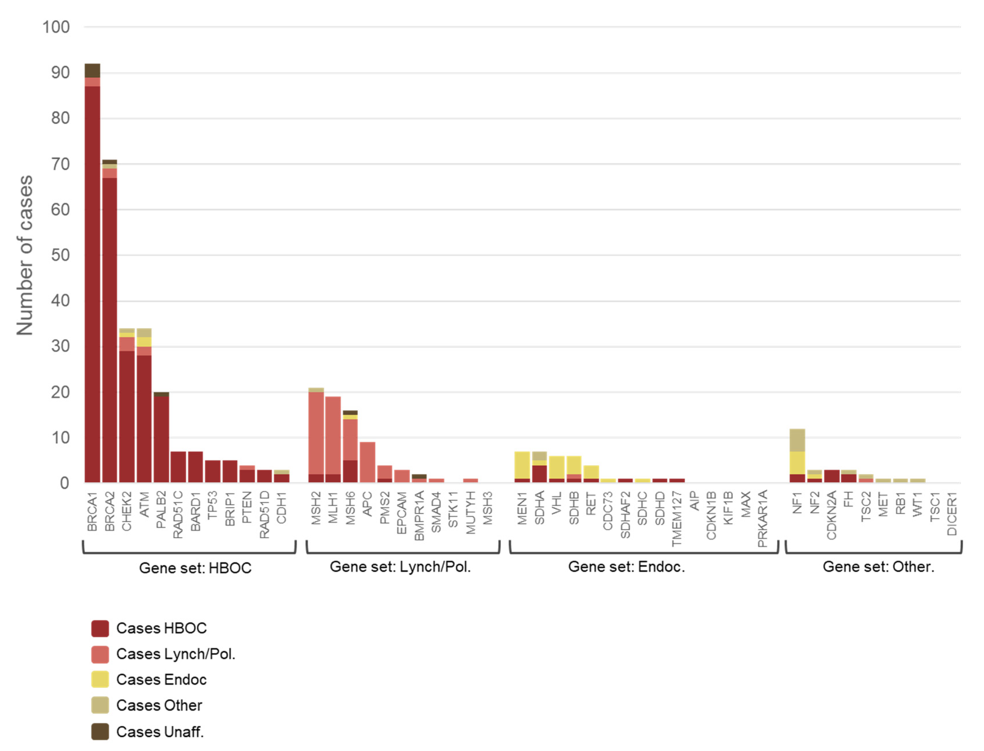
Figure 1. Number of positive cases according to affected genes, categorised by phenotype. Genes are grouped based on the syndrome they are associated with.
Ijms 26 08390 g001
Figure 2. Illustration of combinations of cancer susceptibility genes in DH patients, categorised by the associated syndrome (A) and by gene penetrance (B). The first column includes genes with variants most relevant to the patient’s phenotype. In cases with three P/LP variants in the same patient, all possible gene pairs were included. Nodes represent the syndrome associated with each gene (A) or the level of penetrance of genes (B). High penetrance genes: BRCA1, BRCA2, CDH1, PALB2, SDHAF2; Moderate penetrance genes: ATM, BARD1, BRIP1, CHEK2, MSH6, SDHB.; Low penetrance genes: SDHA.
Ijms 26 08390 g002
Figure 3. Boxplot showing the age of disease onset among patients suspected of HBOC syndrome predisposition. Patients were grouped based on the number of P/LP variants carried: none, one, or two (or more).
Ijms 26 08390 g003
Table 1. Demographic characteristics, prevalence of P/LP variants and ratio of DH status among patients grouped by suspected tumour syndrome. n.a.: not applicable.
HBOCLynch + Pol.EndocrineOtherUnaffectedSum
Case number149626814684562050
%73.013.17.14.12.7100
Gender
Female (number)14281878645561802
%95.569.858.953.6100.087.9
Male (number)688160390248
%4.530.241.146.40.012.1
Mean age (years)49.551.941.241.9n.a.
SD11.515.418.319.4n.a.
Median age (years)48544244n.a.48
min1850 a0 bn.a.0 b
max85788380n.a.85
Muliple primaries20349522n.a.279
%13.618.33.426.2n.a.13.6
Number of P/LP cases2777331186405
%18.527.221.221.410.719.8
Number of DH cases13101116
%0.90.40.01.21.80.8
a age: 6 months. b age: 1 month.
Table 2. (A) Clinical characteristics of DH patients. (B) Variant description and genomic consequences in DH cases. Patients’ order is the same as in (A).
(A)
#SexSuspected SyTumour_1AgeTumour_2AgeMPsTumour HistologyFamily History
1FEMHBOCbr44 IDC; ER +; PR +; Her2 -; Ki67 80%br (52 + 56); br (58)
2FEMLynch/Polyposisend36 endometrioid cc with squamous differentiationLung (?); cr (43); st (37); st (53); br (60) + ov (60)
3FEMHBOCbr22 IDC; ER -; PR -; Her2 -; Ki67 70%Skin (45); br (77); cr (70)
4FEMHBOCbr37 IDC; ER +; PR -; Her2 +; Ki67 80%br (58); br (59 + 65); st (49); st (86)
5FEMunaffunaff unaffectedBr (42); br (?)
6FEMHBOCbr45NHL50 IDC; ER -; PR -; Her2 -; Ki67 ?%cr (60); br (57); gb (?); cr (?); ut (51) + ov (51)
7FEMHBOCbr47 IDC; ER +; PR +; Her2 -; Ki67 40–50%br (44)
8FEMHBOCbr30 IDC; ER +; PR +; Her2 ?; Ki67 20%br (53); br (66); cr (58); cr (50)
9MALHBOCprostate60 adenocc.br (49); nb (2)
10FEMHBOCend54breast66yadenocc.; LCIS + ILC; ER +; PR +; Her2 -; Ki67 1–2%br (42); brain (43); ren (?); brain (?); st (?)
11FEMHBOCbr35 IDC; ER -; PR -; Her2 -; Ki67 50%br (68); lung (54); bone (?)
12FEMHBOCbr42 IDC + DCIS; ER -; PR -; Her2 +; Ki67 10%br (?); lung (?)
13FEMHBOCbr33 IDC; ER -; PR -; Her2 -; Ki67 80%br (55)
14FEMHBOCbr (bilat)37 yILC; ER +; PR +; Her2 +; Ki67 10–15%pros (46); panc (46); liver (46)
15FEMHBOCbr43 IDC; ER +; PR +; Her2 -; Ki67 10%Cervix (44)
16FEMotherut (leiomyoma)32 FH deficient ut leiomyomabr (48); br (66); larynx (52)
(B)
#Gene_1HGVS
Transcript_1
HGVS
Protein_1
Gene_2HGVS
Transcript_2
HGVS
Protein_2
Gene_3HGVS Transcript_3HGVS
Protein_3
1ATMNM_000051.4:c.6679C>TNP_000042.3:p.(Arg2227Cys)BARD1NM_000465.4:c.1932_1933delNP_000456.2:p.(Cys645Ter)
2MSH2NM_000251.3:c.2068C>TNP_000242.1:p.(Gln690Ter)SDHBNM_003000.3:c.148G>TNP_002991.2:p.(Asp50Tyr)
3BRCA1NM_007294.4:c.5266dupNP_009225.1:p.(Gln1756ProfsTer74)MSH6NM_000179.3:c.3261delNP_000170.1:p.(Phe1088SerfsTer2)
4CHEK2NM_007194.4: del(exon 9–10)NP_009125.1:p.?CHEK2NM_007194.4:c.499G>ANP_009125.1:p.(Gly167Arg)
5BRCA1NM_007294.4:c.181T>GNP_009225.1:p.(Cys61Gly)MSH6NM_000179.3:c.3379_3438+5delNP_000170.1:p.?
6PALB2NM_024675.4: del(exon 11)NP_078951.2:p.?SDHANM_004168.4:c.91C>TNP_004159.2:p.(Arg31Ter)
7BRCA2NM_000059.4:c.5682C>GNP_000050.3:p.(Tyr1894Ter)BRIP1NM_032043.3:c.3525dupNP_114432.2:p.(Ile1176TyrfsTer13)
8BRCA2NM_000059.4:c.8249_8251delNP_000050.3:p.(Lys2750del)ATMNM_000051.4:c.1564_1565delNP_000042.3:p.(Glu522IlefsTer43)
9BRCA1NM_007294.4:c.181T>GNP_009225.1:p.(Cys61Gly)BRCA2NM_000059.4:c.6295A>TNP_000050.3:p.(Arg2099Ter)
10CHEK2NM_007194.4:c.1100delNP_009125.1:p.(Thr367MetfsTer15)SDHAF2NM_017841.4:c.446_447delNP_060311.1:p.(Lys149ArgfsTer10)
11BRCA2NM_000059.4:c.5073dupNP_000050.3:p.(Trp1692MetfsTer3)MSH6NM_000179.3:c.3379_3438+5delNP_000170.1:p.?
12BRCA1NM_007294.4:c.181T>GNP_009225.1:p.(Cys61Gly)ATMNM_000051.4:c.7630-2A>CNP_000042.3:p.?SDHAdel (whole gene)NP_004159.2:p.?
13BRCA1NM_007294.4:c.181T>GNP_009225.1:p.(Cys61Gly)BRIP1NM_032043.3:c.1741C>TNP_114432.2:p.(Arg581Ter)
14BRCA2NM_000059.4:c.5645C>ANP_000050.3:p.(Ser1882Ter)CDH1NM_004360.5:c.1711+5G>ANP_004351.1:p.?
15CHEK2NM_007194.4:c.1337delNP_009125.1:p.(Asn446ThrfsTer23)CHEK2NM_007194.4:c.277delNP_009125.1:p.(Trp93GlyfsTer17)
16ATMNM_000051.4:c.381delNP_000042.3:p.(Val128Ter)CDH1NM_004360.5:c.1063_1066dupNP_004351.1:p.(Ser356IlefsTer13)
Abbreviations: cc carcinoma; cr colorectal; br breast; ccRCC clear cell renal cell carcinoma; end endometrial carcinoma; ov ovarial; nb neuroblastoma; panc pancreas; pros prostate; PTC papillary thyroid carcinoma; st stomach; ut uterus. '?' indicates that the age at tumour onset is unknown.
Disclaimer/Publisher’s Note: The statements, opinions and data contained in all publications are solely those of the individual author(s) and contributor(s) and not of MDPI and/or the editor(s). MDPI and/or the editor(s) disclaim responsibility for any injury to people or property resulting from any ideas, methods, instructions or products referred to in the content.