Review

Silent Inflammation, Loud Consequences: Decoding NLR Across Renal, Cardiovascular and Metabolic Disorders

Department of Health Promotion, Mother and Child Care, Internal and Specialistic Medicine, University of Palermo, 90127 Palermo, Italy
*
Author to whom correspondence should be addressed.
Int. J. Mol. Sci. 2025, 26(17), 8256; https://doi.org/10.3390/ijms26178256
Submission received: 22 July 2025 / Revised: 20 August 2025 / Accepted: 22 August 2025 / Published: 26 August 2025

Abstract

The neutrophil-to-lymphocyte ratio (NLR) has emerged as a readily accessible, cost-effective biomarker reflecting systemic inflammation. Chronic low-grade inflammation plays a pivotal role in the pathogenesis and progression of metabolic and cardiovascular disorders including chronic kidney disease (CKD), hypertension, diabetes mellitus, and cardiovascular disease (CVD). This review critically evaluates the current evidence on NLR as a prognostic marker across these interconnected conditions. A comprehensive literature search was conducted focusing on clinical and epidemiological studies investigating the association between NLR and CKD, hypertension, diabetes, and cardiovascular outcomes. Mechanistic insights into inflammation-driven pathophysiology and the predictive utility of NLR in disease progression and adverse events were synthesized. Elevated NLR is consistently associated with increased risk and severity of CKD, correlating with glomerular filtration decline, proteinuria, and mortality. In hypertension, higher NLR levels are linked to non-dipper blood pressure patterns, arterial stiffness, and increased cardiovascular risk. Among diabetic patients, NLR correlates with poor glycemic control and vascular complications. In cardiovascular disease, elevated NLR predicts major adverse cardiovascular events (MACE) and all-cause mortality, reflecting underlying immune dysregulation and endothelial dysfunction. Despite promising findings, direct comparisons with established inflammatory biomarkers remain limited, and heterogeneity exists across populations. NLR represents a simple yet powerful inflammatory biomarker with significant prognostic value in CKD, hypertension, diabetes, and cardiovascular disease. Its integration into clinical risk stratification models could enhance personalized medicine approaches. Future research should focus on longitudinal studies, validation in diverse cohorts, and comparative analyses with other inflammatory markers to fully delineate NLR’s clinical utility.

1. Introduction

Chronic low-grade inflammation, often termed “silent” or subclinical inflammation, represents a fundamental pathophysiological process underpinning a range of chronic diseases, including chronic kidney disease (CKD), diabetes mellitus, cardiovascular disease (CVD), and hypertension [1,2,3]. Unlike overt inflammatory conditions characterized by acute immune activation and clinical symptoms, silent inflammation is a persistent, low-intensity immune response that progressively contributes to tissue injury and organ dysfunction over time [4]. In CKD, silent inflammation accelerates renal fibrosis and functional decline through sustained activation of innate immune cells and pro-inflammatory cytokines [5,6]. Similarly, in diabetes mellitus, chronic subclinical inflammation is implicated in insulin resistance, β-cell dysfunction, and the development of microvascular and macrovascular complications [7]. Cardiovascular disease is driven, in part, by inflammation-mediated endothelial dysfunction, atherosclerotic plaque formation, and destabilization [8,9]. Hypertension itself is increasingly recognized as an inflammatory disorder, where immune activation promotes vascular remodeling and stiffness [9,10]. This shared inflammatory milieu establishes silent inflammation as a unifying mechanism that links metabolic, renal, and cardiovascular pathologies, highlighting the need for biomarkers and therapeutic strategies targeting this covert immune dysregulation.
The neutrophil-to-lymphocyte ratio (NLR) has gained considerable attention as a readily available biomarker reflecting systemic inflammation. Derived from routine complete blood counts, NLR offers a simple, cost-effective, and widely accessible measure that integrates innate and adaptive immune system components [11,12]. Neutrophils, key mediators of acute and chronic inflammatory responses, tend to increase during systemic inflammation [4], whereas lymphocytes often decrease in settings of physiological stress and immune dysregulation [13]. This dynamic shift results in an elevated NLR, which correlates with the degree of underlying inflammation. Compared to traditional inflammatory markers such as C-reactive protein (CRP) or interleukin-6 (IL-6), NLR benefits from its low cost, rapid availability, and ease of interpretation, making it a practical tool for clinical and research applications across diverse patient populations.
This review aims to comprehensively explore the potential of the neutrophil-to-lymphocyte ratio (NLR) as a versatile biomarker of subclinical inflammation across CKD), diabetes mellitus, cardiovascular disease, and hypertension. By synthesizing existing evidence, we seek to underscore the utility of NLR in risk stratification, disease monitoring, and prognosis within these interconnected conditions. Furthermore, the review highlights the current gaps and limitations in the literature, including inconsistencies in threshold values, mechanistic understanding, and clinical applicability, to guide future research directions and promote standardized use of NLR in clinical practice.

2. Materials and Methods

This work is a narrative review aiming to synthesize current evidence on the role of NLR as a marker of immune dysregulation and inflammation in the context of diabetes mellitus, CKD, and cardiovascular disease, with a focus on shared pathophysiological mechanisms such as endothelial dysfunction, oxidative stress, and systemic immune imbalance. A non-systematic, yet comprehensive literature search was conducted using PubMed, Web of Science and Google Scholar in May 2025. The following keywords and Boolean operators were used to retrieve relevant publications: (“neutrophil-to-lymphocyte ratio” OR NLR) AND (diabetes OR “chronic kidney disease” OR CKD OR “cardiovascular disease” OR CVD).
Ijms 26 08256 i001
We included original research articles, meta-analyses, and narrative or systematic reviews published up to 1 June 2025, without language or publication date restrictions. Priority was given to studies reporting on human subjects and discussing pathophysiological mechanisms linking NLR with one or more of the four conditions (diabetes, CKD, hypertension and CVD), either separately or in combination. Studies were selected based on their relevance to the review’s aims, methodological rigor, and contribution to understanding shared inflammatory and immune-mediated pathways. Given the narrative nature of the review, no formal risk of bias assessment or meta-analysis was performed. Instead, findings were grouped thematically and discussed in relation to key pathophysiological domains: endothelial dysfunction, oxidative stress, and immune dysregulation, with reference to clinical correlates such as disease progression and cardiovascular or renal outcomes.

3. NLR and Inflammation: Biological Basis

Neutrophils and lymphocytes represent two fundamental arms of the immune system—innate and adaptive, respectively—but their functions are deeply intertwined in shaping the host inflammatory response. Their recruitment, activation, and effector profiles define not only the initial immune engagement but also the trajectory of chronic inflammation in cardio-renal-metabolic disorders [14,15].
Neutrophils, the most abundant circulating leukocytes, act as the first responders of innate immunity. They are rapidly recruited to sites of infection or tissue injury in response to chemokine gradients and danger-associated molecular patterns [16]. Upon activation, neutrophils engage in phagocytosis, degranulation, production of reactive oxygen species (ROS) [17], and release of extracellular traps (NETs) and pro- and anti-inflammatory cytokines [18]. While these responses are essential for pathogen clearance, dysregulated neutrophil activation may result in bystander tissue injury and contribute to chronic inflammation. A significant contributor to neutrophil-mediated oxidative stress is the formation of reactive carbonyl species—electrophilic compounds with aldehyde and ketone groups—produced through lipid peroxidation and protein oxidation [19]. These reactive carbonyls can modify cellular proteins through carbonylation, altering signaling pathways and exacerbating tissue damage.
Recent insights have redefined neutrophils as modulators of immunity rather than mere terminal effectors. Through cytokine secretion and dynamic cross-talk with dendritic cells and lymphocytes, neutrophils influence the magnitude and quality of adaptive immune responses. They may also carry an “immunological blueprint” that shapes downstream immune cascades [20,21]. Sustained neutrophil activation has been implicated in chronic inflammatory states such as atherosclerosis, autoimmunity, cancer, and CVD. Neutrophil death pathways, including NETosis and pyroptosis, further amplify inflammation and may expose autoantigens, perpetuating immune dysregulation [22].
Lymphocytes—primarily T and B cells—form the backbone of adaptive immunity. CD4+ T-helper subsets (Th1, Th2, Th17) orchestrate immune responses against specific pathogen classes. Th1 cells drive macrophage activation via interferon-gamma (IFN-γ), Th2 cells promote eosinophilic responses through IL-4, IL-5, and IL-13, and Th17 cells recruit neutrophils through IL-17 and IL-22 [23]. Regulatory T cells (Tregs) are crucial for maintaining immune tolerance and suppressing excessive inflammation [24]. A decline in circulating lymphocytes—due to apoptosis, sequestration, or exhaustion—is frequently observed in chronic inflammatory conditions such as diabetes and CKD, and is associated with adverse clinical outcomes [25].
Additionally, innate lymphoid cells (ILCs) have emerged as critical players in bridging innate and adaptive immunity. Though antigen-independent, ILCs mirror the cytokine profiles of T-helper subsets. Group 1 ILCs (e.g., NK cells) produce IFN-γ; Group 2 ILCs (e.g., nuocytes) produce IL-5 and IL-13; and Group 3 ILCs produce IL-17 and IL-22, paralleling Th17 responses. These cells contribute to tissue-specific immune responses and may play roles in mucosal defense and chronic inflammation. Their development relies on the transcription factor Id2 and common γ-chain cytokines such as IL-7, indicating shared lineage with classical lymphocytes [26,27].
However, the balance between neutrophils and lymphocytes is also tightly regulated by transcriptional programs that orchestrate inflammatory signaling. Among them, NF-κB, AP-1, STAT-3, and Nrf2 are particularly relevant.
NF-κB and AP-1 act as pro-inflammatory hubs, driving expression of chemokines, adhesion molecules, and survival factors that prolong neutrophil activity, thereby favoring neutrophilia. STAT-3, activated downstream of IL-6 and related cytokines, promotes Th17 differentiation and neutrophil recruitment while contributing to lymphocyte exhaustion. In contrast, Nrf2 provides a counter-regulatory mechanism by inducing antioxidant defenses and limiting oxidative stress; its dysfunction enhances neutrophil-driven damage and accelerates lymphocyte loss. Importantly, redox-sensitive pathways involving these transcription factors can be therapeutically modulated—for instance, polyphenols have been shown to activate Nrf2 and dampen NF-κB/AP-1 signaling, reducing systemic inflammation [28].
The neutrophil-to-lymphocyte ratio (NLR) has emerged as a practical biomarker representing this complex immunological equilibrium. A high NLR reflects a dominance of innate pro-inflammatory responses over adaptive regulatory mechanisms—a hallmark of immune dysregulation and “inflammaging,” the chronic low-grade inflammation observed in aging, metabolic stress, and endothelial dysfunction [29,30]. Elevated NLR has been linked to disease progression in hypertension [31], diabetes [32], CKD [33,34], and CVD [35], suggesting its role extends beyond a surrogate of inflammation to a window into pathophysiological processes driving organ-specific injury.
In metabolic cardiorenal syndrome—where diabetes, CVD, and CKD coexist in a synergistic and self-amplifying pathophysiological loop—NLR might provide critical information about systemic immune activation. An elevated NLR reflects increased neutrophil-mediated endothelial dysfunction, atherosclerotic instability, and renal fibrosis, coupled with lymphopenia arising from metabolic stress and immune suppression. This imbalance marks a shift toward innate immune predominance, which promotes vascular injury, impairs reparative responses, and accelerates multiorgan deterioration [36]. The pathophysiological interplay between CKD, diabetes mellitus, and CVD is underpinned by a constellation of shared mechanisms, notably endothelial dysfunction, oxidative stress, and immune dysregulation [37]. Hyperglycemia, uremic toxins, and dyslipidemia synergistically impair endothelial integrity, promoting vascular stiffness and atherogenesis. Concurrently, elevated levels of ROS and systemic inflammation perpetuate oxidative stress, exacerbating tissue injury across the renal, cardiovascular, and metabolic systems [38]. Within this context, NLR has emerged as a clinically accessible marker reflecting this pro-inflammatory and immune-altered milieu. Increased neutrophil counts reflect activation of innate immune pathways and oxidative burst activity, while decreased lymphocyte levels may result from chronic metabolic stress and immunosuppressive signaling. This imbalance is particularly relevant in the metabolic cardiorenal continuum, where NLR correlates with endothelial dysfunction, vascular calcification, and adverse clinical outcomes. As such, NLR not only represents a surrogate of systemic inflammation but also integrates key pathophysiological features common to CKD, diabetes, and CVD [14,39] (Figure 1).

4. NLR and Diabetes

In individuals with diabetes, chronic low-grade inflammation and immune system dysfunction plays a pivotal role in both disease onset and progression [40]. This inflammatory milieu is closely linked to the development of insulin resistance, β-cell dysfunction, and vascular damage [41]. Type 2 diabetes frequently develops in the context of obesity, advancing age, and sedentary lifestyle. The disease arises from the progressive inability of pancreatic β-cells to adequately compensate for insulin resistance. Several mechanisms have been proposed to account for impaired insulin action and secretion, including oxidative and endoplasmic reticulum stress, deposition of amyloid within pancreatic islets, abnormal fat accumulation in liver, muscle, and pancreas, as well as lipotoxic and glucotoxic effects [34,35]. These metabolic and cellular disturbances not only trigger inflammatory pathways but are also aggravated by pre-existing inflammation.
Evidence of innate immune activation has been detected in the circulation, insulin-sensitive tissues, and pancreatic islets of individuals with type 2 diabetes, reinforcing the concept that inflammation contributes to disease onset and progression. Proposed drivers of this pro-inflammatory state include adipose tissue expansion with hypoxia and cell death, activation of the nuclear factor-κB (NF-κB) and c-Jun N-terminal kinase (JNK) signaling cascades, stimulation of interleukin-1β (IL-1β), and recruitment of immune cells into metabolic tissues [42]. Clinical studies using anti-inflammatory agents such as IL-1 antagonists or salsalate have provided encouraging results, highlighting inflammation as a promising therapeutic target [43].
A growing body of evidence suggests that inflammatory processes play a significant role in the pathogenesis of type 2 diabetes, although their precise contribution and therapeutic implications are still being clarified. The neutrophil-to-lymphocyte ratio (NLR) has repeatedly been linked to diabetes within this context, with higher NLR values reported in patients compared to healthy controls [44,45,46]. Interestingly, elevated NLR has also been documented in individuals with prediabetes, indicating that immune imbalance may be detectable even at early stages of metabolic dysfunction.
A large prospective study based on NHANES data included 13,024 adults with prediabetes, followed over approximately 8.5 years. Participants were stratified into NLR tertiles (≤1.60, 1.60–2.29, ≥2.29). After adjusting for confounders, each one-unit increase in NLR was associated with a 12% higher risk of all-cause mortality (HR 1.12; 95% CI, 1.05–1.19) and a 24% increase in cardiovascular mortality (HR 1.24; 95% CI, 1.11–1.37) compared to the lowest NLR group. Individuals in the highest NLR tertile showed a significantly increased risk (HR ~1.21 for all-cause mortality, ~1.49 for cardiovascular mortality) compared to those in the lowest tertile [42].
In addition, a 3-year follow-up study on individuals with diabetes and prediabetes found that elevated NLR was an independent predictor of worsening renal function [43].
These findings support a significant prognostic role for NLR even at the early stages of glucose metabolism dysregulation, suggesting its potential utility not only in established diabetes but also in prediabetes, for risk stratification and clinical monitoring.
Most of the existing literature on NLR in the context of diabetes has predominantly focused on its association with glycemic control. Elevated NLR values have been consistently linked to higher HbA1c levels, poor metabolic control, and increased insulin resistance, suggesting that systemic low-grade inflammation plays a role in the deterioration of glycemic homeostasis. A 2023 meta-analysis by Adane et al., which included 13 studies, demonstrated that patients with poor glycemic control exhibited significantly higher NLR values compared to those with adequate control [44]. These findings reinforce the association between elevated NLR and increased HbA1c levels in individuals with type 2 diabetes. Consequently, NLR may be considered a complementary marker to HbA1c for assessing glycemic control in T2DM patients.
However, despite this growing body of evidence, relatively few longitudinal or prospective studies have explored the prognostic value of NLR in predicting diabetes-related complications, such as nephropathy, retinopathy, or cardiovascular events. Beyond glycemic control, higher NLR levels have been identified as independent predictors of diabetic nephropathy, retinopathy, and cardiovascular mortality in patients with T2DM.
A recent study using data from the NHANES 2009–2018 cycles investigated the association between inflammatory biomarkers and clinical outcomes in individuals with diabetic retinopathy (DR). The analysis included 572 adults with DR and examined the prognostic value of three inflammation-based indices: the neutrophil-to-lymphocyte ratio (NLR), monocyte-to-lymphocyte ratio (MLR), and the systemic inflammation response index (SIRI). Results showed that elevated levels of these biomarkers were significantly associated with increased all-cause and diabetes-related cardiovascular mortality. Specifically, participants with NLR ≥ 1.516, MLR ≥ 0.309, or SIRI ≥ 0.756 had markedly higher mortality risks compared to those below these thresholds. The associations were even more pronounced in individuals aged 60 years or older. Furthermore, the combined use of NLR, MLR, and SIRI slightly improved predictive accuracy for mortality compared to individual markers alone [45]. These findings support the potential role of inflammatory markers as prognostic tools in the diabetic retinopathy population, highlighting the contribution of systemic inflammation to adverse outcomes in this high-risk group. Similar findings were reported by Wang and colleagues, who demonstrated that elevated NLR and PLR are significantly associated with diabetic retinopathy (DR) in patients without a family history of diabetes or hypertension. NLR emerged as an independent risk factor, while high PLR levels were linked to increased DR risk in the upper quantile. The inclusion of NLR and PLR improved DR risk prediction beyond hemoglobin, enhancing model discrimination and reclassification [46]
These findings may suggest that NLR may serve not only as a marker of systemic inflammation but also as a prognostic indicator for long-term outcomes in diabetic populations. Additionally, its role in risk stratification and its potential integration into multiparametric models for personalized prevention and treatment strategies remain largely unexplored. This highlights the need for broader investigation beyond glycemic indices to fully elucidate the clinical utility of NLR in diabetes care. Table 1 summarized the main studies reviewed in this section.

5. NLR and CKD

The relationship between systemic inflammation CKD has long been recognized, with a growing body of literature indicating that inflammation is not merely a consequence but a central driver of CKD progression. Among inflammatory markers, NLR has emerged as a particularly promising indicator, offering a cost-effective, accessible, and dynamic tool for assessing inflammatory burden in CKD patients.
Multiple studies have documented a progressive increase in NLR across CKD stages, correlating with declining glomerular filtration rate (GFR), increased proteinuria, and worsening histological damage such as interstitial fibrosis and tubular atrophy. In a pivotal prospective study by Yoshitomi et al. [47] involving 256 non-dialysis CKD patients followed for nearly three years, elevated baseline NLR was independently associated with a higher risk of renal outcomes, including significant eGFR decline and initiation of dialysis. Notably, these findings remained robust after adjusting for traditional risk factors, underscoring the independent prognostic role of inflammatory activation in renal deterioration.
Other large cohort studies have reinforced these associations. In a study of 966 patients with biopsy-proven IgA nephropathy, those with NLR ≥ 2.67 were significantly more likely to progress to end-stage kidney disease, and elevated NLR was closely linked to histopathologic findings such as glomerulosclerosis and interstitial fibrosis [48].
In a large cross-sectional study involving 2846 apparently healthy adults (1777 men and 1069 women), the association between NLR and CKD was evaluated with a focus on the impact of body weight. Participants were categorized into normal-weight and overweight/obese groups based on BMI criteria. The study found that higher NLR values were significantly associated with increased CKD prevalence in overweight and obese individuals—both in men (adjusted OR = 1.37, 95% CI: 1.03–1.82, p = 0.03) and women (adjusted OR = 1.77, 95% CI: 1.08–2.90, p = 0.023)—but not in those with normal weight.
Further, in overweight/obese participants with mildly to moderately reduced eGFR (50–70 mL/min/1.73 m2), NLR was inversely correlated with eGFR in both sexes, suggesting a link between systemic inflammation and early renal function decline. These findings point to a pathophysiological interplay between adiposity-related low-grade inflammation and renal injury, where elevated NLR may reflect a heightened inflammatory burden that contributes to glomerular damage, endothelial dysfunction, and progressive renal impairment.
The absence of a significant association in normal-weight individuals reinforces the hypothesis that metabolic inflammation—characteristic of obesity and insulin resistance—may potentiate the deleterious renal effects of immune dysregulation. NLR may thus serve as a valuable early indicator of CKD risk in overweight and obese populations [49].
Mechanistically, elevated NLR in CKD appears to reflect the confluence of several pathophysiological processes. Neutrophil activation in renal disease promotes endothelial dysfunction, capillary rarefaction, and the release of reactive oxygen species and neutrophil extracellular traps (NETs), all of which accelerate glomerular and tubulointerstitial injury. In contrast, lymphopenia reflects impaired regulatory immune responses and reduced capacity to resolve inflammation, creating a chronic pro-inflammatory state that fuels fibrosis. This dysregulated immunological environment fosters progression of CKD and increases susceptibility to cardiovascular events, a leading cause of death in this population.
The prognostic relevance of NLR extends to patients undergoing renal replacement therapy. In a meta-analysis encompassing over 116,000 CKD and dialysis patients, high NLR was significantly associated with increased all-cause and cardiovascular mortality [50]. In a Romanian end-stage renal disease cohort, individuals in the high-NLR group exhibited a 30-day mortality rate exceeding 40%, compared to less than 2% among those with low NLR, indicating the profound prognostic impact of inflammatory imbalance in this population [51].
In a cohort of 225 patients with stage 3–5 chronic kidney disease (CKD) followed for an average of 39 months, elevated neutrophil-to-lymphocyte ratio (NLR) was found to be a significant predictor of both fatal and nonfatal cardiovascular events (CVE). NLR increased progressively with advancing CKD stage and was independently associated with endothelial dysfunction, as measured by flow-mediated dilation (FMD), regardless of hsCRP levels. A baseline NLR ≥ 3.76 predicted composite CVE with 80.3% sensitivity and 91.8% specificity, and patients with higher NLR had significantly reduced survival time. These findings suggest that NLR is a useful independent marker of cardiovascular risk in moderate to severe CKD [52].
Recent work by Carollo and colleagues, further contributed to the field by examining the prognostic value of NLR in a cohort of 236 chronic hemodialysis patients. Their findings confirmed that patients in the highest tertile of NLR not only had worse nutritional and iron parameters, but also significantly higher mortality risk. Interestingly, NLR demonstrated superior prognostic accuracy compared to C-reactive protein (CRP), with a higher area under the curve in ROC analysis, supporting its potential as a more sensitive marker of chronic inflammation in dialysis settings [53].
Emerging evidence supports the association between increased NLR and accelerated decline in renal function, underscoring the role of inflammation in CKD progression. This pro-inflammatory environment not only contributes to renal parenchymal injury but also interferes with erythropoiesis, leading to reduced responsiveness to erythropoietin (EPO) therapy [54]. Resistance to EPO, characterized by a suboptimal hematologic response to ESAs, correlates with higher inflammatory burden as indicated by NLR elevation [55]. The mechanistic pathways involve inflammatory cytokines such as interleukin-6 and tumor necrosis factor-alpha, which inhibit erythroid progenitor proliferation and disrupt iron homeostasis, thereby attenuating EPO efficacy [56].
Notably, EPO resistance serves as an independent predictor of adverse renal outcomes, with patients exhibiting both high NLR and EPO resistance experiencing more rapid CKD progression and increased cardiovascular morbidity [57]. Thus, integrating NLR assessment with evaluation of ESA responsiveness may enhance risk stratification and guide therapeutic strategies in CKD.
In conclusion, the interplay between systemic inflammation, measured by NLR, and erythropoietin resistance constitutes a critical axis influencing CKD progression. Targeted anti-inflammatory interventions aimed at improving EPO responsiveness hold promise in mitigating disease advancement and improving clinical outcomes.
Regarding CV and all-cause mortality in CKD patients, while it is well known that inflammation is common in CKD, it is less clear how well certain inflammatory markers can predict long-term survival in these patients.
To explore this, researchers analyzed data from nearly 7000 CKD patients collected over almost 20 years from the National Health and Nutrition Examination Survey (NHANES). They looked at four blood-based inflammatory indicators: the systemic immune-inflammation index (SII), NLR, PLR and LMR [58].
The findings showed that higher levels of SII, NLR, and PLR were linked to a greater risk of death, whereas higher LMR levels appeared to have a protective effect. Among these, NLR and LMR stood out as the strongest predictors of survival in CKD patients. This suggests that measuring these markers could help doctors better identify patients who are at higher risk of poor outcomes.
Moreover, the relationship between these inflammatory markers and mortality risk was not straightforward, indicating that changes in inflammation levels may affect survival in complex ways. Catabay and colleagues investigated whether NLR and PLR, could help predict mortality in patients starting hemodialysis [59]. Among over 108,000 patients in a large U.S. dialysis network (2007–2011), NLR was significantly associated with increased mortality, particularly when measured over time. PLR showed a J-shaped association but did not significantly improve mortality prediction models.
NLR modestly improved predictive models beyond standard clinical parameters (like demographics, comorbidities, and serum albumin), as shown by improvements in AUROC, net reclassification index (NRI), and adjusted R2. In contrast, PLR provided little to no added predictive value.
From a translational standpoint, NLR represents more than a passive biomarker. Its integration into clinical risk models could enhance the precision of prognostication in CKD, particularly when combined with other clinical, biochemical, and possibly genomic indicators. Despite this promise, several critical knowledge gaps remain. First, direct comparisons between NLR and other inflammatory markers—such as (IL-6, tumor necrosis factor-alpha (TNF-α), or high-sensitivity C-reactive protein (hsCRP)—are limited. While such comparisons have been extensively explored in the context of acute inflammatory states, particularly COVID-19 [60,61], similar head-to-head evaluations are scarce in the setting of chronic kidney disease. In nephrology, most comparative studies involving NLR are conducted against other blood cell–derived inflammatory indices, such PLR, MLR, or the composite lymphocyte-to-C-reactive protein ratio (LCR), rather than against canonical cytokine-based inflammatory markers. This limits our understanding of NLR’s relative performance and mechanistic specificity in capturing the complex inflammatory milieu characteristic of progressive renal dysfunction.
Secondly, the majority of current research on this topic is based on observational studies, and there remains a lack of interventional trials specifically targeting pathways related to NLR modulation, such as through anti-inflammatory or immunomodulatory treatments. To address this gap, findings from five recent randomized controlled trials—CANTOS, JUPITER, SPIRE-1, SPIRE-2, and CIRT—have been examined. These studies evaluated the effects of canakinumab, rosuvastatin, bococizumab, and methotrexate compared to placebo in patients with atherosclerosis. The data indicated that lipid-lowering agents did not produce meaningful changes in NLR levels. In contrast, treatment with canakinumab, an interleukin-1β–targeting monoclonal antibody, resulted in a significant reduction in NLR, suggesting a distinct effect of anti-inflammatory therapies on this biomarker. These observations highlight the need for further research into the influence of other immunomodulatory strategies, particularly those targeting interleukin pathways, on NLR dynamics [62]. Finally, population-based studies in healthy individuals are needed to better define normal reference ranges and thresholds for pathological significance.
Despite the growing evidence supporting the role of NLR as a prognostic marker in CKD, several studies have reported no significant association between NLR and renal disease progression or related clinical outcomes. For instance, in a retrospective 5-year study of 740 CKD stage 2–4 patients from Altunoren and colleagues, higher baseline NLR was associated with greater inflammation and faster GFR decline, but NLR was not an independent predictor of CKD progression. Instead, diabetes, younger age, and lower baseline eGFR were stronger predictors of reaching end-stage [63]. Similar results were reported by Yuan and colleagues, who investigated the association between NLR and progression to ESRD, cardiovascular events, and all-cause mortality in Chinese patients with stages 1–4 CKD. Their multicenter study demonstrated that NLR was associated with the risk of ESRD only in patients with stage 4 CKD, while no significant associations were observed between NLR and the risk of cardiovascular events or mortality in the overall cohort or within CKD stage subgroups [64].
These discrepant results may be attributable to several factors, including heterogeneity in study designs, differences in patient populations, and the multifactorial nature of inflammation in CKD. In particular, the inflammatory state in advanced CKD and dialysis patients can be influenced by comorbid conditions, infections, medication use, and dialysis-related factors, which may confound NLR measurements and diminish its specificity as a biomarker for renal disease progression. Consequently, while NLR remains a promising and accessible marker of systemic inflammation, its role as a reliable prognostic indicator in CKD progression requires further validation through well-designed longitudinal studies accounting for these confounders.
In summary, NLR encapsulates the inflammatory burden of CKD and offers independent prognostic value for disease progression and cardiovascular morbidity. As inflammation becomes an increasingly recognized therapeutic target in nephrology, NLR may serve both as a stratification tool and a surrogate endpoint in future clinical trials. Its routine implementation in nephrology practice will require robust validation, standardization of thresholds, and integration into multifactorial predictive models to support personalized risk assessment and tailored therapeutic strategies. Table 2 summarizes the main studies reviewed in this section.

6. NLR and Cardiovascular Disease

The relationship between the NLR and cardiovascular disease represents one of the most extensively investigated areas in the study of inflammation-based prognostic biomarkers. A growing body of evidence supports the role of NLR as a reliable, independent predictor of both acute and long-term cardiovascular outcomes, including major adverse cardiovascular events (MACE), heart failure, arrhythmia, and cardiovascular mortality.
Large-scale meta-analyses and cohort studies have demonstrated that elevated NLR levels are consistently associated with worse prognosis in patients with acute coronary syndromes (ACS), particularly in ST-elevation myocardial infarction (STEMI) treated with percutaneous coronary intervention (PCI). In a meta-analysis of over 10,000 STEMI patients, higher admission NLR was linked to a markedly increased risk of in-hospital and long-term mortality (RR ≈ 3.3), MACE (RR ≈ 2.0), stent thrombosis (RR ≈ 2.7), and heart failure [65].
Similarly, in patients with non-ST-elevation myocardial infarction (NSTEMI), elevated NLR was shown to enhance long-term prognostic stratification. In a cohort study involving 678 NSTEMI patients undergoing PCI, combined elevation of NLR and MLR provided greater predictive accuracy for MACE than either marker alone [66].
Finally in another meta-analysis evaluated the prognostic utility of NLR in patients with ST-segment elevation myocardial infarction (STEMI) undergoing percutaneous coronary intervention (PCI), NLR, a marker of systemic inflammation, has been increasingly recognized as a potential predictor of adverse outcomes in cardiovascular disease. The primary objective of this study was to assess whether elevated NLR is associated with increased in-hospital and long-term mortality and major cardiovascular events in this high-risk patient population.
The pooled analysis demonstrated that higher baseline NLR was significantly associated with increased in-hospital all-cause mortality, cardiovascular mortality, and MACE. Moreover, elevated NLR was correlated with higher long-term mortality and MACE, as well as prolonged hospital stay. The association between elevated NLR and long-term cardiovascular mortality appeared particularly strong. These findings remained consistent across studies and statistical models, supporting the robustness of the association [67].
Caimi et al. investigated the neutrophil-to-lymphocyte ratio (NLR) in a cohort of 123 young patients (mean age 39.4 ± 5.8 years) with acute myocardial infarction (AMI), assessing values at the initial stage and at 3 and 12 months post-event [68]. They found that NLR was significantly elevated in young AMI patients compared to healthy controls (2.38 ± 0.87 vs. 1.82 ± 0.71, p < 0.0001). Notably, NLR did not differ significantly between STEMI and non-STEMI patients, nor between diabetic and non-diabetic individuals. However, smokers exhibited lower NLR values compared to non-smokers. Initial NLR levels were not correlated with the number of cardiovascular risk factors or the extent of coronary artery disease. Over time, a significant reduction in neutrophil count led to decreased NLR at both 3 and 12 months post-AMI, while lymphocyte counts remained stable. The same group observed in young patients with acute myocardial infarction both NLR and plasma viscosity (PV) increased during the acute phase, but their trajectories diverged over time. NLR significantly decreased at 3 and 12 months follow-up, while PV remained persistently elevated, suggesting different underlying mechanisms. The sustained rise in PV, unlike NLR, appeared linked to genetic predisposition and was associated with cardiovascular complications during follow-up [69].
The prognostic significance of NLR extends beyond ischemic heart disease. In patients hospitalized for acute decompensated heart failure, elevated NLR was independently associated with increased in-hospital mortality (OR ≈ 2.2) and three-year post-discharge mortality (OR ≈ 1.44), even after adjusting for age, renal function, and ejection fraction [70].
Moreover, in patients presenting with cardiogenic shock following acute MI, NLR > 7.3 at admission predicted a significantly higher 30-day mortality rate (73.7% vs. 26.3%) and was confirmed as an independent predictor of both all-cause death and MACE (HR ≈ 2.8) [71].
The pathophysiological rationale for these associations lies in the dual roles of neutrophils and lymphocytes during cardiovascular injury. Neutrophils exacerbate myocardial and vascular damage via the release of reactive oxygen species, proteolytic enzymes, and inflammatory mediators. Lymphocytes, on the other hand, are essential for resolving inflammation and facilitating tissue repair. Thus, an elevated NLR reflects a systemic inflammatory imbalance, skewed toward injury and inadequate repair—contributing to plaque instability, thrombosis, and maladaptive remodeling [72].
Beyond coronary syndromes and heart failure, elevated NLR has also shown prognostic relevance in atrial fibrillation (AF). A 2024 meta-analysis found that higher NLR levels were significantly associated with increased all-cause mortality (OR ≈ 1.87) and stroke (OR ≈ 1.56) in patients with AF, highlighting its broader cardiovascular implications [73].
Regarding atherosclerosis a large-scale study involving more than 100,000 participants examined the relationship between neutrophil levels and the risk of nine distinct cardiovascular outcomes using both observational and genetic methodologies. The findings indicated that elevated neutrophil counts were consistently linked to an increased risk across all atherosclerotic cardiovascular disease (ASCVD) endpoints, with comparable associations observed in both male and female subjects [74]. A retrospective study involving 4000 participants found that both the Chinese Visceral Adiposity Index (VAI) and NLR were independently associated with an increased risk of carotid atherosclerosis. Additionally, both markers demonstrated a significant positive correlation with the 10-year ASCVD (atherosclerotic cardiovascular disease) risk score, with all associations reaching strong statistical significance (p < 0.001) [75]. Wang et al. investigated the predictive value of the neutrophil-to-lymphocyte ratio (NLR) and plasma lipoprotein (a) [Lp (a)] levels for identifying unstable coronary plaques in a cohort of 1618 individuals with atherosclerotic cardiovascular disease (ASCVD), as assessed through coronary computed tomography angiography (CTA) [76]. In a similar context, several studies have recognized the neutrophil-to-lymphocyte ratio (NLR) as a promising and informative biomarker for detecting vulnerable carotid plaques [77], as demonstrated through carotid ultrasonography. These findings support the potential of NLR in assessing the likelihood of carotid plaque development and instability [78].
Collectively, these findings support the role of NLR as a readily available, cost-effective marker that could be integrated into risk stratification frameworks for patients with cardiovascular disease. However, further comparative studies are needed to assess its additive value over established markers such as high-sensitivity CRP, troponins, NT-proBNP, and interleukin-6. Table 3 summarizes the main studies reviewed in this section.

7. NLR and Hypertension

Hypertension is increasingly recognized not solely as a hemodynamic disorder but as a chronic, low-grade inflammatory condition. Sustained elevations in blood pressure induce mechanical stress on the vascular wall, promoting endothelial dysfunction, oxidative stress, and immune activation. These processes initiate and perpetuate a pro-inflammatory milieu characterized by increased circulating levels of interleukin (IL)-1β, IL-6, and tumor necrosis factor-alpha (TNF-α), which contribute to arterial remodeling, vascular stiffening, and progressive target organ damage [4,31,79].
A growing body of evidence supports the role of the NLR as a surrogate marker of systemic inflammation in hypertensive populations. Multiple epidemiological studies and meta-analyses have consistently demonstrated significantly higher NLR levels in hypertensive individuals compared to normotensive controls. This difference is particularly pronounced in “non-dipper” hypertensive patients, whose nocturnal blood pressure fails to decline physiologically—a pattern associated with greater cardiovascular risk and subclinical organ damage [80]. In a resistant hypertension cohort, NLR remained substantially elevated even after adjusting for confounders and showed a positive correlation with both office and ambulatory systolic and diastolic blood pressure values [81].
NLR has also been independently associated with left ventricular hypertrophy (LVH) in hypertensive patients. Yu et al. found that higher NLR levels were significantly linked to LVH (odds ratio = 1.506; 95% CI: 1.086–2.089; p = 0.014), with an optimal diagnostic cutoff of 2.185 (AUC = 0.626), comparable to CRP (AUC = 0.639) and BNP (AUC = 0.645) in terms of diagnostic performance [82].
The predictive value of NLR for the development of hypertension has also been demonstrated. In a prospective cohort study of 28,850 initially normotensive individuals, those in the highest NLR quintile had a significantly increased risk of developing hypertension over approximately six years (HR = 1.23; 95% CI: 1.06–1.43; p for trend < 0.01) compared to the lowest quintile, even after full adjustment [83].
A 2021 meta-analysis of 21 studies confirmed that hypertensive individuals had significantly higher NLR levels than normotensive controls (WMD = 0.40; 95% CI: 0.22–0.57; p < 0.0001). NLR was also significantly higher in non-dipper versus dipper hypertensive patients (WMD = 0.58; 95% CI: 0.19–0.97; p = 0.003), suggesting a potential link between blood pressure patterns, systemic inflammation, and cardiovascular risk [31].
A cross-sectional NHANES analysis (1999–2010; n = 22,290) further supported the association between NLR and hypertension prevalence. Per natural log-unit increase in NLR, the odds of hypertension increased (OR = 1.087; 95% CI: 1.008–1.173). Individuals in the highest NLR tertile had a 1.11-fold higher risk of hypertension compared to those in the lowest. While the systemic immune-inflammation index (SII) demonstrated even stronger associations, other markers such as platelet-to-lymphocyte ratio (PLR) and lymphocyte-to-monocyte ratio (LMR) were not significantly associated with hypertension [84].
In a large cohort of 6278 Taiwanese adults, higher NLR levels were significantly associated with prevalent hypertension. After adjusting for covariates, individuals in the highest NLR tertile had a 28% higher risk of hypertension (HR = 1.28; 95% CI: 1.03–1.59), with particularly strong associations in older adults (HR = 1.88; 95% CI: 1.19–2.96) and men over 60 years (HR = 1.84; 95% CI: 1.06–3.18), indicating the relevance of inflammatory markers in age-related hypertension risk [85].
The relationship between NLR and target organ damage has also been extensively investigated. In patients with resistant hypertension, NLR positively correlated with systolic and diastolic blood pressure measures, suggesting a direct link with vascular stress and structural adaptation. In one study of 150 individuals, NLR levels were significantly higher in resistant hypertension (RHT) compared to controlled hypertensives (CHT) (p = 0.03) and normotensives (NT) (p < 0.001). Multivariate analysis confirmed both NLR and absolute neutrophil count as independent predictors of RHT (p < 0.001), reinforcing the hypothesis of inflammatory involvement in blood pressure resistance [81].
In patients with H-type hypertension, characterized by elevated homocysteine levels, each unit increase in NLR was associated with a 51% higher risk of renal dysfunction, defined by reductions in eGFR and elevations in blood urea nitrogen and serum creatinine [86].
Further supporting its vascular relevance, in a study of 217 hypertensive patients and 132 normotensive controls, NLR levels were significantly higher in individuals with isolated systolic hypertension and combined systolic-diastolic hypertension. Brachial-ankle pulse wave velocity (baPWV), a surrogate marker of arterial stiffness, was elevated across all hypertensive subtypes and positively correlated with NLR. Multivariate analysis confirmed NLR as an independent predictor of baPWV, suggesting that systemic inflammation—as indexed by NLR—may actively contribute to arterial stiffening and atherosclerotic changes [87].
Importantly, NLR also has prognostic value. In a cohort of 3067 hypertensive adults from NHANES (2009–2014), an elevated NLR (>3.5) was independently associated with increased all-cause mortality (HR = 1.96; 95% CI: 1.52–2.52) and cardiovascular mortality (HR = 2.33; 95% CI: 1.54–3.51) over a median follow-up of 92 months. A clear dose–response relationship was observed, and eGFR partially mediated this association (5.4% for all-cause and 4.7% for cardiovascular mortality). Time-dependent AUCs for NLR predicting 3-, 5-, and 10-year survival ranged from 0.64 to 0.70, underscoring its moderate prognostic value [88].
These findings were subsequently validated in a larger cohort using a lower NLR threshold (>2.0), which remained significantly associated with both all-cause (HR ≈ 1.47) and cardiovascular mortality (HR ≈ 2.08) [89].
Overall, NLR emerges as a robust, inexpensive, and readily accessible biomarker that integrates the inflammatory dimension of hypertension. Its consistent associations with hypertension incidence, blood pressure patterns, target organ damage, vascular stiffness, and long-term mortality make it a promising candidate for clinical risk stratification and potentially for guiding anti-inflammatory strategies in hypertension management. However, despite its growing relevance, direct comparative studies between NLR and other established or emerging inflammatory biomarkers—such as high-sensitivity CRP, interleukin-6, or the systemic immune-inflammation index (SII)—remain limited. Future research is needed to evaluate the relative diagnostic, prognostic, and therapeutic value of NLR in comparison to these markers, particularly in diverse hypertensive subpopulations. Such data will be essential to clarify whether NLR should be adopted as a first-line inflammatory marker or used in conjunction with a broader panel in clinical practice. Table 4 summarises the main studied reviewed in this section.

8. Current Limitations and Gaps

Current limitations and gaps in the literature underscore the need for further standardization and validation of inflammatory markers such as the neutrophil-to-lymphocyte ratio (NLR) in clinical practice. Notably, there is a lack of universally accepted and standardized cut-off values for NLR, both in healthy populations and in patients with chronic diseases, which hampers its broader clinical implementation. Furthermore, data on NLR distribution in healthy individuals are limited, and population profiling studies aimed at establishing normative reference ranges are still lacking. For instance, diurnal variation has been reported, with neutrophil counts peaking in the morning and lymphocyte counts being relatively higher later in the day, leading to fluctuating NLR [90]. In addition, acute infections, surgery, trauma, and psychological or physical stress can transiently elevate NLR, reflecting short-term immune activation rather than chronic low-grade inflammation. Pharmacological interventions, including corticosteroids, immunosuppressive agents, or chemotherapy, also alter leukocyte subpopulations and therefore affect NLR independently of disease activity [14]. Such sources of variability should be acknowledged when using NLR for risk stratification, particularly in longitudinal or comparative studies.
Most available evidence derives from retrospective or single-center studies, with a particular scarcity of large-scale, prospective, multicenter investigations—especially in populations with diabetes mellitus.
Although this is a narrative review, we have considered the methodological quality of the available studies. Most of the evidence derives from observational studies with heterogeneous populations and variable adjustment for confounding factors. Only a limited number of randomized clinical trials are available, which reduces the strength of causal inferences. Nevertheless, the consistency of findings across diverse clinical settings supports the robustness of the association between NLR and cardio-renal-metabolic disorders. Moreover, NLR is not currently integrated into established composite cardiovascular risk models, such as SCORE or the Framingham Risk Score, limiting its utility in risk stratification. Defining clinically meaningful thresholds tailored to high-risk subgroups (e.g., patients with chronic kidney disease, diabetes, or established cardiovascular disease) is essential. Additionally, no interventional studies have yet assessed whether targeting NLR can improve clinical outcomes. Preliminary evidence suggests that integrating NLR with other biomarkers—such as high-sensitivity C-reactive protein (hs-CRP) or troponins—may enhance predictive accuracy, warranting further investigation.

9. Future Perspectives and Conclusions

The neutrophil-to-lymphocyte ratio (NLR) has emerged as a simple yet powerful inflammatory biomarker with broad potential applications in cardiovascular, renal, and metabolic medicine. Given its accessibility, cost-effectiveness, and robust association with adverse outcomes in hypertension, chronic kidney disease (CKD), and diabetes, NLR represents a promising candidate for integration into personalized medicine frameworks.
In the context of personalized risk stratification, NLR may serve as a valuable tool for identifying individuals at heightened risk of adverse events, particularly in those with overlapping cardio-metabolic comorbidities. When combined with clinical parameters—such as blood pressure profiles, renal function indices, and glycemic markers—and genetic or epigenetic signatures, NLR could enhance secondary prevention strategies, allowing for earlier and more targeted interventions.
Moreover, the incorporation of NLR into risk prediction algorithms and clinical decision-support systems may facilitate refined triage and monitoring protocols. Its dynamic responsiveness to inflammatory shifts suggests utility in assessing therapeutic efficacy, particularly in settings of resistant hypertension, diabetic nephropathy, or cardiorenal syndromes. The potential to track longitudinal changes in NLR could help guide medication adjustments, lifestyle interventions, or escalation of care in high-risk individuals.
Despite its promise, several gaps remain. Notably, direct comparative studies between NLR and other validated inflammatory or prognostic biomarkers—such as high-sensitivity C-reactive protein (hsCRP), interleukin-6, or novel immune-metabolic indices—are lacking, especially on a disease-specific basis. Such comparisons are essential to contextualize the unique value of NLR in clinical decision-making. Table 5 provides a comparison of NLR and other inflammatory markers’ prognostic role.
Additionally, normative data for NLR in healthy populations across age, sex, and ethnicity are sparse. Establishing reference ranges and population-specific thresholds is crucial for distinguishing pathologic inflammation from physiological variability, particularly in asymptomatic or early-stage patients.
Ultimately, realizing the full clinical utility of NLR will depend on translational research efforts and well-designed prospective validation studies. These should aim to define cut-off values, evaluate predictive performance in diverse clinical settings, and explore integration with other biomarkers in multi-parametric approaches. As the field moves toward precision cardiometabolic care, NLR holds significant potential—but its adoption must be guided by rigorous scientific validation.

Author Contributions

Conceptualization, all.; methodology, all.; investigation, all.; writing—original draft preparation, all.; writing—review and editing: all visualization, all.; supervision, all.; project administration: all. All authors have read and agreed to the published version of the manuscript.

Funding

This research received no external funding.

Conflicts of Interest

The authors declare no conflicts of interest.

References

  1. Targeting Innate Immunity-Driven Inflammation in CKD and Cardiovascular Disease; Nature Reviews Nephrology. Available online: https://www.nature.com/articles/s41581-022-00621-9 (accessed on 21 July 2025).
  2. Margioris, A.N.; Dermitzaki, E.; Venihaki, M.; Tsatsanis, C. 4-Chronic low-grade inflammation. In Diet, Immunity and Inflammation; Calder, P.C., Yaqoob, P., Eds.; Woodhead Publishing Series in Food Science, Technology and Nutrition; Woodhead Publishing: Sawston, UK, 2013; pp. 105–120. ISBN 978-0-85709-037-9. [Google Scholar]
  3. Cachofeiro, V.; Goicochea, M.; de Vinuesa, S.G.; Oubiña, P.; Lahera, V.; Luño, J. Oxidative stress and inflammation, a link between chronic kidney disease and cardiovascular disease: New strategies to prevent cardiovascular risk in chronic kidney disease. Kidney Int. 2008, 74, S4–S9. [Google Scholar] [CrossRef]
  4. Chen, L.; Deng, H.; Cui, H.; Fang, J.; Zuo, Z.; Deng, J.; Li, Y.; Wang, X.; Zhao, L. Inflammatory responses and inflammation-associated diseases in organs. Oncotarget 2017, 9, 7204–7218. [Google Scholar] [CrossRef]
  5. Kadatane, S.P.; Satariano, M.; Massey, M.; Mongan, K.; Raina, R. The Role of Inflammation in CKD. Cells 2023, 12, 1581. [Google Scholar] [CrossRef]
  6. Plonsky-Toder, M.; Magen, D.; Pollack, S. Innate Immunity and CKD: Is There a Significant Association? Cells 2023, 12, 2714. [Google Scholar] [CrossRef] [PubMed]
  7. Rohm, T.V.; Meier, D.T.; Olefsky, J.M.; Donath, M.Y. Inflammation in obesity, diabetes, and related disorders. Immunity 2022, 55, 31–55. [Google Scholar] [CrossRef] [PubMed]
  8. Henein, M.Y.; Vancheri, S.; Longo, G.; Vancheri, F. The Role of Inflammation in Cardiovascular Disease. Int. J. Mol. Sci. 2022, 23, 12906. [Google Scholar] [CrossRef]
  9. Mechanisms of Vascular Inflammation and Potential Therapeutic Targets: A Position Paper from the ESH Working Group on Small Arteries. Hypertension. Available online: https://www.ahajournals.org/doi/10.1161/HYPERTENSIONAHA.123.22483 (accessed on 22 July 2025).
  10. Savoia, C.; Sada, L.; Zezza, L.; Pucci, L.; Lauri, F.M.; Befani, A.; Alonzo, A.; Volpe, M. Vascular Inflammation and Endothelial Dysfunction in Experimental Hypertension. Int. J. Hypertens. 2011, 2011, 281240. [Google Scholar] [CrossRef] [PubMed]
  11. Balta, S.; Ozturk, C.; Balta, I.; Demirkol, S.; Demir, M.; Celik, T.; Iyisoy, A. The Neutrophil–Lymphocyte Ratio and Inflammation. Angiology 2016, 67, 298–299. [Google Scholar] [CrossRef]
  12. Islam, M.M.; Satici, M.O.; Eroglu, S.E. Unraveling the clinical significance and prognostic value of the neutrophil-to-lymphocyte ratio, platelet-to-lymphocyte ratio, systemic immune-inflammation index, systemic inflammation response index, and delta neutrophil index: An extensive literature review. Turk. J. Emerg. Med. 2024, 24, 8–19. [Google Scholar] [CrossRef]
  13. Alotiby, A. Immunology of Stress: A Review Article. J. Clin. Med. 2024, 13, 6394. [Google Scholar] [CrossRef]
  14. Obeagu, E.I. Stress, neutrophils, and immunity: A dynamic interplay. Ann. Med. Surg. 2025, 87, 3573–3585. [Google Scholar] [CrossRef]
  15. Rosales, C.; Demaurex, N.; Lowell, C.A.; Uribe-Querol, E. Neutrophils: Their Role in Innate and Adaptive Immunity. J. Immunol. Res. 2016, 2016, 1469780. [Google Scholar] [CrossRef]
  16. Yang, L.; Shi, F.; Cao, F.; Wang, L.; She, J.; He, B.; Xu, X.; Kong, L.; Cai, B. Neutrophils in Tissue Injury and Repair: Molecular Mechanisms and Therapeutic Targets. MedComm 2025, 6, e70184. [Google Scholar] [CrossRef]
  17. Winterbourn, C.C.; Kettle, A.J.; Hampton, M.B. Reactive Oxygen Species and Neutrophil Function. Annu. Rev. Biochem. 2016, 85, 765–792. [Google Scholar] [CrossRef]
  18. Zhu, S.; Yu, Y.; Ren, Y.; Xu, L.; Wang, H.; Ling, X.; Jin, L.; Hu, Y.; Zhang, H.; Miao, C.; et al. The emerging roles of neutrophil extracellular traps in wound healing. Cell Death Dis. 2021, 12, 984. [Google Scholar] [CrossRef]
  19. Behaviour of Carbonyl Groups in Several Clinical Conditions: Analysis of Our Survey—Gregorio Caimi, Eugenia Hopps, Maria Montana, Caterina Carollo, Vincenzo Calandrino, Eleonora Gallà, Baldassare Canino, Rosalia Lo Presti; 2020. Available online: https://journals.sagepub.com/doi/abs/10.3233/CH-190689 (accessed on 22 July 2025).
  20. Li, Y.; Wang, W.; Yang, F.; Xu, Y.; Feng, C.; Zhao, Y. The regulatory roles of neutrophils in adaptive immunity. Cell Commun. Signal. 2019, 17, 147. [Google Scholar] [CrossRef]
  21. Lee, M.; Lee, S.Y.; Bae, Y.-S. Emerging roles of neutrophils in immune homeostasis. BMB Rep. 2022, 55, 473–480. [Google Scholar] [CrossRef] [PubMed]
  22. Tu, H.; Ren, H.; Jiang, J.; Shao, C.; Shi, Y.; Li, P. Dying to Defend: Neutrophil Death Pathways and their Implications in Immunity. Adv. Sci. 2023, 11, 2306457. [Google Scholar] [CrossRef]
  23. Zhu, X.; Zhu, J. CD4 T Helper Cell Subsets and Related Human Immunological Disorders. Int. J. Mol. Sci. 2020, 21, 8011. [Google Scholar] [CrossRef] [PubMed]
  24. Singer, M.; Elsayed, A.M.; Husseiny, M.I. Regulatory T-cells: The Face-off of the Immune Balance. Front. Biosci. (Landmark Ed.) 2024, 29, 377. [Google Scholar] [CrossRef] [PubMed]
  25. Lymphocyte Depletion and Subset Alteration Correlate to Renal Function in Chronic Kidney Disease Patients. Available online: https://www.tandfonline.com/doi/full/10.3109/0886022X.2015.1106871 (accessed on 22 July 2025).
  26. Nagasawa, M.; Spits, H.; Ros, X.R. Innate Lymphoid Cells (ILCs): Cytokine Hubs Regulating Immunity and Tissue Homeostasis. Cold Spring Harb. Perspect. Biol. 2018, 10, a030304. [Google Scholar] [CrossRef]
  27. Nouari, W.; Aribi, M. Innate lymphoid cells, immune functional dynamics, epithelial parallels, and therapeutic frontiers in infections. Int. Rev. Immunol. 2025, 1–28, ahead of print. [Google Scholar] [CrossRef]
  28. Kostenko, V.; Akimov, O.; Gutnik, O.; Kostenko, H.; Kostenko, V.; Romantseva, T.; Morhun, Y.; Nazarenko, S.; Taran, O. Modulation of redox-sensitive transcription factors with polyphenols as pathogenetically grounded approach in therapy of systemic inflammatory response. Heliyon 2023, 9, e15551. [Google Scholar] [CrossRef] [PubMed]
  29. Salminen, A. Increased immunosuppression impairs tissue homeostasis with aging and age-related diseases. J. Mol. Med. 2021, 99, 1–20. [Google Scholar] [CrossRef] [PubMed]
  30. Paganelli, R.; Iorio, A.D. The neutrophil-to-lymphocyte ratio in aging and immunosenescence. Explor. Immunol. 2025, 5, 1003200. [Google Scholar] [CrossRef]
  31. Sarejloo, S.; Dehesh, M.; Fathi, M.; Khanzadeh, M.; Lucke-Wold, B.; Ghaedi, A.; Khanzadeh, S. Meta-analysis of differences in neutrophil to lymphocyte ratio between hypertensive and non-hypertensive individuals. BMC Cardiovasc. Disord. 2023, 23, 283. [Google Scholar] [CrossRef] [PubMed]
  32. Wang, Y.; Liu, X.; Xiao, Z. Diagnostic Accuracy of Neutrophil-to-Lymphocyte Ratio in Type 2 Diabetic Nephropathy: A Meta-Analysis. Front. Endocrinol. 2025, 16, 1564170. [Google Scholar] [CrossRef]
  33. Aneez, F.A.; Shariffdeen, N.; Haleem, F.A.; Thangarajah, B.R.; Rasaratnam, K. Correlation between neutrophil to lymphocyte ratio and platelet to lymphocyte ratio with proteinuria in different stages of chronic kidney disease. Egypt. J. Intern. Med. 2024, 36, 6. [Google Scholar] [CrossRef]
  34. Zhao, W.-M.; Tao, S.-M.; Liu, G.-L. Neutrophil-to-lymphocyte ratio in relation to the risk of all-cause mortality and cardiovascular events in patients with chronic kidney disease: A systematic review and meta-analysis. Ren. Fail. 2020, 42, 1059–1066. [Google Scholar] [CrossRef]
  35. Ghasempour Dabaghi, G.; Rabiee Rad, M.; Mortaheb, M.; Darouei, B.; Amani-Beni, R.; Mazaheri-Tehrani, S.; Izadan, M.; Touhidi, A. The Neutrophil-to-Lymphocyte Ratio Predicts Cardiovascular Outcomes in Patients With Diabetes: A Systematic Review and Meta-Analysis. Cardiol. Rev. 2025, 33, 202–211. [Google Scholar] [CrossRef]
  36. Penna, C.; Pagliaro, P. Endothelial Dysfunction: Redox Imbalance, NLRP3 Inflammasome, and Inflammatory Responses in Cardiovascular Diseases. Antioxidants 2025, 14, 256. [Google Scholar] [CrossRef]
  37. Usman, M.S.; Khan, M.S.; Butler, J. The Interplay Between Diabetes, Cardiovascular Disease, and Kidney Disease. In Chronic Kidney Disease and Type 2 Diabetes; ADA Clinical Compendia Series; American Diabetes Association: Arlington, VA, USA, 2021. Available online: http://www.ncbi.nlm.nih.gov/books/NBK571718/ (accessed on 22 July 2025).
  38. Caturano, A.; Rocco, M.; Tagliaferri, G.; Piacevole, A.; Nilo, D.; Di Lorenzo, G.; Iadicicco, I.; Donnarumma, M.; Galiero, R.; Acierno, C.; et al. Oxidative Stress and Cardiovascular Complications in Type 2 Diabetes: From Pathophysiology to Lifestyle Modifications. Antioxidants 2025, 14, 72. [Google Scholar] [CrossRef]
  39. Buonacera, A.; Stancanelli, B.; Colaci, M.; Malatino, L. Neutrophil to Lymphocyte Ratio: An Emerging Marker of the Relationships between the Immune System and Diseases. Int. J. Mol. Sci. 2022, 23, 3636. [Google Scholar] [CrossRef] [PubMed]
  40. Liu, S.; Zheng, H.; Zhu, X.; Mao, F.; Zhang, S.; Shi, H.; Li, Y.; Lu, B. Neutrophil-to-lymphocyte ratio is associated with diabetic peripheral neuropathy in type 2 diabetes patients. Diabetes Res. Clin. Pract. 2017, 130, 90–97. [Google Scholar] [CrossRef]
  41. Donath, M.Y.; Shoelson, S.E. Type 2 diabetes as an inflammatory disease. Nat. Rev. Immunol. 2011, 11, 98–107. [Google Scholar] [CrossRef]
  42. Chen, H.L.; Wu, C.; Cao, L.; Wang, R.; Zhang, T.Y.; He, Z. The association between the neutrophil-to-lymphocyte ratio and type 2 diabetes mellitus: A cross-sectional study. BMC Endocr. Disord. 2024, 24, 107. [Google Scholar] [CrossRef]
  43. Li, J.; Li, T.; Wang, H.; Yan, W.; Mu, Y. Neutrophil-lymphocyte ratio as a predictor of kidney function decline among individuals with diabetes and prediabetes: A 3-year follow-up study. J. Diabetes 2019, 11, 427–430. [Google Scholar] [CrossRef]
  44. Adane, T.; Melku, M.; Worku, Y.B.; Fasil, A.; Aynalem, M.; Kelem, A.; Getawa, S. The Association between Neutrophil-to-Lymphocyte Ratio and Glycemic Control in Type 2 Diabetes Mellitus: A Systematic Review and Meta-Analysis. J. Diabetes Res. 2023, 2023, 3117396. [Google Scholar] [CrossRef] [PubMed]
  45. Si, Y.; Chen, Q.; Xiong, X.; Zheng, M. The association of inflammatory biomarkers with clinical outcomes in diabetic retinopathy participants: Data from NHANES 2009-2018. Diabetol. Metab. Syndr. 2024, 16, 181. [Google Scholar] [CrossRef]
  46. Wang, J.-R.; Chen, Z.; Yang, K.; Yang, H.-J.; Tao, W.-Y.; Li, Y.-P.; Jiang, Z.-J.; Bai, C.-F.; Yin, Y.-C.; Duan, J.-M.; et al. Association between neutrophil-to-lymphocyte ratio, platelet-to-lymphocyte ratio, and diabetic retinopathy among diabetic patients without a related family history. Diabetol. Metab. Syndr. 2020, 12, 55. [Google Scholar] [CrossRef] [PubMed]
  47. Yoshitomi, R.; Nakayama, M.; Sakoh, T.; Fukui, A.; Katafuchi, E.; Seki, M.; Tsuda, S.; Nakano, T.; Tsuruya, K.; Kitazono, T. High neutrophil/lymphocyte ratio is associated with poor renal outcomes in Japanese patients with chronic kidney disease. Ren. Fail. 2019, 41, 238–243. [Google Scholar] [CrossRef]
  48. Wang, S.; Dong, L.; Pei, G.; Jiang, Z.; Qin, A.; Tan, J.; Tang, Y.; Qin, W. High Neutrophil-To-Lymphocyte Ratio Is an Independent Risk Factor for End Stage Renal Diseases in IgA Nephropathy. Front. Immunol. 2021, 12. [Google Scholar] [CrossRef]
  49. Lin, C.-H.; Li, Y.-H.; Wang, Y.-Y.; Chang, W.-D. Higher Neutrophil-To-Lymphocyte Ratio Was Associated with Increased Risk of Chronic Kidney Disease in Overweight/Obese but Not Normal-Weight Individuals. Int. J. Environ. Res. Public Health 2022, 19, 8077. [Google Scholar] [CrossRef] [PubMed]
  50. Ao, G.; Wang, Y.; Qi, X.; Wang, F.; Wen, H. Association of neutrophil-to-lymphocyte ratio and risk of cardiovascular or all-cause mortality in chronic kidney disease: A meta-analysis. Clin. Exp. Nephrol. 2021, 25, 157–165. [Google Scholar] [CrossRef] [PubMed]
  51. Mureșan, A.V.; Russu, E.; Arbănași, E.M.; Kaller, R.; Hosu, I.; Arbănași, E.M.; Voidăzan, S.T. The Predictive Value of NLR, MLR, and PLR in the Outcome of End-Stage Kidney Disease Patients. Biomedicines 2022, 10, 1272. [Google Scholar] [CrossRef]
  52. Solak, Y.; Yilmaz, M.I.; Sonmez, A.; Saglam, M.; Cakir, E.; Unal, H.U.; Gok, M.; Caglar, K.; Oguz, Y.; Yenicesu, M.; et al. Neutrophil to lymphocyte ratio independently predicts cardiovascular events in patients with chronic kidney disease. Clin. Exp. Nephrol. 2013, 17, 532–540. [Google Scholar] [CrossRef]
  53. Carollo, C.; Mancia, E.; Sorce, A.; Altieri, C.; Altieri, D.; Brunori, G.; Mulè, G. Prognostic impact of neutrophil-to-lymphocyte ratio and vascular access in patients on chronic hemodialysis. J. Nephrol. 2025, 38, 717–723. [Google Scholar] [CrossRef] [PubMed]
  54. Zhang, J.; Lu, X.; Wang, S.; Li, H. Neutrophil-to-Lymphocyte Ratio and Erythropoietin Resistance among Maintenance Hemodialysis Patients. Available online: https://dx.doi.org/10.1159/000519644 (accessed on 22 July 2025).
  55. Carollo, C.; Sorce, A.; Mancia, E.; Cirafici, E.; Ciuppa, M.E.; De Biasio, B.; Mulè, G.; Brunori, G. Assessing the Impact of Inflammation on Erythropoietin Resistance in Hemodialysis: The Role of the NLR. J. Clin. Med. 2025, 14, 3411. [Google Scholar] [CrossRef]
  56. Morceau, F.; Dicato, M.; Diederich, M. Pro-Inflammatory Cytokine-Mediated Anemia: Regarding Molecular Mechanisms of Erythropoiesis. Mediat. Inflamm. 2009, 2009, 405016. [Google Scholar] [CrossRef]
  57. Pineault, J.; Lamarche, C.; Bell, R.; Lafrance, J.-P.; Ouellet, G.; Leblanc, M.; Pichette, V.; Bezzaoucha, S.; Vallée, M. Association of Neutrophil-to-Lymphocyte Ratio with Inflammation and Erythropoietin Resistance in Chronic Dialysis Patients. Can. J. Kidney Health Dis. 2017, 4, 2054358117735563. [Google Scholar] [CrossRef]
  58. Chen, Y.; Nie, Y.; Wu, J.; Li, C.; Zheng, L.; Zhu, B.; Min, Y.; Ling, T.; Liu, X. Association between systemic inflammatory indicators with the survival of chronic kidney disease: A prospective study based on NHANES. Front. Immunol. 2024, 15, 1365591. [Google Scholar] [CrossRef]
  59. Catabay, C.; Obi, Y.; Streja, E.; Soohoo, M.; Park, C.; Rhee, C.M.; Kovesdy, C.P.; Hamano, T.; Kalantar-Zadeh, K. Lymphocyte Cell Ratios and Mortality among Incident Hemodialysis Patients. Am. J. Nephrol. 2017, 46, 408–416. [Google Scholar] [CrossRef]
  60. Carollo, C.; Benfante, A.; Sorce, A.; Montalbano, K.; Cirafici, E.; Calandra, L.; Geraci, G.; Mulè, G.; Scichilone, N. Predictive Biomarkers of Acute Kidney Injury in COVID-19: Distinct Inflammatory Pathways in Patients with and Without Pre-Existing Chronic Kidney Disease. Life 2025, 15, 720. [Google Scholar] [CrossRef]
  61. Regolo, M.; Vaccaro, M.; Sorce, A.; Stancanelli, B.; Colaci, M.; Natoli, G.; Russo, M.; Alessandria, I.; Motta, M.; Santangelo, N. Neutrophil-to-lymphocyte ratio (NLR) is a promising predictor of mortality and admission to intensive care unit of COVID-19 patients. J. Clin. Med. 2022, 11, 2235. [Google Scholar] [CrossRef]
  62. Adamstein, N.H.; MacFadyen, J.G.; Rose, L.M.; Glynn, R.J.; Dey, A.K.; Libby, P.; Tabas, I.A.; Mehta, N.N.; Ridker, P.M. The neutrophil-lymphocyte ratio and incident atherosclerotic events: Analyses from five contemporary randomized trials. Eur. Heart J. 2021, 42, 896–903. [Google Scholar] [CrossRef]
  63. Altunoren, O.; Akkus, G.; Sezal, D.T.; Ciftcioglu, M.; Guzel, F.B.; Isiktas, S.; Torun, G.I.; Uyan, M.; Sokmen, M.F.; Sevim, H.A.; et al. Does neutrophyl to lymphocyte ratio really predict chronic kidney disease progression? Int. Urol. Nephrol. 2019, 51, 129–137. [Google Scholar] [CrossRef] [PubMed]
  64. Yuan, Q.; Wang, J.; Peng, Z.; Zhou, Q.; Xiao, X.; Xie, Y.; Wang, W.; Huang, L.; Tang, W.; Sun, D.; et al. Neutrophil-to-lymphocyte ratio and incident end-stage renal disease in Chinese patients with chronic kidney disease: Results from the Chinese Cohort Study of Chronic Kidney Disease (C-STRIDE). J. Transl. Med. 2019, 17, 86. [Google Scholar] [CrossRef] [PubMed]
  65. Zhang, S.; Diao, J.; Qi, C.; Jin, J.; Li, L.; Gao, X.; Gong, L.; Wu, W. Predictive value of neutrophil to lymphocyte ratio in patients with acute ST segment elevation myocardial infarction after percutaneous coronary intervention: A meta-analysis. BMC Cardiovasc Disord. 2018, 18, 75. [Google Scholar] [CrossRef] [PubMed]
  66. Fan, Z.; Li, Y.; Ji, H.; Jian, X. Prognostic utility of the combination of monocyte-to-lymphocyte ratio and neutrophil-to-lymphocyte ratio in patients with NSTEMI after primary percutaneous coronary intervention: A retrospective cohort study. BMJ Open 2018, 8, e023459. [Google Scholar] [CrossRef]
  67. Ul Hussain, H.; Kumar, K.A.; Zahid, M.; Husban Burney, M.; Khan, Z.; Asif, M.; Rehan, S.T.; Ahmad Cheema, H.; Swed, S.; Yasmin, F.; et al. Neutrophil to lymphocyte ratio as a prognostic marker for cardiovascular outcomes in patients with ST-segment elevation myocardial infarction after percutaneous coronary intervention: A systematic review and meta-analysis. Medicine 2024, 103, e38692. [Google Scholar] [CrossRef]
  68. Caimi, G.; Lo Presti, R.; Canino, B.; Ferrera, E.; Hopps, E. Behaviour of the neutrophil to lymphocyte ratio in young subjects with acute myocardial infarction. Clin. Hemorheol. Microcirc. 2016, 62, 239–247. [Google Scholar] [CrossRef]
  69. Caimi, G.; Montana, M.; Andolina, G.; Hopps, E.; Lo Presti, R. Plasma Viscosity and NLR in Young Subjects with Myocardial Infarction: Evaluation at the Initial Stage and at 3 and 12 Months. Clin. Med. Insights Cardiol. 2019, 13, 1179546819849428. [Google Scholar] [CrossRef]
  70. Cho, J.H.; Cho, H.-J.; Lee, H.-Y.; Ki, Y.-J.; Jeon, E.-S.; Hwang, K.-K.; Chae, S.C.; Baek, S.H.; Kang, S.-M.; Choi, D.-J.; et al. Neutrophil-Lymphocyte Ratio in Patients with Acute Heart Failure Predicts In-Hospital and Long-Term Mortality. J. Clin. Med. 2020, 9, 557. [Google Scholar] [CrossRef] [PubMed]
  71. Sasmita, B.R.; Zhu, Y.; Gan, H.; Hu, X.; Xue, Y.; Xiang, Z.; Huang, B.; Luo, S. Prognostic value of neutrophil-lymphocyte ratio in cardiogenic shock complicating acute myocardial infarction: A cohort study. Int. J. Clin. Pract. 2021, 75, e14655. [Google Scholar] [CrossRef] [PubMed]
  72. Lian, Y.; Lai, X.; Wu, C.; Wang, L.; Shang, J.; Zhang, H.; Jia, S.; Xing, W.; Liu, H. The roles of neutrophils in cardiovascular diseases. Front. Cardiovasc. Med. 2025, 12, 1526170. [Google Scholar] [CrossRef]
  73. Peng, L.; Liu, L.; Chai, M.; Cai, Z.; Wang, D. Predictive value of neutrophil to lymphocyte ratio for clinical outcome in patients with atrial fibrillation: A systematic review and meta-analysis. Front Cardiovasc. Med. 2024, 11, 1461923. [Google Scholar] [CrossRef] [PubMed]
  74. Luo, J.; Thomassen, J.Q.; Nordestgaard, B.G.; Tybjærg-Hansen, A.; Frikke-Schmidt, R. Neutrophil counts and cardiovascular disease. Eur. Heart J. 2023, 44, 4953–4964. [Google Scholar] [CrossRef]
  75. Li, B.; Lai, X.; Yan, C.; Jia, X.; Li, Y. The associations between neutrophil-to-lymphocyte ratio and the Chinese Visceral Adiposity Index, and carotid atherosclerosis and atherosclerotic cardiovascular disease risk. Exp. Gerontol. 2020, 139, 111019. [Google Scholar] [CrossRef] [PubMed]
  76. Wang, X.; Chen, X.; Wang, Y.; Peng, S.; Pi, J.; Yue, J.; Meng, Q.; Liu, J.; Zheng, L.; Chan, P.; et al. The Association of Lipoprotein(a) and Neutrophil-to-Lymphocyte Ratio Combination with Atherosclerotic Cardiovascular Disease in Chinese Patients. Int. J. Gen. Med. 2023, 16, 2805–2817. [Google Scholar] [CrossRef]
  77. Li, X.; Li, J.; Wu, G. Relationship of Neutrophil-to-Lymphocyte Ratio with Carotid Plaque Vulnerability and Occurrence of Vulnerable Carotid Plaque in Patients with Acute Ischemic Stroke. Biomed Res. Int. 2021, 2021, 6894623. [Google Scholar] [CrossRef]
  78. Corriere, T.; Di Marca, S.; Cataudella, E.; Pulvirenti, A.; Alaimo, S.; Stancanelli, B.; Malatino, L. Neutrophil-to-Lymphocyte Ratio is a strong predictor of atherosclerotic carotid plaques in older adults. Nutr. Metab. Cardiovasc. Dis. 2018, 28, 23–27. [Google Scholar] [CrossRef]
  79. Immunomodulatory Activity of Cytokines in Hypertension: A Vascular Perspective. Hypertension. Available online: https://www.ahajournals.org/doi/10.1161/HYPERTENSIONAHA.124.21712 (accessed on 21 July 2025).
  80. Sunbul, M.; Gerin, F.; Durmus, E.; Kivrak, T.; Sari, I.; Tigen, K.; Cincin, A. Neutrophil to lymphocyte and platelet to lymphocyte ratio in patients with dipper versus non-dipper hypertension. Clin. Exp. Hypertens. 2014, 36, 217–221. [Google Scholar] [CrossRef]
  81. Belen, E.; Sungur, A.; Sungur, M.A.; Erdoğan, G. Increased Neutrophil to Lymphocyte Ratio in Patients with Resistant Hypertension. J. Clin. Hypertens. 2015, 17, 532–537. [Google Scholar] [CrossRef]
  82. Yu, X.; Xue, Y.; Bian, B.; Wu, X.; Wang, Z.; Huang, J.; Huang, L.; Sun, Y. NLR-A Simple Indicator of Inflammation for the Diagnosis of Left Ventricular Hypertrophy in Patients with Hypertension. Int. Heart J. 2020, 61, 373–379. [Google Scholar] [CrossRef]
  83. Liu, X.; Zhang, Q.; Wu, H.; Du, H.; Liu, L.; Shi, H.; Wang, C.; Xia, Y.; Guo, X.; Li, C.; et al. Blood Neutrophil to Lymphocyte Ratio as a Predictor of Hypertension. Am. J. Hypertens. 2015, 28, 1339–1346. [Google Scholar] [CrossRef]
  84. Xu, J.-P.; Zeng, R.-X.; Zhang, Y.-Z.; Lin, S.-S.; Tan, J.-W.; Zhu, H.-Y.; Mai, X.-Y.; Guo, L.-H.; Zhang, M.-Z. Systemic inflammation markers and the prevalence of hypertension: A NHANES cross-sectional study. Hypertens. Res. 2023, 46, 1009–1019. [Google Scholar] [CrossRef]
  85. Jhuang, Y.-H.; Kao, T.-W.; Peng, T.-C.; Chen, W.-L.; Li, Y.-W.; Chang, P.-K.; Wu, L.-W. Neutrophil to lymphocyte ratio as predictor for incident hypertension: A 9-year cohort study in Taiwan. Hypertens. Res. 2019, 42, 1209–1214. [Google Scholar] [CrossRef] [PubMed]
  86. Chen, Z.-N.; Huang, Y.-R.; Chen, X.; Liu, K.; Li, S.-J.; Yang, H.; Chen, W.; Ren, B.-Q.; Luo, Z.-H. Value of neutrophil-to-lymphocyte ratio as a marker of renal damage in patients with H-type hypertension. Biomark. Med. 2021, 15, 637–646. [Google Scholar] [CrossRef] [PubMed]
  87. Wang, H.; Hu, Y.; Geng, Y.; Wu, H.; Chu, Y.; Liu, R.; Wei, Y.; Qiu, Z. The relationship between neutrophil to lymphocyte ratio and artery stiffness in subtypes of hypertension. J. Clin. Hypertens. 2017, 19, 780–785. [Google Scholar] [CrossRef]
  88. Zhang, X.; Wei, R.; Wang, X.; Zhang, W.; Li, M.; Ni, T.; Weng, W.; Li, Q. The neutrophil-to-lymphocyte ratio is associated with all-cause and cardiovascular mortality among individuals with hypertension. Cardiovasc. Diabetol. 2024, 23, 117. [Google Scholar] [CrossRef] [PubMed]
  89. Hong, S.; He, H.; Fang, P.; Liu, S.; Chen, C. Association of neutrophil-to-lymphocyte ratio and risk of cardiovascular and all-cause mortality in hypertension patients. Heliyon 2024, 10, e27517. [Google Scholar] [CrossRef] [PubMed]
  90. Bertouch, J.V.; Roberts-Thomson, P.J.; Bradley, J. Diurnal variation of lymphocyte subsets identified by monoclonal antibodies. Br. Med. J. (Clin. Res. Ed.) 1983, 286, 1171–1172. [Google Scholar] [CrossRef] [PubMed]
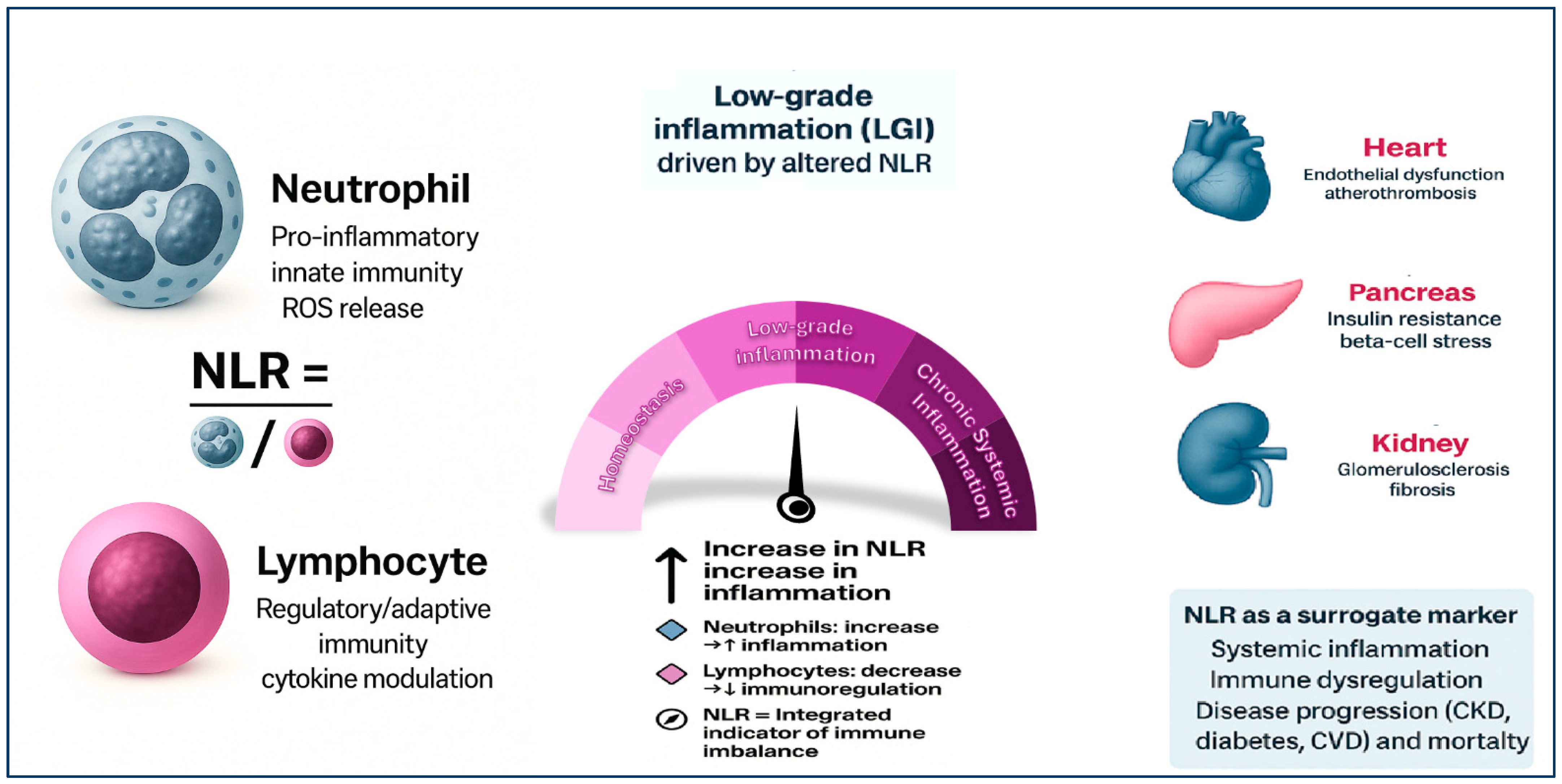
Figure 1. The neutrophil-to-lymphocyte ratio (NLR) as an integrated marker of immune imbalance and chronic low-grade inflammation (LGI). NLR reflects the balance between pro-inflammatory neutrophils and regulatory lymphocytes, integrating signals from both the innate and adaptive immune systems. An elevated NLR—driven by neutrophilia, lymphopenia, or both—indicates immune dysregulation and promotes a state of low-grade inflammation (LGI), which may evolve into chronic systemic inflammation. This immune imbalance contributes to the pathogenesis of major cardio-renal-metabolic diseases. In hypertension, elevated NLR is associated with endothelial dysfunction and vascular remodeling. In diabetes, it correlates with insulin resistance and β-cell stress. In CKD, increased NLR is linked to glomerulosclerosis, fibrosis, and progression of renal impairment. In CVD, NLR predicts atherogenesis, plaque instability, and adverse cardiovascular outcomes. Therefore, NLR serves as a low-cost, accessible surrogate marker of systemic inflammation, immune activation, and disease progression across multiple chronic conditions.
Ijms 26 08256 g001
Table 1. Summary of the main studies reviewed, highlighting key characteristics such as study design, sample size, population, methodology, and main findings.
Author, YearPopulationMain OutcomesKey Findings
Li et al., 2019 [43]3000+ T2DM patientsNLR correlation with renal functionNLR significantly higher in patients with diabetic nephropathy
Adane et al, 2023 [44]13 studies meta-analysisRelationship between NLR and glycemic controlNLR positively correlated with HbA1c and poor glycemic control
Rui-Wang et al., 2020 [46]470 T2DM and controlsAssociation between NLR, PLR and DRNLR independently associated with diabetic retinopathy as continuous and categorical variable
Si et al. 2024 [45]572 adults with diabetic retinopathy (NHANES 2009–2018)All-cause mortality, diabetes-related cardiovascular mortalityElevated NLR (≥1.516), MLR (≥0.309), and SIRI (≥0.756) independently associated with increased mortality risk. Combined marker (NLR + MLR + SIRI) improved prognostic performance. J-shaped mortality curves observed.
Table 2. Summary of the main studies reviewed, highlighting key characteristics such as study design, sample size, population, methodology, and main findings.
Study (Author, Year)PopulationMain OutcomesKey Findings
Yoshitomi et al., 2019 [47]256 CKD stage 3–5, JapaneGFR decline, renal replacement therapyHigher NLR associated with faster renal decline and dialysis initiation
Wang et al., 2021 [48]966 IgA nephropathy patientsESKD progression, histological severityNLR ≥ 2.67 linked to glomerulosclerosis and fibrosis, higher progression risk
Muresan et al., 2023 [51]ESKD Romanian patients (2016–2019)NLR/MLR/PLR at admission vs. 30-day mortalityHigh NLR: 30-day mortality = 40.12% vs. 1.97% (p < 0.0001); independent predictor of early death
Yuan et al., 2021 [64]938 Chinese CKD patients, stages 1–4ESKD progression, CV and all-cause mortalityNLR associated with ESKD risk only in stage 4 CKD; no significant association with CVD events or all-cause mortality across all stages
Chen et al., 2024 [58]3500 NHANES CKD participantsAll-cause mortalityElevated NLR associated with higher 10-year mortality (HR > 1.5)
Carollo et al. 2025 [53]236 chronic hemodialysis patients12-month mortality, inflammation/nutrition markersHigh NLR linked to increased mortality and worse nutritional profile
Lin et al., 2021 [49]3000+ patients, TaiwaneGFR decline, albuminuria, inflammationHigher NLR linked to progression in both diabetic and non-diabetic CKD
Ao, et al., 2021 [50]116,709 patients with CKD, including those on dialysis (13 studies)CV and all-cause mortalityElevated NLR was significantly associated with increased all-cause mortality (HR 1.93) and cardiovascular mortality (HR 1.45); among dialysis patients, all-cause mortality HR 1.94
Catabay et al., 2017 [59]Incident HD patients (2007–2011, USA)Short- & long-term mortalityHigh NLR predicted mortality; improved AUROC and R2; PLR had no predictive value
Table 3. Summary of the main studies reviewed, highlighting key characteristics such as study design, sample size, population, methodology, and main findings.
Setting/StudyDesign & SampleNLR Cut-Off/QuartileOutcome(s)Key Prognostic Findings
Ul Hussain et al. 2024 [67]STEMI patients post-PCI; 28,756 individuals (35 studies)High vs. low NLR (varied)In-hospital & long-term mortality, CV mortality, MACEHigh NLR: in-hospital mortality RR = 3.52; long-term mortality HR ≈ 1.07; CV mortality RR ≈ 2.66; MACE RR ≈ 2.92
Cho et al., 2018 [70]Acute HFElevated NLR (thresholds unspecified)In-hospital mortality, three years mortality Elevated NLR independently associated with increased in-hospital mortality (OR ≈ 2.2) and three-year post-discharge mortality (OR ≈ 1.44)
Caimi et al. 2024 [68]123 young AMI patients (mean age 39.4 ± 5.8 years)NLR ≥ 6.5STEMI/Non STEMI Long term outcomeNLR significantly elevated in AMI patients vs. controls (2.38 ± 0.87 vs. 1.82 ± 0.71, p < 0.0001). No difference in NLR between STEMI vs. non-STEMI or diabetic vs. non-diabetic. NLR not correlated with number of CV risk factors or CAD extent. Neutrophil count decreased over time leading to reduced NLR; lymphocyte counts stable.
Peng et al. 2024 [73]AF cohort meta-analysis; ~59,000 patientsHigh NLR (varied)AF recurrence, stroke, mortality, left atrial thrombusHigh NLR predicted AF recurrence and stroke; linked to mortality and thrombus
Cho et al., 2020 [70]Decompensated HF; 5580 patientsQuartile 4 (not specified)In-hospital & 3-year mortalityHighest quartile NLR: OR ~2.2 for in-hospital mortality; OR ~1.44 at 3 years
Table 4. Summary of the main studies reviewed, highlighting key characteristics such as study design, sample size, population, methodology, and main findings.
Study (Author, Year)Design/PopulationMain Focus/OutcomeKey Findings
Zhang et al., 2024
[88]
NHANES 2009–2014; 3067 hypertensive adultsNLR and mortalityNLR > 3.5 associated with higher all-cause (HR = 1.96) and CV mortality (HR = 2.33); AUC 0.64–0.70
Hong et al., 2024 [89]NHANES (extended sample)NLR thresholds and mortalityNLR > 2.0 linked to all-cause (HR~1.47) and CV mortality (HR~2.08)
Liu et al., 2023
[83]
Prospective cohort; 28,850 normotensivesIncident hypertensionHighest NLR quintile: HR = 1.23 for new-onset hypertension
Sarejiloo et al., 2021 [31]21 studiesNLR in hypertensive vs. normotensiveWMD = 0.40; Non-dippers showed higher NLR (WMD = 0.58)
Yu et al., 2020 [82]Cross-sectional; hypertensive patientsNLR and LVHNLR independently associated with LVH (OR = 1.506); AUC = 0.626
Wang et al., 2017 [87]217 hypertensive + 132 controlsNLR and arterial stiffness (baPWV)NLR higher in ISH and SDH; NLR independently predicted baPWV
Belen et al. [81]Cohort of resistant hypertensionNLR and BP severityNLR correlated with office and ambulatory BP; elevated in RHT
Chen et al. [86]H-type hypertension patientsNLR and renal function1 unit ↑ NLR = 51% ↑ risk of renal dysfunction
NHANES 1999–2010 [84]Cross-sectional; n = 22,290NLR and HTN prevalenceln-NLR: OR = 1.087 for HTN; SII stronger; PLR and LMR not significant
Jhuang et al., 2019 [85]6278 adultsNLR and prevalent hypertensionHR = 1.28 overall; stronger in older adults (HR = 1.88)
Sunbul et al., 2014 [80]83 Non-dipper hypertensivesNLR and BP patternNLR > 2.7 predicted non-dipping (83% sens., 65% spec.)
ISH: Isolated Systolic Hypertension; SDH: Systolic-Diastolic Hypertension; LVH: Left Ventricular Hypertrophy; baPWV: Brachial-Ankle Pulse Wave Velocity; CV: Cardiovascular.
Table 5. Comparison of NLR and other inflammatory/prognostic biomarkers.
BiomarkerBiological
Significance
Biological
Variability
AdvantagesLimitationsMain Prognostic
Evidence
NLR (Neutrophil-to-Lymphocyte Ratio)Ratio between neutrophils (innate inflammation) and lymphocytes (adaptive response)Influenced by circadian rhythm, infections, stress, corticosteroids, other therapiesSimple, inexpensive, derived from standard blood count; reflects immune balanceNon-specific, intra-individual variability; lack of standardized cut-off valuesAssociated with prognosis in sepsis, cardiovascular diseases, cancer, COVID-19
CRP (C-reactive protein)Acute-phase protein produced by the liver in response to IL-6Relatively stable; rises within 6–8 h after inflammatory stimulusWidely validated; available in labs and point-of-care; standardized markerCannot differentiate acute vs. chronic inflammation; less informative on immune statusStrong marker of acute inflammatory severity and cardiovascular risk
IL-6Central pro-inflammatory cytokine, stimulates CRP productionHigh intra-individual variability; short half-life; rapid peaksSensitive marker of early inflammatory activationRequires dedicated assays; costly; less accessible in routine clinical useStrongly associated with severity in sepsis, ARDS, COVID-19
TNF-αKey cytokine in systemic inflammation and tumor necrosisHigh variability; low basal levels; transient peaksEarly indicator of systemic inflammationMeasurement complex, less standardized; high costInvolved in sepsis and autoimmune diseases; prognostic potential but less established compared with CRP/IL-6
Disclaimer/Publisher’s Note: The statements, opinions and data contained in all publications are solely those of the individual author(s) and contributor(s) and not of MDPI and/or the editor(s). MDPI and/or the editor(s) disclaim responsibility for any injury to people or property resulting from any ideas, methods, instructions or products referred to in the content.