Correction: Kalimuthu et al. Regulated Mesenchymal Stem Cells Mediated Colon Cancer Therapy Assessed by Reporter Gene Based Optical Imaging. Int. J. Mol. Sci. 2018, 19, 1002
- Error in Figures

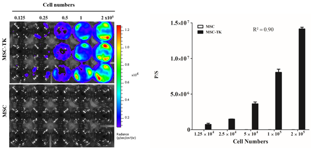
- Figure 1B

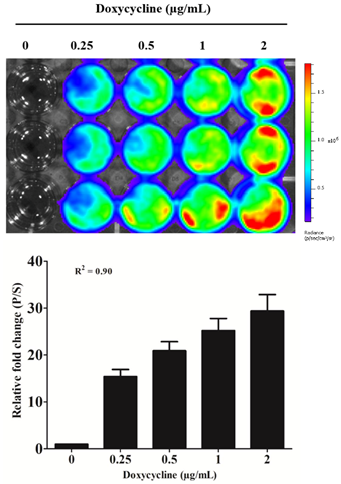
- Figure 1C

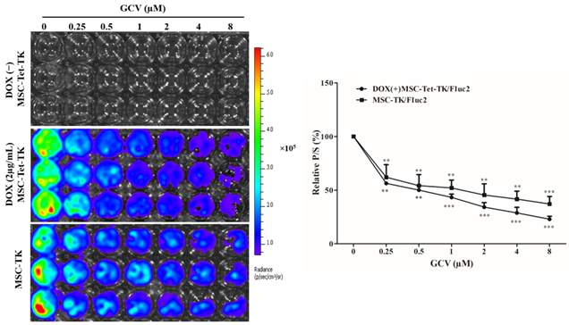
- Figure 2. (DOX(−) MSC-Tet-TK).
- Error in Figure Legends
- Figure 2. Fluc activity of MSC-Tet-TK and MSC-TK cells after ganciclovir (GCV) treatment for 48 h. Fluc activity was measured by bioluminescent imaging (BLI), and the quantitation for MSC-Tet-TK and MSC-TK cells is shown in the right-hand panel. Values obtained from three experiments are expressed as the mean ± standard deviation (SD), ** p < 0.01, *** p < 0.001 (by Student’s t-test). p/s, photons/second.
- Figure 3. Bystander effect of MSC-Tet-TK and MSC-TK cells. (A) Rluc activity in co-cultures (1:1) of naive MSCs and CT26/Rluc cells treated with the indicated concentrations of GCV for 48 h. (B) BLI images of the Rluc activity and quantitation data of CT26/Rluc in co-cultures (1:1) of MSC-TK or MSC-Tet-TK cells in the absence or presence of doxycycline (DOX(−) and DOX 2 μg/mL, respectively). Three experiments are expressed as the mean ± standard deviation (SD), * p < 0.05, ** p < 0.01, *** p < 0.001 (by Student’s t-test). p/s, photons/second.
- Text Correction
- 2.1. Characterization of MSC-Tet-TK and MSC-TK
Reference
- Kalimuthu, S.; Zhu, L.; Oh, J.M.; Lee, H.W.; Gangadaran, P.; Rajendran, R.L.; Baek, S.H.; Jeon, Y.H.; Jeong, S.Y.; Lee, S.-W.; et al. Regulated Mesenchymal Stem Cells Mediated Colon Cancer Therapy Assessed by Reporter Gene Based Optical Imaging. Int. J. Mol. Sci. 2018, 19, 1002. [Google Scholar] [CrossRef]
Disclaimer/Publisher’s Note: The statements, opinions and data contained in all publications are solely those of the individual author(s) and contributor(s) and not of MDPI and/or the editor(s). MDPI and/or the editor(s) disclaim responsibility for any injury to people or property resulting from any ideas, methods, instructions or products referred to in the content. |
© 2025 by the authors. Licensee MDPI, Basel, Switzerland. This article is an open access article distributed under the terms and conditions of the Creative Commons Attribution (CC BY) license (https://creativecommons.org/licenses/by/4.0/).
Share and Cite
Kalimuthu, S.; Zhu, L.; Oh, J.M.; Lee, H.W.; Gangadaran, P.; Rajendran, R.L.; Baek, S.H.; Jeon, Y.H.; Jeong, S.Y.; Lee, S.-W.; et al. Correction: Kalimuthu et al. Regulated Mesenchymal Stem Cells Mediated Colon Cancer Therapy Assessed by Reporter Gene Based Optical Imaging. Int. J. Mol. Sci. 2018, 19, 1002. Int. J. Mol. Sci. 2025, 26, 8225. https://doi.org/10.3390/ijms26178225
Kalimuthu S, Zhu L, Oh JM, Lee HW, Gangadaran P, Rajendran RL, Baek SH, Jeon YH, Jeong SY, Lee S-W, et al. Correction: Kalimuthu et al. Regulated Mesenchymal Stem Cells Mediated Colon Cancer Therapy Assessed by Reporter Gene Based Optical Imaging. Int. J. Mol. Sci. 2018, 19, 1002. International Journal of Molecular Sciences. 2025; 26(17):8225. https://doi.org/10.3390/ijms26178225
Chicago/Turabian StyleKalimuthu, Senthilkumar, Liya Zhu, Ji Min Oh, Ho Won Lee, Prakash Gangadaran, Ramya Lakshmi Rajendran, Se Hwan Baek, Yong Hyun Jeon, Shin Young Jeong, Sang-Woo Lee, and et al. 2025. "Correction: Kalimuthu et al. Regulated Mesenchymal Stem Cells Mediated Colon Cancer Therapy Assessed by Reporter Gene Based Optical Imaging. Int. J. Mol. Sci. 2018, 19, 1002" International Journal of Molecular Sciences 26, no. 17: 8225. https://doi.org/10.3390/ijms26178225
APA StyleKalimuthu, S., Zhu, L., Oh, J. M., Lee, H. W., Gangadaran, P., Rajendran, R. L., Baek, S. H., Jeon, Y. H., Jeong, S. Y., Lee, S.-W., Lee, J., & Ahn, B.-C. (2025). Correction: Kalimuthu et al. Regulated Mesenchymal Stem Cells Mediated Colon Cancer Therapy Assessed by Reporter Gene Based Optical Imaging. Int. J. Mol. Sci. 2018, 19, 1002. International Journal of Molecular Sciences, 26(17), 8225. https://doi.org/10.3390/ijms26178225


