Article

α-Cyclodextrin/Moringin Impacts Actin Cytoskeleton Dynamics with Potential Implications for Synaptic Organization: A Preliminary Transcriptomic Study in NSC-34 Motor Neurons

1
IRCCS Centro Neurolesi “Bonino-Pulejo”, Via Provinciale Palermo, Contrada Casazza, 98124 Messina, Italy
2
Institute of Organic and Analytical Chemistry (ICOA), Université d’Orléans, UMR 7311, BP 6759, F-45067 Orléans, France
3
Department of Food Quality and Nutrition, Research and Innovation Centre, Fondazione Edmund Mach (FEM), Via E. Mach 1, 38098 San Michele all’Adige, Italy
4
Department of Medical, Oral and Biotechnological Sciences, University “G. D’Annunzio” Chieti-Pescara, 66100 Chieti, Italy
*
Author to whom correspondence should be addressed.
These authors contributed equally to this work.
Int. J. Mol. Sci. 2025, 26(17), 8220; https://doi.org/10.3390/ijms26178220
Submission received: 21 July 2025 / Revised: 21 August 2025 / Accepted: 22 August 2025 / Published: 24 August 2025

Abstract

α-Cyclodextrin/Moringin (α-CD/MOR) is an isothiocyanate showing neuroprotective and antioxidant properties. In this work, we studied in differentiated NSC-34 motor neurons cell line the molecular pathways activated following a treatment of 96 h with α-CD/MOR at different doses, namely 0.5, 5 and 10 μM. Taking advantage of comparative transcriptomic analysis, we retrieved the differentially expressed genes (DEGs) and we mapped DEGs to synaptic genes using the SynGO database. Then, we focused on the biological pathways in which they are involved. We observed that the prolonged treatment with α-CD/MOR significantly modulated biological processes and cellular components associated with synaptic organization. Interestingly, the KEGG pathway “Regulation of actin cytoskeleton” was overrepresented, alongside pathways related to synapses and axon guidance. Specifically, SPIA analysis indicated that the “Regulation of actin cytoskeleton” pathway was found to be activated with the highest dose of α-CD/MOR. Moreover, α-CD/MOR also modulated transcription factors involved in synaptic plasticity, such as Creb1. These results could indicate that α-CD/MOR can influence synaptic functions and organization, being involved in synaptic plasticity through the modulation of actin dynamics.

1. Introduction

Isothiocyanates (ITCs) are among the most recently studied phytocompounds. ITCs are a class of organic compounds formed as a result of the glucosinolates (GLs) hydrolysis through the enzyme myrosinase. GLs are present in different plants of the Cruciferae family (Brassicaceae), such as broccoli, mustard, cabbage, and cauliflower, and in the Moringaceae family. GLs are stable molecules in plant cells. The conversion from GLs to ITCs can occur as a defense mechanism after an injury to plant tissues (for instance, after the attack of predators or pathogens) [1]. In humans, after ingestion, GLs may be hydrolyzed by plant-derived myrosinase, and the resulting products are absorbed, while unmetabolized GL reach the colon, where they are processed by the gut microbiota [2].
Depending on the type of GLs, different ITCs are formed: glucoraphanin (GRA) and glucomoringin (GMG) produce sulforaphane (SFN) and moringin (MOR), respectively. In addition to the neuroprotective effects, ITCs can also exert anti-inflammatory, antioxidant, anticancer, and cardioprotective actions [3,4].
MOR is an ITC found in the plant Moringa oleifera [4]. M. oleifera is one of 12 species of the Moringaceae family. It is a tropical plant commonly called the “miraculous tree”. M. oleifera is widely used as a dietary supplement because of its rich nutritional composition, which includes vitamins, essential amino acids, minerals, and oleic acids [5]. It also contains bioactive substances that could support its therapeutic properties and offer positive effects on human health. One of the most studied bioactive substances is MOR. Indeed, several studies, both in vitro and in vivo, showed its antimicrobial [6], antioxidant [7,8], anti-inflammatory [9], antitumor [10], and neuroprotective [11,12] effects. In particular, MOR exerts protective effects in different models of neurodegenerative disorders [12,13,14].
Different phytochemicals were shown to promote neurite extension, synaptic transmission and formation, and the development of cytoskeletal structures, resulting in improved synaptic functionality [15]. The cytoskeleton is a highly dynamic structure that is composed of microtubules, actin filaments, and neurofilaments, and its rearrangement plays a key role in the regulation of synaptic plasticity. Specifically, actin dynamics is important in synaptic plasticity, modulating synapse organization and functions [16]. Synaptic stimulation rapidly changes actin dynamics, and many actin regulators play roles in neuronal functions. On the contrary, an abnormal synapse stabilization or pruning can lead to neurological complications. Accordingly, defects in the regulation of the actin cytoskeleton in neurons have been implicated in neurological disorders [17]. Cytoskeletal remodeling associated with synaptic functions is also relevant in neurodegenerative diseases and in aging-related processes [18]. Alterations of actin-binding proteins, which participate in actin cytoskeleton dynamics, could therefore represent a common feature of different synaptopathies, such as amyotrophic lateral sclerosis (ALS), frontotemporal dementia, and Alzheimer’s disease (AD) [19,20,21]. As an example, it is common to observe a compromised neuronal plasticity after traumatic events such as traumatic brain injury in which the cytoskeleton proteins undergo irreversible reactions that alter their functionality [22]. Synaptic plasticity also becomes impaired during aging, with changes occurring in the shape and density of dendritic spines [23]. Studies on AD post-mortem tissues and AD experimental models suggested that AD pathology negatively impacts actin cytoskeleton pathways. Indeed, actin polymerization influences clathrin-mediated endocytosis, which in turn controls Aβ production through the colocalization of the amyloid cascade proteins. So, the actin cytoskeleton represents a crossroad of pathways contributing to AD pathogenesis [19]. Motor neurons show long axonal projections, that require the integrity of neuronal cytoskeleton to maintain axonal stability, anterograde and retrograde transport, and signaling between neurons. For these reasons, protein aggregates containing cytoskeletal proteins, which represent a pathological feature of ALS, may alter the functions of neurons [20].
In this study, we investigated the effect of the prolonged treatment with the conjugated form α-cyclodextrin (CD)/MOR on the expression of genes involved in molecular pathways associated with synapses. Specifically, we differentiated NSC-34 cells, and treated them with the 0.5, 5 and 10 μM doses of α-CD/MOR for 96 h. The conjugated form α-CD/MOR showed an increased stability and solubility compared to MOR, which shows a low solubility in water [24]. We used transcriptomic analysis to investigate in more detail how this treatment can modulate gene ontology and pathways related to synaptic genes.

2. Results

2.1. Comparative Analysis of α-CD/MOR Against Control Group

The comparative analysis of the control (CTRL) group versus α-CD/MOR-treated samples for 96 h with 0.5 μM (CTRL vs. α-CD/MOR0.5-96), 5 μM (CTRL vs. α-CD/MOR5-96), or 10 μM (CTRL vs. α-CD/MOR10-96) is presented item by item in Figure 1 using Volcano plots.
The amount of differentially expressed genes (DEGs) in the three groups and the number of up- or downregulated genes were of the same order of magnitude and increased proportionally to the used dose of α-CD/MOR. In detail, 5005, 5320, and 5964 DEGs were found in CTRL vs. α-CD/MOR0.5-96, CTRL vs. α-CD/MOR5-96, and CTRL vs. α-CD/MOR10-96, respectively (Tables S1–S3). Also, the amount of upregulated and downregulated DEGs was 2524 and 2481 for CTRL vs. α-CD/MOR0.5-96, 2611 and 2709 for CTRL vs. α-CD/MOR5-96, and 2957 and 3007 for CTRL vs. α-CD/MOR10-96, respectively.

2.2. α-CD/MOR Modulated Synaptic Ontologies

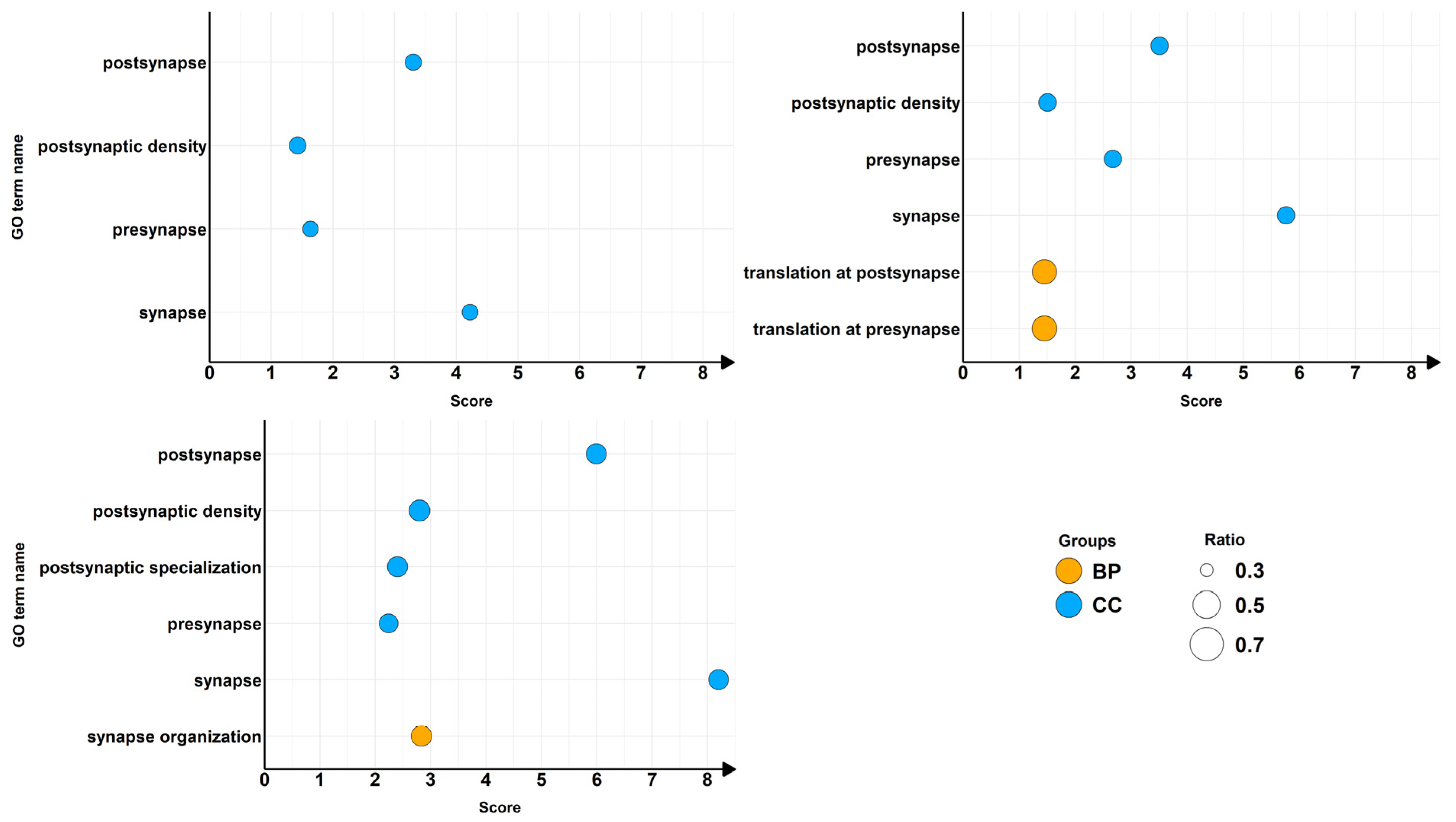
The focus of our study was to observe in differentiated motor neuronal NSC-34 cells the effects of the prolonged exposure to α-CD/MOR on synaptic genes. In this sense, we took advantage of the curated SynGO database [22] accessible at https://syngoportal.org (30 June 2025), to associate our DEGs to a specific neurological context. Since SynGO is curated on human genes, it provides the mapping tool for alias conversion from other species. After mapping, SynGO was able to provide an association for 4872 DEGs in CTRL vs. α-CD/MOR0.5-96, 5196 DEGs in CTRL vs. α-CD/MOR5-96, and 5815 DEGs in CTRL vs. α-CD/MOR10-96. In particular, the DEGs mapped on 302 terms (Tables S4–S6). For each term, the q-value was obtained using false discovery rate as post hoc correction. We set the threshold of 0.05 for the q-value to discriminate statistically significant terms. As shown in Table 1, eight terms were under the threshold, and they were three biological process (BP) and five cellular component (CC) terms.
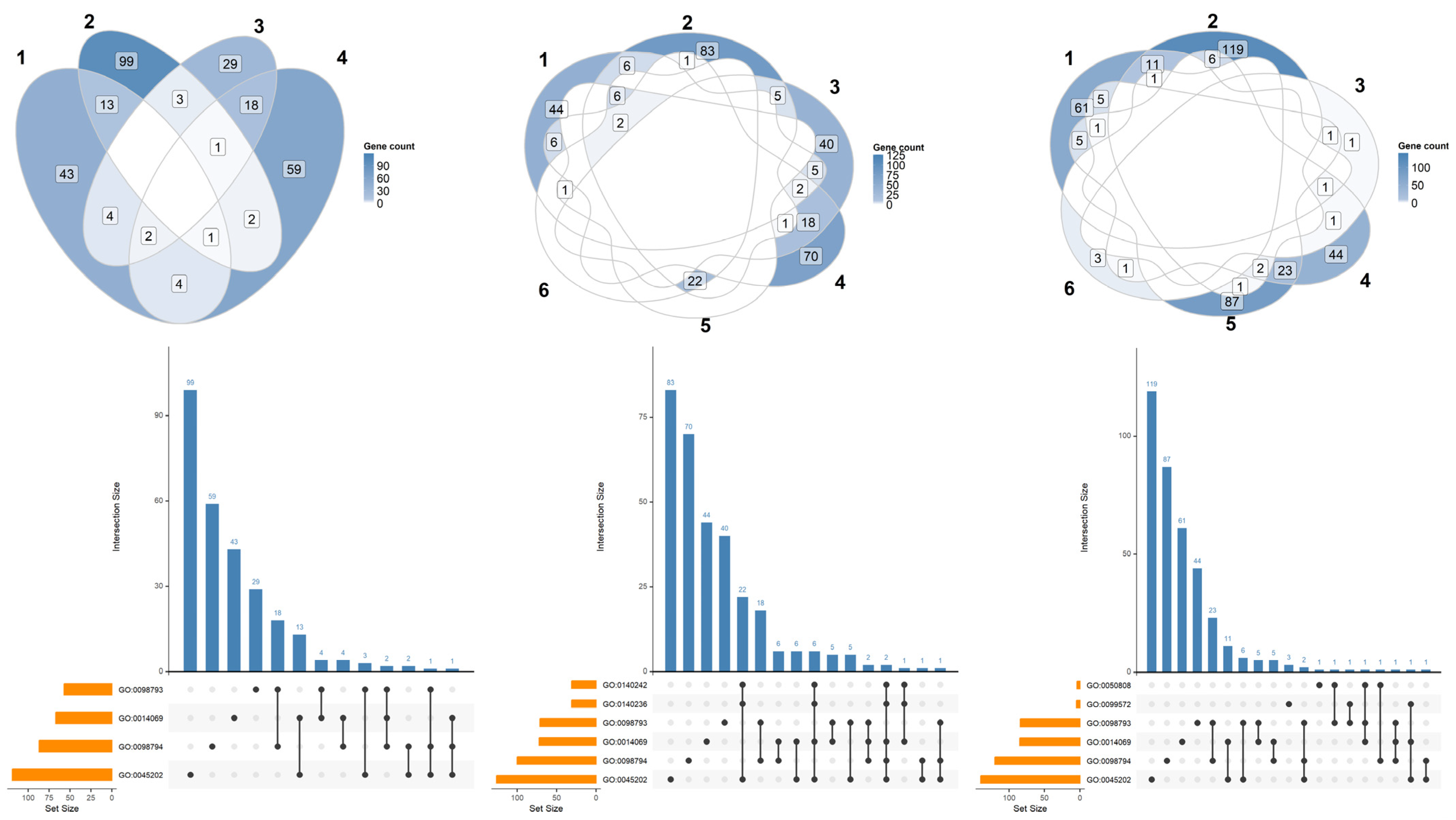
To inspect how the DEGs are distributed among the overrepresented terms, we computed the Venn plots in Figure 2 along with upset distribution for each comparison. No comparison showed any DEG taking place in all the terms. Most DEGs were specific to the particular term, and in some cases, they are shared among the terms. In this sense, the terms were quite specific and independent from each other for each comparison. In detail, among the significant terms of the Gene Ontology dictionary, CTRL vs. α-CD/MOR0.5-96 included four terms: synapse (GO:0045202), presynapse (GO:0098793), postsynapse (GO:0098794), and postsynaptic density (GO:0014069). CTRL vs. α-CD/MOR5-96 included six terms: synapse (GO:0045202), presynapse (GO:0098793), postsynapse (GO:0098794), postsynaptic density (GO:0014069), translation at presynapse (GO:0140236), and translation at postysnapse (GO:0140242). CTRL vs. α-CD/MOR10-96 included six terms: synapse (GO:0045202), presynapse (GO:0098793), postsynapse (GO:0098794), postsynaptic specialization (GO:0099572), postsynaptic density (GO:0014069), and synapse organization (GO:0050808). Thus, the general terms GO:0045202, GO:0098793, GO:0098794, and GO:0014069 were shared among all the comparisons.
We then inspected, as shown in Figure 3, the different categories to which each overrepresented term belongs and the relative amount represented as the ratio of the DEGs mapped in the terms over the number of genes included in the term.

2.3. α-CD/MOR Modulated Synaptic Pathways

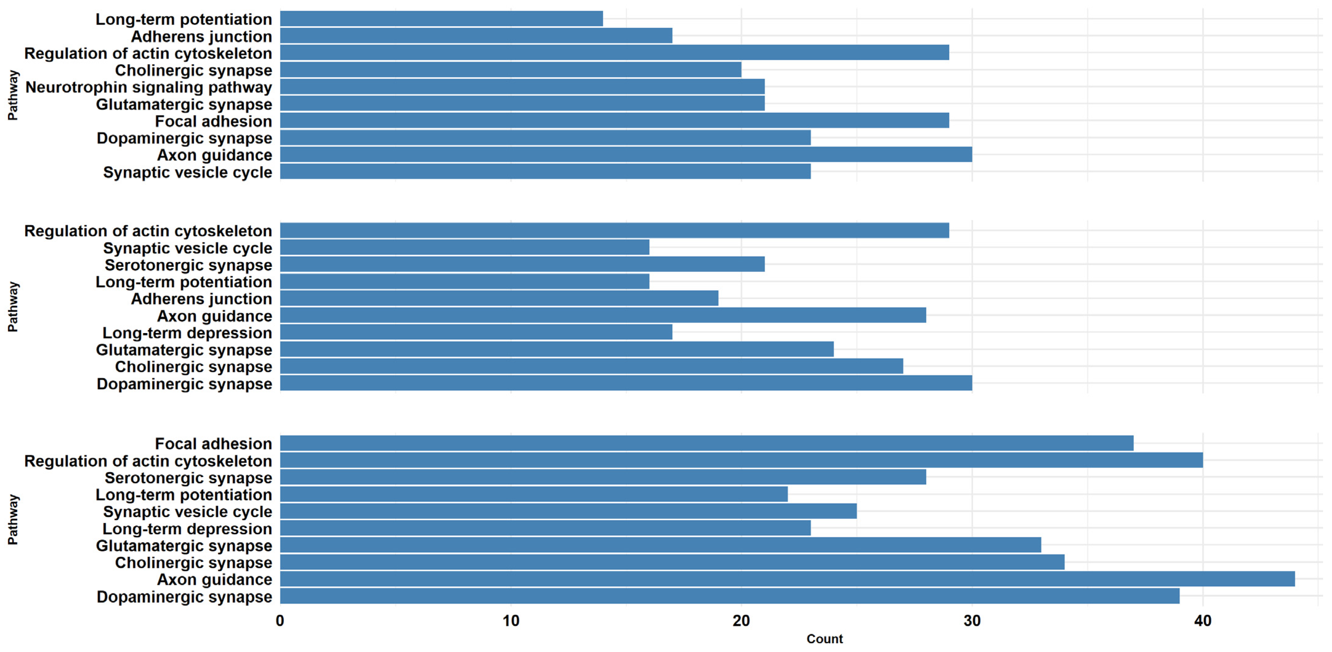
Then, for each comparison, we performed the pathway overrepresentation analysis of the DEGs mapped on SynGO using the KEGG database [25], considering the same threshold of q-value (0.05) (Tables S7–S9). Among all the pathways represented in KEGG, we kept all the pathways related to our experiment that are included under the categories “Cell growth and death”, “Cell motility”, “Cellular community—eukaryotes”, “Development and regeneration”, “Nervous system”, and “Neurodegenerative disease”. Finally, CTRL vs. α-CD/MOR0.5-96 and CTRL vs. α-CD/MOR5-96 counted 21 pathways, while CTRL vs. α-CD/MOR10-96 counted 26 pathways. In order to obtain a better representation, we selected the 10 pathways with the lowest q-values from each comparison group and represented them in Figure 4. The majority of the pathways were common between the comparisons. Notably, the “Axon guidance” and “Regulation of actin cytoskeleton” pathways were among those with more genes in all the comparisons.
Given the importance of the pathway “Regulation of actin cytoskeleton” in synapse function and organization, we evaluated the activation state of this pathway in the different comparisons using SPIA. SPIA analysis revealed that this pathway was activated only in the comparison CTRL vs. α-CD/MOR10-96, while in the others, it was inhibited.

2.4. α-CD/MOR-Treated Cells Expressed Neuronal Markers

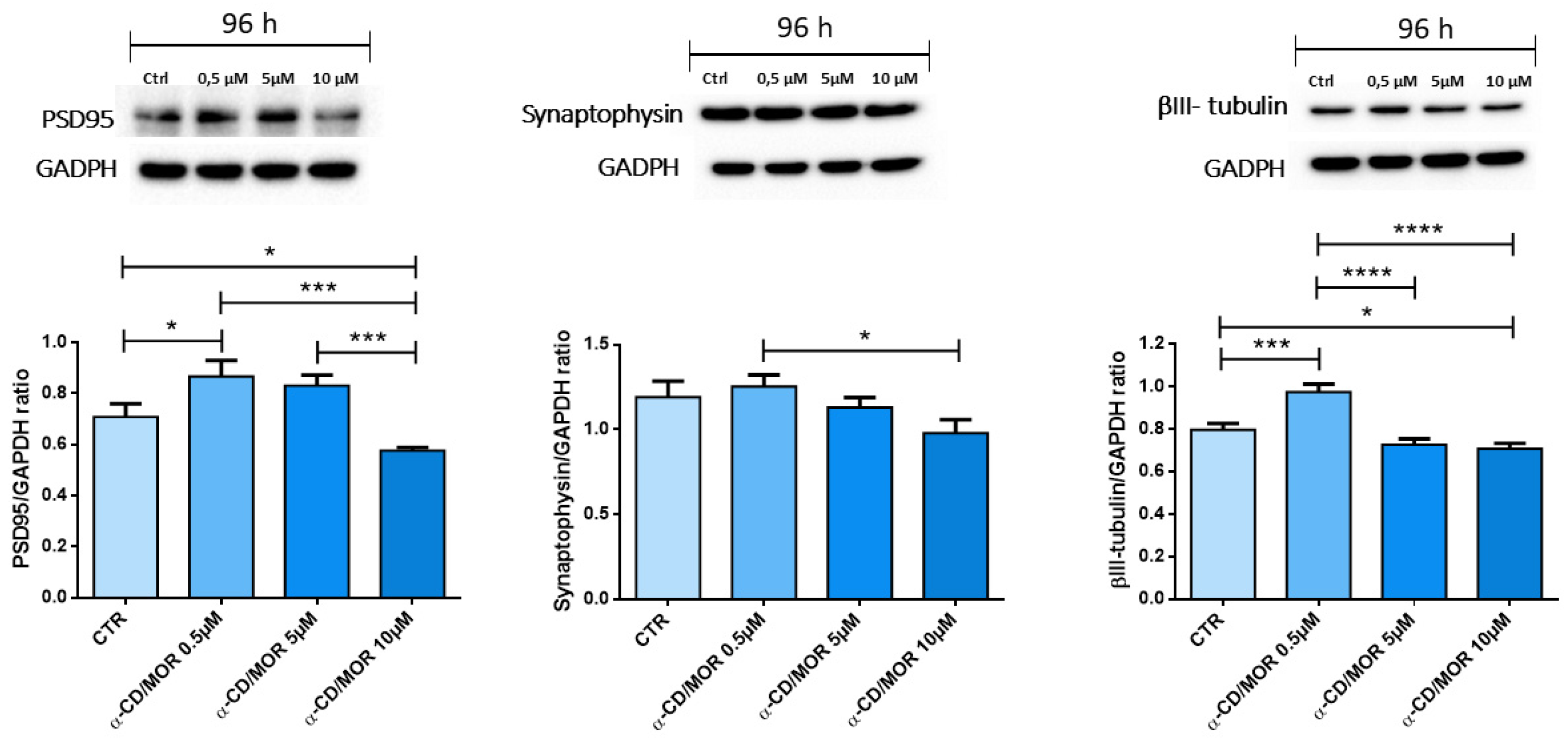
We performed the Western blot analysis of neuronal markers PSD95, synaptophysin, and βIII-tubulin (Figure 5).
PSD95 and βIII-tubulin significantly increased in α-CD/MOR0.5-96, while their protein levels decreased in the α-CD/MOR10-96 group compared to the control. Synaptophysin was expressed in all groups and showed no significant differences compared to the control.

2.5. Integration of Synaptic Genes into Cytoskeleton Regulation

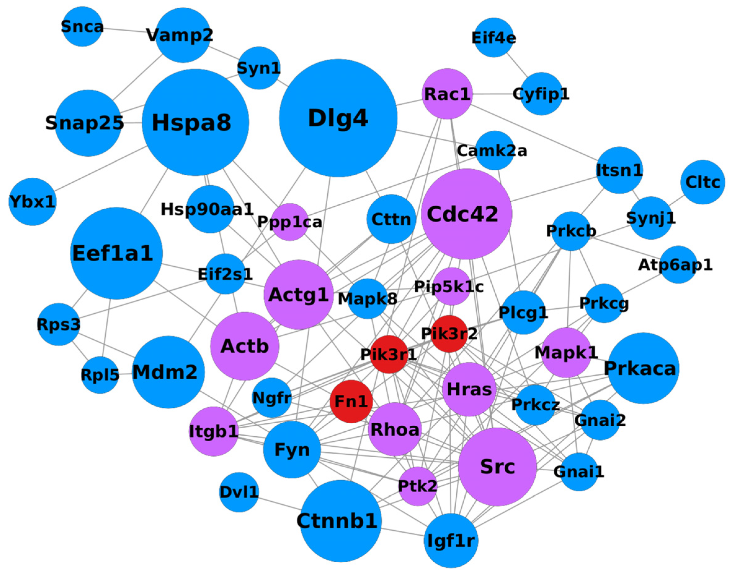
Interaction networks were generated for each comparison using the STRING database and analyzed in Cytoscape.
The initial network consisted of 574 nodes for the CTRL vs. α-CD/MOR0.5-96 comparison, 644 nodes for the CTRL vs. α-CD/MOR5-96 comparison, and 726 nodes for the CTRL vs. α-CD/MOR10-96 comparison. After removing proteins not connected to the main network, the nodes were reduced to 372 for the CTRL vs. α-CD/MOR0.5-96 comparison, 411 for the CTRL vs. α-CD/MOR5-96 comparison, and 494 for the CTRL vs. α-CD/MOR10-96 comparison.
Subsequently, for each network, proteins with a betweenness centrality above the 90th percentile were plotted. The final filtered network for the CTRL vs. α-CD/MOR10-96 comparison is reported in Figure 6, while the networks for the comparisons CTRL vs. α-CD/MOR0.5-96 and CTRL vs. α-CD/MOR5-96 were reported in Figures S1 and S2, respectively. All the tables with the information related to all the nodes of the different networks are provided in Supplementary Table S10.

2.6. Transcription Factors Involved in α-CD/MOR Effects and Analysis of DEGs Associated with Neurodegenerative, Neurological, and Neuropsychiatric Diseases

In order to identify the transcription factors that could be involved in the modulation of the transcriptomic profile of the inspected groups, we used the web tool Enrichr (https://maayanlab.cloud/Enrichr/, accessed on 5 August 2025). Specifically, we took advantage of the ChEA database with default parameters to highlight the overrepresented transcription factors. The chosen threshold of 0.05 for p-value, adjusted by post hoc correction, showed 588 transcription factors for α-CD/MOR0.5-96, 624 transcription factors for α-CD/MOR5-96, and 656 transcription factors for α-CD/MOR10-96 (Supplementary Table S11).
Additionally, the same tool was used to identify overrepresented disorders in which our identified DEGs were involved for each comparison with the same statistical significance. In particular, the database DisGeNET showed 150 diseases for α-CD/MOR0.5-96, 178 diseases for α-CD/MOR5-96, and 350 diseases for α-CD/MOR10-96 (Supplementary Table S12). Among them, 14 diseases for α-CD/MOR0.5-96, 18 diseases for α-CD/MOR5-96, and 35 diseases for α-CD/MOR10-96 were related to neurodegenerative, neurological, and neuropsychiatric diseases.
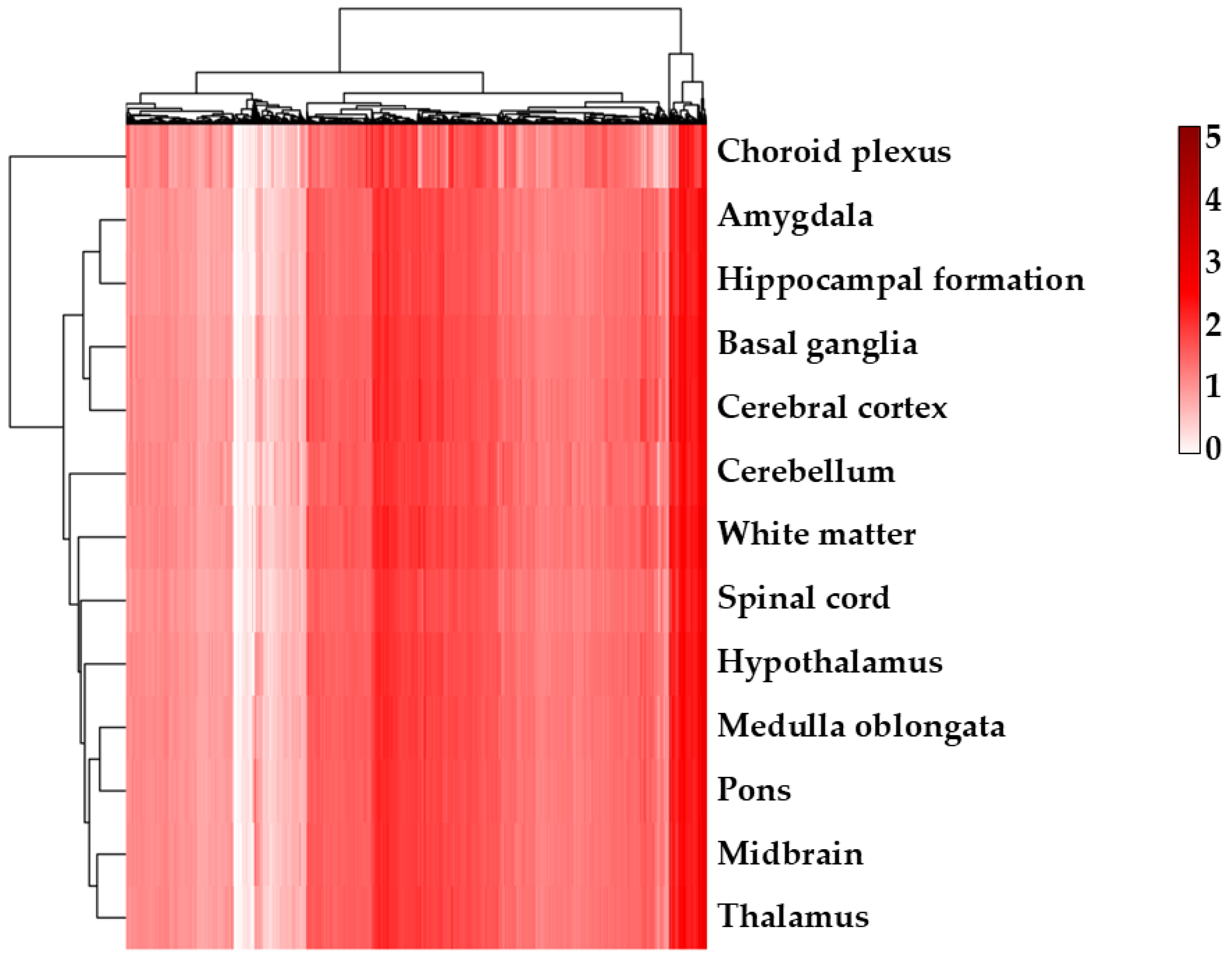
Given the amount of neurological disorders found as overrepresented, we observed for each comparison how the expression level of transcripts identified as DEGs in our experiments were distributed among the different brain regions in the Human Protein Atlas to identify possible cross-interactions (Figure 7; Supplementary Figures S3 and S4 for CTRL vs. α-CD/MOR0.5-96 and CTRL vs. α-CD/MOR5-96 comparisons, respectively).

3. Discussion

α-CD/MOR demonstrated several protective effects and neuroprotective properties. In a previous study, we showed that α-CD/MOR at 10 μM for 96 h exhibited antioxidant and neuroprotective effects, modulating the expression of NRF2 and its interactors in differentiated motor neuron NSC-34 cells [26]. Here, we evaluated the effects of α-CD/MOR on synaptic genes in differentiated NSC-34 cells after a prolonged exposure. For this reason, we treated differentiated NSC-34 cells for 96 h with different α-CD/MOR concentrations, namely 0.5, 5 and 10 μM. SynGO database shows a significant overrepresentation of both biological process (BP) and cellular component (CC) terms in our comparisons, except for the CTRL vs. 0.5 μM α-CD/MOR. Indeed, in comparison CTRL vs. α-CD/MOR0.5-96, only CC terms were overrepresented. The different terms shared few genes, while the majority were unique for each term. In particular, the CC “synapse” was significantly overrepresented in all the groups, highlighting that α-CD/MOR was able to modulate synaptic genes at all the concentrations. Interestingly, even if “presynapse” was overrepresented in all the comparisons, the postsynaptic compartment was more interested by α-CD/MOR effects. Indeed, the “postsynapse” term had a higher number of DEGs. Moreover, the terms “postsynapse” and “postsynaptic density” were overrepresented in all comparisons, while “postsynaptic specialization” was overrepresented in comparison CTRL vs. 10 μM α-CD/MOR. These results indicated that α-CD/MOR may influence synapse organization, and especially postsynaptic region, influencing the spatial and functional organization of postsynaptic proteins, such as anchoring and scaffolding molecules, neurotransmitter receptors, enzymes, and cytoskeletal components. In accordance with that, previous reports indicated that M. oleifera extracts promoted synaptic plasticity [27,28].
Interestingly, KEGG analysis also revealed that some pathways were significantly overrepresented in all the comparisons, such as synaptic pathways, “long-term potentiation”, “Axon guidance”, and “Regulation of actin cytoskeleton”. Interestingly, “Regulation of actin cytoskeleton” pathway was one of the most represented ones in all comparisons in terms of the number of included genes. This result is in accordance with GO results which indicated that α-CD/MOR modulated genes involved in synapse modulation and organization. Cytoskeleton plays a crucial role in cellular physiology, especially in processes such as cell motility, cell shape, and intercellular communication. In the context of motor neurons, the regulation of the cytoskeleton becomes even more critical, as it is essential for the growth, extension, and branching of neurites. These processes are fundamental for the formation of neuronal connections and the promotion of synaptic plasticity. Specifically, the synaptic actin cytoskeleton is a key element in synapse stability. Actin has a main role in regulating synapse structure and functions, such as spine morphology, post-synaptic density organization, long-term potentiation, anchoring, and trafficking of post-synaptic receptors. Then, synaptic plasticity induces remodeling of the actin cytoskeleton at the synapse, including pre- and post-synaptic compartments [16].
Given that “Regulation of actin cytoskeleton” emerged from the KEGG analysis as one of the pathways with the highest number of genes, these results indicate that the regulation of the actin cytoskeleton plays a central and fundamental role in the modulation of synaptic organization mediated by α-CD/MOR. Moreover, the pathway “Regulation of actin cytoskeleton” represents a base for all other pathways, given that actin cytoskeleton influences all others, including synapses, axon guidance, LTP, and junctions. Among these, the “Axon guidance” pathway was one of the bigger pathways overrepresented in all comparisons. Axon outgrowth is guided by growth cones, high motility structures enriched in filamentous actin at the axon distal tips. Growth cones can scan the environment using their F-actin protrusions in order to sense guidance cues and respond to them controlling actin dynamics. Actin dynamics in growth cones steers the axon towards attractants and away from repellents [29]. Among the main guidance cues that induce actin cytoskeletal modifications are slit and its Robo receptors, semaphorins and their plexin and neuropilin receptors, and ephrin and its Eph receptors. They initiate a cascade of events to modulate the growth cone membrane and the cytoskeleton to modulate axon growth and guidance [30].
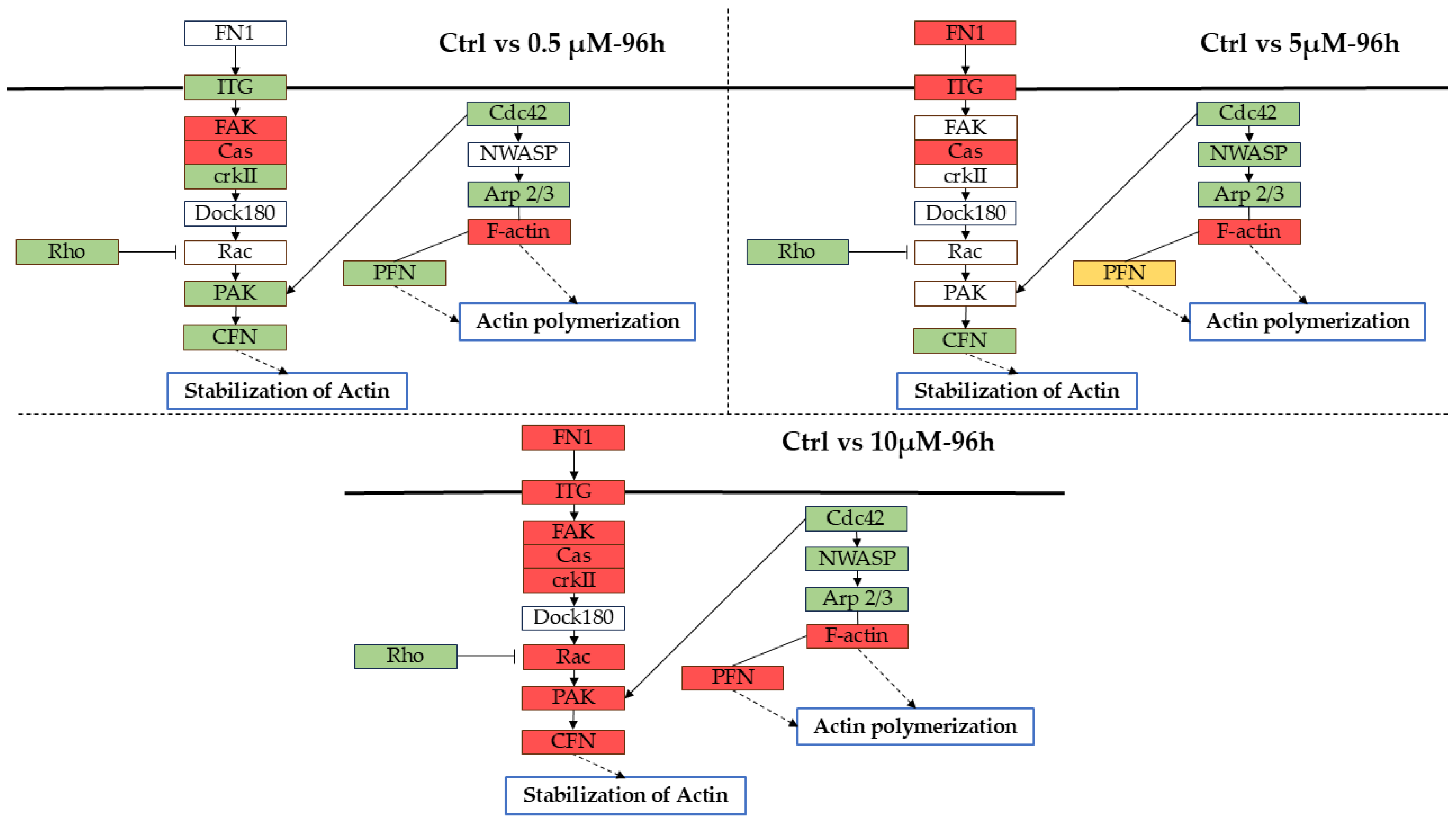
Interestingly, the pathway “Regulation of actin cytoskeleton” was overrepresented at all the tested concentrations. However, the SPIA analysis revealed that it was activated only with the highest α-CD/MOR concentration, suggesting that lower ones were not sufficient to exert this action. Indeed, some key genes involved in actin dynamics were DEGs or upregulated only at this concentration (Figure 8). For this reason, we focused on the concentration of 10 µM α-CD/MOR.
Integrin receptors make essential contributions to neurite outgrowth and axon elongation. Activated integrins engage extracellular matrix components allowing the growth cone to form contact points, which link the extracellular substrate to dynamic intracellular protein complexes [31]. In our study, we observed that the genes Itga2, Itgb1, and Itgb3, encoding the different integrin subunits, were upregulated, while Itga5 was downregulated. Among them, Itgb1, and Itgb3 are directly correlated to the nervous system. Specifically, Itgb1 plays an important role in the formation of intraneuronal connections, promoting neurite growth, axon guidance, and synapse formation and maturation [32]. Itgb3 is essential for a normal dendritic morphology in pyramidal neurons [33].
Integrin-mediated signaling recruits the complex Cas/CrkII/FAK. In our study, Crk and Ptk2 were involved in its formation, and they were upregulated. The protein Cas forms a complex with non-receptor focal adhesion kinases (FAK) and SRC family kinases (SFKs), particularly the CrkII protein, encoded by Crk. In vitro studies demonstrated the important role of the Cas protein in mediating the integrin signal during neuronal development and the axon guidance activation [34]. The protein FAK, encoded by Ptk2, is broadly expressed in the mammalian brain and highly enriched in neuronal growth cones. In vitro and in vivo studies showed that FAK expression positively modulates neurite outgrowth and synaptic plasticity. It is also involved in the formation and preservation of long-term spatial memory [35].
Rho family GTPases are fundamental regulatory molecules, linking surface receptors with actin cytoskeleton organization. Specifically, Rho GTPases are involved in the regulation of neuronal morphology, including dendritic arborization, spine morphogenesis, growth cone development, and axon guidance [36]. RhoA, Rac, and Cdc42 were indicated as important regulators of axonal and dendrite morphogenesis. Rho proteins are known to inhibit neurite extension; in contrast, Cdc42 and Rac are positive regulators of neurite outgrowth and dendritic spine formation. In particular, they promote protrusions through actin filament assembly [37]. Studies confirmed an important role of Rac1 in neurite outgrowth and axonal pathfinding, as well as neuronal migration [36]. In our study, only Rac1 was upregulated, while Rhoa and Cdc42 were downregulated in CTRL vs. α-CD/MOR10-96. Interestingly, Rac1 was upregulated only in CTRL vs. α-CD/MOR10-96, while it was not differentially expressed with lower α-CD/MOR concentrations. A study on motor neurons showed that reduced Rac activity is associated with increased death. In addition, it showed that Rac is downregulated in patients with sporadic ALS. On the other hand, RhoA expression may cause growth cone collapse by inhibiting neurite extension through stress fiber formation. Several studies suggested that an imbalance between the expression of RhoA and Rac could be one of the contributing factors to ALS [38]. Interestingly, Rho family GTPases play a role also in neuron death and survival. Specifically, Rho indirectly inactivates pro-survival proteins suppressing neuronal survival. In contrast, Rac enhances neuronal survival also via the activation of PAK [36].
An effector of Rac1, Pak2 gene, encoding for Pak protein, was upregulated only in the comparison CTRL vs. α-CD/MOR10-96. Pak2 belongs to the group I Paks along with Pak1 and Pak3, and they are very similar in structure. Several studies showed that Paks, particularly PAK1, are involved in neuronal polarization, differentiation, and migration. A study in PAK1 knockout mice showed a reduction in the number of pyramidal neurons and impaired neuronal migration [39]. Very little is known about Pak2, but their similarities in sequence and structure suggest that both proteins perform similar functions in cytoskeleton regulation. Moreover, Pak2 haploinsufficiency caused a decrease in synapse densities, defective long-term potentiation, and autism-related behaviors in mice, but also alterations in actin polymerization [40].
The ability of cofilin to modulate actin cytoskeleton rearrangements confers an important role in neurite outgrowth and guidance. Indeed, growth cone motility is based on the dynamic assembly and disassembly of actin filaments [41]. The function of cofilin is spatially and temporally regulated within the growth cone. In fact, in the anterior part of the growth cone, actin polymerizes, enabling forward movement. In contrast, in the posterior part of the growth cone, actin filaments undergo depolymerization to ensure forward movement [42]. Cofilin is capable of regulating actin dynamics, and in this study, Cfl1 gene was upregulated. At low cofilin/actin ratios, cofilin severs F-actin, increasing the ADP-actin monomer dissociation rate. At high cofilin/actin ratios, cofilin stabilizes F-actin or even the nucleation of new filaments. Inactive phosphorylated cofilin does not significantly bind to F-actin, and actin severing or depolymerization is low [43].
Profilin is a monomeric (G-)actin-binding protein needed in all non-muscle cells to maintain a G-actin pool necessary for the fast actin dynamics of these cells. More isoforms exist in mammals. Profilin 1 is ubiquitously expressed [44]. It binds G-actin in a 1:1 ratio, positively regulating actin polymerization. Profilin1, encoded by the Pfn1 gene, was upregulated in our study. It is an important regulator of synaptic plasticity thanks to its contribution to actin dynamics and cytoskeletal integrity and is needed in synaptogenesis [45].
Arp2/3 complex genes Actr2 and Arpc2 were downregulated. A study demonstrated that inhibiting the Arp2/3 complex in neurons has no effect on the dynamics of actin in the growth cone and, as a result, has no negative impact on neurite formation [46]. The genes encoding for actin (Actb and Actg1) were upregulated in CTRL vs. α-CD/MOR10-96.
Interestingly, only at the concentration of 10 μM α-CD/MOR, after 96 h, key genes involved in actin dynamics, such as Rac1, Cfl1, and Pak2, were upregulated. Instead, at lower concentrations, they were downregulated or not differentially expressed.
We also constructed an interaction network among the DEGs related to actin remodeling to highlight how they integrate with synaptic components and identify central regulatory nodes. It is not surprising that synaptic genes that also take part in the pathway “Regulation of actin cytoskeleton” represented important nodes of the network, such as Cdc42, Actg1, and Actb.
We evaluated the expression of neuronal markers using Western blot. βIII-tubulin is one of the earliest markers of neuronal differentiation of both the central and peripheral nervous systems. Tubb3 expression reaches a peak during axonal guidance and neuronal maturation, and its levels decreased in the central nervous system with maturity, while they were maintained high in the peripheral nervous system [47]. In our study, Tubb3 expression was downregulated with the concentration 10 µM α-CD/MOR and Western blot analysis confirmed the reduced protein levels. A study showed that reduced Tubb3 levels accelerated microtubule growth in axons and dendrites, and Tubb3 knockdown induced a specific upregulation of Tubb4 gene expression [48]. These data are in line with our transcriptomic results, in which Tubb3 downregulation is associated with Tubb4b upregulation in cells treated with 10 µM α-CD/MOR.
PSD-95 is a major component of the post synaptic density, an electron-dense specialization of excitatory postsynaptic membranes containing high concentrations of glutamate receptors and associated signaling and cytoskeletal proteins. PSD95 interacts with postsynaptic glutamate receptors and is important for their synaptic signaling [49]. PSD-95 acts as a scaffolding protein during synaptogenesis, regulates synaptic maturation [50], and maintains excitatory synapse balance [51]. However, PSD95 also has a role in excitotoxicity triggered by NMDARs. Indeed, in cultured cortical neurons, the suppression of PSD95 expression attenuated excitotoxicity caused by NMDA receptors [52]. PSD95 gene silencing delayed cell death in postischemic rat hippocampus [53]. A study also showed that PSD95-gene-specific siRNAs may relieve neuropathic pain [54]. In this study, we found a decrease in PSD95 after a 96 h treatment with the concentration 10 µM α-CD/MOR, in accordance with the transcriptomic analysis that evidenced the downregulation of the gene Dlg4 encoding for PSD95. Its decrease may indicate the maintenance of excitatory versus inhibitory balance and the absence of excitotoxicity, that is a hallmark of neurodegeneration. Then, α-CD/MOR may also be helpful in those conditions characterized by excitotoxcity.
Synaptophysin is a presynaptic protein and the second most abundant cargo on the synaptic vesicles. Synaptophysin is involved in the formation of synaptic vesicles and their exocytosis and drives the synapsis formation. Synaptophysin seems to coordinate the retrieval of synaptobrevin 2 during synaptic vesicle endocytosis [55]. In our study, synaptophysin is expressed in all groups; however, there were no significant differences compared to control, confirming transcriptomic analysis. This result is in line with GO results, which indicated a less prominent role of α-CD/MOR at the presynaptic level.
We also identified upstream transcription factors known to regulate gene expression of our DEGs to deeply evaluate the mechanism of action of α-CD/MOR. Among the most significant transcription factors, we found the members of the cAMP response element binding protein family, CREB and CREM [56]. CREB is a key component in diverse physiological processes, including nervous system development, neuronal survival, postnatal hippocampal neurogenesis, synaptic plasticity, and neurite outgrowth, and it has neuroprotective properties [57]. Also the transcription factor YY1 was identified, involved in proliferation, differentiation, and apoptosis; specifically it plays a role in neural development, neuronal function, and myelination [58]. A recent study evidenced that Yy1 controls murine cerebral cortex development in a stage-dependent manner. Specifically, it maintains the proliferation and survival of neural progenitor cells at early stages of brain development, while the dependence on Yy1 decreases during corticogenesis [59]. Also, Runx2 was among the significant transcription factors. A study indicated that Runx2 may be involved in neurite outgrowth, Schwann cell differentiation, and migration after sciatic nerve injury [60]. Interestingly, in our study, the transcription factors Creb1 and Runx2 were upregulated, suggesting that another mechanism that mediates α-CD/MOR effects on synaptic genes may be associated with the upregulation of these transcription factors.
Interestingly, DEGs modulated by α-CD/MOR were shown to be involved in different neurological, neurodegenerative, and neuropsychiatric conditions and to be expressed in different brain areas. This result may be of interest and support the use of α-CD/MOR as an integrative therapy in different synaptopathies associated with cytoskeletal alterations.
Our study is in accordance with previous studies which demonstrated ITCs effects on synaptic plasticity and cytoskeleton remodeling. SFN, through the modulation of Nrf2, enhanced white matter plasticity, improving the pyramidal tract plasticity and regeneration of oligodendrocytes after ischemic stroke [61]. SFN administration during postnatal brain development in mice enhanced synaptic plasticity and spatial learning skills by increasing the proteasome activity [62]. SFN attenuated scopolamine-induced deterioration of memory in rats, in association with the hippocampal induction of BDNF and CREB expression and the enhancement of hippocampal synaptic activity [63]. Also, bioactive RS-GRA-treatment protected against the neuronal functional disruption, restoring dendritic spine count in a Parkinson’s disease model [64]. A study evidenced that Phenethyl ITC can induce cytoskeletal changes [65]. Then, our study confirms the role of ITCs in the modulation of synaptic plasticity and cytoskeleton regulation. Moringin, like other isothiocyanates, is known to activate the TRPA1 ion channel at the concentrations used in this study [66]. TRPA1 is expressed in motoneurons and oligodendrocytes, and its activation has been linked to neuromuscular symptoms such as cramps and fasciculations, particularly in individuals carrying hyperactive TRPA1 variants [67,68]. TRPA1 activation, or possibly desensitization, might have contributed to the transcriptional changes we observed, potentially through calcium signaling or oxidative stress pathways, in which it is already known to be involved [69]. Notably, the upregulation of transcription factors such as CREB, which is responsive to calcium and ROS, may point in this direction [70]. Further studies will be needed to explore this potential mechanism. Taken together, these observations suggest that TRPA1 may represent a relevant modulatory factor in motoneuron physiology. Its responsiveness to electrophilic compounds and its involvement in calcium and oxidative stress signaling indicate that it could play a contributory role in the observed transcriptomic effect, warranting further targeted investigation.
It is important to note that in this preliminary study we used differentiated NSC-34 motor neurons. These cells after differentiation exhibit morphological and physiological characteristics of primary motor neurons, such as long branching processes and neurite outgrowth and the expression of neurofilament proteins. Moreover, differentiated NSC-34 cells also showed the upregulation of AChE and accumulation of synaptophysin in growth cone-like structures [71]. However, these transcriptomic changes should also be verified in primary motor neurons.

4. Materials and Methods

4.1. Synthesis of the α-CD/MOR Complex

MOR was produced by hydrolyzing Moringa oleifera seeds (cake powder PKM2 supplied by Indena India Pvt. Ltd., Bangalore, India) thanks to the use of myrosinase of GMG. Reverse-phase chromatography was used to purify it. The preparation procedure is detailed in [72,73]. The α-CD/MOR combination has been developed in accordance with [11] and described by Mathiron et al. [24].

4.2. NSC-34 Culture, Differentiation, and Treatment

The Cedarlane Corporation, located in Burlington, ON, Canada, provided the NSC-34 cell line. The maintenance medium consisted of DMEM High Glucose enriched with 10% Fetal Bovine Serum (FBS), 1% penicillin/streptomycin, and 1% L-Glutamine (Sigma-Aldrich, Merck KGaA, Darmstadt, Germany). Six-well plates were used to seed NSC-34 cells. At 24 h after seeding, NSC-34 cells were cultured for 5 days using the following medium to induce cell differentiation: 1:1 DMEM/F-12 (Ham), 1% FBS, 1% L-glutamine, 0.5% penicillin/streptomycin, and 1 µM retinoic acid (Sigma-Aldrich, Saint Louis, MO, USA). At the conclusion of the differentiation process, cells were treated with varying concentrations of α-CD/MOR (0.5 µM, 5 µM, and 10 µM) for 96 h. α-CD/MOR was directly dissolved in the medium, and the medium was replaced with fresh medium containing α-CD/MOR every 24 h throughout the duration of the treatment. Based on previous studies, differentiated NSC-34 cells exhibit morphological and physiological characteristics of primary motor neurons (neurite outgrowth and expression of neurofilament proteins, synthesis and storage of Ach) [71].

4.3. Library Preparation and Sequencing

After 96 h of treatment with the different concentrations of α-CD/MOR, cells were harvested, and the Maxwell® RSC Simply RNA Cells Kit (Promega, Madison, WI, USA) was used to extract total RNA. Following the protocol, a cDNA library for transcriptome analysis was created using the TruSeq RNA Exome, and sequencing was made using NextSeq 550 instrument (Illumina, San Diego, CA, USA).

4.4. Comparative Transcriptomic and In Silico Analysis

The quality score of each base of the runs was checked using The FastQC tool (version 0.11.9, Babraham Institute, Cambridge, UK) was used to analyze the base quality score and Trimmomatic (version 0.40-rc1, Usadel Lab, Aachen, Germany) [74] to cut the adapters from each sequence.
STAR RNA-seq aligner (version 2.7.10a_alpha_220207, New York, NY, USA) [75] was run against the genome of Mus Musculus (version vM28) obtained in GENCODE to map the reads while HTSeq (version 0.13.5) [76] to count the reads. The comparative analysis, performed in R version 4.2.0 (R Core Team) with DESeq2 library version 1.36.0 [77] allowed us to extract genes that were defined as DEGs when the p-value corrected using the Benjamini–Hochberg method was lower than 0.05 to remove false-positive genes.
All DEGs were imported in SynGO (30 June 2025). Since SynGO needs human alias, we used the built-in convert tool removing the unmappable genes. Then, the mapped alias DEGs were enriched by SynGO. The overrepresented terms were inspected with AmiGO2 (30 June 2025).
The packages BiomaRt (version 2.58.2), clusterProfiler (version 4.6.2) and org.Mm.eg.db (version 3.16.0) were used to obtain the list of ortholog genes and perform KEGG overrepresentation, and SPIA (version 2.54.0) to show the activation or inhibition of the KEGG pathways. Data manipulation was made using packages dplyr (version 1.1.4) and tidyverse (version 2.0.0). The packages UpSetR (version 1.4.0) and ggplot2 (version 3.5.0) were used to plot data.
All the DEGs from different comparisons were uploaded to STRING databases [78] to inspect the connections across all the proteins and build an interaction network. The networks resulted from STRING were analyzed using Cytoscape v.3.10.3 [79]. We first removed all proteins not involved in the main network, and then we filtered out all the proteins with a betweenness centrality below the 90th percentile.
The Enrichr tool from online web page was used in our experiments to perform the overrepresentation of transcription factors and diseases using the ChEA and DisGeNET database, respectively. The levels of expression of the transcripts in the different brain areas were obtained from the Human Protein Atlas.

4.5. Protein Extraction and WTestern Blot

After the 96 h treatments with the different α-CD/MOR concentrations, NSC-34 cells were harvested, and protein extraction was carried out with the NE-PER™ Nuclear and Cytoplasmic Extraction Reagents (Thermo Scientific™, Waltham, MA, USA), according to the manufacturer’s instructions. Bradford assay (Bio-Rad, Hercules, CA, USA) was used to obtain protein concentrations. Thirty micrograms of cytoplasmatic proteins were heated for 5 min at 95 °C and then loaded and resolved using SDS-polyacrylamide gel electrophoresis (SDS-PAGE). Proteins were transferred onto a PVDF membrane (Immobilon-P, Millipore, Burlington, MA, USA). Membranes were blocked with 5% skim milk dissolved in TBS for 1 h at room temperature. Then, membranes were incubated overnight at 4 °C with the following antibodies: synaptophysin (1:10000; Abcam, Cambridge, UK); PSD95 (1:1000; Abcam, Cambridge, UK); βIII tubulin (1:1000; Cell Signaling Technology, Danvers, MA, USA). Membranes were washed with TBS 1×, and incubated with HRP-conjugated anti-rabbit (1:2000; Santa Cruz Biotechnology Inc., Dallas, TX, USA) or anti-mouse antibody (1:2000; ThermoFisher Scientific, Rockford, IL, USA) for 1 h at room temperature. The protein bands were visualized using the Immobilion Forte Western HRP Substrate (Millipore Corporation, Burlington, MA, USA). Protein band acquisition was obtained using the ChemiDoc™ XRS + System (Bio-Rad, Hercules, CA, USA), while protein band quantification was carried out using ImageJ 1.53t software. Restore Western Blot buffer (Thermo Scientific, Meridian, Rockford, IL, USA) was used to strip the membranes, and then they were incubated with GAPDH HRP-conjugated antibody (1:1000; Cell Signaling Technology, Danvers, MA, USA) used as loading controls. The original uncropped blots are available in Figures S5–S7.

4.6. Statistical Analysis

Western blot statistical analysis was performed with GraphPad Prism version 10.2.3 software (GraphPad Software, La Jolla, CA, USA). One-way ANOVA test and the Bonferroni post hoc test were used for multiple comparisons among the groups. A p-value ≤ 0.05 was considered statistically significant. The results were represented as the mean ± standard deviation (SD).

5. Conclusions

In this preliminary study, we found that prolonged treatment (96 h) with α-CD/MOR modulated synaptic genes and pathways in differentiated NSC-34 cells. Moreover, the α-CD/MOR dose of 10 μM is the most effective in promoting the modulation of genes involved in cytoskeleton regulatory pathway, that is associated with synaptic organization and functions. Our study showed that α-CD/MOR also modulated transcription factors associated with synaptic plasticity. Therefore, α-CD/MOR may activate a transcriptional program which modulates actin dynamics and reinforce synaptic plasticity. Interestingly, the genes modulated by α-CD/MOR are involved in various neurological, neurodegenerative, and neuropsychiatric disorders. This result could be of interest because of the potential use of α-CD/MOR as an integrative therapy for improving synaptic plasticity in different synaptopathies associated with cytoskeletal alteration. In the future, these transcriptomic changes should also be verified in primary motor neurons to confirm α-CD/MOR effects.

Supplementary Materials

The following supporting information can be downloaded at https://www.mdpi.com/article/10.3390/ijms26178220/s1.

Author Contributions

Conceptualization, L.C.; methodology, G.C. and A.G.; software, L.C. and S.D.; validation, L.C. and A.G.; formal analysis, L.C. and S.D.; investigation, G.C.; resources, P.R., D.P. and R.I.; data curation, L.C.; writing—original draft preparation, G.C., A.G. and L.C.; writing—review and editing, L.C., A.G. and E.M.; visualization, L.C. and G.C.; supervision, L.C. and E.M. All authors have read and agreed to the published version of the manuscript.

Funding

This study was supported by Current Research Funds 2025 (RRC-2025-23686388), Ministry of Health, Italy.

Institutional Review Board Statement

Not applicable.

Informed Consent Statement

Not applicable.

Data Availability Statement

The data supporting the findings of this study are openly available in the NCBI Sequence Read Archive at BioProject, accession number PRJNA1117421.

Conflicts of Interest

The authors declare no conflicts of interest. The funders had no role in the design of the study; in the collection, analyses, or interpretation of data; in the writing of the manuscript; or in the decision to publish the results.

Abbreviations

The following abbreviations are used in this manuscript:
ITCsIsothiocyanates
GLsGlucosinolates
GRAGlucoraphanin
SFNSulforaphane
GMGGlucomoringin
MORMoringin
ALSAmyotrophic lateral sclerosis
ADAlzheimer’s disease
CDα-cyclodextrin
CTRLControl
DEGsDifferentially expressed genes

References

  1. Palliyaguru, D.L.; Yuan, J.M.; Kensler, T.W.; Fahey, J.W. Isothiocyanates: Translating the Power of Plants to People. Mol. Nutr. Food Res. 2018, 62, e1700965. [Google Scholar] [CrossRef]
  2. Barba, F.J.; Nikmaram, N.; Roohinejad, S.; Khelfa, A.; Zhu, Z.; Koubaa, M. Bioavailability of Glucosinolates and Their Breakdown Products: Impact of Processing. Front. Nutr. 2016, 3, 24. [Google Scholar] [CrossRef]
  3. Na, G.; He, C.; Zhang, S.; Tian, S.; Bao, Y.; Shan, Y. Dietary Isothiocyanates: Novel Insights into the Potential for Cancer Prevention and Therapy. Int. J. Mol. Sci. 2023, 24, 1962. [Google Scholar] [CrossRef]
  4. Kamal, R.M.; Abdull Razis, A.F.; Mohd Sukri, N.S.; Perimal, E.K.; Ahmad, H.; Patrick, R.; Djedaini-Pilard, F.; Mazzon, E.; Rigaud, S. Beneficial Health Effects of Glucosinolates-Derived Isothiocyanates on Cardiovascular and Neurodegenerative Diseases. Molecules 2022, 27, 624. [Google Scholar] [CrossRef]
  5. Azlan, U.K.; Khairul Annuar, N.A.; Mediani, A.; Aizat, W.M.; Damanhuri, H.A.; Tong, X.; Yanagisawa, D.; Tooyama, I.; Wan Ngah, W.Z.; Jantan, I.; et al. An insight into the neuroprotective and anti-neuroinflammatory effects and mechanisms of Moringa oleifera. Front. Pharmacol. 2022, 13, 1035220. [Google Scholar] [CrossRef]
  6. Wen, Y.; Li, W.; Su, R.; Yang, M.; Zhang, N.; Li, X.; Li, L.; Sheng, J.; Tian, Y. Multi-Target Antibacterial Mechanism of Moringin From Moringa oleifera Seeds Against Listeria monocytogenes. Front. Microbiol. 2022, 13, 925291. [Google Scholar] [CrossRef]
  7. Jaafaru, M.S.; Nordin, N.; Shaari, K.; Rosli, R.; Abdull Razis, A.F. Isothiocyanate from Moringa oleifera seeds mitigates hydrogen peroxide-induced cytotoxicity and preserved morphological features of human neuronal cells. PLoS ONE 2018, 13, e0196403. [Google Scholar] [CrossRef]
  8. Manjunath, S.H.; Nataraj, P.; Swamy, V.H.; Sugur, K.; Dey, S.K.; Ranganathan, V.; Daniel, S.; Leihang, Z.; Sharon, V.; Chandrashekharappa, S.; et al. Development of Moringa oleifera as functional food targeting NRF2 signaling: Antioxidant and anti-inflammatory activity in experimental model systems. Food Funct. 2023, 14, 4734–4751. [Google Scholar] [CrossRef] [PubMed]
  9. Giacoppo, S.; Rajan, T.S.; Iori, R.; Rollin, P.; Bramanti, P.; Mazzon, E. The alpha-cyclodextrin complex of the Moringa isothiocyanate suppresses lipopolysaccharide-induced inflammation in RAW 264.7 macrophage cells through Akt and p38 inhibition. Inflamm. Res. 2017, 66, 487–503. [Google Scholar] [CrossRef] [PubMed]
  10. Rajan, T.S.; De Nicola, G.R.; Iori, R.; Rollin, P.; Bramanti, P.; Mazzon, E. Anticancer activity of glucomoringin isothiocyanate in human malignant astrocytoma cells. Fitoterapia 2016, 110, 1–7. [Google Scholar] [CrossRef] [PubMed]
  11. Silvestro, S.; Chiricosta, L.; Gugliandolo, A.; Iori, R.; Rollin, P.; Perenzoni, D.; Mattivi, F.; Bramanti, P.; Mazzon, E. The Moringin/alpha-CD Pretreatment Induces Neuroprotection in an In Vitro Model of Alzheimer’s Disease: A Transcriptomic Study. Curr. Issues Mol. Biol. 2021, 43, 197–214. [Google Scholar] [CrossRef]
  12. Giacoppo, S.; Rajan, T.S.; De Nicola, G.R.; Iori, R.; Rollin, P.; Bramanti, P.; Mazzon, E. The Isothiocyanate Isolated from Moringa oleifera Shows Potent Anti-Inflammatory Activity in the Treatment of Murine Subacute Parkinson’s Disease. Rejuvenation Res. 2017, 20, 50–63. [Google Scholar] [CrossRef]
  13. Galuppo, M.; Giacoppo, S.; Iori, R.; De Nicola, G.R.; Bramanti, P.; Mazzon, E. Administration of 4-(alpha-L-rhamnosyloxy)-benzyl isothiocyanate delays disease phenotype in SOD1(G93A) rats: A transgenic model of amyotrophic lateral sclerosis. BioMed Res. Int. 2015, 2015, 259417. [Google Scholar] [CrossRef]
  14. Giacoppo, S.; Soundara Rajan, T.; De Nicola, G.R.; Iori, R.; Bramanti, P.; Mazzon, E. Moringin activates Wnt canonical pathway by inhibiting GSK3beta in a mouse model of experimental autoimmune encephalomyelitis. Drug Des. Dev. Ther. 2016, 10, 3291–3304. [Google Scholar] [CrossRef]
  15. Verma, H.; Kaur, S.; Kaur, S.; Gangwar, P.; Dhiman, M.; Mantha, A.K. Role of Cytoskeletal Elements in Regulation of Synaptic Functions: Implications Toward Alzheimer’s Disease and Phytochemicals-Based Interventions. Mol. Neurobiol. 2024, 61, 8320–8343. [Google Scholar] [CrossRef] [PubMed]
  16. Pinho, J.; Marcut, C.; Fonseca, R. Actin remodeling, the synaptic tag and the maintenance of synaptic plasticity. IUBMB Life 2020, 72, 577–589. [Google Scholar] [CrossRef] [PubMed]
  17. Hlushchenko, I.; Koskinen, M.; Hotulainen, P. Dendritic spine actin dynamics in neuronal maturation and synaptic plasticity. Cytoskeleton 2016, 73, 435–441. [Google Scholar] [CrossRef]
  18. Gordon-Weeks, P.R.; Fournier, A.E. Neuronal cytoskeleton in synaptic plasticity and regeneration. J. Neurochem. 2014, 129, 206–212. [Google Scholar] [CrossRef]
  19. Pelucchi, S.; Stringhi, R.; Marcello, E. Dendritic Spines in Alzheimer’s Disease: How the Actin Cytoskeleton Contributes to Synaptic Failure. Int. J. Mol. Sci. 2020, 21, 908. [Google Scholar] [CrossRef]
  20. Theunissen, F.; West, P.K.; Brennan, S.; Petrovic, B.; Hooshmand, K.; Akkari, P.A.; Keon, M.; Guennewig, B. New perspectives on cytoskeletal dysregulation and mitochondrial mislocalization in amyotrophic lateral sclerosis. Transl. Neurodegener. 2021, 10, 46. [Google Scholar] [CrossRef] [PubMed]
  21. Sjogren, M.; Wallin, A. Pathophysiological aspects of frontotemporal dementia--emphasis on cytoskeleton proteins and autoimmunity. Mech. Ageing Dev. 2001, 122, 1923–1935. [Google Scholar] [CrossRef] [PubMed]
  22. Zhang, Q.; Zhang, M.; Huang, X.; Liu, X.; Li, W. Inhibition of cytoskeletal protein carbonylation may protect against oxidative damage in traumatic brain injury. Exp. Ther. Med. 2016, 12, 4107–4112. [Google Scholar] [CrossRef]
  23. Richardson, B.; Goedert, T.; Quraishe, S.; Deinhardt, K.; Mudher, A. How do neurons age? A focused review on the aging of the microtubular cytoskeleton. Neural Regen. Res. 2024, 19, 1899–1907. [Google Scholar] [CrossRef]
  24. Mathiron, D.; Iori, R.; Pilard, S.; Soundara Rajan, T.; Landy, D.; Mazzon, E.; Rollin, P.; Djedaini-Pilard, F. A Combined Approach of NMR and Mass Spectrometry Techniques Applied to the alpha-Cyclodextrin/Moringin Complex for a Novel Bioactive Formulation (dagger). Molecules 2018, 23, 1714. [Google Scholar] [CrossRef]
  25. Kanehisa, M.; Goto, S. KEGG: Kyoto encyclopedia of genes and genomes. Nucleic Acids Res. 2000, 28, 27–30. [Google Scholar] [CrossRef] [PubMed]
  26. Gugliandolo, A.; Cali, G.; Muscara, C.; Artimagnella, O.; Rollin, P.; Perenzoni, D.; Iori, R.; Mazzon, E.; Chiricosta, L. alpha-Cyclodextrin/Moringin Induces an Antioxidant Transcriptional Response Activating Nrf2 in Differentiated NSC-34 Motor Neurons. Antioxidants 2024, 13, 813. [Google Scholar] [CrossRef] [PubMed]
  27. Mahaman, Y.A.R.; Feng, J.; Huang, F.; Salissou, M.T.M.; Wang, J.; Liu, R.; Zhang, B.; Li, H.; Zhu, F.; Wang, X. Moringa Oleifera Alleviates Abeta Burden and Improves Synaptic Plasticity and Cognitive Impairments in APP/PS1 Mice. Nutrients 2022, 14, 4284. [Google Scholar] [CrossRef]
  28. Zeng, K.; Li, Y.; Yang, W.; Ge, Y.; Xu, L.; Ren, T.; Zhang, H.; Zhuo, R.; Peng, L.; Chen, C.; et al. Moringa oleifera seed extract protects against brain damage in both the acute and delayed stages of ischemic stroke. Exp. Gerontol. 2019, 122, 99–108. [Google Scholar] [CrossRef]
  29. Schneider, F.; Metz, I.; Rust, M.B. Regulation of actin filament assembly and disassembly in growth cone motility and axon guidance. Brain Res. Bull. 2023, 192, 21–35. [Google Scholar] [CrossRef]
  30. Zang, Y.; Chaudhari, K.; Bashaw, G.J. New insights into the molecular mechanisms of axon guidance receptor regulation and signaling. Curr. Top. Dev. Biol. 2021, 142, 147–196. [Google Scholar] [CrossRef]
  31. Davis-Lunn, M.; Goult, B.T.; Andrews, M.R. Clutching at Guidance Cues: The Integrin-FAK Axis Steers Axon Outgrowth. Biology 2023, 12, 954. [Google Scholar] [CrossRef]
  32. Cui, Y.; Rolova, T.; Fagerholm, S.C. The role of integrins in brain health and neurodegenerative diseases. Eur. J. Cell Biol. 2024, 103, 151441. [Google Scholar] [CrossRef]
  33. Handwerk, C.J.; Denzler, C.J.; Kalinowski, A.R.; Cook, H.N.; Rodriguez, H.V.; Bland, K.M.; Brett, C.A.; Swinehart, B.D.; Vinson, E.C.; Vidal, G.S. Integrin beta3 regulates apical dendritic morphology of pyramidal neurons throughout hippocampal CA3. Hippocampus 2023, 33, 936–947. [Google Scholar] [CrossRef] [PubMed]
  34. Huang, Z.; Yazdani, U.; Thompson-Peer, K.L.; Kolodkin, A.L.; Terman, J.R. Crk-associated substrate (Cas) signaling protein functions with integrins to specify axon guidance during development. Development 2007, 134, 2337–2347. [Google Scholar] [CrossRef]
  35. Monje, F.J.; Kim, E.J.; Pollak, D.D.; Cabatic, M.; Li, L.; Baston, A.; Lubec, G. Focal adhesion kinase regulates neuronal growth, synaptic plasticity and hippocampus-dependent spatial learning and memory. Neuro-Signals 2012, 20, 1–14. [Google Scholar] [CrossRef] [PubMed]
  36. Stankiewicz, T.R.; Linseman, D.A. Rho family GTPases: Key players in neuronal development, neuronal survival, and neurodegeneration. Front. Cell. Neurosci. 2014, 8, 314. [Google Scholar] [CrossRef]
  37. Auer, M.; Hausott, B.; Klimaschewski, L. Rho GTPases as regulators of morphological neuroplasticity. Ann. Anat.-Anat. Anz. Off. Organ Anat. Ges. 2011, 193, 259–266. [Google Scholar] [CrossRef]
  38. Stankiewicz, T.R.; Pena, C.; Bouchard, R.J.; Linseman, D.A. Dysregulation of Rac or Rho elicits death of motor neurons and activation of these GTPases is altered in the G93A mutant hSOD1 mouse model of amyotrophic lateral sclerosis. Neurobiol. Dis. 2020, 136, 104743. [Google Scholar] [CrossRef]
  39. Pan, X.; Chang, X.; Leung, C.; Zhou, Z.; Cao, F.; Xie, W.; Jia, Z. PAK1 regulates cortical development via promoting neuronal migration and progenitor cell proliferation. Mol. Brain 2015, 8, 36. [Google Scholar] [CrossRef]
  40. Wang, Y.; Zeng, C.; Li, J.; Zhou, Z.; Ju, X.; Xia, S.; Li, Y.; Liu, A.; Teng, H.; Zhang, K.; et al. PAK2 Haploinsufficiency Results in Synaptic Cytoskeleton Impairment and Autism-Related Behavior. Cell Rep. 2018, 24, 2029–2041. [Google Scholar] [CrossRef] [PubMed]
  41. Dent, E.W.; Gupton, S.L.; Gertler, F.B. The growth cone cytoskeleton in axon outgrowth and guidance. Cold Spring Harb. Perspect. Biol. 2011, 3, a001800. [Google Scholar] [CrossRef]
  42. Gomez, T.M.; Letourneau, P.C. Actin dynamics in growth cone motility and navigation. J. Neurochem. 2014, 129, 221–234. [Google Scholar] [CrossRef]
  43. Namme, J.N.; Bepari, A.K.; Takebayashi, H. Cofilin Signaling in the CNS Physiology and Neurodegeneration. Int. J. Mol. Sci. 2021, 22, 10727. [Google Scholar] [CrossRef] [PubMed]
  44. Witke, W.; Podtelejnikov, A.V.; Di Nardo, A.; Sutherland, J.D.; Gurniak, C.B.; Dotti, C.; Mann, M. In mouse brain profilin I and profilin II associate with regulators of the endocytic pathway and actin assembly. EMBO J. 1998, 17, 967–976. [Google Scholar] [CrossRef] [PubMed]
  45. Alkam, D.; Feldman, E.Z.; Singh, A.; Kiaei, M. Profilin1 biology and its mutation, actin(g) in disease. Cell. Mol. Life Sci. CMLS 2017, 74, 967–981. [Google Scholar] [CrossRef]
  46. Strasser, G.A.; Rahim, N.A.; VanderWaal, K.E.; Gertler, F.B.; Lanier, L.M. Arp2/3 is a negative regulator of growth cone translocation. Neuron 2004, 43, 81–94. [Google Scholar] [CrossRef] [PubMed]
  47. Duly, A.M.P.; Kao, F.C.L.; Teo, W.S.; Kavallaris, M. betaIII-Tubulin Gene Regulation in Health and Disease. Front. Cell Dev. Biol. 2022, 10, 851542. [Google Scholar] [CrossRef]
  48. Radwitz, J.; Hausrat, T.J.; Heisler, F.F.; Janiesch, P.C.; Pechmann, Y.; Rubhausen, M.; Kneussel, M. Tubb3 expression levels are sensitive to neuronal activity changes and determine microtubule growth and kinesin-mediated transport. Cell. Mol. Life Sci. CMLS 2022, 79, 575. [Google Scholar] [CrossRef]
  49. Zhu, J.; Shang, Y.; Zhang, M. Mechanistic basis of MAGUK-organized complexes in synaptic development and signalling. Nat. Rev. Neurosci. 2016, 17, 209–223. [Google Scholar] [CrossRef]
  50. Keith, D.; El-Husseini, A. Excitation Control: Balancing PSD-95 Function at the Synapse. Front. Mol. Neurosci. 2008, 1, 4. [Google Scholar] [CrossRef]
  51. Merlo, L.; Cimino, F.; Angileri, F.F.; La Torre, D.; Conti, A.; Cardali, S.M.; Saija, A.; Germano, A. Alteration in synaptic junction proteins following traumatic brain injury. J. Neurotrauma 2014, 31, 1375–1385. [Google Scholar] [CrossRef] [PubMed]
  52. Sattler, R.; Xiong, Z.; Lu, W.Y.; Hafner, M.; MacDonald, J.F.; Tymianski, M. Specific coupling of NMDA receptor activation to nitric oxide neurotoxicity by PSD-95 protein. Science 1999, 284, 1845–1848. [Google Scholar] [CrossRef]
  53. Hou, X.Y.; Zhang, G.Y.; Wang, D.G.; Guan, Q.H.; Yan, J.Z. Suppression of postsynaptic density protein 95 by antisense oligonucleotides diminishes postischemic pyramidal cell death in rat hippocampal CA1 subfield. Neurosci. Lett. 2005, 385, 230–233. [Google Scholar] [CrossRef]
  54. Le, S.; Xu, L.; Wen, C.; Li, X.; Wei, L.; Xue-rong, Y.; Yu-guang, H. PSD95 gene specific siRNAs attenuate neuropathic pain through modulating neuron sensibility and postsynaptic CaMKIIalpha phosphorylation. Chin. Med. Sci. J. = Chung-Kuo I Hsueh K’o Hsueh Tsa Chih 2011, 26, 201–207. [Google Scholar] [CrossRef]
  55. Cousin, M.A. Synaptophysin-dependent synaptobrevin-2 trafficking at the presynapse-Mechanism and function. J. Neurochem. 2021, 159, 78–89. [Google Scholar] [CrossRef]
  56. Lonze, B.E.; Ginty, D.D. Function and regulation of CREB family transcription factors in the nervous system. Neuron 2002, 35, 605–623. [Google Scholar] [CrossRef]
  57. Sakamoto, K.; Karelina, K.; Obrietan, K. CREB: A multifaceted regulator of neuronal plasticity and protection. J. Neurochem. 2011, 116, 1–9. [Google Scholar] [CrossRef]
  58. He, Y.; Casaccia-Bonnefil, P. The Yin and Yang of YY1 in the nervous system. J. Neurochem. 2008, 106, 1493–1502. [Google Scholar] [CrossRef] [PubMed]
  59. Zurkirchen, L.; Varum, S.; Giger, S.; Klug, A.; Hausel, J.; Bossart, R.; Zemke, M.; Cantu, C.; Atak, Z.K.; Zamboni, N.; et al. Yin Yang 1 sustains biosynthetic demands during brain development in a stage-specific manner. Nat. Commun. 2019, 10, 2192. [Google Scholar] [CrossRef] [PubMed]
  60. Ding, D.; Zhang, P.; Liu, Y.; Wang, Y.; Sun, W.; Yu, Z.; Cheng, Z.; Wang, Y. Runx2 was Correlated with Neurite Outgrowth and Schwann Cell Differentiation, Migration After Sciatic Nerve Crush. Neurochem. Res. 2018, 43, 2423–2434. [Google Scholar] [CrossRef]
  61. Li, Q.; Fadoul, G.; Ikonomovic, M.; Yang, T.; Zhang, F. Sulforaphane promotes white matter plasticity and improves long-term neurological outcomes after ischemic stroke via the Nrf2 pathway. Free Radic. Biol. Med. 2022, 193, 292–303. [Google Scholar] [CrossRef]
  62. Sunkaria, A.; Bhardwaj, S.; Yadav, A.; Halder, A.; Sandhir, R. Sulforaphane attenuates postnatal proteasome inhibition and improves spatial learning in adult mice. J. Nutr. Biochem. 2018, 51, 69–79. [Google Scholar] [CrossRef] [PubMed]
  63. Park, H.S.; Hwang, E.S.; Choi, G.Y.; Kim, H.B.; Park, K.S.; Sul, J.Y.; Hwang, Y.; Choi, G.W.; Kim, B.I.; Park, H.; et al. Sulforaphane enhances long-term potentiation and ameliorate scopolamine-induced memory impairment. Physiol. Behav. 2021, 238, 113467. [Google Scholar] [CrossRef] [PubMed]
  64. Galuppo, M.; Iori, R.; De Nicola, G.R.; Bramanti, P.; Mazzon, E. Anti-inflammatory and anti-apoptotic effects of (RS)-glucoraphanin bioactivated with myrosinase in murine sub-acute and acute MPTP-induced Parkinson’s disease. Bioorganic Med. Chem. 2013, 21, 5532–5547. [Google Scholar] [CrossRef]
  65. Pawlik, A.; Szczepanski, M.A.; Klimaszewska, A.; Gackowska, L.; Zuryn, A.; Grzanka, A. Phenethyl isothiocyanate-induced cytoskeletal changes and cell death in lung cancer cells. Food Chem. Toxicol. 2012, 50, 3577–3594. [Google Scholar] [CrossRef]
  66. Borgonovo, G.; De Petrocellis, L.; Schiano Moriello, A.; Bertoli, S.; Leone, A.; Battezzati, A.; Mazzini, S.; Bassoli, A. Moringin, A Stable Isothiocyanate from Moringa oleifera, Activates the Somatosensory and Pain Receptor TRPA1 Channel In Vitro. Molecules 2020, 25, 976. [Google Scholar] [CrossRef] [PubMed]
  67. Nirenberg, M.J.; Chaouni, R.; Biller, T.M.; Gilbert, R.M.; Paisan-Ruiz, C. A novel TRPA1 variant is associated with carbamazepine-responsive cramp-fasciculation syndrome. Clin. Genet. 2018, 93, 164–168. [Google Scholar] [CrossRef]
  68. Bali, A.; Schaefer, S.P.; Trier, I.; Zhang, A.L.; Kabeche, L.; Paulsen, C.E. Molecular mechanism of hyperactivation conferred by a truncation of TRPA1. Nat. Commun. 2023, 14, 2867. [Google Scholar] [CrossRef]
  69. Piciu, F.; Balas, M.; Badea, M.A.; Cucu, D. TRP Channels in Tumoral Processes Mediated by Oxidative Stress and Inflammation. Antioxidants 2023, 12, 1327. [Google Scholar] [CrossRef]
  70. Zhao, L.; Xia, W.; Jiang, P. CREB1 and ATF1 Negatively Regulate Glutathione Biosynthesis Sensitizing Cells to Oxidative Stress. Front. Cell Dev. Biol. 2021, 9, 698264. [Google Scholar] [CrossRef]
  71. Maier, O.; Bohm, J.; Dahm, M.; Bruck, S.; Beyer, C.; Johann, S. Differentiated NSC-34 motoneuron-like cells as experimental model for cholinergic neurodegeneration. Neurochem. Int. 2013, 62, 1029–1038. [Google Scholar] [CrossRef] [PubMed]
  72. Brunelli, D.; Tavecchio, M.; Falcioni, C.; Frapolli, R.; Erba, E.; Iori, R.; Rollin, P.; Barillari, J.; Manzotti, C.; Morazzoni, P.; et al. The isothiocyanate produced from glucomoringin inhibits NF-kB and reduces myeloma growth in nude mice in vivo. Biochem. Pharmacol. 2010, 79, 1141–1148. [Google Scholar] [CrossRef]
  73. Waterman, C.; Cheng, D.M.; Rojas-Silva, P.; Poulev, A.; Dreifus, J.; Lila, M.A.; Raskin, I. Stable, water extractable isothiocyanates from Moringa oleifera leaves attenuate inflammation in vitro. Phytochemistry 2014, 103, 114–122. [Google Scholar] [CrossRef]
  74. Bolger, A.M.; Lohse, M.; Usadel, B. Trimmomatic: A flexible trimmer for Illumina sequence data. Bioinformatics 2014, 30, 2114–2120. [Google Scholar] [CrossRef] [PubMed]
  75. Dobin, A.; Davis, C.A.; Schlesinger, F.; Drenkow, J.; Zaleski, C.; Jha, S.; Batut, P.; Chaisson, M.; Gingeras, T.R. STAR: Ultrafast universal RNA-seq aligner. Bioinformatics 2013, 29, 15–21. [Google Scholar] [CrossRef]
  76. Anders, S.; Pyl, P.T.; Huber, W. HTSeq--a Python framework to work with high-throughput sequencing data. Bioinformatics 2015, 31, 166–169. [Google Scholar] [CrossRef] [PubMed]
  77. Love, M.I.; Huber, W.; Anders, S. Moderated estimation of fold change and dispersion for RNA-seq data with DESeq2. Genome Biol. 2014, 15, 550. [Google Scholar] [CrossRef]
  78. von Mering, C.; Jensen, L.J.; Snel, B.; Hooper, S.D.; Krupp, M.; Foglierini, M.; Jouffre, N.; Huynen, M.A.; Bork, P. STRING: Known and predicted protein-protein associations, integrated and transferred across organisms. Nucleic Acids Res. 2005, 33, D433–D437. [Google Scholar] [CrossRef] [PubMed]
  79. Shannon, P.; Markiel, A.; Ozier, O.; Baliga, N.S.; Wang, J.T.; Ramage, D.; Amin, N.; Schwikowski, B.; Ideker, T. Cytoscape: A software environment for integrated models of biomolecular interaction networks. Genome Res. 2003, 13, 2498–2504. [Google Scholar] [CrossRef] [PubMed]
Figure 1. From left to right, the Volcano plots are related to the comparisons between CTRL vs. α-CD/MOR0.5-96, CTRL vs. α-CD/MOR5-96, and CTRL vs. α-CD/MOR10-96. The overrepresented DEGs (more expressed in α-CD/MOR) are highlighted in red if they have a fold change higher than 2 and in dark red otherwise. On the other hand, the downregulated DEGs (more expressed in CTRL), are depicted in green when the fold change is lower than −2, and in dark green if it is not. Non-statistically significant genes are represented in gray. The fold change is computed as the log2(α-CD/MOR/CTRL). The score is computed as the log10(p-adjusted). The threshold is set to the p-adjusted 0.05.
Ijms 26 08220 g001
Figure 2. The comparisons CTRL vs. α-CD/MOR0.5-96, CTRL vs. α-CD/MOR5-96, and CTRL vs. α-CD/MOR10-96, from left to right, are depicted using Venn diagram in the upper panels and the relative upset distributions in the bottom panels. The scale color is related to the number of genes that are included in each term. Regarding the Venn plots, the terms referred for CTRL vs. α-CD/MOR0.5-96 to 1-GO:0045202, 2-GO:0098793, 3-GO:0098794, 4-GO:0014069; for CTRL vs. α-CD/MOR5-96 to 1-GO:0045202, 2-GO:0098793, 3-GO:0098794, 4-GO:0014069, 5-GO:0140236, 6-GO:0140242; and for CTRL vs. α-CD/MOR10-96 to 1-GO:0045202, 2-GO:0098793, 3-GO:0098794, 4-GO:0099572, 5-GO:0014069, 6-GO:0050808. In the bottom panel, the barplots shows the number of genes in each combination of terms while the black dot plot highlights the combination of term for each bar.
Ijms 26 08220 g002
Figure 3. Overrepresented terms of DEGs mapped on SynGO related to CTRL vs. α-CD/MOR0.5-96 (upper left panel), CTRL vs. α-CD/MOR5-96 (upper right panel) and CTRL vs. α-CD/MOR10-96 (bottom panel). The terms highlighted in the plot have a q-value lower than 0.05. The score is obtained as −log10 q-value. The orange bubbles represent biological process terms, while the blue bubbles represent cellular component terms. The size of the bubbles is obtained as the ratio of the DEGs mapped in the terms over the number of genes included in the term.
Ijms 26 08220 g003
Figure 4. From top to bottom, the comparisons CTRL vs. α-CD/MOR0.5-96, CTRL vs. α-CD/MOR5-96 and CTRL vs. α-CD/MOR10-96 are depicted using barplots. For each comparison, we represented the 10 pathways, sorted by lowest q-value, obtained with KEGG overrepresentation of DEGs mapped on SynGO. The count refers to the number of DEGs observed in the pathway.
Ijms 26 08220 g004
Figure 5. Western blot analysis of the neuronal markers PSD95, synaptophysin, and βIII-tubulin. Treatment with α-CD/MOR0.5-96 significantly increased PSD95 and βIII-tubulin, while α-CD/MOR10-96 decreased their levels. α-CD/MOR10-96 did not significantly change synaptophysin protein levels compared to control. * p < 0.05; *** p < 0.001; **** p < 0.0001.
Ijms 26 08220 g005
Figure 6. The network highlights nodes with betweenness centrality above the 90th percentile in the CTRL vs. α-CD/MOR10-96 comparison. Inside each node is reported the name of the related protein. The size of each node is proportional to its betweenness centrality. Blue nodes are related to proteins mapped in SynGO, red nodes are related to proteins involved in the “Regulation of actin cytoskeleton” KEGG pathways, while purple nodes are related to proteins mapped in SynGO that also take part in the “Regulation of actin cytoskeleton” pathway.
Ijms 26 08220 g006
Figure 7. Heatmap with expression level of transcripts identified as DEGs in CTRL vs. α-CD/MOR10-96 comparison in the different brain regions of the Human Protein Atlas. Each line of the dendogram on x-axis represent a DEG. Conversely, each line on y-axis stands for brain region.
Ijms 26 08220 g007
Figure 8. “Regulation of actin cytoskeleton” (mmu04810) KEGG pathway at different α-CD/MOR doses. A schematic representation of a comparison of the main DEGs involved in the pathway related to the regulation of actin cytoskeleton for CTRL vs. α-CD/MOR 0.5 μM-96 h, CTRL vs. α-CD/MOR 5 μM-96 h, CTRL vs. α-CD/MOR 10 μM-96 h. Red background is related to upregulated proteins while the green one is related to downregulated proteins. The yellow background is present to highlight a protein made by both upregulated and downregulated genes. Solid line represent direct connection between protein while dashed one highlight undirected activation of processes.
Ijms 26 08220 g008
Table 1. Statistically significant gene ontology terms according to SynGO.
GO Term ID GO
Domain
GO Term Name GSEA Count Background GSEA Count Input Comparisons
GO:0045202 CC synapse 1478 506CTRL vs. α-CD/MOR0.5-96
551CTRL vs. α-CD/MOR5-96
639CTRL vs. α-CD/MOR10-96
GO:0098793 CC presynapse 692 233CTRL vs. α-CD/MOR0.5-96
258CTRL vs. α-CD/MOR5-96
282CTRL vs. α-CD/MOR10-96
GO:0098794 CC postsynapse 911 321CTRL vs. α-CD/MOR0.5-96
341CTRL vs. α-CD/MOR5-96
405CTRL vs. α-CD/MOR10-96
GO:0014069 CC postsynaptic density 348 125CTRL vs. α-CD/MOR0.5-96
132CTRL vs. α-CD/MOR5-96
162CTRL vs. α-CD/MOR10-96
GO:0140236BPtranslation at presynapse5131CTRL vs. α-CD/MOR5-96
GO:0140242BPtranslation at postsynapse5633CTRL vs. α-CD/MOR5-96
GO:0099572 CCpostsynaptic specialization415 183CTRL vs. α-CD/MOR10-96
GO:0050808 BPsynapse organization 424 195CTRL vs. α-CD/MOR10-96
The list of gene ontology terms that are obtained by gene set enrichment analysis using SynGO. In GO domain column, CC stands for terms associated with cellular component, while BP stands for terms in the biological process category.
Disclaimer/Publisher’s Note: The statements, opinions and data contained in all publications are solely those of the individual author(s) and contributor(s) and not of MDPI and/or the editor(s). MDPI and/or the editor(s) disclaim responsibility for any injury to people or property resulting from any ideas, methods, instructions or products referred to in the content.