Article

Long Non-Coding RNA 74687 Regulates Meiotic Progression and Gonadal Development in Rainbow Trout (Oncorhynchus mykiss) via the miR-15a-5p–ccne1 Regulatory Axis

1
State Key Laboratory of Mariculture Biobreeding and Sustainable Goods, Heilongjiang River Fisheries Research Institute, Chinese Academy of Fishery Sciences, Harbin 150070, China
2
College of Animal Science and Technology, Northeast Agricultural University, Harbin 150030, China
*
Author to whom correspondence should be addressed.
These authors contributed equally to this work.
Int. J. Mol. Sci. 2025, 26(16), 8036; https://doi.org/10.3390/ijms26168036
Submission received: 8 July 2025 / Revised: 12 August 2025 / Accepted: 18 August 2025 / Published: 20 August 2025
(This article belongs to the Section Molecular Biology)

Abstract

High-throughput transcriptomic analyses have identified numerous candidate miRNA–mRNA and long non-coding RNA (lncRNA) regulatory networks in teleosts, but most remain without systematic functional validation or mechanistic definition. Here, by interrogating miRNA–lncRNA networks in rainbow trout (Oncorhynchus mykiss) gonads, we define their roles in meiotic progression and gonadal development. From preliminary screening, we identified lncRNA74687 as a central node and characterised its function. Subcellular localisation showed predominant nuclear enrichment of lncRNA74687 in gonadal cells. Dual-luciferase assays confirmed miR-15a-5p targeting of Cyclin E (CCNE1) and lncRNA74687. Functional studies showed that concurrent overexpression of lncRNA74687 and inhibition of miR-15a-5p synergistically increased the CCNE1 protein to maximal levels. 5-ethynyl-2′-deoxyuridine (EdU) assays showed that knockdown of lncRNA74687 and CCNE1 in rainbow trout gonadal (RTG-2) cells reduced proliferation by 36.4% and 41.2%, respectively (p < 0.05). Immunofluorescence indicated that lncRNA74687 increased Synaptonemal Complex Protein 1 (SYCP1) signalling 6.93-fold in gonadal cells via CCNE1. In vivo, lncRNA74687 knockdown increased miR-15a-5p expression 6.34-fold relative to the wild-type controls (p < 0.01). Transcriptomic profiling revealed broad downregulation of meiosis-related genes in lncRNA74687-deficient gonads, with the strongest reduction in mstrg1 expression, indicating a key role of lncRNA74687 in germ-cell meiotic progression. Together, these data show that lncRNA74687 enhances CCNE1 mRNA and the CCNE1 protein in rainbow trout by competitively binding miR-15a-5p. This lncRNA74687–miR-15a-5p–CCNE1 axis regulates gonadal cell proliferation and meiotic gene expression during gonadal development.

1. Introduction

Long non-coding RNAs (lncRNAs) are epigenetic regulatory molecules measuring longer than 200 nucleotides (nt) [1]. They are structurally similar to mRNAs but lack protein-coding capacity and show low sequence conservation [2]. As key regulators of gene expression, lncRNAs participate in essential biological processes such as cell proliferation, differentiation, metastasis, and apoptosis through diverse mechanisms [3,4]. Based on the relationship between their genomic loci and functional genes, lncRNAs are classified into six major categories: sense, antisense, bidirectional, enhancer, intronic, and long intergenic [5]. Mechanistically, some lncRNAs act as competitive endogenous RNAs (ceRNAs) that regulate mRNA expression by competing with mRNAs for miRNA binding, whereas others serve as miRNA precursors, contributing to miRNA biogenesis and regulating target mRNA expression [6,7].
Fertility and reproductive development are fundamental biological processes that ensure species continuity, with meiosis serving as a critical step in gamete formation in sexually reproducing organisms [8]. In fish, chromosomes become indistinct during the late pachytene stage of meiosis, making them invisible under microscopy [9,10]. Cyclin E (CCNE), a key factor in cell cycle regulation, is involved in assembling the chromatin replication pre-complex and plays essential roles in coordinating cell cycle progression, promoting cell growth, and maintaining normal cell cycle function [11]. Overexpression of CCNE1 induces chromosomal instability in mouse hepatocytes [12], and the CCNE1–CDK2 complex plays a critical role in ovarian development and oogenesis in the spotted shrimp Penaeus monodon [13]. Regulatory network studies on CCNE have expanded to the lncRNA–miRNA–mRNA axis. For example, the SENP3–EIF4A1–miR-195-5p axis in humans promotes carcinogenesis by regulating CCNE1 expression [14].
Rainbow trout (Oncorhynchus mykiss), an economically important cold-water species, is widely farmed in the Americas, Europe, and Asia [15]. It is also a widely used model organism in toxicology, nutrition, immunology, and reproductive physiology [16]. Our previous work identified an lncRNA–miRNA–mRNA network comprising miR-15a-5p, ccne1, and three lncRNAs (lncRNA74687.3, lncRNA76205.1, and lncRNA30919.1) that regulate meiosis and gonadal development, and play a key role in meiotic arrest [17].
To investigate the regulatory mechanism of key miRNA–lncRNA interaction networks in rainbow trout gonads and their role in meiosis and gonadal development, we used a dual-luciferase reporter assay to verify the targeting relationship between miR-15a-5p and ccne1, as well as the three lncRNAs. We then examined the regulatory functions of lncRNA74687, ccne1, and miR-15a-5p in meiosis and gonadal development at both the cellular and organismal levels using quantitative real-time PCR (qPCR), protein immunoblotting (Western blot), and immunofluorescence (IF). These findings provide a theoretical basis for understanding the molecular mechanisms of reproductive regulation in fish.

2. Results

2.1. Defining the Target Genes of miR-15a-5p

To elucidate the molecular mechanism of the miRNA–lncRNA interaction network regulating gonadal development in rainbow trout, the predicted binding sites of miR-15a-5p in the ccne1 gene and three candidate lncRNAs (lnc76205.1, lnc30919.1, and lnc74687.3) were identified (Figure 1A). Wild-type and mutant dual-luciferase vectors (pmirGLO-ccne1-WT/MUT and pmirGLO-lncRNAs-WT/MUT) were constructed for ccne1, lnc76205.1, lnc30919.1, and lnc74687.3. Sequencing confirmed that all vector sequences matched the target sequences (Figure 1B).
To validate the interaction between miR-15a-5p and the predicted targets and to identify the lncRNAs involved in the miRNA–lncRNA interaction network, we co-transfected the dual-luciferase vectors with miR-15a-5p mimic, miR-15a-5p inhibitor, mimic NC, or inhibitor NC into 293T cells and measured the relative light units (RLU). The RLU of the miR-15a-5p mimic group was 1.16 times higher than that of the ccne1 wild-type group (p < 0.05), and the RLU of the ccne1 wild-type group was 1.27 times higher than that of the miR-15a-5p inhibitor group (p < 0.01) (Figure 1C). No significant effect of miR-15a-5p was observed on lnc30919.1 or lnc76205.1 (Figure 1D,E). For lnc74687.3, the RLU of the wild-type group was 1.26 times higher than that of the miR-15a-5p mimic group (p < 0.01), and the RLU of the miR-15a-5p inhibitor group was 1.23 times higher than that of the wild-type group (p < 0.05) (Figure 1F). In summary, miR-15a-5p inhibited bis-luciferase activity by targeting the 3′UTR of ccne1 and lnc74687.3, identifying these two transcripts as target genes of miR-15a-5p.

2.2. Characterisation of lnc74687.3

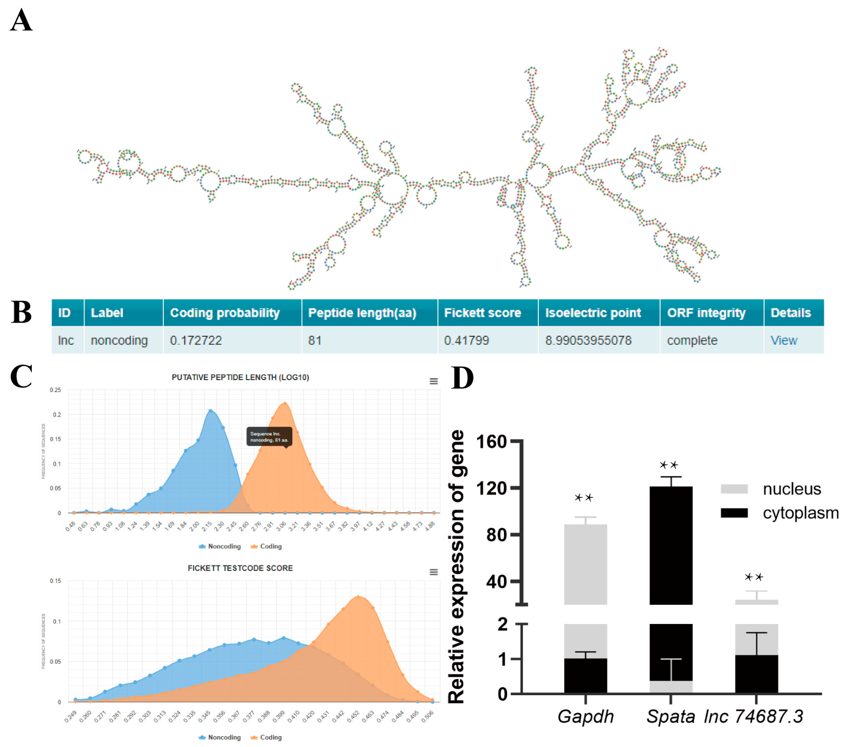
Based on the 1648 nt full-length sequence of lnc74687.3 from rainbow trout gonads obtained in our previous study [17], the minimum free energy (MFE) secondary structure was predicted using RNAfold (default parameters). The results showed that lnc74687.3 formed a complex stem–loop structure (MFE = −410.70 kcal/mol), indicating a stable secondary structure (Figure 2A). Coding potential, a key criterion for identifying lncRNAs, was assessed using CPC2.0 Online website, which indicated that lnc74687.3 has no coding capacity (Figure 2B). Sequence conservation analysis using the NCBI database retrieved no homologous sequences, suggesting that lnc74687.3 is weakly conserved. Subcellular localisation of lncRNAs is closely related to their biological functions and potential molecular roles [18]. Therefore, nuclear and cytoplasmic RNAs were separately isolated from gonadal tissues. Using β-actin as an internal reference, RT-PCR was performed to verify the expression of the nuclear marker gene (Gapdh) and the cytoplasmic marker gene (Spata), as well as to assess the relative expression of lnc74687.3. The results showed that lnc74687.3 is predominantly localised in the nucleus (Figure 2C,D). In conclusion, lnc74687.3 is a long non-coding RNA predominantly expressed in the nucleus of rainbow trout gonocytes.

2.3. Co-Expression of miR-15a-5p, Ccne1, and lncRNA74687.3

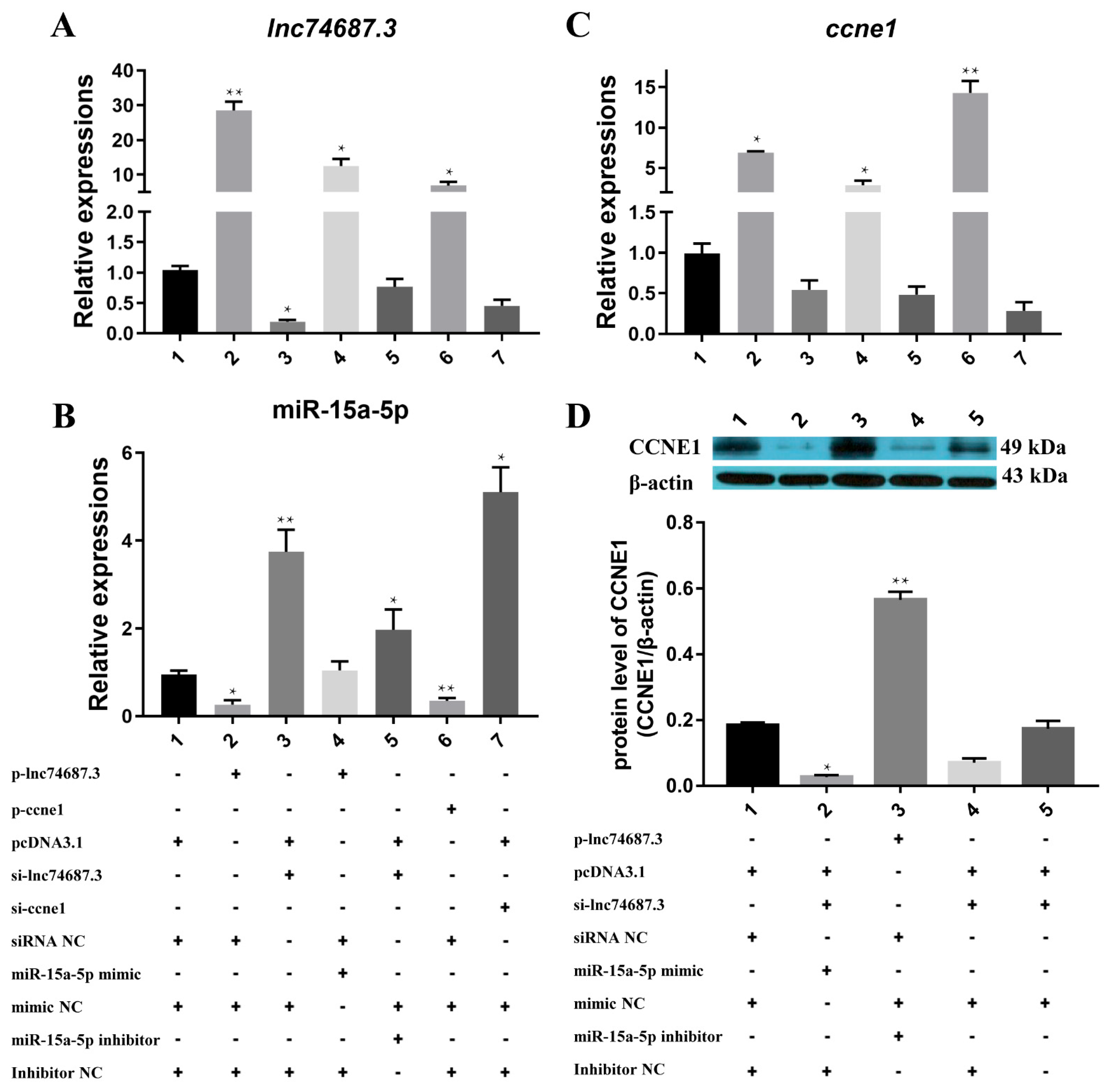
To investigate the relationship between miR-15a-5p, ccne1, and lncRNA74687.3, different combinations of expression vectors (pcDNA3.1, p-ccne1, p-lnc74687.3) and regulatory molecules (siRNA, miRNA mimic/inhibitor, and their controls) were co-transfected into rainbow trout gonadal (RTG-2) cells, and gene expression was assessed by quantitative real-time PCR. Overexpression of lnc74687.3 increased its transcript level by 28.49-fold, decreased miR-15a-5p expression to 0.26-fold, and increased ccne1 expression by 6.93-fold. Conversely, knockdown of lnc74687.3 increased miR-15a-5p expression by 3.74-fold but did not significantly affect ccne1 transcript levels. Co-overexpression of miR-15a-5p and lnc74687.3 resulted in 12.54-fold and 2.91-fold increases in their respective expression levels. Overexpression of ccne1 alone elevated lnc74687.3 expression by 6.86-fold and reduced miR-15a-5p expression to 0.35-fold (Figure 3A–C). The effects of lnc74687.3 and miR-15a-5p manipulation on CCNE1 protein expression were further examined by Western blot (Figure 3D). Knockdown of lnc74687.3 reduced CCNE1 protein to 15% compared with 18% in the control, whereas co-overexpression of lnc74687.3 and miR-15a-5p increased CCNE1 protein to 56%. These findings indicate that lnc74687.3 may alleviate the inhibitory effect on ccne1 and CCNE1 by competitively binding miR-15a-5p.

2.4. lnc74687.3 and Ccne1 Promote Gonocyte Proliferation in Rainbow Trout

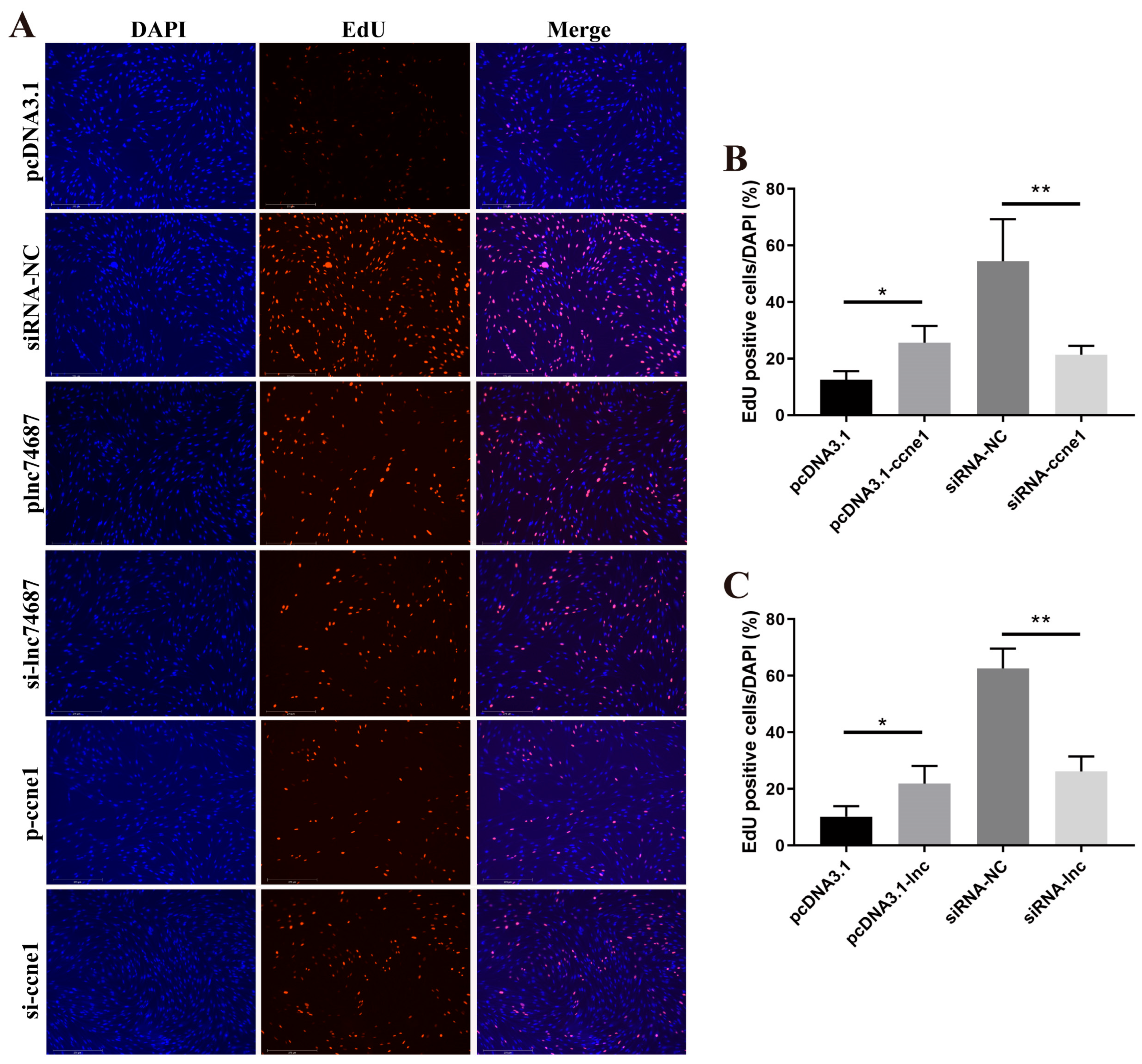
The proliferative effects of lnc74687.3 and ccne1 on rainbow trout gonadal RTG-2 cells were assessed using 5-ethynyl-2′-deoxyuridine (EdU) assays. Overexpression of lnc74687.3 increased the proliferation rate to 21.9%, significantly higher than that of the empty vector control group. In contrast, the proliferation rate in the siRNA-NC control group was 62.6%, whereas lnc74687.3 knockdown significantly reduced it to 26.2% (Figure 4B). Overexpression of ccne1 increased the proliferation rate to 25.6%, while in the siRNA-NC control group, it was 54.5%; knockdown of ccne1 significantly reduced the rate to 21.4% (Figure 4C). In conclusion, lnc74687.3 and ccne1 promote gonadal cell proliferation.

2.5. lnc74687.3 Affects the Expression of Synaptonemal Complex Protein 1 (SYCP1)

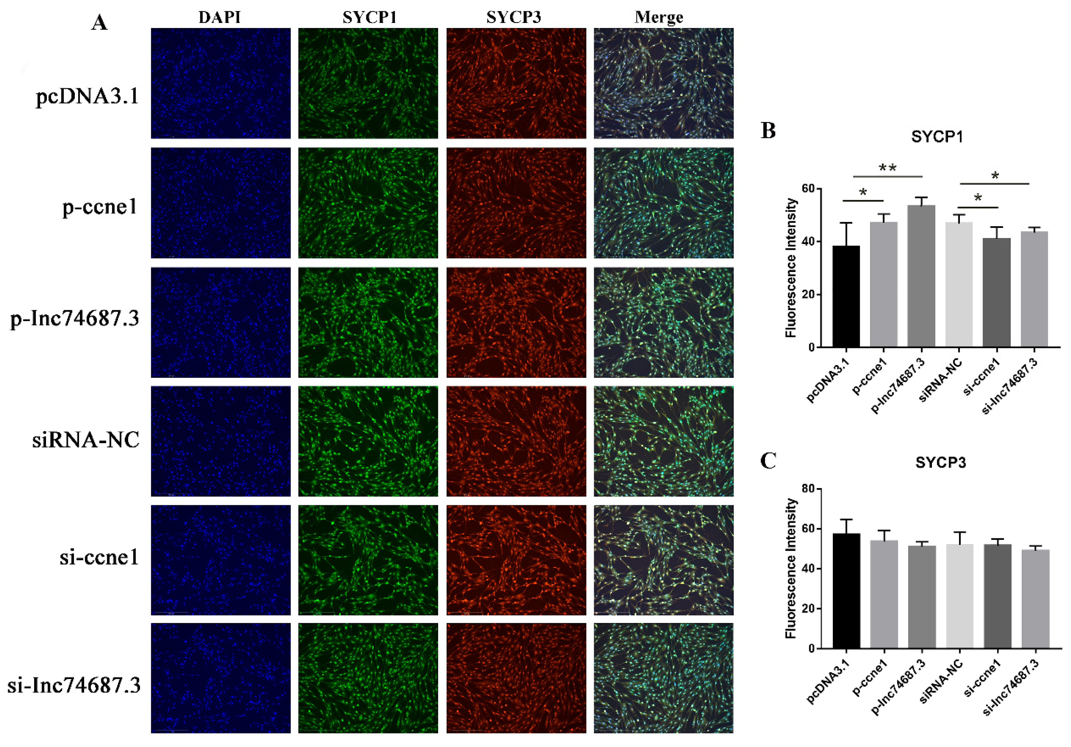
Synaptonemal complexes (SCs) are associated with homologous chromosome synapsis [19], and SYCP1 and SYCP3 serve as markers for meiosis in fish [20]. Using cellular immunofluorescence, we examined the effects of overexpression or knockdown of lnc74687.3 and ccne1 on SYCP1 and SYCP3 protein expression. SYCP1 fluorescence intensity in groups overexpressing ccne1 or lnc74687.3 was significantly higher than in empty vector controls by 1.18-fold and 1.23-fold, respectively. In contrast, inhibition of ccne1 or lnc74687.3 significantly reduced SYCP1 fluorescence intensity in RTG-2 cells relative to the siRNA-NC controls, with the control intensities being 1.18-fold and 1.05 times higher, respectively (Figure 5A). However, neither overexpression nor knockdown of lnc74687.3 or ccne1 affected SYCP3 fluorescence intensity in RTG-2 cells (Figure 5B,C).

2.6. lnc74687.3 Regulates Ccne1 Gene Expression In Vivo by Targeting miR-15a-5p

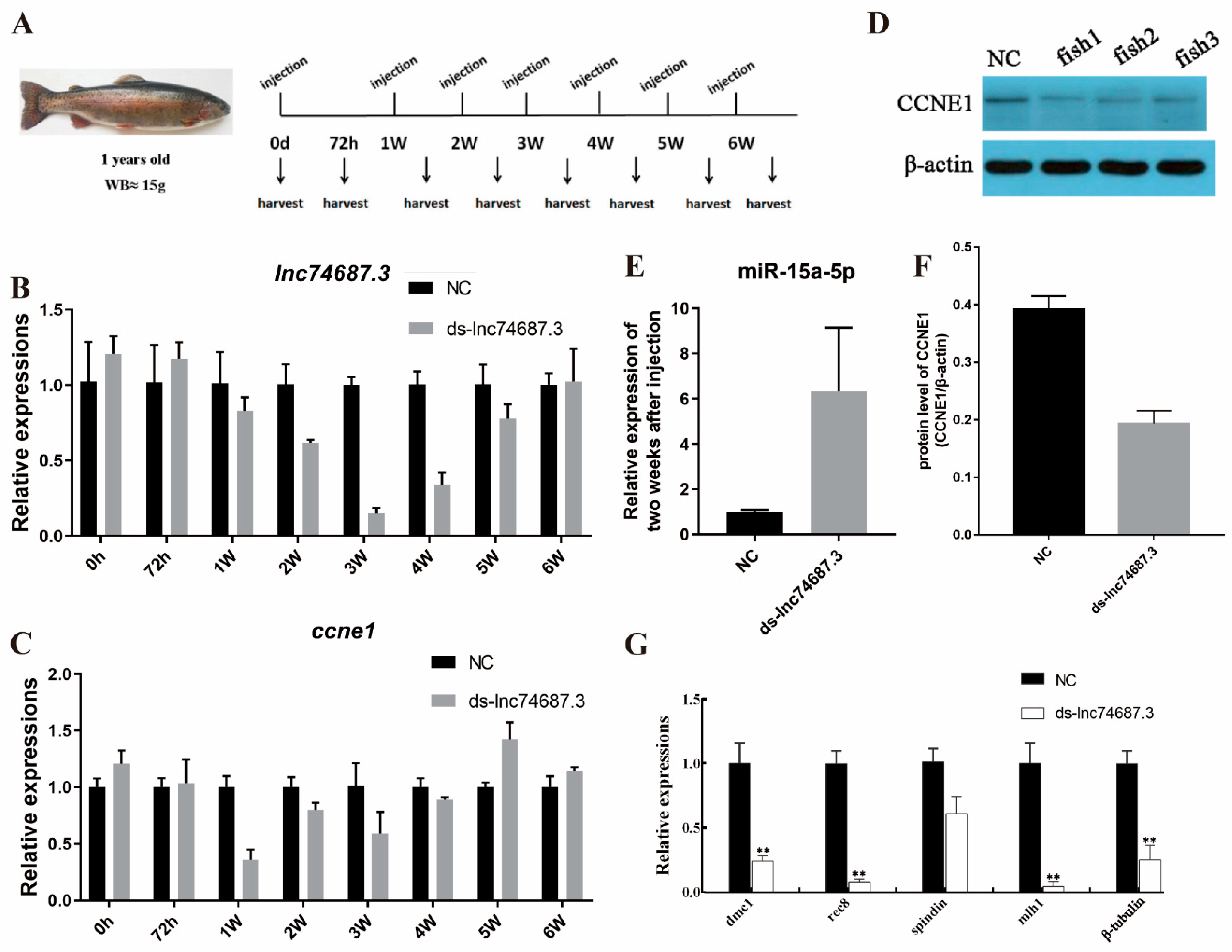
Oocytes in the gonadal tissues of 1-year-old juvenile female rainbow trout are at the pre-minus I diplotene lineage stage or coarser. Double-stranded lnc74687.3 RNA (ds-lnc74687.3) generated by in vitro transcription was administered via seven intraperitoneal injections over six consecutive weeks, with samples collected 72 h after each injection (Figure 6A). Quantitative PCR analysis of gonadal tissue showed that ds-lnc74687.3 significantly reduced lnc74687.3 expression by 1.63-fold, 6.71-fold, and 2.94-fold compared with the control group from weeks 2 to 4 post-injection (Figure 6B). ccne1 mRNA levels were significantly reduced at weeks 1 and 2 post-injection, decreasing by 2.73-fold and 1.25-fold, respectively, but increased to 1.4 times the control level at week 5 (Figure 6C). Western blot analysis showed that knockdown of lnc74687.3 significantly reduced CCNE1 protein levels in gonadal tissues, with CCNE1 expression in the control group being 2.03 times higher than in the knockdown group (Figure 6D,F). Knockdown of lnc74687.3 also significantly increased miR-15a-5p expression in gonadal tissues (Figure 6E). In conclusion, at the organismal level, lnc74687.3 regulates ccne1 mRNA and CCNE1 protein expression in rainbow trout gonads by targeting miR-15a-5p.
To further verify whether lnc74687.3 influences gonadal meiosis in rainbow trout, we examined the expression levels of the meiosis-related genes DNA meiotic recombinase 1 (dmc1), recombination 8 (rec8), spindlin family member (spindlin), β-tubulin, and MutL homolog 1 (mlh1) in gonadal tissues after somatic knockdown of lnc74687.3. Knockdown markedly reduced the expression of mlh1, rec8, dmc1, and β-tubulin to 0.04, 0.08, 0.22, and 0.26, respectively, relative to the controls, while spindlin1 expression showed only a moderate decrease to 0.59 (Figure 6G).

3. Discussion

3.1. lnc74687.3 and Ccne1 Are Target Genes of miR-15a-5p

Research on fish fertility control has primarily focused on histology, cytology, and reproductive endocrinology. Key genes such as cytochrome P450 family 19 subfamily A polypeptide 1a (cyp19a1a), anti-Müllerian hormone (amh), forkhead box L2a (foxl2a), and doublesex and mab-3-related transcription factor 1 (dmrt1) play central roles in reproductive regulation [20,21,22]. Studies on the role of lncRNAs in fish reproduction remain limited, with existing work largely confined to preliminary screening and validation [23,24,25], and no in-depth studies have been conducted to systematically analyse the molecular mechanism of fish lncRNA. Our previous comparative transcriptome analysis of diploid and triploid rainbow trout gonads led to the first prediction and identification of miRNA–mRNA/lncRNA interaction networks potentially involved in gonadal development, including several key lncRNAs that may regulate gonadal cell proliferation and meiosis [17]. Building on these findings, the present study focused on verifying the biological functions of ccne1 and lnc74687.3 as miR-15a-5p target genes, and systematically elucidating the molecular mechanisms of the ccne1lnc74687.3–miR-15a-5p regulatory network in rainbow trout reproductive development, through multi-dimensional experiments at both the cellular and organismal levels.

3.2. lnc74687.3 as a Nuclear Non-Coding RNA May Be Involved in Transcriptional Regulation

Although significant progress has been made in whole-genome and transcriptome sequencing and in the screening of lncRNAs in rainbow trout, experimental validation of lncRNA function remains limited. Classical molecular biology techniques still play an irreplaceable role in lncRNA functional verification [26]. In this study, lnc74687.3, a key regulator of reproductive development in rainbow trout, was found to have a unique secondary structure comprising three main domains: a multifurcated head, an intermediate trunk, and a re-furcated tail. It is localised in the nucleus of gonadal cells and is non-coding, suggesting a potential role in transcriptional regulation. The structure and subcellular localization of an lncRNA can reflect its expression site and coding potential. A subset of lncRNAs is distributed in the nucleus and chromatin, where they can interact with DNA, RNA, and proteins to regulate chromosome structure and function [19,27], or influence mRNA splicing, stability, and translation by modulating gene transcription [28].

3.3. lnc74687.3 Affects Germ-Cell Meiotic Progression by Regulating Cell Cycle- and Meiosis-Related Protein Expression in Gonadal Cells

In multicellular organisms, precise regulation of cell division and differentiation is essential for organ function [29,30]. Regulation of cell proliferation during gonadal development, from the early to mature stages, was found to be particularly critical, with the cell cycle regulator CCNE1 playing a central role in coordinating cell cycle progression and maintaining normal cell division [13]. Since both lnc74687.3 and ccne1 influenced gonadal cell proliferation, it was inferred that lnc74687.3 may affect rainbow trout fertility by modulating gonadal cell proliferation. Abnormal gonadal development disrupts meiosis, resulting in impaired gamete production. The transverse filament protein SYCP1 is essential for meiotic recombination and SC assembly, both of which are key to meiosis [31,32]. Knockdown of lnc74687.3 significantly reduced SYCP1 expression in RTG-2 cells, suggesting that lnc74687.3 may influence germ-cell meiotic progression by regulating the expression of key meiotic proteins, thereby affecting gametogenesis.

3.4. lnc74687.3 Affects Gamete Production by Regulating the miR-15a-5p/ccne1/CCNE1 Pathway

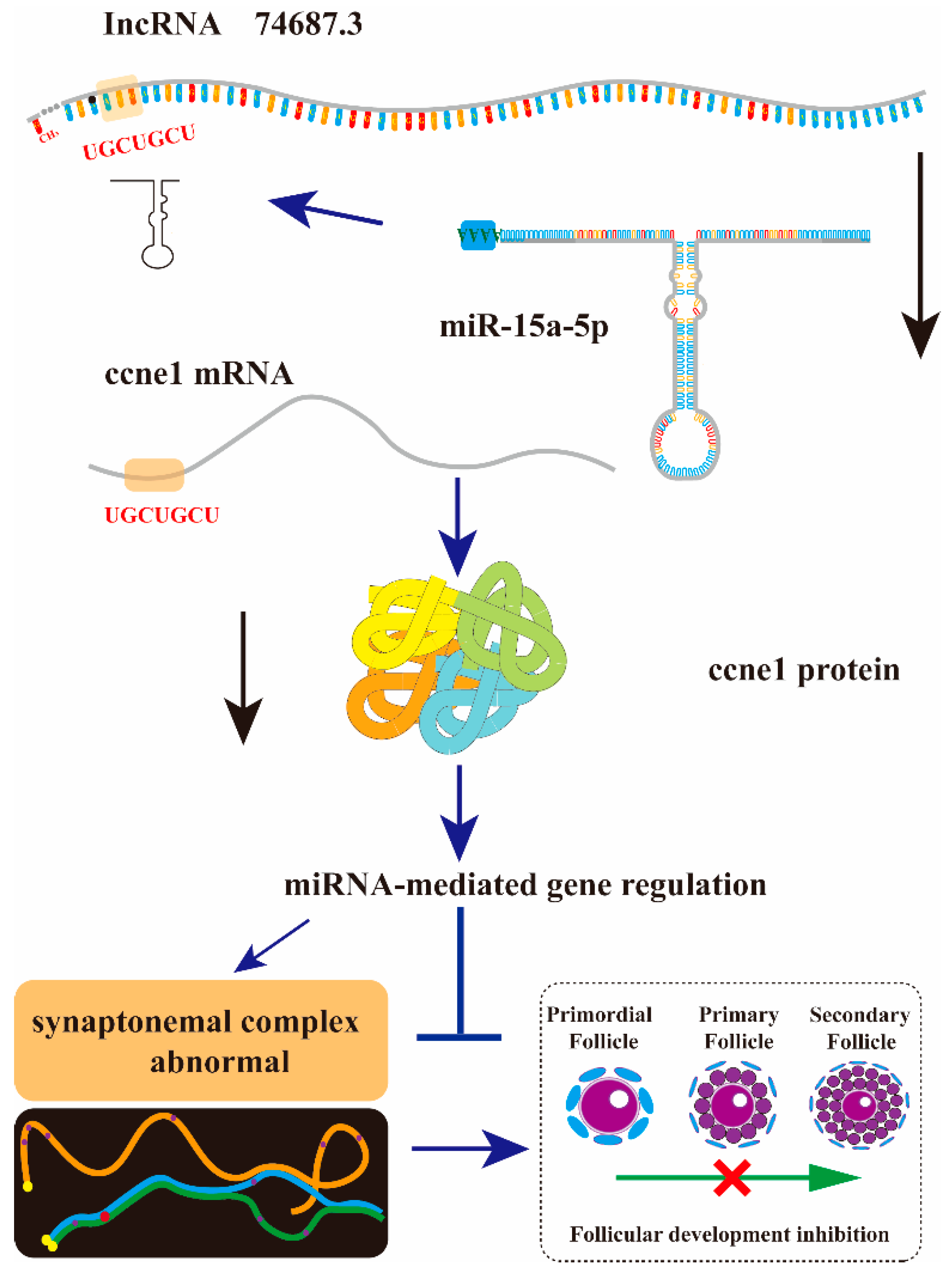
The gonads of rainbow trout at one year of age are in an early developmental stage, during which primordial germ cells (PGCs) differentiate into oogonia and undergo an asynchronous transition from mitosis to meiosis [33]. Knockdown of lnc74687.3 in rainbow trout resulted in an abnormal increase in ccne1 expression at week 5, which may reflect a conserved mechanism for initiating and maintaining gene expression during development. As an important regulator of chromosome behaviour in meiosis, the dmc1 gene encodes a recombinase essential for synapsis [8]. Rec8 localises to the axial core of meiotic chromosomes, where it mediates cohesion between sister chromatids and is critical for homologous chromosome pairing [34,35]. As part of the DNA mismatch repair machinery, mlh1 is essential for maintaining chromosome structure during the first meiotic division [36]. Abnormal meiosis in rainbow trout ovaries may also be linked to altered expression of Spindlin-1 [37]. Additionally, the microtubule protein β-tubulin affects meiosis by influencing spindle formation, with its dimer structure serving as an essential spindle component [38]. Our previous research found that CCNE1 balance meiosis and gamete development through specific regulatory mechanisms, and their dysregulation may be a key factor underlying meiosis inhibition and reproductive development abnormalities [39]. In vivo validation confirmed our cellular findings: knockdown of lnc74687.3 increased miR-15a-5p expression, while ccne1 mRNA and CCNE1 protein were significantly reduced due to miR-15a-5p activity. Reduced expression of dmc1, rec8, mlh1, and β-tubulin correlated with a marked decrease in SYCP1 signal intensity in RTG-2 cells. Collectively, lnc74687.3 regulates meiosis in gonadal cells via the miR-15a-5p/ccne1/CCNE1 pathway, thereby inhibiting cell proliferation and ultimately reducing gamete production (Figure 7).

4. Materials and Methods

4.1. Ethics Statement

All animal experiments were approved by the Ethics Committee for Animal Experiments of the Heilongjiang River Fisheries Research Institute, Chinese Academy of Fisheries Sciences (Approval No. 20250305-001), and conducted in strict compliance with the Guidelines for Ethical Review of Laboratory Animal Welfare in China.

4.2. Animals

The rainbow trout used in this study were supplied by the Bohai Sea Cold Water Fish Experimental Station (Mudanjiang), Heilongjiang River Fisheries Research Institute, Chinese Academy of Fisheries Sciences. All fish were acclimated for two weeks before the experiment. They were reared in a 1 m3 aquarium with a recirculating water system, with water temperature maintained at 10 ± 0.5 °C, dissolved oxygen at 7.8–10.0 mg/L, and pH at 7.2–7.5.
In vivo injection experiments were conducted using 48 one-year-old rainbow trout, divided into control and knockdown groups, housed separately in the same aquarium. Fish were anaesthetised with MS-222 (250 mg/L) before each injection to prevent pain. After the start of the experiment, three individuals from each group were randomly selected weekly for euthanasia and sampling. All euthanasia procedures involved MS-222 (250 mg/L) anaesthesia followed by bloodletting to ensure a painless death. Collected tissue samples were immediately frozen in liquid nitrogen and then stored at −80 °C.

4.3. Cell Culture

Rainbow trout gonadal cells (RTG-2) and human embryonic kidney epithelial cells (293T) were maintained in our laboratory. RTG-2 cells were cultured in Minimum Essential Medium (MEM, Sigma, St. Louis, MO, USA) supplemented with 10% foetal bovine serum (FBS, Zeta Life, Adams Drive, CA, USA), 100 U/mL penicillin, and 100 µg/mL streptomycin (Biosharp, Hefei, China) at 18 °C in a low-temperature incubator with 5% CO2 and 21% O2. 293T cells were cultured in Dulbecco’s Modified Eagle Medium (DMEM, Sigma, St. Louis, MO, USA) supplemented with 10% FBS (Zeta Life, Adams Drive, CA, USA), 100 U/mL penicillin, and 100 µg/mL streptomycin (NCM Biotech, Suzhou, China) at 37 °C in an incubator with 5% CO2 and 21% O2. RTG-2 and 293T cells reached 80–90% confluence after 48 h and 24 h, respectively, and were passaged using 0.25% trypsin (NCM Biotech, Suzhou, China).

4.4. Dual-Luciferase Reporter Assays

The potential binding sites of miR-15a-5p in the ccne1 gene and lncRNAs (lnc76205.1, lnc30919.1, and lnc74687.3) were predicted using TargetScan (https://www.targetscan.org/vert_42/, accessed 15 December 2019). Based on these predictions, primers containing the binding sites were designed with XhoI and SalI restriction sites at both ends, and the amplified fragments were cloned into pmirGLO dual-luciferase reporter vectors to generate wild-type and mutant constructs for ccne1 and each lncRNA. Small interfering RNAs (siRNAs) targeting miR-15a-5p, ccne1, lnc76205.1, lnc30919.1, and lnc74687.3 were synthesised by Jimagene. At 48 h post-transfection, double luciferase activity was measured using the dual-luciferase reporter assay system (Promega, Madison, WI, USA), and relative fluorescence activity was calculated as the ratio of firefly luciferase to Renilla luciferase activity. Target genes of miR-15a-5p were confirmed by comparing the relative fluorescence activity changes across reporter constructs.

4.5. Subcellular Fractionation

Subcellular RNA isolation was performed under clean conditions using a Cytoplasmic and Nuclear Fractionation RNA Purification Kit (Norgen, Thorold, ON, Canada). Briefly, gonadal tissues frozen at −80 °C were ground in liquid nitrogen, homogenised in Lysis Buffer J, and centrifuged at 14,000× g for 10 min. The supernatant (cytoplasmic fraction) and pellet (nuclear fraction) were separated. Buffer SK and anhydrous ethanol were added to each fraction, combined, washed, and eluted through silica gel membrane columns to obtain high-purity cytoplasmic and nuclear RNA. The subcellular distribution of lnc74687.3 was quantified by RT-qPCR.

4.6. RNA Extraction and Real-Time PCR Analysis

Total RNA from tissues was extracted using a Simply P Total RNA Extraction Kit (BioFlux, San Francisco, CA, USA) following the manufacturer’s instructions. cDNA was synthesised using the BioFlux reverse transcription kit (BioFlux, San Francisco, CA, USA). Gene expression was quantified by RT-qPCR using SYBR Green (NovaBio, Shanghai, China) according to the manufacturer’s protocol: 95 °C for 10 min; 39 cycles of 95 °C for 10 s and 60 °C for 30 s; and a melting curve from 65 °C to 95 °C with increments of 0.5 °C/s. Relative gene expression was calculated using the 2−ΔΔCt method, with β-actin as the internal reference. Specific primers were designed using Primer Premier 5 (Table 1).

4.7. Western Blot Analysis

Cell or tissue extracts were mixed with 2× SDS-PAGE loading buffer (Beyotime Shanghai, China), boiled for 10 min, and separated by SDS-PAGE, followed by transfer to PVDF membranes (Biosharp, Hefei, China). After blocking for 1.5 h in tris-buffered saline with tween-20 (TBST) containing 5% BSA and 0.5% Triton X-100, the membranes were incubated with primary antibodies against CCNE1 (1:10,000 in TBST; ABclonal Technology, Wuhan, China) and β-actin (1:10,000 in TBST; Immunoway, Plano, TX, USA). Goat anti-rabbit IgG secondary antibody (1:2000 in TBST; Cell Signalling Technology, Danvers, MA, USA) was then applied. After each step, the membranes were washed five times with TBST. All incubation and washing solutions fully covered the PVDF membranes. Protein bands were visualised using an Odyssey imaging system (LI-COR Inc., Lincoln, NE, USA) and quantified with ImageJ.

4.8. Cell Proliferation Assay

Cell proliferation was assessed using an EdU Cell Proliferation Kit with Alexa Fluor 555 (Beyotime, Shanghai, China) following the manufacturer’s instructions with minor optimisation. Briefly, cells were incubated overnight with EdU working solution 48 h after transfection, fixed in 4% paraformaldehyde for 30 min at room temperature, permeabilised with 0.5% Triton X-100 in PBS for 15 min, and reacted with Click reaction solution (containing Alexa Fluor 555) for 30 min in the dark. Nuclei were counterstained with Hoechst (Beyotime, Shanghai, China) for 5 min. Between steps, cells were washed three times with PBS (5 min each). Images were captured under a fluorescence microscope. EdU-positive cells were quantified using ImageJ, and proliferation rates were calculated using Hoechst-stained nuclei as a reference.

4.9. Bioinformatics Analysis

Minimum free energy secondary structures were predicted using RNAfold (http://rna.tbi.univie.ac.at/, accessed 27 May 2024) with default parameters. The coding potential of lncRNAs was assessed using CPC2 software (http://cpc2.cbi.pku.edu.cn/, accessed 27 May 2024).

4.10. Statistical Analysis

All independent experiments included at least three parallel groups. Data were analysed and plotted using IBM SPSS Statistics v27.0 (IBM, Armonk, NY, USA) and GraphPad Prism v8.0.2 (GraphPad Software, San Diego, CA, USA). The results are presented as the mean ± standard deviation. Comparisons between two groups were performed using unpaired t-tests, and comparisons among multiple groups were performed using one-way ANOVA.

5. Conclusions

In summary, we report the first identified mechanism of lncRNA action in fish. In rainbow trout, lnc74687.3 is a long non-coding RNA localised in the nucleus of gonadal cells. lnc74687.3 regulates ccne1 mRNA and CCNE1 protein expression by targeting miR-15a-5p, thereby influencing cell proliferation and meiosis. This lncRNA is a potential key factor in regulating meiosis and gonadal development in rainbow trout gonadal cells. The validation of this key lncRNA regulatory factor provides a target element for future in-depth research into rainbow trout reproductive capacity and lays crucial groundwork for understanding targeted regulatory mechanisms and advancing reproductive regulation technology.

Author Contributions

T.H. designed and performed the experiments; R.X., E.L. and Y.S. analysed the data and checked the manuscript; K.G., W.G. and G.W. cultured and sampled the fish; B.C., D.L. and T.H. drafted the article; P.F. and G.X. reviewed the manuscript. All authors contributed to the manuscript at various stages. All authors have read and agreed to the published version of the manuscript.

Funding

This study was supported by the STI2030—Major Projects (2023ZD0405505), China Agriculture Research System of MOF and MARA (CARS-46), National Natural Science Foundation of China (32403019), and Natural Science Foundation Project of Heilongjiang Province (LH2023C054). The funding agencies played no part in the study design, data collection and analysis, decision to publish the manuscript, or manuscript preparation.

Institutional Review Board Statement

All experiments in this study were approved by the Animal Husbandry Department of the Heilongjiang Animal Care and Use Committee. All fish involved in this research were bred following the guidelines of the Animal Husbandry Department of Heilongjiang, China.

Data Availability Statement

All data generated or analysed during this study are included in this article.

Conflicts of Interest

The authors declare no conflicts of interest.

References

  1. Zhao, X.F.; Liang, L.Q.; Liew, H.J.; Chang, Y.M.; Sun, B.; Wang, S.Y.; Mi, B.H.; Zhang, L.M. Identification and analysis of long non-coding RNAs in Leuciscus waleckii adapted to highly alkaline conditions. Front. Physiol. 2021, 12, 665268. [Google Scholar] [CrossRef]
  2. Ning, X.; Sun, L. Identification and characterization of immune-related lncRNAs and lncRNA-miRNA-mRNA networks of Paralichthys olivaceus involved in Vibrio anguillarum infection. BMC Genom. 2021, 22, 447. [Google Scholar]
  3. Hao, R.; Zhu, X.; Tian, C.; Jiang, M.; Huang, Y.; Li, G.; Zhu, C. LncRNA–miRNA–mRNA ceRNA network of different body colors in Plectropomus leopardus. Front. Mar. Sci. 2023, 10, 1170762. [Google Scholar]
  4. Li, L.; Jia, X.; Liu, Y.; He, Y.; Pang, Y.; Shen, Y.; Xu, X.; Li, J. LncRNA-SUMO3 and lncRNA-HDMO13 modulate the inflammatory response by binding miR-21 and miR-142a-3p in grass carp. Dev. Comp. Immunol. 2021, 121, 104082. [Google Scholar] [PubMed]
  5. Deng, Q.; Zhao, N.; Zhu, C.; Zhang, B. Long non-coding RNAs in the physiology of aquaculture animals: A perspective update. Rev. Fish Biol. Fish. 2022, 32, 1103–1122. [Google Scholar] [CrossRef]
  6. Mahajan, A.; Hong, J.; Krukovets, I.; Shin, J.; Tkachenko, S.; Espinosa-Diez, C.; Owens, G.K.; Cherepanova, O.A. Integrative analysis of the lncRNA-miRNA-mRNA interactions in smooth muscle cell phenotypic transitions. Front. Genet. 2024, 15, 1356558. [Google Scholar]
  7. He, Y.; Zhou, H.; Wang, W.; Xu, H.; Cheng, H. Construction of a circRNA-miRNA-mRNA Regulatory Network Reveals Potential Mechanism and Treatment Options for Osteosarcoma. Front. Genet. 2021, 12, 632359. [Google Scholar]
  8. Qin, Q.; Zhou, Y.; Wang, C.; Zhang, M.; Qin, H.; Zhao, C.; Liu, S. Analysis on the meiosis-related gene (Dmc1, Ph1) expression in autotriploid Carassius auratus. Mar. Biotechnol. 2019, 21, 753–761. [Google Scholar] [CrossRef]
  9. Wang, H.; Yu, H.; Li, Q.; Liu, S. Transcription analysis for core networks of lncRNAs–mRNAs: Implication for potential role in sterility of Crassostrea gigas. Biology 2022, 11, 378. [Google Scholar]
  10. Zhang, C.; Li, Q.; Zhu, L.; He, W.; Yang, C.; Zhang, H.; Sun, Y.; Zhou, L.; Sun, Y.; Zhu, S. Abnormal meiosis in fertile and sterile triploid cyprinid fish. Sci. China Life Sci. 2021, 64, 1917–1928. [Google Scholar] [CrossRef]
  11. Honda, R.; Tanaka, H.; Yasuda, H. Oncoprotein MDM2 is a ubiquitin ligase E3 for tumor suppressor p53. FEBS Lett. 1997, 420, 25–27. [Google Scholar] [CrossRef] [PubMed]
  12. Aziz, K.; Limzerwala, J.F.; Sturmlechner, I.; Hurley, E.; Zhang, C.; Jeganathan, K.B.; Nelson, G.; Bronk, S.; Velasco, R.O.F.; van Deursen, E.; et al. Ccne1 Overexpression Causes Chromosome Instability in Liver Cells and Liver Tumor Development in Mice. Gastroenterology 2019, 157, 210–226. [Google Scholar] [CrossRef] [PubMed]
  13. Zhang, S.; Pang, Z.; Gao, J.; Dai, Q.; Liu, X.; Shen, Y.; Baloch, W.A.; Noonari, S.; Wang, P.; Gao, H. Functional analysis of the cell cycle protein E gene (ccne) in ovarian development of the white ridgetail prawn, Exopalaemon carinicauda. Aquac. Rep. 2023, 32, 101716. [Google Scholar] [CrossRef]
  14. Chen, L.; Miao, X.; Si, C.; Qin, A.; Zhang, Y.; Chu, C.; Li, Z.; Wang, T.; Liu, X. Long non-coding RNA SENP3-eif4a1 functions as a sponge of miR-195-5p to drive triple-negative breast cancer progress by overexpressing CCNE1. Front. Cell Dev. Biol. 2021, 9, 647527. [Google Scholar]
  15. Bao, S.; Zhuo, L.; Qi, D.; Tian, H.; Wang, D.; Zhu, B.; Meng, Y.; Ma, R. Comparative study on the fillet nutritional quality of diploid and triploid rainbow trout (Oncorhynchus mykiss). Aquac. Rep. 2023, 28, 101431. [Google Scholar]
  16. Nynca, J.; Malinowska, A.; Świderska, B.; Wiśniewska, J.; Dobosz, S.; Ciereszko, A. Triploidization of rainbow trout affects proteins related to ovary development and reproductive activity. Aquaculture 2023, 565, 739145. [Google Scholar]
  17. Huang, T.; Gu, W.; Liu, E.; Shi, X.; Wang, B.; Wu, W.; Dong, F.; Xu, G. Comprehensive analysis of miRNA-mRNA/lncRNA during gonadal development of triploid female rainbow trout (Oncorhynchus mykiss). Genomics 2021, 113, 3533–3543. [Google Scholar]
  18. Iwai, T.; Yoshii, A.; Yokota, T.; Sakai, C.; Hori, H.; Kanamori, A.; Yamashita, M. Structural components of the synaptonemal complex, SYCP1 and SYCP3, in the medaka fish Oryzias latipes. Exp. Cell Res. 2006, 312, 2528–2537. [Google Scholar] [CrossRef]
  19. Zhang, Y.; Wang, W.; Duan, C.; Li, M.; Gao, L. Novel Long Non-Coding RNA (lncRNA) Transcript AL137782. 1 Promotes the Migration of Normal Lung Epithelial Cells through Positively Regulating LMO7. Int. J. Mol. Sci. 2023, 24, 13904. [Google Scholar]
  20. Liebermann, J.; Tucker, M.J. Effect of carrier system on the yield of human oocytes and embryos as assessed by survival and developmental potential after vitrification. Reproduction 2002, 124, 483–489. [Google Scholar] [CrossRef]
  21. Brown, M.S.; Evans, B.S.; Afonso, L.O. Developmental changes in gene expression and gonad morphology during sex differentiation in Atlantic salmon (Salmo salar). Gene 2022, 823, 146393. [Google Scholar] [CrossRef]
  22. Zhang, X.; Min, Q.; Li, M.; Liu, X.; Li, M.; Wang, D. Mutation of cyp19a1b results in sterile males due to efferent duct obstruction in Nile tilapia. Mol. Reprod. Dev. 2019, 86, 1224–1235. [Google Scholar]
  23. Qi, S.; Dai, S.; Zhou, X.; Wei, X.; Chen, P.; He, Y.; Kocher, T.D.; Wang, D.; Li, M. Dmrt1 is the only male pathway gene tested indispensable for sex determination and functional testis development in tilapia. PLoS Genet. 2024, 20, e1011210. [Google Scholar]
  24. Yan, D.; Long, X.; Zhang, X.; Dong, X.; Wang, Z.; Jiang, H.; An, M.; Chen, J.; Gan, L. Identification and characterization of long non-coding RNAs in intestinal immune regulation of largemouth bass, Micropterus salmoides, under acute heat stress. Comp. Biochem. Physiol. Part D Genom. Proteom. 2023, 48, 101132. [Google Scholar]
  25. Li, Y.; Yang, B.; Shi, C.; Tan, Y.; Ren, L.; Mokrani, A.; Li, Q.; Liu, S. Integrated analysis of mRNAs and lncRNAs reveals candidate marker genes and potential hub lncRNAs associated with growth regulation of the Pacific Oyster, Crassostrea gigas. BMC Genom. 2023, 24, 453. [Google Scholar]
  26. Fan, K.; Shen, Y.; Xu, X.; Tao, L.; Bao, T.; Li, J. LncRNA-WAS and lncRNA-C8807 interact with miR-142a-3p to regulate the inflammatory response in grass carp. Fish Shellfish Immunol. 2021, 111, 201–207. [Google Scholar]
  27. Dykes, I.M.; Emanueli, C. Transcriptional and post-transcriptional gene regulation by long non-coding RNA, Genomics. Proteom. Bioinform. 2017, 15, 177–186. [Google Scholar]
  28. Statello, L.; Guo, C.; Chen, L.; Huarte, M. Gene regulation by long non-coding RNAs and its biological functions. Nat. Rev. Mol. Cell Biol. 2021, 22, 96–118. [Google Scholar] [CrossRef]
  29. Padilla, L.F.A.; Castañeda-Cortés, D.C.; Rosa, I.F.; Acosta, O.D.M.; Hattori, R.S.; Nóbrega, R.H.; Fernandino, J.I. Cystic proliferation of germline stem cells is necessary to reproductive success and normal mating behavior in medaka. eLife 2021, 10, e62757. [Google Scholar]
  30. Marinović, Z.; Lujić, J.; Bajec, S.S.; Djurdjevič, I.; Snoj, A.; Hoitsy, G.; Urbányi, B.; Horváth, Á. Evaluation of triploid rainbow trout Oncorhynchus mykiss as a surrogate parent for brown trout Salmo trutta m. fario and grayling Thymallus thymallus. Aquac. Rep. 2022, 24, 101163. [Google Scholar] [CrossRef]
  31. Imai, Y.; Saito, K.; Takemoto, K.; Velilla, F.; Kawasaki, T.; Ishiguro, K.; Sakai, N. Sycp1 is not required for subtelomeric DNA double-strand breaks but is required for homologous alignment in zebrafish spermatocytes. Front. Cell Dev. Biol. 2021, 9, 664377. [Google Scholar] [CrossRef]
  32. Liang, M.; Wang, H.; He, C.; Zhang, K.; Hu, K. LncRNA-Gm2044 is transcriptionally activated by a-MYB and regulates Sycp1 expression as a miR-335-3p sponge in mouse spermatocyte-derived GC-2spd (ts) cells. Differentiation 2020, 114, 49–57. [Google Scholar]
  33. Garcia-Alonso, L.; Lorenzi, V.; Mazzeo, C.I.; Alves-Lopes, J.P.; Roberts, K.; Sancho-Serra, C.; Engelbert, J.; Marečková, M.; Gruhn, W.H.; Botting, R.A. Single-cell roadmap of human gonadal development. Nature 2022, 607, 540–547. [Google Scholar] [CrossRef]
  34. Wang, Q.; Lin, F.; He, Q.; Huang, Q.; Duan, X.; Liu, X.; Xiao, S.; Yang, H.; Zhao, H. Cloning and characterization of rec8 gene in orange-spotted grouper (Epinephelus coioides) and Dmrt1 regulation of rec8 promoter activity. Fish Physiol. Biochem. 2021, 47, 393–407. [Google Scholar] [CrossRef]
  35. Lin, F.; Tong, F.; He, Q.; Xiao, S.; Liu, X.; Yang, H.; Guo, Y.; Wang, Q.; Zhao, H. In vitro effects of androgen on testicular development by the AR-foxl3-rec8/fbxo47 axis in orange-spotted grouper (Epinephelus coioides). Gen. Comp. Endocrinol. 2020, 292, 113435. [Google Scholar] [PubMed]
  36. Leal, M.C.; Feitsma, H.; Cuppen, E.; França, L.R.; Schulz, R.W. Completion of meiosis in male zebrafish (Danio rerio) despite lack of DNA mismatch repair gene mlh1. Cell Tissue Res. 2008, 332, 133–139. [Google Scholar] [CrossRef] [PubMed]
  37. Wang, F.; Guo, F.; Ma, W. Abnormal expression of miR-1388-5p and its target spindlin-1 in female triploid rainbow trout (Oncorhynchus mykiss). Aquac. Rep. 2020, 18, 100420. [Google Scholar]
  38. Needleman, D. Developing cell biology. eLife 2013, 2, e00571. [Google Scholar]
  39. Liu, E.; Song, H.; Gu, W.; Wang, G.; Fan, P.; Ge, K.; Sun, Y.; Li, D.; Xu, G.; Huang, T. Functional Analysis of the Cyclin E Gene in the Reproductive Development of Rainbow Trout (Oncorhynchus mykiss). Biology 2025, 14, 862. [Google Scholar] [CrossRef]
Figure 1. Construction of dual-luciferase vectors and analysis of relative light units. (A) Target sites in ccne1, lnc76205.1, lnc30919.1, and lnc74687.3 wild-type and mutant sequences (red words). (B) Structure of pmirGLO-sequence-WT/MUT plasmid. (CF) Relative fluorescence activity of ccne1, lnc76205.1, lnc30919.1, and lnc74687.3 transfected into 293T cells. Data are expressed as mean ± SD (n = 3), with differences indicated by asterisks (** p < 0.01, * p < 0.05).
Ijms 26 08036 g001
Figure 2. Structure and expression distribution of lnc74687.3. (A) Predicted secondary structure. (B,C) Coding capacity prediction. (D) Expression of Gapdh, Spata, and lnc74687.3 in nucleus and cytoplasm of gonadal cells. Data are expressed as mean ± SD (n = 3), with differences indicated by asterisks (** p < 0.01).
Ijms 26 08036 g002
Figure 3. Co-expression analysis of miR-15a-5p, ccne1, and lncRNA74687.3. (AC) Expression levels of lnc74687.3, ccne1, and miR-15a-5p under different combinations, with lower left table showing reagent combinations corresponding to horizontal axis (+, reagent added; -, reagent not added). (D) CCNE1 protein expression levels, with lower right table showing protein expression combinations (+, reagent added; -, reagent not added). Data are expressed as mean ± SD (n = 3), with differences indicated by asterisks (** p < 0.01, * p < 0.05).
Ijms 26 08036 g003
Figure 4. 5-ethynyl-2′-deoxyuridine (EdU) assay of effect of ccne1 on proliferative capacity of rainbow trout gonadal (RTG)-2 cells. (A) EdU results for RTG-2 cells under different transfection conditions. (B) Quantification of lnc74687.3’s effects on proliferation by ImageJ (ImageJ, 1.53q). (C) Quantification of ccne1’s effects on proliferation by ImageJ. Data are expressed as mean ± SD (n = 3), with differences indicated by asterisks (** p < 0.01, * p < 0.05).
Ijms 26 08036 g004
Figure 5. Cellular immunofluorescence detection of CCNE1, and lnc74687.3’s effects on expression of Synaptonemal Complex Protein 1 (SYCP1) and Synaptonemal Complex Protein 3 (SYCP3). (A) Immunofluorescence images of SYCP1 and SYCP3 in RTG-2 cells under different transfection conditions. (B) Quantification of SYCP1 fluorescence intensity by ImageJ. (C) Quantification of SYCP3 fluorescence intensity by ImageJ. Data are expressed as mean ± SD (n = 3), with differences indicated by asterisks (** p < 0.01, * p < 0.05).
Ijms 26 08036 g005
Figure 6. lnc74687.3 targeting miR-15a-5p at the organismal level affects ccne1 and CCNE1 expression. (A) Injection and sampling scheme; (B) lnc74687.3 expression in rainbow trout gonads at different time points; (C) ccne1 expression in rainbow trout gonads at different time points; (D) CCNE1 protein expression levels at weeks 1, 2, and 5; (E) miR-15a-5p expression two weeks after lnc74687.3 knockdown; (F) CCNE1 protein expression levels; (G) effect of lnc74687.3 knockdown on meiosis-related genes dmc1, rec8, spindlin, β-tubulin, and mlh1. Data are expressed as mean ± SD (n = 3), with differences indicated by asterisks (** p < 0.01).
Ijms 26 08036 g006
Figure 7. Mechanistic map of the lnc74687.3/miR-15a-5p/ccne1 axis regulating gonocyte meiosis and gonadal development. lnc74687.3 in rainbow trout gonads affects ccne1 and CCNE1 expression by targeting miR-15a-5p, ultimately influencing gonadal cell meiosis and gonadal development.
Ijms 26 08036 g007
Table 1. Oligonucleotide primers used for RT-qPCR.
Primer NameNucleotide Sequence (5′-3′)Accession Number
dmc1-FGCACGAGCGTCAAACATTCCXM 021575970.2
dmc1-RCGACCTCATCCTCCACAACT
rec8b-FGCAGTATGGGGTGGTTATXM 021562658.2
rec8b-RCTCGTCATGGAATTGGTC
mlh1-FCAGGATTGTGGAGGTGGTGAXM 021606940.2
mlh1-RATGGGTGAGAGTTCTTGGGA
β-tubulin-FCGACCCCACTGGCACATAXM 021601756.2
β-tubulin-RCCTCACCACATCCAAAAC
spindling-FCCATACCAGGCGGTGATACAXM 021588921.2
spindling-RCAACCACAGGAGGCTGAAGA
ccne1-FAACGGCAACGTCTGATTTTCLOC 110486705
ccne1-RCGTGGGATATAATGCCAAGC
gapdh-FCATTGAGGGTCTGATGAGCANM 001124209.1
gapdh -RCCTCCACAGCTTTCCAGAAG
spata4-FATTTTGTAAGCGTGTGTGNM 001124526.2
spata4-RTTTGGTTGGAAGTGATGT
lnc74687.3-FCAGGCCTCCATTCCAAATTAMSTRG. 74687
lnc74687.3-RACGTGAGTTGAACACGCAAC
β-actin-FCTCACCGACTACCTGATGAAGATCLOC 100136352
β-actin-RGTAGCACAGCTTCTCCTTGATGTC
miR-15a-5pTAGCAGCACAGAATGGTTTGTGA
Disclaimer/Publisher’s Note: The statements, opinions and data contained in all publications are solely those of the individual author(s) and contributor(s) and not of MDPI and/or the editor(s). MDPI and/or the editor(s) disclaim responsibility for any injury to people or property resulting from any ideas, methods, instructions or products referred to in the content.