Article

Bioactive Properties of Selected European Phellinus Species: A Comprehensive Study

by
Grzegorz Świderski
1,*,
Monika Kalinowska
1,
Ewa Zapora
2,
Marek Wołkowycki
2,
Marcin Stocki
2,
Ewa Ciszkowicz
3,
Aleksandra Bocian
3,
Marcin Jaromin
3,
Mirosław Tyrka
3,
Katarzyna Lecka-Szlachta
3,
Elżbieta Wołejko
1,
Urszula Wydro
1,
Małgorzata Pawłowska
4,
Paweł Golianek
4,
Małgorzata Zawadzka
1,
Qëndrim Ramshaj
5,
Carolina Elena Girometta
6 and
Mitko Karadelev
7
1
Department of Chemistry, Biology and Biotechnology, Bialystok University of Technology, Wiejska 45E, 15351 Białystok, Poland
2
Department of Silviculture and Forest Utilization, Bialystok University of Technology, Wiejska 45E, 15351 Bialystok, Poland
3
Department of Biotechnology and Bioinformatics, Faculty of Chemistry, Rzeszow University of Technology, Powstańców Warszawy 6, 35959 Rzeszow, Poland
4
Faculty of Environmental Engineering, Lublin University of Technology, Nadbystrzycka 40B, 20618 Lublin, Poland
5
Department of Biology, Faculty of Mathematics and Natural Sciences, University of Prishtina, St. “Eqrem Çabej”, 10 000 Prishtina, Kosovo
6
Department of Earth and Environmental Sciences, University of Pavia, Via Sant’Epifanio 14, 27100 Pavia, Italy
7
Institute of Biology, Faculty of Natural Sciences and Mathematics, St. Cyril and Methodius University, Arhimedova 3, 1000 Skopje, North Macedonia
*
Author to whom correspondence should be addressed.
Int. J. Mol. Sci. 2025, 26(16), 8013; https://doi.org/10.3390/ijms26168013
Submission received: 8 July 2025 / Revised: 29 July 2025 / Accepted: 15 August 2025 / Published: 19 August 2025
(This article belongs to the Special Issue Molecular Insight into Plant Bioactive Compounds)

Abstract

This study conducted a multi-directional evaluation of the chemical potential and biological properties of selected European fungal species of the genus Phellinus. We investigated 30 samples belonging to 22 Phellinus species. Fruiting bodies were collected, among other specimens, in the Białowieża Forest (Poland); Village Kozhle (North Macedonia); Estremadura, Sesimbra, and Lagoa de Albufeira (Portugal); Zlatari close to Prishtina (Kosovo); and Spoleto and the Bosco Siro Negri State Nature Reserve (Italy). Morphological identification of the collected fungi was carried out, and genetic tests were performed to confirm the identity of the collected specimens. Methanol extracts for biological activity tests were prepared. Screening of antimicrobial activity of 30 methanolic extracts was performed on strains of bacteria (Klebsiella pneumoniae, Pseudomonas aeruginosa, Staphylococcus aureus, Staphylococcus epidermidis, and Kocuria rhizophila) and fungi (Candida albicans). Antioxidant activity tests (DPPH and ABTS) were also performed. The three most biologically active fungi species were selected (Phellinus igniarius, Fomitiporia robusta, and Porodaedalea pini) for further research. The chemical composition of the extracts was determined using GC-MS analysis. Thermal decomposition studies and spectroscopic analysis of the dry fruiting bodies were performed. The extracts were tested for their antimicrobial activity against antibiotic-resistant bacteria. Cytotoxic activity was also tested.

1. Introduction

The genus Phellinus sensu lato is a significant group within the family Hymenochaetaceae Imazeki & Toki, comprising approximately 220 species globally [1,2]. These fungi are predominantly lignicolous, meaning they grow on wood, and are known for their roles in forest ecosystems as both decomposers and pathogens [3]. Phellinus species are found in diverse habitats, including temperate and tropical forests, where they contribute to the decomposition of wood and nutrient cycling [4]. In Europe, several species of Phellinus are recognized for their ecological and medicinal importance, with some species having been used in traditional medicine for centuries [5].
Taxonomically, Phellinus sensu lato is a complex genus with a rich diversity of species, characterized by their perennial fruiting bodies and tough, woody consistency [4]. The genus has been subject to extensive taxonomic revisions, with recent studies employing molecular phylogenetic analyses to better understand the relationships within the group [6]. These analyses have led to the reclassification of some species and the identification of new ones, highlighting the genus’s diversity and evolutionary history [7].

1.1. Chemical Composition

The genus Phellinus, a group of medicinal fungi, is known for its diverse chemical composition—particularly its secondary metabolites. These fungi produce a wide array of compounds, including polysaccharides, flavones, coumarins, terpenes, steroids, and styrylpyranones, which have been extensively studied for their potential health benefits [1]. Among these, polysaccharides are the most prominent bioactive macromolecules, exhibiting various health-promoting effects such as immunomodulatory, anticancer, and antioxidant activities [8,9]. Specific species such as Phellinus igniarius (L.) Quél., Porodaedalea pini (Brot.) Murrill [former Phellinus pini (Brot.) A. Ames], Phellinus tuberculosus (Baumg.) Niemelä [syn. P. pomaceus (Pers.) Maire], and Fomitiporia robusta (P. Karst.) Fiasson & Niemelä [former P. robustus (P. Karst.) Bourdot & Galzin] have been found to contain polysaccharides, phenolic compounds, and terpenoids that contribute to their biological activities, including anticancer and antiviral effects [5]. Sanghuangporus baumii (Pilát) L.W. Zhou & Y.C. Dai [former Phellinus baumii Pilát] has yielded unique sesquiterpenes and alkyl-benzene derivatives, such as phellinbaumins A and B, which have shown moderate anti-inflammatory activity [10]. Fuscoporia gilva (Schwein.) T. Wagner & M. Fisch. [former Phellinus gilvus (Schwein.) Pat.] contains significant amounts of carbohydrates, proteins, and phenols, which contribute to its antioxidant potential [11]. Tropicoporus linteus (Berk. & M.A. Curtis) L.W. Zhou & Y.C. Dai [former Tropicoporus linteus (Berk. & M.A. Curtis) Teng] is notable for its acidic proteo-heteroglycan—a complex molecule composed of polysaccharides and proteins—which has been isolated and characterized for its unique structural properties [12]. Additionally, P. igniarius has been found to produce pyrano [4,3-c] isochromen-4-one derivatives, such as phelligridins H, I, and J, which possess novel carbon skeletons and exhibit cytotoxic activity against cancer cell lines [13]. These findings highlight the chemical diversity within the genus Phellinus and underscore the potential of these fungi as sources of novel bioactive compounds.

1.2. Bioactivity

Anticancer activity
Phellinus sensu lato species, particularly Tropicoporus linteus and Phellinus nigricans, (Fr.) P. Karst. (the latter being currently embedded into P. igniarius according to both Mycobank and Index Fungorum) have been extensively studied for their anticancer properties, with several in vivo studies highlighting their potential. In an in vivo study, polysaccharides extracted from PT. linteus significantly reduced tumor growth in a mouse model by modulating the Wnt/β-catenin signaling pathway, which is crucial for cell proliferation and survival [14]. Additionally, P. linteus extracts have been found to selectively induce apoptosis in cancer cells while exhibiting low toxicity towards normal cells, suggesting a potential for targeted cancer therapy [15]. Proteoglycans isolated from the mycelium of P. nigricans have demonstrated antitumor effects in vivo against Sarcoma 180 in mice. These effects were primarily attributed to the immunomodulatory properties of the proteoglycans, which enhanced the immune response rather than directly killing the tumor cells. The study by the authors of [16] reported increased spleen and thymus weights and elevated levels of tumor necrosis factor-alpha (TNF-α), indicating an activated immune system capable of combating tumor growth. Moreover, hispolon—a compound found in Tropicoporus linteus—has been identified as a potent anticancer agent. In vivo studies have shown that hispolon can induce apoptosis in cancer cells by disrupting mitochondrial membrane potential and activating caspase pathways, which are essential for programmed cell death [17]. This compound has also been isolated from other Phellinus species, including P. igniarius, Sanghuangporus. lonicerinus, (Bondartsev) Sheng H. Wu, L.W. Zhou & Y.C. Dai [fromer P. lonicerinus (Bondartsev) Bondartsev & Singer], Fulvifomes. merrillii, (Murrill) Baltazar & Gibertoni [former P. merrillii (Murrill) Ryvarden], and Inonotus hispidus,(Bull.) P. Karst. and has been shown to modulate several signaling pathways involved in cancer progression, such as cell cycle arrest and inhibition of angiogenesis and metastasis [18].
Immunomodulatory activity
Selected Phellinus species also demonstrate notable immunomodulatory effects. Polysaccharides have been shown to enhance both hormonal and cell-mediated immune functions, which can help in managing various immune-related conditions [2,9,19], Tropicoporus linteus polysaccharides have demonstrated significant anti-inflammatory effects by inhibiting pathways such as MAPK and NF-kappaB, which are involved in the expression of inflammatory cytokines. This inhibition helps to reduce inflammation in various models, including those induced by lipopolysaccharides (LPSs) [20,21,22,23]. Additionally, Tropicoporus linteus has been shown to modulate immune responses by increasing the production of cytokines such as IL-2, IL-12, and IFN-gamma, which are crucial for immune function [20,21,22,23]. Sanghuangporus baumii also exhibits anti-inflammatory properties, particularly in conditions such as inflammatory bowel disease. Its polysaccharides can decrease the expression of pro-inflammatory genes and increase anti-inflammatory markers, contributing to tissue repair and immune modulation [24,25]. This species has been noted for its potential in enhancing immune responses, especially in immunosuppressed conditions, by improving lymphocyte proliferation and natural killer cell activity [24].
Antioxidant activity
The genus Phellinus is known for its diverse species that exhibit significant antioxidant activities, which have been the focus of various studies. Porodaedalea pini and Phellinus sp. collected in Foshan have demonstrated potent antioxidant activities, with IC50 values for DPPH and hydroxyl radicals comparable to ascorbic acid, indicating their strong radical scavenging capabilities [26]. Similarly, Fuscoporia torulosa (Pers.) T. Wagner & M. Fisch. [former Phellinus torulosus (Pers.) Bourdot & Galzin] and P. igniarius have been used to fortify yogurt, enhancing its antioxidant profile significantly, with P. torulosus showing superior DPPH inhibition and lipid peroxidation neutralizing activities [27]. Sanghuangporus baumii extracts, which are rich in phenolics, have shown high antioxidant and free radical scavenging activities, particularly in methanol and hot water extracts, exhibiting 80–90% inhibition rates in various assays [28]. The ethanolic extracts of S. baumii have been effective in protecting PC12 cells from oxidative damage, with strong correlations between flavonoid content and antioxidant capacity [29]. Phellinus rimosus (Berk.) Pilát has also been noted for its significant antioxidant and antihepatotoxic activities, with its ethyl acetate extract showing potent free radical scavenging and liver protective effects [30]. Tropicoporus linteus, extracted with ethanol, has demonstrated strong antioxidant activities, comparable to vitamin C, and has shown potential antitumor effects due to its anti-angiogenic properties [31]. In studies conducted in Northeast Thailand, Fuscoporia torulosa and Porodaedalea pini exhibited potent radical scavenging activities, with high total phenolic content correlating with their antioxidant efficacy [32]. Additionally, Phellinus rimosus has shown excellent antioxidant and antimutagenic activities, particularly in ethanol extracts, suggesting its potential in disease prevention and treatment [33].
Antiviral activity
Among the species studied, T. linteus and P. igniarius have shown promising antiviral effects. Tropicoporus linteus, for instance, has been investigated for its potential against dengue virus type-2, where compounds such as phelignan A and dimeric ferulates exhibited significant inhibitory activity [34]. Additionally, Tropicoporus linteus has demonstrated potential in inhibiting SARS-CoV-2, with key metabolites such as hispidin and hypholomine B reducing viral entry by affecting angiotensin-converting enzyme 2 gene expression [35]. Phellinus igniarius has also been highlighted for its antiviral properties. A new sesquiterpenoid isolated from this species showed significant activity against the H5N1 influenza virus, suggesting its potential as a neuraminidase inhibitor [14]. Furthermore, polysaccharides and flavonoids derived from Phellinus igniarius have been shown to enhance immune responses in immunocompromised mice, which could indirectly contribute to antiviral defense [19].
Antidiabetic effects
Phellinus spp. have shown promising antidiabetic effects in various in vivo studies. Tropicoporus linteus polysaccharides (PLPs) have been demonstrated to ameliorate insulin resistance in mice fed a high-fat, high-fructose diet. The administration of PLP significantly reduced fasting blood glucose levels and improved glucose intolerance. This effect was attributed to the modification of hepatic phospholipid metabolism and the enhancement of insulin signaling transduction, as well as the stimulation of gut bacteria capable of synthesizing vitamin B12, which plays a role in metabolic processes [36]. Similarly, S. baumii exopolysaccharides (EPSs) have exhibited substantial hypoglycemic effects in streptozotocin-induced diabetic rats. The EPS treatment led to a significant reduction in plasma glucose levels and improved liver function, as indicated by decreased activities of liver enzymes such as alanine aminotransferase and aspartate aminotransferase. These findings suggest that S. baumii EPS could be beneficial for managing diabetes mellitus [37]. Phellinus igniarius has also been studied for its antidiabetic properties. A polyphenol-rich extract from this species was shown to lower fasting blood glucose levels and improve glucose tolerance in KK-Ay mice with spontaneous type 2 diabetes. The extract enhanced the expression of GLUT4 and activated the AMPK pathway; these processes are crucial for glucose uptake and metabolism [38]. Furthermore, S. baumii extract (PBE) demonstrated hypoglycemic effects in diabetic mice by reducing fasting blood glucose levels and improving insulin sensitivity. The extract modulated the gut microbiota, promoting beneficial bacteria and inhibiting harmful ones, which may contribute to its antidiabetic effects [39].
Phellinus species are valuable medicinal fungi with a wide array of health-promoting properties, including antitumor, immunomodulatory, anti-inflammatory, antioxidant, and antidiabetic effects. These properties are primarily attributed to their rich composition of bioactive compounds, making them a significant focus of research for developing functional foods and pharmaceuticals.
The aim of this study was a multi-directional evaluation of the chemical potential and biological properties of selected European fungal species of the genus Phellinus.
As part of the project, 30 specimens of Phellinus mushrooms were collected. Morphological identification of the collected fungi was carried out, and genetic tests were performed to confirm the identity of the collected specimens. Methanol extracts for biological activity tests were prepared. Screening of the antimicrobial activity of 30 methanolic extracts was performed on strains of bacteria (Klebsiella pneumoniae, Pseudomonas aeruginosa, Staphylococcus aureus, Staphylococcus epidermidis, and Kocuria rhizophila) and fungi (Candida albicans). Antioxidant activity tests (DPPH and ABTS) were also performed.
Three species of fungi with the highest activity were selected for further research (Phellinus igniarius, Fomitiporia robusta, and Porodaedalea pini) The chemical composition of the extracts was determined using GC-MS analysis. Thermal decomposition studies and spectroscopic analysis of the dry fruiting bodies were performed. The extracts were tested for their antimicrobial activity against antibiotic-resistant bacteria. Cytotoxic activity was also tested.

2. Results and Discussion

2.1. Antimicrobial Activity—Preliminary Studies

The antimicrobial activity of 30 samples of methanolic extracts of fruiting bodies of the genus Phellinus was tested. The antimicrobial properties of the tested fungal extracts are summarized in Table 1. Based on the obtained results, it was observed that the highest antimicrobial activity was shown by Fomitiporia robusta (mainly on strains C. albicans—MIC = 25 mg/mL, S. aureus—MIC = 25 mg/mL, and K. rhizophila—MIC = 25 mg/mL); in turn, Porodaedalea pini inhibited the growth of strains C. albicans (MIC = 12.5 mg/mL), S. aureus (MIC = 25 mg/mL), K. rhizophila (MIC = 25 mg/mL), and S. epidermidis (MIC = 25 mg/mL). Extract from Phellinus igniarius was the most effective against S. aureus (MIC = 6.3 mg/mL), S. epidermidis (MIC = 25 mg/mL), and C. albicans (MIC = 25 mg/mL).

2.2. Elemental Analysis

The highest amounts of macroelements (P, K, and Ca) were found in Fomitiporia robusta extract (2930.88 mg/kg, 3218.03 mg/kg, and 324.89 mg/kg of extract weight) (Table 2). The potassium content in the extracts from P. pini and P. igniarius was at a similar level, but was significantly lower than in the extract from F. robusta. The lowest phosphorus content was recorded in the extract from P. pini (1224.77 mg/kg); in the extract from P. igniarius, the content of this element was almost twice as high. The calcium content was also the lowest in P. pini and amounted to 128.02%, which is almost 2.5 times less than in the extract from F. robusta. The iron content in the extracts from P. pini and P. igniarius was at a similar level of about 5 mg/kg of extract weight. The iron content in the F. robusta extract was twice as high. The F. robusta extract contained the most zinc, while the P. pini extract contained the most copper. Interestingly, the F. robusta and P. pini extracts did not contain magnesium while, in the Phellinus igniarius extract, the content of this element was over 1 g/kg of extract weight. The highest silicon and sulfur content was noted in the F. robusta extract, and the lowest in P. igniarius. The tested extracts also contained arsenic of about 1–2 mg per kg of extracts. The presence of bromine and chlorine was also noted in the tested extracts. The latter element was observed in significant amounts in the P. pini and P. igniarius extracts—over 3 g/kg of extract weight—while in the F. robusta extract, the chlorine content was over 25 times lower. Small amounts of nickel were also observed in the extracts of P. pini and P. igniarius.

2.3. Thermal Analysis

Thermal analysis of dried fruiting bodies of Fomitiporia robusta (Figure 1A), Porodaedalea pini (Figure 1B), and Phellinus igniarius (Figure 1C) was performed. Dried and powdered fruiting bodies were heated in the temperature range of 30–660 °C in a nitrogen atmosphere. The first stage of thermal decomposition of the sample is evaporation of the remaining water. This process occurs in the temperature range of 50–200 °C. A rather mild decrease in mass is observed on the TG curves (Figure 1A–C) in this temperature range.
Based on the mass loss, it can be stated that the dried sample of Fomitiporia robusta contained about 4.1% water, Porodaedalea pini contained 2.2% water, and the water content in Phellinus igniarius was 4.8%. The peak on the DTG curve reached a minimum at 93.0 °C (F. robusta), 86.7 °C (P. pini), and 90.3 °C (P. igniarius).
Further heating of the samples led to the decomposition of organic substances contained in the samples—mainly sugars, carboxylic acids, and sterols. The maximum peak on the DTG decomposition curve of the F. robusta sample was 302.7.0 °C, while for the P. pini sample it was 307.9 °C, and for the P. igniarius sample it was 313.0 °C. The thermal decomposition process was carried out to a temperature of 660 °C. At a temperature of 640 °C, the thermal decomposition curves began to flatten out. The masses of the sample residues after thermal decomposition were 43.57% (Figure 1A), 43.22% (Figure 1B), and 42.30% (Figure 1C), respectively. The residue from the decomposition process of the tested dried fruiting body samples is the residue of inorganic carbon and mineral compounds contained in the tested samples. The thermal decomposition curves for all three species of the Phellinus genus have a similar course, which indicates a similar composition of the matter constituting the fruiting bodies of the tested fungi.

2.4. FTIR Spectroscopy

Infrared spectroscopy (FTIR) was used to determine the main groups of organic compounds in the tested samples. Infrared spectra of dried fruiting bodies, Phellinus igniarius, Fomitiporia robusta, and Porodaedalea pini, and spectra of their extracts were recorded using the multireflection ATR technique in the spectral range of 2000–600 cm−1. The spectra are presented in Figure 2, while the position of the bands and their assignment are provided in Table 3.
In the FTIR spectra of the dried fruiting body samples and their extracts, characteristic bands of high intensity are present, which are a result of the stretching vibrations of the carbonyl group, νC=O. In the spectra of dried fruiting bodies, these bands are present in the spectral range of 1647–1645 cm−1, while in the spectra of fungal extracts, the bands are present in the range of 1735–1675 cm−1. The shift of these bands in the spectra of extracts is caused by the presence of carboxylic acid dimers in the dried fruiting body samples. Other characteristic bands present in the spectra are related to the stretching vibrations, νC-O. These bands are located in the wavenumber range of 1055–1024 cm−1. The presence of the νC=O and νC-O bands indicates the presence of compounds from the carboxylic acid group in the tested fruiting bodies and their extracts. Additionally, the spectra show bands associated with the vibrations of C-H and C-C bonds in aliphatic and aromatic systems. This indicates that the tested samples contain aromatic carboxylic acids and aliphatic carboxylic acids, including fatty acids.
Carbonylic group (νC-O) bands may also indicate the presence of carbohydrates, especially since the recorded spectra additionally show vibration bands originating from CH2OH groups. These include the δCH2OH bending vibration bands located in the wavenumber range of 1301–1242 cm−1. Other bands present in the spectra of dried fruiting bodies and extracts associated with the presence of carbohydrates are the δCOH, δCCH, and δOCH bending vibration bands located at wavenumbers 946–779 cm−1. In the spectra of the tested products, many bands originating from vibrations of the aromatic system—related to the presence of aromatic compounds—are also observed. These include bands originating from deformation vibrations δCHring in the wavenumber range of 1168–1088 cm−1, bands related to stretching vibrations νCC (1556–1514 cm−1), as well as bands related to deformation of the aromatic ring or de-fringing (697–624 cm−1).

2.5. GC/MS Analysis

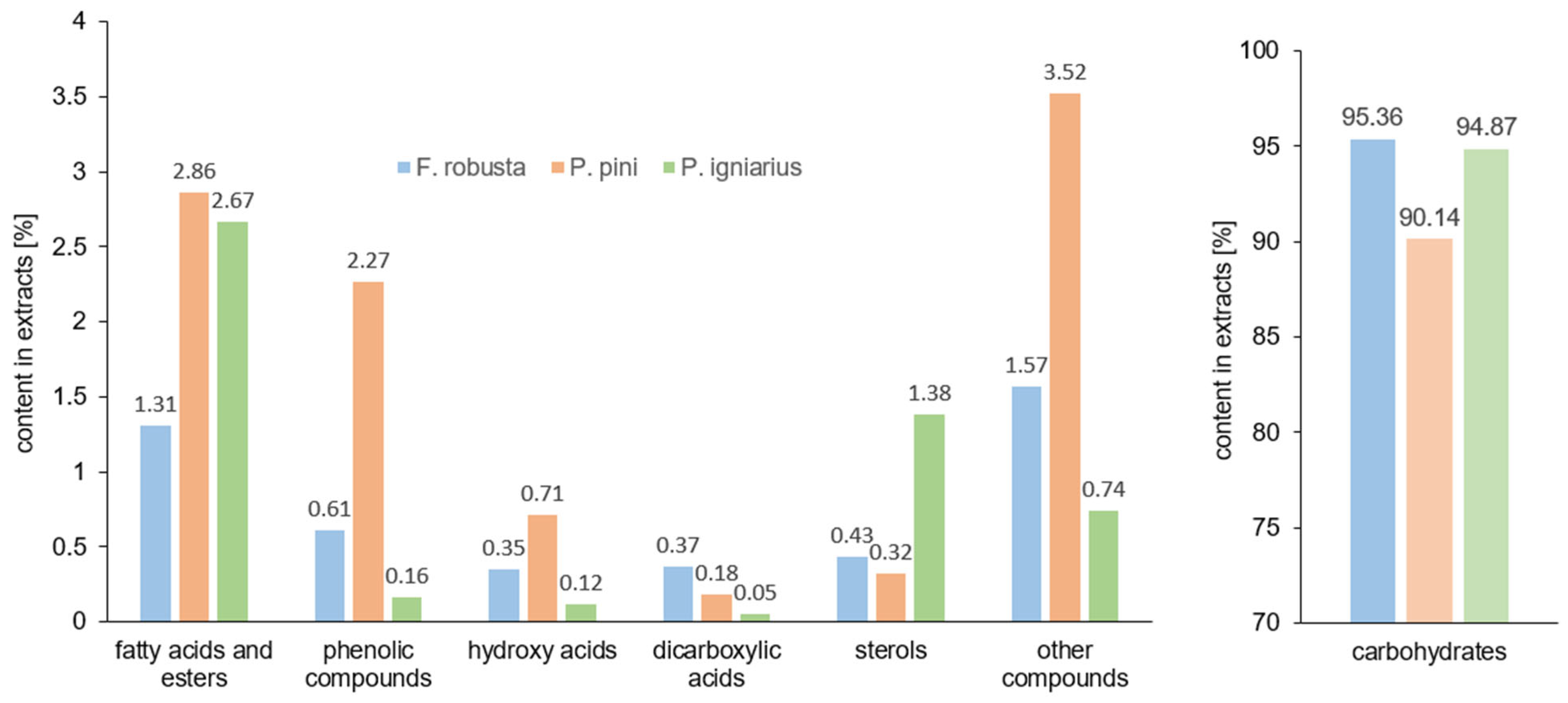
The results of GC/MS analysis showed that the main components of the extracts of Phellinus igniarius, Fomitiporia robusta, and Porodaedalea pini are carbohydrates. The contents of monosaccharides and disaccharides constituting the polysaccharide fraction are 95.36% (F. robusta), 94.87% (P. igniarus), and 90.14% (P. pini) (Figure 3). The structural characteristics of polysaccharides from samples of Phellinus spp. are generally defined by their average molecular weight, monosaccharide composition, and chemical structure [40,41]. The main disaccharide building block of the polysaccharides of the tested samples is trehalose, the content of which is about 58% in F. robusta and P. pini, and as much as 68% in P. igniarius. High contents of polyhydric alcohols were also noted, i.e., the content of mannitol was about 19% in F. robusta and P. pini and 13% in P. igniarius; the content of ribitol was almost 15% in the extract from F. robusta, 11% in P. igniarius, and 4% in P. pini. In the extract of P. igniarius, small amounts of sugars such as arabinitol, β-fructofuranose, α-glucopyranose, β-glucopyranose, myo-inositol, xylitol, and galactitol (with a total content of less than 1%) were also noted. In the extract of F. robusta, sugars such as arabinitol, α-glucopyranose, β-glucopyranose, myo-inositol, deoxy-inositol, galactitol, and sucrose were determined. In the extract of P. pini, the presence of the same sugars as in the extract of F. robusta was noted, as well as additional sugars such as pinitol (1.83%), threitol (0.74%), D-ribose (0.23%), fructose (0.23%), β-L-fucopyranose, and smaller amounts of β-mannopyranose and α-galactopyranose.
The content of fatty acids and their esters in the extracts of Phellinus igniarius, Fomitiporia robusta, and Porodaedalea pini was 2.67%, 1.31%, and 2.86%, respectively. The main acids identified in the extracts tested were palmitic acid (0.03%, 0.10%, and 0.59%, respectively), linoleic acid (0.20%, 0.35%, and 0.89%), and esters methyl palmitate (0.24%, 0.16%, and 0.25%) and methyl linoleate (1.07%, 0.47%, and 0.26%). In the extracts of Phellinus igniarius and Porodaedalea pini, the content of oleic acid (0.11% and 0.16%) was determined.
The highest content of phenolic acids was recorded in the extract from P. pini (2.27%), including 3-hydroxybenzoic acid (0.77%), protocatechuic acid (0.94%), caffeic acid (0.30%), and vanillic acid (0.10%), as well as small amounts of protocatechuic acid esters. These compounds are characterized by high antioxidant potential [42]. The F. robusta extract contained 0.61% of phenolic compounds, including 0.37% of 3-hydroxybenzoic acid, 0.12% of protocatechuic acid, and 0.05% of syringic acid. The lowest content of phenolic compounds was recorded in the extract from P. igniarius (0.16%), including 0.08% of 3-hydroxybenzoic acid, 0.05% of protocatechuic acid, and 0.04% of caffeic acid.
The extracts tested also contained compounds from the group of aliphatic hydroxy acids. The P. igniarius extract contained 0.12% of compounds from this group (lactic acid and malic acid), the F. robusta extract contained 0.35% (lactic acid, glycolic acid, malic acid, and citric acid), while the P. pini extract contained 0.71% (lactic acid, glycolic acid, glyceric acid, and malic acid).
The dicarboxylic acids present in the extracts tested were oxalic acid (only in F. robusta); succinic acid, which was present in all extracts in amounts below 0.15%; and fumaric acid, which was present in the amount of 0.27% in the F. robusta extract and 0.03% in the P. igniarius extract.
The sterol content in the P. igniarius extract was 1.38% (including ergosterol—0.9%, 3-hydroxyergosta-7,22-diene—0.27%, and 3-hydroxyergost-7-ene—0.12%). In the F. robusta extract, 0.43% sterols were noted (including 0.2% ergosterol, 0.14% 3-hydroxyergosta-7,22-diene, and 0.1% 3-hydroxyergost-7-ene). The P. pini extract contained 0.2% ergosterol and 0.11% 3-hydroxyergost-7-ene.
The extracts tested also contained small amounts of ethylene glycol (below 0.2%), 1,2-propanediol (below 0.15%), and pyroglutamic acid (below 0.2%). The glycerol content in P. igniarius was 0.51%, in F. robusta 0.19%, and in the extract from P. pini 1.42%. The extract from P. pini contained N-acetylglucosamine (0.59%), which is a component of biopolymers that build fungal cell walls.

2.6. Antioxidant Study

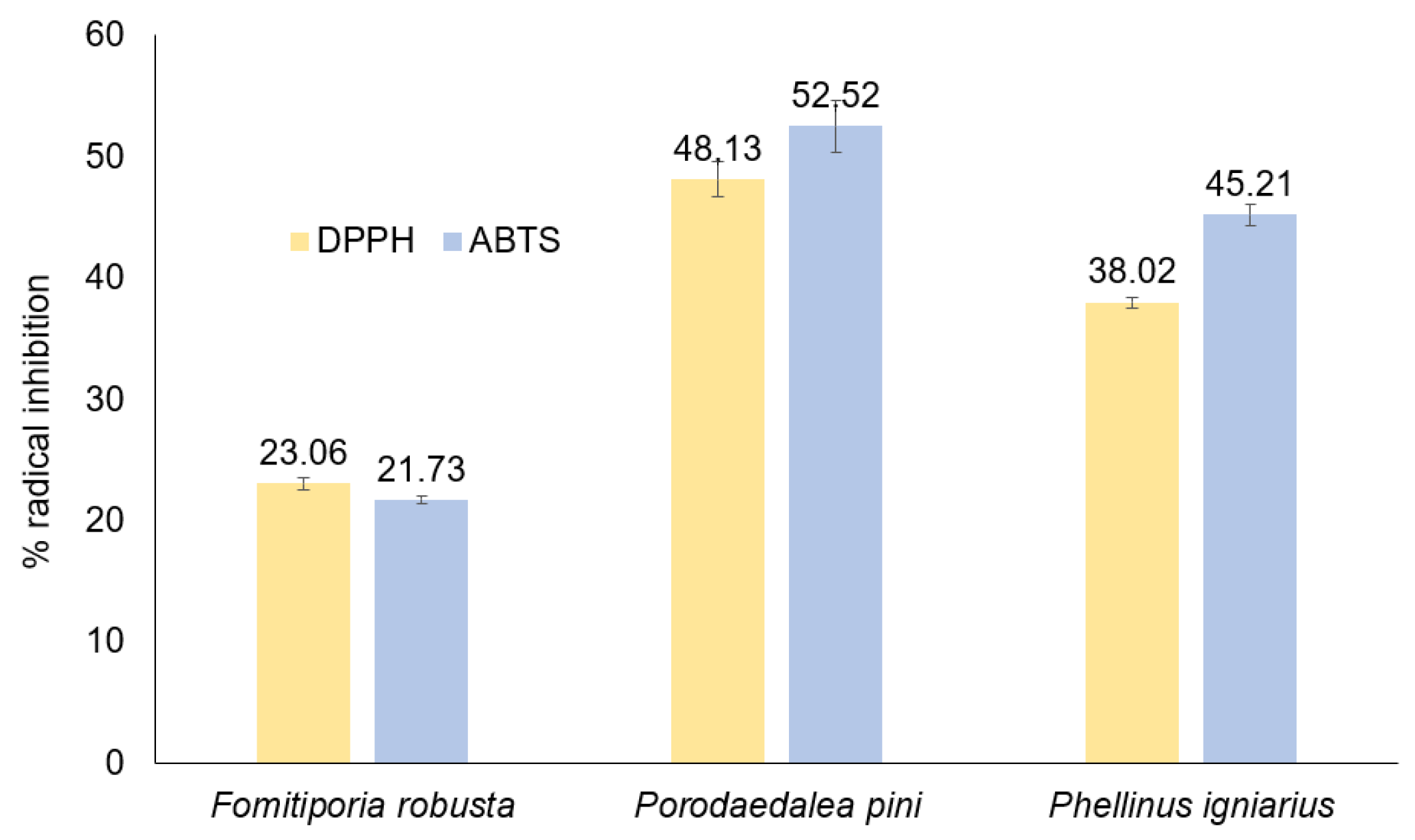
The antioxidant activity of methanolic extracts of Fomitiporia robusta, Porodoedalea pini, and Phellinus igniarius determined through tests with DPPH and ABTS radicals is presented in Figure 4. The concentration of extracts for which the level of radical inhibition was measured was 54 mg/L. The highest antioxidant activity in tests with ABTS and DPPH radicals was demonstrated by the extract from P. pini. Over 50% level scavenging of the ABTS radical and 48% level scavenging of the DPPH radical were noted. This extract was characterized by the highest content of phenolic compounds, which are mainly responsible for antioxidant activity. The lowest content of phenolic compounds was noted in the extract of P. igniarius; however, this extract did not show the lowest antioxidant potential among the extracts tested. A high content of sterols was found in this extract, which may also demonstrate antioxidant potential [43,44]. The level of ABTS radical neutralization by the P. igniarius extract was 45%, while that of the DPPH radical was 38%. The lowest antioxidant activity was demonstrated by the F. robusta extract, with 23% and 21.7% of radical removal, respectively, in the DPPH and ABTS tests. This extract is characterized by a low content of phenolic compounds, as well as a low content of sterols. For comparison, gallic acid at a concentration of 0.85 mg/L removes almost 100% of radicals in the ABTS test, and about 50% of radicals in the DPPH test.
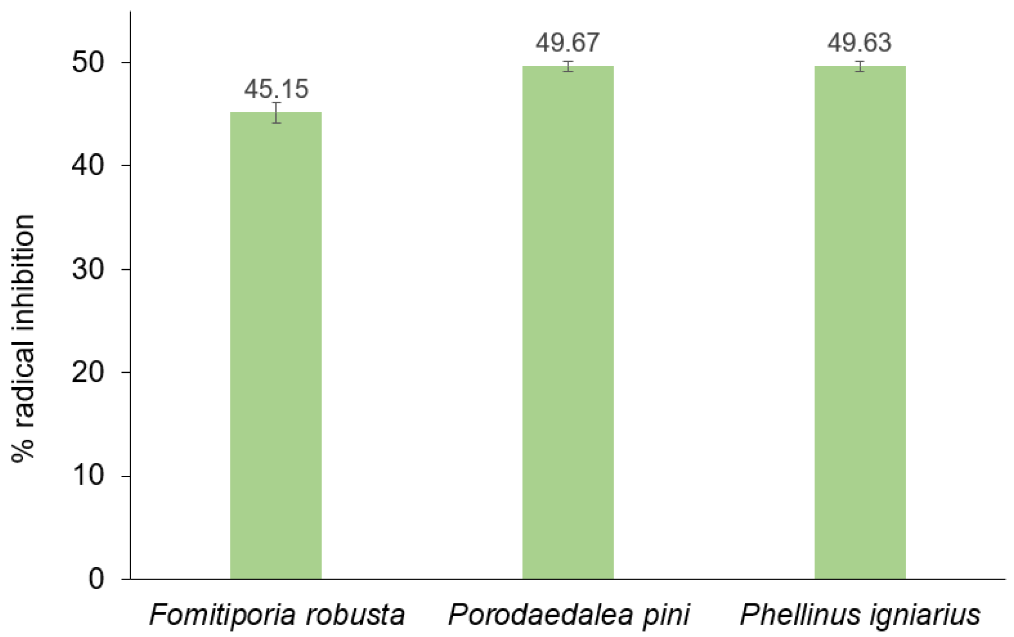
Figure 5 presents the results of the fat oxidation inhibition test. The tested extracts at a concentration of 54 mg/L of P. pini and P. igniarius extracts caused nearly 50% inhibition of fat oxidation in the 5-day test. The F. robusta extract showed a slightly lower inhibition of oxidation fats (approximately 45%). The high sterol content in the Phellinus igniarius extract has a major impact on its antioxidant properties against fats.

2.7. Antimicrobial Study

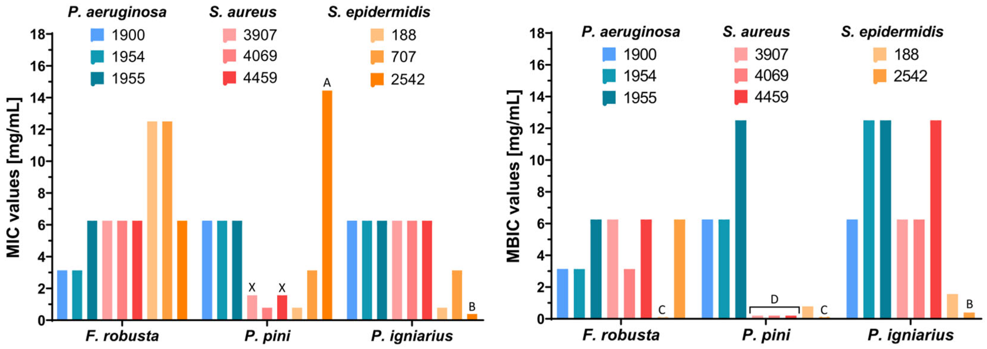
To evaluate the therapeutic potential of tested fungal extracts, antimicrobial activity was assessed by determining the minimum inhibitory concentration (MIC), minimum bactericidal concentration (MBC), and minimum biofilm inhibitory concentration (MBIC). The MIC—the lowest concentration inhibiting bacterial growth—and the MBC—the concentration achieving bactericidal effects—were determined as described previously [45]. The MBIC is a concentration of tested agents that inhibits the formation of bacterial biofilm, which creates a protective barrier for bacteria, rendering them significantly more resistant to antibiotics [46]. Fungal extracts that inhibit biofilm formation can restore bacterial susceptibility to antibiotics, enhancing treatment efficacy. Porodoedalea pini (against S. aureus and S. epidermidis) and P. igniarius (against S. epidermidis) showed the highest anti-biofilm properties with MBIC values between 0.78 and 1.56 mg/mL against Staphylococcus epidermidis and <0.20 mg/mL against S. aureus clinical strains. S. aureus 4459 and 3907 strains showed increased resistance to the tested antibiotic, ERT, with MIC ≥ 0.50 mg/mL. Interestingly, in contrast to the other extracts tested, only the P. pini extract exhibited bactericidal activity against these two bacterial strains, as evidenced by its minimum bactericidal concentration (MBC) of 6.25 mg/mL. Furthermore, this extract demonstrated significant antibacterial activity with a minimum inhibitory concentration (MIC) of 1.56 mg/mL and a minimum biofilm inhibitory concentration (MBIC) below 0.20 mg/mL (Table 4, Figure 6). Planktonic bacteria—which are individual, free-floating cells—are more susceptible to antibacterial agents than biofilm bacteria, which form complex communities encased in a self-produced extracellular polymeric substance matrix. This matrix makes biofilm bacteria significantly more resistant, even up to 1000-fold [47]. Phellinus iginarius extract showed the lowest activity in inhibition of P. aeruginosa and S. aureus biofilm formation, which can be seen by comparing the MBIC results for all tested extracts. The lowest concentrations that inhibited biofilm formation of S. aureus and S. epidermidis were observed after treatment with P. pini extract, while F. robusta extract was the most effective against strains of P. aeruginosa (Figure 6).

2.8. Cytotoxicity

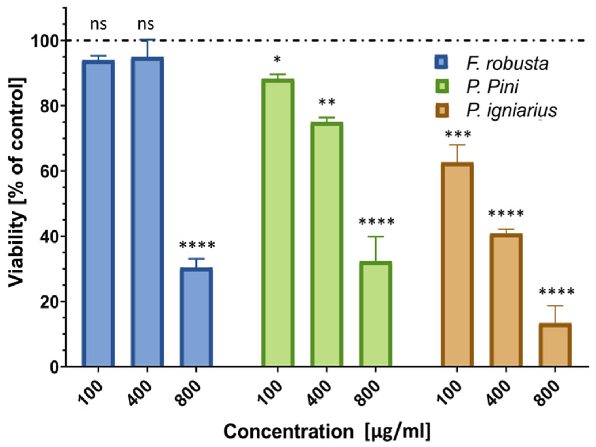
The cytotoxicity of the examined fungal extracts was evaluated using the Neutral Red (NR) assay, which measures cell viability based on the uptake of NR dye by functional lysosomes [48]. Only F. robusta extract showed a lack of cytotoxicity against human keratinocytes at 100 and 400 µg/mL; however, the highest concentration (800 µg/mL) of this extract caused an approximately 70% reduction in cell viability. The other two extracts (P. pini and P. igniarius) showed varying cytotoxicity at all three concentrations, with the P. igniarius extract causing approximately 9, 43, and 69% decreases in HaCaT cell viability at concentrations of 100, 400, and 800, respectively (Figure 7).

2.9. Reactive Oxygen Species

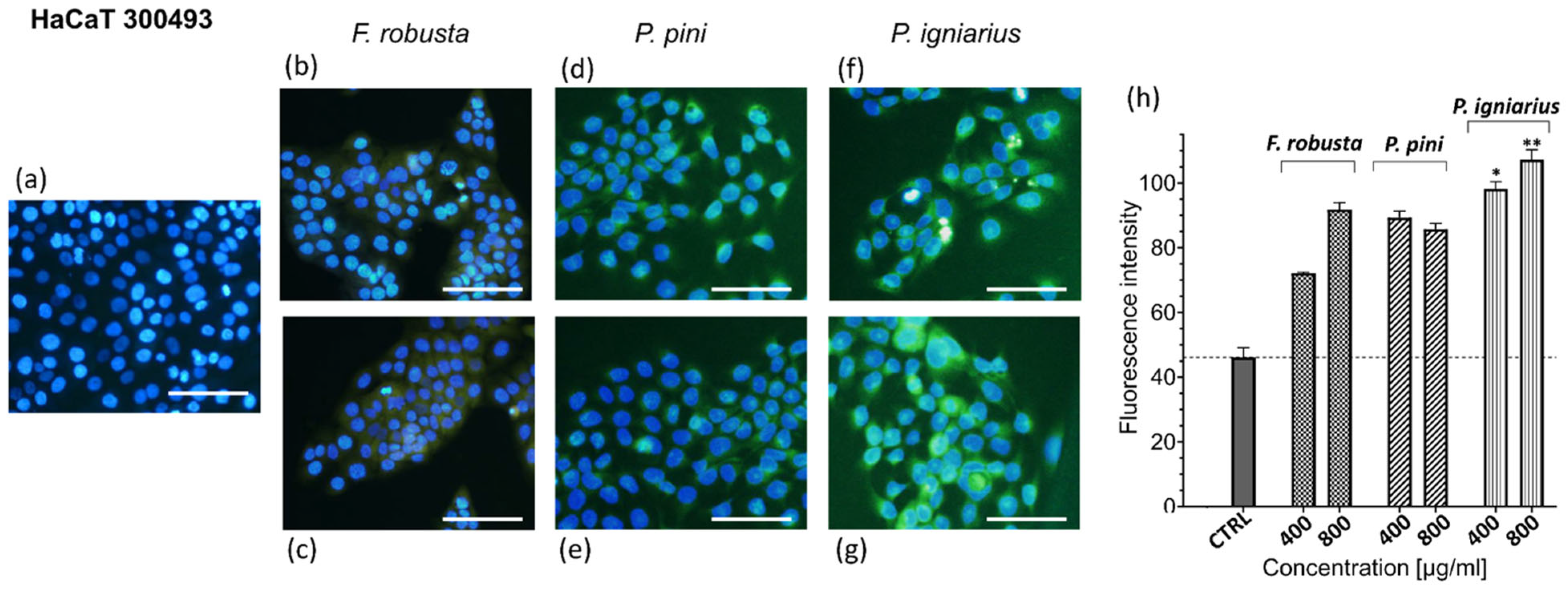
Reactive oxygen species (ROS) generation is a critical mediator of cellular stress and oxidative damage that is implicated in diverse pathologies [49]. To investigate the effects of fungal extracts on intracellular ROS levels, the CellROX Green assay, a fluorogenic probe quantifying ROS through oxidation-induced fluorescence, was utilized. Only incubation with P. igniarius extract showed a statistically significant pro-oxidative effect against HaCaT cells (Figure 8).

3. Materials and Methods

3.1. Collection of Fungal Samples in the Field

This study investigated 30 samples belonging to 22 species of the genus Phellinus sensu lato (Table 5). Fruiting bodies were collected, among other specimens, in the Białowieża Forest (Poland); Village Kozhle (North Macedonia); Estremadura, Sesimbra, and Lagoa de Albufeira (Portugal); Zlatari close to Prishtina (Kosovo); and Spoleto and the Bosco Siro Negri State Nature Reserve (Italy). Each fruiting body found in the field was collected together with the substrate. Prior to collection, a photograph was taken under natural conditions, documenting the general habit and features of the fruiting body that may be lost during drying. The specimens were collected in paper envelopes on which the place and date of collection; the in situ fruiting body photo numbers; the type of substrate and host; and the habitat data were recorded. Additionally, in the case of saprobes growing on dead wood, the species was identified and information on the stage of decomposition of the substrate was recorded.

3.2. Morphological Identification

Slides were prepared under an OPTA-TECH MI6 binocular (OPTA-TECH) magnifier at 5–25x magnification. Thin sections of the fruiting body were obtained with a microtome or razor blade. If necessary, a preparation was made from the relevant part of the fruiting body—i.e., tube and context—and, for some species, also from the cap cover. The tube preparation was prepared in such a way as to make the following characteristics visible: the arrangement of the filaments in the trama; the appearance of the bristles in the hymenium and context, characteristic of the genus Phellinus; the appearance of the filament tips on the blades of the inner tube septa; and the appearance and elements of the hymenium at the bottom of the tubes. An OPTATECH LAB-40 light microscope with variable phase contrast and a NIKON ECLIPSE Ni with Nomarski contrast were used to observe microscopic features.
Preparations from fresh specimens were made in water and from dry specimens in 3–10% KOH. All available features were observed in these media. To improve observational comfort and obtain a more contrasting image, the slides were stained with Congo Red. This dye stains the cell walls and, less intensely, the cell contents. Given that microscopic features in the genus Phellinus s.l. have a dark brown color, lactophenol was used to lighten them. The amyloid and dextrorotatory properties of the microstructural elements were checked in Melzer’s reagent and in 0.1% Cotton Blue in 60% lactic acid to determine the degree of cyanophilicity of the spore walls and filaments.
The primary sources of information for the taxonomic analysis of fungi of the genus Phellinus s.l. were the following keys and monographic studies: Domanski et al. (1967, 1973) [50,51]; Ryvarden and Gilbertson (1993, 1994) [52,53]; Larsen and Cobb-Poulle (1990) [54]; and Sell (2008) [55]. The nomenclature was adopted from MycoBank(available online: http://www.mycobank.org (accessed on 26 March 2025)). The correctness of the spelling used and the author of the name at the genus level was confirmed with the latest edition of the Dictionary of the Fungi (Kirk et al. 2008) [56]. Acronyms of herbaria follow the Index Herbariorum. Documentary material (dry fungal specimens) was deposited in the herbarium of the Institute of Forest Sciences, Bialystok University of Technology (BLS).

3.3. Genetic Identification

In order to confirm genetic identity, DNA was isolated from the fungal samples with the Plant & Fungi DNA Purification Kit (EURx, Gdańsk, Poland). The obtained preparations were characterized spectrophotometrically (Varioskan LUX, Thermo Fisher Scientific, Waltham, MA, USA) and fluorometrically (Qubit 2.0, Thermo Fisher Scientific, US). Whole genome sequencing was performed using the DNA library prepared with the ITD xGen DNA EZ kit on an Illumina NovaSeq S4 system. Paired reads (2 × 150 bp) were cleared and then subjected to de novo assembly using MEGAHIT v. 1.2.9 and CLC Genomics Workbench v. 12.0.3. The resulting contigs were searched to identify reference sequences, including the small subunit ribosomal RNA gene fragment, the internal transcribed spacer 1, the 5.8 S ribosomal RNA gene, the internal transcribed spacer 2, and the partial large subunit ribosomal RNA gene used in taxonomic analyses. Sequences were deposited at NCBI (PV125346.1 and PV186754.1). The fungal material was dried.

3.4. Preparation of Extracts

The fruiting bodies were cleaned and separated from the remaining substrate and then dried at a temperature not exceeding 40 ˚C. The fruiting bodies were crushed in a laboratory grinder. The samples were then macerated in methanol (99.8%; POCH Avantor Performance Materials, Poland) at a ratio of 1:6 [g/mL]. The maceration period was no less than 2 months, and the samples were shaken systematically.
The finished tinctures were filtered through 80 g/m2 filter paper, and the solvent was evaporated in two steps. In the first step, most of the solvent was removed in a rotary evaporator Rotavapor® R-100 (Büchi, Switzerland) at 46 °C, rotation speed 3–4 units, and under the reduced pressure from 300 to 100 mbar. In the second step, the extract was collected from the walls of the round-bottomed extraction flask together with a small amount of methanol and placed in the glass extraction cells of a parallel evaporation system, MultivaporTM P-12 (Büchi), where it was incubated at 46 °C and under reduced pressure from 400 to 100 mbar. Each solid-state extract was stored in the dark at 10 °C in the Fungi Extract Bank® collection (available online: https://fungiextractbank.com/en (accessed on 1 April 2025)).
For fungal samples where the residue in the flask did not dissolve in methanol, the flask was rinsed with distilled water, then frozen at −25 °C, and lyophilized at −40 °C at a pressure of 0.04 mbar; these samples were stored for other tests. The methanolic extracts obtained had a plastic consistency, high viscosity, and colors from brown to dark brown. Dry methanolic extracts were used for further studies.

3.5. Antimicrobial Activity—Preliminary Studies

Preliminary screening tests of the antimicrobial activity of 30 samples of fungi were performed. Antimicrobial assays of fungal extracts were conducted as part of the project. For each extract, the MIC (minimum concentration of a substance that inhibits microbial growth) was determined using the microdilution method in Mueller–Hinton Broth II (MH II) medium using serial two-fold dilutions of the fungal extract, according to the procedure described by Balouiri et al. and Rašeta et al. [57,58]. The MIC value was expressed as the lowest concentration of fungal extract (in mg/mL) that completely inhibited the growth of the microorganism, as identified based on the turbidity of the culture after 24 h. To confirm the MIC value, cultures were performed on plates of three dilutions that showed the lowest or no turbidity. This study was performed on bacterial strains Klebsiella pneumoniae (ATCC-13883), Pseudomonas aeruginosa (ATCC-27853), Staphylococcus aureus (ATCC-25923), Staphylococcus epidermidis, and Kocuria rhizophila (ATCC-9341), and fungi Candida albicans (ATCC-1023), which were obtained from the ATCC collection. Based on the MIC results, three extracts of Phellinus species were selected for further studies: Fomitiporia robusta, Phellinus igniarius, and Porodaedalea pini.

3.6. Elemental Analysis

The determination of selected elements in fungal extracts (Phellinus igniarius, Fomitiporia robusta, and Porodaedalea pini (Figure 9)) was performed using X-ray spectrofluorimetry. An amount of 900 µL of the aqueous extract solution was collected in an Eppendorf tube, and 100 µL of internal standard (Ga at 10 mg/L) was added to obtain a final concentration of 1 mg/L. An amount of 10 µL of the prepared solution was applied to measuring slides and dried at 50 °C. The prepared samples were analyzed using a TXRF (Total X-Ray Reflection Fluorescence) S2 PICOFOX apparatus, Bruker, Berlin, Germany.

3.7. Thermal Analysis

Thermal analysis of dried fruiting body samples of Phellinus igniarius, Fomitiporia robusta, and Porodaedalea pini was performed on a Libra thermal analyzer by TG 209 F1 (Netzsch company, Selb, Germany). Dried material weighing 10 ± 0.1 g was heated in ceramic crucibles in a nitrogen atmosphere (15 mL/min) with a temperature increase of 10 °C/min. The thermal decomposition process was carried out in the temperature range from 30 to 660 °C. The TG and DTG curves were recorded.

3.8. Gas Chromatography–Mass Spectrometry (GC-MS) Analysis

The fungus extract chemical composition was analyzed using an Agilent 7890A gas chromatograph coupled with an Agilent 5975C mass spectrometer. The methanol extract from fungus (10 mg) was diluted in 1 mL of pyridine, and 0.1 mL N,O-bis (trimethylsilyl)-trifluoroacetamide (BSTFA) was added. The sample was heated for 30 min at 60 °C. After silylation, 1 μL of the sample was entered into a GC/MS injector. The injector was set to a 1:10 split, and the temperature was 300 °C. The separation of compounds was conducted on a silica column HP-5MS (30 m × 0.25 mm × 0.25 μm), and the rate of helium flow was 1 mL/min. The starting column temperature was 50 °C, increased to 325 °C at 3 °C/min, and the final temperature was held for 10 min. The quadrupole and ion source temperatures were 150 °C and 230 °C, respectively. The energy of ionization was 70 eV, and detection was performed in a range of 41–800 units in a full scan mode. After peak integration, the percentage of every compound in the total ion current (% TIC) was calculated. Both mass spectra and retention indices were used for the identification of chemical compounds. The Isidorov (2020) [59], NIST (2020) [60], and Wiley (2020) [61] libraries of mass spectra were used for analysis. The experimental retention indices (RIexp.) were calculated in relation to the retention times of C8-C40 n-alkanes. The RIexp. values were compared with the literature retention indices (RIlit.).

3.9. Spectroscopy

The FTIR spectra of both extracts and dried fruiting bodies of 3 selected fungal samples, Phellinus igniarius, Fomitiporia robusta, and Porodaedalea pini, were obtained using the ATR multireflection technique. The spectra were recorded in the range of 2000–600 cm−1 using an Alfa spectrometer (Bruker, Billerica, MA, USA).

3.10. Antioxidant Properties and Lipid Peroxidation Analysis

The antioxidant properties were determined using DPPH and ABTS radical assays.
DPPH radical test
The determination of antioxidant activity via the DPPH radical assay was conducted following the method described in [62]. Briefly, 1 mL of the extract was mixed with 2 mL of a DPPH radical solution. A control sample was prepared simultaneously, in which 1 mL of solvent (methanol) was used instead of the extract. The samples were incubated for 1 h. After incubation, absorbance was measured at 516 nm using a UV–VIS spectrophotometer (NANOCOLOR® VIS, MACHEREY-NAGEL). Each sample was analyzed in triplicate. The free radical scavenging activity of DPPH was calculated based on absorbance measurements, with the average percentage of inhibition determined using the following equation:
% I = A c 516 A s 516 A c 516 · 100 %
%I—percentage of inhibition; Ac516—absorbance of the control sample at 516 nm; and As516—absorbance of the sample containing the extract at 516 nm.
ABTS radical test
The antioxidant activity via the ABTS radical assay was determined based on the study by the authors of [63]. ABTS was prepared at a concentration of 7 mM and mixed with 2.45 mM of potassium persulfate (K2S2O8) in a 1:1 ratio, followed by incubation in the dark at 20 °C for approximately 12 h to generate the ABTS+ radical. For the assay, 1.5 mL of the working ABTS+ solution was diluted with 30 mL of methanol. Then, 1 mL of the extract was mixed with 1 mL of the ABTS+ solution. A control sample was prepared simultaneously, in which the extract was replaced with methanol. The samples were incubated for 7 min at 20 °C. After incubation, absorbance was measured at 734 nm using a UV–VIS spectrophotometer. Each sample was analyzed in triplicate. The average percentage of inhibition of the ABTS+ cation radical was calculated using the following equation:
% I = A c 734 A s 734 A c 734 · 100 %
%I—percentage of inhibition; Ac734—absorbance of the control sample at 734 nm; As734—absorbance of the sample containing the extract at 734 nm.
Lipid peroxidation
To evaluate the ability of extracts to inhibit lipid peroxidation, following the method described in [64], a linoleic acid emulsion was prepared by mixing 0.312 mL of linoleic acid, 0.256 mL of Tween 20, and 0.05 M of phosphate buffer in a 50 mL volumetric flask, with the final volume adjusted using the buffer. Then, 1 mL of each extract, prepared at a concentration of 45 mg/25 mL, was added to 1.5 mL of the emulsion. Each extract was tested in five replicates, and control samples were prepared in parallel by replacing the extract with 1 mL of methanol. The test tubes were sealed and incubated at 40 °C. Once the target temperature was reached, 0.1 mL of the reaction mixture was sampled from each tube and mixed with 4.7 mL of 75% ethanol and 0.05 mL of 30% ammonium thiocyanate. After a 3-min reaction period, 0.05 mL of 0.02 M FeCl2 solution in 3.5% HCl was added. Absorbance was measured at 500 nm using a UV–VIS spectrophotometer, with 75% ethanol used as a reference. Measurements were performed every 24 h for 5 days, with 0.1 mL volumes withdrawn from the same emulsions at each time point. The percentage inhibition of linoleic acid peroxidation (%I) was calculated using the following formula:
% I = A c 500 A s 500 A c 500 · 100 %
%I—percentage of peroxidation inhibition; Ac500—absorbance of the control sample at 500 nm; and As500—absorbance of the sample containing the extract at 500 nm.

3.11. Antibacterial Activity

Bacterial Strains and Culture Conditions
Clinical bacterial isolates, specifically Pseudomonas aeruginosa (1900, 1954, and 1955), Staphylococcus aureus (3907, 4069, and 4459), and Staphylococcus epidermidis (188, 707, and 2542), were obtained from the Department of Medical Laboratory Diagnostics, Provincial Specialist Hospital in Rzeszow, and deposited at the Department of Biotechnology and Bioinformatics, Faculty of Chemistry, Rzeszow University of Technology. All bacterial strains were cultured at 37 °C in a New Brunswick Innova 40 Shaker (Eppendorf AG, Hamburg, Germany) until reaching a turbidity equivalent to 0.5 McFarland standard (approximately 108 colony-forming units per mL [CFU/mL]). The obtained cultures were then diluted to a final concentration of 105 CFU/mL. All bacterial cultures were prepared under aseptic conditions within an ESCO Airstream Laminar Flow Cabinet.
Antimicrobial Activity Assays
Minimum inhibitory concentrations (MICs) were determined using a micro-broth dilution method in Mueller–Hinton Broth (MHB), as previously described [65]. Serial two-fold dilutions of extracts (0.39–25 mg/mL) were prepared in MHB. Following 24-h incubation at 37 °C, the MIC was defined as the lowest concentration inhibiting visible growth, confirmed by OD600 measurement (BIO-RAD microplate reader). Minimum bactericidal concentrations (MBCs) were determined by subculturing MIC-, 2xMIC-, and 4xMIC-treated cultures onto Mueller–Hinton Agar (MHA) for 24 h at 37 °C. The MBC was defined as a ≥ 99.9% reduction in CFU compared with the untreated controls. Erythromycin (ERT) susceptibility was assessed using the microdilution method (0.03–500 µg/mL).
Anti-Biofilm Activity
The ability of extracts to inhibit biofilm formation was assessed using the MTT assay. Bacterial cultures, conditions, and density of the culture, as well as the 96-well plate preparation with extract dilutions, were the same as for the MIC analysis (see above in Section 2.5 Antimicrobial Activity—Preliminary Studies). After 24-h incubation, non-adherent cells were removed, and adherent cells were stained with 0.5% MTT in PBS for 2 h. If bacteria in biofilm are alive and metabolically active, their oxidoreductase enzymes convert the yellow MTT dye into insoluble purple formazan crystals. These crystals accumulate within the cells or on the biofilm surface. To quantify the formazan, a solubilization solution (DMSO) was added to dissolve the crystals, forming a homogeneous purple solution [65]. Subsequently, the absorbance was measured at 600 nm (Varioskan™ LUX multimode microplate reader, Thermo Scientific, Waltham, MA, USA). The minimum biofilm inhibitory concentration (MBIC) was defined as the lowest concentration inhibiting biofilm formation compared with media-only controls. The experiments were conducted in triplicate.

3.12. Cytotoxic Study

Cytotoxicity Assessment
Human immortalized epidermal cells (HaCaT 300493, Cytion, Eppelheim, Germany) were cultured in DMEM supplemented with 10% FBS and 1% penicillin–streptomycin, at 37 °C with 5% CO2. Cell viability was determined via trypan blue exclusion using an Automatic Cell Counter TC20™ (Hercules, CA, USA) (Cytotoxicity was evaluated using the Neutral Red (NR) assay [48]. The cells were seeded in 96-well plates and exposed to extracts (100, 400, and 800 µg/mL in DMSO, final DMSO ≤1%) for 24 h. NR solution (0.033%) was added for 2 h; afterwards, plates were washed with PBS, and then destaining solution (50% ethanol, 49% water, and 1% acetic acid) was added. Absorbance was measured at 540 nm (Varioskan™ LUX multimode microplate reader, ThermoFisher Scientific, Waltham, MA, USA). Cell viability was expressed as a percentage of the untreated controls.
Reactive Oxygen Species (ROS) Quantification
HaCaT cells were treated with Fomitiporia robusta, Porodaedalea pini, and Phellinus igniarius extracts (400 and 800 µg/mL) for 2 h. Non-treated cells were used as a negative control. ROS production was assessed using the CellROX™ Green Oxidative Stress kit (ThermoFisher Scientific, Waltham, MA, USA). The cells were incubated with 5 µM CellROX Green for 30 min, fixed with 4% paraformaldehyde, and counterstained with Hoechst 33342. Fluorescence imaging was performed using the cellSens Software 4.3 (Olympus, Center Valley, PA, USA). CellROX fluorescence intensity was quantified using ImageJ 1.54 g [66].

3.13. Statistical Analysis

The final results obtained from triplicate experiments are presented as means ± SD. GraphPad Prism version 8.0.1 (GraphPad Software, Boston, MA, USA, available online: http://www.graphpad.com (accessed on 20 April 2025)) was used to compare the differences between groups. Statistical significance was indicated for p-values < 0.05.

4. Conclusions

Antimicrobial activity studies of 30 samples of methanol extracts (22 Phellinus species) of fungi from the genus Phellinus allowed for the identification of extracts with the highest activity. The identified extracts were subjected to further detailed studies. The antimicrobial activity of fungi from the Porodaedalea pini, Fomitiporia robusta, and Phellinus igniarius species was tested on strains of multidrug-resistant bacteria. It was observed that the habitat and origin of a given species have a significant influence on biological activity (related to the content of biologically active compounds). Studies of the content of active compounds in the analyzed extracts showed that each of the three selected extracts was characterized by a high content of sugars (over 90% of the extract mass). Porodaedalea pini extract was characterized by the highest content of phenolic compounds among the three samples analyzed in detail, which determined the highest antioxidant activity of the extract of this mushroom in antioxidant tests.
Antimicrobial tests performed on antibiotic-resistant bacteria (Pseudomonas aeruginosa, Staphylococcus aureus, and Staphylococcus epidermidis) showed that P. pini extract was characterized by the highest activity in neutralizing these microorganisms among the three extracts tested. Cytotoxicity tests of the three selected extracts from fungi of the Phellinus species against human keranocytes showed that only the F. robusta extract did not show a cytotoxic effect, while the remaining two extracts (P. pini and P. igniarius) were biocidal against keranocyte cell lines.
The effects of the three mushroom extracts on the level of reactive oxygen species (ROS) generation—which are critical mediators of cellular stress and oxidative damage associated with various pathologies—were also studied. The studies were performed on the human keratinocyte HaCaT cell line. It was observed that only the P. igniarius extract showed a statistically significant pro-oxidant effect against HaCaT cells.
The conducted experiments provided promising results that are expected to form the basis of further studies on the biological activity of Phellinus sensu lato fungi. It is anticipated that the use of other methods for extracting biologically active compounds from Phellinus fungi will allow for obtaining extracts with high biological activity, which may result in the application of these extracts as compounds with high antimicrobial, antioxidant, and/or anticancer potential.

Author Contributions

Conceptualization, G.Ś., M.K. (Monika Kalinowska), E.Z., E.C. and M.P.; methodology, G.Ś., M.P., E.C., E.Z., M.K. (Monika Kalinowska), E.W., U.W. and M.Z.; validation, G.Ś., M.P., E.C., E.Z. and M.K. (Monika Kalinowska); formal analysis, G.Ś., M.P., E.C., E.Z., M.K. (Monika Kalinowska), A.B., M.J., M.T., P.G., M.S., M.W. and M.Z.; investigation, G.Ś., M.P., E.C., E.Z., M.K. (Monika Kalinowska), A.B., M.J., M.T., P.G., K.L.-S., M.S. and M.K. (Mitko Karadelev); resources, G.Ś., M.P., E.C., E.Z. and M.K. (Monika Kalinowska); A.B., Q.R., C.E.G. and M.K. (Mitko Karadelev); data curation, M.P., G.Ś. and E.C.; writing—original draft preparation, G.Ś., M.P., E.C., E.Z., M.W., M.T., U.W. and E.W.; writing—review and editing, G.Ś., M.P., E.C., E.Z. and M.K. (Monika Kalinowska); visualization, G.Ś., M.P., E.C., M.W. and E.Z.; supervision, G.Ś.; project administration, G.Ś. All authors have read and agreed to the published version of the manuscript.

Funding

This study received funding from the commissioned task titled “VIA CARPATIA Universities of Technology Network named after the President of the Republic of Poland Lech Kaczyński” under the special purpose grant from the Minister of Science, contract no. MEiN/2022/DPI/2577/VC/WB-IIŚ/2/2023, as part of action “ISKRA—building inter-university research teams”.

Institutional Review Board Statement

Not applicable.

Informed Consent Statement

Not applicable.

Data Availability Statement

The data presented in this study are available upon request from the corresponding author.

Acknowledgments

The authors thank the Bosco Siro Negri State Nature Reserve (University of Pavia) for authorization to collect samples and for sharing specimens. The authors thank Konrad Wilamowski and Slavica Tofilovska for collecting the samples for the study.

Conflicts of Interest

The authors declare no conflicts of interest.

References

  1. He, P.; Zhang, Y.; Li, N. The phytochemistry and pharmacology of medicinal fungi of the genus: Phellinus: A review. Food Funct. 2021, 12, 1856–1881. [Google Scholar] [CrossRef]
  2. Zhang, H.; Jiang, F.; Li, L.; Liu, X.; Yan, J.-K. Recent advances in the bioactive polysaccharides and other key components from Phellinus spp. and their pharmacological effects: A review. Int. J. Biol. Macromol. 2022, 222, 3108–3128. [Google Scholar] [CrossRef] [PubMed]
  3. Pošta, A.; Matočec, N.; Kušan, I.; Tkalčec, Z.; Mešić, A. The Lignicolous Genus Entonaema: Its Phylogenetic–Taxonomic Position within Hypoxylaceae (Xylariales, Fungi) and an Overview of Its Species, Biogeography, and Ecology. Forests 2023, 14, 1764. [Google Scholar] [CrossRef]
  4. Nankone, S.; Sanon, E.; Sawadogo, B.R.; Dabire, K.; Guissou, M.-L.K. Ecology and morphological characterization of the genus Phellinus sensu-lato (Basidiomycetes, Hymenochaetaceae) in Burkina Faso. Afr. J. Plant Sci. 2020, 14, 451–460. [Google Scholar]
  5. Sułkowska-Ziaja, K.; Balik, M.; Muszyńska, B. Selected Species of the Genus Phellinus–Chemical Composition, Biological Activity, and Medicinal Applications. Chem. Biodivers. 2021, 18, e2100609. [Google Scholar] [CrossRef] [PubMed]
  6. Salvador-Montoya, C.A.; Elias, S.G.; Popoff, O.F.; Robledo, G.L.; Urcelay, C.; Góes-Neto, A.; Martínez, S.; Drechsler-Santos, E.R. Neotropical Studies on Hymenochaetaceae: Unveiling the Diversity and Endemicity of Phellinotus. J. Fungi 2022, 8, 216. [Google Scholar] [CrossRef]
  7. Zhou, L.W.; Vlasák, J.; Dai, Y.C. Taxonomy and phylogeny of Phellinidium (Hymenochaetales, Basidiomycota): A redefinition and the segregation of Coniferiporia gen. nov. for forest pathogens. Fungal Biol. 2016, 120, 988–1001. [Google Scholar] [CrossRef]
  8. Luan, F.; Peng, X.; Zhao, G.; Zeng, J.; Zou, J.; Rao, Z.; Liu, Y.; Zhang, X.; Ma, H.; Zeng, N. Structural diversity and bioactivity of polysaccharides from medicinal mushroom Phellinus spp.: A review. Food Chem. 2022, 397, 133731. [Google Scholar] [CrossRef]
  9. Yan, J.K.; Pei, J.J.; Ma, H.L.; Wang, Z.-B.; Liu, Y.-S. Advances in antitumor polysaccharides from phellinus sensu lato: Production, isolation, structure, antitumor activity, and mechanisms. Crit. Rev. Food Sci. Nutr. 2017, 57, 1256–1269. [Google Scholar] [CrossRef]
  10. Zhao, Y.Q.; Dong, X.Y.; Ning, J.L.; Jin, Y.-X.; Wang, G.-K.; Feng, T. Chemical constituents from the fungus Phellinus baumii and their anti-inflammatory activity. Phytochem. Lett. 2023, 54, 18–22. [Google Scholar] [CrossRef]
  11. Azeem, U. Antioxidant efficacy of a wood degrading fungus Phellinus gilvus (Schwein.) Pat., from Indian fungal flora. J. Pharmacogn. Phytochem. 2018, 7, 1017–1021. [Google Scholar]
  12. Kim, G.Y.; Park, H.S.; Nam, B.H.; Lee, S.-J.; Lee, J.-D. Purification and characterization of acidic proteo-heteroglycan from the fruiting body of Phellinus linteus (Berk. & M.A. Curtis) Teng. Bioresour. Technol. 2003, 89, 81–87. [Google Scholar] [CrossRef] [PubMed]
  13. Wang, Y.; Shang, X.Y.; Wang, S.J.; Mo, S.-Y.; Li, S.; Yang, Y.-C.; Ye, F.; Shi, J.-G.; He, L. Structures, biogenesis, and biological activities of pyrano [4,3-c] isochromen-4-one derivatives from the fungus Phellinus igniarius. J. Nat. Prod. 2007, 70, 296–299. [Google Scholar] [CrossRef]
  14. Song, A.R.; Sun, X.L.; Kong, C.; Zhao, C.; Qin, D.; Huang, F.; Yang, S. Discovery of a new sesquiterpenoid from Phellinus ignarius with antiviral activity against influenza virus. Arch. Virol. 2014, 159, 753–760. [Google Scholar] [CrossRef]
  15. Gao, M.; Huang, Y.; Hu, C.; Hu, J.; Wang, Y.; Chen, Y.; Huang, Y.; Song, G.; Song, Z.; Wang, Z. Selective Anticancer Effect of Phellinus Linteus on Epidermoid Cell Lines Studied by Atomic Force Microscopy: Anticancer Activity on A431 Cancer Cells and Low Toxicity on HaCat Normal Cells. IEEE Nanotechnol. Mag. 2021, 15, 4–16. [Google Scholar] [CrossRef]
  16. Li, X.; Jiao, L.L.; Zhang, X.; Tian, W.-M.; Chen, S.; Zhang, L.-P. Anti-tumor and immunomodulating activities of proteoglycans from mycelium of Phellinus nigricans and culture medium. Int. Immunopharmacol. 2008, 8, 909–915. [Google Scholar] [CrossRef]
  17. Chen, W.; He, F.Y.; Li, Y.Q. The apoptosis effect of hispolon from Phellinus linteus (Berkeley & Curtis) Teng on human epidermoid KB cells. J. Ethnopharmacol. 2006, 105, 280–285. [Google Scholar] [CrossRef]
  18. Islam, M.T.; Ali, E.S.; Khan, I.N.; Shaw, S.; Uddin, S.J.; Rouf, R.; Dev, S.; Saravi, S.S.; Das, N.; Tripathi, S.; et al. Anticancer Perspectives on the Fungal-Derived Polyphenolic Hispolon. Anti-Cancer Agents Med. Chem. 2020, 20, 1636–1647. [Google Scholar] [CrossRef]
  19. Zhu, X.; Guo, R.; Su, X.; Shang, K.; Tan, C.; Ma, J.; Zhang, Y.; Lin, D.; Ma, Y.; Zhou, M.; et al. Immune-enhancing activity of polysaccharides and flavonoids derived from Phellinus igniarius YASH1. Front. Pharmacol. 2023, 14, 1124607. [Google Scholar] [CrossRef] [PubMed]
  20. Kim, H.G.; Yoon, D.H.; Lee, W.H.; Han, S.K.; Shrestha, B.; Kim, C.H.; Lim, M.H.; Chang, W.; Lim, S.; Choi, S.; et al. Phellinus linteus inhibits inflammatory mediators by suppressing redox-based NF-κB and MAPKs activation in lipopolysaccharide-induced RAW 264.7 macrophage. J. Ethnopharmacol. 2007, 114, 307–315. [Google Scholar] [CrossRef] [PubMed]
  21. Xie, Z.; Wang, Y.; Huang, J.; Qian, N.; Shen, G.; Chen, L. Anti-inflammatory activity of polysaccharides from Phellinus linteus by regulating the NF-κB translocation in LPS-stimulated RAW264.7 macrophages. Int. J. Biol. Macromol. 2019, 129, 61–67. [Google Scholar] [CrossRef]
  22. Shin, M.R.; Lee, J.H.; Lee, J.A.; Kim, M.J.; Park, H.-J.; Park, B.W.; Seo, S.B.; Roh, S.-S. Immunomodulatory and anti-inflammatory effects of Phellinus linteus mycelium. BMC Complement. Med. Ther. 2021, 21, 269. [Google Scholar] [CrossRef] [PubMed]
  23. Hu, T.; Lin, Q.; Guo, T.; Yang, T.; Zhou, W.; Deng, X.; Yan, J.-K.; Luo, Y.; Ju, M.; Luo, F. Polysaccharide isolated from Phellinus linteus mycelia exerts anti-inflammatory effects via MAPK and PPAR signaling pathways. Carbohydr. Polym. 2018, 200, 487–497. [Google Scholar] [CrossRef] [PubMed]
  24. Yoo, J.H.; Lee, Y.S.; Ku, S.K.; Lee, H.J. Phellinus baumii enhances the immune response in cyclophosphamide-induced immunosuppressed mice. Nutr. Res. 2020, 75, 15–31. [Google Scholar] [CrossRef]
  25. Sun, Y.; Huo, J.; Zhong, S.; Zhu, J.; Li, Y.; Li, X. Chemical structure and anti-inflammatory activity of a branched polysaccharide isolated from Phellinus baumii. Carbohydr. Polym. 2021, 268, 118214. [Google Scholar] [CrossRef]
  26. Zhu, F.; Lu, W.; Feng, W.; Song, Z.; Wang, C.; Chen, X. Preliminary Investigation on the Chemical Constituents and Antioxidant Activities of Two Phellinus Mushrooms Collected in Foshan. Int. J. Org. Chem. 2017, 7, 25–33. [Google Scholar] [CrossRef]
  27. Dimitrova-Shumkovska, J.; Kosharkoska-Spasovska, F.; Krstanoski, L.; Karadelev, M. Antioxidant properties of fortified yogurt with medicinal mushrooms from Phellinus species. J. Food Biochem. 2022, 46, e14364. [Google Scholar] [CrossRef]
  28. Shon, M.Y.; Kim, T.H.; Sung, N.J. Antioxidants and free radical scavenging activity of Phellinus baumii (Phellinus of Hymenochaetaceae) extracts. Food Chem. 2003, 82, 593–597. [Google Scholar] [CrossRef]
  29. Yang, Y.; Wang, Q.; Liu, Y.; Wang, W.; Feng, N.; Wu, D. Antioxidant activities of extracts from the genus Phellinus species. In Proceedings of the 7th International Conference on Mushroom Biology and Mushroom Products (ICMBMP7), Arcachon, France, 4–7 October 2011. [Google Scholar]
  30. Ajith, T.A.; Janardhanan, K.K. Antioxidant and antihepatotoxic activities of Phellinus rimosus (Berk) Pilat. J. Ethnopharmacol. 2002, 81, 387–391. [Google Scholar] [CrossRef]
  31. Song, Y.S.; Kim, S.H.; Sa, J.H.; Jin, C.; Lim, C.-J.; Park, E.-H. Anti-angiogenic, antioxidant and xanthine oxidase inhibition activities of the mushroom Phellinus linteus. J. Ethnopharmacol. 2003, 88, 113–116. [Google Scholar] [CrossRef] [PubMed]
  32. Seephonkai, P.; Samchai, S.; Thongsom, A.; Sunaart, S.; Kiemsanmuang, B.; Chakuton, K. DPPH radical scavenging activity and total phenolics of Phellinus mushroom extracts collected from northeast of Thailand. Chin. J. Nat. Med. 2011, 9, 441–445. [Google Scholar] [CrossRef]
  33. Laovachirasuwan, P.; Judprakob, C.; Sinaphet, B.; Phadungkit, M. In vitro antioxidant and antimutagenic activities of different solvent extracts of Phellinus spp. Int. Food Res. J. 2016, 23, 2608. [Google Scholar]
  34. Kim, Y.H.; Choi, D.C.; Ki, D.W.; Seo, W.-G.; Song, J.-H.; Ko, H.-J.; Lee, I.-K.; Yun, B.-S. Three undescribed dimeric ferulates from the culture broth of Phellinus linteus and their dengue virus type-2 inhibition activity. Phytochem. Lett. 2024, 60, 229–233. [Google Scholar] [CrossRef]
  35. Li, T.J.; Lin, T.W.; Lu, T.Y.; Tseng, C.-K.; Lin, C.-K.; Chu, H.-T.; Li, I.-C.; Chen, C.-C. Phellinus linteus mycelia extract in COVID-19 prevention and identification of its key metabolic compounds profiling using UPLC-QTOF-MS/MS spectrometry. Fitoterapia 2023, 171, 105695. [Google Scholar] [CrossRef]
  36. Feng, H.; Zhang, S.; Wan, J.M.F.; Gui, L.; Ruan, M.; Li, N.; Zhang, H.; Liu, Z.; Wang, H. Polysaccharides extracted from Phellinus linteus ameliorate high-fat high-fructose diet induced insulin resistance in mice. Carbohydr. Polym. 2018, 200, 144–153. [Google Scholar] [CrossRef]
  37. Hwang, H.J.; Kim, S.W.; Lim, J.M.; Joo, J.-H.; Kim, H.-O.; Kim, H.-M.; Yun, J.-W. Hypoglycemic effect of crude exopolysaccharides produced by a medicinal mushroom Phellinus baumii in streptozotocin-induced diabetic rats. Life Sci. 2005, 76, 3069–3080. [Google Scholar] [CrossRef] [PubMed]
  38. Zheng, S.; Deng, S.; Huang, Y.; Huang, M.; Zhao, P.; Ma, X.; Wen, Y.; Wang, Q.; Yang, X. Anti-diabetic activity of a polyphenol-rich extract from: Phellinus igniarius in KK-Ay mice with spontaneous type 2 diabetes mellitus. Food Funct. 2018, 9, 614–623. [Google Scholar] [CrossRef] [PubMed]
  39. Zheng, M.; Wang, L.; Sun, Y.; Pi, X.; Zhang, W.; Gao, P.; Lu, S.; Liu, W. Hypoglycemic effect of the Phellinus baumii extract with α-glucosidase-inhibited activity and its modulation to gut microbiota in diabetic patients. Biomed. Pharmacother. 2023, 158, 114130. [Google Scholar] [CrossRef] [PubMed]
  40. Liu, J.; Wang, Y.; Wu, J.; Georgiev, M.I.; Xu, B.; Wong, K.-H.; Bai, W.; Tian, L. Isolation, Structural Properties, and Bioactivities of Polysaccharides from Mushrooms Termitomyces: A Review. J. Agric. Food Chem. 2022, 70, 21–33. [Google Scholar] [CrossRef]
  41. Wang, Q.; Wang, F.; Xu, Z.; Ding, Z. Bioactive mushroom polysaccharides: A review on monosaccharide composition, biosynthesis and regulation. Molecules 2017, 22, 955. [Google Scholar] [CrossRef] [PubMed]
  42. Kalinowska, M.; Bielawska, A.; Lewandowska-Siwkiewicz, H.; Priebe, W.; Lewandowski, W. Apples: Content of phenolic compounds vs. variety, part of apple and cultivation model, extraction of phenolic compounds, biological properties. Plant Physiol. Biochem. 2014, 84, 169–188. [Google Scholar] [CrossRef] [PubMed]
  43. Wang, T.; Hicks, K.B.; Moreau, R. Antioxidant activity of phytosterols, oryzanol, and other phytosterol conjugates. J. Am. Oil Chem. Soc. 2002, 79, 1201–1206. [Google Scholar] [CrossRef]
  44. Yoshida, Y.; Niki, E. Antioxidant effects of phytosterol and its components. J. Nutr. Sci. Vitaminol. 2003, 49, 277–280. [Google Scholar] [CrossRef]
  45. Zapała, L.; Ciszkowicz, E.; Kosińska-Pezda, M.; Maciołek, U.; Kozioł, A.E.; Miłoś, A.; Woźnicka, E.; Bocian, A.; Zapała, W.; Rydel-Ciszek, K.; et al. Novel silver(I) complexes with fenamates: Insights into synthesis, spectral characterization, and bioactivity. J. Inorg. Biochem. 2025, 266, 112846. [Google Scholar] [CrossRef] [PubMed]
  46. Thieme, L.; Hartung, A.; Tramm, K.; Klinger-Strobel, M.; Jandt, K.D.; Makarewicz, O.; Pletz, M.W. MBEC Versus MBIC: The Lack of Differentiation between Biofilm Reducing and Inhibitory Effects as a Current Problem in Biofilm Methodology. Biol. Proced. Online 2019, 21, 18. [Google Scholar] [CrossRef]
  47. Bocian, A.; Ciszkowicz, E.; Hus, K.K.; Buczkowicz, J.; Lecka-Szlachta, K.; Pietrowska, M.; Petrilla, V.; Petrillova, M.; Legáth, Ľ.; Legáth, J. Antimicrobial activity of protein fraction from Naja ashei venom against Staphylococcus epidermidis. Molecules 2020, 25, 293. [Google Scholar] [CrossRef]
  48. Repetto, G.; del Peso, A.; Zurita, J.L. Neutral red uptake assay for the estimation of cell viability/cytotoxicity. Nat. Protoc. 2008, 3, 1125–1131. [Google Scholar] [CrossRef]
  49. Geddo, F.; Antoniotti, S.; Gallo, M.P.; Querio, G. Indole-3-Propionic Acid, a Gut Microbiota-Derived Tryptophan Metabolite, Promotes Endothelial Dysfunction Impairing Purinergic-Induced Nitric Oxide Release in Endothelial Cells. Int. J. Mol. Sci. 2024, 25, 3389. [Google Scholar] [CrossRef]
  50. Domański, S.; Orłaś, H.; Skirgiełło, A. Fungi. Polyporaceae II (pileatae) Mucronoporaceae II (pileatae) Ganodermataceae, Bon darzewiaceae, Boletopsidaceae, Fistulinaceae (Grzyby), revised ed.; Translated from Polish; Foreign Scientific Publications Department of the National Center for Scientific, Technical, and Economic Information: Warsaw, Poland, 1973.
  51. Domański, S.; Orłoś, H.; Skirgiełło, A. Podstawczaki (Basidiomycetes), bezblaszkowe (Aphyllophorales), żagwiowate II (Polyporaceae pileatae), szczecinkowate II (Mucroporonaceae pileatae), lakownicowate (Ganodermataceae), bondarcewowate (Bondarzewiaceae), boletkowate (Boletopsidaceae), ozorkowate (Fistulinaceae); Flora Polska. Rośliny zarodnikowe Polski i ziem ościennych. Grzyby (Mycota); Polska Akademia Nauk, Instytut Botaniki, Państwowe Wydawnictwo Naukowe: Warsaw, Poland, 1973; Volume 3. [Google Scholar]
  52. Ryvarden, L.; Gilbertson, R.L. European Polypores 1. Synop. Fungorum 1993, 6, 1–387. [Google Scholar]
  53. Ryvarden, L.; Gilbertson, R.L. European Polypores 2. Synop. Fungorum 1994, 7, 394–743. [Google Scholar]
  54. Larsen, M.J.; Cobb Poulle, L.A. Phellinus (Hymenochaetaceae).: A survey of the world taxa. Synop. Fungorum 1990, 3, 1. [Google Scholar]
  55. Sell, I. Taxonomy of the species in the Phellinus igniarius group. Mycotaxon 2008, 104, 337–347. [Google Scholar]
  56. Kirk, P.M.; Cannon, P.F.; Minter, D.W.; Stalpers, J.A. Ainsworth and Bisby’s Dictionary of the Fungi; CAB International: Wallingford, UK, 2008. [Google Scholar]
  57. Balouiri, M.; Sadiki, M.; Ibnsouda, S.K. Methods for in vitro evaluating antimicrobial activity: A review. J. Pharm. Anal. 2016, 6, 71–79. [Google Scholar] [CrossRef]
  58. Rašeta, M.; Mišković, J.; Čapelja, E.; Zapora, E.; Fabijan, A.P.; Knežević, P.; Karaman, M. Do Ganoderma Species Represent Novel Sources of Phenolic Based Antimicrobial Agents? Molecules 2023, 28, 3264. [Google Scholar] [CrossRef]
  59. Isidorov, V.A. GC-MS of Biologically and Environmentally Significant Organic Compounds; TMS Derivatives: Hoboken, NJ, USA, 2020. [Google Scholar]
  60. Stephen, E.S. NIST/EPA/NIH Mass Spectral Library; National Institute of Standards and Technology: Gaithersburg, MD, USA, 2020.
  61. Stenhagen, E.A.; McLafferty, F.; Abrahamsson, S. Wiley Registry of Mass Spectral Data, 12th ed.; John Wiley & Sons: Hoboken, NJ, USA, 2020. [Google Scholar]
  62. Kedare, S.B.; Singh, R.P. Genesis and development of DPPH method of antioxidant assay. J. Food Sci. Technol. 2011, 48, 412–422. [Google Scholar] [CrossRef]
  63. Re, R.; Pellegrini, N.; Proteggente, A.; Pannala, A.; Yang, M.; Rice-Evans, C. Antioxidant activity applying an improved ABTS radical cation decolorization assay. Free. Radic. Biol. Med. 1999, 26, 1231–1237. [Google Scholar] [CrossRef] [PubMed]
  64. Amel, B.; Seddik, K.; Shtaywy, A.; Saliha, D.; Mussa, A.Z.; Assia, B.; Saliha, D.; Abderahmane, B.; Smain, A. Phytochemical analysis, antioxidant activity and hypotensive effect of algerian azarole (Crataegus azarolus L.) leaves extracts. Res. J. Pharm. Biol. Chem. Sci. 2014, 5, 286–305. [Google Scholar]
  65. Karcz, D.; Starzak, K.; Ciszkowicz, E.; Lecka-Szlachta, K.; Kamiński, D.; Creaven, B.; Miłoś, A.; Jenkins, H.; Ślusarczyk, L.; Matwijczuk, A. Design, Spectroscopy, and Assessment of Cholinesterase Inhibition and Antimicrobial Activities of Novel Coumarin–Thiadiazole Hybrids. Int. J. Mol. Sci. 2022, 23, 6314. [Google Scholar] [CrossRef] [PubMed]
  66. Rasband, W.S. ImageJ; National Institutes of Health: Bethesda, MD, USA, 2015. Available online: https://imagej.net/ij/ (accessed on 18 March 2025).
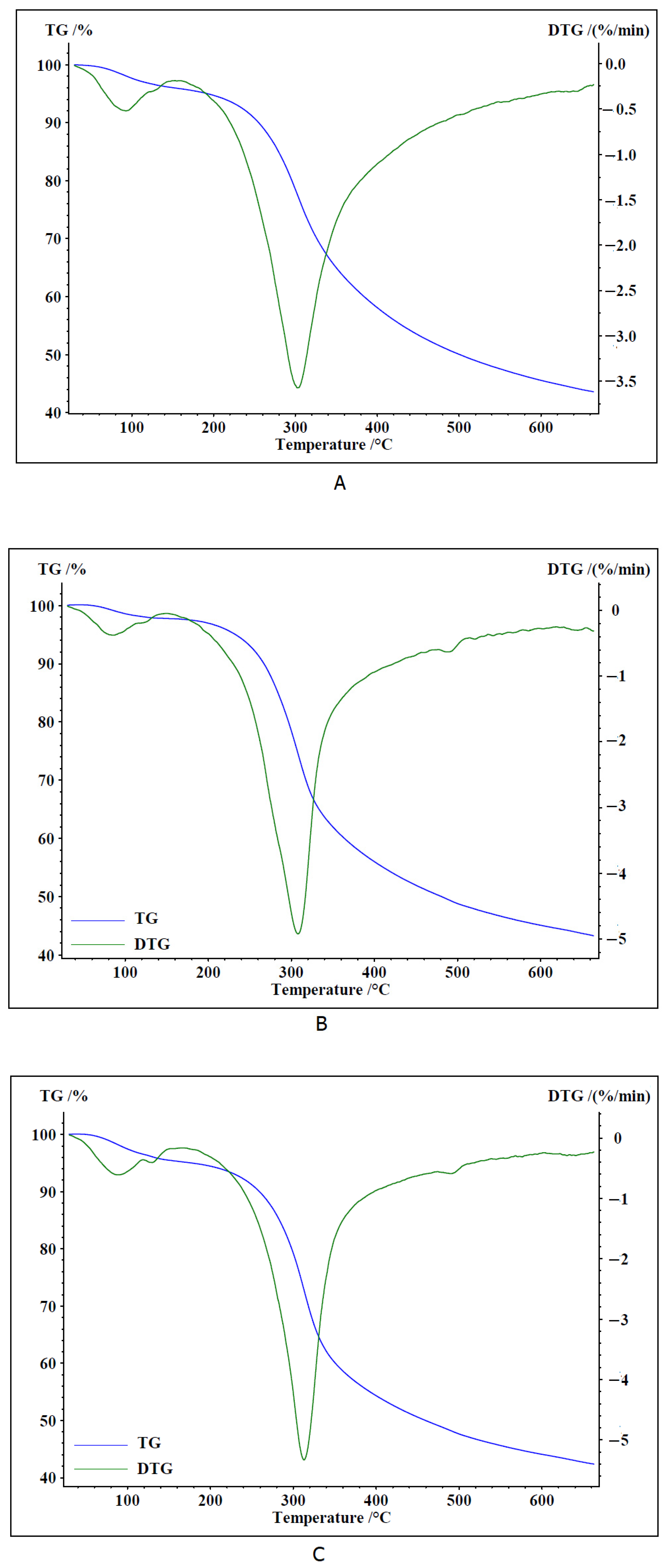
Figure 1. TG and DTG curves of dried fungal samples ((A)—F. robusta, (B)—P. pini, and (C)—P. igniarius).
Ijms 26 08013 g001
Figure 2. FTIR ATR spectra of F. robusta (black line), P. pini (blue line), and P. igniarius (green line). Continuous line—spectra of dried fruiting body; dashed line-spectra of methanolic extract.
Ijms 26 08013 g002
Figure 3. Main groups of compounds in the tested extracts of F. robusta, P. pini, and P. igniarius.
Ijms 26 08013 g003
Figure 4. Antioxidant activity of Phellinus spp. extracts.
Ijms 26 08013 g004
Figure 5. Lipid peroxidation inhibition after 5 days of reaction by Phellinus sample extracts.
Ijms 26 08013 g005
Figure 6. Antibacterial properties (MIC and MBIC) of Fomitiporia robusta, Porodoedalea pini, and Phellinus igniarius methanolic extracts against clinical bacterial strains; A—MIC > 12.5 mg/mL; B—MIC < 0.78 mg/mL; C—no inhibition of biofilm formation; D—MIC < 0.20 mg/mL; X—only for S. aureus 3907 and 4459 strains the MBC value was obtained (6.25 mg/mL).
Ijms 26 08013 g006
Figure 7. HaCaT cell viability after 24-h treatment with Fomitiporia robusta, Porodoedalea pini, and Phellinus igniarius methanolic extracts in the following concentrations: 100, 400, and 800 µg/mL. Viability was evaluated with the use of the NR assay. Statistical differences between the untreated control and cells treated with extracts were evaluated with the use of one-way ANOVA: * p < 0.05, ** p < 0.005, *** p < 0.002, and **** p < 0.0001.
Ijms 26 08013 g007
Figure 8. Fomitiporia robusta, Porodoedalea pini, and Phellinus igniarius extracts affect ROS production in HaCaT cells. Representative fluorescent images (40 ×) of CellROX ®Green Reagent-labeled cells, treated for 30 min: (a) untreated cells; 400 µg/mL (b) and 800 µg/mL (c) extracts of F. robusta; 400 µg/mL (d) and 800 µg/mL (e) extracts of P. pini; and 400 µg/mL (f) and 800 µg/mL (g) extracts of Phellinus igniarius; scale bar = 100 μm. Bar graph (h) summarizing the effect on ROS production after treatment with different extracts at concentrations of 400 and 800 µg/mL. Data are represented as mean ± SD; the presented results are not statistically significant, unless otherwise stated (* p < 0.05; ** p < 0.01).
Ijms 26 08013 g008
Figure 9. Fruiting bodies: Phellinus pini syn., Porodaedalea pini., Phellinus robustus syn. Fomitiporia robusta, and Phellinus igniarius (Photography: M. Wołkowycki and M. Karadelev).
Ijms 26 08013 g009
Table 1. Antimicrobial properties (MIC) of examined Phellinus spp. methanolic extracts.
Sample No.Species NameC.
albicans
[mg/mL]
P.
aeruginosa
[mg/mL]
S.
aureus
[mg/mL]
S.
epidermidis
[mg/mL]
K.
pneumoniae
[mg/mL]
K.
rhizophila
[mg/mL]
16.DPorodaedalea chrysoloma>50>5050>5050>50
17.DPorodaedalea pini>505025505050
18.DPhellinus tremulae>505012,5505050
19.DPhellinus laevigatus>50>5050>50>50>50
21.DPhellinus tuberculosus>50>5025>5050>50
22.DFomitiporia robusta>505025255025
25.DPhellinopsis conchata25>5025>505050
38.DPhellinus igniarius>50>5025>50>5050
118.DPhellinus alni25>5025>50>5050
160.DFuscoporia ferruginosa505025505025
254.DPhellinus alni25>5025>50>50>50
255.DPhellinus alni50>50>5050>5050
256.DPhellinidium ferrugineofuscum25>5050>50>5050
257.DFomitiporia punctata>505050505050
258.DFomitiporia punctata>5050>50502525
261.DPhellinus nigricans>50>502550>50>50
262.DPhellinus populicola>505012.5>505050
266.DFomitiporia robusta>50>5025505025
261.DPhellinus nigricans>50>5012.5505050
268.DFomitiporia hartigii>505025255025
279.DPhellinus tuberculosus50>5025>50>50>50
280.DFomitiporia robusta255025505025
298.DPorodaedalea pini12.55025255025
300.DPhellinus nigricans25>502550>50>50
300.DaPhellinus nigricans>50>506.350>5050
301.DPhellinus igniarius25>506.3255050
319.DFuscoporia torulosa50>50505050>50
320.DFuscoporia torulosa25>50>50505050
322.DFomitiporia mediterranea>505050505050
329.DPorodaedalea chrysoloma50>5012.5505050
Table 2. Elemental composition of examined Phellinus spp. extracts.
ElementContent of Elements [mg/kg of Extract]
Fomitiporia robusta* SDPorodaedalea pini* SDPhellinus igniarius* SD
Cu17.500.7523.180.836.010.22
Fe10.840.695.350.225.040.88
Mn10.860.791.690.10--
Ni--0.410.140.570.14
Zn18.520.2716.150.796.010.22
Ti5.760.284.920.278.091.02
Rb12.810.5511.470.7018.270.72
K3218.030.502349.735.622287.272.91
Ca324.891.44128.022.91146.061.70
Mg----1051.271.90
P2930.881.871224.772.402117.810.94
S710.190.64581.220.94252.631.13
Cl139.010.353194.661.003240.800.62
Si202.470.8988.551.9070.702.40
As1.780.481.410.090.990.11
Br3.800.2321.542.224.180.38
* SD: standard deviation.
Table 3. Wavenumbers (cm−1), intensities, and assignments of bands occurring in the FTIR spectra of dried fruiting body samples and extracts of F. robusta, P. pini, and P. igniarius.
Fomitiporia robustaPorodaedalea piniPhellinus igniariusType of VibrationGroups of Chemical Compounds
Dried Fruiting BodyExtractDried Fruiting BodyExtractDried Fruiting BodyExtract
1645 s1733 s1647 s1711 vs1647 s1735 sνC = OCarboxylic acid, esters, fatty acids, and carbohydrates
1675 m
1556 m1554 m1555 m1552 m1549 m1553 wνCCringAromatic/phenolic compounds
1515 m1520 m1514 m1517 m1515 m1519 wνCCringAromatic/phenolic compounds
-1454 m-1451 s-1453 mδCH2, δCH3Aromatic and aliphatic compounds
--1415 m1410 m1416 m-δCH2, δCH3, δOHAromatic and aliphatic compounds, and carbohydrates
1372 m1374 m1365 m1374 m1366 m1374 mγCHringAromatic/phenolic compounds
1301 m-1310 w1280 m 1306 mδCH2OHCarbohydrates
1242 w 1245 m1243 mδCH2OHCarbohydrates
1206 w1202 m-1202 m1205 m1205 mνCO, νCC, δCOHCarbohydrates
1168 m1151 m-1155 m- δCHringAromatic/phenolic compounds
1113 m1116 m1107 m-1123 mδCHringAromatic/phenolic compounds
-1075 m-1075 m-1088 sδCHringAromatic/phenolic compounds
1054 vs, 1037 vs1029 vs1038 s1024 s1041 s1055 sνC-OCarboxylic acids, fatty acids, and carbohydrates
-993 m---1000 s
913 w946 w915 w-899 m-δCOH, δCCH, δOCHCarbohydrates
862 m871 m860 m874 m858 m865 mδCOH, δCCH, δOCHCarbohydrates
779 m779 m-809 m-800 mδCOH, δCCH, δOCHCarbohydrates
691 m691 s697 s--695 mDe-fringAromatic/phenolic compounds
632 s657 s624 m633 m627 s635 s mDe-fringAromatic/phenolic compounds
ν—stretching, δ—deforming in plane, γ—deforming out of plane, ring—vibrations of atoms of an aromatic ring, and intensities: vs—very strong, s—strong, m—medium, w—weak.
Table 4. Antibacterial properties (MIC, MBC, and MBIC) of Fomitiporia robusta, Porodoedalea pini, and Phellinus igniarius methanolic extracts against clinical bacterial strains.
Methanolic
Extract
Concentration
[mg/mL]
Clinical Strains
Pseudomonas
aeruginosa
Staphylococcus aureusStaphylococcus
epidermidis
1900195419553907406944591887072542
F. robustaMIC3.133.136.256.256.256.25>6.25>6.256.25
MBCBS aBS aBS aBS aBS aBS aBS aBS aBS a
MBIC3.133.136.256.253.136.25- c- b6.25
P. piniMIC6.256.256.251.560.781.560.783.13>12.5
MBCBS aBS aBS a6.25BS a6.25BS aBS aBS a
MBIC6.256.2512.50<0.20<0.20<0.200.78- b- c
P. igniariusMIC6.256.256.256.256.256.250.783.13<0.78
MBCBS aBS aBS aBS aBS aBS aBS aBS aBS a
MBIC6.2512.5012.506.256.2512.501.56- b<0.78
ERTMIC0.060.060.060.502 × 10−4>0.502 × 10−42 × 10−32 × 10−4
MBCBS aBS aBS aBS aBS a>0.504 × 10−32 × 10−24 × 10−3
MBIC0.060.060.060.502 × 10−4- c2 × 10−4- b2 × 10−4
a Bacteriostatic (BS) properties of compounds for which the MIC value was determined. b This bacterial strain does not form biofilm. c No inhibition of biofilm formation.
Table 5. Characteristics of evidentiary specimens of Phellinus spp. samples.
Sample No.Evidence Specimen NumberSpecies NameSubstrate/Host
(Species Name)
Collection SiteCollected and Identified
16.DBLS-M-05505Porodaedalea chrysoloma
(Fries) Fiasson & Niemelä [syn. Phellinus chrysoloma (Fr.) Donk]
Picea abiesPoland, Białowieża ForestLeg. et det. Marek Wołkowycki
17.DBLS-M-05498Porodaedalea pini (Brot.) Murrill [syn.
Phellinus pini (Brot.) A. Ames]
Pinus sylvestrisPoland, Białowieża ForestLeg. et det. Marek Wołkowycki
18.DBLS-M-05506Phellinus tremulae (Bondartsev) Bondartsev & P.N. BorissovPopulus
tremula
Poland, Białowieża ForestLeg. et det. Marek Wołkowycki
19.DBLS-M-05512Phellinus laevigatus
(Fries) Bourdot & Galzin
Betula pendulaPoland, Białowieża ForestLeg. et det. Marek Wołkowycki
21.DBLS-M-05493Phellinus tuberculosus 
(Baumgarten) Niemelä [syn. Phellinus pomaceus (Pers.) Maire]
Prunus
domestica
Poland, Białowieża ForestLeg. et det. Marek Wołkowycki
22.DBLS-M-05497Fomitiporia robusta 
(P. Karst.) Fiasson & Niemelä [syn. Phellinus robustus (P. Karst.) Bourdot & Galzin]
Quercus roburPoland, Białowieża ForestLeg. et det. Marek Wołkowycki
25.DBLS-M-02596Phellinopsis conchata (Persoon) Dai [syn. Phellinus conchatus (Pers.: Fr.) Quél]Salix capreaPoland, Białowieża ForestLeg. et det. Marek Wołkowycki
38.DBLS-M-01027Phellinus igniarius 
(L.) Quél.
Salix fragilisPoland, Sokolskie Hill,
Losiniany
Leg. et det. Marek Wołkowycki
118.DBLS-M-05609Phellinus alni 
(Bondartsev) Parmasto
Alnus glutinosaPoland, Białowieża ForestLeg. et det. Marek Wołkowycki
160.DBLS-M-05495Fuscoporia ferruginosa 
(Schrad ex J.F. Gmelin) Murill [syn. Phellinus ferruginosus (Schrad.: Fr.) Pat.]
Fraxinus
excelsior
Poland, Białowieża ForestLeg. et det. Marek Wołkowycki
254.DBLS-M-05492Phellinus alni 
(Bondartsev) Parmasto
Malus
domestica
Poland, Białowieża ForestLeg. et det. Marek Wołkowycki
255.DBLS-M-05490Phellinus alni 
(Bondartsev) Parmasto
Carpinus
betulus
Poland, Białowieża ForestLeg. et det. Marek Wołkowycki
256.DBLS-M-05448Phellinidium ferrugineofuscum 
(P. Karst) Fiasson & Niemelä [syn. Phellinus ferrugineofuscus (P. Karst.) Bourdot]
Picea abiesPoland, Białowieża ForestLeg. et det. Marek Wołkowycki
257.DBLS-M-05494Fomitiporia punctata 
(Fries ex P. Karsten) Murill [syn. Phellinus punctatus (P.Karst.) Pilát]
Corylus avellanaPoland, Białowieża ForestLeg. et det. Marek Wołkowycki
258.DBLS-M-05499Fomitiporia punctata 
(Fries ex P. Karsten) Murill [syn. Phellinus punctatus (P.Karst.) Pilát]
Sorbus
aucuparia
Poland, Białowieża ForestLeg. et det. Marek Wołkowycki
261.DBLS-M-05509Phellinus nigricans 
(Fries) P. Karsten
Betula pendulaPoland, Białowieża ForestLeg. et det. Marek Wołkowycki
262.DBLS-M-05507Phellinus populicola 
Niemelä
Populus
tremula
Poland, Białowieża ForestLeg. et det. Marek Wołkowycki
266.DBLS-M-02977Fomitiporia robusta 
(P. Karst.) Fiasson & Niemelä [syn. Phellinus robustus (P. Karst.) Bourdot & Galzin]
Robinia
pseudoacacia
Poland, Białowieża ForestLeg. et det. Marek Wołkowycki
261.DBLS-M-05509Phellinus nigricans 
(Fries) P. Karsten
Betula
pubescens
Poland, Białowieża ForestLeg. et det. Marek Wołkowycki
268.DBLS-M-05496Fomitiporia hartigii 
(Allesch. & Schnabl) Fiasson & Niemelä [syn. Phellinus hartigii (Allesch. & Schnabl) Pat]
Abies albaPoland, Beskid NiskiLeg. et det. Anna Hreczka rev. Marek Wołkowycki
279.DBLS-M-05599Phellinus tuberculosus 
(Baumgarten) Niemelä [syn. Phellinus pomaceus (Pers.) Maire]
Cerasus aviumPoland, Kuraszewo,
wild fruit orchard
Leg. Konrad Wilamowski det. Marek Wołkowycki
280.DBLS-M-05610Fomitiporia robusta 
(P. Karst.) Fiasson & Niemelä [syn. Phellinus robustus (P. Karst.) Bourdot & Galzin]
Quercus rubraPoland, Białowieża ForestLeg. et det. Marek Wołkowycki
298.DBLS-M-10167Porodaedalea pini (Brot.) Murrill (syn. Porodaedalea pini (Brot.) A. Ames)Pinus pineaPortugal, Estremadura,
Sesimbra, Lagoa de Albufeira
Leg. et det. Mitko Karadelev
300.DBLS-M-10094Phellinus nigricans 
(Fries) P. Karsten
Fagus sylvaticaPoland, Białowieża ForestLeg. Konrad Wilamowski det. Marek Wołkowycki
300.DaBLS-M-10164Phellinus nigricans 
(Fries) P. Karsten
Fagus sylvaticaPoland, Baltic Coast,
“Zagorska Struga” Valley
Leg. et det. Mirosław Wantoch-Rekowski
rev. Marek Wołkowycki
301.DBLS-M-10093Phellinus igniarius 
(L.) Quél.
Sorbus
intermedia
Poland, Romnicka ForestLeg. Konrad Wilamowski det. Marek Wołkowycki
319.DBLS-M-10412Fuscoporia torulosa 
(Pers.) T. Wagner & M. Fisch [syn. Phellinus torulosus (Pers.) Bourdot & Galzin]
Quercus ilexItaly, Perugia, Sacro Bosco di Monteluco, SpoletoLeg. et det. Carolina Girometta
320.DBLS-M-10413Fuscoporia torulosa 
(Pers.) T. Wagner & M. Fisch [syn. Phellinus torulosus (Pers.) Bourdot & Galzin]
Robinia
pseudoacacia
Italy, Pavia, “Bosco Siro Negri” State Nature ReserveLeg. et det. Carolina Girometta
322.DBLS-M-10414Fomitiporia mediterranea 
M. Fisch. [syn. Phellinus punctatus (P. Karst.) Pilát]
Robinia
pseudoacacia
Italy, Pavia, “Bosco Siro Negri” State Nature Reserve Leg. et det. Carolina Girometta
329.DBLS-M-05505Porodaedalea chrysoloma 
(Fr.) [syn. Porodaedalea abietis]
[syn. Phellinus chrysoloma (Fr.) Donk]
Picea abiesPoland, Białowieża ForestLeg. et det. Marek Wołkowycki
Disclaimer/Publisher’s Note: The statements, opinions and data contained in all publications are solely those of the individual author(s) and contributor(s) and not of MDPI and/or the editor(s). MDPI and/or the editor(s) disclaim responsibility for any injury to people or property resulting from any ideas, methods, instructions or products referred to in the content.