Article

Molecular Characterization of Hemopexin in the Siberian Sturgeon (Acipenser baerii): Evolutionary Insights and Differential Expression Under Immune and Thermal Stresses

1
Faculty of Fisheries Life Sciences, Pukyong National University, Busan 48513, Republic of Korea
2
Department of Aquatic Life Medicine, Pukyong National University, Busan 48513, Republic of Korea
*
Author to whom correspondence should be addressed.
Int. J. Mol. Sci. 2025, 26(16), 7934; https://doi.org/10.3390/ijms26167934 (registering DOI)
Submission received: 7 July 2025 / Revised: 12 August 2025 / Accepted: 13 August 2025 / Published: 17 August 2025
(This article belongs to the Section Molecular Biology)

Abstract

Hemopexin (HPX) is a heme-binding glycoprotein involved in detoxification, oxidative stress regulation, and immune defense. In teleosts, gene duplication gave rise to Wap65-1 and Wap65-2, but the structure and function of ancestral HPX in basal actinopterygians remain poorly understood. Here, we characterized HPX from the Siberian sturgeon (Acipenser baerii), a slow-evolving chondrostean species that diverged prior to the teleost-specific duplication. Structural modeling and superimposed 3D alignment revealed high similarity between sturgeon HPX, human HPX, and Wap65-2, but not Wap65-1. Phylogenetic analysis placed sturgeon HPX in a distinct basal clade within the Actinopterygian lineage, positioned below the divergence of Wap65 paralogs. Tissue expression was liver-dominant but also evident in skin and kidney, and HPX transcripts increased during larval development. Under Aeromonas hydrophila challenge, HPX was strongly upregulated in immune-relevant tissues. Thermal challenge also induced modest, tissue-specific changes, particularly in peripheral tissues. These results indicate that ancestral HPX possessed multivalent stress responsiveness—primarily immune-related—with supplementary thermal sensitivity. The observed functional flexibility is consistent with the hypothesis that ancestral HPX functions were partitioned following Wap65 gene duplication in teleosts, with Wap65-1 evolving toward a specialized thermal response role.

1. Introduction

Hemopexin (HPX) is a vertebrate-specific glycoprotein that binds free heme with high affinity, playing a crucial role in heme detoxification, iron homeostasis, and inflammation modulation during infection and tissue injury. As a classical acute-phase protein (APP), HPX limits heme-driven oxidative stress and prevents host tissue damage, particularly under hemolytic or septic conditions [1,2,3]. In mammals, HPX is predominantly synthesized in the liver and is systemically upregulated during inflammation [1].
In teleost fish, HPX orthologs were initially identified as warm-temperature acclimation-associated proteins (Wap65), named after a 65 kDa protein upregulated during thermal stress [4,5,6]. Following the teleost-specific third round of whole-genome duplication (3R WGD), two paralogous genes—Wap65-1 and Wap65-2—emerged [7]. Comparative analyses suggest that Wap65-2 retains high sequence and functional homology with mammalian HPX, including conserved heme-binding domains and immune-responsive regulation. In contrast, Wap65-1 appears to have diverged functionally, contributing more prominently to thermal acclimation and general stress responses [8,9,10]. This gene duplication has served as a model for subfunctionalization and neofunctionalization, raising important questions about the ancestral state, tissue specificity, and regulatory divergence of HPX-related genes in vertebrates [7,10].
Among basal actinopterygians (i.e., lineages that diverged before the 3R WGD), sturgeons (Acipenseriformes) are members of the ancient Chondrostei clade, representing an evolutionary lineage that predates the teleost WGD but exhibits independent lineage-specific polyploidy [11,12]. Their unique phylogenetic position offers valuable insight into the pre-duplication architecture and ancestral functions of HPX-like genes [13,14]. Additionally, given their endangered status and growing importance in aquaculture for caviar production, understanding immune- and stress-responsive molecules such as HPX/Wap65 is not only of evolutionary interest but also of practical relevance to sustainable farming practices [15,16,17].
The Siberian sturgeon (Acipenser baerii), belonging to the ancient family Acipenseridae, is a key species in both conservation and global aquaculture. Native populations are distributed across the rivers of Siberia from the Ob to the Kolyma and occur in both semi-anadromous and strictly freshwater forms, with Lake Baikal harboring an isolated lacustrine population [18]. Historically, annual commercial catches exceeded 1700 metric tons, but overfishing and other anthropogenic pressures have driven severe population declines [19,20,21]. To counteract these losses, large-scale aquaculture was initiated in the 1970s and is now practiced in multiple countries, including Russia, France, Germany, Italy, Austria, Japan, and Korea, making A. baerii one of the principal sturgeon species in the global farming industry [22,23,24]. From a phylogenetic perspective, sturgeons diverged prior to the teleost radiation and thus occupy a basal position within Actinopterygii. Their genomes, including A. baerii are not expected to contain both Wap65-1 and Wap65-2 paralogs, but instead likely retain a single ancestral HPX gene. Despite this, molecular studies on HPX in sturgeons remain scarce, with only limited work focusing on sequence identity or immune challenge-related expression [25,26]. Crucially, the broader functional roles of HPX—such as thermal responsiveness or tissue-specific transcriptional regulation—have yet to be fully characterized in this lineage.
To address these gaps, the present study aimed to (1) characterize the structural and phylogenetic relationships of Siberian sturgeon (A. baerii) HPX to vertebrate HPX orthologs and teleost Wap65 paralogs, including three-dimensional protein modeling; and (2) investigate both basal expression patterns (tissue distribution and ontogenetic stage) and differential expression in response to bacterial infection and thermal challenge.

2. Results

2.1. Characterization of Acipenser baerii HPX cDNA and Deduced Amino Acid Sequences

The cloned cDNA sequence of Acipenser baerii HPX was 1800 bp in length, excluding the poly(A+) tail. It consisted of a 5′ untranslated region (5′-UTR) of 23 bp, an open reading frame (ORF) of 1368 bp including the TAG stop codon, and a 3′ untranslated region (3′-UTR) of 399 bp excluding the stop codon and poly(A+) tail. A canonical polyadenylation signal (AATAAA) was identified 21 bp upstream of the poly(A+) tail. The ORF encodes a polypeptide of 455 amino acids. The theoretical molecular weight of the deduced protein was calculated to be 57.8 kDa, and the predicted isoelectric point (pI) was 6.03.
A signal peptide comprising 19 amino acids was predicted by SignalP 6.0, with a cleavage site between Ala19 and Ala20. Conserved domain searches using InterPro and CDD identified two hemopexin (HX) domains spanning residues 65–250 and 266–453, respectively. Six HX-like repeat regions were detected (Figure 1). Eight residues involved in metal binding were identified: in the N-domain, D69 and I71 (DAI), D110 and A112 (DAA), D163 and A165 (DAA), and T208 and A210 (TSA); in the C-domain, D273 and F275 (DAF), N318 and V320 (NAV), D365 and A367 (DAA), and D408 and A410 (DAA). Two conserved histidine residues related to heme binding were located at His255 and His302. Five cysteine pairs were predicted to form disulfide bonds, and eight potential N-glycosylation sites were predicted at N99, N140, N152, N160, N206, N372, N445, and N455 with moderate scores (0.525–0.7288) (Figure 1).
Multiple sequence alignment showed that A. baerii HPX shares greater similarity with Wap65-2 than with Wap65-1. Several residues were conserved exclusively between HPX and Wap65-2 but not Wap65-1. The heme-binding histidine in the N-domain was uniquely conserved in HPX/Wap65-2, while the corresponding histidine in the C-domain was present in both Wap65-2 and several Wap65-1 orthologs (Figure 1). Pairwise comparison of amino acid sequence identity and similarity revealed the highest similarity with Acipenser ruthenus (98.0% identity, 98.5% similarity), followed by Lepisosteus oculatus (55.4% identity, 65.4% similarity). The average identity and similarity with tetrapod HPX orthologs were 37.2 ± 2.7% and 50.8 ± 3.5%, respectively. Comparisons with Wap65-1 showed 41.4 ± 2.1% identity and 56.9 ± 2.1% similarity, while those with Wap65-2 yielded 48.9 ± 1.6% identity and 63.5 ± 1.7% similarity. The identity and similarity scores with Wap65-2 were significantly higher than those with Wap65-1 (p < 0.05). No lineage-specific differences were observed across Ostariophysi, Protacanthopterygii, Stomiatii, or Neoteleostei groups (Supplementary Figure S1).

2.2. Predicted 3D Structure of A. baerii HPX and Superimposed Visualization of Conserved Motifs with Human HPX

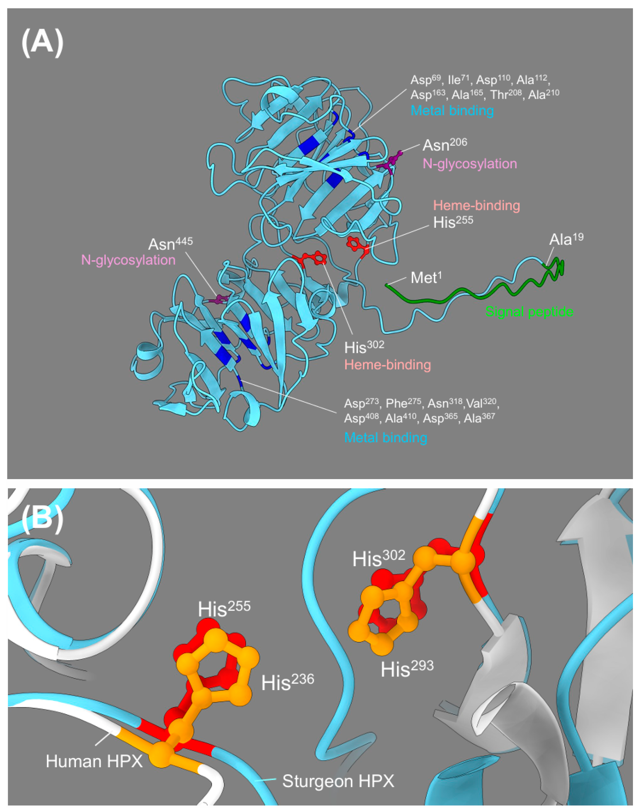
The three-dimensional structure of Acipenser baerii HPX was predicted using AlphaFold2. The resulting model exhibits a characteristic two-domain β-propeller-like fold, consistent with known hemopexin family structures. An unstructured region was observed immediately following the signal peptide, a feature commonly found in other HPX and Wap65 orthologs. The N- and C-domains are organized in a circular fashion, forming a central cavity, typical of heme-binding hemopexin proteins (Figure 2A; Supplementary Figure S2).
To evaluate structural conservation across species, the A. baerii HPX model was superimposed with the crystal structure of human HPX. Despite the relatively low sequence identity between the two proteins (33.7%), the superimposed models showed remarkable similarity in overall architecture. Key functional motifs involved in heme binding, metal coordination, and disulfide bonding were structurally conserved in both proteins (Figure 2B, Supplementary Figure S3).
Superimposed comparison of heme-binding residues revealed that A. baerii HPX shares two conserved histidine residues—His255 and His302—corresponding to His236 and His293 in human HPX. In contrast, human HPX contains four known histidine residues involved in heme coordination (Supplementary Figure S3A). Residues involved in metal binding were also structurally conserved. Eight key sites (Asp69/Ile71, Asp110/Ala112, Asp163/Ala165, Thr208/Ala210, Asp273/Phe275, Asn318/Val320, Asp365/Ala367, and Asp408/Ala410) were all positioned similarly in both models, indicating conserved spatial arrangement (Supplementary Figure S3B). Regarding disulfide bonding, the ten predicted cysteine residues in A. baerii HPX showed perfect pairwise matching with those in human HPX. All disulfide bridges appeared conserved in position and orientation, supporting strong structural conservation between the two species (Supplementary Figure S3C). Although both A. baerii and human HPX exhibit multiple potential N-glycosylation sites, only two sites—Asn206 and Asn455 in sturgeon, corresponding to Asn187 and Asn453 in human HPX—were conserved in both sequence position and spatial localization in the 3D models (Supplementary Figure S3D).

2.3. Molecular Phylogenetic Analysis of Vertebrate HPX and Wap65 Orthologs

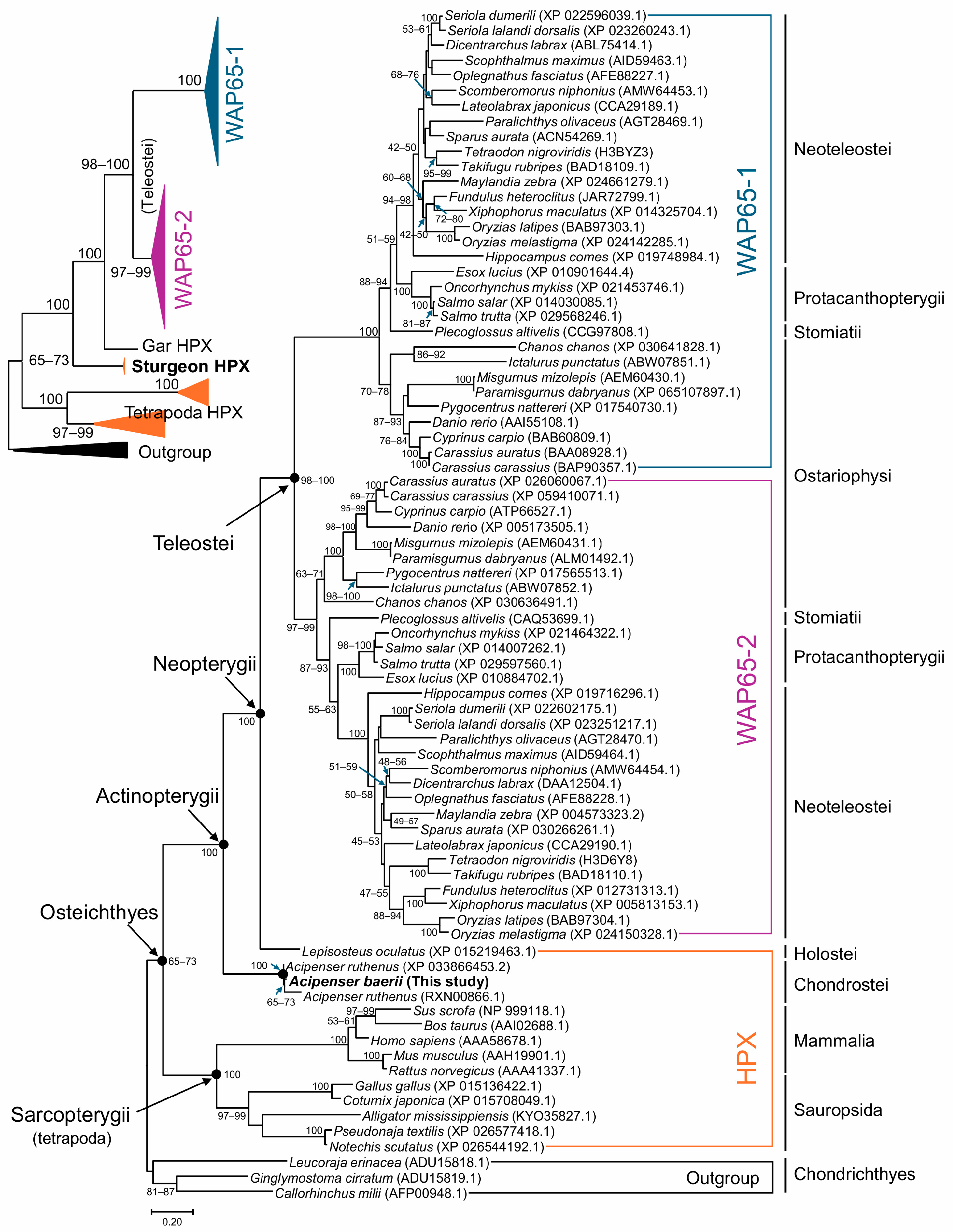
A maximum likelihood (ML) phylogenetic tree was constructed using a dataset of vertebrate HPX and Wap65 amino acid sequences, rooted with three cartilaginous fish HPX sequences as the outgroup. The overall tree topology was consistent with the established taxonomic hierarchy of vertebrates. In the teleost lineage, Wap65 sequences were grouped by isoform (Wap65-1 and Wap65-2) rather than by species, indicating that isoform divergence occurred prior to the radiation of extant teleosts. Within each isoform clade, sequences clustered into subgroups reflecting taxonomic divisions such as Ostariophysi, Stomiatii, Protacanthopterygii, and Neoteleostei, supporting phylogenetic coherence across major teleost groups (Figure 3).
The Wap65 clades formed a large teleostean cluster that was connected to the non-teleost HPX gene of the holostean Lepisosteus oculatus (gar), which in turn was connected to the HPX genes of Acipenser baerii and A. ruthenus, representing the Chondrostei subclass. This positioning reflects the basal placement of Acipenseriformes within the superclass Actinopterygii (ray-finned fishes). The actinopterygian clade was then joined with Sarcopterygii (lobe-finned fishes and tetrapods), forming a monophyletic Osteichthyes (bony fish) group (Figure 3).
To evaluate the robustness of tree topology, additional phylogenetic reconstructions were performed using the neighbor-joining (NJ) method under varying option settings (e.g., substitution models, rate heterogeneity, gap/missing data treatment). Although some minor differences in internal subgrouping were observed among teleost Wap65 isoform clades, the relative placement of A. baerii HPX remained consistent—basal within the actinopterygian lineage and distinct from Wap65 isoforms—across all NJ tree topologies (Supplementary Figure S4A,B).

2.4. Structural Comparison of A. baerii HPX with Other HPX and Wap65 Orthologs

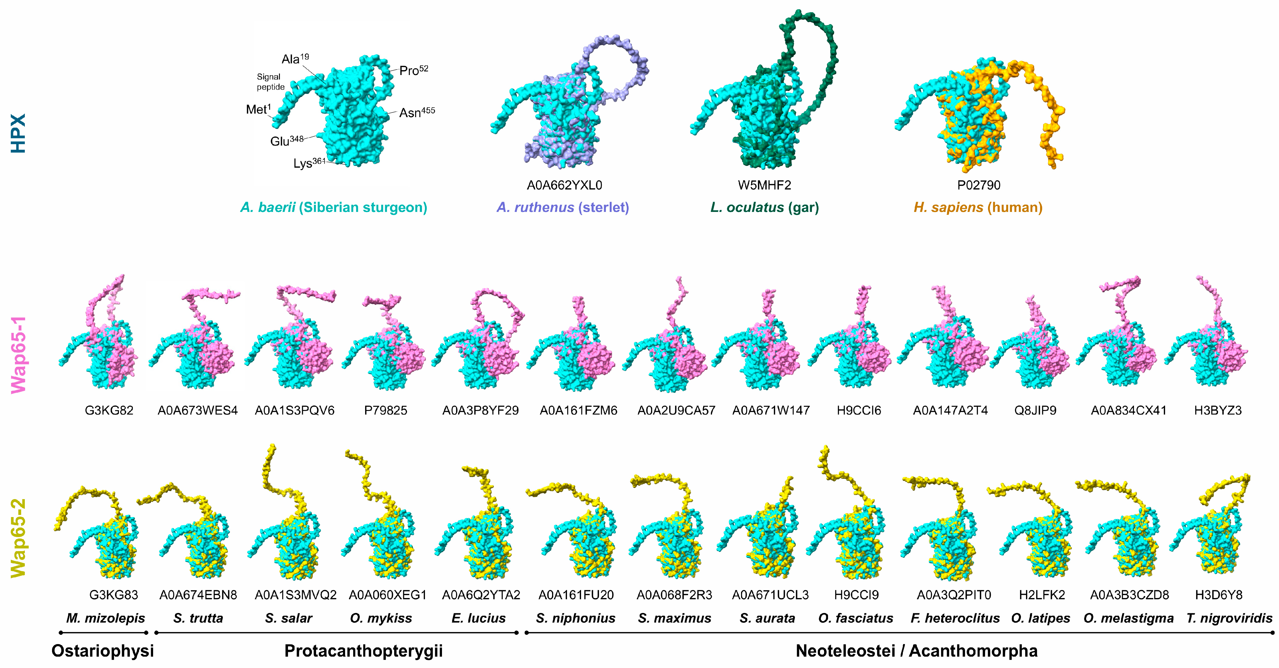
The predicted 3D structure of Acipenser baerii HPX was first compared with HPX orthologs from Acipenser ruthenus (sterlet) and Lepisosteus oculatus (gar). The model showed a high degree of structural similarity with A. ruthenus HPX, consistent with their close evolutionary relationship. The holostean HPX from L. oculatus also exhibited strong structural conservation with A. baerii, particularly in the arrangement of the two β-propeller-like domains (Figure 4). These findings support the phylogenetic positioning of sturgeon HPX as a basal member of the actinopterygian clade.
Further comparisons were made with representative teleost Wap65 isoforms. Superimposition of A. baerii HPX with multiple Wap65-2 models from species across Ostariophysi, Protacanthopterygii, and Neoteleostei showed consistent overall structural similarity. In contrast, all comparisons with Wap65-1 isoforms revealed a distinct structural deviation in the C-domain, where a non-aligned or uniquely diverged region was consistently observed. This suggests that the C-domain has undergone isoform-specific structural divergence in Wap65-1, differentiating it from the ancestral HPX fold retained in A. baerii (Figure 4).

2.5. Tissue Distribution of HPX mRNA Expression in Fingerling A. baerii

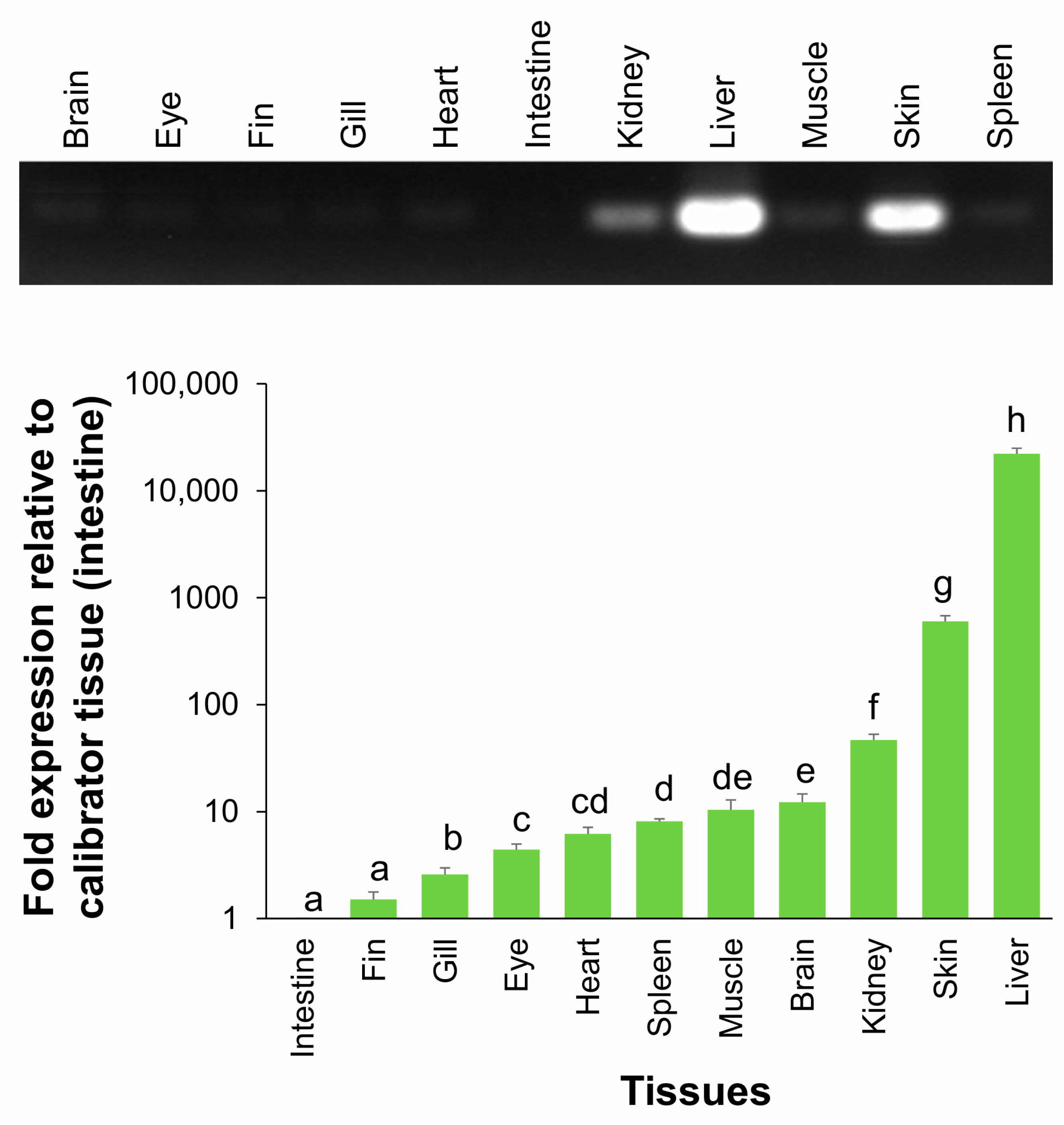
To evaluate the tissue-specific expression profile of HPX in fingerling Acipenser baerii, RT-qPCR analysis was conducted using RNA extracted from multiple tissues. HPX mRNA was detected in all examined tissues, but transcript abundance varied markedly. The liver showed overwhelmingly dominant expression, followed by skin and kidney, whereas tissues such as intestine, fin, and gill exhibited only trace levels (Figure 5). Normalization using each of the three selected reference genes yielded consistent expression ranking among tissues, although the absolute fold differences varied slightly depending on the reference (Supplementary Figure S5). When the intestine—identified as the tissue with the lowest expression—was used as the calibrator, HPX expression in the liver averaged 22,128 ± 2690-fold higher. The expression levels in skin and kidney were also elevated, measured at 600.6 ± 77.2 and 46.8 ± 6.2-fold, respectively, compared to the intestine.

2.6. Ontogenetic Expression of HPX During Early Development in A. baerii

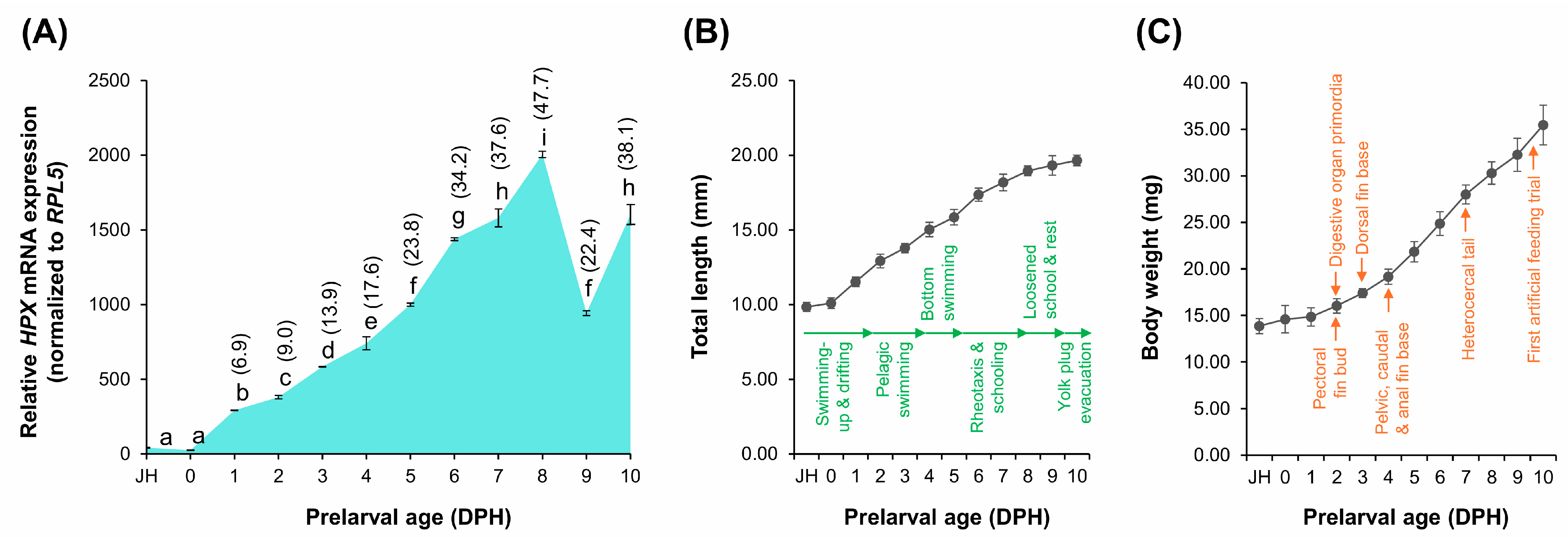
To examine the developmental regulation of HPX transcription, HPX mRNA levels were measured in A. baerii prelarvae and larvae from the just-hatched (JH) stage through 10 days post-hatching (DPH). During this period, the fish exhibited characteristic early life stage transitions, including fin and digestive primordia formation and behavioral shifts such as the pelagic-to-benthic transition and the onset of schooling behavior. Normalization was performed using RPL5, previously validated as a stable internal control for developmental samples. HPX expression remained minimal through the JH and 0 DPH stages but began to increase significantly from 1 DPH onward (p < 0.05). Expression continued to rise and peaked at 8 DPH. A marked, transient reduction was observed at 9 DPH, followed by partial recovery at 10 DPH (Figure 6A). This decline in HPX expression occurred despite continuous increases in total length and body weight throughout the same period (Figure 6B,C), indicating that the transcriptional fluctuation was not associated with general somatic growth.

2.7. Dose-Dependent Modulation of HPX Expression Following A. hydrophila Challenge in A. baerii Fingerlings

During the survival/mortality test across a range of Aeromonas hydrophila concentrations (2 × 10⁴ to 2 × 10⁹ CFU/g body weight), mortality occurred rapidly and was dose-dependent, with all deaths occurring within 36 h post-injection. Complete mortality was observed at 2 × 10⁹ CFU/g, while no significant mortality occurred at 2 × 10⁴ CFU/g and below. The 2 × 10⁵ CFU/g dose resulted in approximately 50% survival, indicating an LD50 near this level and defining 2 × 10⁴ CFU/g as the upper threshold of the sublethal range (Supplementary Figure S6).
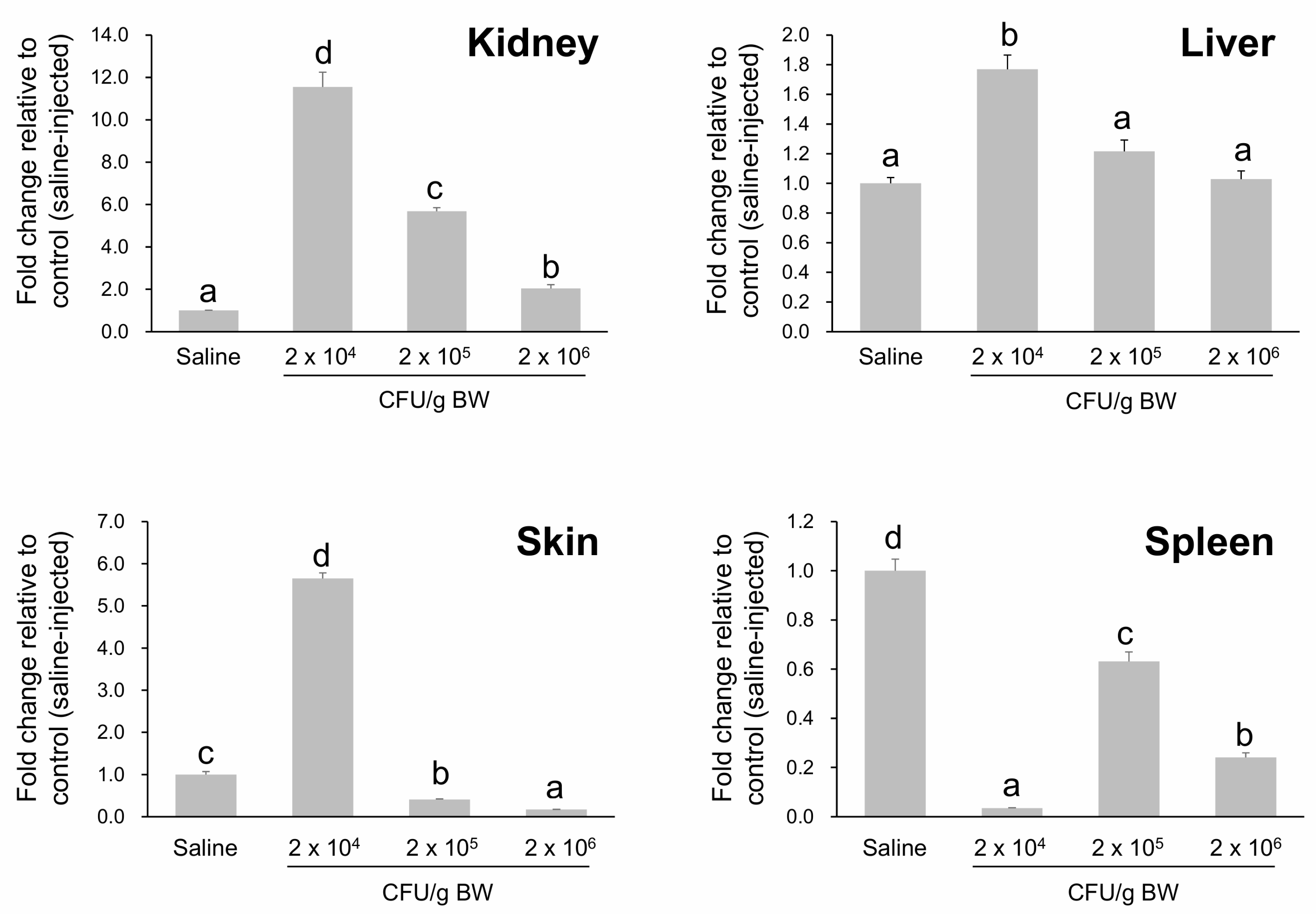
To examine early transcriptional responses, HPX mRNA levels were measured 12 h post-injection (HPI) following exposure to three bacterial doses: 2 × 10⁴, 2 × 10⁵, and 2 × 10⁶ CFU/g. A clear dose-dependent pattern of HPX modulation was observed across tissues (Figure 7). In the kidney, HPX expression was strongly induced at 2 × 10⁴ CFU/g (12.6-fold relative to control) but decreased progressively at higher doses. The skin showed a similar trend, with peak induction at the lowest dose and marked downregulation at the highest dose. In the liver, a modest increase was observed at 2 × 10⁴ CFU/g, with no further upregulation at higher concentrations. Notably, HPX expression in the spleen was significantly suppressed at all tested doses, suggesting an immunosuppressive or exhaustion-related response. These results indicate that excessive bacterial burden can override the capacity for HPX upregulation, particularly in mucosal and hematopoietic tissues. Based on both survival data and gene expression profiles, the 2 × 10⁴ CFU/g dose was selected as an optimal sublethal challenge condition for subsequent immune response analyses.

2.8. Temporal Expression Dynamics of HPX and TF Following A. hydrophila Challenge

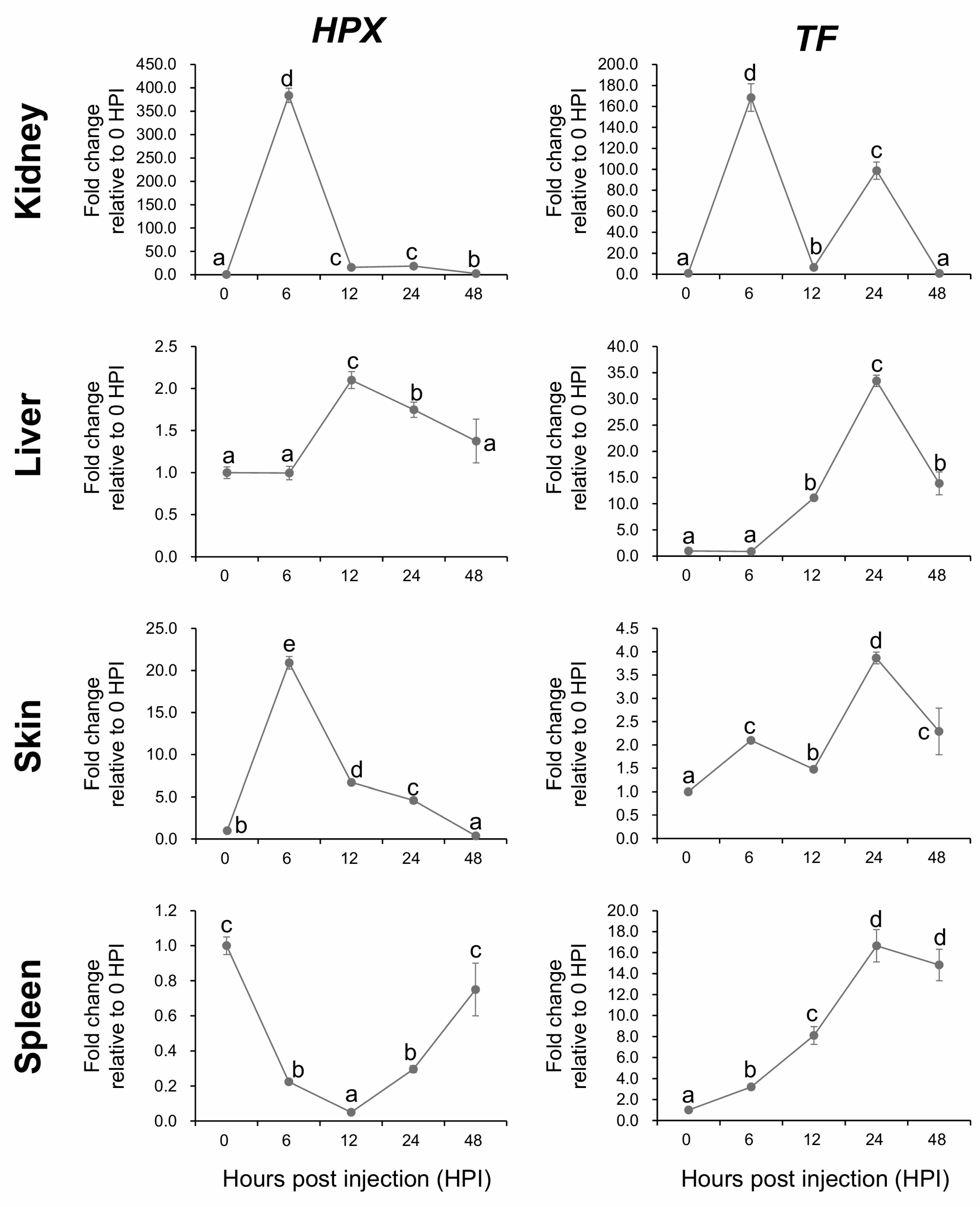
Following the initial dose optimization, the temporal patterns of HPX expression were examined in fingerling A. baerii during the early immune response to A. hydrophila. Transcript levels were measured at 0, 6, 12, 24, and 48 h post-injection (HPI), alongside expression of the known immune transcription factor transferrin (TF), across four tissues: kidney, liver, skin, and spleen (Figure 8).
In the kidney, HPX was rapidly and robustly upregulated, reaching a peak of nearly 400-fold relative to baseline at 6 HPI, before gradually decreasing at later timepoints, although remaining elevated above baseline throughout the period. This early upregulation closely mirrored the pattern observed for TF, which also peaked at 6 HPI with over 160-fold induction, followed by a second peak at 24 HPI. In contrast, HPX expression in the liver showed a delayed onset, with no significant upregulation at 6 HPI but reaching a peak at 12 HPI. Thereafter, expression steadily declined, returning to baseline by 48 HPI. The TF response in the liver was similarly delayed, with no early upregulation at 6 HPI and maximal induction observed at 24 HPI.
In the skin, HPX expression followed a pattern similar to that in the kidney, with strong induction at 6 HPI followed by a decline over time. TF in the skin was modestly induced at 6 HPI and showed a second, stronger peak at 24 HPI, again resembling the kidney pattern. In the spleen, the expression dynamics differed notably. HPX was rapidly downregulated following challenge, reaching its lowest level at 12 HPI and returning to baseline by 48 HPI. In contrast, TF expression in the spleen steadily increased, peaking at 24 HPI. This divergence suggests that spleen-specific regulation of HPX may involve immune suppression or exhaustion mechanisms distinct from those influencing TF (Figure 8).

2.9. HPX Expression Under Mild and Severe Thermal Stress in A. baerii Fingerlings

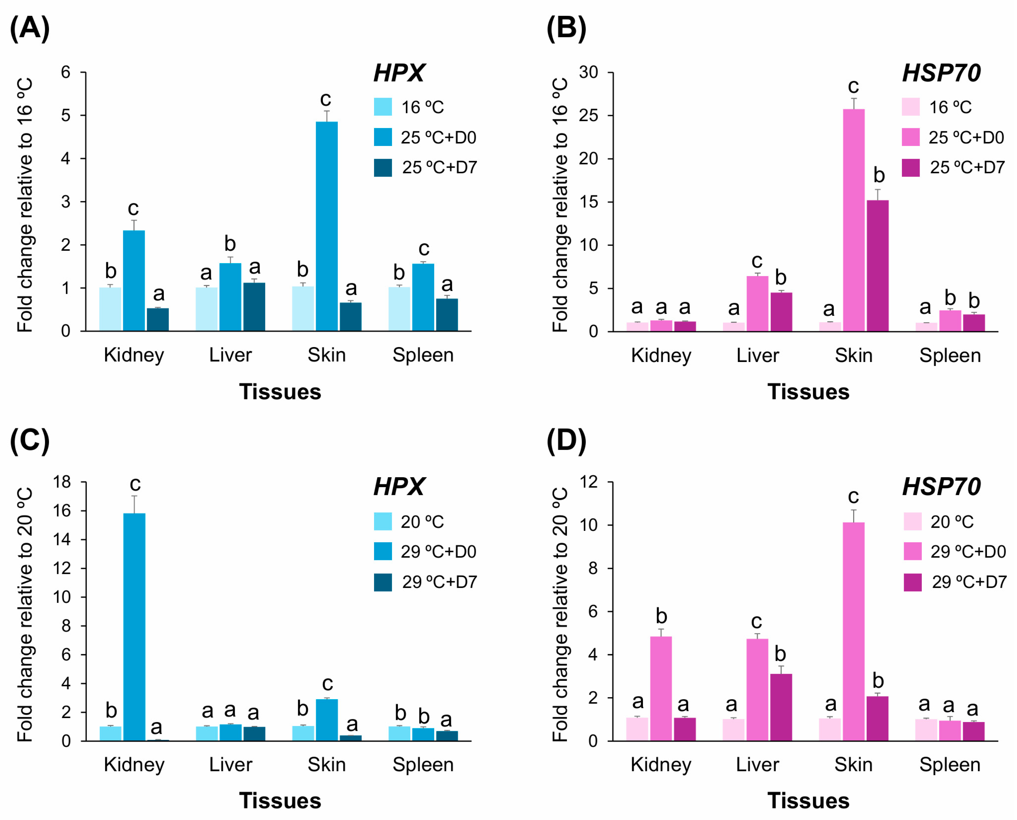
To compare transcriptional responses of HPX under high-stress temperature conditions, fingerling A. baerii were exposed to two thermal regimes: (1) a mild elevation from 16 °C to 25 °C, and (2) a severe elevation from 20 °C to 29 °C. Gene expression was assessed in the kidney, liver, skin, and spleen at baseline, immediately after temperature increase (D0), and after one week of continuous exposure (D7). HPX expression was compared alongside HSP70, a canonical thermal stress marker (Figure 9A–D).
Under the mild treatment (16 °C → 25 °C), HPX expression was upregulated in all four tissues at D0. The skin showed the highest induction (4.85-fold), followed by moderate increases in the kidney (2.3-fold), liver, and spleen (each <2-fold). However, by D7, HPX expression decreased markedly in all tissues—dropping below baseline in kidney and skin and returning to near-control levels in liver and spleen. In contrast, HSP70 expression showed more sustained activation. While the skin exhibited a dramatic increase (>25-fold at D0 and >15-fold at D7), liver and spleen also maintained significantly elevated expression at D7. The kidney showed no significant HSP70 response, highlighting a divergence from HPX activation patterns (Figure 9A,B).
Under the severe treatment (20 °C → 29 °C), HPX was highly induced in the kidney at D0 (15.8-fold) but sharply downregulated at D7 (<0.1-fold). In the skin, a 2.9-fold increase at D0 was followed by near-complete suppression at D7. In liver and spleen, HPX expression remained largely unresponsive or suppressed throughout. HSP70 expression, in contrast, was broadly upregulated across kidney, liver, and skin at D0, and remained elevated (particularly in liver) through D7. The spleen showed only modest HSP70 activation (Figure 9C,D).

3. Discussion

3.1. Structural Features and Evolutionary Context of A. baerii HPX

The structural analysis of A. baerii HPX confirms that it retains all canonical features typical of vertebrate hemopexins. Conserved heme-binding histidines, multiple metal-binding residues, disulfide-forming cysteine pairs, and a predicted β-propeller-like domain organization strongly suggest that this ancestral form remains structurally competent. These motifs are critical for heme sequestration and transport, supporting the preservation of detoxification potential in the sturgeon lineage.
While it is well established that teleost fish possess two HPX-derived paralogs, Wap65-1 and Wap65-2, sequence alignment and similarity analyses in this study clearly position A. baerii HPX closer to Wap65-2. This pattern is consistent with previous studies identifying Wap65-2 as the isoform more functionally aligned with mammalian HPX, particularly in heme-binding and immune-responsive roles [8,9,27]. This relationship is further supported by the conservation of key residues, including the two canonical heme-binding histidines. In human HPX, at least four histidines have been implicated in heme coordination; however, only two are consistently recognized as essential for direct and high-affinity binding, while others are likely to play flexible or condition-dependent roles [28,29]. The retention of these two critical residues in A. baerii HPX—at positions directly corresponding to their human counterparts—underscores the conserved functional core preserved across distant vertebrate lineages.
The structural comparison further emphasizes the divergence between ancestral HPX and teleost Wap65-1. Three-dimensional superimposition revealed high congruence between A. baerii HPX and other vertebrate HPX/Wap65-2 orthologs, particularly in the spatial arrangement of binding residues and overall domain organization. In contrast, Wap65-1 consistently exhibited a structurally divergent region in the C-domain that failed to align with the HPX scaffold. This C-domain modification may reflect isoform-specific structural remodeling associated with post-duplication neofunctionalization and/or thermal specialization in teleost lineages [10].
Phylogenetic analyses reinforce this interpretation. Both maximum likelihood (ML) and neighbor-joining (NJ) trees consistently placed A. baerii HPX basal to the teleost Wap65-1 and Wap65-2 clades, forming a distinct lineage alongside the holostean L. oculatus. Although A. baerii HPX shares significantly greater sequence identity and similarity with teleost Wap65-2 than with Wap65-1, it did not cluster within either paralogous clade. This reflects the fact that phylogenetic topology captures deeper evolutionary divergence patterns, beyond simple pairwise similarity. Importantly, the sturgeon HPX was resolved within the basal actinopterygian radiation, clearly separated from sarcopterygian HPXs and the neopterygian Wap65 paralogs. Based on current phylogenomic estimates, Chondrostei (e.g., sturgeons) diverged from Neopterygii approximately 360–330 million years ago, while Holostei (e.g., gars) split from Teleostei later, around 300–250 million years ago. The teleost-specific third round of whole-genome duplication (3R WGD), which gave rise to Wap65-1 and Wap65-2, likely occurred after these divergences, approximately around 210–200 million years ago [30,31,32].
Together, these lines of evidence support the conclusion that A. baerii HPX represents a pre-duplication, ancestral form of vertebrate hemopexin. Furthermore, sturgeons are known to exhibit unusually slow molecular evolution, with significantly lower substitution rates than most teleosts across nuclear loci [11,33]. This slow evolving genomic context, combined with the early phylogenetic position and retention of a single-copy HPX gene, suggests that A. baerii HPX preserves an evolutionarily informative prototype for understanding ancestral structural and functional features from which the teleost Wap65 paralogs diverged through sub- and neofunctionalization

3.2. Basal Expression Patterns of HPX in Tissues and Early Ontogenic Development

The basal expression profile of A. baerii HPX shows distinct tissue-specific patterns, with particularly high transcript abundance in the liver, followed by the skin and kidney. Its predominant expression in the liver aligns with both mammalian HPX and teleostean Wap65, reinforcing a conserved function in systemic heme detoxification and redox regulation. However, considerable HPX mRNA levels in the skin and kidney—mucosal and excretory tissues exposed to external stressors—suggest its additional roles in localized functions associated with oxidative defense or barrier protection [34,35]. Meanwhile, among diverged teleostean lineages, tissue expression patterns of Wap65 isoforms have shown considerable variation, indicating species- or lineage-specific regulation rather than a conserved expression framework—barring the consistent and predominant hepatic expression observed across both Wap65 isoforms [8,9,36,37].
During early development, A. baerii HPX expression increased progressively from hatching to 8 DPH. This ontogenic rise coincides with key behavioral and physiological transitions, including the development of fins, digestive organs, and elevated swimming activity. From 1 to 3 DPH, prelarvae exhibit phototactic pelagic swimming and heightened locomotor activity, likely associated with increased oxygen consumption and reactive oxygen species (ROS) production [24]. Between 5 and 8 DPH, larvae transition to benthic swimming, characterized by strong rheotaxis and active schooling—also energetically demanding behaviors [24,38]. The sustained upregulation of HPX may reflect anticipatory protection against oxidative stress linked to these high-energy phases and to the expansion of vascular structures, including branchial circulation and external gill development [39,40].
These patterns support the interpretation that HPX functions as a developmentally regulated oxidative buffer—not limited to hemolysis or infection but integrated into the broader physiological adaptations required during early life-stage transitions. Notably, the ontogenetic increase of A. baerii HPX expression mirrors the early developmental upregulation of Wap65-2 reported in several teleosts [8,41,42], suggesting a functional persistence of redox-regulatory or protective roles. In contrast, Wap65-1 tends to show less consistent activation during early development, reinforcing the closer expression alignment of HPX with Wap65-2.
A transient decline in HPX expression was observed at 9 DPH, despite continued increases in total length and body weight. This stage coincides with the developmental transition from exclusive reliance on endogenous yolk reserves to the initiation of exogenous feeding. At this point, prelarvae evacuate the yolk plug and begin ingesting artificial diets, a process that involves substantial metabolic and physiological reorganization. Behaviorally, this period is often marked by reduced locomotion (“post-schooling rest”) [38], and similar suppression has been reported for multiple immune-related genes at this interval [14,39]. These findings suggest that HPX expression during development is modulated in a non-linear fashion, likely reflecting shifts in metabolic and physiological priorities—particularly the transition from endogenous and exogenous nutrient utilization in early development.
Taken together, the liver-dominant yet extrahepatic tissue expression, combined with ontogenetic regulation during key transitional phases, supports the view that A. baerii HPX contributes to baseline physiological homeostasis in multiple tissues. These roles likely include systemic heme clearance, localized oxidative stress buffering, and early-life adaptation to rising metabolic demands.

3.3. Multivalent Stress Responsiveness: HPX Under Infection and Heat Challenge

The challenge experiments demonstrate that A. baerii HPX expression is dynamically modulated by both immune and thermal stresses. Under A. hydrophila challenge, HPX showed a dose- and tissue-dependent regulation [25,26]. At sublethal doses, strong upregulation occurred in the kidney and skin—tissues involved in immune surveillance and hemolysis mitigation—while the liver and spleen exhibited only modest or even suppressed responses. At higher bacterial loads, HPX expression decreased or became inhibited, particularly in the spleen, suggesting an exhaustion-like response under overwhelming stress [14,43,44].
The temporal regulation of HPX revealed further tissue-specific dynamics. Kidney and skin showed rapid and strong induction at 6 h post-injection, consistent with early-phase, local immune activation at barrier sites. Liver upregulation was delayed, reflecting relatively slower, systemic acute-phase signaling [45,46]. In contrast, splenic expression of HPX was transiently downregulated despite continued upregulation of TF, indicating gene-specific regulatory divergence during immune activation. Although the precise basis for this pattern remains unclear, it may involve tissue-specific transcriptional controls, differences in inflammatory sensitivity, or compartmentalized immune signaling [47,48]. Overall, HPX appears to function as a context-sensitive acute-phase protein, with both systemic and peripheral roles varying by tissue and stimulus intensity.
In thermal stress experiments, both mild (16 °C → 25 °C) and severe (20 °C → 29 °C) temperature shifts induced HPX expression at the acute phase (D0). The strongest response occurred in the skin (mild stress) and kidney (severe stress). However, in the liver—where teleost Wap65-1 is typically upregulated under thermal stress—HPX levels remained unchanged or minimally altered, unlike the robust hepatic induction of HSP70. This suggests that the thermal responsiveness of HPX in A. baerii is localized and non-systemic, likely linked to transient oxidative imbalance or inflammation in peripheral tissues rather than coordinated hepatic regulation [49,50].
However, the elevated expression at D0 markedly declined or even suppressed at D7. Compared to relatively sustained expression of HSP70, the downregulation of HPX occurred more rapidly during thermal acclimation, supporting its role as an early-phase protective or compensatory factor. Previous studies have noted that chronic thermal stress disrupts immune regulation and physiological balance in Acipenseriform species, with species- and context-specific variation in stress responses [17,51,52]. While HPX plays a role during the acute heat response, its transient expression suggests a transition to other long-term stress management systems during thermal adaptation.
While our qPCR-based analysis provides robust and reproducible quantification of HPX transcripts, it is limited to mRNA-level measurements and does not address potential post-transcriptional or post-translational regulation. In the polyploid genomic context of A. baerii, the possibility of undetected paralogous loci or sequence variants cannot be fully excluded, although the inclusion of conserved-region primer testing supports the reliability of our quantification. Future work incorporating protein-level assays (e.g., Western blotting, targeted proteomics) and functional studies will be important to further elucidate the regulatory mechanisms and physiological roles of HPX in sturgeons.
Taken together, HPX exhibits transcriptional responsiveness to both bacterial and thermal stimuli, with stressor- and tissue-specific modulation. While its immune-related regulation is robust and evident across multiple tissues, thermal sensitivity is more modest and localized. These findings support the view that ancestral HPX possessed multivalent stress reactivity, with immune-regulatory function as the dominant feature and heat responsiveness as a supplementary, context-dependent trait. This functional plasticity is consistent with the hypothesis that ancestral HPX functions were partitioned following Wap65 gene duplication in teleosts, with Wap65-1 evolving toward a specialized, heat-responsive role.

4. Materials and Methods

4.1. Molecular Cloning of Acipenser baerii Hemopexin (HPX) cDNA

Putative HPX/Wap65-related sequences were identified from a local A. baerii transcriptomic resource comprising paired-end Illumina short-read assemblies and full-length transcript isoforms generated via PacBio Iso-Seq from multiple tissue sources. Contigs showing high similarity to known vertebrate hemopexin (HPX) or warm-temperature acclimation-associated 65 kDa protein (Wap65) orthologs were assembled using Unipro UGENE software v52.1 (Unipro, LLC, Novosibirsk, Russia; https://ugene.net/, accessed on 13 August 2025).To isolate the presumed full-length open reading frame (ORF), gene-specific primers targeting the 5′- and 3′-untranslated regions (UTRs) were designed based on assembled contigs. PCR amplification was performed using 0.5 μg of total RNA extracted from the liver of a female A. baerii as the template. Oligonucleotide primer sequences are listed in Supplementary Table S1. The amplified product was visualized by electrophoresis on a 1% agarose gel stained with ethidium bromide. A single specific band was gel-purified and sequenced bidirectionally to obtain the representative cDNA sequence of A. baerii HPX.

4.2. Bioinformatic Sequence Analysis

4.2.1. Sequence Characterization

The open reading frame (ORF) of A. baerii HPX was identified using the ORF Finder tool (National Center for Biotechnology Information, Bethesda, MD, USA; https://www.ncbi.nlm.nih.gov/orffinder/, accessed on 13 August 2025), and the deduced amino acid sequence was analyzed with the ExPASy ProtParam tool (Swiss Institute of Bioinformatics, Lausanne, Switzerland; https://web.expasy.org/protparam/, accessed on 13 August 2025) to calculate molecular weight and theoretical isoelectric point (pI). The signal peptide and its predicted cleavage site were assessed using SignalP 6.0 (v6.0; Technical University of Denmark, Lyngby, Denmark; https://services.healthtech.dtu.dk/services/SignalP-6.0/, accessed on 13 August 2025). Conserved domain architecture and structural features—including heme-binding, metal-binding, and disulfide-forming motifs—were annotated using InterPro (European Bioinformatics Institute, Cambridge, UK; https://www.ebi.ac.uk/interpro/, accessed on 13 August 2025), SMART (European Molecular Biology Laboratory, Heidelberg, Germany; http://smart.embl-heidelberg.de/, accessed on 13 August 2025), and the NCBI Conserved Domain Database (CDD; National Center for Biotechnology Information, Bethesda, MD, USA; https://www.ncbi.nlm.nih.gov/Structure/cdd/wrpsb.cgi, accessed on 13 August 2025). Potential N-glycosylation sites were predicted using NetNGlyc 1.0 (v1.0; Technical University of Denmark, Lyngby, Denmark; https://services.healthtech.dtu.dk/services/NetNGlyc-1.0/, accessed on 13 August 2025).

4.2.2. Multiple Sequence Alignments and Molecular Phylogeny

A total of 79 HPX/Wap65 protein sequences from 46 vertebrate species were retrieved from GenBank based on BLASTP homology search (National Center for Biotechnology Information, Bethesda, MD, USA; https://blast.ncbi.nlm.nih.gov/Blast.cgi, accessed on 13 August 2025). These included three cartilaginous fishes (Chondrichthyes), ten sarcopterygian tetrapods, one holostean, two chondrosteans (including A. baerii), and 31 teleosts, each represented by two Wap65 isoforms where available (Supplementary Table S2). Due to inconsistent nomenclature in public databases, sequence inclusion was based on alignment similarity rather than annotated gene names. Multiple sequence alignments were performed using ClustalW (Kyoto University Bioinformatics Center, Kyoto, Japan; https://www.genome.jp/tools-bin/clustalw, accessed on 13 August 2025) to assess conserved regions and diagnostic motifs. Pairwise identity and similarity were calculated using the Ident and Sim tool from the Sequence Manipulation Suite v2 (University of Alberta, Edmonton, AB, Canada; https://www.bioinformatics.org/sms2/, accessed on 13 August 2025).
Phylogenetic trees were constructed using MEGA v12 (Temple University, Philadelphia, PA, USA; Tokyo Metropolitan University, Tokyo, Japan; https://www.megasoftware.net/). Maximum likelihood (ML) analysis was conducted under the JTT model with gamma distribution and invariant sites (G+I), using all alignment sites. Heuristic search was performed with the nearest-neighbor interchange (NNI) method. Node confidence was evaluated via both adaptive bootstrap (threshold = 5.0) and conventional bootstrap (1000 replicates). For comparison, neighbor-joining (NJ) trees were reconstructed under different substitution models (JTT, Poisson, Dayhoff, and p-distance), rate heterogeneity settings (uniform and gamma-distributed), and gap/missing data strategies (pairwise and complete deletion). NJ topologies were supported by 1000 bootstrap replicates.

4.2.3. Three-Dimensional Structural Modeling and Superimposed Analysis

The full-length amino acid sequence, including the signal peptide, was submitted to ColabFold v1.55 (an AlphaFold2-based platform using MMseqs2; Max Planck Institute for Multidisciplinary Sciences, Göttingen, Germany; https://colab.research.google.com/github/sokrypton/ColabFold/blob/main/AlphaFold2.ipynb, accessed on 13 August 2025) to generate de novo 3D structural models. Structural confidence was evaluated by pLDDT scores, MSA coverage, and predicted aligned error (PAE) plots for the top five ranked models. The highest-ranking structure (PDB format) was visualized and annotated using UCSF ChimeraX v1.10 (University of California, San Francisco, CA, USA; https://www.rbvi.ucsf.edu/chimerax), enabling exploration of domain features, loops, and functional motifs.
Structural superimposition was performed using the MatchMaker tool in ChimeraX, comparing A. baerii HPX with homologous HPX and Wap65 structures from other vertebrates, obtained from the AlphaFold Protein Structure Database (European Bioinformatics Institute, Cambridge, UK; https://alphafold.ebi.ac.uk/, accessed on 13 August 2025). Conserved features including heme-binding histidines, cysteine pairs, metal-binding motifs, and glycosylation sites were examined for spatial conservation and divergence.

4.3. Tissue and Prelarval Sampling for Basal Expression Analysis

To assess the tissue-specific distribution of HPX mRNA, eleven somatic tissues (brain, eye, fin, gill, intestine, heart, kidney, liver, muscle, skin, and spleen) were collected from healthy A. baerii fingerlings (n = 6; average total length = 10.5 ± 1.8 cm). Fish were euthanized with buffered MS-222 (tricaine methanesulfonate; 1000 ppm; Sigma-Aldrich, St. Louis, MO, USA) and sampled. All individuals were artificially bred by hormone-induced spawning and reared under standard aquaculture conditions (19–20 °C) in a semi-recirculating freshwater system at the Experimental Fish Culture Station, Pukyong National University (PKNU), Busan, South Korea. Immediately upon excision, tissue samples were snap-frozen in dry ice and stored at −85 °C until RNA extraction.
For ontogenetic expression analysis, prelarvae were reared under the same environmental conditions from hatching through 10 days post-hatching (DPH), encompassing the endogenous-to-exogenous feeding transition. Morphometric traits (wet body weight to the nearest 0.1 mg and total length to the nearest 0.01 mm) were measured daily, and key developmental milestones (e.g., fin formation, phototaxis, rheotaxis) were recorded. At each time point, three biological replicates were collected, each comprising pooled whole-body samples of 3–4 randomly selected prelarvae. Samples were immediately frozen in dry ice and stored at −85 °C for subsequent RNA-based analysis.

4.4. Bacterial and Thermal Challenges for Differential Expression Analyses

4.4.1. Bacterial Challenge

Healthy A. baerii fingerlings (average total length = 11.4 ± 2.1 cm), reared at the PKNU Experimental Fish Culture Station under standard conditions (see Section 4.3), were used for all bacterial challenge experiments. Prior to the trial, absence of Aeromonas species was confirmed in the tank water and fish tissue samples (n = 6) using Aeromonas-specific isolation agar (Millipore; Merck KGaA, Darmstadt, Germany). All experimental tanks received filtered (1.0 μm) groundwater (pH 7.0–7.3; NH3 < 0.05 mg/L) at 19 ± 0.5 °C, with dissolved oxygen maintained between 6 and 7 mg/L using both air blowers and supplemental oxygenation.
The challenge pathogen, Aeromonas hydrophila (KCTC 2358; Gram-negative), was administered via intraperitoneal (IP) injection in saline (100 μL) at doses ranging from 2 × 10⁴ to 2 × 10⁹ CFU/g body weight (10-fold intervals). Each dose group (n = 12) was housed in a 100 L aerated tank (0.5 × 1.0 × 0.3 m) with independent sand filtration. A saline-injected control group was identically prepared. Post-injection mortality was monitored at 2 h intervals for 48 h.
To assess dose-dependent HPX expression, three bacterial concentrations were selected based on the survival test: a sublethal dose (2 × 10⁴ CFU/g), approximate LD50 (2 × 10⁵ CFU/g), and a high-lethality dose (2 × 10⁶ CFU/g). At 12 h post-injection (HPI), liver, kidney, skin, and spleen tissues were collected from six randomly selected individuals per group, immediately frozen, and stored at −85 °C for gene expression analysis. For temporal expression profiling, fish were injected at 2 × 10⁴ CFU/g (sublethal dose) as above. Tissue samples (liver, kidney, skin, spleen) were collected at 0, 6, 12, 24, and 48 HPI from four randomly selected individuals per time point. The 0 h group received saline injection and served as baseline control.

4.4.2. Thermal Treatment

Two consecutive thermal challenge experiments were conducted using fingerlings previously acclimated to lower baseline temperatures. In the first experiment (mild stress), fish were pre-acclimated at 16 °C for 7 days and then subjected to a gradual thermal increase to 25 °C—considered non-lethal but physiologically stressful under prolonged exposure. Two replicate tanks (n = 12 fish per tank) were used for the thermal elevation group (16 °C → 25 °C; 1 °C increase per 3 h), while two control tanks (n = 12 each) were maintained at 16 °C. Tank size and environmental parameters were consistent with those used in bacterial challenge experiments, except for the temperature variable. For gene expression analysis, four individuals were randomly sampled from each tank at three time points: (1) prior to heat ramping (baseline at 16 °C), (2) immediately after reaching 25 °C (designated 25 °C + D0), and (3) following 7-day exposure at 25 °C (designated 25 °C + D7). Tissues collected included liver, kidney, skin, and spleen, and stored at −85 °C for gene expression analysis.
In the second experiment (severe stress), a separate group of fingerlings maintained at 20 °C was subjected to thermal elevation to 29 °C. This temperature is known to be highly stressful for A. baerii fingerlings, inducing physiological dysregulation and potential mortality upon prolonged exposure. Although no mortality occurred during the experiment, fish exhibited abnormal bottom-dwelling and reduced locomotor activity. Two replicate treatment tanks (29 °C) and two control tanks (20 °C) were prepared as described above. Sampling was conducted at baseline (at 20 °C), immediately after reaching 29 °C (29 °C + D0), and following 7-day exposure at 29 °C (29 °C + D7), using four randomly selected individuals per tank. Same kinds of tissues (liver, kidney, skin, and spleen) were collected.

4.5. Nucleic Acid Preparation and RT-qPCR

4.5.1. RNA Extraction and cDNA Synthesis

Total RNA was extracted using TRIzol reagent (Thermo Fisher Scientific, Waltham, MA, USA), followed by on-column purification and DNase treatment with the RNeasy Mini Kit (Qiagen, Hilden, Germany) according to the manufacturers’ protocols. RNA concentration and quantity were determined spectrophotometrically at 260 nm using a NanoDrop ND-1000 (Thermo Fisher Scientific). RNA purity was assessed based on A260/A280 and A260/A230 ratios, with acceptable values ≥1.9 and ≥2.0, respectively.
For reverse transcription, 2 μg of total RNA was used to synthesize cDNA with the Omniscript RT Kit (Qiagen), using a primer mix consisting of oligo(dT)20 and random nonamers according to manufacturer’s instruction. Resulting cDNA products were diluted 10-fold in nuclease-free water and stored at −85 °C until used as templates for quantitative PCR.

4.5.2. Reference Gene Selection and Quantitative PCR Conditions

To select stable internal reference genes for normalization, eight candidate genes were tested across multiple tissue types: β-2-microglobulin (B2M), ribosomal protein L4 (RPL4), ribosomal protein L7A (RPL7A), succinate dehydrogenase complex flavoprotein subunit A (SDHA), ubiquitin-conjugating enzyme (UBE), glyceraldehyde-3-phosphate dehydrogenase (GAPDH), ribosomal protein L5 (RPL5), and ribosomal protein L7 (RPL7). RPL5, RPL7, and RPL7A were identified as the most stable, each showing a coefficient of variation (%CV) below 3% in CT values (unpublished data). To ensure robust and reproducible quantification of HPX transcripts, three primer pairs were evaluated: one study-specific pair designed from the A. baerii HPX sequence obtained in this study for use across all experimental samples, and two pairs designed at conserved regions of publicly available Acipenser HPX-like sequences to maximize inclusiveness across potential interspecific or allelic variants. The study-specific pair was applied to all samples, whereas the two conserved pairs were tested on a representative subset of 15 cDNA samples spanning four experimental sets to validate cross-lineage amplification performance. Reliability was assessed by comparing relative quantification (RQ) values between primer pairs for the same samples, yielding high concordance correlation coefficients (CCC > 0.99) and strong linear relationships (Pearson’s r ≥ 0.99) with regression slopes close to 1.0. These results indicate that all tested primer pairs produce highly comparable quantification across diverse tissue types and experimental conditions. Full methodological and result details of the validation experiments are provided in Supplementary Figure S7 and Table S3.
Quantitative PCR reactions were performed in a LightCycler 480 Real-Time PCR System (Roche Applied Science, Penzberg, Germany) using the LightCycler 480 SYBR Green I Master Mix. Each reaction contained 2 μL of diluted cDNA and 5 μM (final concentration) of each forward and reverse primer. Primer efficiency for all gene targets, including HPX and normalization controls, was confirmed to range from 92 to 102%, based on standard curves generated from five log-fold serial dilutions of pooled cDNA.
All qPCR assays were conducted in technical triplicates. Mean CT values were calculated per sample and used to compute biological replicates. Relative transcript levels for tissue distribution and ontogenetic expression were calculated using the 2−ΔCT method [48,49]. For bacterial and thermal challenge experiments, fold changes in expression relative to control groups were determined using the 2−ΔΔCT method [53,54].

4.6. Statistical Analysis

Statistical analyses were conducted using IBM SPSS Statistics software (version 29.0.2; IBM Corp., Armonk, NY, USA). Differences among group means for amino acid sequence identity/similarity, prelarval growth parameters (total length and body weight), and gene expression levels were assessed by one-way analysis of variance (ANOVA), followed by Tukey’s honestly significant difference (HSD) post hoc test. Statistical significance was accepted at p < 0.05. Survival differences among groups exposed to various bacterial challenge doses were evaluated using the Kaplan–Meier survival analysis with log-rank test, as implemented in the same software package. Statistical significance for survival comparisons was also set at p < 0.05.

5. Conclusions

This study provides the integrative characterization of hemopexin (HPX) in the Siberian sturgeon (Acipenser baerii), a basal actinopterygian lineage. The encoded protein retains conserved structural features, including motifs for heme binding, metal coordination, and disulfide bonding, and shows high structural similarity to mammalian HPX and teleost Wap65-2, but not to Wap65-1. Phylogenetic and 3D structural analyses position sturgeon HPX within a distinct basal clade of the Actinopterygian lineage, representing a pre-duplication prototype of the HPX/Wap65 family. Expression profiling revealed dominant hepatic transcription, strong immune-responsive induction, and modest, tissue-specific thermal sensitivity. These patterns indicate a multivalent but predominantly immune-related functional role, with thermal responsiveness more likely representing a component of a broader immune–stress response than a primary specialization. While our results are compatible with the possibility that Wap65-1 diverged from an ancestral HPX through sub- and/or neofunctionalization, potentially incorporating limited thermal responsiveness into its functional repertoire, this interpretation remains speculative and requires further evidence to confirm. The immune-related activities observed here are more directly supported by our data. Sturgeon HPX thus offers an evolutionarily informative reference for understanding the structural and regulatory foundations from which HPX/Wap65 paralogs diversified in teleost fishes.

Supplementary Materials

The following supporting information can be downloaded at: https://www.mdpi.com/article/10.3390/ijms26167934/s1.

Author Contributions

Conceptualization, Y.K.N.; methodology, E.J.K.; software, E.J.K.; validation, Y.K.N. and K.H.K.; formal analysis, E.J.K.; investigation, E.J.K.; resources, Y.K.N.; data curation, K.H.K.; writing—original draft preparation, Y.K.N.; project administration, K.H.K.; funding acquisition, K.H.K. and Y.K.N. All authors have read and agreed to the published version of the manuscript.

Funding

This work was supported by the National Research Foundation of Korea (NRF) grant funded by the Korea government (Ministry of Science and ICT; No. RS-2024-00405986).

Institutional Review Board Statement

The animal study protocol was approved by the Institutional Animal Care and Use Committee (IACUC) of Pukyong National University (Approval Nos. PKNU2018-16 and PKNU2018-18; approved on 30 August 2018).

Informed Consent Statement

Not applicable.

Data Availability Statement

The cDNA sequence of Acipenser baerii HPX generated in this study has been deposited in GenBank under accession number (PV871511).

Acknowledgments

The authors thank Chulhong Park of Dinoville AquaFarm (Hamyang, Gyeongnam, South Korea) for his valuable assistance in conducting the induced hormonal breeding of sturgeon spawners.

Conflicts of Interest

The authors declare no conflicts of interest.

Abbreviations

HPXHemopexin
Wap65Warm-temperature acclimation-associated 65 kDa protein
TFTransferrin
HSP70Heat shock protein 70
RPL5Ribosomal protein L5
RPL7Ribosomal protein L7
RPL7ARibosomal protein L7A
DPHDays post-hatching
HPIHours post-injection
CFUColony-forming unit
CTThreshold cycle

References

  1. Tolosano, E.; Fagoonee, S.; Morello, N.; Vinchi, F.; Fiorito, V. Heme scavenging and the other facets of hemopexin. Antioxid. Redox Signal. 2010, 12, 305–320. [Google Scholar] [CrossRef] [PubMed]
  2. Matalon, S. The long road to Ithaca: A physiologist’s journey. Am. J. Physiol. Cell Physiol. 2025, 328, C1526–C1534. [Google Scholar] [CrossRef] [PubMed]
  3. Li, Y.; Chen, R.; Wang, C.; Deng, J.; Luo, S. Double-edged functions of hemopexin in hematological related diseases: From basic mechanisms to clinical application. Front. Immunol. 2023, 14, 1274333. [Google Scholar] [CrossRef] [PubMed]
  4. Kikuchi, K.; Watabe, S.; Suzuki, Y.; Aida, K.; Nakajima, H. The 65-kDa cytosolic protein associated with warm temperature acclimation in goldfish, Carassius auratus. J. Comp. Physiol. B 1993, 163, 349–354. [Google Scholar] [CrossRef]
  5. Watabe, S.; Kikuchi, K.; Aida, K. Cold-and Warm-temperature Acclimation Induces Specific Cytosolic Proteins in Goldfish and Carp. Nippon Suisan Gakkaishi 1993, 59, 151–156. [Google Scholar] [CrossRef]
  6. Kikuchi, K.; Watabe, S.; Aida, K. The Wap65 gene expression of goldfish (Carassius auratus) in association with warm water temperature as well as bacterial lipopolysaccharide (LPS). Fish Physiol. Biochem. 1997, 17, 423–432. [Google Scholar] [CrossRef]
  7. Dooley, H.; Buckingham, E.B.; Criscitiello, M.F.; Flajnik, M.F. Emergence of the acute-phase protein hemopexin in jawed vertebrates. Mol. Immunol. 2010, 48, 147–152. [Google Scholar] [CrossRef]
  8. Cho, Y.S.; Kim, B.S.; Kim, D.S.; Nam, Y.K. Modulation of warm-temperature-acclimation-associated 65-kDa protein genes (Wap65-1 and Wap65-2) in mud loach (Misgurnus mizolepis, Cypriniformes) liver in response to different stimulatory treatments. Fish Shellfish Immunol. 2012, 32, 662–669. [Google Scholar] [CrossRef]
  9. Lee, S.Y.; Kim, B.S.; Noh, C.H.; Nam, Y.K. Genomic organization and functional diversification of two warm-temperature-acclimation-associated 65-kDa protein genes in rockbream (Oplegnathus fasciatus; Perciformes). Fish Shellfish Immunol. 2014, 37, 11–21. [Google Scholar] [CrossRef]
  10. Machado, J.P.; Vasconcelos, V.; Antunes, A. Adaptive functional divergence of the warm temperature acclimation related protein (WAP65) in fishes and the ortholog Hemopexin (HPX) in Mammals. J. Hered. 2014, 105, 237–252. [Google Scholar] [CrossRef]
  11. Du, K.; Stöck, M.; Kneitz, S.; Klopp, C.; Woltering, J.M.; Adolfi, M.C.; Feron, R.; Prokopov, D.; Makunin, A.; Kichigin, I.; et al. The sterlet sturgeon genome sequence and the mechanisms of segmental rediploidization. Nat. Ecol. Evol. 2020, 4, 841–852. [Google Scholar] [CrossRef]
  12. Shedko, S.V. Molecular dating of phylogeny of sturgeons (Acipenseridae) based on total evidence analysis. Russ. J. Genet. 2022, 58, 718–729. [Google Scholar] [CrossRef]
  13. Kim, C.H.; Kim, E.J.; Nam, Y.K. Chondrostean sturgeon hepcidin: An evolutionary link between teleost and tetrapod hepcidins. Fish Shellfish Immunol. 2019, 88, 117–125. [Google Scholar] [CrossRef]
  14. Kim, C.H.; Kim, E.J.; Nam, Y.K. Subfunctionalization and evolution of liver-expressed antimicrobial peptide 2 (LEAP2) isoform genes in Siberian sturgeon (Acipenser baerii), a primitive chondrostean fish species. Fish Shellfish Immunol. 2019, 93, 161–173. [Google Scholar] [CrossRef]
  15. Höhne, C.; Prokopov, D.; Kuhl, H.; Du, K.; Klopp, C.; Wuertz, S.; Trifonov, V.; Stöck, M. The immune system of sturgeons and paddlefish (Acipenseriformes): A review with new data from a chromosome-scale sturgeon genome. Rev. Aquac. 2021, 13, 1709–1729. [Google Scholar] [CrossRef]
  16. Aversa-Marnai, M.; Castellano, M.; Quartiani, I.; Conijesky, D.; Perretta, A.; Villarino, A.; Silva-Álvarez, V.; Ferreira, A.M. Different response of Acipenser gueldenstaedtii CRP/SAP and SAA to bacterial challenge and chronic thermal stress sheds light on the innate immune system of sturgeons. Fish Shellfish Immunol. 2022, 121, 404–417. [Google Scholar] [CrossRef] [PubMed]
  17. Ferreira, A.M.; Aversa-Marnai, M.; Villarino, A.; Silva-Álvarez, V. Innate immune and chronic heat stress responses in sturgeons: Advances and insights from studies on Russian sturgeons. Fish Shellfish Immunol. Rep. 2023, 5, 100121. [Google Scholar] [CrossRef] [PubMed]
  18. Afanasyev, S.G. Ecological and biological characteristics of the Siberian sturgeon (Acipenser baeri Brandt, 1869) in Lake Baikal. Nat. Inn. Asia 2025, 1, 6–19. (In Russian) [Google Scholar]
  19. Ruban, G.I. On the structure of the Siberian sturgeon species Acipenser baerii Brandt. J. Ichthyol. (Vopr. Ikhtiol.) 1998, 38, 307–327. [Google Scholar]
  20. Ruban, G.I. Siberian sturgeon Acipenser baerii Brandt: Species Structure and Ecology, 1st ed.; GEOS: Moscow, Russia, 2008; pp. 1–235. [Google Scholar]
  21. Ruban, G.I. Adaptive ecological and morphological features of the Siberian sturgeon (Acipenser baerii Brandt). Biol. Inland Waters 2019, 2, 71–78. [Google Scholar] [CrossRef]
  22. Ruban, G.I. Geographical distribution, ecological and biological characteristics of the Siberian sturgeon species. In The Siberian Sturgeon (Acipenser baerii, Brandt, 1869) Volume 1—Biology; Williot, P., Nonnotte, G., Vizziano-Cantonnet, D., Chebanov, M., Eds.; Springer: Cham, Switzerland, 2018; pp. 3–28. [Google Scholar] [CrossRef]
  23. Park, C.; Lee, S.Y.; Kim, D.S.; Nam, Y.K. Embryonic development of Siberian Sturgeon Acipenser baerii under hatchery conditions: An image guide with embryological descriptions. Fish. Aquat. Sci. 2013, 16, 15–23. [Google Scholar] [CrossRef]
  24. Kim, E.J.; Park, C.; Nam, Y.K. Behavioral characteristics of a chondrostean sturgeon species Acipenser baerii prelarvae in response to different environmental light intensities in a diel photoperiodic cycle. J. Anim. Reprod. Biotechnol. 2020, 35, 245–257. [Google Scholar] [CrossRef]
  25. Mugue, N.; Terekhanova, N.; Afanasyev, S.; Krasnov, A. Transcriptome sequencing of hybrid bester sturgeon: Responses to poly (I:C) in the context of comparative immunogenomics. Fish Shellfish Immunol. 2019, 93, 888–894. [Google Scholar] [CrossRef]
  26. Castellano, M.; Silva-Álvarez, V.; Aversa-Marnai, M.; Lamas-Bervejillo, M.; Quartiani, I.; Perretta, A.; Villarino, A.; Ferreira, A.M. Serum amyloid A is a positive acute phase protein in Russian sturgeon challenged with Aeromonas hydrophila. Sci. Rep. 2020, 17, 22162. [Google Scholar] [CrossRef]
  27. Kim, Y.O.; Park, E.M.; Moon, J.Y.; Nam, B.H.; Kim, D.G.; Kong, H.J.; Kim, W.J.; Lee, Y.J.; Lee, S.J. Genetic organization of two types of Flounder warm-temperature acclimation-associated 65 kDa protein and their gene expression profile. Biosci. Biotechnol. Biochem. 2013, 77, 2065–2072. [Google Scholar] [CrossRef]
  28. Detzel, M.S.; Schmalohr, B.F.; Steinbock, F.; Hopp, M.; Ramoji, A.; George, A.A.P.; Neugebauer, U.; Imhof, D. Revisiting the interaction of heme with hemopexin. Biol. Chem. 2021, 402, 675–691. [Google Scholar] [CrossRef]
  29. Hopp, M.; George, A.A.P.; Ramoji, A.; Pepanian, A.; Detzel, M.S. A model peptide reveals insights into the interaction of human Hemopexin with Heme. Int. J. Pept. Res. Ther. 2022, 28, 129. [Google Scholar] [CrossRef]
  30. Near, T.J.; Eytan, R.I.; Dornburg, A.; Kuhn, K.L.; Moore, J.A.; Davis, M.P.; Wainwright, P.C.; Friedman, M.; Smith, W.L. Resolution of ray-finned fish phylogeny and timing of diversification. Proc. Natl. Acad. Sci. USA 2012, 109, 13698–13703. [Google Scholar] [CrossRef]
  31. Hughes, L.C.; Ortí, G.; Huang, Y.; Sun, Y.; Baldwin, C.C.; Thompson, A.W.; Arcila, D.; Betancur-R, R.; Li, C.; Becker, L.; et al. Comprehensive phylogeny of ray-finned fishes (Actinopterygii) based on transcriptomic and genomic data. Proc. Natl. Acad. Sci. USA 2018, 115, 6249–6254. [Google Scholar] [CrossRef]
  32. Harvey, V.L.; Keating, J.N.; Buckley, M. Phylogenetic analyses of ray finned fishes (Actinopterygii) using collagen type I protein sequences. R. Soc. Open Sci. 2021, 8, 201955. [Google Scholar] [CrossRef] [PubMed]
  33. Krieger, J.; Fuerst, P.A. Evidence for a slowed rate of molecular evolution in the order Acipenseriformes. Mol. Biol. Evol. 2002, 19, 891–897. [Google Scholar] [CrossRef] [PubMed]
  34. Delanghe, J.R.; Langlois, M.R. Hemopexin: A review of biological aspects and the role in laboratory medicine. Clin. Chim. Acta 2001, 312, 13–23. [Google Scholar] [CrossRef] [PubMed]
  35. Tolosano, E.; Altruda, F. Hemopexin: Structure, function, and regulation. DNA Cell Biol. 2002, 21, 297–306. [Google Scholar] [CrossRef]
  36. Sha, Z.; Xu, P.; Takano, T.; Liu, H.; Terhune, J.; Liu, Z. The warm temperature acclimation protein Wap65 as an immune response gene: Its duplicates are differentially regulated by temperature and bacterial infections. Mol. Immunol. 2008, 45, 1458–1469. [Google Scholar] [CrossRef]
  37. Hirayama, M.; Kobiyama, A.; Kinoshita, S.; Watabe, S. The occurrence of two types of hemopexin-like protein in medaka and differences in their affinity to heme. J. Exp. Biol. 2004, 207, 1387–1398. [Google Scholar] [CrossRef]
  38. Gisbert, E.; Nam, Y.K. Early Ontogeny in the Siberian Sturgeon. In The Siberian Sturgeon (Acipenser baerii, Brandt, 1869) Volume 1—Biology; Williot, P., Nonnotte, G., Vizziano-Cantonnet, D., Chebanov, M., Eds.; Springer: Cham, Switzerland, 2018; pp. 131–158. [Google Scholar] [CrossRef]
  39. Kim, C.H.; Kim, E.J.; Nam, Y.K. Superoxide dismutase multigene family from a primitive chondrostean sturgeon, Acipenser baerii: Molecular characterization, evolution, and antioxidant defense during development and pathogen infection. Antioxidants 2021, 10, 232. [Google Scholar] [CrossRef]
  40. Kim, E.J.; Nam, Y.K. Anesthetic protocol for microinjection-related handling of Siberian sturgeon (Acipenser baerii; Acipenseriformes) prolarvae. PLoS ONE 2018, 13, e0209928. [Google Scholar] [CrossRef] [PubMed]
  41. Nakaniwa, M.; Hirayama, M.; Shimizu, A.; Sasaki, T.; Asakawa, S.; Shimizu, N.; Watabe, S. Genomic sequences encoding two types of medaka hemopexin-like protein Wap65, and their gene expression profiles in embryos. J. Exp. Biol. 2005, 208, 1915–1925. [Google Scholar] [CrossRef]
  42. Sarropoulou, E.; Fernandes, J.M.O.; Mitter, K.; Magoulas, A.; Mulero, V.; Sepulcre, M.P.; Figueras, A.; Novoa, B.; Kotoulas, G. Evolution of a multifunctional gene: The warm temperature acclimation protein Wap65 in the European seabass Dicentrarchus labrax. Mol. Phylogenet. Evol. 2010, 55, 640–649. [Google Scholar] [CrossRef]
  43. Kumar, N.; Thorat, S.T.; Gunaware, M.A.; Kumar, P.; Reddy, K.S. Unraveling gene regulation mechanisms in fish: Insights into multistress responses and mitigation through iron nanoparticles. Front. Immunol. 2024, 15, 1410150. [Google Scholar] [CrossRef] [PubMed]
  44. Alotiby, A. Immunology of stress: A Review article. J. Clin. Med. 2024, 13, 6394. [Google Scholar] [CrossRef]
  45. Gomez, D.; Sunyer, J.O.; Salinas, I. The mucosal immune system of fish: The evolution of tolerating commensals while fighting pathogens. Fish Shellfish Immunol. 2013, 35, 1729–1739. [Google Scholar] [CrossRef]
  46. Salinas, I.; Ding, Y.; Fernández-Montero, Á.; Sunyer, J.O. Mucosal Immunity in Fish. In Principles of Fish Immunology: From Cells and Molecules to Host Protection; Buchmann, K., Secombes, C.J., Eds.; Springer: Cham, Switzerland, 2022; pp. 387–444. [Google Scholar]
  47. Secombes, C.J.; Wang, T. The innate and adaptive immune system of fish. In Infectious Disease in Aquaculture: Prevention and Control; Austin, B., Ed.; Woodhead Publishing: Cambridge, UK, 2012; pp. 3–68. [Google Scholar]
  48. Dalmo, R.A.; Bøgwald, J. Innate Immunity. In Principles of Fish Immunology: From Cells and Molecules to Host Protection; Buchmann, K., Secombes, C.J., Eds.; Springer: Cham, Switzerland, 2022; pp. 31–104. [Google Scholar]
  49. Wang, W.; Deng, D.; Riu, N.D.; Moniello, G.; Hung, S.S.O. Heat shock protein 70 (HSP70) responses in tissues of White Sturgeon and Green Sturgeon exposed to different stressors. N. Am. J. Aquac. 2013, 75, 164–169. [Google Scholar] [CrossRef]
  50. Yang, S.; Xu, W.; Tan, C.; Li, M.; Li, D.; Zhang, C.; Feng, L.; Chen, Q.; Jiang, J.; Li, Y.; et al. Heat stress weakens the skin barrier function in sturgeon by decreasing mucus secretion and disrupting the mucosal microbiota. Front. Microbiol. 2022, 13, 860079. [Google Scholar] [CrossRef] [PubMed]
  51. Bugg, W.S.; Yoon, G.R.; Schoen, A.N.; Weinrauch, A.M.; Jeffries, K.M.; Anderson, W.G. Elevated temperatures dampen the innate immune capacity of developing lake sturgeon (Acipenser fulvescens). J. Exp. Biol. 2023, 226, jeb245335. [Google Scholar] [CrossRef] [PubMed]
  52. Penny, F.M.; Bugg, W.S.; Kieffer, J.D.; Jeffries, K.M.; Pavey, S.A. Atlantic sturgeon and shortnose sturgeon exhibit highly divergent transcriptomic responses to acute heat stress. Comp. Biochem. Physiol. D 2023, 45, 101058. [Google Scholar] [CrossRef] [PubMed]
  53. Livak, K.J.; Schmittgen, T.D. Analysis of relative gene expression data using Real-Time Quantitative PCR and the 2 −ΔΔCT method. Methods 2001, 25, 402–408. [Google Scholar] [CrossRef]
  54. Schmittgen, T.D.; Livak, K.J. Analyzing real-time PCR data by the comparative CT method. Nat. Protoc. 2008, 3, 1101–1108. [Google Scholar] [CrossRef]
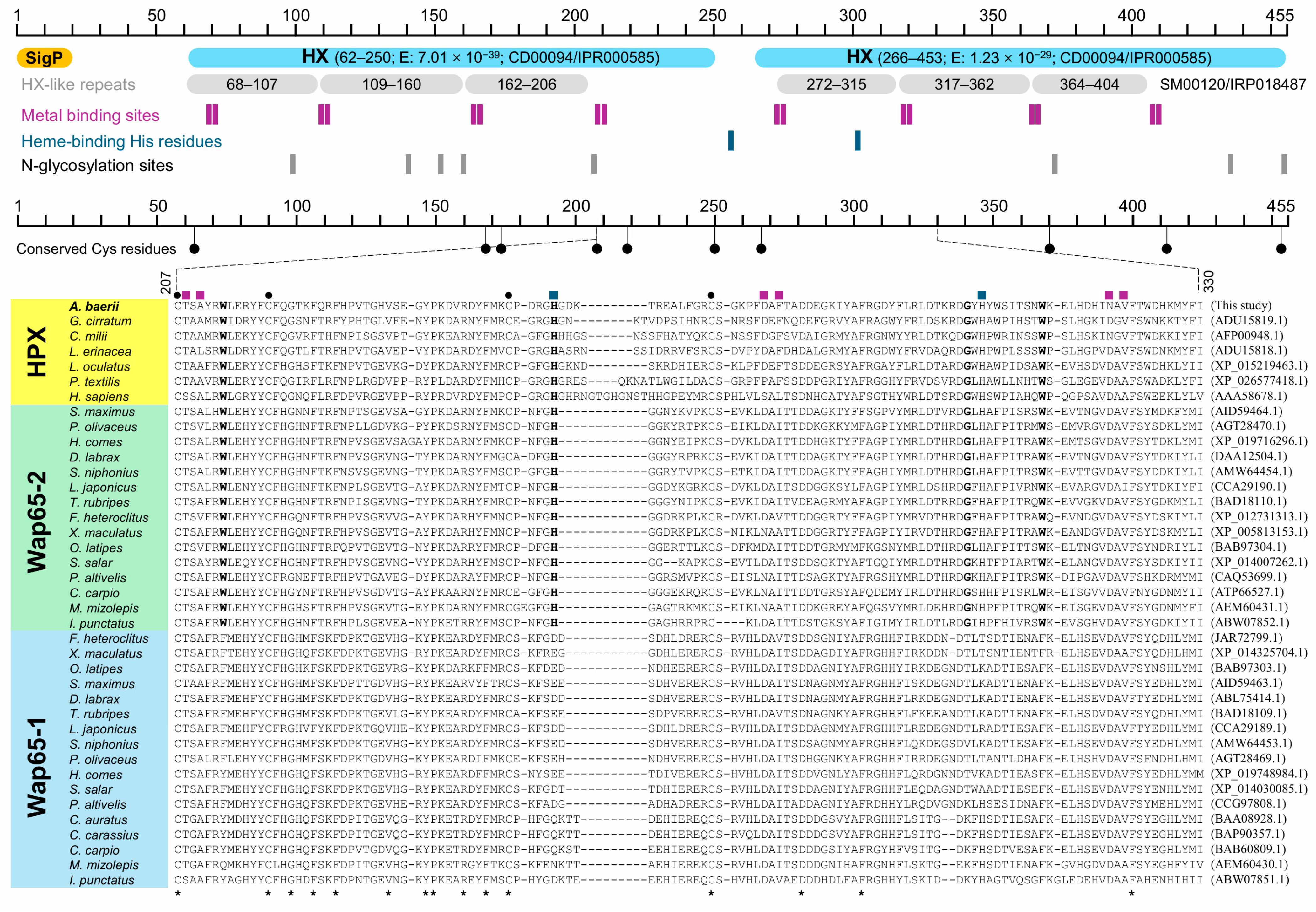
Figure 1. Schematic representation of the conserved domains and functional motifs in the deduced amino acid sequence of A. baerii HPX (top). The diagram highlights the predicted signal peptide (SigP), two conserved hemopexin (HX) domains, six HX-like repeats, eight putative metal-binding sites, two conserved heme-binding histidine residues, and eight predicted N-glycosylation sites. A partial multiple sequence alignment (bottom) with representative HPX, Wap65-1, and Wap65-2 orthologs from vertebrates illustrates the relative conservation of key motifs across species. Five disulfide-bond-forming cysteine pairs are also annotated. A. baerii HPX showing higher similarity to Wap65-2 than to Wap65-1. In the alignment, residues conserved for all the species aligned are indicated with asterisks at bottom, while residues conserved only between HPX and Wap65-2 are bolded. Black dots indicate conserved cysteine residues forming disulfide bonds; colored squares denote key motif positions as in the top schematic. The exact positions of residues and/or motifs can be referred to main text.
Ijms 26 07934 g001
Figure 2. Predicted 3D structure of A. baerii HPX and superimposed comparison with human HPX. (A) AlphaFold2-predicted tertiary structure of A. baerii HPX, revealing a two-domain β-propeller-like architecture characteristic of vertebrate hemopexins. Conserved functional residues are labeled, including signal peptide cleavage site (Ala19–Ala20), two heme-binding histidines (His255 and His302), eight metal-binding sites (blue-colored for N- and C-domains), and representative N-glycosylation sites (Asn206 and Asn445). The quality assessment of modeling can be referred to Supplementary Figure S2. (B) Structural superimposition of A. baerii and human HPX (P02790) models. Despite low overall sequence identity (33.7%), key heme-binding histidines (human/A. baerii; His236/His255 and His293/His302) and structurally conserved motifs align well, supporting functional conservation (see Supplementary Figure S3).
Ijms 26 07934 g002
Figure 3. Maximum likelihood tree constructed using amino acid sequences of HPX, Wap65-1, and Wap65-2 orthologs from representative vertebrate species under the JTT model with gamma distribution and invariant sites (G+I), using all alignment sites. The tree is rooted with three cartilaginous fish (Chondrichthyes) sequences used as the outgroup. Support values shown at nodes represent adaptive bootstrap (threshold = 5.0) percentages. A simplified schematic of the major clade topology is provided in the top left panel for reference. A. baerii HPX is placed basal to the teleost Wap65-1 and Wap65-2 clades, forming a distinct branch alongside holostean (Lepisosteus oculatus) HPX. Phylogenetic trees constructed with a neighbor-joining (NJ) algorithm also displayed the similar tree topologies (see Supplementary Figure S4).
Ijms 26 07934 g003
Figure 4. Structural superimposition of A. baerii HPX with representative vertebrate HPX and Wap65 isoforms. Unlike superimposition with Wap65-2, Wap65-1 displays a unique structurally divergent region in the C-domain across all comparisons, consistent with isoform-specific divergence following gene duplication. Protein IDs used for modeling and superimposition are shown for each sequence.
Ijms 26 07934 g004
Figure 5. Tissue distribution patterns and expression levels of HPX mRNA in A. baerii fingerlings. Quantitative RT-PCR analysis of HPX transcript levels across 11 tissues from healthy fingerlings. Expression levels were normalized using the geometric mean of three reference genes (RPL5, RPL7, and RPL7A) and are presented as fold differences relative to the calibrator tissue (intestine). Data are shown as mean ± SD (n = 6; biological replications). Statistically significant differences among tissues were determined using one-way ANOVA followed by Tukey’s post hoc test. Different lowercase letters indicate significant differences (p < 0.05). A representative image of ethidium bromide-stained agarose gel (1%) from end-point RT-PCR amplification is also shown on the top. Expression data normalized with each reference control can be referred to Supplementary Figure S5.
Ijms 26 07934 g005
Figure 6. Ontogenetic expression pattern of HPX mRNA during prelarval intervals in A. baerii. (A) Relative HPX mRNA expression levels measured by RT-qPCR from the just-hatched (JH) stage through 10 days post-hatching (DPH), normalized to RPL5. Figures in parentheses are fold change of expression levels relative to that at JH. Different lowercase letters denote statistically significant differences among time points (p < 0.05, ANOVA with Tukey’s post hoc test), based on triplicate biological replications (each comprising of 3–4 pooled prelarvae). (B,C) Corresponding growth curves for total length (B) and body weight (C) over the same developmental window. Developmental milestones for behavior and metamorphosis are also indicated.
Ijms 26 07934 g006
Figure 7. Differential expression of HPX mRNA in responsive to experimental infection with different doses of A. hydrophila in A. baerii fingerlings. Quantitative RT-PCR analysis of HPX transcript levels at 12 h post-injection (HPI) in kidney, liver, skin, and spleen tissues following intraperitoneal injection of A. hydrophila at 2 × 10⁴, 2 × 10⁵, or 2 × 10⁶ CFU/g body weight (BW). Expression values were normalized to RPL5 and are shown as fold change relative to saline-injected controls. Data represent mean ± SD (n = 6). Statistically significant differences among dose groups within each tissue were evaluated using one-way ANOVA followed by Tukey’s test. Different letters indicate significant differences (p < 0.05).
Ijms 26 07934 g007
Figure 8. Temporal modulation of HPX and TF mRNA expression following A. hydrophila injection in A. baerii fingerling tissues (kidney, liver, skin and spleen), assessed at 0, 6, 12, 24, 48 h post-injection (HPI) based on RT-qPCR. Expression values were normalized to RPL5 and are shown as fold change relative to 0 HPI. Data represent mean ± SD (n = 4). Different lowercase letters indicate significant differences among time points within each tissue (p < 0.05, ANOVA with Tukey’s test).
Ijms 26 07934 g008
Figure 9. Tissue-specific expression of HPX and HSP70 under mild and severe thermal stress in A. baerii fingerlings. (A,B) Fold change in HPX (A) and HSP70 (B) mRNA expression in kidney, liver, skin, and spleen after exposure to mild thermal stress (16 °C to 25 °C). Expression was measured immediately after reaching the target temperature elevation (25 °C + D0) and after additional exposure at 25 °C for 1 week (25 °C + D7). (C,D) HPX (C) and HSP70 (D) mRNA expression following severe thermal stress (20 °C to 29 °C) at the same time points (20 °C, 29 °C + D0 and 29 °C + D7). Gene expression levels were normalized to RPL5 and shown as fold change relative to baseline temperatures (16 °C or 20 °C). Data are shown as mean ± SD (n = 4). Different lowercase letters denote statistically significant differences within each tissue across treatments (p < 0.05, ANOVA with Tukey’s post hoc test).
Ijms 26 07934 g009
Disclaimer/Publisher’s Note: The statements, opinions and data contained in all publications are solely those of the individual author(s) and contributor(s) and not of MDPI and/or the editor(s). MDPI and/or the editor(s) disclaim responsibility for any injury to people or property resulting from any ideas, methods, instructions or products referred to in the content.