Article

Significant Interplay Between Lipids, Cytokines, Chemokines, Growth Factors, and Blood Cells in an Outpatient Cohort

by
Mats B. Eriksson
1,2,*,
Lars B. Eriksson
1 and
Anders O. Larsson
3
1
Department of Surgical Sciences, Uppsala University, Uppsala University Hospital, SE-751 85 Uppsala, Sweden
2
NOVA Medical School, New University of Lisbon, 1099-085 Lisbon, Portugal
3
Department of Medical Sciences, Uppsala University, Uppsala University Hospital, SE-751 85 Uppsala, Sweden
*
Author to whom correspondence should be addressed.
Int. J. Mol. Sci. 2025, 26(16), 7746; https://doi.org/10.3390/ijms26167746
Submission received: 2 July 2025 / Revised: 2 August 2025 / Accepted: 8 August 2025 / Published: 11 August 2025
(This article belongs to the Special Issue Interplay Between Blood Cells and Cytokines)

Abstract

Cardiovascular disease (CVD) remains the leading global cause of morbidity and mortality, largely driven by atherosclerosis, a chronic inflammatory process involving lipids and immune cells. Although traditional lipid biomarkers such as low-density lipoprotein (LDL) and high-density lipoprotein (HDL) are well-established in CVD risk stratification, the interplay between cytokines, chemokines, growth factors (CCGFs), lipid metabolism, and hematological parameters in non-cardiac populations remains underexplored. We investigated associations between plasma cytokines and lipid-related biomarkers and their relationships with circulating blood cell counts in a cohort of 164 essentially healthy adults aged 18–44 years. CCGF profiling was performed using a proximity extension assay (PEA), and statistical correlations were adjusted for multiple testing using false discovery rate (FDR) correction. The CCGFs that were associated with HDL and apolipoprotein A1 all displayed negative associations. Several pro-inflammatory cytokines, including CCL3, IL-6, and TNFSF10, showed strong positive associations with triglycerides, remnants, non-HDL, and body mass index (BMI). Furthermore, triglycerides and remnants were consistently correlated with elevated leukocyte, neutrophil, and platelet counts. HGF and FGF-21, mainly considered as anti-inflammatory, were positively associated with BMI and negatively associated with HDL, which is compliant with a multitude of actions, depending on the local milieu and the cellular interplay. Our results support the existence of a complex immunometabolic network involving lipids, CCGFs, and blood cells, even in non-diseased individuals. The observed patterns underscore the importance of understanding the intricate cytokine–lipid–cell interactions that may occur in early pathophysiological processes and highlight their potential utility in refining cardiovascular risk assessment beyond traditional lipid metrics.

1. Introduction

Cardiovascular disease (CVD), a leading cause of morbidity and mortality, affects more than 500 million people globally, and its prevalence seems to be increasing. Atherosclerotic diseases are the main mediators of CVD. Approximately 50% of CVD deaths are attributed to ischemic heart disease and an additional 25% are caused by ischemic stroke. CVD not only is associated with increased morbidity and mortality, but also confers a substantial economic burden to the health-care system [1,2,3,4,5,6,7].
Atherosclerosis is an inflammatory disease of the arterial intima, where the balance between pro-inflammatory and anti-inflammatory mechanisms is crucial for the clinical outcome. Intimal infiltration and modification of plasma lipoproteins, particularly LDL, and their uptake by macrophages are key events in the development of atherosclerosis. Arterial LDL accumulation is an important step in atherosclerotic plaque formation. Low-density lipoprotein (LDL) concentrations in the arterial intima far exceed concentrations in other connective tissues. Genetic variations and host immune–inflammatory responses can modulate the pro-atherogenic effect of elevated LDL-cholesterol. High-density lipoprotein (HDL) particles have cardiovascular-protective effects, primarily attributed to their ability to transport cholesterol to the liver, where it can be excreted or converted into bile acids. HDL particles also have antioxidant and anti-inflammatory roles. There is a causal relationship between triglycerides, triglyceride-rich lipoproteins, and their remnants in atherosclerotic disease, especially in patients with obesity, metabolic syndrome, diabetes, and chronic kidney disease [8,9,10,11].
Pro-inflammatory cytokines are linked to several types of CVDs. The most important cytokines in this respect include interleukin-6 (IL-6), tumor necrosis factor (TNF) alpha, and the interleukin-1 (IL-1) family. Inflammation is involved in numerous pathophysiological processes such as oxidative stress and calcium-related signaling events that may facilitate leukocyte–endothelial cell interactions, indicating the dynamic nature of pro-inflammatory cytokines in several CVDs. As part of the inflammation-induced endothelial dysfunction, there is increased permeability to lipoproteins, leading to deposition in the subendothelial space, leukocyte migration, and platelet activation. Once inside the arterial wall, LDL-cholesterol undergoes oxidation, while triglyceride-rich lipoproteins and remnant lipoproteins exert pro-inflammatory effects. Furthermore, psychological stress may increase the risk of cardiovascular disease, mediated by the release of inflammatory cytokines. Furthermore, inflammation is a link between aging and cardiovascular disease, as aging presents with systemic low-grade chronic inflammation and elevated concentrations of mediators such as IL-6, TNFα, and C-reactive protein (CRP) [12,13,14].
High levels of erythrocytes are thrombogenic, which may not only be due to impaired blood flow, since erythrocytes may also up-regulate IL-8 mRNA even if IL-6 and VEGF mRNA expression appears down-regulated. IL-8 plays a crucial role in neutrophil recruitment, which may induce potent cytotoxic effects through neutrophil extracellular trap (NET) formation and the release of proteolytic enzymes. Erythrocytes from healthy individuals regulate immune cell activity and bind more than 50 cytokines, hereby playing an active role in cytokine signaling and regulation. Neutrophil cells have important functions, including not only cardiovascular inflammation and repair, but also atherogenesis, plaque destabilization, and plaque erosion. Activated platelets are highly involved in inflammatory processes, where they express a plethora of pro- and anti-inflammatory molecules that attract circulating leukocytes. Furthermore, platelets can directly influence adaptive immune responses. Significant associations between cytokines in saliva and peripheral blood cells were recently published by our group [15,16,17,18,19,20,21,22].
The aims of the study were to (1) investigate associations between cytokines, chemokines, and growth factors (CCGFs) and selected biomarkers of cardiovascular disorders in a non-cardiac cohort; and (2) explore potential relationships between these biomarkers and circulating blood cells in the same cohort.

2. Results

2.1. Patient Characteristics

The cohort consisted of 164 individuals (53 males). The mean age was 29 years and the range was 18–44 years.
Prevalences of the CCGFs analyzed in our cohort are displayed in Table 1.

2.2. Cytokine Values Below the Assay’s Standard Curve Limits

Table 2 displays the number of results below the lowest standard point for each of the Olink markers. There were no values exceeding the assay’s highest standard points.

2.3. Biomarkers vs. Cytokines

All significant associations, after adjustment for multiplicity, between CCGFs and other biomarkers are shown in Supplementary Table S1.
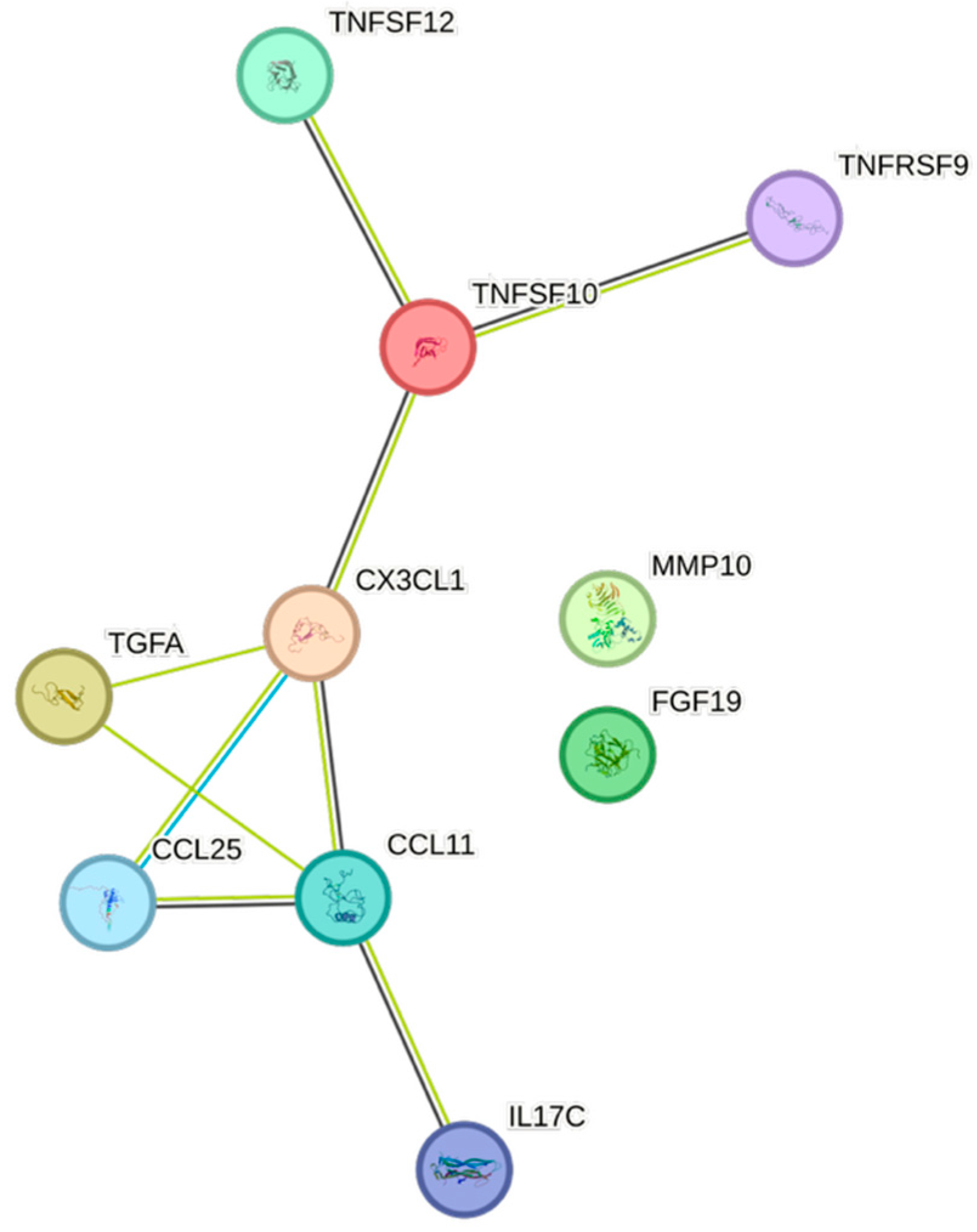
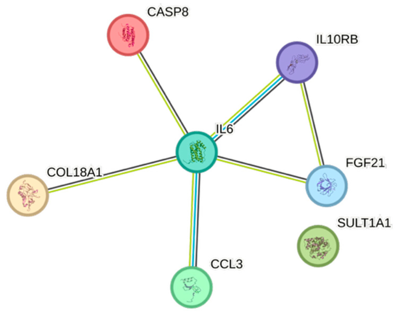
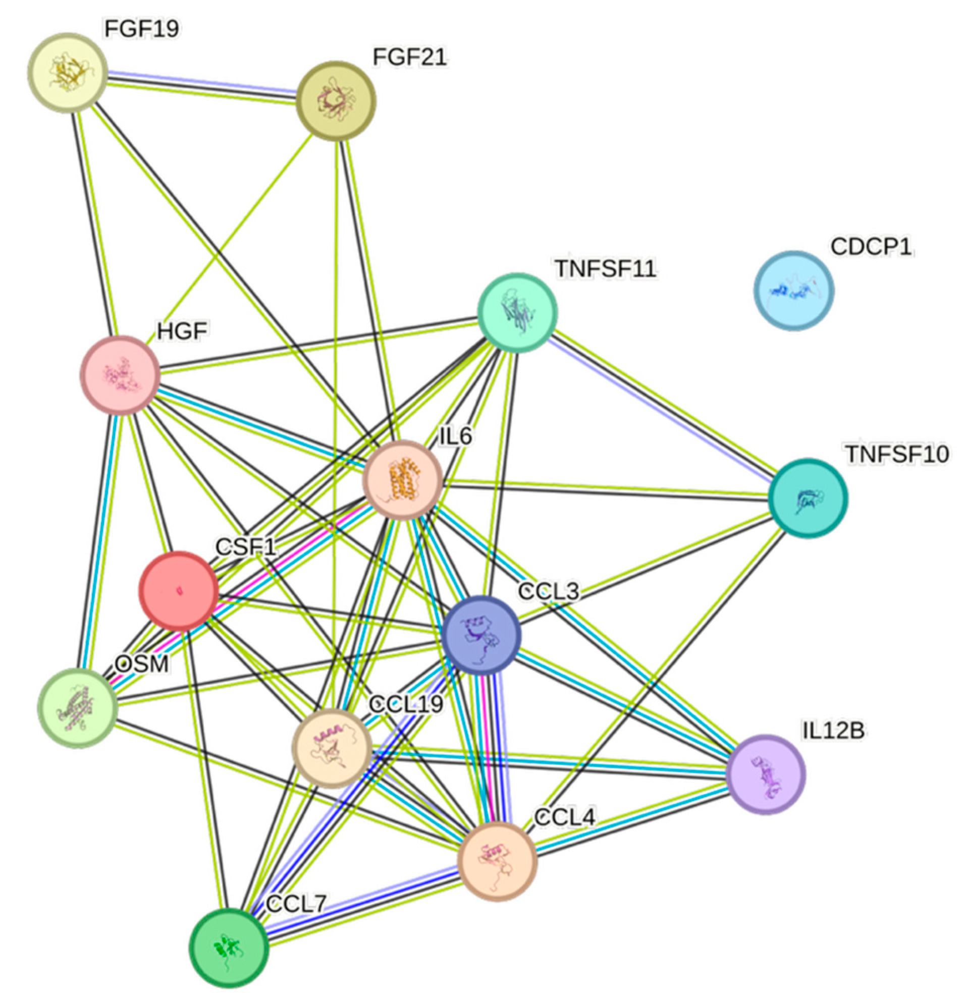
HDL exhibited 12 significant associations out of the quantified CCGFs (Figure 1), all of them negative. CCL3 was the one that was most negatively associated with HDL, whereas TNFRSF9 was the cytokine that exhibited the least expressed negative association with HDL.
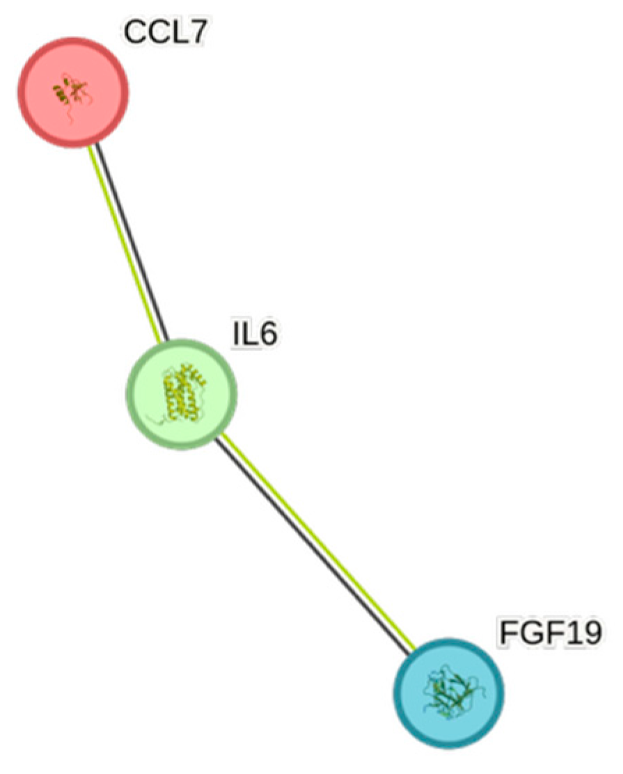
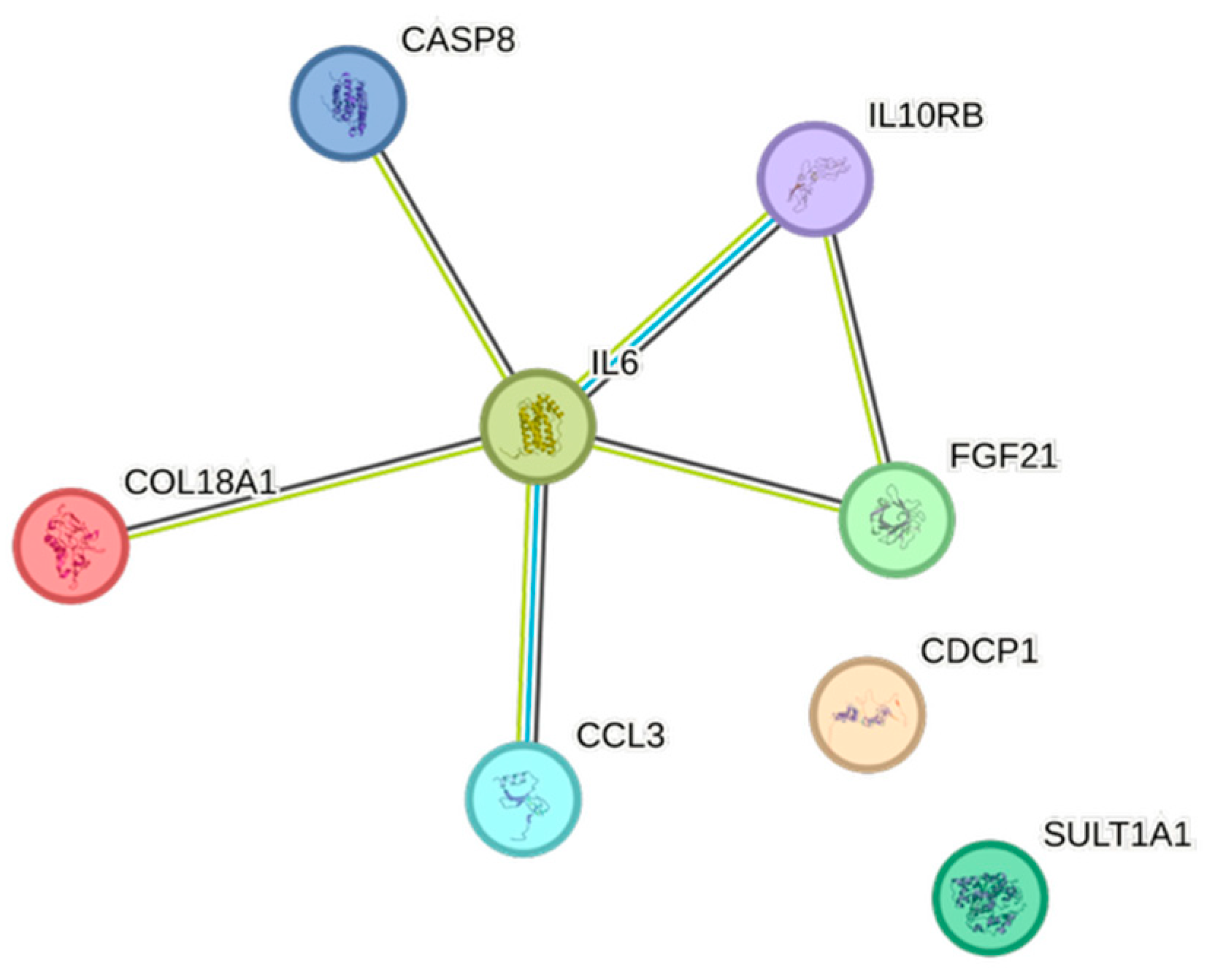
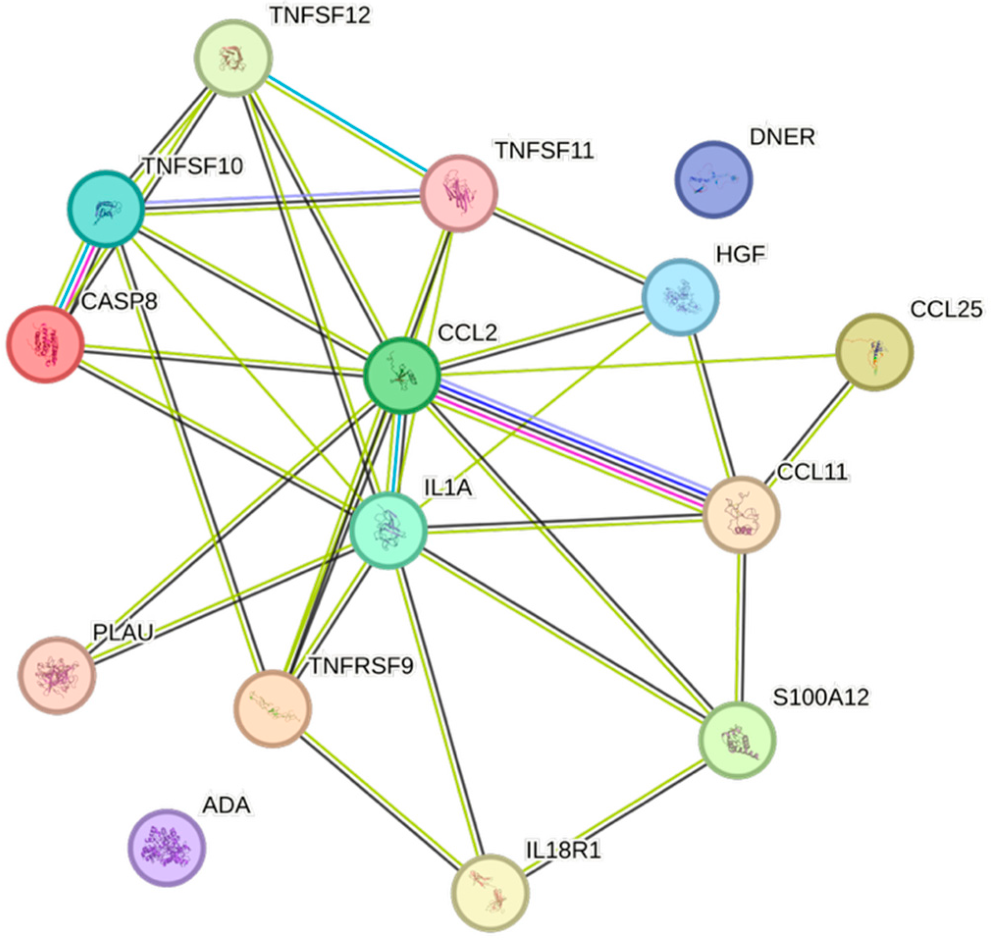
Apolipoprotein A1 was negatively associated with nine cytokines (Figure 2). TNFSF10 was the cytokine that was the least negatively associated, whereas PLAU was the most negatively associated one.
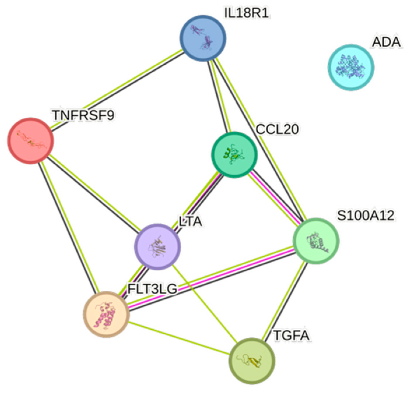
Creatinine was positively associated with 11 CCGFs (Figure 3), and eGFRcreatinine (Figure 4) was associated with 3 CCGFs. FGF-19 was the second-most associated with creatinine, but was negatively associated with eGFRcreatinine.
Albumin was associated with nine CCGFs (Figure 5). Five of these associations were positive.
Apolipoprotein B was positively associated with seven CCGFs (Figure 6), where CCL3 showed the strongest association and TNFSF10 the weakest one.
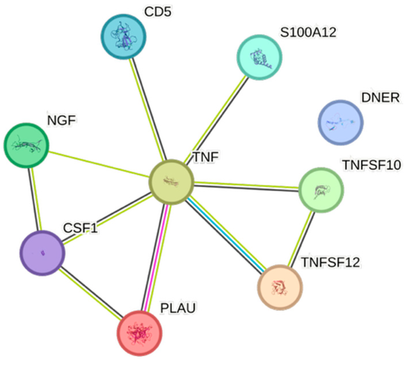
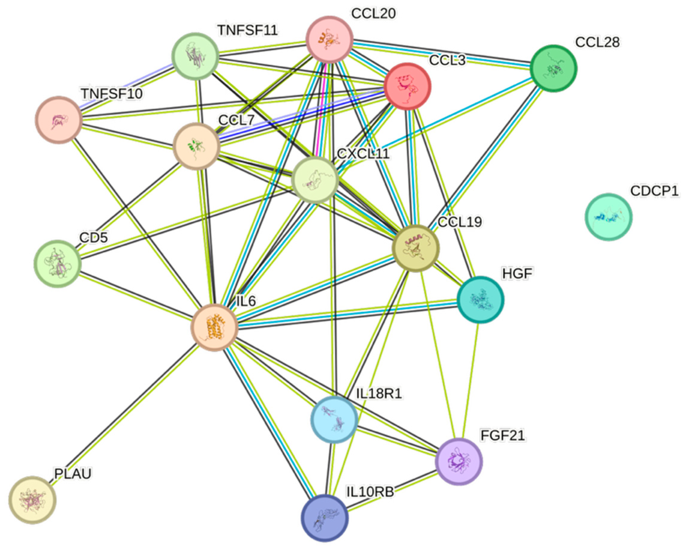
Triglycerides were associated with eight of the analyzed CCGFs. The strongest associations between triglycerides and CCGFs were noted for FGF-21, followed by CCL3 (Figure 7).
Remnants (=Non-HDL–LDL) exhibited several similarities with and were almost identical to the triglycerides, except for the presence of CDCP1 in remnants (Figure 8).
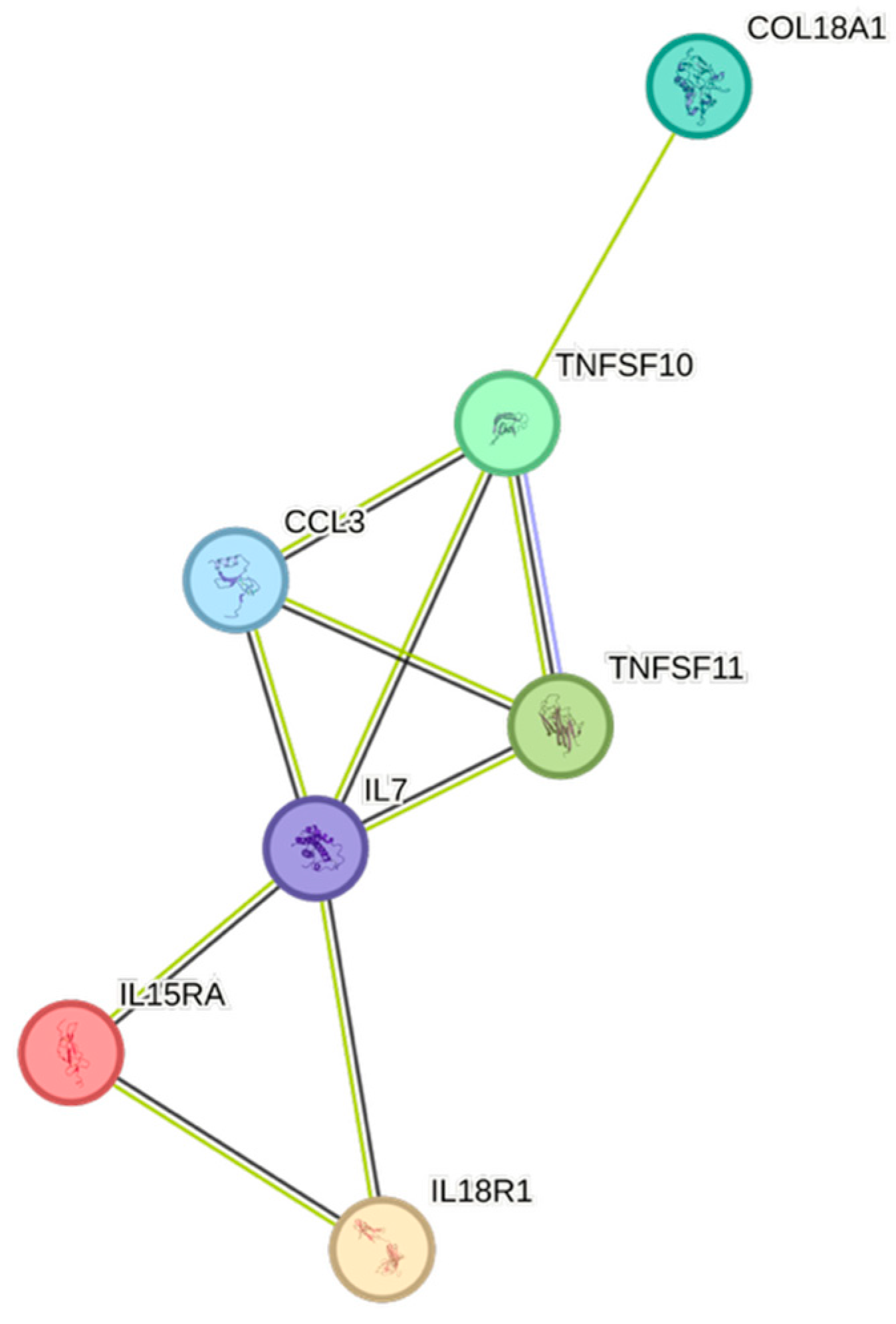
Non-HDL was equally and strongly associated with both CCL3 and CDCP1, followed by TNFSF10 (Figure 9).
LDL was merely associated with both TNFSF10 and CDCP1.
Total cholesterol was not associated with any of the assessed CCGFs.
Age exhibited eight associations. The only CCGF that was positively associated with age was Flt3L, whereas the most negative association with age was noted for IL-18R1 (Figure 10).
Both weight and body mass index displayed complex association patterns, comprising sixteen and fourteen associations, respectively. The most expressed associations for both of them were with IL6 (Figure 11 and Figure 12, respectively).
Gender was associated with fifteen CCGFs (Figure 13), with multiple inter-cytokine interactions. All associations were negative, and TNFSF10 was the cytokine that exhibited the strongest negative association with gender.

2.4. Biomarkers vs. Hematology

All significant associations, after adjustment for multiplicity, between biomarkers and hematological data are displayed in Supplementary Table S2.
Both remnants and triglycerides were strongly associated with the erythrocyte count, leukocytes, platelets, hemoglobin, erythrocyte volume fraction, neutrophils, and mean corpuscular volume (negative associations). Except for the fact that weight was associated with mean corpuscular hemoglobin concentration (MCHC), there were definite similarities between this biomarker and both remnants and triglycerides. BMI was associated with leukocytes, neutrophils, platelets, and the erythrocyte count. Total cholesterol was associated with erythrocyte count, platelets, and mean corpuscular hemoglobin (MCH), whereas total cholesterol was not associated with any of the cytokines that were quantified.
eGFRcreatinine was not associated with any of the hematological data.

2.5. CCGF Correlations

Correlations between the proteins in plasma are shown in three correlation matrices, displaying Spearman rank values for each association in the entire cohort, among men, and among women, respectively (Supplementary Tables S3–S5). Values in red denote p < 0.05.

3. Discussion

Although cytokines can exert diverse effects depending on the biological context, most exhibit predominantly pro- or anti-inflammatory functions. From an evolutionary perspective, it would have been disadvantageous to develop signaling molecules that are not, under specific conditions, beneficial to the host. Nevertheless, inflammatory responses are often Janus-faced, reflecting both protective and potentially harmful roles, and their specific regulatory features may have shifted over time as new species have emerged.
Independent of their predominant pro- or anti-inflammatory functions, all cytokines assessed in this study were negatively correlated with apolipoprotein A and HDL levels. From a quantitative aspect, TNFSF10 appears to be the most important cytokine in our study. This pro-inflammatory cytokine has been linked to improved survival in cancers with high tumor-associated macrophage content, which may reflect its capacity to induce cell death or alternatively to activate survival-promoting pathways depending on the tumor context [23]. Furthermore, TNFSF10 was the cytokine that had the strongest negative association with apolipoprotein A1 and exhibited the most positive association with LDL.
CCL3, the second-most frequently found cytokine in our cohort, is an inflammatory cytokine, secreted by monocytes and macrophages, having a fundamental role when tumor-associated macrophages have an impact on tumor development [24]. CCL3 was negatively associated with apolipoprotein A1 and HDL, but positively associated with apolipoprotein B, BMI, non-HDL, remnants, triglycerides, and weight.
TNFSF11 is an inflammatory cytokine that correlates with age-related macular degeneration [25]. TNFSF11 was significantly negatively associated with apolipoprotein A1, gender, and HDL. Positive associations were noted between TNFSF11 and apolipoprotein B, BMI, non-HDL, and weight.
IL6, conventionally seen as a pro-inflammatory cytokine, was positively associated with risk factors for cardiovascular disease, e.g., BMI, eGFRcreatinine, remnants, triglycerides, and weight, but not with increasing age, which in a previous study has been associated with elevated levels of IL6 [26]. IL6 was negatively associated with HDL, indicating an immunometabolic interplay that affects the risk of cardiovascular events [27,28]. The inflammatory member of the tumor necrosis factor ligand superfamily, TNFRSF9 [29], was negatively associated with age, apolipoprotein A1, gender, and HDL, but positively associated with creatinine, which overall may suggest a negative impact of this cytokine.
The inflammatory cytokine Flt3L was associated with age, a finding in agreement with a previous study in 94 strictly healthy volunteers, aged 18–80 years old [30].
The two most frequent cytokines, which mainly have anti-inflammatory properties, noted in our cohort were HGF [31] and FGF-21 [32], respectively. HGF was positively associated with BMI, non-HDL, and weight, whereas HGF was negatively associated with gender, and, somewhat surprisingly, also negatively associated with HDL. FGF-21 exhibited a similar pattern, were BMI, remnants, triglycerides, and weight were positively associated, while gender and HDL, once again, were negatively associated.
IL-10RB [33], FGF-19 [34], and COL18A1 (VEGFA) [35], having at least partly anti-inflammatory properties, exhibited three significant associations each. Both IL-10RB and COL18A1 were positively associated with remnants as well as triglycerides. IL-17C exhibited the strongest Spearman correlation against creatinine in our cohort. This cytokine is known to have a pathogenic role in renal damage, and IL-17C neutralization protects the kidney against both acute and chronic injury [36,37]. Thus, it is remarkable that even in an essentially healthy cohort, significant associations between potentially nephrotoxic cytokines and creatinine were observed. FGF-19 showed the second-strongest positive association with creatinine, while also being negatively associated with eGFRcreatinine. In this context, it is noteworthy that high levels of FGF19 were found in non-diabetic patients with chronic kidney disease [38]. A bidirectional relationship exists between the heart and the kidneys, whereby dysfunction in one organ system can lead to dysfunction in the other [39]. Cytokines play a crucial role in the development of this cardiorenal syndrome [39].
The most striking finding when biomarkers were associated with hematological data was that WBC (especially neutrophil cells) and TPK were frequently related to BMI, remnants, triglycerides, and weight. This is not surprising, since WBC, neutrophils, and platelets are all related to triglycerides [40,41]. Leukocytes, in particular neutrophils, and platelets secrete inflammatory cytokines, which are related to chronic low-grade inflammation of adipose tissue [42]. This is a complex, multifaceted process, where adipocytes secrete inflammatory adipokines, cytokines, and chemokines. Leukocytosis and increased platelet counts are driven by this inflammatory state [42,43,44]. In this context it is noteworthy that statins significantly reduce plasma levels of CRP, as well as several pro-inflammatory cytokines, thereby exerting beneficial effects by lowering the levels of these inflammatory markers [45], a finding in alignment with our results on the associations between CCGFs and blood lipids.
Leukocytes and especially neutrophil cells have a distinct role in this context, as increased numbers of white blood cells and neutrophils will also increase the formation of neutrophil extracellular traps (NETs). NETs are web-like structures composed of DNA, histones, and granule proteins that are released by activated neutrophils. This process, known as NETosis, is a distinct form of cell activation that allows neutrophils to trap and kill pathogens extracellularly. While NETs serve a protective role in host defense, dysregulated or excessive NET formation has been implicated in the pathogenesis of various diseases, particularly in the cardiovascular and renal systems [46,47].
NETs contribute to several aspects of cardiovascular pathology. NETs promote plaque formation and destabilization in atherosclerosis by activating macrophages and endothelial cells. They serve as a scaffold for platelet adhesion and coagulation factor activation, thus contributing to thrombus formation.
NET components such as histones and myeloperoxidase can damage cardiomyocytes and propagate inflammation post-infarction. In recent years, growing evidence has highlighted the strong link between coagulation and inflammation, leading to the emergence of the concept of immunothrombosis. NETs are found within venous thrombi and facilitate fibrin deposition and coagulation, linking inflammation and thrombosis (termed immunothrombosis).
NLRP3 is a key component of the innate immune system and forms part of the inflammasome that has been implicated in chronic low-grade inflammation associated with metabolic syndrome, including obesity, insulin resistance, and type 2 diabetes [48]. NLRP3 elicits maturation of the cytokines IL-1β and IL-18 [49]. We noted that IL18R1, a receptor for IL-18, is negatively associated with age and gender, but positively associated with Apo B and weight.
This process involves coordinated interactions between leukocytes, platelets, and coagulation factors. Neutrophil extracellular traps (NETs) play a pivotal role by offering a scaffold that promotes platelet activation, thrombin generation, and fibrin deposition, thereby contributing to clot formation. NET formation has been shown to induce inflammation and cardiac injury [46]. NET formation has also been shown to cause kidney injury [47]. In ischemia–reperfusion injury or sepsis, excessive NET formation promotes microvascular thrombosis, endothelial damage, and inflammation, all contributing to renal dysfunction [47]. During NET formation the nuclear and granule membranes break down, allowing the release of proteins stored within the neutrophil [50]. Given their roles in defense and disease, NETs have become targets for novel therapeutic approaches, including anti-inflammatory agents that reduce NETosis.
Furthermore, lipids have signaling roles in platelets and regulate how lipids generated by platelets influence other cells [51,52]. Lipids also play an important role in cell fate decisions during hematopoiesis [52], which may be in alignment with our finding that platelets were also associated with apolipoprotein B and total cholesterol, respectively. We have recently shown that cytokines, in both peripheral blood [21] and human saliva [22], are associated with blood cell counts. These associations between circulating cytokines and peripheral blood cell counts are in agreement with the present paradigm on the complex interrelations between lipids, cytokines, and peripheral blood cells. For example, we noted strong associations between platelet count and IL-6 [21], which, together with our present study, strengthens the postulate that platelets play a crucial role in the inflammatory process and that inter-cytokine interactions across different cytokine families reflect a part of the large amount of crosstalk that is a part of immunologic homeostasis [53].

Limitations and Strengths

This study has several limitations. It is a single-site study from a region where the majority of the patients were Caucasians. There is a distinct female preponderance among the subjects. Also, the cohort is fairly uniform in age and was clinically evaluated to be essentially healthy, without any acute or systemic severe illness.
The STRING images visualize predicted protein–protein interactions within an integrated network, thus facilitating biological interpretation and hypothesis generation, which might be of translational relevance in future studies.

4. Materials and Methods

4.1. Population

Patients referred to the Department of Oral and Maxillofacial Surgery at the Falu County Hospital, Sweden, were recruited by LBE and offered to participate in this study, which focused on men and women from 18 to 44 years old, with a bodyweight of 50 to 120kg. Healthy patients, or those with well-compensated systemic disease, were accepted for screening. Inclusion and exclusion criteria and the surgical procedure have previously been described in detail [54,55]. After successful screening, those who had signed a written informed consent form were included.
The cohort is briefly described in Table 3, in compliance with GDPR (EU 2016/679).

4.2. Sampling Procedures

Blood samples were collected in vacutainer tubes while the patient was in the supine position. All blood samples were obtained prior to surgery. Complete blood counts were analyzed in EDTA–blood at the clinical laboratory at Falun Hospital. Conventional biochemical analyses, including estimated glomerular filtration rate (eGFRcreatinine) [56], were performed. Plasma cytokine samples were frozen and stored at −80 °C until analysis.

4.3. Ethics

This study was conducted in accordance with the principles of the Helsinki Declaration [57]. Blood sampling was the only difference from clinical routine treatment, which implied a further invasive step. The study was approved by the Swedish Ethical Review Authority (Dnr 2015/378) on 2 December 2015 and registered in the European Union Regulating Authorities Clinical Trials Database (EudraCT) under number 2014-004235-39 on 29 September 2014. Furthermore, the trial was listed on ClinicalTrials.gov with the ID NCT04459377 on 8 July 2020.

4.4. Proximity Extension Assay

The proximity extension assay (PEA) was performed using the Proseek Multiplex Inflammation kit [Olink Bioscience, Uppsala, Sweden; (v3024)] [58,59,60]. The PEA seems to reliably reflect protein plasma levels, as compared to conventional assays [61]. In brief, 1 µL of plasma was combined with 3 µL of incubation mix, containing two probes (antibodies conjugated with unique DNA oligonucleotides), and incubated at 8 °C overnight.
Following incubation, 96 µL of extension mix, which included the PEA enzyme and PCR reagents, was added. The samples were then incubated at room temperature for 5 min before undergoing 17 cycles of DNA amplification in a thermal cycler.
A 96.96 Dynamic Array IFC (Fluidigm, South San Francisco, CA, USA) was prepared and primed according to the manufacturer’s guidelines. In a separate plate, 2.8 µL of the sample mixture was combined with 7.2 µL of detection mix, and 5 µL of this solution was loaded into the right side of the primed 96.96 Dynamic Array IFC. Unique primer pairs for each cytokine were loaded into the left side of the array. The protein expression analysis was then performed using the Fluidigm Biomark reader, following the Proseek protocol. The Proseek kit quantified 92 proteins, which are listed in Supplementary Table S6 along with their full names, UniProtIDs, and corresponding encoding genes.

4.5. STRING Images

Cytokine, chemokine, and growth factor interactions and concentration patterns are visualized using images generated from the STRING database [62]. The edge weights are calculated using the STRING database based on the associations found in our study. Protein names are displayed to create interaction networks, and edge thicknesses are indicators of confidence, indicating how likely STRING judges an interaction to be true given the available evidence. The images were exported in high-resolution format for inclusion in figures. This approach enabled a simultaneous representation of both quantitative concentration data and qualitative information about potential interactions.

4.6. Statistical Analysis

Coefficients of variation were analyzed using Spearman rank correlations in Statistica (StatSoft, v14; Tulsa, OK, USA). Absolute cytokine values below the lowest standard point were included in the statistical analysis. There were no values exceeding the assay’s standard curve limits when set to the highest or lowest standard value, respectively. To account for the increased risk of false positives due to multiple comparisons, p-values were adjusted using the false discovery rate (FDR) approach [63]. Adjusted p-values below 0.10, corresponding to an expected FDR of ≤10%, were considered statistically significant.

5. Conclusions

This study demonstrates significant associations between circulating cytokines, lipid-related biomarkers, and peripheral blood cell parameters in a healthy, young adult population. Most cytokines were inversely correlated with HDL and apolipoprotein A1, while several positive associations were observed with triglycerides, non-HDL, BMI, and blood cells secreting inflammatory cytokines. These findings underscore the complex interplay between lipids, cytokine signaling, and hematological components, even in a non-cardiac cohort. STRING images provide a visual summary of predicted protein–protein interactions by integrating experimental data, known pathways, and computational predictions. By illustrating interaction networks, they facilitate understanding of complex molecular relationships that extend beyond isolated analyte changes.
Our results may suggest that pre-existing continuous crosstalk between the lipid metabolism and immune inflammatory pathways may lead to subsequent immunometabolic dysregulation, with potential implications for cardiovascular risk assessment.

Supplementary Materials

The following supporting information can be downloaded at https://www.mdpi.com/article/10.3390/ijms26167746/s1.

Author Contributions

M.B.E.: conceptualization, methodology, formal analysis, writing—original draft, and writing—review and editing. L.B.E.: conceptualization, methodology, investigation, data curation, formal analysis, visualization, writing—review and editing, project administration, and resources. A.O.L.: conceptualization, methodology, formal analysis, writing—original draft, and writing—review and editing. All authors have read and agreed to the published version of the manuscript.

Funding

This work was supported by the Centre for Clinical Research, Dalarna, Uppsala-Örebro Regional Research Council, and Public Dental Care Dalarna, Sweden.

Institutional Review Board Statement

This study was conducted in accordance with the ethical principles originating from the 1975 Declaration of Helsinki, which was revised in 2013 [57], and was consistent with ICH/GCP E6 (R2) guidelines and received approval from the Swedish Ethical Review Authority on 2 December 2015 (Dnr 2015/378). The European Union Regulating Authorities Clinical Trials Database (EudraCT) number (2014-004235-39) was obtained on 29 September 2014. Furthermore, the study was registered on ClinicalTrials.gov under the ID NCT04459377 on 8 July 2020.

Informed Consent Statement

All patients received oral information and signed the informed consent form at screening before any study-specific procedures commenced.

Data Availability Statement

The dataset used and analyzed during the current study is available from the corresponding author on reasonable request.

Acknowledgments

We are grateful to Charina Brännström for skilled technical assistance.

Conflicts of Interest

The authors declare no conflicts of interest.

References

  1. Nedkoff, L.; Briffa, T.; Zemedikun, D.; Herrington, S.; Wright, F.L. Global Trends in Atherosclerotic Cardiovascular Disease. Clin. Ther. 2023, 45, 1087–1091. [Google Scholar] [CrossRef]
  2. Ziaeian, B.; Fonarow, G.C. Epidemiology and aetiology of heart failure. Nat. Rev. Cardiol. 2016, 13, 368–378. [Google Scholar] [CrossRef]
  3. Steen Carlsson, K.; Nilsson, K.; Wolden, M.L.; Faurby, M. Economic burden of atherosclerotic cardiovascular disease: A matched case-control study in more than 450,000 Swedish individuals. BMC Cardiovasc. Disord. 2023, 23, 483. [Google Scholar] [CrossRef] [PubMed]
  4. Liu, S.; Li, Y.; Zeng, X.; Wang, H.; Yin, P.; Wang, L.; Liu, Y.; Liu, J.; Qi, J.; Ran, S.; et al. Burden of Cardiovascular Diseases in China, 1990-2016: Findings From the 2016 Global Burden of Disease Study. JAMA Cardiol. 2019, 4, 342–352. [Google Scholar] [CrossRef]
  5. Mennini, F.S.; Scortichini, M.; Colivicchi, F.; Maggioni, A.P.; Sciattella, P. The Economic Burden of Atherosclerotic Cardiovascular Disease in Italy. Clin. Drug Investig. 2024, 44, 739–747. [Google Scholar] [CrossRef]
  6. Joynt Maddox, K.E.; Elkind, M.S.V.; Aparicio, H.J.; Commodore-Mensah, Y.; de Ferranti, S.D.; Dowd, W.N.; Hernandez, A.F.; Khavjou, O.; Michos, E.D.; Palaniappan, L.; et al. Forecasting the Burden of Cardiovascular Disease and Stroke in the United States Through 2050-Prevalence of Risk Factors and Disease: A Presidential Advisory From the American Heart Association. Circulation 2024, 150, e65–e88. [Google Scholar] [CrossRef]
  7. Kazi, D.S.; Elkind, M.S.V.; Deutsch, A.; Dowd, W.N.; Heidenreich, P.; Khavjou, O.; Mark, D.; Mussolino, M.E.; Ovbiagele, B.; Patel, S.S.; et al. Forecasting the Economic Burden of Cardiovascular Disease and Stroke in the United States Through 2050: A Presidential Advisory From the American Heart Association. Circulation 2024, 150, e89–e101. [Google Scholar] [CrossRef] [PubMed]
  8. Bornfeldt, K.E.; Linton, M.F.; Fisher, E.A.; Guyton, J.R. JCL roundtable: Lipids and inflammation in atherosclerosis. J. Clin. Lipidol. 2021, 15, 3–17. [Google Scholar] [CrossRef] [PubMed]
  9. Chyu, K.Y.; Dimayuga, P.C.; Shah, P.K. Immunogenetics of Atherosclerosis-Link between Lipids, Immunity, and Genes. Curr. Atheroscler. Rep. 2020, 22, 53. [Google Scholar] [CrossRef]
  10. Bäck, M.; Yurdagul, A., Jr.; Tabas, I.; Öörni, K.; Kovanen, P.T. Inflammation and its resolution in atherosclerosis: Mediators and therapeutic opportunities. Nat. Rev. Cardiol. 2019, 16, 389–406. [Google Scholar] [CrossRef]
  11. Xu, D.; Xie, L.; Cheng, C.; Xue, F.; Sun, C. Triglyceride-rich lipoproteins and cardiovascular diseases. Front. Endocrinol. 2024, 15, 1409653. [Google Scholar] [CrossRef]
  12. Zhang, H.; Dhalla, N.S. The Role of Pro-Inflammatory Cytokines in the Pathogenesis of Cardiovascular Disease. Int. J. Mol. Sci. 2024, 25, 1082. [Google Scholar] [CrossRef]
  13. Henein, M.Y.; Vancheri, S.; Longo, G.; Vancheri, F. The Role of Inflammation in Cardiovascular Disease. Int. J. Mol. Sci. 2022, 23, 12906. [Google Scholar] [CrossRef]
  14. De Maeyer, R.P.H.; Chambers, E.S. The impact of ageing on monocytes and macrophages. Immunol. Lett. 2021, 230, 1–10. [Google Scholar] [CrossRef]
  15. Silvestre-Roig, C.; Braster, Q.; Ortega-Gomez, A.; Soehnlein, O. Neutrophils as regulators of cardiovascular inflammation. Nat. Rev. Cardiol. 2020, 17, 327–340. [Google Scholar] [CrossRef]
  16. Antonelli, A.; Scarpa, E.S.; Magnani, M. Human Red Blood Cells Modulate Cytokine Expression in Monocytes/Macrophages Under Anoxic Conditions. Front. Physiol. 2021, 12, 632682. [Google Scholar] [CrossRef]
  17. Ren, G.; Dewald, O.; Frangogiannis, N.G. Inflammatory mechanisms in myocardial infarction. Curr. Drug Targets Inflamm. Allergy 2003, 2, 242–256. [Google Scholar] [CrossRef] [PubMed]
  18. Karsten, E.; Herbert, B.R. The emerging role of red blood cells in cytokine signalling and modulating immune cells. Blood Rev. 2020, 41, 100644. [Google Scholar] [CrossRef] [PubMed]
  19. Bakogiannis, C.; Sachse, M.; Stamatelopoulos, K.; Stellos, K. Platelet-derived chemokines in inflammation and atherosclerosis. Cytokine 2019, 122, 154157. [Google Scholar] [CrossRef] [PubMed]
  20. Maouia, A.; Rebetz, J.; Kapur, R.; Semple, J.W. The Immune Nature of Platelets Revisited. Transfus. Med. Rev. 2020, 34, 209–220. [Google Scholar] [CrossRef]
  21. Eriksson, L.B.; Eriksson, M.B.; Gordh, T.; Larsson, A.O. Significant Associations Between Blood Cell Counts and Plasma Cytokines, Chemokines, and Growth Factors. Int. J. Mol. Sci. 2025, 26, 4065. [Google Scholar] [CrossRef]
  22. Eriksson, L.B.; Eriksson, M.; Gordh, T.; Larsson, A. Significant Associations Between Blood Cell Counts and a Large Number of Salivary Cytokines, Chemokines, and Growth Factors. J. Interferon Cytokine Res. 2025, 45, 254–262. [Google Scholar] [CrossRef]
  23. Gunalp, S.; Helvaci, D.G.; Oner, A.; Bursalı, A.; Conforte, A.; Güner, H.; Karakülah, G.; Szegezdi, E.; Sag, D. TRAIL promotes the polarization of human macrophages toward a proinflammatory M1 phenotype and is associated with increased survival in cancer patients with high tumor macrophage content. Front. Immunol. 2023, 14, 1209249. [Google Scholar] [CrossRef]
  24. Qin, R.; Ren, W.; Ya, G.; Wang, B.; He, J.; Ren, S.; Jiang, L.; Zhao, S. Role of chemokines in the crosstalk between tumor and tumor-associated macrophages. Clin. Exp. Med. 2023, 23, 1359–1373. [Google Scholar] [CrossRef]
  25. Wang, T.; Chen, J.; Wang, J.; Zhang, Y.; Mao, W.; Yi, Q. 91 Circulating inflammatory proteins and the risk of age-related macular degeneration: A bidirectional Mendelian randomization study. Int. Immunopharmacol. 2024, 139, 112678. [Google Scholar] [CrossRef]
  26. Koelman, L.; Pivovarova-Ramich, O.; Pfeiffer, A.F.H.; Grune, T.; Aleksandrova, K. Cytokines for evaluation of chronic inflammatory status in ageing research: Reliability and phenotypic characterisation. Immun. Ageing 2019, 16, 11. [Google Scholar] [CrossRef] [PubMed]
  27. Nagao, M.; Nakajima, H.; Toh, R.; Hirata, K.I.; Ishida, T. Cardioprotective Effects of High-Density Lipoprotein Beyond its Anti-Atherogenic Action. J. Atheroscler. Thromb. 2018, 25, 985–993. [Google Scholar] [CrossRef] [PubMed]
  28. Liu, Y.; Chen, Y.; Lin, Y.; Wei, B.; Luo, Z. Impacts of pro-inflammatory cytokines variant on cardiometabolic profile and premature coronary artery disease: A systematic review and meta-analysis. J. Cell. Mol. Med. 2024, 28, e18311. [Google Scholar] [CrossRef] [PubMed]
  29. Chen, Z.; Guo, Y.; Sun, H.; Zhang, W.; Hou, S.; Guo, Y.; Ma, X.; Meng, H. Exploration of the causal associations between circulating inflammatory proteins, immune cells, and neuromyelitis optica spectrum disorder: A bidirectional Mendelian randomization study and mediation analysis. Front. Aging Neurosci. 2024, 16, 1394738. [Google Scholar] [CrossRef]
  30. Ravalet, N.; Guermouche, H.; Hirsch, P.; Picou, F.; Foucault, A.; Gallay, N.; Martignoles, J.A.; Beaud, J.; Suner, L.; Deswarte, C.; et al. Modulation of bone marrow and peripheral blood cytokine levels by age and clonal hematopoiesis in healthy individuals. Clin. Immunol. 2023, 255, 109730. [Google Scholar] [CrossRef]
  31. Rong, S.L.; Wang, X.L.; Wang, Y.C.; Wu, H.; Zhou, X.D.; Wang, Z.K.; Wang, Y.C.; Xue, C.S.; Li, B.; Gao, D.L. Anti-inflammatory activities of hepatocyte growth factor in post-ischemic heart failure. Acta Pharmacol. Sin. 2018, 39, 1613–1621. [Google Scholar] [CrossRef]
  32. Fisher, F.M.; Maratos-Flier, E. Understanding the Physiology of FGF21. Annu. Rev. Physiol. 2016, 78, 223–241. [Google Scholar] [CrossRef] [PubMed]
  33. Hoffmann, D.; Sens, J.; Brennig, S.; Brand, D.; Philipp, F.; Vollmer Barbosa, P.; Kuehle, J.; Steinemann, D.; Lenz, D.; Buchegger, T.; et al. Genetic Correction of IL-10RB Deficiency Reconstitutes Anti-Inflammatory Regulation in iPSC-Derived Macrophages. J. Pers. Med. 2021, 11, 221. [Google Scholar] [CrossRef]
  34. Henriksson, E.; Andersen, B. FGF19 and FGF21 for the Treatment of NASH-Two Sides of the Same Coin? Differential and Overlapping Effects of FGF19 and FGF21 From Mice to Human. Front. Endocrinol. 2020, 11, 601349. [Google Scholar] [CrossRef]
  35. Huang, M.-j.; Ji, Y.-w.; Chen, J.-w.; Li, D.; Zhou, T.; Qi, P.; Wang, X.; Li, X.-f.; Zhang, Y.-f.; Yu, X.; et al. Targeted VEGFA therapy in regulating early acute kidney injury and late fibrosis. Acta Pharmacol. Sin. 2023, 44, 1815–1825. [Google Scholar] [CrossRef]
  36. Wang, F.; Yin, J.; Lin, Y.; Zhang, F.; Liu, X.; Zhang, G.; Kong, Y.; Lu, Z.; Wu, R.; Wang, N.; et al. IL-17C has a pathogenic role in kidney ischemia/reperfusion injury. Kidney Int. 2020, 97, 1219–1229. [Google Scholar] [CrossRef]
  37. Zhang, F.; Yin, J.; Liu, L.; Liu, S.; Zhang, G.; Kong, Y.; Wang, Y.; Wang, N.; Chen, X.; Wang, F. IL-17C neutralization protects the kidney against acute injury and chronic injury. eBioMedicine 2023, 92, 104607. [Google Scholar] [CrossRef]
  38. Marchelek-Mysliwiec, M.; Dziedziejko, V.; Dolegowka, K.; Pawlik, A.; Safranow, K.; Stepniewska, J.; Wisniewska, M.; Malyszko, J.; Ciechanowski, K. Association of FGF19, FGF21 and FGF23 with carbohydrate metabolism parameters and insulin resistance in patients with chronic kidney disease. J. Appl. Biomed. 2020, 18, 61–69. [Google Scholar] [CrossRef]
  39. Rangaswami, J.; Bhalla, V.; Blair, J.E.A.; Chang, T.I.; Costa, S.; Lentine, K.L.; Lerma, E.V.; Mezue, K.; Molitch, M.; Mullens, W.; et al. Cardiorenal Syndrome: Classification, Pathophysiology, Diagnosis, and Treatment Strategies: A Scientific Statement From the American Heart Association. Circulation 2019, 139, e840–e878. [Google Scholar] [CrossRef] [PubMed]
  40. Sawant, S.; Tucker, B.; Senanayake, P.; Waters, D.D.; Patel, S.; Rye, K.A.; Ong, K.L.; Cochran, B.J. The association between lipid levels and leukocyte count: A cross-sectional and longitudinal analysis of three large cohorts. Am. Heart J. Plus 2021, 4, 100024. [Google Scholar] [CrossRef] [PubMed]
  41. Nkambule, B.B.; Chan, M.V.; Lachapelle, A.R.; Grech, J.; Thibord, F.; Chen, M.H.; Johnson, A.D. The association between platelet reactivity and lipoprotein levels in Framingham Heart Study participants. Thromb. Res. 2023, 225, 103–109. [Google Scholar] [CrossRef]
  42. Kawai, T.; Autieri, M.V.; Scalia, R. Adipose tissue inflammation and metabolic dysfunction in obesity. Am. J. Physiol. Cell Physiol. 2021, 320, C375–C391. [Google Scholar] [CrossRef]
  43. Farhangi, M.A.; Keshavarz, S.A.; Eshraghian, M.; Ostadrahimi, A.; Saboor-Yaraghi, A.A. White blood cell count in women: Relation to inflammatory biomarkers, haematological profiles, visceral adiposity, and other cardiovascular risk factors. J. Health Popul. Nutr. 2013, 31, 58–64. [Google Scholar] [CrossRef]
  44. Purdy, J.C.; Shatzel, J.J. The hematologic consequences of obesity. Eur. J. Haematol. 2021, 106, 306–319. [Google Scholar] [CrossRef]
  45. Tabrizi, R.; Tamtaji, O.R.; Mirhosseini, N.; Lankarani, K.B.; Akbari, M.; Dadgostar, E.; Borhani-Haghighi, A.; Peymani, P.; Ahmadizar, F.; Asemi, Z. The effects of statin use on inflammatory markers among patients with metabolic syndrome and related disorders: A systematic review and meta-analysis of randomized controlled trials. Pharmacol. Res. 2019, 141, 85–103. [Google Scholar] [CrossRef] [PubMed]
  46. Cedervall, J.; Herre, M.; Dragomir, A.; Rabelo-Melo, F.; Svensson, A.; Thålin, C.; Rosell, A.; Hjalmar, V.; Wallén, H.; Lindman, H.; et al. Neutrophil extracellular traps promote cancer-associated inflammation and myocardial stress. Oncoimmunology 2022, 11, 2049487. [Google Scholar] [CrossRef] [PubMed]
  47. Cedervall, J.; Dragomir, A.; Saupe, F.; Zhang, Y.; Ärnlöv, J.; Larsson, E.; Dimberg, A.; Larsson, A.; Olsson, A.K. Pharmacological targeting of peptidylarginine deiminase 4 prevents cancer-associated kidney injury in mice. Oncoimmunology 2017, 6, e1320009. [Google Scholar] [CrossRef] [PubMed]
  48. Mastrocola, R.; Aragno, M.; Alloatti, G.; Collino, M.; Penna, C.; Pagliaro, P. Metaflammation: Tissue-Specific Alterations of the NLRP3 Inflammasome Platform in Metabolic Syndrome. Curr. Med. Chem. 2018, 25, 1294–1310. [Google Scholar] [CrossRef]
  49. Fu, J.; Wu, H. Structural Mechanisms of NLRP3 Inflammasome Assembly and Activation. Annu. Rev. Immunol. 2023, 41, 301–316. [Google Scholar] [CrossRef]
  50. Bennike, T.B. Advances in proteomics: Characterization of the innate immune system after birth and during inflammation. Front. Immunol. 2023, 14, 1254948. [Google Scholar] [CrossRef]
  51. O’Donnell, V.B.; Murphy, R.C.; Watson, S.P. Platelet lipidomics: Modern day perspective on lipid discovery and characterization in platelets. Circ. Res. 2014, 114, 1185–1203. [Google Scholar] [CrossRef] [PubMed]
  52. Bansal, P.; Dahate, P.; Raghuwanshi, S.; Sharma, D.S.; Kovuru, N.; Gutti, U.; Yarla, N.S.; Gutti, R.K. Current Updates on Role of Lipids in Hematopoiesis. Infect. Disord. Drug Targets 2018, 18, 192–198. [Google Scholar] [CrossRef] [PubMed]
  53. Tanaka, T.; Narazaki, M.; Kishimoto, T. IL-6 in inflammation, immunity, and disease. Cold Spring Harb. Perspect. Biol. 2014, 6, a016295. [Google Scholar] [CrossRef]
  54. Eriksson, L.B.; Gordh, T.; Karlsten, R.; LoMartire, R.; Thor, A.; Tegelberg, Å. Intravenous S-ketamine’s analgesic efficacy in third molar surgery. A randomized placebo-controlled double-blind clinical trial. Br. J. Pain. 2024, 18, 197–208. [Google Scholar] [CrossRef] [PubMed]
  55. Eriksson, L.B.; Gordh, T.; Karlsten, R.; Thor, A.; Tegelberg, Å. Patient safety of adjunct pre-operative intravenous S-ketamine for pain relief in third molar surgery—A randomised, placebo-controlled, double-blind trial. Br. J. Pain. 2024, 18, 450–460. [Google Scholar] [CrossRef]
  56. Nyman, U.; Grubb, A.; Larsson, A.; Hansson, L.O.; Flodin, M.; Nordin, G.; Lindström, V.; Björk, J. The revised Lund-Malmö GFR estimating equation outperforms MDRD and CKD-EPI across GFR, age and BMI intervals in a large Swedish population. Clin. Chem. Lab. Med. 2014, 52, 815–824. [Google Scholar] [CrossRef]
  57. WMA Declaration of Helsinki—Ethical Principles for Medical Research Involving Human Subjects. Available online: https://www.wma.net/policies-post/wma-declaration-of-helsinki-ethical-principles-for-medical-research-involving-human-subjects (accessed on 23 March 2022).
  58. What is PEA? Available online: https://olink.com/technology/what-is-pea (accessed on 11 June 2025).
  59. Documents. User manual. Available online: https://olink.com/knowledge/documents?query=user%20manual (accessed on 11 June 2025).
  60. Assarsson, E.; Lundberg, M.; Holmquist, G.; Björkesten, J.; Thorsen, S.B.; Ekman, D.; Eriksson, A.; Rennel Dickens, E.; Ohlsson, S.; Edfeldt, G.; et al. Homogenous 96-plex PEA immunoassay exhibiting high sensitivity, specificity, and excellent scalability. PLoS ONE 2014, 9, e95192. [Google Scholar] [CrossRef]
  61. Skau, E.; Wagner, P.; Leppert, J.; Ärnlöv, J.; Hedberg, P. Are the results from a multiplex proteomic assay and a conventional immunoassay for NT-proBNP and GDF-15 comparable? Clin. Proteom. 2023, 20, 5. [Google Scholar] [CrossRef]
  62. STRING: Functional Protein Association Networks. Available online: https://string-db.org (accessed on 1 February 2025).
  63. Benjamini, Y.; Hochberg, Y. Controlling the False Discovery Rate: A Practical and Powerful Approach to Multiple Testing. J. R. Stat. Soc. Ser. B (Methodol.) 1995, 57, 289–300. [Google Scholar] [CrossRef]
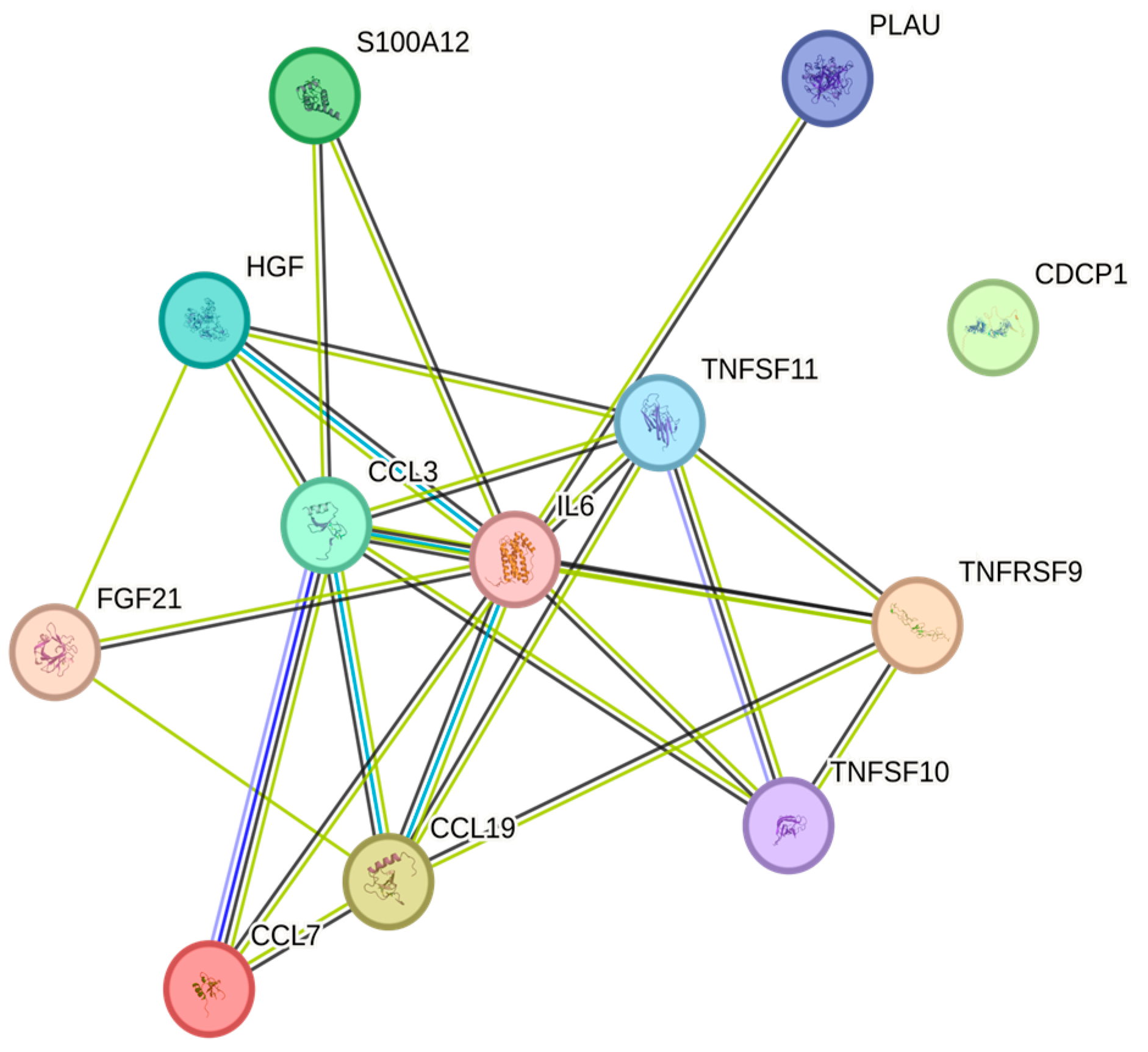
Figure 1. This figure represents a network of interactions among cytokines, demonstrating significant associations between circulating cytokine levels and HDL. The nodes (CCGFs) are connected by edges of varying thickness, indicating confidence of interactions.
Ijms 26 07746 g001
Figure 2. This figure represents a network of interactions among CCGFs, demonstrating significant associations between circulating CCGF levels and apolipoprotein A1. The nodes (CCGFs) are connected by edges of varying thickness, indicating confidence of interactions.
Ijms 26 07746 g002
Figure 3. This figure represents a network of interactions among CCGFs, demonstrating significant associations between circulating CCGF levels and creatinine. The nodes (CCGFs) are connected by edges of varying thickness, indicating confidence of interactions.
Ijms 26 07746 g003
Figure 4. This figure represents a network of interactions among CCGFs, demonstrating significant associations between circulating cytokine levels and eGFRcreatinine. The nodes (CCGFs) are connected by edges of varying thickness, indicating confidence of interactions.
Ijms 26 07746 g004
Figure 5. This figure represents a network of interactions among CCGFs, demonstrating significant associations between circulating cytokine levels and albumin. The nodes (CCGFs) are connected by edges of varying thickness, indicating confidence of interactions.
Ijms 26 07746 g005
Figure 6. This figure represents a network of interactions among CCGFs, demonstrating significant associations between circulating cytokine levels and apolipoprotein B. The nodes (CCGFs) are connected by edges of varying thickness, indicating confidence of interactions.
Ijms 26 07746 g006
Figure 7. This figure represents a network of interactions among CCGFs, demonstrating significant associations between circulating CCGF levels and non-HDL. The nodes (CCGFs) are connected by edges of varying thickness, indicating confidence of interactions.
Ijms 26 07746 g007
Figure 8. This figure represents a network of interactions among CCGFs, demonstrating significant associations between circulating cytokine levels and remnants. The nodes (CCGFs) are connected by edges of varying thickness, indicating confidence of interactions.
Ijms 26 07746 g008
Figure 9. This figure represents a network of interactions among CCGFs, demonstrating significant associations between circulating cytokine levels and non-HDL. The nodes (CCGFs) are connected by edges of varying thickness, indicating confidence of interactions.
Ijms 26 07746 g009
Figure 10. This figure represents a network of interactions among CCGFs, demonstrating significant associations between circulating cytokine levels and age. The nodes (CCGFs) are connected by edges of varying thickness, indicating confidence of interactions.
Ijms 26 07746 g010
Figure 11. This figure represents a network of interactions among CCGFs, demonstrating significant associations between circulating cytokine levels and weight. The nodes (CCGFs) are connected by edges of varying thickness, indicating confidence of interactions.
Ijms 26 07746 g011
Figure 12. This figure represents a network of interactions among CCGFs, demonstrating significant associations between circulating cytokine levels and BMI. The nodes (cytokines) are connected by edges of varying thickness, indicating confidence of interactions.
Ijms 26 07746 g012
Figure 13. This figure represents a network of interactions among CCGFs, demonstrating significant associations between circulating cytokine levels and gender. The nodes (CCGFs) are connected by edges of varying thickness, indicating confidence of interactions.
Ijms 26 07746 g013
Table 1. This table summarizes the prevalence of the CCGFs in our study cohort. Prevalences are given in absolute numbers. Abbreviations are explained in Supplementary Table S6, where UniprotIDs are also given.
CytokinePrevalenceCytokinePrevalenceCytokinePrevalence
TNFSF1010CASP-83CGCP11
CCL38COL18A13CXCL111
TNFSF117TNFSF123CX3CL11
CDCP16CSF-12Flt3L1
IL66SULT1A12IL-17C1
FGF-215CD52OSM1
S100A125TGF-alpha2CCL41
TNFRSF95DNER2IL-12B1
PLAU5ADA2IL-15RA1
HGF5CCL112IL71
CCL75CCL202CD61
IL-18R14CCL252TNF1
CCL194MMP-101Beta-NGF1
IL-10RB3IL-1 alpha1LTA1
FGF-193CCL21CCL281
Table 2. Number of CCGFs below the assay’s standard points.
CCGFUniProt N°Olink Assay IDN° Below Limit of Quantification
Olink Target 96 InflammationIL8P10145OID004710
Olink Target 96 InflammationVEGFAP15692OID004720
Olink Target 96 InflammationCD8AP01732OID051240
Olink Target 96 InflammationMCP-3P80098OID0047474
Olink Target 96 InflammationGDNFP39905OID004751
Olink Target 96 InflammationCDCP1Q9H5V8OID004760
Olink Target 96 InflammationCD244Q9BZW8OID004770
Olink Target 96 InflammationIL7P13232OID004781
Olink Target 96 InflammationOPGO00300OID004790
Olink Target 96 InflammationLAP TGF-beta-1P01137OID004800
Olink Target 96 InflammationuPAP00749OID004810
Olink Target 96 InflammationIL6P05231OID004820
Olink Target 96 InflammationIL-17CQ9P0M4OID0048312
Olink Target 96 InflammationMCP-1P13500OID004840
Olink Target 96 InflammationIL-17AQ16552OID0048555
Olink Target 96 InflammationCXCL11O14625OID004860
Olink Target 96 InflammationAXIN1O15169OID004870
Olink Target 96 InflammationTRAILP50591OID004880
Olink Target 96 InflammationIL-20RAQ9UHF4OID00489125
Olink Target 96 InflammationCXCL9Q07325OID004900
Olink Target 96 InflammationCST5P28325OID004910
Olink Target 96 InflammationIL-2RBP14784OID0049257
Olink Target 96 InflammationIL-1 alphaP01583OID00493142
Olink Target 96 InflammationOSMP13725OID004940
Olink Target 96 InflammationIL2P60568OID00495155
Olink Target 96 InflammationCXCL1P09341OID004960
Olink Target 96 InflammationTSLPQ969D9OID00497128
Olink Target 96 InflammationCCL4P13236OID004980
Olink Target 96 InflammationCD6P30203OID004990
Olink Target 96 InflammationSCFP21583OID005000
Olink Target 96 InflammationIL18Q14116OID005010
Olink Target 96 InflammationSLAMF1Q13291OID005020
Olink Target 96 InflammationTGF-alphaP01135OID005030
Olink Target 96 InflammationMCP-4Q99616OID005040
Olink Target 96 InflammationCCL11P51671OID005050
Olink Target 96 InflammationTNFSF14O43557OID005060
Olink Target 96 InflammationFGF-23Q9GZV9OID005070
Olink Target 96 InflammationIL-10RAQ13651OID0050852
Olink Target 96 InflammationFGF-5P12034OID0050970
Olink Target 96 InflammationMMP-1P03956OID005100
Olink Target 96 InflammationLIF-RP42702OID005110
Olink Target 96 InflammationFGF-21Q9NSA1OID005126
Olink Target 96 InflammationCCL19Q99731OID005130
Olink Target 96 InflammationIL-15RAQ13261OID00514102
Olink Target 96 InflammationIL-10RBQ08334OID005150
Olink Target 96 InflammationIL-22 RA1Q8N6P7OID00516142
Olink Target 96 InflammationIL-18R1Q13478OID005170
Olink Target 96 InflammationPD-L1Q9NZQ7OID005180
Olink Target 96 InflammationBeta-NGFP01138OID00519155
Olink Target 96 InflammationCXCL5P42830OID005200
Olink Target 96 InflammationTRANCEO14788OID005210
Olink Target 96 InflammationHGFP14210OID005220
Olink Target 96 InflammationIL-12BP29460OID005230
Olink Target 96 InflammationIL-24Q13007OID00524151
Olink Target 96 InflammationIL13P35225OID00525134
Olink Target 96 InflammationARTNQ5T4W7OID00526130
Olink Target 96 InflammationMMP-10P09238OID005270
Olink Target 96 InflammationIL10P22301OID005280
Olink Target 96 InflammationTNFP01375OID055480
Olink Target 96 InflammationCCL23P55773OID005300
Olink Target 96 InflammationCD5P06127OID005310
Olink Target 96 InflammationCCL3P10147OID005320
Olink Target 96 InflammationFlt3LP49771OID005330
Olink Target 96 InflammationCXCL6P80162OID005340
Olink Target 96 InflammationCXCL10P02778OID005350
Olink Target 96 Inflammation4E-BP1Q13541OID005360
Olink Target 96 InflammationIL-20Q9NYY1OID00537157
Olink Target 96 InflammationSIRT2Q8IXJ6OID005380
Olink Target 96 InflammationCCL28Q9NRJ3OID005390
Olink Target 96 InflammationDNERQ8NFT8OID012130
Olink Target 96 InflammationEN-RAGEP80511OID005410
Olink Target 96 InflammationCD40P25942OID00542162
Olink Target 96 InflammationIL33O95760OID005430
Olink Target 96 InflammationIFN-gammaP01579OID055470
Olink Target 96 InflammationFGF-19O95750OID005450
Olink Target 96 InflammationIL4P05112OID00546116
Olink Target 96 InflammationLIFP15018OID00547156
Olink Target 96 InflammationNRTNQ99748OID00548149
Olink Target 96 InflammationMCP-2P80075OID005490
Olink Target 96 InflammationCASP-8Q14790OID005500
Olink Target 96 InflammationCCL25O15444OID005510
Olink Target 96 InflammationCX3CL1P78423OID005520
Olink Target 96 InflammationTNFRSF9Q07011OID005530
Olink Target 96 InflammationNT-3P20783OID005543
Olink Target 96 InflammationTWEAKO43508OID005550
Olink Target 96 InflammationCCL20P78556OID005560
Olink Target 96 InflammationST1A1P50225OID005572
Olink Target 96 InflammationSTAMBPO95630OID005580
Olink Target 96 InflammationIL5P05113OID00559127
Olink Target 96 InflammationADAP00813OID005600
Olink Target 96 InflammationTNFBP01374OID005610
Olink Target 96 InflammationCSF-1P09603OID005620
Table 3. Interquartile range (=IQR). Erythrocyte volume fraction (EVF), white blood cell count (WBC), platelet count (Plt), albumin (Alb), and creatinine (Crea).
Valid NMedianIQR
Sex 68% females
Ageyear1642912
Weightkg16472.917
BMIkg/m216424.35
Hbg/L16413416
EVF%164414
WBC×109/L1645.52
Plt×109/L16423966
Albg/L164424
Creamicromol/L1646717
Cortisolnanomol/L164360199
Disclaimer/Publisher’s Note: The statements, opinions and data contained in all publications are solely those of the individual author(s) and contributor(s) and not of MDPI and/or the editor(s). MDPI and/or the editor(s) disclaim responsibility for any injury to people or property resulting from any ideas, methods, instructions or products referred to in the content.