Review

The Role of PAR2 in MASLD Progression and HCC Development

1
Department of Medicine, University of Padova, Via Giustiniani 2, 35128 Padova, Italy
2
Azienda Ospedale-Università Padova, Via Giustiniani 2, 35128 Padova, Italy
*
Author to whom correspondence should be addressed.
Int. J. Mol. Sci. 2025, 26(15), 7076; https://doi.org/10.3390/ijms26157076
Submission received: 17 June 2025 / Revised: 13 July 2025 / Accepted: 16 July 2025 / Published: 23 July 2025
(This article belongs to the Special Issue Obesity and Cancer Risk: Molecular Mechanisms and Perspectives)

Abstract

Metabolic dysfunction-associated steatotic liver disease (MASLD) has recently become the leading cause of chronic liver disease and can progress to hepatocellular carcinoma (HCC) through multiple pathogenic mechanisms. Protease-activated receptor 2 (PAR2) is a G-protein-coupled receptor activated by proteases such as trypsin, tryptase or coagulation factors VII and Xa. Recent studies have shown that PAR2 expression is increased in the liver of patients with MASLD or liver fibrosis. Its activation is linked to metabolic dysfunction through several pathways, including SREBP1c activation, AMPK inhibition and Akt-induced insulin resistance. Inhibition of PAR2 has been effective in reducing MASLD progression in different animal models. Notably, PAR2 blockade has also been effective in more advanced stages of the disease by dampening chronic inflammation and fibrogenesis through the inhibition of hepatic stellate cell activation and of TGF-β and SerpinB3 production. PAR2 also plays a role in cancer development, promoting tumour proliferation, angiogenesis and expression of immune checkpoint inhibitors (like PD-L1, CD47 and CD24). Due to its multifaceted involvement in liver disease, PAR2 is emerging as a key therapeutic target in this clinical context. This review aims to summarise current knowledge on PAR2′s role in MASLD and its potential as a therapeutic target.

1. Introduction

Protease-activated receptor 2 (PAR2) is a member of the G-protein-coupled receptor (GPCR) family. Unlike classical GPCRs that are activated by ligand binding, PAR2 and other members of the PAR family are uniquely activated through proteolytic cleavage of their N-terminal region by specific serine proteases. This cleavage unveils a tethered ligand, which interacts with the receptor itself to trigger a conformational change and initiate intracellular signalling. Among the four known PARs, PAR2 is selectively activated by various enzymes, including trypsin, tryptase and coagulation factors VIIa and Xa, among others [1].
PAR2 plays a crucial role in inflammation and fibrosis deposition and has recently been identified as a key regulator of metabolism. Consequently, it has been extensively studied in the context of obesity, liver disease progression and cancer development (Figure 1). In this review, we explore the pathophysiological roles of PAR2 in chronic liver disease progression and its potential as a therapeutic target.

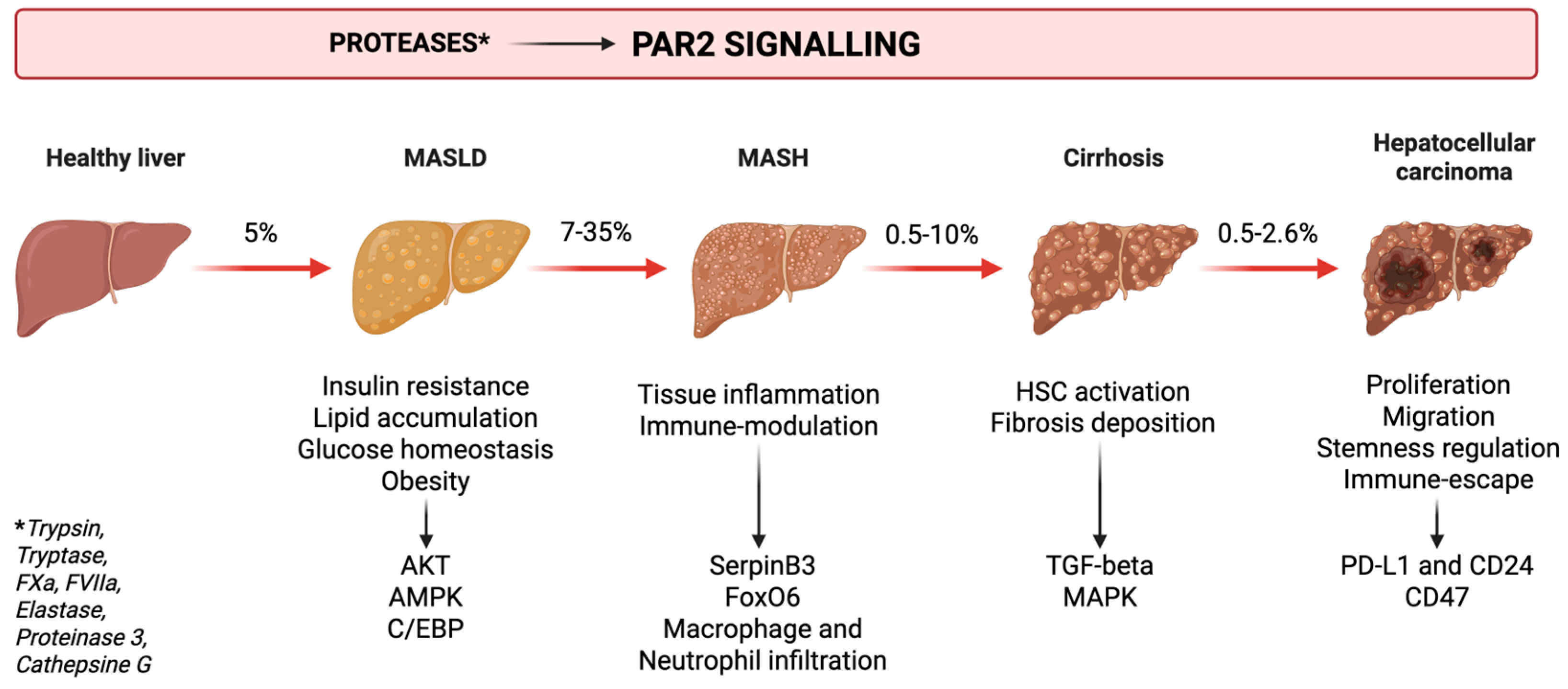
2. Metabolic Dysfunction—Associated Liver Disease and Liver Cancer

Metabolic dysfunction-associated steatotic liver disease (MASLD), formerly known as non-alcoholic fatty liver disease (NAFLD), has recently emerged as the leading cause of chronic liver disease, with an estimated global prevalence of 30% [2]. It encompasses a spectrum of progressive stages, beginning with steatotic liver disease (SLD)—characterised by lipid accumulation in the hepatocytes—and advancing to inflammation and metabolic dysfunction-associated steatohepatitis (MASH), previously referred to as non-alcoholic steatohepatitis (NASH). The inflammatory process promotes fibrotic tissue deposition, which may progress to liver cirrhosis and, ultimately, hepatocellular carcinoma (HCC) [2] (Figure 1). Due to its central role in the pathogenesis of liver cirrhosis, MASLD has been extensively investigated as a driver of liver-related complications. Moreover, growing evidence links MASLD to cardiovascular disease and extrahepatic malignancies [3], which are now recognised as the leading causes of death among these patients [4,5].
MASLD progression to HCC does not depend on cirrhotic evolution, as it is a recognised risk factor even in the absence of liver cirrhosis [6]. It has been estimated that in Italy, nearly 50% of patients with HCC in the context of metabolic liver disease are not cirrhotic [7]. There are different risk factors for cancer development, including obesity, metabolic dysfunction, type 2 diabetes, metabolic syndrome and insulin resistance [8].
MASLD can progress to HCC through different pathogenetic mechanisms. In brief, metabolic dysfunction, which may occur in genetically predisposed individuals, leads to an imbalance in lipid metabolism, resulting in increased oxidative stress and DNA damage. This process is accompanied by chronic inflammation, which impairs immune surveillance and promotes fibrosis deposition and HCC progression [9,10]. Obesity is a driver in MASLD progression to cancer, since it is associated with systemic inflammation and insulin resistance. These factors lead to deterioration in the regulation of immune surveillance and lipid metabolism in the liver. In fact, obesity alone doubles the risk of developing HCC, and this risk quadruples in individuals with a body mass index (BMI) exceeding 35 kg/m2 [6].
The following sections review how PAR2 activation contributes to the onset and progression of metabolic dysfunction, inflammation, fibrosis and ultimately, to cancer development.

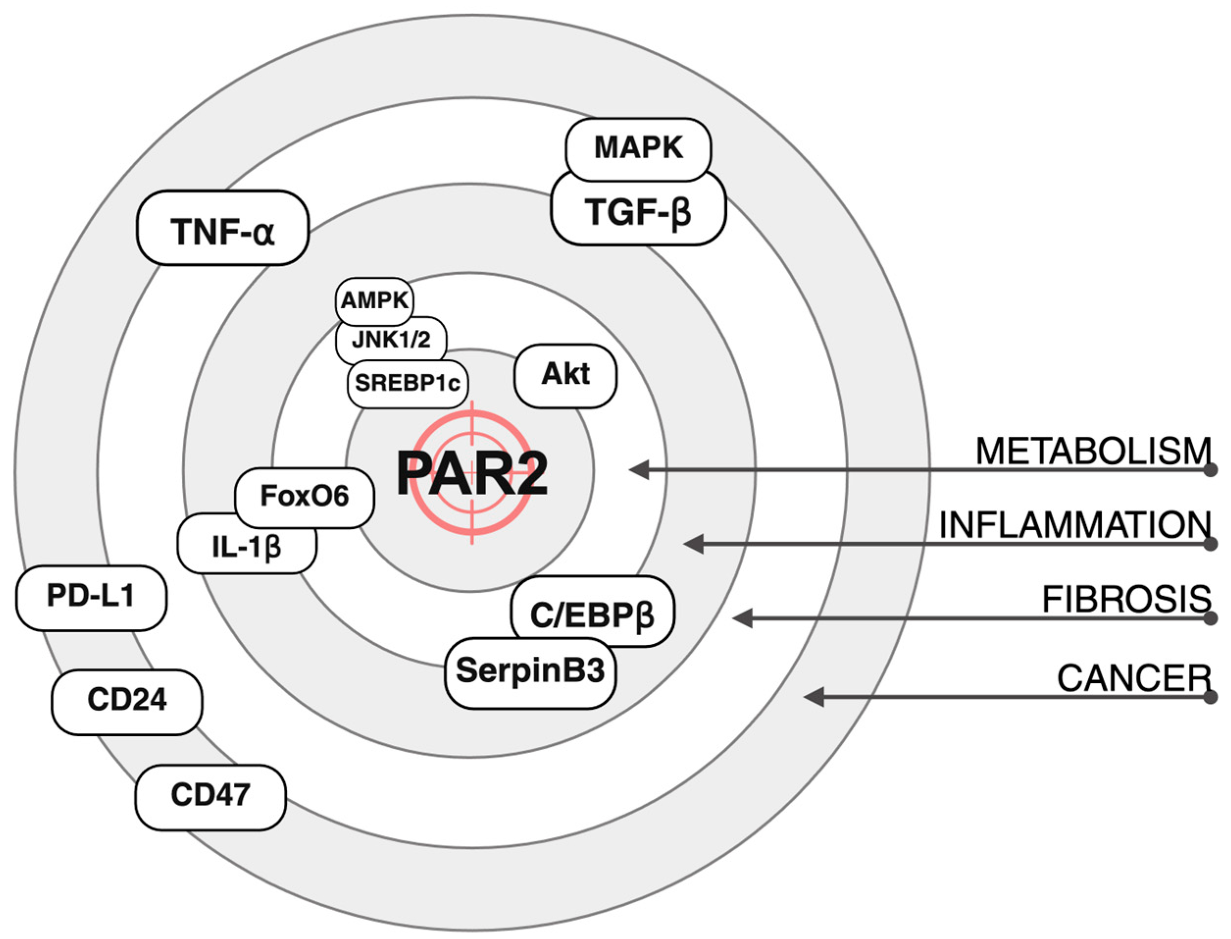
3. PAR2 and Metabolism

Given the central role of metabolic dysfunction in the onset of MASLD, increasing attention has been directed toward molecular mediators that link these processes. Among them, PAR2 has emerged as a key player in metabolic regulation (Figure 1) and different studies have proposed it as a possible therapeutic target (Table 1).

3.1. Glucose Metabolism and Insulin Resistance

Insulin resistance, a hallmark of type 2 diabetes and obesity, is central in the pathogenesis of metabolic dysfunction and hepatic steatosis. Impaired insulin signalling leads to a reduction in protein kinase B (Akt) activation, exacerbating dysregulation of glucose and lipid metabolism. A recent study investigated the role of PAR2 in promoting insulin resistance [11]. The authors observed an increased expression of PAR2 in hepatocytes from patients with concurrent diabetes and hepatic steatosis. Using a murine model of steatosis and diabetes, they also demonstrated that genetic deletion of PAR2 (PAR2-KO) improved histological markers of steatosis activity and led to a reduction in plasma glucose and insulin levels. These effects were primarily mediated by an increased expression of the glucose transporter type 2 (GLUT2), which permits glucose uptake and glycogen storage in the hepatocytes. Mechanistically, they showed that PAR2 activation inhibited insulin-Akt signalling, promoting insulin resistance, whereas pharmacological inhibition or genetic silencing of PAR2 restored insulin sensitivity.
The contribution of PAR2 to insulin resistance was also reported in another study where the authors found elevated levels of forkhead transcription factor (FoxO) 6 in insulin-resistant rats [12]. FoxO6 upregulation was accompanied by increased PAR2 expression. Deletion of FoxO6 improved insulin sensitivity and reduced PAR2 expression. The mechanistic link between FoxO6 and PAR2 was found in IL-1β, a cytokine, induced by FoxO6 signalling, which in turn stimulated PAR2 expression and inhibited insulin signalling.

3.2. Lipid Metabolism and Obesity

Insulin resistance promotes lipid uptake, de novo lipogenesis and lipid storage, ultimately leading to hepatic steatosis. Lipid metabolism represents another critical pathway in the pathogenesis of MASLD; therefore, the role of PAR2 was also evaluated in lipid homeostasis. A recent study observed that hepatic PAR2 expression was elevated in steatotic livers and that patients with high PAR2 levels in the liver had significantly increased plasma LDL cholesterol [13]. In PAR2-KO mice fed a high-fat diet (HFD), PAR2 deficiency was associated with lower hepatic and plasma cholesterol levels. This effect was mediated by three mechanisms: (a) reduced cholesterol synthesis, (b) increased hepatic cholesterol uptake and (c) enhanced biliary cholesterol excretion, as demonstrated by PCR analysis of gene transcription. Despite unchanged plasma triglyceride levels, PAR2-KO mice exhibited reduced hepatic accumulation of triglyceride and fatty acid. This was accompanied by downregulation of key lipogenic enzymes and upregulation of genes associated with β-oxidation. Mechanistically, the authors proved that PAR2-induced activation of JNK1/2 promoted sterol regulatory element binding protein 1 (SREBP1c) activation and inhibited AMP-activated protein kinase (AMPK), a key regulator in lipid catabolism.
Further evidence supporting the link between PAR2 and AMPK came from Kim et al., who demonstrated that PAR2-KO mice were protected from developing hepatic steatosis when fed on an HFD. PAR2-KO mice showed higher AMPK activation compared to wild-type controls. Conversely, in vitro, overexpression of PAR2 was associated with AMPK inhibition. The downregulation of AMPK by PAR2 resulted in an impairment of autophagy, contributing to hepatic lipid accumulation [14].
Badeanlou et al. reported that genetic deletion of PAR2 or tissue factor (TF), which mediates coagulation factor VIIa-induced PAR2 activation, protected mice from developing obesity and insulin resistance when subjected to an HFD [15]. Furthermore, PAR2 deletion was associated with a reduction in macrophage infiltration in the adipose tissue, supporting the role of PAR2 in the development of tissue inflammation. Interestingly, the researchers uncovered a dual role for PAR2. Specific deletion of PAR2 or TF in non-hematopoietic cells led to a reduction in weight gain. Conversely, deletion of these targets in immune cells did not affect obesity development but led to decreased markers of insulin resistance and inflammation.

3.3. Liver Steatosis and Pathology

Some years later, the same group explored the role of TF–PAR2 signalling in the liver [16]. Genetic deletion or pharmacological inhibition of TF, PAR2 or both led to reductions in gluconeogenesis, lipogenesis, and liver inflammation in a murine model of diet-induced obesity. Notably, these beneficial effects were also observed when TF or PAR2 deletion was restricted to hematopoietic cells, further supporting the role of this receptor in the crosstalk between the immune system and target tissue. Interestingly, wild-type (WT) mice fed an HFD exhibited increased hepatic infiltration of CD11b+CD11c+ macrophages and CD8+ lymphocytes, compared to mice fed a low-fat diet (LFD). Accordingly, TF deletion decreased CD11b+CD11c+ macrophage infiltration, while PAR2 deletion reduced the CD8+ T cell population.
A novel mechanism involving PAR2 in liver disease was recently identified by Villano et al., who demonstrated an interaction between PAR2 and SerpinB3 [17]. PAR2 activation induced the expression of the transcription factor CCAAT/enhancer-binding protein β (C/EBPβ), a known regulator of SerpinB3. In turn, SerpinB3, a member of the serine protease inhibitor family, was shown to be essential for PAR2 activation. Importantly, the authors found that PAR2 activity could be effectively inhibited using a small molecule called 1-Piperidine Propionic Acid (1-PPA): treatment with 1-PPA prevented lipid accumulation, inflammation and liver fibrosis in mice overexpressing SerpinB3. Additionally, it suppressed C/EBPβ expression in both THP-1 and HepG2 cells.
SerpinB3 has been previously implicated in the pathogenesis of steatotic liver disease. In a recent study, due to its involvement in lipid accumulation and inflammation, it was proposed as a new hepatokine [18]. In this study, two mouse models were used: one overexpressing SerpinB3 (TG/SB3) and the other expressing an isoform lacking its antiprotease activity (KO/SB3). Mice were fed on two different steatogenic diets: methionine–choline-deficient (MCD) or choline-deficient, L-amino acid-defined (CDAA). Overexpression of SerpinB3 resulted in increased hepatic lipid accumulation and inflammation, while loss of its activity conferred protection compared to WT mice. Moreover, TG/SB3 mice exhibited increased hepatic accumulation of crown-like structures formed by macrophages, a characteristic histological feature of steatotic liver disease, mirroring the findings of Wang et al. regarding TF–PAR2 signalling and CD11b+CD11c+ macrophage recruitment [16].
Lipid accumulation, when excessive, can lead to hepatotoxicity. This process, together with insulin resistance and obesity, is often accompanied by the development of chronic inflammation.

4. PAR2 and Inflammation

Inflammation is a process that allows the organism to cope with external damage. However, its dysregulation can be the trigger of various diseases. In the context of liver disease, problems begin when the inflammatory response becomes chronic. This drives the progression from benign MASLD to more advanced forms, such as MASH. PAR2 has been associated with several signalling pathways that contribute to the onset of inflammation in the liver (Figure 1) and other organs. Complementing these findings, various animal models have suggested it as a potential therapeutic target (Table 2).

4.1. Lung and Airways

Recent evidence has highlighted its involvement in promoting antigen responses during allergic reactions. A recent study demonstrates that inhibition of PAR2 reduces the production of inflammatory cytokines in a model of airway hypersensitivity [32]. It has also been shown that PAR2 antagonism effectively reduces airway hyperresponsiveness and inflammation in a murine model of allergic reaction [19]. Similar results were obtained in a mouse model of allergen-induced asthma [20], and in vitro [33].

4.2. Sepsis and Infection

In line with these findings, our research team recently published a study evaluating the efficacy of PAR2 antagonism using 1-PPA in a murine model of sepsis induced by intraperitoneal injection of lipopolysaccharide [21]. Treatment with 1-PPA significantly attenuated the inflammatory response and vasodilation, consequently enhancing cardiac function, reducing organ damage and alleviating clinical symptoms, which ultimately led to improved survival rates. Similar results were previously obtained in a model of liver damage induced by LPS administration. The authors reported an increased expression of PAR2 in Kupffer cells, tissue resident macrophages involved in pathogen-response and immune-activation or tolerance. Notably, PAR2 inhibition resulted in a suppression of TNFα (tumour necrosis factor) and an improved healing process [22].
The role of PAR2 in the response to microorganisms was also highlighted by Chu et al., who described a novel PAR2-dependent mechanism of neutrophil activation, driving inflammation and giving the rationale for inhibiting this pathway in the treatment of neutrophil-mediated inflammatory disease [34]. Other evidence demonstrated that PAR2 inhibition can prevent infection by Candida albicans [35] and periodontal inflammation induced by Porphyromonas gingivalis [23].

4.3. Neuroinflammation

Our research team recently published findings on the effects of PAR2 inhibition using 1-PPA in neuroinflammation, demonstrating its efficacy in reducing amyloid deposition and neuroglial inflammation in fibroblasts in patients with Parkinson’s disease [36]. Consistently, it has been shown that inhibition of mast cell tryptase effectively reduced PAR2-driven neuroinflammation in a murine model of cardiac arrest [24].

4.4. Gut and Microbiome

Several studies have also explored the role of PAR2 in intestinal diseases. It has been reported that a microbiome with high proteolytic activity can trigger colitis via PAR2 activation in mice [25]. Bacterial proteolytic activation of PAR2 has also been identified as a potential therapeutic target in inflammatory bowel disease [26] and in mediating both inflammation and pain in colitis [37]. However, Ke et al. showed that PAR2 deficiency in myeloid-derived suppressor cells enhanced their immunosuppressive activity, suggesting a dual role for this receptor in this context [27].

4.5. Kidney and Vascular System

Of particular relevance to this review is the link between PAR2, inflammation and metabolism, an interplay that involves not only the liver but also other organs. Ha et al. showed that PAR2-deficient mice were protected from HFD-induced kidney inflammation and injury [28]. In a subsequent study, the same group demonstrated that PAR2-KO mice were also protected from age-related kidney inflammation and senescence [29].
Two different studies further demonstrated the efficacy of PAR2 inhibition in preventing LDL-induced vascular damage in models of atherosclerosis, reinforcing the role of this receptor in the crosstalk between metabolism and inflammation [38,39].

4.6. PAR2 Dual Role in Inflammation

More recent studies have provided further insights into the complex interaction between the immune system and target organs (Figure 2). Reches et al. addressed this issue by investigating the role of PAR2 in two distinct murine models of liver injury: one immune-mediated (using concanavalin A) and one toxin-induced (with carbon tetrachloride, CCL4). The authors compared wild-type and PAR2 knock-out mice and repeated the experiments after bone marrow transplantation between the two strains. Their results demonstrated that PAR2 expression in liver tissue was essential for hepatocyte regeneration after toxic injury, whereas PAR2 expression in immune cells enhanced inflammation and worsened immune-mediated damage [30].
This dual role of PAR2 was later confirmed by the same group in a model of autoimmune diabetes. In this context, PAR2 expression in pancreatic β-cells was protective against immune damage, while its expression in lymphocytes promoted β-cell destruction and diabetes onset [31].
This dichotomous role of PAR2 in immune and target tissues warrants further investigation, particularly given the emerging importance in tumour development of this receptor.

5. PAR2 and Fibrosis

Chronic inflammation ultimately leads to fibrotic tissue deposition. Currently, the only therapeutic approach capable of halting—or, in certain cases, reversing—fibrosis relies on the removal or control of the underlying etiological factor. Owing to its critical role in the development of liver-related complications, fibrosis remains a major focus of translational research. Figure 1 outlines the mechanistic involvement of PAR2 in the initiation and perpetuation of fibrogenesis, while Table 3 summarises key preclinical studies investigating PAR2 inhibition as a potential antifibrotic strategy.

5.1. Liver Fibrosis

Inhibition of PAR2 using the pepducin PZ-235 was reported as effective in preventing liver fibrosis in a two-murine model, one fed on an HFD and the other treated with CCL4. PZ-235 directly reduced hepatic stellate cell (HSC) activation and decreased hepatocyte death, thereby limiting the main stimulus for chronic inflammation [40].
Other studies have confirmed the involvement of PAR2 in modulating HSC function. Knight et al. showed that PAR2-KO mice were protected from fibrosis induced by CCl4, a protection associated with reduced expression of TGF-β, the main mediator of fibrogenesis [41]. In a later study, the same authors investigated the relationship between TF activation and liver fibrosis using three different genetically modified murine models lacking PAR2, the cytoplasmic domain of TF, or both. Deletion of either PAR2 or TF conferred protection against fibrosis, and simultaneous deletion of both did not produce any additive benefit, suggesting that TF-PAR2 signalling is a shared pathway driving fibrosis [42].
In vitro studies also support this evidence. HSCs upregulated both PAR1 and PAR2 expression when cultured on plastic to promote myofibroblastic phenotype transformation. Stimulation of either receptor increased proliferation and activation of mitogen-activated protein kinase (MAPK), while their inhibition had the opposite effect [49]. Another in vitro study explored the role of PAR2 in apoptosis. Activation-induced cell death, triggered by phorbol myristate acetate, was suppressed by concurrent PAR2 stimulation with tryptase, suggesting a protective anti-apoptotic role of PAR2 in activated HSCs [50].

5.2. Other Organs Fibrosis
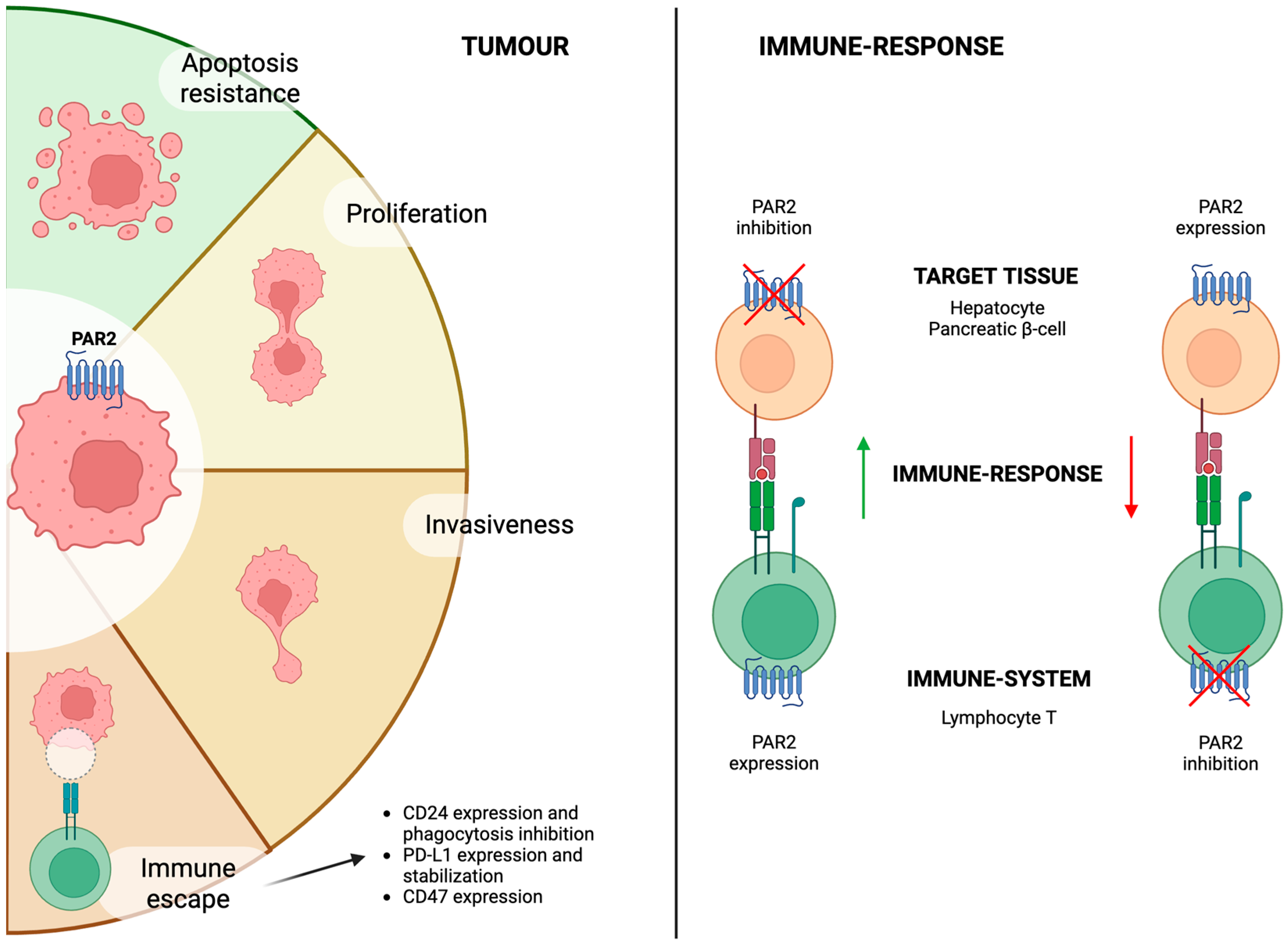
The pro-fibrotic activity of PAR2 is not limited to the liver. Tisch et al. recently reported that genetic deletion of PAR2 reduced airway fibrosis following allergen exposure [43]. Similar protective effects were observed in the kidney, where PAR2KO mice were protected from renal inflammation and fibrosis [44]. Along the same lines, both PAR2 and PAR1 contribute to kidney injury and fibrosis in a diabetic mouse model [45].
Additional studies have examined the effects of PAR2 on intestinal fibrosis. Mast cell-derived tryptase activated PAR2 on intestinal fibroblasts, promoting their transition to myofibroblasts and mirroring the findings in hepatic HSCs [46]. Two additional studies confirmed that PAR2 inhibition protected against intestinal fibrosis [47,48].
Collectively, these findings underscore the role of PAR2 in orchestrating immune responses and promoting fibrosis across multiple organ systems [51]. This has important implications, as sustained inflammation and fibrotic signalling may contribute to tumour development via activation of oncogenic pathways.

6. PAR2 and Cancer

PAR2 expression and activation has also been associated with intracellular signalling pathways associated with cancer development and immune-evasion (Figure 1 and Figure 2). Table 4 summarises studies investigating the role of PAR2 in this context and its inhibition as a potential therapeutic target.

6.1. Cancer Progression and Growth

In the liver, PAR2 expression in HSCs promotes HCC growth and angiogenesis in a murine xenograft model, whereas genetic deletion of PAR2 suppresses these effects. Furthermore, PAR2-deficient HSCs exhibited reduced activation following TGF-β stimulation and Hep3B-conditioned medium, indicating that PAR2 expression in HSCs contributes not only to fibrogenesis but also to HCC progression [52].
Moreover, a recent study showed that high PAR2 expression in HCC tissue samples after radical resection was associated with more aggressive clinical features. Patients with elevated PAR2 levels had larger tumours, more advanced stages and a higher incidence of microvascular invasion. Building on these findings, the authors demonstrated in two in vitro hepatic cancer cell models that PAR2 overexpression enhanced both proliferation and metastatic potential, while silencing of PAR2 reversed these effects. In vivo, PAR2 silencing reduced the extent of metastatic spread in a mouse model of liver metastasis [53].
In another study, the authors showed that secreted cathepsin S interacts with PAR2 to regulate the transition of cancer stem cells in HCC. Cathepsin S was secreted by CD47+ cells displaying stemness characteristics. Suppression of CD47 reduced HCC cell proliferation, and disruption of the cathepsin S/PAR2 axis increased chemosensitivity, suggesting a role for this pathway in therapy resistance [54].

6.2. Immune Surveillance and Immunotherapy

Immune checkpoint inhibitors (ICIs) have become the first-line therapy for various cancers. Given its role in modulating immune responses, PAR2 has been investigated in this context. In a recent study, a novel gene signature predictive of response to immunotherapy across multiple cancer types was identified [59]. Interestingly, among the genes analysed, PAR2, together with RNA-binding motif protein 9, was associated with poorer response to ICI therapy and T cell dysfunction.
The role of PAR2 in regulating anti-tumour immune responses was further explored in a study on metastatic colorectal cancer [55]. The authors found that PAR2 expression correlated with reduced macrophage-mediated phagocytosis of malignant cells. This effect was driven by increased expression of the “don’t eat me” signal, CD24. Conversely, neutrophil elastase, which cleaves and inhibits PAR2, downregulates CD24 expression, thereby enhancing phagocytosis. Similar findings were also reported in breast cancer, where activation of PAR2 signalling via coagulation factor VIIa led to reduced phagocytosis in an in vitro model [56]. Moreover, genetic deletion of PAR2 in vivo resulted in a smaller tumour size. Both studies highlighted a synergistic effect between PAR2 inhibition (whether pharmacological or genetic) and ICI treatment.
Apart from phagocytosis, other authors have focused on the effects of PAR2 in regulating T-cell response. A recent study reported that activation of PAR2 by coagulation factor Xa (FXa) conferred resistance to anoikis in HCC cells, thereby promoting metastasis. FXa-mediated PAR2 activation also increased PD-L1 expression while simultaneously inhibiting CD8+ T cell infiltration into tumours. Inhibition of FXa in vivo resulted in decreased metastasis and downregulation of PD-L1. These effects were further enhanced by concomitant administration of anti-PD-L1 antibodies [57]. Similar results were reported in the context of breast cancer, where PAR2 activation leads to increased production and stabilisation of PD-L1 and a reduction in CD8+ T cell activity [58].
These findings suggest that PAR2 expression in cancer cells not only promotes tumour progression and invasion but also suppresses the immune response. On the other hand, as previously reported, PAR2 expression in the immune system may activate an immune response (Figure 2). However, to the best of our knowledge, no study has directly investigated the impact of PAR2 activation on immune cells within the tumour microenvironment.

7. Discussion and Future Perspectives

7.1. PAR2 as a Therapeutic Target in Liver Disease Progression

The role of PAR2 in promoting metabolic dysfunction has been extensively investigated. Reducing insulin resistance may represent a promising therapeutic strategy for obesity, diabetes and MASLD, as it constitutes one of the key pathogenetic drivers of these conditions [60]. Several preclinical studies have demonstrated the beneficial effects of PAR2 inhibition, with no significant side effects or adverse reactions reported (Table 1). Moreover, these findings are consistent with the observed upregulation of PAR2 expression in liver biopsies from patients with diabetes, MASLD or fibrosis [11,13].
As lipid accumulation progresses, it can lead to hepatocellular injury and ultimately to chronic inflammation, resulting in fibrotic tissue deposition. There is growing evidence supporting the efficacy of PAR2 inhibitors in attenuating liver fibrosis (Table 3). Notably, the antifibrotic effects of PAR2 inhibition are not solely attributable to the reduction in hepatic steatosis but also involve the direct modulation of chronic inflammation and fibrogenesis, particularly through the suppression of hepatic stellate cell activation and TGF-β production. Indeed, PAR2 inhibition has been effective in reducing fibrosis both in dietary preclinical models of hepatic steatosis (e.g., HFD or CDAA diets) and in toxin-induced liver injury models (e.g., CCl4) (Table 3). It is important to note that, among PAR2 antagonists, PZ-235 pepducin (Oasis Pharmaceuticals) is currently undergoing clinical development for various indications. This compound has advanced to phase 1 clinical trials for non-alcoholic steatohepatitis (NCT05680233). Meanwhile, the clinical development process for 1-PPA is still in progress.

7.2. PAR2 as a Therapeutic Target in HCC

A critical consequence of chronic liver inflammation is the increased risk of tumour development. The incidence of HCC is known to rise not only in the advanced stages of cirrhosis but also in the setting of MASH. Several mechanisms contribute to this elevated oncogenic risk, including enhanced DNA damage, persistent proliferative signalling and impaired immune surveillance [2].
Taken together, the aforementioned findings highlight the fundamentally pro-inflammatory role of PAR2. Non-tissue-specific inhibition of PAR2 has been shown to exert beneficial effects in various conditions characterised by uncontrolled inflammation (Table 2). PAR2 blockade has demonstrated efficacy in preclinical models of asthma, autoimmune diseases and septic shock, among others.
The dual role of PAR2—stimulating the immune system when expressed by immune cells and suppressing immune responses when expressed by target tissue cells—seems to function as a balance that shifts depending on the clinical context. Indeed, the observation that systemic inhibition of PAR2 in pro-inflammatory settings confers therapeutic benefit suggests that, in these conditions, immune activation is dysregulated and that PAR2 inhibition contributes to the restoration of immune homeostasis. However, under specific conditions of acute tissue injury, PAR2 activation has been associated with increased survival of organ-specific cells and protection against tissue damage and cell death. These findings imply that PAR2 may exert context-dependent protective effects in the acute or chronic setting.
Extensive evidence has investigated the role of PAR2 in cancer, demonstrating that its activation promotes cancer proliferation, migratory capacity and invasiveness of tumour cells (Figure 2). On the same line, PAR2 inhibition has been shown to reduce the risk of metastasis as well as the growth of various tumour types, including HCC. Additionally, PAR2 contributes to immune escape mechanisms when expressed in the tumoural cells, consistent with its ability to upregulate different immune checkpoint inhibitors (Figure 2). On the other hand, PAR2 appears to enhance inflammation in other clinical contexts, and studies reported an increased immune response when it was activated in immune system cells. Given that current oncological strategies increasingly focus on modulating the immune system, it is important to highlight the potential contribution of PAR2 in this context. However, studies on PAR2’s potential activation within the immune-system cells are lacking. The current available evidence suggests that inhibiting PAR2 in tumoural cells enhances therapeutic response.

7.3. PAR2 as a Prognostic Marker in HCC

Given its broad biological functions, PAR2 expression could be investigated as a potential prognostic biomarker in liver disease and cancer. However, current evidence in this field remains limited.
Rana et al. [13] and Shearer et al. [11] have shown that PAR2 is overexpressed in liver biopsies from patients with metabolic liver disease, with expression levels increasing alongside the severity of fibrosis and inflammation. These findings suggest a correlation between PAR2 expression and disease progression, although direct prognostic implications have not been fully explored.
In this context, different molecules should also be considered as surrogate markers or readouts of PAR2 activation. For example, SerpinB3 is known to induce PAR2 synthesis and activation and has been described as a prognostic factor in various liver conditions. Numerous studies have supported SerpinB3 association with HCC development [61,62] and worse outcomes in patients with liver cirrhosis [63,64], cholangiocarcinoma [65,66] and HCC [67,68]. Also, Li et al. [57] have reported that the prognosis of patients with HCC is influenced by the expression of coagulation factor Xa, a known activator of PAR2. However, these studies did not directly investigate PAR2 expression in hepatic tissue, highlighting a significant gap in our understanding of the mechanistic link and underscoring the need for further research in this area.
To the best of our knowledge, only two studies have directly associated PAR2 expression with clinical outcomes in HCC. Tsai et al. found that high PAR2 expression correlates with features of more aggressive tumours at diagnosis and poorer prognosis in HCC patients after liver resection [69], while Barenche et al. reported that PAR2 expression is associated with reduced response to immunotherapy in different types of cancer [59].
Nevertheless, these findings require confirmation through larger, well-designed observational clinical studies to validate the prognostic role of PAR2.

8. Conclusions

In conclusion, recent research has thoroughly examined the crucial role of PAR2 in the progression of metabolic dysfunction-associated steatotic liver disease (MASLD). PAR2 acts as an upstream regulator in various pathogenetic mechanisms associated with MASLD. Consequently, inhibiting PAR2 emerges as a promising therapeutic strategy, potentially interrupting simultaneously multiple disease-driving pathways (Figure 3). However, clinical validation through prospective studies is warranted to confirm these promising preclinical findings and to establish the efficacy and safety of PAR2 inhibition in humans.
Increasing evidence also points to a key role for PAR2 in promoting tumour growth and immune escape. Notably, PAR2 acts upstream of multiple signalling pathways whose downstream effectors are currently being targeted by tailored therapies (Figure 3). Unlike these single-pathway inhibitors, targeting PAR2 may allow for a broader therapeutic effect, intercepting several pro-inflammatory, pro-fibrogenic and pro-tumourigenic signals, as already described. Nevertheless, PAR2′s role in immuno-oncology remains underexplored, presenting a significant opportunity for further investigation. This emerging area of research is particularly promising, given the expanding role of immunotherapy in cancer treatment and the potential for PAR2 to modulate immune responses within the tumour microenvironment.

9. Patents

The authors declare that P.P. and M.A. are listed as inventors of the following patents: IT 102017000026858, EP 20709715.5, US 11628163, IT 102019000012930, EP 20709715.5, US 17/629.791 pending; P.P. is listed as inventor of the patents IT 102022000014593, PTC/IB2023/057138 pending. All the patents have been filed by the University of Padova.

Author Contributions

Conceptualisation, P.G. and P.P.; methodology, P.G. and A.M.; writing—original draft preparation, P.G.; writing—review and editing, P.P. and A.M.; supervision, P.P. All authors have read and agreed to the published version of the manuscript.

Funding

This review received no external funding.

Conflicts of Interest

The authors declare no conflict of interest.

Abbreviations

The following abbreviations are used in this manuscript:
1-PPA1-Piperidie Propionic Acid
AktProtein Kinase B
AMPKAMP-Activated Protein Kinase
AOMAzoxymethane
BMIBody Mass Index
C/EBP-βCCAAT/Enhancer-Binding Protein β
CCL4Carbon Tetrachloride
CDAACholine-Deficient, L-Amino Acid-Defined
DSSDextran Sulfate Sodium
FoxO6Forkhead Transcription Factor 6
FXaCoagulation Factor Xa
GPCRG-Protein Coupled Receptor
GLUT2Glucose Transporte Type 2
HCCHepatocellular Carcinoma
HFDHigh-Fat Diet
HSCHepatic Stellate Cell
ICIImmune-Checkpoint Inhibitor
KOKnock-Out
LFDLow-Fat Diet
LPSLipopolysaccharide
MAPKMitogen-Activated Protein Kinase
MASHMetabolic Dysfunction-Associated Steatohepatitis
MASLDMetabolic Dysfunction-Associated Steatotic Liver Disease
MCDMethionine–Choline-Deficient
NAFLDNon-Alcoholic Fatty Liver Disease
NASHNon-Alcoholic Steatohepatitis
PAR2Protease-Activated Receptor 2
SLDSteatotic Liver Disease
SREBP1cSterol Regulatory Element Binding Protein 1
TFTissue Factor
TGF-βTransforming Growth Factor β
TNBS2,4,6-Trinitrobenzene Sulfonic Acid
TNF-αTumour Necrosis Factor α
WTWild-Type

References

  1. Villano, G.; Pontisso, P. Protease Activated Receptor 2 as a Novel Druggable Target for the Treatment of Metabolic Dysfunction-Associated Fatty Liver Disease and Cancer. Front. Immunol. 2024, 15, 1397441. [Google Scholar] [CrossRef] [PubMed]
  2. European Association for the Study of the Liver (EASL); European Association for the Study of Diabetes (EASD); European Association for the Study of Obesity (EASO). EASL-EASD-EASO Clinical Practice Guidelines on the Management of Metabolic Dysfunction-Associated Steatotic Liver Disease (MASLD). Obes. Facts 2024, 17, 374–444. [Google Scholar] [CrossRef] [PubMed]
  3. Thomas, J.A.; Kendall, B.J.; El-Serag, H.B.; Thrift, A.P.; Macdonald, G.A. Hepatocellular and Extrahepatic Cancer Risk in People with Non-Alcoholic Fatty Liver Disease. Lancet Gastroenterol. Hepatol. 2024, 9, 159–169. [Google Scholar] [CrossRef] [PubMed]
  4. Chalasani, N.; Younossi, Z.; Lavine, J.E.; Charlton, M.; Cusi, K.; Rinella, M.; Harrison, S.A.; Brunt, E.M.; Sanyal, A.J. The Diagnosis and Management of Nonalcoholic Fatty Liver Disease: Practice Guidance from the American Association for the Study of Liver Diseases. Hepatology 2018, 67, 328–357. [Google Scholar] [CrossRef] [PubMed]
  5. Paik, J.M.; Henry, L.; De Avila, L.; Younossi, E.; Racila, A.; Younossi, Z.M. Mortality Related to Nonalcoholic Fatty Liver Disease Is Increasing in the United States. Hepatol. Commun. 2019, 3, 1459–1471. [Google Scholar] [CrossRef] [PubMed]
  6. Younossi, Z.M.; Henry, L. Epidemiology of Non-Alcoholic Fatty Liver Disease and Hepatocellular Carcinoma. JHEP Rep. 2021, 3, 100305. [Google Scholar] [CrossRef] [PubMed]
  7. Piscaglia, F.; Svegliati-Baroni, G.; Barchetti, A.; Pecorelli, A.; Marinelli, S.; Tiribelli, C.; Bellentani, S.; on behalf of the HCC-NAFLD Italian Study Group. Clinical Patterns of Hepatocellular Carcinoma in Nonalcoholic Fatty Liver Disease: A Multicenter Prospective Study. Hepatology 2016, 63, 827–838. [Google Scholar] [CrossRef] [PubMed]
  8. Wang, C.; Wang, X.; Gong, G.; Ben, Q.; Qiu, W.; Chen, Y.; Li, G.; Wang, L. Increased Risk of Hepatocellular Carcinoma in Patients with Diabetes Mellitus: A Systematic Review and Meta-analysis of Cohort Studies. Int. J. Cancer 2012, 130, 1639–1648. [Google Scholar] [CrossRef] [PubMed]
  9. Anstee, Q.M.; Reeves, H.L.; Kotsiliti, E.; Govaere, O.; Heikenwalder, M. From NASH to HCC: Current Concepts and Future Challenges. Nat. Rev. Gastroenterol. Hepatol. 2019, 16, 411–428. [Google Scholar] [CrossRef] [PubMed]
  10. Wang, X.; Zhang, L.; Dong, B. Molecular Mechanisms in MASLD/MASH-Related HCC. Hepatology 2024. [Google Scholar] [CrossRef] [PubMed]
  11. Shearer, A.M.; Wang, Y.; Fletcher, E.K.; Rana, R.; Michael, E.S.; Nguyen, N.; Abdelmalek, M.F.; Covic, L.; Kuliopulos, A. PAR2 Promotes Impaired Glucose Uptake and Insulin Resistance in NAFLD through GLUT2 and Akt Interference. Hepatology 2022, 76, 1778–1793. [Google Scholar] [CrossRef] [PubMed]
  12. Kim, D.H.; Lee, B.; Lee, J.; Kim, M.E.; Lee, J.S.; Chung, J.H.; Yu, B.P.; Dong, H.H.; Chung, H.Y. FoxO6-Mediated IL-1β Induces Hepatic Insulin Resistance and Age-Related Inflammation via the TF/PAR2 Pathway in Aging and Diabetic Mice. Redox Biol. 2019, 24, 101184. [Google Scholar] [CrossRef] [PubMed]
  13. Rana, R.; Shearer, A.M.; Fletcher, E.K.; Nguyen, N.; Guha, S.; Cox, D.H.; Abdelmalek, M.; Wang, Y.; Baleja, J.D.; Covic, L.; et al. PAR2 Controls Cholesterol Homeostasis and Lipid Metabolism in Nonalcoholic Fatty Liver Disease. Mol. Metab. 2019, 29, 99–113. [Google Scholar] [CrossRef] [PubMed]
  14. Kim, B.M.; Kim, D.H.; Park, Y.J.; Ha, S.; Choi, Y.J.; Yu, H.S.; Chung, K.W.; Chung, H.Y. PAR2 Promotes High-Fat Diet-Induced Hepatic Steatosis by Inhibiting AMPK-Mediated Autophagy. J. Nutr. Biochem. 2021, 95, 108769. [Google Scholar] [CrossRef] [PubMed]
  15. Badeanlou, L.; Furlan-Freguia, C.; Yang, G.; Ruf, W.; Samad, F. Tissue Factor–Protease-Activated Receptor 2 Signaling Promotes Diet-Induced Obesity and Adipose Inflammation. Nat. Med. 2011, 17, 1490–1497. [Google Scholar] [CrossRef] [PubMed]
  16. Wang, J.; Chakrabarty, S.; Bui, Q.; Ruf, W.; Samad, F. Hematopoietic Tissue Factor–Protease-Activated Receptor 2 Signaling Promotes Hepatic Inflammation and Contributes to Pathways of Gluconeogenesis and Steatosis in Obese Mice. Am. J. Pathol. 2015, 185, 524–535. [Google Scholar] [CrossRef] [PubMed]
  17. Villano, G.; Novo, E.; Turato, C.; Quarta, S.; Ruvoletto, M.; Biasiolo, A.; Protopapa, F.; Chinellato, M.; Martini, A.; Trevellin, E.; et al. The Protease Activated Receptor 2—CCAAT/Enhancer-Binding Protein Beta—SerpinB3 Axis Inhibition as a Novel Strategy for the Treatment of Non-Alcoholic Steatohepatitis. Mol. Metab. 2024, 81, 101889. [Google Scholar] [CrossRef] [PubMed]
  18. Novo, E.; Cappon, A.; Villano, G.; Quarta, S.; Cannito, S.; Bocca, C.; Turato, C.; Guido, M.; Maggiora, M.; Protopapa, F.; et al. SerpinB3 as a Pro-Inflammatory Mediator in the Progression of Experimental Non-Alcoholic Fatty Liver Disease. Front. Immunol. 2022, 13, 910526. [Google Scholar] [CrossRef] [PubMed]
  19. Schiff, H.V.; Rivas, C.M.; Pederson, W.P.; Sandoval, E.; Gillman, S.; Prisco, J.; Kume, M.; Dussor, G.; Vagner, J.; Ledford, J.G.; et al. β-Arrestin-biased Proteinase-activated Receptor-2 Antagonist C781 Limits Allergen-induced Airway Hyperresponsiveness and Inflammation. Br. J. Pharmacol. 2023, 180, 667–680. [Google Scholar] [CrossRef] [PubMed]
  20. De Matos, N.A.; Lima, O.C.O.; Da Silva, J.F.; Piñeros, A.R.; Tavares, J.C.; Lemos, V.S.; Alves-Filho, J.C.; Klein, A. Blockade of Protease-Activated Receptor 2 Attenuates Allergen-Mediated Acute Lung Inflammation and Leukocyte Recruitment in Mice. J. Biosci. 2022, 47, 2. [Google Scholar] [CrossRef]
  21. Luisetto, R.; Scarpa, M.; Villano, G.; Martini, A.; Quarta, S.; Ruvoletto, M.; Guerra, P.; Scarpa, M.; Chinellato, M.; Biasiolo, A.; et al. 1-Piperidine Propionic Acid Protects from Septic Shock Through Protease Receptor 2 Inhibition. IJMS 2024, 25, 11662. [Google Scholar] [CrossRef] [PubMed]
  22. Jesmin, S.; Gando, S.; Zaedi, S.; Sakuraya, F. Chronological Expression of PAR Isoforms in Acute Liver Injury and Its Amelioration by PAR2 Blockade in a Rat Model of Sepsis. Thromb. Haemost. 2006, 96, 830–838. [Google Scholar] [CrossRef] [PubMed]
  23. Francis, N.; Sanaei, R.; Ayodele, B.A.; O’Brien-Simpson, N.M.; Fairlie, D.P.; Wijeyewickrema, L.C.; Pike, R.N.; Mackie, E.J.; Pagel, C.N. Effect of a Protease-activated Receptor-2 Antagonist (GB88) on Inflammation-related Loss of Alveolar Bone in Periodontal Disease. J. Periodontal Res. 2023, 58, 544–552. [Google Scholar] [CrossRef] [PubMed]
  24. Ocak, U.; Eser Ocak, P.; Huang, L.; Xu, W.; Zuo, Y.; Li, P.; Gamdzyk, M.; Zuo, G.; Mo, J.; Zhang, G.; et al. Inhibition of Mast Cell Tryptase Attenuates Neuroinflammation via PAR-2/P38/NFκB Pathway Following Asphyxial Cardiac Arrest in Rats. J. Neuroinflamm. 2020, 17, 144. [Google Scholar] [CrossRef] [PubMed]
  25. Santiago, A.; Hann, A.; Constante, M.; Rahmani, S.; Libertucci, J.; Jackson, K.; Rueda, G.; Rossi, L.; Ramachandran, R.; Ruf, W.; et al. Crohn’s Disease Proteolytic Microbiota Enhances Inflammation through PAR2 Pathway in Gnotobiotic Mice. Gut Microbes 2023, 15, 2205425. [Google Scholar] [CrossRef] [PubMed]
  26. Rondeau, L.E.; Da Luz, B.B.; Santiago, A.; Bermudez-Brito, M.; Hann, A.; De Palma, G.; Jury, J.; Wang, X.; Verdu, E.F.; Galipeau, H.J.; et al. Proteolytic Bacteria Expansion during Colitis Amplifies Inflammation through Cleavage of the External Domain of PAR2. Gut Microbes 2024, 16, 2387857. [Google Scholar] [CrossRef] [PubMed]
  27. Ke, Z.; Wang, C.; Wu, T.; Wang, W.; Yang, Y.; Dai, Y. PAR2 Deficiency Enhances Myeloid Cell-Mediated Immunosuppression and Promotes Colitis-Associated Tumorigenesis. Cancer Lett. 2020, 469, 437–446. [Google Scholar] [CrossRef] [PubMed]
  28. Ha, S.; Yang, Y.; Kim, B.M.; Kim, J.; Son, M.; Kim, D.; Yu, H.S.; Im, D.; Chung, H.Y.; Chung, K.W. Activation of PAR2 Promotes High-Fat Diet-Induced Renal Injury by Inducing Oxidative Stress and Inflammation. Biochim. Biophys. Acta (BBA)—Mol. Basis Dis. 2022, 1868, 166474. [Google Scholar] [CrossRef] [PubMed]
  29. Ha, S.; Kim, H.W.; Kim, K.M.; Kim, B.M.; Kim, J.; Son, M.; Kim, D.; Kim, M.; Yoo, J.; Yu, H.S.; et al. PAR2-mediated Cellular Senescence Promotes Inflammation and Fibrosis in Aging and Chronic Kidney Disease. Aging Cell 2024, 23, e14184. [Google Scholar] [CrossRef] [PubMed]
  30. Reches, G.; Blondheim Shraga, N.R.; Carrette, F.; Malka, A.; Saleev, N.; Gubbay, Y.; Ertracht, O.; Haviv, I.; Bradley, L.M.; Levine, F.; et al. Resolving the Conflicts around Par2 Opposing Roles in Regeneration by Comparing Immune-Mediated and Toxic-Induced Injuries. Inflamm. Regen. 2022, 42, 52. [Google Scholar] [CrossRef] [PubMed]
  31. Reches, G.; Khoon, L.; Ghanayiem, N.; Malka, A.; Piran, R. Controlling Autoimmune Diabetes Onset by Targeting Protease-Activated Receptor 2. Biomed. Pharmacother. 2024, 175, 116622. [Google Scholar] [CrossRef] [PubMed]
  32. Wang, Y.-J.; Yu, S.-J.; Tsai, J.-J.; Yu, C.-H.; Liao, E.-C. Antagonism of Protease Activated Receptor-2 by GB88 Reduces Inflammation Triggered by Protease Allergen Tyr-P3. Front. Immunol. 2021, 12, 557433. [Google Scholar] [CrossRef] [PubMed]
  33. Rivas, C.M.; Schiff, H.V.; Moutal, A.; Khanna, R.; Kiela, P.R.; Dussor, G.; Price, T.J.; Vagner, J.; DeFea, K.A.; Boitano, S. Alternaria Alternata-Induced Airway Epithelial Signaling and Inflammatory Responses via Protease-Activated Receptor-2 Expression. Biochem. Biophys. Res. Commun. 2022, 591, 13–19. [Google Scholar] [CrossRef] [PubMed]
  34. Chu, T.-Y.; Zheng-Gérard, C.; Huang, K.-Y.; Chang, Y.-C.; Chen, Y.-W.; I, K.-Y.; Lo, Y.-L.; Chiang, N.-Y.; Chen, H.-Y.; Stacey, M.; et al. GPR97 Triggers Inflammatory Processes in Human Neutrophils via a Macromolecular Complex Upstream of PAR2 Activation. Nat. Commun. 2022, 13, 6385. [Google Scholar] [CrossRef] [PubMed]
  35. Kumar, R.; Rojas, I.G.; Edgerton, M. Candida Albicans Sap6 Initiates Oral Mucosal Inflammation via the Protease Activated Receptor PAR2. Front. Immunol. 2022, 13, 912748. [Google Scholar] [CrossRef] [PubMed]
  36. Quarta, S.; Sandre, M.; Ruvoletto, M.; Campagnolo, M.; Emmi, A.; Biasiolo, A.; Pontisso, P.; Antonini, A. Inhibition of Protease-Activated Receptor-2 Activation in Parkinson’s Disease Using 1-Piperidin Propionic Acid. Biomedicines 2024, 12, 1623. [Google Scholar] [CrossRef] [PubMed]
  37. Latorre, R.; Hegron, A.; Peach, C.J.; Teng, S.; Tonello, R.; Retamal, J.S.; Klein-Cloud, R.; Bok, D.; Jensen, D.D.; Gottesman-Katz, L.; et al. Mice Expressing Fluorescent PAR2 Reveal That Endocytosis Mediates Colonic Inflammation and Pain. Proc. Natl. Acad. Sci. USA 2022, 119, e2112059119. [Google Scholar] [CrossRef] [PubMed]
  38. Sun, L.; Gai, J.; Shi, S.; Zhao, J.; Bai, X.; Liu, B.; Li, X. Protease-Activated Receptor 2 (PAR-2) Antagonist AZ3451 Mitigates Oxidized Low-Density Lipoprotein (Ox-LDL)-Induced Damage and Endothelial Inflammation. Chem. Res. Toxicol. 2021, 34, 2202–2208. [Google Scholar] [CrossRef] [PubMed]
  39. Liu, Y.; Wei, M.; Liu, G.; Song, C.; Yang, M.; Cao, Z.; Zheng, M. Silencing Protease-Activated Receptor 2 Alleviates Ox-LDL-Induced Lipid Accumulation, Inflammation, and Apoptosis via Activation of Wnt/β-Catenin Signaling. Gen. Physiol. Biophys. 2020, 39, 437–448. [Google Scholar] [CrossRef] [PubMed]
  40. Shearer, A.M.; Rana, R.; Austin, K.; Baleja, J.D.; Nguyen, N.; Bohm, A.; Covic, L.; Kuliopulos, A. Targeting Liver Fibrosis with a Cell-Penetrating Protease-Activated Receptor-2 (PAR2) Pepducin. J. Biol. Chem. 2016, 291, 23188–23198. [Google Scholar] [CrossRef] [PubMed]
  41. Knight, V.; Tchongue, J.; Lourensz, D.; Tipping, P.; Sievert, W. Protease-Activated Receptor 2 Promotes Experimental Liver Fibrosis in Mice and Activates Human Hepatic Stellate Cells. Hepatology 2012, 55, 879–887. [Google Scholar] [CrossRef] [PubMed]
  42. Knight, V.; Lourensz, D.; Tchongue, J.; Correia, J.; Tipping, P.; Sievert, W. Cytoplasmic Domain of Tissue Factor Promotes Liver Fibrosis in Mice. World J. Gastroenterol. 2017, 23, 5692. [Google Scholar] [CrossRef] [PubMed]
  43. Tisch, L.J.; Bartone, R.D.; Antoniak, S.; Bonner, J.C. Protease-Activated Receptor-2 (PAR2) Mutation Attenuates Airway Fibrosis in Mice during the Exacerbation of House Dust Mite-induced Allergic Lung Disease by Multi-walled Carbon Nanotubes. Respir. Res. 2025, 26, 90. [Google Scholar] [CrossRef] [PubMed]
  44. Ha, S.; Chung, K.W.; Lee, J.; Chung, H.Y.; Moon, H.R. Renal Tubular PAR2 Promotes Interstitial Fibrosis by Increasing Inflammatory Responses and EMT Process. Arch. Pharm. Res. 2022, 45, 159–173. [Google Scholar] [CrossRef] [PubMed]
  45. Mitsui, S.; Oe, Y.; Sekimoto, A.; Sato, E.; Hashizume, Y.; Yamakage, S.; Kumakura, S.; Sato, H.; Ito, S.; Takahashi, N. Dual Blockade of Protease-Activated Receptor 1 and 2 Additively Ameliorates Diabetic Kidney Disease. Am. J. Physiol.-Ren. Physiol. 2020, 318, F1067–F1073. [Google Scholar] [CrossRef] [PubMed]
  46. Liu, B.; Yang, M.-Q.; Yu, T.-Y.; Yin, Y.-Y.; Liu, Y.; Wang, X.-D.; He, Z.-G.; Yin, L.; Chen, C.-Q.; Li, J.-Y. Mast Cell Tryptase Promotes Inflammatory Bowel Disease–Induced Intestinal Fibrosis. Inflamm. Bowel Dis. 2021, 27, 242–255. [Google Scholar] [CrossRef] [PubMed]
  47. Liu, J.; Xu, L.; Wang, L.; Wang, Q.; Yu, L.; Zhang, S. Naringin Alleviates Intestinal Fibrosis by Inhibiting ER Stress–Induced PAR2 Activation. Inflamm. Bowel Dis. 2024, 30, 1946–1956. [Google Scholar] [CrossRef] [PubMed]
  48. Xie, Y.; Fontenot, L.; Chupina Estrada, A.; Nelson, B.; Wang, J.; Shih, D.Q.; Ho, W.; Mattai, S.A.; Rieder, F.; Jensen, D.D.; et al. Elafin Reverses Intestinal Fibrosis by Inhibiting Cathepsin S-Mediated Protease-Activated Receptor 2. Cell. Mol. Gastroenterol. Hepatol. 2022, 14, 841–876. [Google Scholar] [CrossRef] [PubMed]
  49. Gaça, M.D.A.; Zhou, X.; Benyon, R.C. Regulation of Hepatic Stellate Cell Proliferation and Collagen Synthesis by Proteinase-Activated Receptors. J. Hepatol. 2002, 36, 362–369. [Google Scholar] [CrossRef] [PubMed]
  50. Li, J.; Cai, W.; Shen, F.; Feng, Z.; Zhu, G.; Cao, J.; Xu, B. Protease-Activated Receptor-2 Modulates Hepatic Stellate Cell Collagen Release and Apoptotic Status. Arch. Biochem. Biophys. 2014, 545, 162–166. [Google Scholar] [CrossRef] [PubMed]
  51. Guerra, P.; Martini, A.; Pontisso, P.; Angeli, P. Novel Molecular Targets for Immune Surveillance of Hepatocellular Carcinoma. Cancers 2023, 15, 3629. [Google Scholar] [CrossRef] [PubMed]
  52. Mußbach, F.; Ungefroren, H.; Günther, B.; Katenkamp, K.; Henklein, P.; Westermann, M.; Settmacher, U.; Lenk, L.; Sebens, S.; Müller, J.P.; et al. Proteinase-Activated Receptor 2 (PAR2) in Hepatic Stellate Cells—Evidence for a Role in Hepatocellular Carcinoma Growth in Vivo. Mol. Cancer 2016, 15, 54. [Google Scholar] [CrossRef] [PubMed]
  53. Sun, L.; Li, P.-B.; Yao, Y.-F.; Xiu, A.-Y.; Peng, Z.; Bai, Y.-H.; Gao, Y.-J. Proteinase-Activated Receptor 2 Promotes Tumor Cell Proliferation and Metastasis by Inducing Epithelial-Mesenchymal Transition and Predicts Poor Prognosis in Hepatocellular Carcinoma. World J. Gastroenterol. 2018, 24, 1120–1133. [Google Scholar] [CrossRef] [PubMed]
  54. Lee, T.K.-W.; Cheung, V.C.-H.; Lu, P.; Lau, E.Y.T.; Ma, S.; Tang, K.H.; Tong, M.; Lo, J.; Ng, I.O.L. Blockade of Cd47-Mediated Cathepsin S/Protease-Activated Receptor 2 Signaling Provides a Therapeutic Target for Hepatocellular Carcinoma. Hepatology 2014, 60, 179–191. [Google Scholar] [CrossRef] [PubMed]
  55. Liu, Y.; Ma, J.; Ma, Y.; Wang, B.; Wang, Y.; Yuan, J.; Zhang, F.; Zhao, X.; Chen, K.; Zhang, X.; et al. Neutrophil Extracellular Traps Impede Cancer Metastatic Seeding via Protease-Activated Receptor 2-Mediated Downregulation of Phagocytic Checkpoint CD24. J. Immunother. Cancer 2025, 13, e010813. [Google Scholar] [CrossRef] [PubMed]
  56. Ghosh, A.; Bhoumick, A.; Paul, S.; Chatterjee, A.; Mandal, S.; Basu, A.; Mukhopadhyay, S.; Das, K.; Sen, P. FVIIa-PAR2 Signaling Facilitates Immune Escape by Reducing Phagocytic Potential of Macrophages in Breast Cancer. J. Thromb. Haemost. 2025, 23, 903–920. [Google Scholar] [CrossRef] [PubMed]
  57. Li, X.; Gao, L.; Wang, B.; Hu, J.; Yu, Y.; Gu, B.; Xiang, L.; Li, X.; Li, H.; Zhang, T.; et al. FXa-Mediated PAR-2 Promotes the Efficacy of Immunotherapy for Hepatocellular Carcinoma through Immune Escape and Anoikis Resistance by Inducing PD-L1 Transcription. J. Immunother. Cancer 2024, 12, e009565. [Google Scholar] [CrossRef] [PubMed]
  58. Paul, S.; Das, K.; Ghosh, A.; Chatterjee, A.; Bhoumick, A.; Basu, A.; Sen, P. Coagulation Factor VIIa Enhances Programmed Death-Ligand 1 Expression and Its Stability in Breast Cancer Cells to Promote Breast Cancer Immune Evasion. J. Thromb. Haemost. 2023, 21, 3522–3538. [Google Scholar] [CrossRef] [PubMed]
  59. Bareche, Y.; Kelly, D.; Abbas-Aghababazadeh, F.; Nakano, M.; Esfahani, P.N.; Tkachuk, D.; Mohammad, H.; Samstein, R.; Lee, C.-H.; Morris, L.G.T.; et al. Leveraging Big Data of Immune Checkpoint Blockade Response Identifies Novel Potential Targets. Ann. Oncol. 2022, 33, 1304–1317. [Google Scholar] [CrossRef] [PubMed]
  60. Lee, S.-H.; Park, S.-Y.; Choi, C.S. Insulin Resistance: From Mechanisms to Therapeutic Strategies. Diabetes Metab. J. 2022, 46, 15–37. [Google Scholar] [CrossRef] [PubMed]
  61. Pontisso, P.; Quarta, S.; Caberlotto, C.; Beneduce, L.; Marino, M.; Bernardinello, E.; Tono, N.; Fassina, G.; Cavalletto, L.; Gatta, A.; et al. Progressive increase of SCCA-IgM immune complexes in cirrhotic patients is associated with development of hepatocellular carcinoma. Int. J. Cancer 2006, 119, 735–740. [Google Scholar] [CrossRef] [PubMed]
  62. Gil-Gómez, A.; Rojas, Á.; Liu, C.-H.; Gallego-Duran, R.; Muñoz-Hernandez, R.; Fassina, G.; Pontisso, P.; Ampuero, J.; Romero-Gómez, M. Combination of Squamous Cell Carcinoma Antigen Immunocomplex and Alpha-Fetoprotein in Mid- and Long-Term Prediction of Hepatocellular Carcinoma among Cirrhotic Patients. World J. Gastroenterol. 2021, 27, 8343–8356. [Google Scholar] [CrossRef] [PubMed]
  63. Cagnin, M.; Biasiolo, A.; Martini, A.; Ruvoletto, M.; Quarta, S.; Fasolato, S.; Angeli, P.; Fassiona, G.; Pontisso, P. Serum squamous cell carcinoma antigen-immunoglobulin M complex levels predict survival in patients with cirrhosis. Sci. Rep. 2019, 9, 20126. [Google Scholar] [CrossRef] [PubMed]
  64. Martini, A.; Turato, C.; Cannito, S.; Quarta, S.; Biasiolo, A.; Ruvoletto, M.; Novo, E.; Marafatto, F.; Guerra, P.; Tonon, M.; et al. The Polymorphic Variant of SerpinB3 (SerpinB3-PD) Is Associated with Faster Cirrhosis Decompensation. Aliment. Pharmacol. Ther. 2024, 59, 380–392. [Google Scholar] [CrossRef] [PubMed]
  65. Correnti, M.; Cappon, A.; Pastore, M.; Piombanti, B.; Lori, G.; Oliveira, D.V.P.N.; Munoz-Garridos, P.; Lewinska, M.; Andersen, J.B.; Coulouran, C.; et al. The protease-inhibitor SerpinB3 as a critical modulator of the stem-like subset in human cholangiocarcinoma. Liver Int. 2022, 42, 233–248. [Google Scholar] [CrossRef] [PubMed]
  66. Martini, A.; Prasai, K.; Zemla, T.J.; Ahmed, F.Y.; Elnagar, M.B.; Giama, N.H.; Guzzardo, V.; Biasiolo, A.; Fassan, M.; Yin, J.; et al. SerpinB3/4 Expression Is Associated with Poor Prognosis in Patients with Cholangiocarcinoma. Cancers 2024, 16, 225. [Google Scholar] [CrossRef] [PubMed]
  67. Pozzan, C.; Cardin, R.; Piciocchi, M.; Cazzagon, M.; Maddalo, G.; Vanin, V.; Giacomin, A.; Pontisso, P.; Cillo, U.; Farinati, F. Diagnostic and prognostic role of SCCA-IgM serum levels in hepatocellular carcinoma (HCC). J. Gastroenterol. Hepatol. 2014, 8, 1634–1644. [Google Scholar] [CrossRef] [PubMed]
  68. Guerra, P.; Ruvoletto, M.; Quarta, S.; Boninsegna, G.; Biasiolo, A.; Cagnin, S.; Angeli, P.; Pontisso, P.; Martini, A. The Impact of serpinB3-PD Polymorphism on the Prognosis of Patients with Hepatocellular Carcinoma. Transl. Oncol. 2025, 57, 102413. [Google Scholar] [CrossRef] [PubMed]
  69. Tsai, M.-C.; Lin, C.-C.; Chen, D.-W.; Liu, Y.-W.; Wu, Y.-J.; Yen, Y.-H.; Huang, P.-Y.; Yao, C.-C.; Chuang, C.-H.; Hsiao, C.-C. The Role of Protease-Activated Receptor 2 in Hepatocellular Carcinoma after Hepatectomy. Medicina 2021, 57, 574. [Google Scholar] [CrossRef] [PubMed]
Figure 1. Protease-activated receptor 2 (PAR2) signalling role in the development and progression of metabolic dysfunction-associated liver disease (MASLD). The figure illustrates the sequential stages of chronic liver disease from MASLD to metabolic dysfunction-associated steatohepatitis (MASH), cirrhosis and hepatocellular carcinoma (HCC), along with the estimated annual progression rates between each stage. This schematic illustration depicts the role of PAR2 in both the establishment and progression of each stage of liver disease, highlighting the intracellular signalling pathways driven by PAR2 activation.
Ijms 26 07076 g001
Figure 2. The dichotomous roles of PAR2 in cancer biology. Left panel: PAR2 expression in cancer cells promotes tumour progression by enhancing apoptosis resistance, proliferation, invasiveness, and ultimately facilitating immune escape through the upregulation of immune checkpoint inhibitors. Right panel: PAR2 expression in target cells—such as hepatocytes and pancreatic β-cells—can promote immune tolerance. Conversely, its expression in T lymphocytes can stimulate immune response.
Ijms 26 07076 g002
Figure 3. Protease-activated receptor 2 (PAR2) as a therapeutic target for different pathogenetic mechanisms. The figure schematically illustrates the primary signalling pathways affected by PAR2 inhibition, highlighting their contributions to the dysregulation of metabolism, inflammation, fibrosis and cancer. Akt, Protein Kinase B. AMPK, AMP-Activated Protein Kinase. C/EBP-β, CCAAT/Enhancer-Binding Protein β. FoxO6, Forkhead Transcription Factor 6. MAPK, Mitogen-Activated Protein Kinase. SERNPB1c, Sterol Regulatory Element Binding Protein 1. TNF-α, Tumour Necrosis Factor α. TGF-β, transforming growth factor β.
Ijms 26 07076 g003
Table 1. Summary of preclinical studies targeting protease-activated receptor 2 (PAR2) in animal models of metabolic dysfunction. HFD, high-fat diet. KO, knock-out. 1-PPA, 1-Piperidine propionic acid. MCD, methionine–choline deficient. CDAA, choline–deficient, l-amino acid-defined.
ReferenceMouse StrainDiet/ModelPAR2 InhibitionOutcome
METABOLISM
Shearer et al., 2022 [11]C57BL/6J
C57BL/6 Db/Db
HFD,
HFD + streptozotocin
Whole-body KO
PZ-235 (PAR2-inhibitor)
Reduced hepatic steatosis. Increased glucose metabolism.
Kim et al., 2019 [12]C57BL/6JStandard dietWhole-body KOReduced hepatic inflammation and
Rana et al., 2019 [13]C57BL/6JHFDWhole-body KOhepatic steatosis.
Increased lipid metabolism.
Kim et al., 2021 [14]C57BL/6JHFDWhole-body KOReduced hepatic steatosis.
Badeanlou et al., 2011 [15]C57BL/6JHFDWhole-body KO
Bone-marrow chimeras
Reduced obesity development and adipose tissue inflammation, hepatic steatosis and inflammation.
Wang et al., 2015 [16]C57BL/6JHFDWhole-body KO
Bone-marrow chimeras
Reduced insulin resistance and obesity development.
Villano et al., 2024 [17]C57BL/6J, BALB/cMCD and CDAA1-PPA (PAR2-inhibitor)Reduced hepatic steatosis, inflammation and fibrosis.
Table 2. Summary of preclinical studies targeting protease-activated receptor 2 (PAR2) in animal models of inflammation. LPS, lipopolysaccharides. AOM, Azoxymethane. DSS, Dextran Sulfate Sodium. HFD, high-fat diet. CCL4, Carbon Tetrachloride. KO, knock-out. 1-PPA, 1-Piperidine propionic acid.
ReferenceMouse/Rat StrainDiet/ModelPAR2 InhibitionMeasured Outcomes
INFLAMMATION
Schiff et al., 2023 [19]BALB/cAllergen sensitisation and challengeC781 (PAR2 inhibitor)Reduced airway hyperresponsiveness.
De Matos et al., 2022 [20]BALB/cAllergic-ovalbumin lung inflammationENMD1068 (PAR2 inhibitor)Reduced airway inflammation.
Luisetto et al., 2024 [21]C57BL/6JLPS injection1-PPA (PAR2 inhibitor)Reduced organ damage and mortality. Improved cardiac function.
Jesmin et al., 2006 [22]Wistar ratsLPS injection PAR2 blocking peptide (PAR2 BP)Increased tissue healing and decreased fibrosis deposition.
Francis et al., 2023 [23]BALB/cP. gingivalis-induced periodontitisGB88 (PAR2 inhibitor)Reduced bone loss and inflammation.
Ocak et al., 2020 [24]Sprague–Dawley ratsAsphyxia-induced cardiac arrestAPC366 (mast cell tryptase inhibitor)Reduced neurological deficit and neuroinflammation.
Santiago et al., 2023 [25]C57BL/6N, Nod2−/−Crohn’s disease colonising microbiotaR38E-PAR2 (PAR2 cleavage-resistant mouse)Reduced colitis development and severity.
Rondeau et al., 2024 [26]C57BL/6NProteolytic bacteria colonisationR38E-PAR2 (PAR2 cleavage-resistant mouse)Reduced colitis development and severity.
Ke et al., 2020 [27]C57BL/6AOM/DSS colitis-associated cancerWhole-body KOIncreased cancer development.
Ha et al., 2022 [28]C57BL/6JHFDWhole-body KOImproved kidney function.
Ha et al., 2024 [29]C57BL/6JRenal fibrosis (adenine diet or cisplatin injection)Whole-body KOReduced renal fibrosis and inflammation.
Reches et al., 2022 [30]C57BL/6JConcanavalin A
CCl4
Whole-body KO
Bone-marrow chimeras
Reduced hepatic damage.
Reches et al., 2024 [31]C57BL/6J/NODAutoimmune diabetesTissue-specific PAR2 deletionReduced pancreatic damage and T1D onset.
Table 3. Summary of preclinical studies targeting PAR2 in animal models of fibrosis. HFD, high-fat diet. KO, knock-out. 1-PPA, 1-Piperidine propionic acid. CCL4, Carbon Tetrachloride. MCD, methionine–choline deficient. HDM, house dust mite. NOS, endothelial nitric oxide synthase. TNBS, 2,4,6-Trinitrobenzene Sulfonic Acid.
ReferenceMouse StrainDiet/ModelPAR2 InhibitionMeasured Outcomes
FIBROSIS
Shearer et al., 2016 [40] C57BL/6MCD
CCl4
PZ-235 (PAR2 inhibitor)Reduced liver fibrosis.
Knight et al., 2012 [41]C57BL/6CCl4Whole-body KOReduced liver fibrosis.
Knight et al., 2017 [42]C57BL/6CCl4Whole-body KOReduced liver fibrosis.
Tisch et al., 2025 [43]C57BL/6HDMWhole-body KOReduced lung fibrosis.
Ha et al., 2022 [44]Wild-type miceAdenine dietWhole-body KOReduced kidney fibrosis.
Mitsui et al., 2020 [45]Akita mice (eNOS-/-)Diabetic kidney diseaseFSLLRY-NH2 (PAR2 inhibitor)Reduced kidney fibrosis.
Liu et al., 2021 [46]C57BL/6DSS-induced colitis modelENMD-1068 (PAR2 inhibitor)Reduced intestinal fibrosis.
Liu et al., 2024 [47]C57BL/6TNBS-induced colitisWhole-body KOReduced intestinal fibrosis.
Xie et al., 2022 [48]SAMP1/YitFc, 129Sv/J, CD-1Salmonella-infected mice and TNBSElafin (Cathepsin-S inhibitor)Reduced intestinal fibrosis.
Table 4. Summary of preclinical studies targeting PAR2 in animal models of cancer. HSC, hepatic stellate cell. LPS, lipopolysaccharide.
ReferenceMouse StrainDiet/ModelPAR2 InhibitionMeasured Outcomes
CANCER
Mußbach et al., 2016 [52]SCIDXenotransplantation (HSC and LX-2 cells)shRNA silencingReduced tumour growth and angiogenesis.
Sun et al., 2018 [53]BALB/c nudeXenotransplantation (HepG2 and SMMC-7721)shRNA silencingReduced tumour growth and metastasis.
Lee et al., 2014 [54]BALB/c nudeXenograft (PCL/PRF/5 cells)shRNA silencing CD47Reduced chemoresistance and tumour growth.
Liu et al., 2025 [55]BALB/c nudeLPS injection
Xenograft (HT-29 cells)
shRNA silencing,
GB88 (PAR2 inhibitor)
Enhanced phagocytosis (downregulation of CD24).
Ghosh et al., 2025 [56]BALB/cAllograft model (4T1 cells)shRNA silencingEnhanced phagocytosis (downregulation of CD47).
Li et al., 2024 [57]NOD/SCID, C57BL/6Xenograft (MHCC97-H and Hepal-6 cells))FXa inhibitionReduced immune-escape (downregulation of PD-L1).
Paul et al., 2023 [58]BALB/cAllograft model (4T1 cells)shRNA silencingReduced immune-escape (downregulation of PD-L1).
Disclaimer/Publisher’s Note: The statements, opinions and data contained in all publications are solely those of the individual author(s) and contributor(s) and not of MDPI and/or the editor(s). MDPI and/or the editor(s) disclaim responsibility for any injury to people or property resulting from any ideas, methods, instructions or products referred to in the content.

