Article

Ethnic-Specific and UV-Independent Mutational Signatures of Basal Cell Carcinoma in Koreans

1
Translational-Transdisciplinary Research Center, Clinical Research Institute, Kyung Hee University Hospital at Gangdong, Kyung Hee University College of Medicine, Seoul 05278, Republic of Korea
2
Department of Biomedical Science and Technology, Graduate School, Kyung Hee University, Seoul 02447, Republic of Korea
3
Department of Medicine, Kyung Hee University College of Medicine, Seoul 02453, Republic of Korea
4
Division of Tourism & Wellness, Hankuk University of Foreign Studies, Yongin-si 17035, Republic of Korea
5
Department of Bioscience and Biotechnology, Hankuk University of Foreign Studies, Yongin-si 17035, Republic of Korea
6
Department of Dermatology, Kyung Hee University Hospital at Gangdong, Kyung Hee University College of Medicine, Seoul 02453, Republic of Korea
*
Authors to whom correspondence should be addressed.
These authors contributed equally to this work.
Int. J. Mol. Sci. 2025, 26(14), 6941; https://doi.org/10.3390/ijms26146941
Submission received: 30 May 2025 / Revised: 8 July 2025 / Accepted: 15 July 2025 / Published: 19 July 2025
(This article belongs to the Special Issue Skin Cancer: From Molecular Pathophysiology to Novel Treatment)

Abstract

Basal cell carcinoma (BCC), the most common skin cancer, is primarily driven by Hedgehog (Hh) and TP53 pathway alterations. Although additional pathways were implicated, the mutational landscape in Asian populations, particularly Koreans, remains underexplored. We performed whole-exome sequencing of BCC tumor tissues from Korean patients and analyzed mutations in 11 established BCC driver genes (PTCH1, SMO, GLI1, TP53, CSMD1/2, NOTCH1/2, ITIH2, DPP10, and STEAP4). Mutational profiles were compared with Caucasian cohort profiles to identify ethnicity-specific variants. Ultraviolet (UV)-exposed and non-UV-exposed tumor sites were compared; genes unique to non-UV-exposed tumors were further analyzed with protein–protein interaction analysis. BCCs in Koreans exhibited distinct features, including fewer truncating and more intronic variants compared to Caucasians. Korean-specific mutations in SMO, PTCH1, TP53, and NOTCH2 overlapped with oncogenic gain-of-function/loss-of-function (GOF/LOF) variants annotated in OncoKB, with some occurring at hotspot sites. BCCs in non-exposed areas showed recurrent mutations in CSMD1, PTCH1, and NOTCH1, suggesting a UV-independent mechanism. Novel mutations in TAS1R2 and ADCY10 were exclusive to non-exposed BCCs, with protein–protein interaction analysis linking them to TP53 and NOTCH2. We found unique ethnic-specific and UV-independent mutational profiles of BCCs in Koreans. TAS1R2 and ADCY10 may contribute to tumorigenesis of BCC in non-exposed areas, supporting the need for population-specific precision oncology.

1. Introduction

Basal cell carcinoma (BCC) is the most common skin cancer subtype, and its incidence is rapidly increasing worldwide [1]. Although BCC rarely metastasizes or affects mortality, it can cause substantial morbidity through local invasion and tissue destruction. Asians with Fitzpatrick skin phototypes (FSPT) III to IV are at lower risk of BCC than Caucasians with FSPT phototypes I to II. However, BCC incidence in Korea was 7.6 per 100,000 person/year in 2019, representing 7.6-fold increase over the last 20 years [2]. Due to an aging population, increased outdoor activity, and continuing depletion of the ozone layer, the rapid rise in BCC incidence is expected to worsen over the next few decades.
Aberrant activation of the sonic hedgehog (Hh) signaling pathway plays a crucial role in BCC [3]. In physiological skin, the Hh pathway is implicated in maintaining the stem cell population and regulating hair follicle and sebaceous gland development. However, mutations that inactivate patched 1 (PTCH1) or activate smoothened (SMO) result in the translocation of activated glioma-associated transcription factor 1 (GLI1) into the nucleus. The expression of GLI1-targeted genes, such as CCND1/CCND2, MYC, TWIST, Wnt pathway, BCL2, and FOXM1, affects cell proliferation, cell cycle regulation, apoptosis, angiogenesis, and self-renewal, causing BCC [3]. PTCH1 inactivation is the most frequently mutation in BCC, reported in 11–75% of sporadic BCCs [4,5,6,7,8,9,10], while activating mutations of SMO occur in 10–20% of BCCs [9,10,11]. Meanwhile, inactivating mutations of the TP53 tumor suppressor gene have been reported in approximately half of sporadic BCCs [5,7,9,10,12,13,14,15,16,17]. In addition to regulating cell cycle arrest and programmed cell death, the inactivation of TP53 activates the Hh pathway by upregulating SMO [18].
Chronic exposure to ultraviolet (UV) radiation is the primary environmental risk factor and a major driver of mutagenesis in BCC. UV radiation induces C-to-T or CC-to-TT mutations via cyclobutane dimers and pyrimidine (6-4) pyrimidine photoproducts [19,20]. When these signature mutations occur in oncogenes or tumor suppressor genes, BCCs may develop. BCC typically appears on the sun-exposed skin of older adults, particularly on the face and neck. However, a significant proportion (17–26% in Caucasians [21,22], 7–8% in Koreans [23]) of BCCs occur in non-exposed areas such as the trunk or genitalia. While the nodular subtype predominates in the head and neck, superficial BCCs occur predominantly in non-exposed areas [21]. Patients with non-exposed BBCs were younger and had larger tumors than those with head and neck BCCs [21].
Although the activation of the Hh signaling pathway is a defining feature of BCC, recent genomic studies have revealed that additional driver mutations in cancer-related genes are involved. A large-scale study reported that 85% of BCCs harbored at least one mutation other than PTCH1, TP53, and SMO [24]. Interestingly, tumor location and UV exposure influenced these mutational patterns. In a previous study of Caucasian patients, PTCH1 mutations were significantly associated with sun-exposed areas, whereas NOTCH1 mutations were more frequent in non-sun-exposed regions [24]. These findings suggested that UV-related mutagenesis contributed to regional heterogeneity in BCC pathogenesis.
In addition to anatomical differences, ethnic variations in the genomic landscape of BCC have been proposed [25]. However, large-scale genomic studies have focused predominantly on Caucasian populations, and the mutational profile of BCCs in Asian patients remains underexplored.
In the present study, we aimed to investigate the mutational landscape of BCCs in a Korean cohort and compare these findings with published data from Caucasian patients. We analyzed a panel of 13 BCC marker genes previously identified in an Italian study [24], including PTCH1, SMO, GLI1, CSMD1, CSMD2, NOTCH1, NOTCH2, TP53, ITIH2, DPP10, and STEAP4, as well as promoter mutations in TERT and DPH3. However, due to inconsistent annotations and ambiguous genomic coordinates, TERT and DPH3 promoter mutations were excluded from our analysis. Furthermore, we aimed to identify distinct driver mutations in non-sun-exposed tumors compared with sun-exposed tumors, particularly focusing on Korean patients.

2. Results

2.1. Study Cohort

Fourteen sporadic BCCs from fourteen patients (nine males and five females) were included in the study. The mean age at diagnosis was 67.50 ± 8.06 (range, 51–79 years). Demographic features of the patients and clinicopathologic characteristics of the BCCs are described in Table 1.

2.2. Mutations in Caucasians and Koreans Reveal Different Patterns in BCC

To elucidate the genetic alterations underlying BCC in a Korean population, we performed whole-exome sequencing of 11 previously implicated genes and compared the resulting mutation profiles with those reported in the Caucasian cohort [24]. The analyzed genes included Hh pathway genes PTCH1, SMO, GLI1, CSMD1, CSMD2, NOTCH1, NOTCH2, TP53, ITIH2, DPP10, and STEAP4.
PTCH1, a tumor suppressor gene associated with the Hh pathway, harbored Korea-specific mutations, including two missense mutations, four splice mutations, three truncating mutations, and one intron mutation, with mutations located in sterol-sensing domain and a patched family functional protein domain (Figure 1A). Compared with the Caucasian dataset [24], the Korean cohort lacked several PTCH1 alterations, including one in-frame mutation, nineteen splice mutations, seventeen missense mutations, thirteen intron mutations, and thirty-nine truncating mutations.
SMO, an oncogene in the Hh pathway, displayed a single missense mutation within the frizzled family functional domain that was unique to the Korean cohort (Figure 1B). In contrast, the Caucasian dataset harbored additional SMO mutations, two splice mutations, four missense mutations, two truncating mutations, and nine intron mutations that were not identified in Koreans. Meanwhile, GLI, another known oncogene in the Hh pathway, exhibited no novel coding mutations and only two intron mutations specific to the Korean cohort. Meanwhile, seven missense, seventeen truncating, one splice, and four intron mutations were missing compared to Caucasian GLI coding variants (Figure 1C).
TP53 showed two missense mutations and one splice mutation within its DNA-binding domain in the Korean cohort (Figure 1D). However, the Korean cohort lacked the eight missense mutations, ten truncating mutations, and five splice mutations, or thirteen intron mutations identified in the Caucasian cohort. Most other analyzed genes (CSMD1, CSMD2, NOTCH1, NOTCH2, ITIH2, and DPP10) followed a similar pattern; a few novel alterations were specific to the Korean cohort, whereas numerous Caucasian variants were absent (Figure S1). Only STEAP4 showed no mutations in the Korean cohort.

2.3. BCCs in Non-Exposure Areas Exhibited Significant Mutations in CSMD1, PTCH1 and NOTCH1

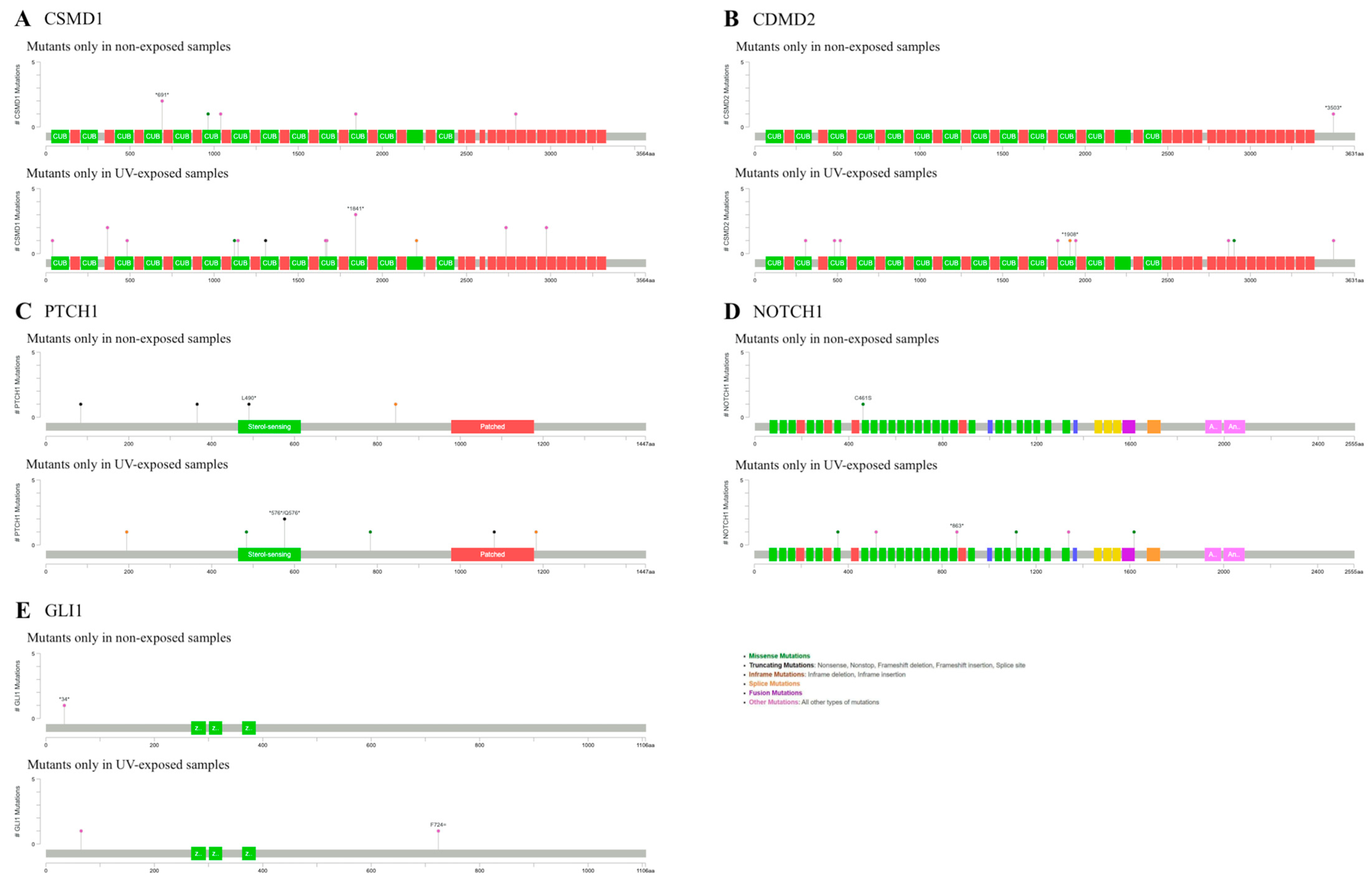
We conducted additional analysis to compare the genetic mutations based on the anatomical sites: UV-exposed and non-exposed areas. We identified different mutation frequencies in targeting BBC marker genes between these groups. SNP analysis revealed that the UV-exposed group harbored more mutations in these genes than the non-exposed group. CSMD1, CSMD2, PTCH1, GLI1, and NOTCH1 showed different mutation positions in both the exposed and non-exposed groups, whereas the others showed different mutated SNPs in the exposed group (Figure 2). These five genes exhibited higher mutation prevalence in the exposed groups.
CSMD1 displayed one missense mutation in the non-exposed group, while the exposed group showed missense and truncating mutations in the exon. However, the mutation in the non-exposed CSMD2 group was located in the intron, which was less important. PTCH1 showed a truncating mutation in the sterol-sensing domain, and another mutation was located in an exon associated with a patched function in the exposed group. Exploring mutations within SMO, GLI1, and ITIH2 revealed a solitary missense mutation in SMO of the exposed group, while no mutations were detected in the exons of GLI1 in either group or ITIH2 in the exposed group. NOTCH1 included one missense mutation in the non-exposed group, and three missense mutations in the exposed group. However, NOTCH2, TP53, and DPP10 displayed mutations only in the exposed group, with TP53 exhibiting missense variants in the P53 domain, which is involved in tumor suppression [26] (Figure S2). Therefore, CSMD1, PTCH1, and NOTCH1 manifested amino acid-altering mutations in the exons of the non-exposed group.

2.4. Mutations of ADCY10 and TAS1R2 Found in the Exposed Areas Interact with Ith BCC Marker Genes

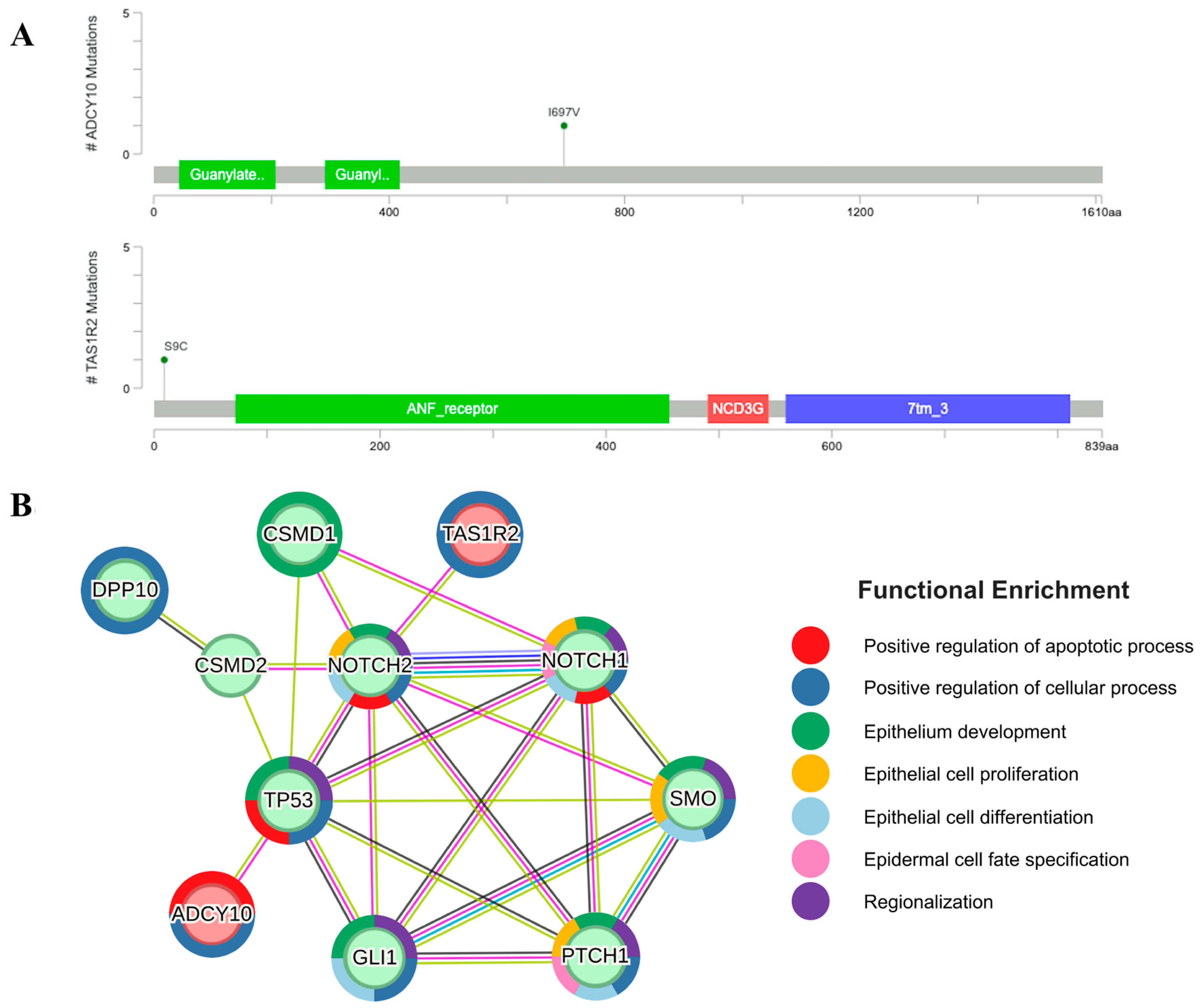
To further explore the difference between the BCCs in exposed and non-exposed areas regarding the significantly mutated genes, we conducted SNP analysis independent of the 11 previously studied BCC marker genes. We identified missense variant mutations in ADCY10 and TAS1R2 in the non-UV-exposed group compared to the UV-exposed group. The frequencies of SNP alleles in the non-UV-exposed group for TAS1R2 and ADCY10 were 0.4167 and 0.3, respectively, while these variants were absent in controls (Table S1). Given the limited prior research on these genes, protein–protein interaction (PPI) analysis was conducted between BCC marker genes and the selected genes. The PPI network, filtered with a confidence of 0.15, is presented in Figure 3, which shows the connections between ADCY10, TAS1R2 and BCC marker genes. Enriched functions from the network were identified by STRING [27] using Gene Ontology biological processes and included functions associated with epithelial cell fate and growth—epithelial development, epithelial cell proliferation, epithelial cell differentiation, epidermal cell fate specification, and cellular process—as well as positive regulation of apoptosis, positive regulation of cellular process, and regionalization. Among BCC marker genes, PTCH1, GLI1, TP53, SMO, NOTCH1, and NOTCH2 interacted with each other and shared common enriched functions, including epithelial development, positive regulation of cellular processes, and regionalization. ADCY10 and TAS1R2 are involved in the positive regulation of cellular processes, whereas ADCY10 is also related to the positive regulation of apoptosis by TP53. Interestingly, TP53 and NOTCH2 displayed experimentally determined connections with ADCY10 and TAS1R2.

3. Discussion

Recent genomic studies identified novel genetic alterations beyond the Hh pathway, contributing to pathogenesis of BCC. CSMD1 and CSMD2, which encode inhibitors of the complement system, have been proposed as tumor suppressor genes, with CSMD1 being the second most frequently mutated gene in BCC [24,28,29]. LOF mutations in NOTCH1, observed in approximately 30–50% of BCCs, are considered to facilitate tumor persistence in sporadic cases [10,30]. Non-coding mutations in the TERT promoter, present in 39–74% of BCCs, are associated with telomere shortening and subsequent telomerase activation, a well-established hallmark of numerous human cancers [31,32,33,34]. Other genes that are frequently mutated in BCC include LATS1 and PTPN14, which are critical components of the Hippo-YAP signaling pathway [10], the DPH3 promoter [34], MYCN [10], PPP6C [10], and STK19 [10]. These findings suggest that BCC harbors a heterogeneous genetic landscape involving a broader network of cancer-associated genes than was previously recognized.
In our analysis, we focused on the 11 genes, including Hh pathway genes and additional genes with mutations previously reported in the Caucasian BCC cohort [24]. We specifically sought to identify mutations that were unique to the Korean cohort. We observed the L412F mutation in SMO, a known cancer hotspot that likely confers a GOF oncogenic effect via aberrant Hh pathway activation. Within PTCH1, several variants, 1183, V1083Wfs4, X844_splice, L490, R459Lfs49, X195_splice, and X132_splice, were predicted to be oncogenic LOF mutations according to OncoKB, further supporting the central role of Hh signaling in BCC pathogenesis in the Korean population. We detected three cancer hotspot mutations in TP53, V274D, R175H, and X126_splice, which potentially resulted in LOF due to OncogenicDB. These findings highlight the critical role of TP53 in the Korean BCC population compared to its established significance in other malignancies [35,36].
Additionally, a potentially oncogenic LOF mutation (OncogenicDB) in NOTCH2 (Q1061) was identified, suggesting that signaling pathways beyond the Hh and TP53 axes may contribute to BCC pathogenesis. NOTCH2 plays a crucial role in epidermal cell differentiation, and its dysregulation has been implicated in BCC, highlighting its role in skin homeostasis and tumorigenesis [37]. While these pathogenic variants correspond to established oncogenic drivers, several Korean-specific mutations remain of uncertain clinical significance (not annotated in OncogenicDB). Further functional and epidemiological studies are needed to elucidate their tumorigenic potential and refine our understanding of population-specific genetic variations in BCC (Table S2).
To further investigate population-specific genetic differences, we analyzed the proportion of mutations unique to Korean and Caucasian cohorts. Intronic mutations represented the most abundant variant class in both cohorts, comprising 54.7% and 31.9% of Korean-specific and European-specific mutations, respectively. The significantly elevated proportion of intronic variants in the Korean cohort suggests potential population-specific differences in gene regulation or splicing efficiency, as intronic mutations may influence gene expression and protein function despite being traditionally classified as noncoding [38]. Missense mutations were less prevalent in the Korean-specific mutation group (17.8%) than in the European-specific group (21.4%). Similarly, mutations in the splice region were observed at a slightly higher frequency in the Korean-specific mutation group (10.6%) than in the European-specific mutation group (9.8%).
A notable difference was observed in the truncating mutations. In the Korean-specific mutation group, only 6.84% of the mutations were truncating, and the majority were nonsense mutations. In contrast, truncating mutations were significantly more prevalent in the Caucasian dataset (26.3%), dominated by frameshift insertions (FS ins) and deletions (FS del). These findings highlight population-specific differences in BCC in the mutational spectrum, where the Caucasian cohort exhibited a higher prevalence of FS ins and FS del, whereas the Korean cohort was more concentrated in intronic regions. Further studies are needed to assess whether these genomic disparities influence disease progression, treatment responses, and clinical presentations.
UV exposure is a well-established etiological factor in the development of skin cancers, particularly BCC. Genetic alterations resulting from UV-induced DNA damage play a critical role in BCC pathogenesis. Additionally, while the impact of UV exposure and BCC occurrence regarding systemic implications was presented by previous research, the mechanism beyond BCC in non-UV-exposed areas is not well-known [39]. In the previous study of Caucasian patients, PTCH1 mutations were significantly associated with sun-exposed areas, whereas NOTCH1 mutations were more frequent in truncal BCCs, a typical non-exposed region [24]. These findings demonstrate that BCC from different anatomical sites may exhibit distinct clinical, histological, and molecular profiles, highlighting BCC heterogeneity and the need to further characterize mutations in non-exposed tumors.
In our comparative analysis of UV-exposed and UV-unexposed BCC samples, we found that BCCs in exposed areas harbored a greater number of SNPs and pathogenic mutations. However, mutations in CSMD1, CSMD2, PTCH1, GLI1, and NOTCH1—all recognized BCC marker genes—were identified in non-UV-exposed groups. CSMD1, PTCH1, and NOTCH1 exhibited missense or truncating mutations that may alter protein structure or function. This suggested that CSMD1 and PTCH1 play significant roles in BCC pathogenesis of BCCs in non-exposed areas. Moreover, PTCH1 mutations in the non-exposed area included splice-site (X844_splice) and nonsense mutation (L490*, Q365*, and Q84*) variants (Table S3). While PTCH1 mutations are well-known in UV-induced carcinogenesis [40,41], our results imply they may also contribute to BCC carcinogenesis independent of UV exposure. PTCH1 and NOTCH1 are key components of the Hh and NOTCH signaling pathways, respectively, highlighting their critical roles in BCC. Additionally, PTCH1 contributed to BCC development in a Caucasian cohort study [24].
In the PPI network between TAS1R2, ADCY10 and BCC marker genes, the core six genes—NOTCH1, NOTCH2, TP53, GLI1, PTCH1, and SMO—were interconnected. Specific interactions were observed between NOTCH2 and TAS1R2, and TP53 and ADCY10. All mutated genes from the non-UV-exposed groups were included in the PPI network, exhibiting edges between CSMD1 and NOTCH1, which share functional enrichment in epithelial development. While TAS1R2 has been associated with solid tumors in previous studies [42,43], its role in BCC progression and carcinogenesis remains unclear. Its interaction with NOTCH2 indicates its potential role in BCC pathogenesis. ADCY10 mutations have been identified in multiple cancers, including Merkel cell carcinoma and breast cancer [44,45]. Interestingly, a previous study has found that adenylyl cyclase, particularly ADCY10, is linked to ferroptosis in lung cancer [46]. Additionally, soluble adenylyl cyclase has been implicated in hyperproliferative skin disease, even though it was similar to normal human skin in BCC [47].
Our study has several limitations. First, while we identified mutations unique to the Korean cohort, functional validation was not performed to confirm their oncogenic effects. Second, our study relied on SNP-based analysis, which may not fully capture structural variants or epigenetic modifications involved in BCC progression. Lastly, the sample size was limited, requiring further large-scale studies to confirm our findings.

4. Materials and Methods

4.1. Sample Collection

BCC tumor tissues (diameter, 5 mm) and matched peripheral blood samples were obtained from the fourteen patients. This study was conducted in accordance with the Declaration of Helsinki and the International Conference on Harmonization and Good Clinical Practice Guidelines and was reviewed and approved by the Institutional Review Board of Kyung Hee University Hospital at Gangdong (KHNMC 2022-04-014). Written informed consent was obtained from all participants before enrolment in the study.

4.2. DNA Extraction from Tumor Samples

We utilized the Agilent SureSelect Target Enrichment protocol for Illumina Paired-End Sequencing Library (Version C2, December 2018) with 0.5 µg input gDNA to generate standard exome capture libraries. All libraries were enriched using SureSelect Human All Exon V6 Probe (Agilent, Santa Clara, CA, USA). DNA quantification and quality were assessed using PicoGreen (Invitrogen, Waltham, MA, USA) and Tapestation gDNA Screentape (Agilent, Santa Clara, CA, USA). We used 1 μg genomic DNA diluted in EB Buffer and sheared to a target peak size of 150–200 bp using the Covaris LE220 focused-ultrasonicator (Covaris, Woburn, MA, USA) according to the manufacturer’s protocol. The DNA was sheared in an 8-microTUBE Strip (Covaris, Woburn, MA, USA) into the tube holder of the ultrasonicator using the following settings: mode, frequency sweeping; duty cycle, 10%; intensity, 5; cycles per burst, 200; duration, 60 s × 6 cycles; temperature, 4–7 °C.
The fragmented DNA was repaired and underwent A-tailing to the 3′ end, followed by ligation of Agilent adapters to the fragments. After verifying ligation, the adapter-ligated product was amplified using PCR. For exome capture, 250 ng of the DNA library was combined with hybridization buffers (Agilent, Santa Clara, CA, USA), blocking mixes (Agilent, Santa Clara, CA, USA), RNase block (Agilent, Santa Clara, CA, USA), and 5 µL of SureSelect all exon capture library, according to the Agilent SureSelect Target Enrichment protocol. Hybridization was performed at 65 °C for 24 h in a thermal cycler lid, with the temperature set to 105 °C. The captured DNA was washed and amplified. The final purified product was quantified by qPCR using the KAPA Library Quantification Kit (Roche, Basel, Switzerland) for Illumina Sequencing Platforms and qualified using the TapeStation D1000 ScreenTape (Agilent, Santa Clara, CA, USA). Sequencing was performed on the NovaSeq6000 platform (Illumina, San Diego, CA, USA).

4.3. Sequencing Data Processing

Integrated primary data analysis was conducted using Illumina’s Real-Time Analysis (RTA, v.3.4.4) software to generate raw images and base calling. The base calling files were converted into FASTQ format using bcl2fastq (v.2.20.0), with the demultiplexing option (barcode mismatches) set to 0. Quality control of raw sequencing was performed using FastQC. Paired-end sequences produced by the NovaSeq Instrument were mapped to the human reference genome using the BWA-MEM algorithm (BWA). The mapping results were generated in BAM format, excluding unordered sequences and alternate haplotypes. PCR duplicates were removed using MarkDuplicates.jar from the Picard-tools package, which identifies duplicate reads in starting positions after coordinate sorting. Base Quality Score Recalibration (BQSR) was applied to recalibrate the BAM files, using machine learning to empirically model sequencing errors and adjust the quality scores accordingly.
Variant genotyping for each sample was performed based on previously generated BAM files using the Haplotype Caller tool from GATK to identify SNPs and short indel candidates at nucleotide resolution. We filtered the variants using the VariantFiltration tool from GATK, which is designed for hard-filtering variant calls based on certain criteria. Records were filtered by modifying the FILTER field, and the filtered records were preserved in the output unless their removal was specified in the command-line parameters. The filtered variants were annotated using SnpEff and filtered using dbSNP and 1000 Genomes Project SNPs. The format of the final product was VCF. Subsequently, an in-house program and SnpEff were used to annotate additional databases, including ESP6500, ClinVar, dbNSFP, and ACMG.

4.4. Variant Calling Analysis and Protein–Protein Interaction Analysis

To identify different mutations in Korean and Caucasian cohorts, a variant calling analysis was performed on BCC marker genes from European and Asian cohorts. In addition to the Asian tissue samples, PBMC data for the corresponding tissue samples were collected. To focus exclusively on mutations in the skin tissue, the intersection of mutations in Asian PBMC and skin samples was excluded from the analysis. To compare mutation calls between Caucasian and Korean cohorts, we used BCFtools isec (v.1.20) [48]. Subsequently, to identify genes with significant mutations in the non-exposed group compared to the exposed group within the Korean cohort, we conducted an analysis using PLINK (v.1.90b6.21) [49]. Mutational profiles were annotated and visualized using MutationMapper in the cBioPortal (version 6.3.3) [50,51,52]. PPI analysis was conducted using STRING (version 12.0), revealing a network cluster between BCC marker genes and the two mutated genes in the non-exposed group [28].

5. Conclusions

Our study provides insights into the unique mutational landscape of BCC in the Korean population, revealing key differences from previously reported Caucasian cohorts. We identified distinct oncogenic mutations in critical Hh pathway genes, including PTCH1 and SMO, as well as in tumor suppressor genes, such as TP53 and NOTCH2. We also discovered population-specific differences in the mutational spectra of BCC.
Importantly, we found that PTCH1 and NOTCH1 may play central roles in BCC pathogenesis, regardless of UV exposure, as some mutations in these genes were observed only in the non-exposed groups. Our findings suggest that while UV radiation is a well-established risk factor for BCC, genetic alterations in specific pathways, particularly Hh and NOTCH signaling, may contribute to tumorigenesis in non-UV-exposed cases.
Additionally, we identified two novel genes, TAS1R2 and ADCY10, as potential contributors to BCC pathogenesis, particularly in BCC of non-exposed areas. These genes have previously been linked to cancer; however, their specific roles in BCC development remain unclear. Their interaction with known BCC driver genes, such as NOTCH2 and TP53, highlights the need for further functional studies to determine their biological relevance.
Our study highlights the population-specific genetic differences in BCC and suggests that PTCH1 and NOTCH1 may serve as key drivers of UV-independent tumorigenesis. Further functional studies and larger cohort analyses are needed to fully elucidate the molecular mechanisms underlying BCC in non-exposed areas and explore potential targeted therapeutic approaches based on these findings. Further analysis should establish ethnic-specific BCC diagnostic algorithms as distinct patterns observed in the Korean population. Comprehensive studies comparing molecular profiles between different ethnic groups are essential to develop medical implications and approaches tailored to Asian genetic backgrounds and environmental factors.

Supplementary Materials

The supporting information can be downloaded at: https://www.mdpi.com/article/10.3390/ijms26146941/s1.

Author Contributions

Conceptualization, M.S.K. and S.-H.K.; investigation, Y.-A.K., S.M., Y.C., M.S.K. and S.-H.K.; formal analysis, Y.-A.K., S.M. and Y.C.; methodology, Y.-A.K., S.M. and Y.C.; software, J.K., Y.L. and K.L.; visualization, Y.-A.K., S.M. and Y.C.; resources, B.-L.L. and S.-H.K.; supervision, M.S.K. and S.-H.K.; writing—original draft preparation, all authors; writing—review and editing, M.S.K. and S.-H.K. All authors have read and agreed to the published version of the manuscript.

Funding

Funded by the Ministry of Health & Welfare, Republic of Korea (grant number: HP23C0146).

Institutional Review Board Statement

This study was conducted in accordance with the Declaration of Helsinki and the International Conference on Harmonization and Good Clinical Practice Guidelines and was reviewed and approved by the Institutional Review Board of Kyung Hee University Hospital at Gangdong (KHNMC 2022-04-014), date of approval: 25 May 2022. Written informed consent was obtained from all participants before enrolment in the study.

Informed Consent Statement

Informed consent was obtained from all subjects involved in the study.

Data Availability Statement

The data presented in this study are available upon request from the corresponding author. Public data used in this study were obtained from the NCBI BioProject repository with the accession PRJNA731355, https://www.ncbi.nlm.nih.gov/Traces/study/?acc=PRJNA731355.

Acknowledgments

This work was supported by a grant of the Korea Health Technology R&D Project grant through the Korea Health Industry Development Institute (KHIDI), funded by the Ministry of Health & Welfare, Republic of Korea (grant number: HP23C0146) and Presidential Fellowship from Kyung Hee University in 2024.

Conflicts of Interest

The authors declare no conflicts of interest.

Abbreviations

The following abbreviations are used in this manuscript:
BCCBasal cancer cell
PPIProtein–protein interaction
HhHedgehog
FSPTFitzpatrick skin phototype
UVUltraviolet
PTCH1Patched 1
SMOSmoothened
GLI1Glioma-associated transcription factor 1
TP53Tumor protein P53
CSMD1CUB And Sushi Multiple Domains 1
CSMD2CUB And Sushi Multiple Domains 2
NOTCH1Notch Receptor 1
NOTCH2Notch Receptor 2
ITIH2Inter-Alpha-Trypsin Inhibitor Heavy Chain 2
DPP10Dipeptidyl Peptidase Like 10
STEAP4Six-Transmembrane Epithelial Antigen of Prostate 4

References

  1. Verkouteren, J.; Ramdas, K.; Wakkee, M.; Nijsten, T. Epidemiology of basal cell carcinoma: Scholarly review. Br. J. Dermatol. 2017, 177, 359–372. [Google Scholar] [CrossRef] [PubMed]
  2. Kwon, S.; Choi, S.; Kim, J.S.; Kim, S.S.; Jue, M.; Seo, S.H.; Park, J.; Roh, M.R.; Mun, J.; Kim, J.Y.; et al. Incidence and survival rates of primary cutaneous malignancies in Korea, 1999–2019: A nationwide population-based study. J. Dermatol. 2024, 51, 532–538. [Google Scholar] [CrossRef] [PubMed]
  3. Pellegrini, C.; Maturo, M.G.; Di Nardo, L.; Ciciarelli, V.; García-Rodrigo, C.G.; Fargnoli, M.C. Understanding the Molecular Genetics of Basal Cell Carcinoma. Int. J. Mol. Sci. 2017, 18, 2485. [Google Scholar] [CrossRef] [PubMed]
  4. Gailani, M.R.; Ståhle-Bäckdahl, M.; Leffell, D.J.; Glyn, M.; Zaphiropoulos, P.G.; Undén, A.B.; Dean, M.; Brash, D.E.; Bale, A.E.; Toftgård, R. The role of the human homologue of Drosophila patched in sporadic basal cell carcinomas. Nat. Genet. 1996, 14, 78–81. [Google Scholar] [CrossRef] [PubMed]
  5. Kim, M.Y.; Park, H.J.; Baek, S.C.; Byun, D.G.; Houh, D. Mutations of the p53 and PTCH gene in basal cell carcinomas: UV mutation signature and strand bias. J. Dermatol. Sci. 2002, 29, 1–9. [Google Scholar] [CrossRef] [PubMed]
  6. Huang, Y.; Bu, D.; Li, X.; Ma, Z.; Yang, Y.; Lin, Z.; Lu, F.; Tu, P.; Li, H. Unique features of PTCH1 mutation spectrum in Chinese sporadic basal cell carcinoma. J. Eur. Acad. Dermatol. Venereol. 2012, 27, 235–241. [Google Scholar] [CrossRef] [PubMed]
  7. Jayaraman, S.S.; Rayhan, D.J.; Hazany, S.; Kolodney, M.S. Mutational Landscape of Basal Cell Carcinomas by Whole-Exome Sequencing. J. Investig. Dermatol. 2014, 134, 213–220. [Google Scholar] [CrossRef] [PubMed]
  8. Teh, M.-T.; Blaydon, D.; Chaplin, T.; Foot, N.J.; Skoulakis, S.; Raghavan, M.; Harwood, C.A.; Proby, C.M.; Philpott, M.P.; Young, B.D.; et al. Genomewide Single Nucleotide Polymorphism Microarray Mapping in Basal Cell Carcinomas Unveils Uniparental Disomy as a Key Somatic Event. Cancer Res. 2005, 65, 8597–8603. [Google Scholar] [CrossRef] [PubMed]
  9. Reifenberger, J.; Wolter, M.; Knobbe, C.B.; Köhler, B.; Schönicke, A.; Scharwächter, C.; Kumar, K.; Blaschke, B.; Ruzicka, T.; Reifenberger, G. Somatic mutations in the PTCH, SMOH, SUFUH and TP53 genes in sporadic basal cell carcinomas. Br. J. Dermatol. 2005, 152, 43–51. [Google Scholar] [CrossRef] [PubMed]
  10. Bonilla, X.; Parmentier, L.; King, B.; Bezrukov, F.; Kaya, G.; Zoete, V.; Seplyarskiy, V.B.; Sharpe, H.J.; McKee, T.; Letourneau, A.; et al. Genomic analysis identifies new drivers and progression pathways in skin basal cell carcinoma. Nat. Genet. 2016, 48, 398–406. [Google Scholar] [CrossRef] [PubMed]
  11. Xie, J.; Murone, M.; Luoh, S.-M.; Ryan, A.; Gu, Q.; Zhang, C.; Bonifas, J.M.; Lam, C.-W.; Hynes, M.; Goddard, A.; et al. Activating Smoothened mutations in sporadic basal-cell carcinoma. Nature 1998, 391, 90–92. [Google Scholar] [CrossRef] [PubMed]
  12. Rady, P.; Scinicariello, F.; Wagner, R.F., Jr.; Tyring, S.K. p53 mutations in basal cell carcinomas. Cancer Res. 1992, 52, 3804–3806. [Google Scholar] [PubMed]
  13. Gailani, M.R.; Leffell, D.J.; Ziegler, A.; Gross, E.G.; Brash, D.E.; Bale, A.E. Relationship Between Sunlight Exposure and a Key Genetic Alteration in Basal Cell Carcinoma. JNCI J. Natl. Cancer Inst. 1996, 88, 349–354. [Google Scholar] [CrossRef] [PubMed]
  14. Ziegler, A.; Leffell, D.J.; Kunala, S.; Sharma, H.W.; Gailani, M.; A Simon, J.; Halperin, A.J.; Baden, H.P.; E Shapiro, P.; E Bale, A. Mutation hotspots due to sunlight in the p53 gene of nonmelanoma skin cancers. Proc. Natl. Acad. Sci. USA 1993, 90, 4216–4220. [Google Scholar] [CrossRef] [PubMed]
  15. Rosenstein, B.S.; Phelps, R.G.; Weinstock, M.A.; Bernstein, J.L.; Gordon, M.L.; Rudikoff, D.; Kantor, I.; Shelton, R.; Lebwohl, M.G. p53 Mutations in Basal Cell Carcinomas Arising in Routine Users of Sunscreens. Photochem. Photobiol. 1999, 70, 798–806. [Google Scholar] [CrossRef] [PubMed]
  16. Bolshakov, S.; Walker, C.M.; Strom, S.S.; Selvan, M.S.; Clayman, G.L.; El-Naggar, A.; Lippman, S.M.; Kripke, M.L.; Ananthaswamy, H.N. p53 mutations in human aggressive and nonaggressive basal and squamous cell carcinomas. Clin. Cancer Res. 2003, 9, 228–234. [Google Scholar] [PubMed]
  17. Wang, Y.M.; Huang, Y.S.; Ma, Z.H.; Bu, D.F.; Tu, P.; Li, H. Frequency and features of TP53 mutation in 30 Chinese patients with sporadic basal cell carcinoma. Clin. Exp. Dermatol. 2014, 39, 829–834. [Google Scholar] [CrossRef] [PubMed]
  18. Wang, G.Y.; Wang, J.; Mancianti, M.L.; Epstein, E.H., Jr. Basal cell carcinomas arise from hair follicle stem cells in Ptch1+/− mice. Cancer Cell 2011, 19, 114–124. [Google Scholar] [CrossRef] [PubMed] [PubMed Central]
  19. Ikehata, H.; Ono, T. The mechanisms of UV mutagenesis. J. Radiat. Res. 2011, 52, 115–125. [Google Scholar] [CrossRef] [PubMed]
  20. Yamamoto, J.; Plaza, P.; Brettel, K. Repair of (6-4) Lesions in DNA by (6-4) Photolyase: 20 Years of Quest for the Photoreaction Mechanism. Photochem. Photobiol. 2017, 93, 51–66. [Google Scholar] [CrossRef] [PubMed]
  21. Scrivener, Y.; Grosshans, E.; Cribier, B. Variations of basal cell carcinomas according to gender, age, location and histopathological subtype. Br. J. Dermatol. 2002, 147, 41–47. [Google Scholar] [CrossRef] [PubMed]
  22. Betti, R.; Bruscagin, C.; Inselvini, E.; Crosti, C. Basal cell carcinomas of covered and unusual sites of the body. Int. J. Dermatol. 1997, 36, 503–505. [Google Scholar] [CrossRef] [PubMed]
  23. Roh, S.G.; Park, J.; Song, K.H.; Nam, K.H.; Yun, S.K.; Kim, H.U. Clinical and histopathological characteristics of extra-facial basal cell carcinoma: Analysis of 35 patients at the Chonbuk National University Hospital in Korea. Australas. J. Dermatol. 2014, 55, e65–e68. [Google Scholar] [CrossRef] [PubMed]
  24. Di Nardo, L.; Pellegrini, C.; Di Stefani, A.; Ricci, F.; Fossati, B.; Del Regno, L.; Carbone, C.; Piro, G.; Corbo, V.; Delfino, P.; et al. Molecular alterations in basal cell carcinoma subtypes. Sci. Rep. 2021, 11, 1–9. [Google Scholar] [CrossRef] [PubMed]
  25. Lobl, M.; Hass, B.; Clarey, D.; Higgins, S.; Sutton, A.; Wysong, A. Basal Cell Carcinoma Gene Mutations Differ Between Asian, Hispanic, and Caucasian Patients: A Pilot Study. J. Drugs Dermatol. 2021, 20, 504–510. [Google Scholar] [CrossRef] [PubMed]
  26. Oren, M. Regulation of the p53 Tumor Suppressor Protein. J. Biol. Chem. 1999, 274, 36031–36034. [Google Scholar] [CrossRef] [PubMed]
  27. Szklarczyk, D.; Kirsch, R.; Koutrouli, M.; Nastou, K.; Mehryary, F.; Hachilif, R.; Gable, A.L.; Fang, T.; Doncheva, N.T.; Pyysalo, S.; et al. The STRING database in 2023: Protein–protein association networks and functional enrichment analyses for any sequenced genome of interest. Nucleic Acids Res. 2022, 51, D638–D646. [Google Scholar] [CrossRef] [PubMed]
  28. Jung, A.R.; Eun, Y.-G.; Lee, Y.C.; Noh, J.K.; Kwon, K.H. Clinical Significance of CUB and Sushi Multiple Domains 1 Inactivation in Head and Neck Squamous Cell Carcinoma. Int. J. Mol. Sci. 2018, 19, 3996. [Google Scholar] [CrossRef] [PubMed]
  29. Zhang, H.; Huang, T.; Ren, X.; Fang, X.; Chen, X.; Wei, H.; Sun, W.; Wang, Y. Integrated pan-cancer analysis of CSMD2 as a potential prognostic, diagnostic, and immune biomarker. Front. Genet. 2022, 13, 918486. [Google Scholar] [CrossRef] [PubMed]
  30. Eberl, M.; Mangelberger, D.; Swanson, J.B.; Verhaegen, M.E.; Harms, P.W.; Frohm, M.L.; Dlugosz, A.A.; Wong, S.Y. Tumor Architecture and Notch Signaling Modulate Drug Response in Basal Cell Carcinoma. Cancer Cell 2018, 33, 229–243.e4. [Google Scholar] [CrossRef] [PubMed]
  31. Griewank, K.G.; Murali, R.; Schilling, B.; Schimming, T.; Möller, I.; Moll, I.; Schwamborn, M.; Sucker, A.; Zimmer, L.; Schadendorf, D.; et al. TERT Promoter Mutations Are Frequent in Cutaneous Basal Cell Carcinoma and Squamous Cell Carcinoma. PLoS ONE 2013, 8, e80354. [Google Scholar] [CrossRef] [PubMed]
  32. Scott, G.A.; Laughlin, T.S.; Rothberg, P.G. Mutations of the TERT promoter are common in basal cell carcinoma and squamous cell carcinoma. Mod. Pathol. 2014, 27, 516–523. [Google Scholar] [CrossRef] [PubMed]
  33. Pópulo, H.; Boaventura, P.; Vinagre, J.; Batista, R.; Mendes, A.; Caldas, R.; Pardal, J.; Azevedo, F.; Honavar, M.; Guimarães, I.; et al. TERT Promoter Mutations in Skin Cancer: The Effects of Sun Exposure and X-Irradiation. J. Investig. Dermatol. 2014, 134, 2251–2257. [Google Scholar] [CrossRef] [PubMed]
  34. Denisova, E.; Heidenreich, B.; Nagore, E.; Rachakonda, P.S.; Hosen, I.; Akrap, I.; Traves, V.; García-Casado, Z.; López-Guerrero, J.A.; Requena, C.; et al. Frequent DPH3 promoter mutations in skin cancers. Oncotarget 2015, 6, 35922–35930. [Google Scholar] [CrossRef] [PubMed]
  35. Lee, E.B.; Jin, G.; Lee, S.Y.; Park, J.Y.; Kim, M.J.; Choi, J.E.; Jeon, H.S.; Cha, S.I.; Cho, S.; Kim, C.H.; et al. TP53 Mutations in Korean Patients with Non-small Cell Lung Cancer. J. Korean Med. Sci. 2010, 25, 698–705. [Google Scholar] [CrossRef] [PubMed]
  36. Kim, Y.; Park, G.S.; Chung, Y.; Lee, J.H. Whole-exome sequencing of secondary tumors arising from nevus sebaceous revealed additional genomic alterations besides RAS mutations. J. Dermatol. 2023, 50, 1072–1075. [Google Scholar] [CrossRef] [PubMed]
  37. Thélu, J.; Rossio, P.; Favier, B. Notch signalling is linked to epidermal cell differentiation level in basal cell carcinoma, psoriasis and wound healing. BMC Dermatol. 2002, 2, 7. [Google Scholar] [CrossRef] [PubMed]
  38. Nawrocka, P.M.; Galka-Marciniak, P.; Urbanek-Trzeciak, M.O.; M-Thirusenthilarasan, I.; Szostak, N.; Philips, A.; Susok, L.; Sand, M.; Kozlowski, P. Profile of Basal Cell Carcinoma Mutations and Copy Number Alterations—Focus on Gene-Associated Noncoding Variants. Front. Oncol. 2021, 11, 752579. [Google Scholar] [CrossRef] [PubMed]
  39. Karampinis, E.; Koumaki, D.; Sgouros, D.; Nechalioti, P.-M.; Toli, O.; Pappa, G.; Papadakis, M.; Georgopoulou, K.-E.; Schulze-Roussaki, A.-V.; Kouretas, D. Non-Melanoma Skin Cancer: Assessing the Systemic Burden of the Disease. Cancers 2025, 17, 703. [Google Scholar] [CrossRef] [PubMed]
  40. Draper, E.; Li, Y.Y.; Mahadevan, N.R.; Laga, A.C.M.; Hanna, J.; Russell-Goldman, E. Clinicopathologic and Molecular Characterization of Basal Cell Carcinoma Arising at Sun-protected Sites. Am. J. Surg. Pathol. 2025, 49, 328–335. [Google Scholar] [CrossRef] [PubMed]
  41. Mancuso, M.; Pazzaglia, S.; Tanori, M.; Hahn, H.; Merola, P.; Rebessi, S.; Atkinson, M.J.; Di Majo, V.; Covelli, V.; Saran, A. Basal cell carcinoma and its development: Insights from radiation-induced tumors in Ptch1-deficient mice. Cancer Res. 2004, 64, 934–941. [Google Scholar] [CrossRef] [PubMed]
  42. Carey, R.M.; Kim, T.; Cohen, N.A.; Lee, R.J.; Nead, K.T. Impact of sweet, umami, and bitter taste receptor (TAS1R and TAS2R) genomic and expression alterations in solid tumors on survival. Sci. Rep. 2022, 12, 8937. [Google Scholar] [CrossRef] [PubMed]
  43. Shaji, C.S.; Saraswathy, R. Taste receptors influencing effective modalities in human health–A cutting edge update on TAS1R and TAS2R receptor polymorphisms in taste perception and disease risk. Nutr. Health 2023. [Google Scholar] [CrossRef] [PubMed]
  44. González-Vela, M.d.C.; Curiel-Olmo, S.; Derdak, S.; Beltran, S.; Santibañez, M.; Martínez, N.; Castillo-Trujillo, A.; Gut, M.; Sánchez-Pacheco, R.; Almaraz, C.; et al. Shared Oncogenic Pathways Implicated in Both Virus-Positive and UV-Induced Merkel Cell Carcinomas. J. Investig. Dermatol. 2017, 137, 197–206. [Google Scholar] [CrossRef] [PubMed]
  45. Mamoor, S. Adenylyl Cyclase 10 Is Differentially Expressed in the Lymph Nodes of Patients with Metastatic Breast Cancer. OSF Preprints 2021. [Google Scholar] [CrossRef]
  46. Zhang, X.; Yu, K.; Ma, L.; Qian, Z.; Tian, X.; Miao, Y.; Niu, Y.; Xu, X.; Guo, S.; Yang, Y.; et al. Endogenous glutamate determines ferroptosis sensitivity via ADCY10-dependent YAP suppression in lung adenocarcinoma. Theranostics 2021, 11, 5650–5674. [Google Scholar] [CrossRef] [PubMed]
  47. Zippin, J.H.; Chadwick, P.A.; Levin, L.R.; Buck, J.; Magro, C.M. Soluble Adenylyl Cyclase Defines a Nuclear cAMP Microdomain in Keratinocyte Hyperproliferative Skin Diseases. J. Investig. Dermatol. 2010, 130, 1279–1287. [Google Scholar] [CrossRef] [PubMed]
  48. Danecek, P.; Bonfield, J.K.; Liddle, J.; Marshall, J.; Ohan, V.; Pollard, M.O.; Whitwham, A.; Keane, T.; McCarthy, S.A.; Davies, R.M.; et al. Twelve years of SAMtools and BCFtools. GigaScience 2021, 10, giab008. [Google Scholar] [CrossRef] [PubMed]
  49. Purcell, S.; Neale, B.; Todd-Brown, K.; Thomas, L.; Ferreira, M.A.R.; Bender, D.; Maller, J.; Sklar, P.; de Bakker, P.I.W.; Daly, M.J.; et al. PLINK: A Tool Set for Whole-Genome Association and Population-Based Linkage Analyses. Am. J. Hum. Genet. 2007, 81, 559–575. [Google Scholar] [CrossRef] [PubMed]
  50. Cerami, E.; Gao, J.; Dogrusoz, U.; Gross, B.E.; Sumer, S.O.; Aksoy, B.A.; Jacobsen, A.; Byrne, C.J.; Heuer, M.L.; Larsson, E.; et al. The cBio cancer genomics portal: An open platform for exploring multidimensional cancer genomics data. Cancer Discov. 2012, 2, 401–404. [Google Scholar] [CrossRef] [PubMed]
  51. Gao, J.; Aksoy, B.A.; Dogrusoz, U.; Dresdner, G.; Gross, B.E.; Sumer, S.O.; Sun, Y.; Jacobsen, A.; Sinha, R.; Larsson, E.; et al. Integrative Analysis of Complex Cancer Genomics and Clinical Profiles Using the cBioPortal. Sci. Signal. 2013, 6, pl1. [Google Scholar] [CrossRef] [PubMed]
  52. de Bruijn, I.; Kundra, R.; Mastrogiacomo, B.; Tran, T.N.; Sikina, L.; Mazor, T.; Li, X.; Ochoa, A.; Zhao, G.; Lai, B.; et al. Analysis and Visualization of Longitudinal Genomic and Clinical Data from the AACR Project GENIE Biopharma Collaborative in cBioPortal. Cancer Res. 2023, 83, 3861–3867. [Google Scholar] [CrossRef] [PubMed]
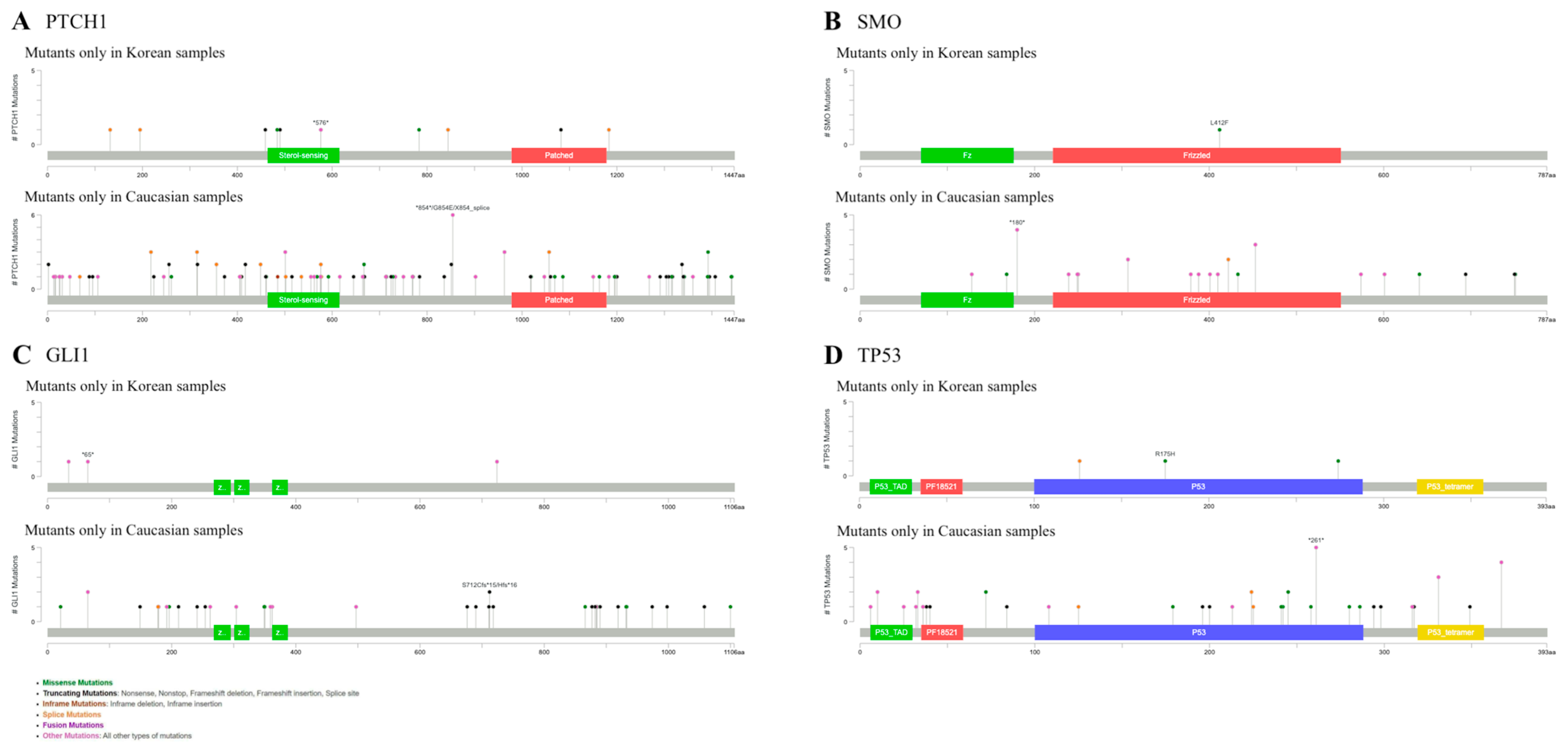
Figure 1. Different mutational profiles in Korean and Caucasian BCC groups. (A) Mutations of PTCH1. (B) Mutations of SMO. (C) Mutations of GLI1. (D) Mutations of TP53.
Ijms 26 06941 g001
Figure 2. Different mutation profiles in the BCCs of exposed and non-exposed areas. (A) Mutations in CSMD1. (B) Mutations in CSMD2. (C) Mutations in PTCH1. (D) Mutations in NOTCH1. (E) Mutations in GLI1.
Ijms 26 06941 g002
Figure 3. (A) Mutational profiles of ADCY10 and TAS1R2. (B) Protein–protein interaction network between ADCY10, TAS1R2 and BCC marker genes, and the functional enrichment of the network.
Ijms 26 06941 g003
Table 1. Demographic features of the patients and clinicopathologic characteristics of the BCCs.
Exposed
(n = 8)
Non-Exposed
(n = 6)
All
(n = 14)
Age (mean ± SD)65.38 ± 8.4270.33 ± 7.2667.50 ± 8.06
Sex, n (%)
 Male6 (75.0)3 (50.0)9 (64.3)
 Female2 (25.0)3 (50.0)5 (35.7)
Ethnicity
 Korean8 (100)6 (100)15 (100)
Skin type, n (%)
 III6 (75.0)5 (83.3)11 (78.6)
 IV2 (25.0)1 (16.7)3 (21.4)
History of other NMSC, n (%)0 (0.0)0 (0.0)0 (0.0)
History of sunburn, n (%)0 (0.0)1 (14.3)1 (6.3)
Immunosuppression, n (%)0 (0.0)0 (0.0)0 (0.0)
Anatomical site, n (%)
 Cheek4 (50.0)0 (0.0)4 (28.6)
 Nose2 (25.0)0 (0.0)2 (14.3)
 Eyelid1 (12.5)0 (0.0)1 (7.1)
 Neck1 (12.5)0 (0.0)1 (7.1)
 Back0 (0.0)2 (33.3)2 (14.3)
 Abdomen0 (0.0)2 (33.3)2 (14.3)
 Scalp0 (0.0)1 (16.7)1 (7.1)
 Calf0 (0.0)1 (16.7)1 (7.1)
Histopathological subtype, n (%)
 Nodular6 (75.0)3 (50.0)9 (64.3)
 Superficial2 (25.0)3 (50.0)5 (35.7)
Pigmentation, n (%)8 (100)6 (100)14 (100)
SD, standard deviation.
Disclaimer/Publisher’s Note: The statements, opinions and data contained in all publications are solely those of the individual author(s) and contributor(s) and not of MDPI and/or the editor(s). MDPI and/or the editor(s) disclaim responsibility for any injury to people or property resulting from any ideas, methods, instructions or products referred to in the content.