Article

Hydrogen Gas Attenuates Toxic Metabolites and Oxidative Stress-Mediated Signaling to Inhibit Neurodegeneration and Enhance Memory in Alzheimer’s Disease Models

1
Department of Global Medical Science, Wonju College of Medicine, Yonsei University, Wonju 26426, Republic of Korea
2
Department of Convergence Medicine, Wonju College of Medicine, Yonsei University, Wonju 26426, Republic of Korea
3
Organelle Medicine Research Center, Wonju College of Medicine, Yonsei University, Wonju 26426, Republic of Korea
4
Department of Laboratory Medicine, Wonju College of Medicine, Yonsei University Wonju 26426, Republic of Korea
5
GOOTZ Co., Ltd., Yuljeongro, 247 beon-gil, Yangju-si 11457, Republic of Korea
6
Department of Biochemistry, Wonju College of Medicine, Yonsei University, Wonju 26426, Republic of Korea
7
Natural Product Research Center, Korea Institute of Science and Technology, Gangneung 25451, Republic of Korea
*
Authors to whom correspondence should be addressed.
These authors contributed equally to this work as first authors.
Int. J. Mol. Sci. 2025, 26(14), 6922; https://doi.org/10.3390/ijms26146922
Submission received: 16 May 2025 / Revised: 28 June 2025 / Accepted: 15 July 2025 / Published: 18 July 2025
(This article belongs to the Special Issue New Advances in Research on Alzheimer’s Disease: 2nd Edition)

Abstract

Alzheimer’s disease (AD) is a neurodegenerative condition in which amyloid-beta (Aβ) plaques trigger oxidative stress (OS) and neuroinflammation, causing memory loss. OS and neurodegeneration can also be caused by reactive astrocytes, thereby promoting AD via toxic metabolite accumulation in the astrocytic urea cycle. However, the effect of molecular hydrogen (H2) on this cycle remains unknown. Therefore, we investigated whether H2 treatment could reduce OS-induced neurodegeneration and memory loss. 5xFAD (n = 14) and wild-type (n = 15) mice were randomized into four groups and treated with either 3% hydrogen gas (H2) or vehicle for 60 days. Cognitive behaviors were evaluated using the Morris water maze and Y-maze tests. In addition, we used biochemical assays to measure ammonia and hydrogen peroxide (H2O2) levels in the hippocampi of the mice and AβO-treated primary mouse astrocytes. Aβ, γ-aminobutyric acid (GABA), and the expression of inflammatory markers were evaluated using immunohistochemistry (IHC) and quantitative real-time polymerase chain reaction (qRT-PCR). We observed that H2 treatment significantly prevented cognitive deficits, oxidative stress, the accumulation of toxic metabolites, and the increase in inflammatory markers in 5xFAD mice. These results suggest that H2 therapy can mitigate toxic metabolites in the astrocytic urea cycle, thereby reducing neurodegeneration and memory loss in AD.

1. Introduction

Memory impairment and neurodegeneration are pathophysiological hallmarks of Alzheimer’s disease (AD), which predominantly manifests in people aged 65 years and older and constitutes a severe public health burden. In AD, amyloid-beta (Aβ) plaques, i.e., the significant deposition of insoluble Aβ fibrils and tau tangles, cause oxidative stress (OS), inflammation, and apoptosis when they accumulate in the brain; thus, they are key markers of AD pathogenesis [1,2,3]. OS is implicated in the onset of neurodegenerative disorders, including AD, Parkinson’s disease, and mild cognitive impairment. It can destroy neuronal lipid membranes, cause DNA damage-related cell death, and stimulate further Aβ accumulation via protein oxidation mechanisms [4]. These effects cause neuronal damage, culminating in the memory loss associated with AD [5] and mitochondrial dysfunction [6,7,8], and resulting in defective cytochrome oxidase activity and altered Ca2+ signaling [9,10,11]. Similarly, Bax, a pro-apoptotic marker, can activate caspases in the mitochondria (e.g., caspase-3, -8, and -9), contributing to neurodegeneration through apoptosis [7]. OS can trigger neuroinflammation via the nuclear factor kappa B (NF-κB) and mitogen-activated protein kinase (MAPK) nexus, causing neurodegeneration and AD [12].
Neurodegeneration and memory loss can also result from reactive astrocytes [13]. Astrocytes become “double-edged swords” by providing neuroprotection and becoming neurotoxic as OS and nitrosative stresses increase [14]. This neurotoxicity is due to the increased accumulation of toxic metabolites, which enhances the reactivity of astrocytes. Toxic metabolites such as ammonia activate the astrocytic urea cycle and trigger the synthesis of putrescine, hydrogen peroxide (H2O2), and gamma (γ)-aminobutyric acid (GABA) [15,16]. In the context of AD, the intracellular accumulation of amyloid-β protein (Aβ) can be cleared by the autophagy–lysosomal pathway, releasing amino acids such as aspartate, which enters the astrocytic urea cycle and is converted to arginosuccinate by arginosuccinate synthetase 1. The arginosuccinate is then transformed into arginine via the catalytic activity of arginosuccinate lyase. Arginase 1 (ARG1) converts arginine to ornithine, and urea is released as a waste product. Ornithine undergoes decarboxylation through ornithine decarboxylase 1 (ODC1) activity to produce putrescine, which goes through a series of enzymatic transformations to produce H2O2 and ammonia as side products and GABA as the final product. The ammonia produced undergoes N-acetylation with glutamate, forming carbamoyl phosphate. Ornithine transcarbamylase (OTC) then converts carbamoyl phosphate to citrulline. Citrulline can also be converted to arginosuccinate, thereby repeating the cycle [13,17,18]. H2O2, a key reactive oxygen species (ROS), and GABA contribute to neurodegeneration and memory impairment, respectively [18]. The key enzymes responsible for producing these toxic metabolites are ARG1, ODC1, and OTC [19].
Ju et al. established the molecular mechanisms underlying the bifunctional role of reactive astrocytes, which comprise a beneficial urea cycle-active pathway that detoxifies metabolites of Aβ breakdown and a harmful cycle that increases ODC1 expression, which facilitates toxic H2O2, ammonia, and GABA accumulation [18]. Elevated astrocyte reactivity can lead to the overexpression of glial fibrillary acidic protein (GFAP) levels. The overexpression of GFAP, an emerging diagnostic marker of AD, signals astrocyte death, also known as astrogliosis [16].
Despite extensive studies on the pathophysiology of AD, there is currently no cure for AD [20]. Therapeutic options, such as cholinesterase inhibitors [21], memantine, lecanemab, and donanemab, can only delay the progression of AD [22,23]. Recently, molecular hydrogen (H2) has been effective against many diseases, including neurodegenerative disorders [17,24,25,26]. H2 effectively reduces OS, inflammation, and apoptosis [27,28,29]. H2 has a small molecular size, making it easy for H2 to penetrate cell membranes and cross the blood–brain barrier, exerting effects on the central nervous system [17,24,25,26]. In neurodegenerative disorders, including AD, H2 therapy has been shown to attenuate OS via selective reduction in ROS and modulating inflammatory and apoptotic signaling pathways [27,30]. Specifically, hydrogen-rich water has been linked to suppressing the NLRP3 inflammasome, an essential component of the innate immune response, which has been implicated in AD [31]. In addition, H2 therapy has been shown to modulate brain-derived neurotrophic factor and estrogen receptor β, which are critical for synaptic plasticity and survival of neurons [32]. Further, in a chemically induced mild cognitive impairment model, H2 has been reported to increase spatial memory and reduce OS, Aβ, Bax, and cleaved caspase-7 levels [29]. Despite H2’s therapeutic benefits, its effect on the astrocytic urea cycle metabolic pathway remains to be explored.
Thus, this study aimed to investigate whether H2 treatment decreases the accumulation of toxic metabolites in the astrocytic urea cycle, thereby slowing neurodegeneration and memory loss. We hypothesized that H2 could downregulate the accumulation of toxic metabolites in the urea cycle, reducing neurodegeneration and memory impairment in the 5 familial AD (5xFAD) mouse model, as assessed using behavioral tests, biochemical assays, Western blot, immunohistochemistry (IHC), immunocytochemistry (ICC), and gene expression analysis.

2. Results

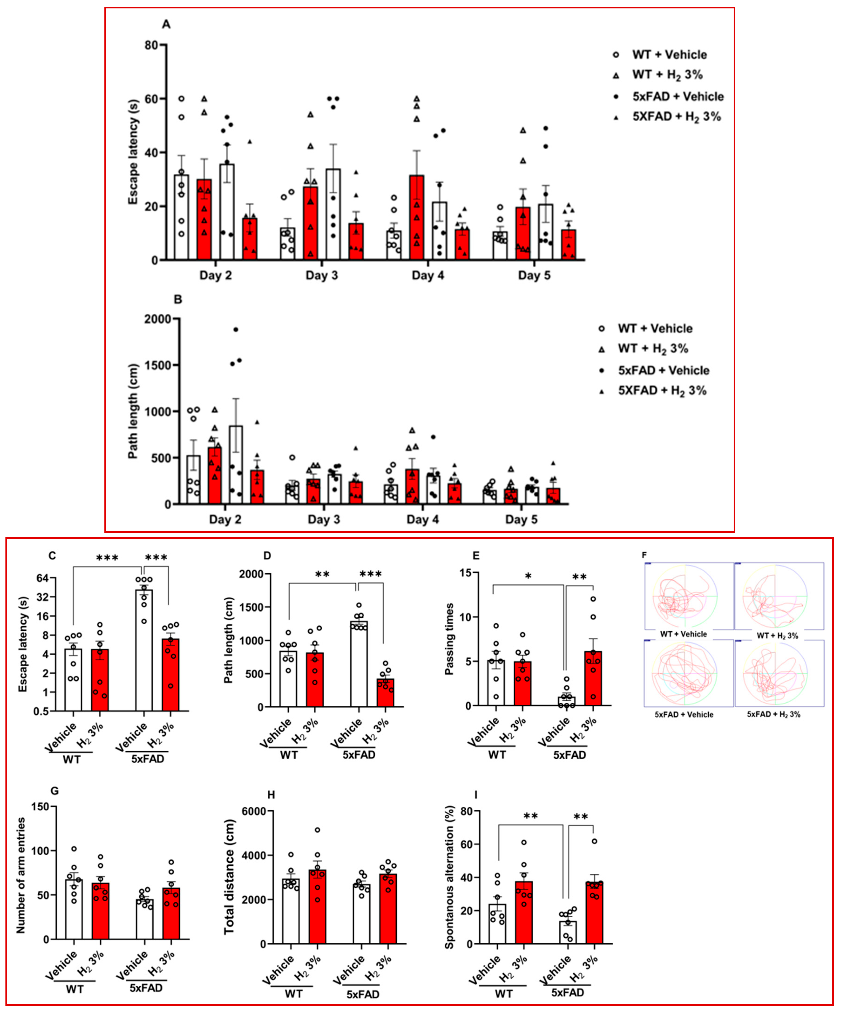
2.1. H2 Inhalation Attenuates Memory Impairment in 5xFAD Mice

We assessed the effect of H2 treatment on hippocampal-dependent spatial learning and memory impairment in 5xFAD mice using the Morris water maze and Y-maze tests. We analyzed escape latency, path length, and passing time from the Morris water maze test. During tests conducted on days 2–5 (hidden platform), we observed that the H2-treated 5xFAD mice showed a 1.4-fold decrease in escape latency time and a 2.0-fold decrease in path length compared with the vehicle-treated mice (Figure 1A,B). In the test trial on day 6, the escape latency time of the H2-treated 5xFAD mice decreased by a factor of 6.0 (p < 0.001; Figure 1C) and path length decreased by a factor of 3.0 (p < 0.001; Figure 1D), compared with the vehicle-treated mice; furthermore, the H2-treated 5xFAD mice traveled into the target quadrant (i.e., passing time) 6.0-fold more often than the vehicle-treated mice (p < 0.01; Figure 1E), as further illustrated by swimming patterns shown in Figure 1F. For the Y-maze test, the arm entry frequency (Figure 1G), total distance (Figure 1H), and the percentage of spontaneous alternation (Figure 1I) were analyzed. The percentage of spontaneous alternation markedly increased by 2.7-fold (p < 0.01) in the H2-treated 5xFAD mice compared with the vehicle-treated mice (Figure 1I).

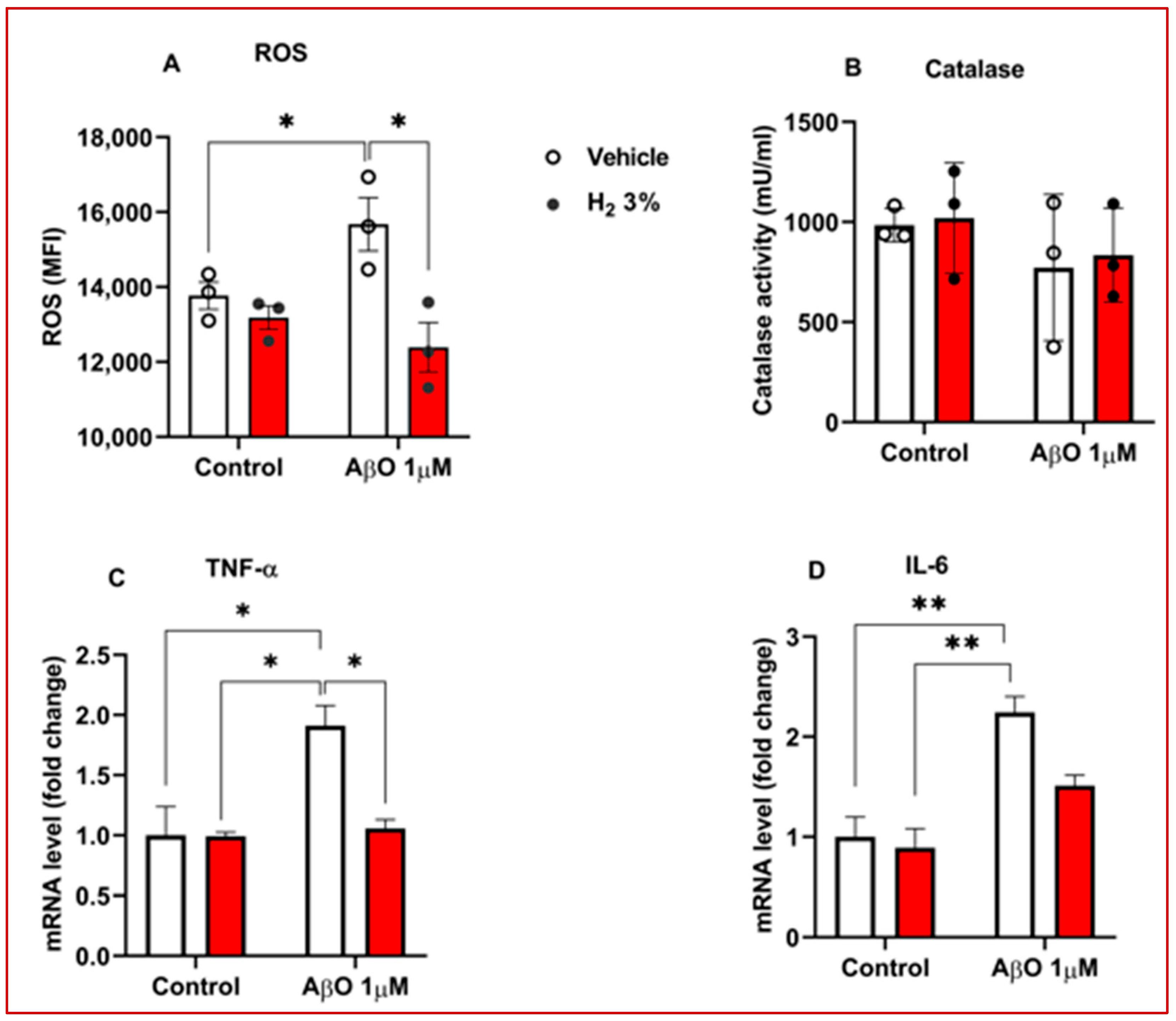
2.2. H2 Reduced OS and Decreased Neuroinflammation in AβO-Treated Astrocytes

Increased oxidative stress and neuroinflammation are characteristic pathophysiological features of Alzheimer’s disease [12]. To evaluate the therapeutic effect of H2 in modulating these pathological markers, we assessed intracellular ROS levels, catalase antioxidant enzyme activity, and pro-inflammatory cytokines in AβO-induced astrocytes. H2 treatment significantly attenuated ROS production in AβO-stimulated primary mouse astrocytes, showing a 1.2-fold reduction compared with vehicle (p < 0.05; Figure 2A). In addition, catalase activity increased by 20% following H2 treatment, although this increase was insignificant (Figure 2B). To further investigate the anti-inflammatory potential of H2 in the AβO-stimulated primary mouse astrocytes, we quantified the expression of TNF-α and IL-6, the common neuro-inflammatory markers, using qRT-PCR. We observed a significant reduction in TNF-α mRNA levels (p < 0.05; Figure 2C) compared with vehicle, but the decrease in IL-6 expression was not significant (Figure 2D).

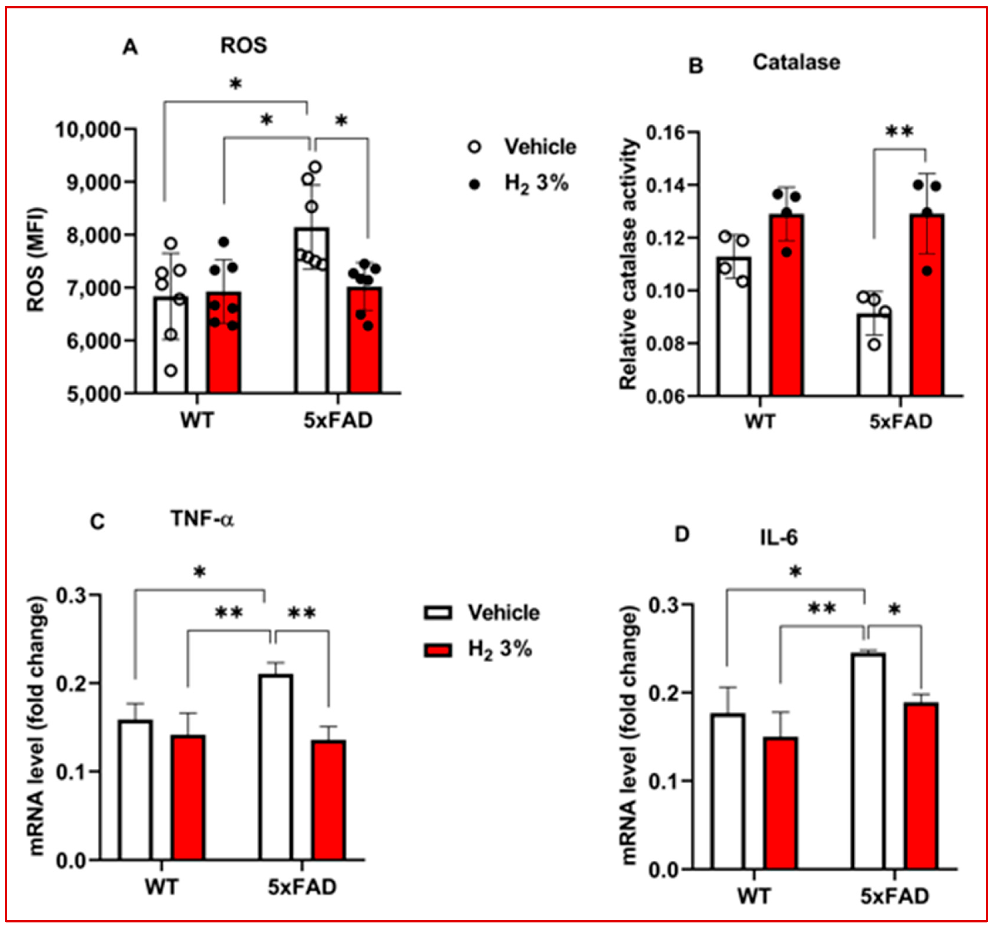
2.3. H2 Reduced OS and Neuroinflammation in 5xFAD Mice

The effects of H2 treatment on intracellular ROS levels, catalase antioxidant enzyme activities, and pro-inflammatory cytokines were also assessed in the hippocampal tissue from 5xFAD mice. We detected a marked effect, with a 1.2-fold decrease in ROS levels (p < 0.05; Figure 3A) and a 1.4-fold increase in catalase activity (p < 0.01; Figure 3B) compared with the vehicle-treated group. H2 treatment markedly suppressed TNF-α and IL-6 mRNA expression by 1.6-fold (p < 0.01) and 1.3-fold (p < 0.05), respectively, as compared to the vehicle-treated group (Figure 3C,D).

2.4. H2 Improved Amyloid Pathology and Reduced Toxic Metabolite Accumulation in 5xFAD Mice and AβO-Induced Primary Astrocytes

Amyloid plaques can activate the astrocytic urea cycle, causing the accumulation of toxic metabolites, resulting in astrocyte reactivation, neurodegeneration, and memory loss [18]. To evaluate the effects of H2 treatment on amyloid pathology, GFAP expression, and toxic metabolites (H2O2, ammonia, and GABA) accumulation, we performed IHC, ICC, and biochemical assays using hippocampal sections and lysates of mice and astrocytes. We first analyzed the ROS levels reflecting H2O2 and found a significant decrease in the H2-treated AβO-induced astrocytes and 5xFAD, compared to their vehicle-treated controls (Figure 2A and Figure 3A). Next, we performed IHC and observed that Aβ plaque accumulation in GFAP decreased by 2.8-fold in the cortex (CTX) (p < 0.001), 2.4-fold in the cornu ammonis 1 (CA1) (p < 0.05), and 2.7-fold in the dentate gyrus (DG), as shown in Figure 4A,B. Similarly, the GABA levels were markedly lower (p < 0.001) in the AβO-induced astrocytes treated with H2 than in those treated with vehicle; this was confirmed with subsequent ICC analyses (Figure 4C,D). Ammonia concentrations were measured and observed to have decreased minimally in vitro, as shown in Figure 4E,F.

3. Discussion

In this study, using 5xFAD transgenic mice and AβO-induced primary astrocytes, we examined the effects of H2 treatment on astrocytic urea cycle metabolites and enzymes after 60 days of 3% H2 exposure in the mice and 3 h in the astrocytes. The main findings of the analysis were as follows: (1) We observed that 5xFAD mice treated with H2 exhibited significant memory improvement, as shown by the significantly higher percentage of passing time in the Morris water maze test and higher spontaneous alternation percentage in the Y-maze test (Figure 1E,I, respectively). These indicate that 3% H2 inhalation significantly improved spatial memory and learning in 5xFAD mice compared with the vehicle. (2) OS (i.e., ROS and catalase activity) and inflammatory markers decreased with H2 treatment in both AβO-induced astrocyte cultures and 5xFAD mice, with significant differences between the treatment and vehicle groups (Figure 2A–D and Figure 3A–D). These findings imply that H2 therapy lowers OS and inflammation by decreasing the H2O2 levels via its antioxidant activity and decreasing TNF-α and IL-6 gene expression via an anti-inflammatory mechanism. Thus, suggesting that H2 induced neuroprotective effects by mitigating OS and downregulating inflammatory cytokine expression in AβO-induced astrocytes and 5xFAD mice. The observed reductions in ROS and pro-inflammatory mediators highlight the therapeutic potential of H2 as an agent targeting redox imbalance and neuroinflammation in AD progression. (3) The levels of toxic metabolites were significantly lower in the treatment group than in the vehicle group, with Aβ plaques (represented by the number of 6E10-positive signals) and GABA showing significant differences (Figure 4A–E). These data demonstrate that H2 treatment attenuated toxic metabolite accumulation, including Aβ, H2O2, and GABA, which were markedly reduced after H2 treatment compared with the vehicle. These suggest that astrocyte function was enhanced after H2 treatment due to reduced astrogliosis, denoted by the decrease in the GFAP level.
The antioxidant defense system is key to H2’s efficacy, which is modulated upstream by nuclear factor erythroid 2-related factor 2 (Nrf2) [33]. Nrf2 translocates to the nucleus under OS conditions, upon Keap1 modification, and binds to the promoter regions of antioxidant response elements. The binding drives the transcription and subsequent expression of many antioxidant enzymes, including catalase, glutathione peroxidase, and superoxide dismutase [34]. Through these mechanisms, Nrf2 prevents cell damage from reactive oxygen and nitrogen species and maintains redox homeostasis [35]. Studies on the medical applications of H2 have increased in recent years, with different methods of H2 administration in pre-clinical models and clinical subjects. These include inhalation, ingestion (dissolved in water), intravenous injection, and gel application to the skin [36,37]. The concentration of H2 in various studies varies. Approximately 1–4% of H2 is deemed safe [36], and 3% of inhaled H2 has been reported to improve cognitive and diffusion tensor imaging scores in patients with AD [38]. Additionally, 2% inhalation has been observed to reduce the risk of AD by downregulating the biomarker levels in healthy adults. These studies have advanced our understanding of the therapeutic efficacy of H2 [36,39]. However, the variations in dosage and routes of administration make these findings difficult to compare [36,38,39]. Moreover, these studies lacked randomization [38], and to date, no one has investigated the mechanisms by which H2 modulates the toxic metabolites associated with the astrocytic urea cycle in AD models.
Recent studies [18,40] have proposed ODC1 as a suitable therapeutic target for AD; however, it has been shown that difluoromethylornithine, an ODC1 inhibitor, requires high dosage and has toxic side effects [41,42]. Furthermore, another study reported no significant differences in ODC1 inhibition between difluoromethylornithine treatment and control [43]. A recent study proposed that tonic GABA may target reactive astrocytes in brain diseases; however, further evidence is needed to support this hypothesis [44]. Our study showed improved learning and memory after treating 5xFAD mice with 3% H2, which may have resulted from reduced OS and inflammatory markers, consistent with the results of earlier studies [37,38,39,45].
H2O2, a primary ROS, is produced as a toxic secondary metabolite in the astrocytic urea cycle; H2 is a scavenger of H2O2, thereby reducing neurodegeneration. The reduction in OS via H2O2 scavenging likely contributed to the observed decrease in the Aβ levels (Figure 4B), consistent with the findings of previous studies [12,46,47]. Additionally, accumulating Aβ in the brain can trigger OS and neuro-inflammatory responses in astrocytes, resulting in neuronal dysfunction and apoptosis [12]. The high levels of OS induce protein oxidation, resulting in Aβ formation [5]. Our study showed a decrease in Aβ, OS (illustrated by the decrease in ROS with the increase in catalase activity), and inflammatory markers such as TNF-α and IL-6, suggesting a decline in neurodegeneration; this enhances intraneuronal signal transmission and prevents the worsening of AD, as shown by memory improvement in mice (Figure 1E,I). The reduced OS also implies a decrease in co-localization of Aβ in the GFAP area (Figure 4A,B), suggesting that astrocyte death was reduced, improving its function. Furthermore, GABA, an inhibitory neurotransmitter that is produced in the final step of the astrocytic urea cycle, causes memory impairment in AD [17]. Our results showed a decrease in the GABA levels (Figure 4D), contributing to the memory improvement observed in the H2-treated 5xFAD mice.
However, the absence of metabolomic and proteomic profiling of specific urea cycle intermediates and enzymes and upstream markers of the antioxidant defense pathway, such as Nrf2, restricts our capacity to broadly evaluate the complete metabolic impact of H2 treatment. Future investigations incorporating integrated proteomic and metabolomic analyses are essential to describe the full scope of the H2-mediated modulation of the astrocytic urea cycle. Nevertheless, our findings indicate that H2 suppresses the accumulation of toxic urea cycle metabolites, thereby mitigating neurodegeneration and memory loss in an AD model.

4. Materials and Methods

4.1. Animal and Cell Culture Models

All animals were managed according to the Institutional Animal Care and Use Committee guidelines of Wonju College of Medicine, Yonsei University, which granted ethical approval for this study (ethical approval no: YWC-240423-2). Male and female mice (n = 29) at 5 weeks old were used, comprising transgenic 5xFAD mice (n = 14) with amyloid precursor protein (APP) and presenilin 1 (PSEN1) overexpression [48,49] and C57BL/6 wild-type (WT) mice (n = 15). The 5xFAD transgenic mice (RRID: MMRRC_034848-JAX) were obtained from The Jackson Laboratory (Bar Harbor, ME, USA; Stock No: 008730). Hemizygous strains were bred by mating transgenic mice with F1 C57BL/6 mice, and their genotypes were confirmed using polymerase chain reaction (PCR). The information on primers used for the genotyping is shown in Table A2. The mice were kept under a 12:12 h light/dark cycle in cages, with food and water access. The treatment groups (WT, n = 8; 5xFAD, n = 7) were administered H2 (3%) by inhalation using an H2-producing device (GOOTZ Co., Ltd., Yangju-si, Republic of Korea) for 60 days. In contrast, the vehicle treatment groups (WT, n = 7; 5xFAD, n = 7) received normal ventilation. One death was recorded in the WT H2-treated group. After 60 days, behavioral tests were performed, the mice were sacrificed, and their brains were removed and preserved for further analysis. Brain lysates were prepared by homogenizing 20 mg of tissue, and protein concentration was determined using a BCA assay. Each sample was normalized to 5 mg/mL of protein.
For in vitro experiments, we prepared primary cortical astrocyte cultures using 1-day-old pups of C57BL/6 mice. Cerebral cortices were dissected, minced, and separated into single cells. We then plated cells in 0.1 mg/mL poly-D-lysine-coated plates. The cells were cultured in Dulbecco’s modified Eagle’s medium (Gibco, Paisley, UK) containing 4.5 g/L glucose, L-glutamine, sodium pyruvate, 10% heat-inactivated fetal bovine serum (SIGMA-ALDRICH, Carlsbad, CA, USA), and 5% penicillin–streptomycin. The cultured cells were maintained under humid conditions at 37 °C, with 5% CO2. The cells were washed with fresh media, floating cell debris was removed, and the medium was replaced after three days [50].
The primary astrocyte culture was then grown in separate 6-well plates (2 × 10⁵ cells/well) for further experiments. AβO enhances AD progression [3]; thus, to evaluate the effects of H2 treatment in AβO-induced astrocytes, the cells in one of the plates were induced with 1 µM AβO (rPeptide, Watkinsville, GA, USA) for 5 days and placed in a FLUXLUK cell treatment chamber connected to the H2-producing device (GOOTZ Co., Ltd., Yangju-si, Republic of Korea) for a total of 3 h at 30 min intervals, with H2 concentration set at 3%. In the other plate, the cells were treated with NH4OH, i.e., vehicle, for a similar duration. AβO was prepared by adding 1% ice-cold ammonium hydroxide (NH4OH) to Aβ monomers aliquoted into 1.5 µL microtubes and stored at −80 °C until needed.
The treatment duration differed between the in vivo and in vitro experiments due to the differences in biological context and experimental constraints. In the mouse model, a prolonged 60-day exposure was necessary to mimic the chronic progression of AD-like pathology. In contrast, primary astrocytes in vitro are more vulnerable to environmental stress, particularly under non-standard incubation conditions such as those in the FLUXLUK chamber. Thus, to avoid inducing cellular stress or death, H2 exposure was limited to short intervals totaling 3 h, which is a commonly used approach in similar in vitro antioxidant treatment studies. This short-term exposure was sufficient to assess the protective or regulatory responses of astrocytes to AβO and H2 treatment.

4.2. Behavior Test

Memory impairment is a hallmark of AD [20]. To assess the effect of H2 cognitive behavior in 5xFAD mice, we used the Morris water maze and Y-maze to evaluate spatial learning and short-term memory [51,52]. All behavior tests were double-blind; unblinding was only performed after the data were analyzed. The Morris water maze test was conducted as published by Bromley-Brits et al. [53]. Briefly, each mouse was carefully positioned in the first quadrant to swim and find the visible platform. Mice that could not locate the platform after 60 s were gently led to it and left for 10 s. This procedure was repeated for each mouse across all quadrants for five days. The platform was hidden from days 2 to 5 to assess spatial learning. On day 6, the platform was removed entirely, and individually, the mice were positioned in the quadrant farthest from the target quadrant to explore for 60 s. The escape latency and path length were determined from the average of five trials for each mouse. A camera (Panlab Harvard Apparatus, Holliston, MA, USA, Panlab SMART video tracking Smart 3.0, Harvard Apparatus) tracked and recorded the movements of the mice, and subsequent data analysis was performed to evaluate their performance. On the sixth day, escape latency, path length, and passing time were analyzed to assess spatial memory and learning abilities.
In the Y-maze test, the mice were placed individually at the maze’s center (Jeongdo B&P Co. Ltd., Seoul, Republic of Korea) with three identical arms (120° from each other) to explore for 8 min. All four limbs of the mouse were within the arm before entry was recorded. We used video tracking as described above and analyzed the frequency of arm entries and spontaneous alternations [52].

4.3. Assessment of Toxic Metabolite Levels

Ju et al. [18] reported that astrocytes remove toxic metabolites such as ammonia, GABA, and H2O2 via the urea cycle under normal conditions. However, these metabolites accumulate in AD, triggering OS, neuroinflammation, and apoptosis, resulting in neurodegeneration and memory loss. We investigated the effects of H2 on the levels of these toxic metabolites through biochemical assays, IHC, and ICC.

4.3.1. ROS and Antioxidant Enzyme Assay

The intracellular ROS levels, specifically H2O2, were measured using 2,7-dichlorofluorescin diacetate (DCF-DA) (EMD Millipore Corp., Beijing, China). Briefly, in vivo, 10 µL of hippocampal lysates (5 mg/mL) and 100 µL of 20 µM DCF-DA were aliquoted into a 96-well plate. The plate was then incubated for 30 min in the dark. After the incubation, a DTX-880 microplate reader (Beckam Counter Inc., Fuller, CA, USA) was used to measure fluorescence at 488 nm excitation/525 nm emission. For in vitro assays, primary astrocytes were cultured and induced with 1 µM AβO (rPeptide, Watkinsville, GA, USA) in 6-well plates (2 × 10⁵ cells/well) for 5 days, then treated with H2 (3%) or 1% NH4OH for 3 h and washed two times in 1× phosphate-buffered saline (PBS) (Lonza, Walkersville, MD, USA); fluorescence detection was conducted as described above.
Antioxidant enzyme activity was quantified using the Amplex Red Catalase Assay Kit (Invitrogen, Carlsbad, CA, USA). Catalase breaks down H2O2, producing water and oxygen, thereby preventing cell damage [54]. Thus, 25 µL of each sample (5 mg/mL) and control were added separately in respective wells on the plate. Next, 25 µL of 40 µM H2O2 solution was added, and the content was kept at approximately 25–37 °C for 30 min. We then added 50 µL of 100 µM Amplex Red reagent/0.4 U/mL horseradish peroxidase, and the plate was incubated for 30 min at 37 °C. Subsequently, the absorbance was measured at 560 nm using a SpectraMax ABS Plus microplate reader (Molecular Devices, San Jose, CA, USA). The change in fluorescence was calculated as the fluorescence of the no-catalase control minus the fluorescence of the samples.

4.3.2. Ammonia Assay

To quantify ammonia concentration in AβO-induced primary astrocyte cultures and mouse brain tissues, we used the Ammonia Assay Kit (Sigma-Aldrich, St Louis, MO, USA) per the product protocol. The cells (2 × 10⁵ cells/well) or hippocampal lysate (5 mg/mL) was made into a homogenous mixture using distilled water and centrifugation. The supernatant was optimized to a pH of 7–8, and 100 µL of this supernatant was placed in cuvettes. The L-glutamate dehydrogenase solution was added to the cuvettes, mixed thoroughly, and incubated for 5 min at 18–35 °C. Then, the absorbance was measured at 340 nm using a microplate reader, as described above.

4.3.3. IHC and ICC

To determine whether 3% H2 treatment decreased Aβ accumulation in the 5xFAD mice hippocampi, we performed IHC as follows. The left hemispheres of excised mice brains were fixed with paraformaldehyde (4%) overnight at 4 °C and dehydrated in sucrose (30%) for 2 days. A cryostat was used to slice 30 µm coronal sections of the hippocampus. The sections were stored at 4 °C; they were washed in PBS and immersed in a blocking buffer, which comprised 5% goat serum in 0.1 M PBS with 0.3% Triton X-100 for 1 h. Next, the slices were incubated in the blocking buffer containing primary antibodies (Dako, Carpinteria, CA, USA) at 4 °C, amidst agitation, until the following day. They were then washed three times to remove unbound antibodies using PBS with 0.03% Triton X-100, and incubation with secondary antibody was carried out at room temperature for 1 h. Washing was repeated, and 4′,6-diamidino-2-phenylindole (DAPI) in PBS (1:1500 dilution) was added; the sections were subsequently mounted with a fluorescence mounting medium (Cell Signaling Technology, Danvers, MA, USA) and allowed to dry. Fluorescent images were captured using a confocal microscope and a Zeiss LSM880 microscope (Zeiss, Jena, Germany). Images were processed using the ImageJ software, version 1.41 (National Institutes of Health, Bethesda, MD, USA). The primary antibody and secondary antibody information are shown in Table A2.
Immunocytochemistry was used to analyze the effect of H2 on GABA accumulation. Briefly, AβO-induced astrocytes treated with 3% H2 or vehicle were fixed with paraformaldehyde (4%) for 15 min and washed repeatedly (three times) in cerebrospinal fluid. PBS containing 0.2% Triton X-100 was used to permeate the fixed cells and blocked for 1 h at room temperature. The cells were then incubated with a mouse anti-GABA primary antibody (Millipore, Bayswater, VIC, Australia) for 1 h at 4 °C. Next, the cells were incubated with a secondary antibody and DAPI (Vectashield®, Newark, CA, USA) for 40 min. After every incubation, three washes were performed with PBS containing 0.1% Triton X-100. Imaging was performed and analyzed as in IHC above.

4.4. RT-qPCR Analysis of Inflammatory Genes

To evaluate how H2 affects the inflammatory cytokine gene expression, we performed RT-qPCR on AβO-induced astrocytes (2 × 10⁵ cells/well) and mice hippocampal lysates (5 mg/mL). First, we extracted RNA from astrocytes and hippocampal lysates using the easy-BLUETM Total RNA Synthesis Kit (iNtRON Biotechnology, Seongnam-si, Republic of Korea). The extracted RNA (normalized to 1 µg) was converted to cDNA using the PrimeScriptTM 1st strand cDNA Synthesis Kit (Takara, Seoul, Republic of Korea). After cDNA library preparation, we conducted RT-qPCR as described by Kwak et al. [55]. Briefly, samples were prepared in triplicate, with a final volume of 10 µL comprising 1 µL of 10 pM primer, 2 µL of cDNA, 5 µL of SYBR Green PCR Master Mix (Applied Biosystems, Warrington, UK), and 2 µL of RNase-free water; these samples were analyzed in QuantStudio 6 Flex Real-Time PCR System (Applied Biosystems®, Marsiling Industrial Estate Road 3, Singapore). GAPDH mRNA was used as a reference, and fold change was determined using 2−∆∆CT. The primers used in this experiment are listed in Table A1.

4.5. Statistical Analysis

We used the GraphPad Prism software (version 8; GraphPad Software, La Jolla, CA, USA) to analyze data. Two-way analysis of variance with multiple comparison tests (Tukey’s post hoc test) was used to detect differences, with a significance level set at p < 0.05. Data from experimental samples are represented as mean and SEM. Individual mice or batches of cultured cells are represented as individual data points and samples, unless otherwise specified in the figure legends.

5. Conclusions

Our findings demonstrate that H2 treatment effectively attenuated the accumulation of toxic metabolites, OS, and inflammatory markers in both AβO-induced astrocytes and the 5XFAD transgenic mouse model of AD. Moreover, 3% H2 administration reduced the levels of H2O2, a toxic by-product of the astrocytic urea cycle, and GABA, thereby promoting neuroprotection. These biochemical improvements were associated with enhanced cognitive performance, as evidenced by improved learning and memory in 5xFAD mice. As we currently understand it, this is the first study to explore the therapeutic potential of H2 in targeting toxic metabolite accumulation within the astrocytic urea cycle in AD models. These findings suggest that H2 may represent a novel and promising strategy for mitigating neurodegeneration by modulating astrocyte-mediated metabolic dysfunction in AD. Nevertheless, detailed proteomic and metabolomic analyses are required to unravel the effects of H2 on the intermediates and enzymes of the astrocytic urea cycle.

Author Contributions

Conceptualization, K.-J.L. and B.A.S.; methodology, S.A.-N.; software, C.T.C., M.H.R. and J.B.; validation, C.-S.K., S.A.-N., M.H.R. and J.B.; formal analysis, S.A.-N. and C.T.C.; investigation, C.-S.K., B.A.S., S.A.-N., C.T.C. and T.T.N.; resources, C.-S.K. and B.A.S.; data curation, S.A.-N., C.-S.K., T.T.N. and C.T.C.; writing—original draft preparation, S.A.-N.; writing—review and editing, S.A.-N., J.B., M.H.R. and C.-S.K.; visualization, C.-S.K., K.H.-U., I.-S.Y. and J.B.; supervision, K.-J.L., B.A.S. and C.-S.K.; project administration, C.-S.K.; funding acquisition, C.-S.K. and K.-J.L. All authors have read and agreed to the published version of the manuscript.

Funding

This research was funded by the National Research Foundation of Korea (NRF), with funding from the government (Ministry of Science and ICT) (grant numbers: RS-2023-00253240 and RS-2022-00166626) and a research grant from Yonsei University Wonju College of Medicine.

Institutional Review Board Statement

All experimental procedures followed the Institutional Animal Care and Use Committee protocol of Yonsei University Wonju College of Medicine (ethical approval no.: YWC-240423-2; approval date: 1 May 2024).

Informed Consent Statement

Not applicable.

Data Availability Statement

The original contributions presented in this study are included in the article. Further inquiries can be directed to the corresponding authors.

Acknowledgments

The authors express their sincere gratitude to GOOTZ Co., Ltd., Yangju-si Gyeonggi-do, Republic of Korea, for providing the experimental hydrogen device and their valuable cooperation in conducting this research.

Conflicts of Interest

Authors Kwon Hwang-Un and You In-Soo were employed by the GOOTZ Co., Ltd., Gyeonggi-do, Republic of Korea. They declare no conflicts of interest. The remaining authors declare that the research was conducted without any commercial or financial relationships that could be construed as a potential conflict of interest.

List of Abbreviations

Amyloid-beta
AβOAmyloid-beta oligomer
ADAlzheimer’s disease
ARG1Arginase 1
CA1Cornu Ammonis 1
CTXCortex
DCF-DA2,7-dichlorofluorescein diacetate
DGDentate gyrus
5xFAD5 Familial Alzheimer’s disease
GABAAminobutyric acid
GFAPGlial fibrillary acidic protein
H2Molecular hydrogen
H2O2Hydrogen peroxide
ICCImmunocytochemistry
IHCImmunohistochemistry
IL-6Interleukin 6
ODC1Ornithine decarboxylase 1
OSOxidative stress
OTCOrnithine transcarboxylase
ROSReactive oxygen species
RT-qPCRQuantitative real-time polymerase chain reaction
TNF-αTumor necrosis factor-alpha

Appendix A

Table A1. List of primers.
GeneForward Primer SequenceReverse Primer Sequence
APP/PS15′-ACC CCC ATG TCA GAG TTC CT-3′5′-CGG GCC TCT TCG CTA TTA C-3′
TNF-α5′-TGTGCTCAGAGCTTTCAACAA -3′5′-CTTGATGGTGGTGCATGAGA-3′
IL-65′-GCTACCAAACTGGATATAATCAGGA-3′5′-CCAGGTAGCTATGGTACTCCAGAA-3′
Table A2. List of antibodies.
AntibodyHostApplicationSourceDilution
Anti-AβPigIFMILLIPORE,1:500
Anti-GABAMouseIFMILLIPORE1:500
Goat anti-rabbit IgGGoatIFInvitrogen1:500
Goat anti-mouse IgGGoatIFInvitrogen1:500
Goat anti-guinea pig IgGGoatIFInvitrogen1:500

References

  1. Goedert, M.; Spillantini, M.G. A Century of Alzheimer’s Disease. Science 2006, 314, 777–781. [Google Scholar] [CrossRef] [PubMed]
  2. Ohta, S.; Ohsawa, I. Dysfunction of Mitochondria and Oxidative Stress in the Pathogenesis of Alzheimer’s Disease: On Defects in the Cytochrome c Oxidase Complex and Aldehyde Detoxification. J. Alzheimer’s Dis. 2006, 9, 155–166. [Google Scholar] [CrossRef] [PubMed]
  3. Chen, G.; Xu, T.; Yan, Y.; Zhou, Y.; Jiang, Y.; Melcher, K.; Xu, H.E. Amyloid Beta: Structure, Biology and Structure-Based Therapeutic Development. Acta Pharmacol. Sin. 2017, 38, 1205–1235. [Google Scholar] [CrossRef] [PubMed]
  4. Picca, A.; Calvani, R.; Coelho-Junior, H.J.; Landi, F.; Bernabei, R.; Marzetti, E. Mitochondrial Dysfunction, Oxidative Stress, and Neuroinflammation: Intertwined Roads to Neurodegeneration. Antioxidants 2020, 9, 647. [Google Scholar] [CrossRef] [PubMed]
  5. Kim, S.; Jung, U.J.; Kim, S.R. Role of Oxidative Stress in Blood–Brain Barrier Disruption and Neurodegenerative Diseases. Antioxidants 2024, 13, 1462. [Google Scholar] [CrossRef] [PubMed]
  6. Montilla, P.; Feijóo, M.; Muñoz, M.C.; Muñoz-Castañeda, J.R.; Bujalance, I.; Túnez, I. Effect of Melatonin on the Oxidative Stress in N1E-115 Cells Is Not Mediated by Mt1 Receptors. J. Physiol. Biochem. 2003, 59, 263–268. [Google Scholar] [CrossRef] [PubMed]
  7. Takuma, K.; Yan, S.S.; Stern, D.M.; Yamada, K. Mitochondrial Dysfunction, Endoplasmic Reticulum Stress, and Apoptosis in Alzheimer’s Disease. J. Pharmacol. Sci. 2005, 97, 312–316. [Google Scholar] [CrossRef] [PubMed]
  8. Peggion, C.; Calì, T.; Brini, M. Mitochondria Dysfunction and Neuroinflammation in Neurodegeneration: Who Comes First? Antioxidants 2024, 13, 240. [Google Scholar] [CrossRef] [PubMed]
  9. Beal, M.F. Mitochondrial Dysfunction in Neurodegenerative Diseases. Biochim. Biophys. Acta (BBA)-Bioenerg. 1998, 1366, 211–223. [Google Scholar] [CrossRef]
  10. Trimmer, P.A.; Keeney, P.M.; Borland, M.K.; Simon, F.A.; Almeida, J.; Swerdlow, R.H.; Parks, J.P.; Parker, W.D.; Bennett, J.P. Mitochondrial Abnormalities in Cybrid Cell Models of Sporadic Alzheimer’s Disease Worsen with Passage in Culture. Neurobiol. Dis. 2004, 15, 29–39. [Google Scholar] [CrossRef] [PubMed]
  11. Sheehan, J.P.; Swerdlow, R.H.; Miller, S.W.; Davis, R.E.; Parks, J.K.; Parker, W.D.; Tuttle, J.B. Calcium Homeostasis and Reactive Oxygen Species Production in Cells Transformed by Mitochondria from Individuals with Sporadic Alzheimer’s Disease. J. Neurosci. 1997, 17, 4612–4622. [Google Scholar] [CrossRef] [PubMed]
  12. Rather, M.A.; Khan, A.; Alshahrani, S.; Rashid, H.; Qadri, M.; Rashid, S.; Alsaffar, R.M.; Kamal, M.A.; Rehman, M.U. Inflammation and Alzheimer’s Disease: Mechanisms and Therapeutic Implications by Natural Products. Mediat. Inflamm. 2021, 2021, 1–21. [Google Scholar] [CrossRef] [PubMed]
  13. Chun, H.; Lee, C.J. Reactive Astrocytes in Alzheimer’s Disease: A Double-Edged Sword. Neurosci. Res. 2018, 126, 44–52. [Google Scholar] [CrossRef] [PubMed]
  14. Chun, H.; Im, H.; Kang, Y.J.; Kim, Y.; Shin, J.H.; Won, W.; Lim, J.; Ju, Y.; Park, Y.M.; Kim, S.; et al. Severe Reactive Astrocytes Precipitate Pathological Hallmarks of Alzheimer’s Disease via H2O2− Production. Nat. Neurosci. 2020, 23, 1555–1566. [Google Scholar] [CrossRef] [PubMed]
  15. Escartin, C.; Galea, E.; Lakatos, A.; O’Callaghan, J.P.; Petzold, G.C.; Serrano-Pozo, A.; Steinhäuser, C.; Volterra, A.; Carmignoto, G.; Agarwal, A.; et al. Reactive Astrocyte Nomenclature, Definitions, and Future Directions. Nat. Neurosci. 2021, 24, 312–325. [Google Scholar] [CrossRef] [PubMed]
  16. Wunderlichová, L.; Buňková, L.; Koutný, M.; Jančová, P.; Buňka, F. Formation, Degradation, and Detoxification of Putrescine by Foodborne Bacteria: A Review. Compr. Rev. Food Sci. Food Saf. 2014, 13, 1012–1030. [Google Scholar] [CrossRef]
  17. Chen, C.-M.A.; Stanford, A.D.; Mao, X.; Abi-Dargham, A.; Shungu, D.C.; Lisanby, S.H.; Schroeder, C.E.; Kegeles, L.S. GABA Level, Gamma Oscillation, and Working Memory Performance in Schizophrenia. Neuroimage Clin. 2014, 4, 531–539. [Google Scholar] [CrossRef] [PubMed]
  18. Ju, Y.H.; Bhalla, M.; Hyeon, S.J.; Oh, J.E.; Yoo, S.; Chae, U.; Kwon, J.; Koh, W.; Lim, J.; Park, Y.M.; et al. Astrocytic Urea Cycle Detoxifies Aβ-Derived Ammonia While Impairing Memory in Alzheimer’s Disease. Cell Metab. 2022, 34, 1104–1120.e8. [Google Scholar] [CrossRef] [PubMed]
  19. Hansmannel, F.; Sillaire, A.; Kamboh, M.I.; Lendon, C.; Pasquier, F.; Hannequin, D.; Laumet, G.; Mounier, A.; Ayral, A.-M.; DeKosky, S.T.; et al. Is the Urea Cycle Involved in Alzheimer’s Disease? J. Alzheimer’s Dis. 2010, 21, 1013–1021. [Google Scholar] [CrossRef] [PubMed]
  20. Alzheimer’s Association. Treatment for Alzheimer’s. Available online: https://www.alz.org/alzheimers-dementia/treatments (accessed on 5 February 2025).
  21. Erkinjuntti, T.; Kurz, A.; Gauthier, S.; Bullock, R.; Lilienfeld, S.; Damaraju, C.V. Efficacy of Galantamine in Probable Vascular Dementia and Alzheimer’s Disease Combined with Cerebrovascular Disease: A Randomised Trial. Lancet 2002, 359, 1283–1290. [Google Scholar] [CrossRef] [PubMed]
  22. Sims, J.R.; Zimmer, J.A.; Evans, C.D.; Lu, M.; Ardayfio, P.; Sparks, J.; Wessels, A.M.; Shcherbinin, S.; Wang, H.; Monkul Nery, E.S.; et al. Donanemab in Early Symptomatic Alzheimer Disease. JAMA 2023, 330, 512. [Google Scholar] [CrossRef] [PubMed]
  23. van Dyck, C.H.; Swanson, C.J.; Aisen, P.; Bateman, R.J.; Chen, C.; Gee, M.; Kanekiyo, M.; Li, D.; Reyderman, L.; Cohen, S.; et al. Lecanemab in Early Alzheimer’s Disease. N. Engl. J. Med. 2023, 388, 9–21. [Google Scholar] [CrossRef] [PubMed]
  24. LeBaron, T.W.; Kura, B.; Kalocayova, B.; Tribulova, N.; Slezak, J. A New Approach for the Prevention and Treatment of Cardiovascular Disorders. Molecular Hydrogen Significantly Reduces the Effects of Oxidative Stress. Molecules 2019, 24, 2076. [Google Scholar] [CrossRef] [PubMed]
  25. Kamimura, N.; Nishimaki, K.; Ohsawa, I.; Ohta, S. Molecular Hydrogen Improves Obesity and Diabetes by Inducing Hepatic FGF21 and Stimulating Energy Metabolism in Db/Db Mice. Obesity 2011, 19, 1396–1403. [Google Scholar] [CrossRef] [PubMed]
  26. Dole, M.; Wilson, F.R.; Fife, W.P. Hyperbaric Hydrogen Therapy: A Possible Treatment for Cancer. Science 1975, 190, 152–154. [Google Scholar] [CrossRef] [PubMed]
  27. Ohsawa, I.; Ishikawa, M.; Takahashi, K.; Watanabe, M.; Nishimaki, K.; Yamagata, K.; Katsura, K.; Katayama, Y.; Asoh, S.; Ohta, S. Hydrogen Acts as a Therapeutic Antioxidant by Selectively Reducing Cytotoxic Oxygen Radicals. Nat. Med. 2007, 13, 688–694. [Google Scholar] [CrossRef] [PubMed]
  28. Lee, K.H.; Cha, M.; Lee, B.H. Neuroprotective Effect of Antioxidants in the Brain. Int. J. Mol. Sci. 2020, 21, 7152. [Google Scholar] [CrossRef] [PubMed]
  29. Jeong, E.-S.; Bajgai, J.; You, I.-S.; Rahman, M.H.; Fadriquela, A.; Sharma, S.; Kwon, H.-U.; Lee, S.-Y.; Kim, C.-S.; Lee, K.-J. Therapeutic Effects of Hydrogen Gas Inhalation on Trimethyltin-Induced Neurotoxicity and Cognitive Impairment in the C57BL/6 Mice Model. Int. J. Mol. Sci. 2021, 22, 13313. [Google Scholar] [CrossRef] [PubMed]
  30. Ohta, S. Molecular Hydrogen as a Preventive and Therapeutic Medical Gas: Initiation, Development and Potential of Hydrogen Medicine. Pharmacol. Ther. 2014, 144, 1–11. [Google Scholar] [CrossRef] [PubMed]
  31. Sim, M.; Kim, C.-S.; Shon, W.-J.; Lee, Y.-K.; Choi, E.Y.; Shin, D.-M. Hydrogen-Rich Water Reduces Inflammatory Responses and Prevents Apoptosis of Peripheral Blood Cells in Healthy Adults: A Randomized, Double-Blind, Controlled Trial. Sci. Rep. 2020, 10, 12130. [Google Scholar] [CrossRef] [PubMed]
  32. Tan, X.; Shen, F.; Dong, W.-L.; Yang, Y.; Chen, G. The Role of Hydrogen in Alzheimer’s Disease. Med. Gas. Res. 2018, 8, 176. [Google Scholar] [CrossRef] [PubMed]
  33. Tonelli, C.; Chio, I.I.C.; Tuveson, D.A. Transcriptional Regulation by Nrf2. Antioxid. Redox Signal 2018, 29, 1727–1745. [Google Scholar] [CrossRef] [PubMed]
  34. Xiang, Q.; Zhao, Y.; Lin, J.; Jiang, S.; Li, W. The Nrf2 Antioxidant Defense System in Intervertebral Disc Degeneration: Molecular Insights. Exp. Mol. Med. 2022, 54, 1067–1075. [Google Scholar] [CrossRef] [PubMed]
  35. Lin, L.; Wu, Q.; Lu, F.; Lei, J.; Zhou, Y.; Liu, Y.; Zhu, N.; Yu, Y.; Ning, Z.; She, T.; et al. Nrf2 Signaling Pathway: Current Status and Potential Therapeutic Targetable Role in Human Cancers. Front. Oncol. 2023, 13, 1184079. [Google Scholar] [CrossRef] [PubMed]
  36. Ohta, S. Recent Progress Toward Hydrogen Medicine: Potential of Molecular Hydrogen for Preventive and Therapeutic Applications. Curr. Pharm. Des. 2011, 17, 2241–2252. [Google Scholar] [CrossRef] [PubMed]
  37. Nakagawa, K.; Kodama, K.; Nagata, W.; Takahashi, S.; Satoh, Y.; Ishizuka, T. Molecular Hydrogen Inhibits Neuroinflammation and Ameliorates Depressive-like Behaviors and Short-Term Cognitive Impairment in Senescence-Accelerated Mouse Prone 8 Mice. Behav. Brain Res. 2025, 478, 115330. [Google Scholar] [CrossRef] [PubMed]
  38. Ono, H.; Nishijima, Y.; Sakamoto, M.; Kitamura, S.; Naitoh, Y.; Suzuki, K.; Fujii, N.; Kikura, R.; Ohta, S. Long-Term Inhalation of Hydrogen Gas for Patients with Advanced Alzheimer’s Disease: A Case Report Showing Improvement in Fecal Incontinence. Med. Res. Arch. 2022, 10, 1–9. [Google Scholar] [CrossRef]
  39. Lin, Y.-T.; Shi, Q.-Q.; Zhang, L.; Yue, C.-P.; He, Z.-J.; Li, X.-X.; He, Q.-J.; Liu, Q.; Du, X.-B. Hydrogen-Rich Water Ameliorates Neuropathological Impairments in a Mouse Model of Alzheimer’s Disease through Reducing Neuroinflammation and Modulating Intestinal Microbiota. Neural Regen. Res. 2022, 17, 409. [Google Scholar] [CrossRef] [PubMed]
  40. Bhalla, M.; Lee, C.J. Long-Term Inhibition of ODC1 in APP/PS1 Mice Rescues Amyloid Pathology and Switches Astrocytes from a Reactive to Active State. Mol. Brain 2024, 17, 3. [Google Scholar] [CrossRef] [PubMed]
  41. Wang, C.C. A Novel Suicide Inhibitor Strategy for Antiparasitic Drug Development. J. Cell Biochem. 1991, 45, 49–53. [Google Scholar] [CrossRef] [PubMed]
  42. McWilliams, M.L. Characterization of the Ototoxicity of Difluoromethylornithine and Its Enantiomers. Toxicol. Sci. 2000, 56, 124–132. [Google Scholar] [CrossRef] [PubMed]
  43. Saulnier Sholler, G.L.; Gerner, E.W.; Bergendahl, G.; MacArthur, R.B.; VanderWerff, A.; Ashikaga, T.; Bond, J.P.; Ferguson, W.; Roberts, W.; Wada, R.K.; et al. A Phase I Trial of DFMO Targeting Polyamine Addiction in Patients with Relapsed/Refractory Neuroblastoma. PLoS ONE 2015, 10, e0127246. [Google Scholar] [CrossRef] [PubMed]
  44. Koh, W.; Lee, C.J. Diagnostic and Therapeutic Potential of Tonic Gamma-aminobutyric Acid from Reactive Astrocytes in Brain Diseases. Clin. Transl. Med. 2024, 14, e1642. [Google Scholar] [CrossRef] [PubMed]
  45. Rahman, M.H.; Bajgai, J.; Sharma, S.; Jeong, E.-S.; Goh, S.H.; Jang, Y.-G.; Kim, C.-S.; Lee, K.-J. Effects of Hydrogen Gas Inhalation on Community-Dwelling Adults of Various Ages: A Single-Arm, Open-Label, Prospective Clinical Trial. Antioxidants 2023, 12, 1241. [Google Scholar] [CrossRef] [PubMed]
  46. de la Torre, J.C. Is Alzheimer’s Disease a Neurodegenerative or a Vascular Disorder? Data, Dogma, and Dialectics. Lancet Neurol. 2004, 3, 184–190. [Google Scholar] [CrossRef] [PubMed]
  47. Pappolla, M.A.; Omar, R.A.; Kim, K.S.; Robakis, N.K. Immunohistochemical Evidence of Oxidative [Corrected] Stress in Alzheimer’s Disease. Am. J. Pathol. 1992, 140, 621–628. [Google Scholar] [PubMed]
  48. De Strooper, B.; Annaert, W. Novel Research Horizons for Presenilins and γ-Secretases in Cell Biology and Disease. Annu. Rev. Cell Dev. Biol. 2010, 26, 235–260. [Google Scholar] [CrossRef] [PubMed]
  49. Selkoe, D.J. Alzheimer’s Disease: Genes, Proteins, and Therapy. Physiol. Rev. 2001, 81, 741–766. [Google Scholar] [CrossRef] [PubMed]
  50. Woo, D.H.; Han, K.-S.; Shim, J.W.; Yoon, B.-E.; Kim, E.; Bae, J.Y.; Oh, S.-J.; Hwang, E.M.; Marmorstein, A.D.; Bae, Y.C.; et al. TREK-1 and Best1 Channels Mediate Fast and Slow Glutamate Release in Astrocytes upon GPCR Activation. Cell 2012, 151, 25–40. [Google Scholar] [CrossRef] [PubMed]
  51. Morris, R.G.M. Spatial Localization Does Not Require the Presence of Local Cues. Learn. Motiv. 1981, 12, 239–260. [Google Scholar] [CrossRef]
  52. Kraeuter, A.-K.; Guest, P.C.; Sarnyai, Z. The Y-Maze for Assessment of Spatial Working and Reference Memory in Mice. In Pre-Clinical Models: Techniques and Protocols; Humana Press: New York, NY, USA, 2019; pp. 105–111. [Google Scholar]
  53. Bromley-Brits, K.; Deng, Y.; Song, W. Morris Water Maze Test for Learning and Memory Deficits in Alzheimer’s Disease Model Mice. J. Vis. Exp. 2011, 53, 2920. [Google Scholar] [CrossRef] [PubMed]
  54. Mueller, S.; Riedel, H.-D.; Stremmel, W. Determination of Catalase Activity at Physiological Hydrogen Peroxide Concentrations. Anal. Biochem. 1997, 245, 55–60. [Google Scholar] [CrossRef] [PubMed]
  55. Kwak, H.; Koh, W.; Kim, S.; Song, K.; Shin, J.-I.; Lee, J.M.; Lee, E.H.; Bae, J.Y.; Ha, G.E.; Oh, J.-E.; et al. Astrocytes Control Sensory Acuity via Tonic Inhibition in the Thalamus. Neuron 2020, 108, 691–706.e10. [Google Scholar] [CrossRef] [PubMed]
Figure 1. Mice behavioral test results. Morris water maze results, including (A) escape latency during trial (days 2–5), (B) path length during trial (days 2–5), (C) escape latency for test trial (day 6), (D) path length for test trial (day 6), (E) passing time for test trial (day 6), and (F) swimming pattern. Y-maze test results: (G) number of arm entries, (H) total distance, and (I) spontaneous alternation. WT and 5xFAD mice were treated with 3% H2 (n = 7 in each group). Significant differences were set at * p < 0.05, ** p < 0.01, and *** p < 0.001. H2, molecular hydrogen; 5xFAD, 5 familial Alzheimer’s disease; WT, wild type.
Ijms 26 06922 g001
Figure 2. Effects of H2 treatment on OS and inflammatory markers in in vitro AD model. (AD) Bar charts of ROS assay, catalase assay, mRNA level of TNF-α, and IL-6 in Aβ-induced astrocytes (2 × 10⁵ cells/well) treated with H2 and vehicle (n = 3 for each group), respectively. H2, molecular hydrogen; IL-6, interleukin 6; OS, oxidative stress; ROS, reactive oxygen species; TNF-α, tumor necrosis factor-alpha. Solid circles and red bars represent H2 treatment and hollow circles in white bars represent vehicle treatment. Statistical significance was set at * p < 0.05, ** p < 0.01.
Ijms 26 06922 g002
Figure 3. Effects of H2 treatment on OS and inflammatory markers in vivo AD model. (AD) Bar charts of ROS assay (n = 7), catalase assay (n = 4), mRNA levels of TNF-α, and IL-6 in mice treated with H2. WT (n = 3) and 5xFAD (n = 3). 5xFAD, 5 familial Alzheimer’s disease; H2, molecular hydrogen; TNF-α, tumor necrosis factor-alpha; IL-6, interleukin 6; OS, oxidative stress; ROS, reactive oxygen species. Solid circles and red bars represent H2 treatment and hollow circles in white bars represent vehicle treatment. Statistical significance was set at * p < 0.05, ** p < 0.01.
Ijms 26 06922 g003
Figure 4. Toxic metabolite levels after H2 treatment. (A) IHC confocal image of mice brain sections. (B) Plot of amyloid-beta signal intensity (n = 4). (C) ICC image for GABA signal detection in astrocytes. (D) Graph of GABA intensity (n = 4). (E,F) Graphs show ammonia concentration for in vitro and in vivo assays. Statistical significance was set at * p < 0.05, *** p < 0.001. H2, molecular hydrogen; CTX, cortex; CA1, cornu ammonis 1; DG, dentate gyrus; 5xFAD, 5 familial Alzheimer’s disease; GABA, γ-aminobutyric acid; ICC, immunocytochemistry; IHC, immunohistochemistry; VEH, vehicle; 6E10 positive indicates amyloid plaques. Arrows show amyloid plaques, and white boxes represent section detected under the microscope.
Ijms 26 06922 g004
Disclaimer/Publisher’s Note: The statements, opinions and data contained in all publications are solely those of the individual author(s) and contributor(s) and not of MDPI and/or the editor(s). MDPI and/or the editor(s) disclaim responsibility for any injury to people or property resulting from any ideas, methods, instructions or products referred to in the content.

