Article

Multi- and Transgenerational Histological and Transcriptomic Outcomes of Developmental TCDD Exposure in Zebrafish (Danio rerio) Ovary

1
Department of Environmental and Global Health, University of Florida, Gainesville, FL 32611, USA
2
Department of Pharmacology, Wayne State University, Detroit, MI 48202, USA
*
Author to whom correspondence should be addressed.
These authors contributed equally to this work.
Int. J. Mol. Sci. 2025, 26(14), 6839; https://doi.org/10.3390/ijms26146839
Submission received: 14 April 2025 / Revised: 5 July 2025 / Accepted: 11 July 2025 / Published: 16 July 2025
(This article belongs to the Special Issue Molecular Research of Reproductive Toxicity)

Abstract

2,3,7,8-Tetrachlorodibenzo-p-dioxin (TCDD) exposure has long been associated with reproductive dysfunction in males and females even at miniscule levels, which can persist across generations. Given the continued industrial use and detection of other aryl hydrocarbon receptor (AhR) agonists in the general population and the demonstrated heritable phenotypes of TCDD exposure, further work is justified to elucidate reproductive pathologies and minimize exposure risk. In females, multi- and transgenerational subfertility has been demonstrated in a zebrafish (Danio rerio) model exposed to 50 pg/mL TCDD once at 3 and 7 weeks post fertilization (wpf). We further characterize the histopathologic, hormonal and transcriptomic outcomes of the mature female zebrafish ovary following early-life TCDD exposure. Exposure was associated with significantly increased ovarian atresia in the F0 and F1, but not F2 generation. Other oocyte staging and vitellogenesis were unaffected in all generations. Exposed F0 females showed increased levels of whole-body triiodothyronine (T3) and 17β-estradiol (E2) levels, but not vitellogenin (Vtg), 11-ketotestosterone (11-KT), cortisol, thyroxine (T4), or testosterone (T). Ovarian transcriptomics were most dysregulated in the F2. Both F0 and F2, but not F1, showed changes in epigenetic-related gene expression. Rho signaling was the top pathway for both F0 and F2.

1. Introduction

As posited by the Developmental Origins of Health and Disease (DOHaD) hypothesis, early life exposure to environmental agents, such as endocrine-disrupting compounds (EDCs), has the potential to interfere with transcriptomic and epigenetic networks during critical windows of development [1]. Such disruption can lead to health effects that present across the lifespan and can be heritable across multiple generations of descendants [2,3,4].
2,3,7,8-Tetrachlorodibenzo-p-dioxin (TCDD) occurs both naturally at low levels by natural processes, such as forest fires, and at higher levels as a by-product of plastic waste burning and other manufacturing processes. It is the most potent and toxic dioxin-class EDC, with highly bioaccumulative and lipophilic properties [5]. This model aryl hydrocarbon receptor (AhR) agonist can represent other dioxin-like compounds found in the general population and environment such as polychlorinated biphenyls (PCBs), furans and polycyclic aromatic hydrocarbons (PAHs) [6].
TCDD has well-established effects on multiple organ systems, including cardiovascular [7], immune [8], skeletal [9], neurobehavioral [10] and reproductive systems. Early-life exposure to TCDD is known to alter the reproductive functioning of both males and females, as demonstrated in the outcomes of residential TCDD exposure in Seveso, Italy: adult males exposed before puberty and male offspring of those exposed showed decreased sperm concentration and motility [11,12], while exposed females demonstrated increased infertility, time to pregnancy and risk of endometriosis [13,14]. When modeled in other mammalian species, developmental TCDD exposure resulted in similar reproductive trends, including effects on testes weight and histopathology, reduction in epididymal sperm reserves and sperm motility and dysregulation of steroidogenic and spermatogenic genes in males [15,16,17]. In females, effects included malformed external genitalia, altered ovarian weight and histopathology, premature ovarian failure, reduced levels of estradiol and LH, impaired folliculogenesis and disruption of follicular development and ovulation pathways [18,19,20,21]. In fact, infertile women have been found to carry higher levels of certain dioxin-like compounds in their follicular fluid [22].
Many factors complicate epidemiological studies of the multigenerational consequences of developmental EDC exposure, including confounding exposures, genetic variability, and the time span between exposure and measured outcomes. Zebrafish (Danio rerio) are useful in modeling such exposures, due to their quick generation time, ease of exposure, production of numerous, and externally fertilized embryos from the same parents and are additionally well suited to reproductive studies due to their emerging presence as a model of gonadal development [23]. At approximately 3 wpf, zebrafish enter gonadal differentiation, where bipotential tissue resembling an immature ovary either initiates the apoptotic transformation to testes, or progresses to oogenesis [24]. Generally, gonadal fate is determined by approximately 7 wpf, but gonads will continue to develop until reproductive maturity at 3–4 months [23]. These F0 fish are then raised to adulthood and spawned to create an indirectly exposed F1 generation. These F1 fish were likewise spawned as adults to produce the completely unexposed F2 generation, where distinctly transgenerational effects would be observed.
In previous work, TCDD-lineage fish demonstrated impaired reproduction across three generations. Outcrosses between TCDD-lineage and control-lineage fish for all three generations revealed transgenerational reproductive deficits present in TCDD-lineage males [25,26], which our group characterized through histological, transcriptomic and methylomic analyses of F0-F2 testes [27,28,29,30]. In contrast, TCDD-lineage females displayed reproductive deficits into the F0 and F1 generations only, suggesting multigenerational but not transgenerational gonadal disruption. A brief examination of H&E-stained F0-F2 adult female ovaries indicated an increased number of atretic follicles in F0 and F1 TCDD-lineage fish alone, further supporting these multi- but not transgenerational findings [25].
In this study, to robustly characterize the long-term reproductive effects on TCDD-lineage females, we carried out an in-depth histological assessment in F0-F2 ovaries, specifically evaluating ovary size, percentage of atresia, presence of egg debris and gonadal staging [31]. In line with previous work, we found that the percentage of atresia was increased in the F0 and F1, but not F2 ovaries. However, there were no differences for other histological endpoints in the F0 (F1 and F2 unassessed). Interestingly, we found gene expression dysregulation in the F0 and F2 generation, but not the F1, suggesting a transgenerational, but not multigenerational, effect for that outcome. The top pathways in the F0 and F2 generations were both Rho signaling-related, again highlighting the transgenerational link. Some epigenetic-related genes were differentially expressed in the F2 generation, which warrants further investigation. Lastly, we measured hormone levels from whole-body lysate. Exposed females showed increased levels of whole-body triiodothyronine (T3) and, unexpectedly, increased 17β-estradiol (E2) levels. This work gives a fuller picture of the effect of low-level, early TCDD exposure and provides opportunities for future research.

2. Results and Discussion

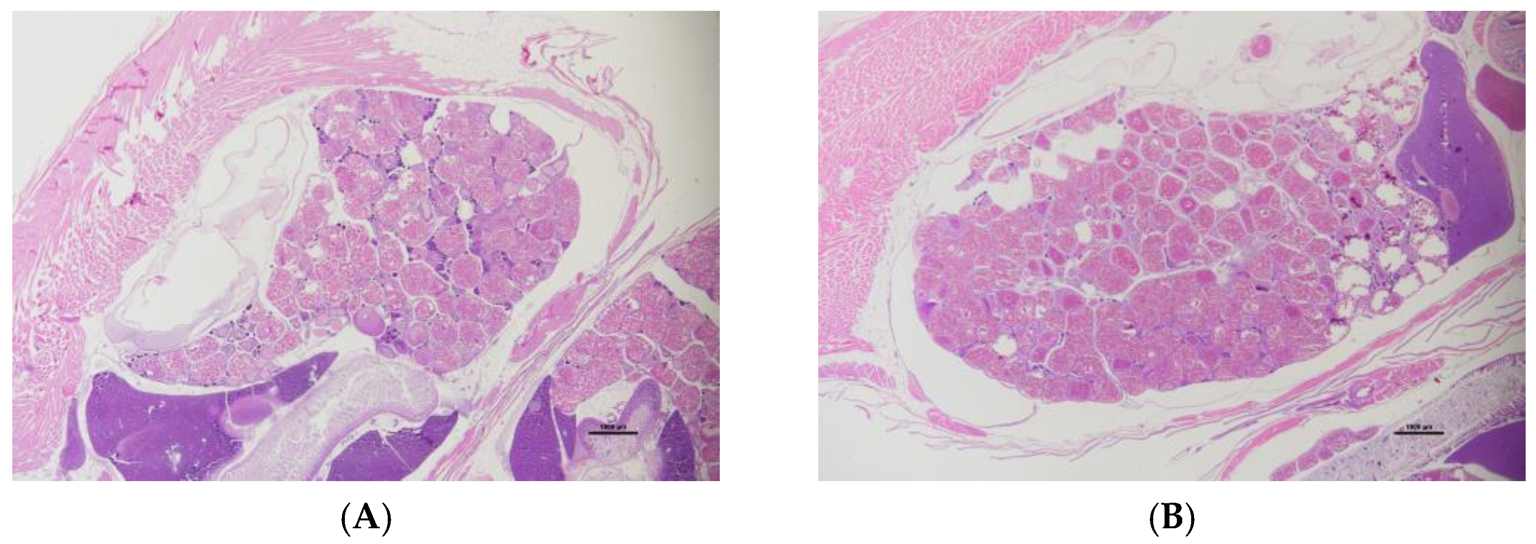
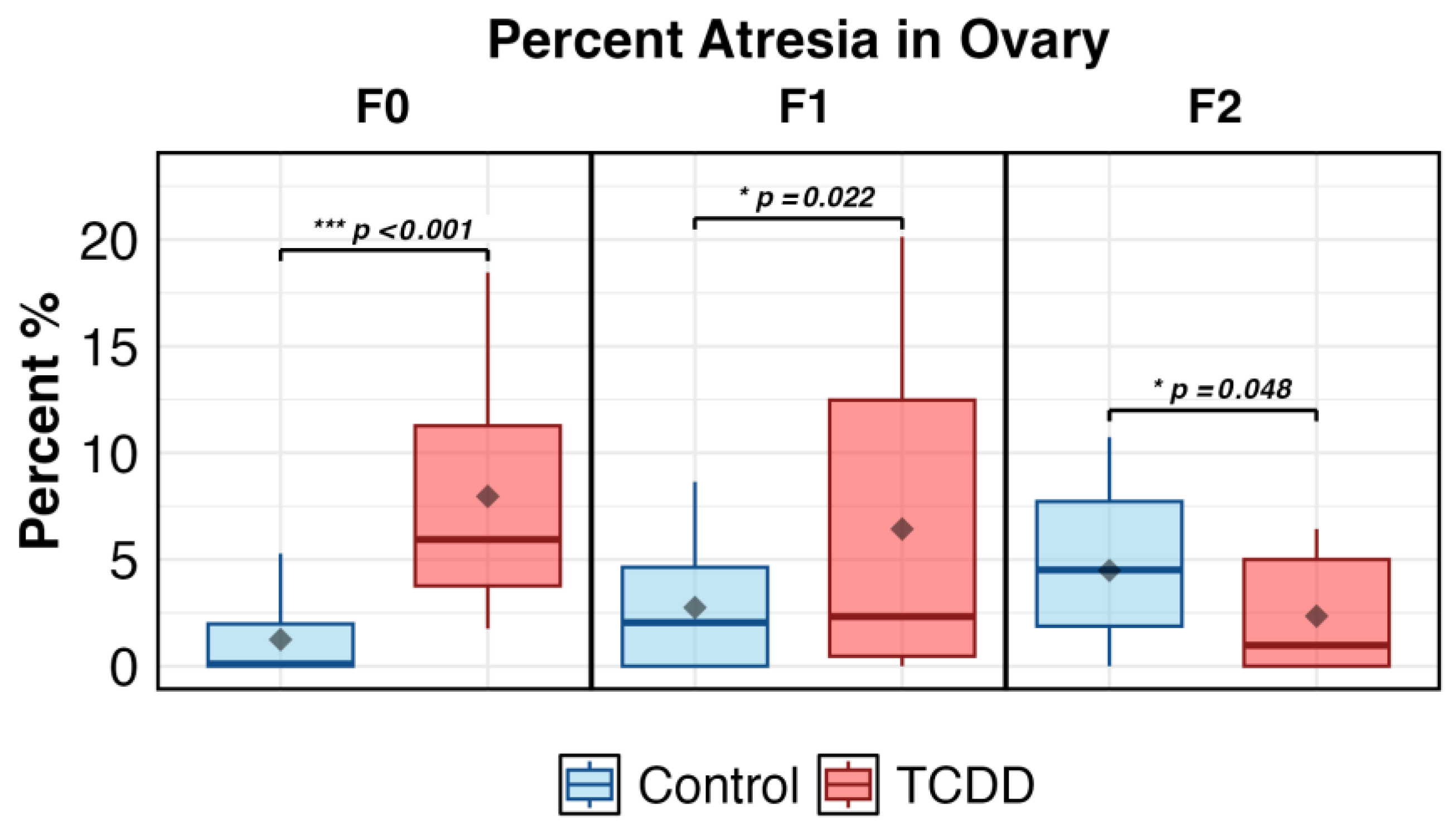
2.1. Ovarian Histology

OECD-defined gonadal staging [31] was performed on H&E-stained sections of F0 fish. Oocytes in each maturation stage (chromatin nucleolar/perinuclear oocytes, cortical alveolar oocytes, vitellogenic oocytes), atretic follicles, presence/absence of egg debris and granulomatous inflammation were measured (Supplemental Table S1). Representative sections are shown in Figure 1.
Ovary size was not affected in F0 (p = 0.687) or F1 (p = 0.989) (Student’s two-sample t-test). Neither egg debris nor granulomatous inflammation area were affected in F0 (p = 0.65 (Fisher’s exact test); p = 0.546 (Student’s two-sample t-test), respectively), and was not assessed in F1 or F2.
Percentage of atretic ovary was increased in the F0 TCDD-exposed fish compared to controls (p < 0.001). No other oocyte staging endpoints were affected. The percentage of atresia was the only staging endpoint further assessed in F1 and F2 generations. In the F1, exposure was associated with a significantly higher percentage of atresia (p < 0.05); in the F2 there was, unexpectedly, a statistically significant (p = 0.048) decrease in atresia compared to controls (Figure 2). This is likely due to the slight increase in control atresia in the F2 compared to F1 and F0, the etiology of which is unknown. While statistically significant, the difference is likely not physiologically relevant in the F2. Similarly, another study found atresia was significantly lower in the F2 exposure group compared to control in TCDD-exposed mice [32], though mice F2 can be considered more comparable to zebrafish F1 [33]. A previous report from this group concurs with the findings of atretic follicles in the F0 and F1, but not F2 generation. Further, spawning failure in the F2 was attributed to male, not female, zebrafish [26]. Thus, we observed a multi- but not trans-generational increase in ovarian atresia following F0 exposure, in line with previous work.
TCDD has been shown to cause atresia in other zebrafish models of TCDD exposure [26,34,35,36,37,38,39]. In rats, number and size of follicles were reduced [40], though mice were unaffected [41]. In mammalian cells, the AhR regulates granulosa cell proliferation [42]. Similar compounds to TCDD such as PAHs have been shown to affect female anovulation, reduced conception, spontaneous abortion, menstrual abnormalities and gonadal developmental defects [43]. Dioxin-like compounds promote the development of endometriosis, a syndrome linked to infertility, pain and irregular menstrual cycles [44,45,46].
Future work will assess gonadal staging in further generations. Though we did not observe altered staging outside of atresia in the F0, TCDD has been associated with decreased mature follicles in zebrafish [35]; multi- and transgenerational effects are yet to be uncovered.

2.2. Differentially Expressed Genes and Pathway Analysis

2.2.1. F0 Generation

The directly exposed F0 generation produced 215 differentially expressed genes with ≥1 absolute log2 fold change and p < 0.01 in the exposed fish (203 upregulated; 12 downregulated; Supplemental Table S2). The highest differential expression was in klf2b (Krueppel-like factor 2b) (log2FC = 2.86), orthologous to human KLF2. In zebrafish, this gene encodes a transcription factor involved in germ layer cell fate and embryonic cardiac development [47,48]. In humans, it is a transcriptional activator involved in many biological processes that overlap with TCDD-disrupted processes including adipogenesis [49,50], embryonic erythropoiesis [51,52] and inflammation [53,54].
When DEGs were restricted to those with FDR < 0.1, eight other DEGs remained. These were involved in basic cellular functions such as enzymes (cathepsins ctsbb and degs1), transporters (slc7a8a and atp1a3b), a chemokine (ccl25b), a transcription factor (rbm47) and part of collagen complex (col12a1a).
Interestingly, stm, which plays a role in egg membrane integrity, ovulation and otolith formation was upregulated (log2FC = 2.03, FDR < 0.1). TCDD exposure has been shown to decrease fertility [25,36], shift sex ratios [25,55], disrupt ovarian histopathology [25,37] and disrupt inner ear development [56]. Otoliths, bone-like structures in the inner ear, are crucial in maintaining balance. Previous work found both skeletal malformations and behavioral alterations in adult fish exposed as juveniles to TCDD [25,57]. A future direction would be to examine what, if any, effect ear development has on subsequent behavior and if TCDD-exposed fish behavior shows any similarity.
Due to TCDD’s known multi- and transgenerational inheritance effects, we next compared the 215 DEGs with the EpiFactors Database [58]. Of the 215 DEGs, 191 had known human orthologs. Three upregulated DEGs had matches with the EpiFactors Database: hdac7, ywhae and mapkapk3. These are interesting candidates for further research, as TCDD exposure has known epigenetic effects [59].
Ingenuity Pathway Analysis (IPA) was conducted on 137 analysis-ready molecules of the 215 DEGs uploaded. Table 1 and Table 2 show pathways with z-score cutoff of ≥|2| and p < 0.05 for Canonical pathways and Diseases and Functions pathways, respectively. RHO GTP pathways stand out, as do increased cancer pathways. Decreases in inflammation pathways are not unexpected, as TCDD and other AhR ligands may support inflammation or its resolution [53]. These pathways should be considered with caution, as most of the DEGs used for analysis were not significant when considering false discovery rate. Additionally, expected dysregulation of transcripts such as ahr2, cyp1a, vtg1 and star [55] was not observed. The full IPA Summary can be found in Supplemental Table S4.

2.2.2. F1 Generation

The indirectly exposed F1 generation displayed six DEGs at the level of ≥1 absolute log2 fold change and p < 0.01; one upregulated (syne2b) and five downregulated (slc35a4, trpc4apb, cxcr4b, stap2b and one uncharacterized). All of these DEGs additionally met the criteria of FDR-adjusted p-value < 0.1. None are known to be related to reproduction. Ingenuity Pathway Analysis was not conducted due to low number of DEGs.

2.2.3. F2 Generation

The unexposed F2 generation exhibited 672 DEGs at the level of ≥1 absolute log2 fold change and p < 0.01 (556 upregulated; 116 downregulated; Supplemental Table S3. Steroid/Thyroid Hormone LC-MS/MS: Electrospray ionization (ESI) parameters). The F2 produced the most DEGs of any generation. To narrow focus on highly relevant DEGs and pathways, we further filtered DEGs using the FDR-adjusted p-value of <0.1. This parameter/filter returned 550 DEGs (454 upregulated; 96 downregulated).
The top upregulated DEG was rxfp2b (human ortholog RXFP2) (log2FC = 4.96). In fish and mammals, RXFP2 (also known as LGR8) is expressed in gonads [60,61]. In males, its loss of function either genetically or via EDCs has been implicated in cryptorchidism and infertility. When bound by its exclusive ligand INSL3 in the ovary, RXFP2 stimulates androstenedione and thus downstream estrogens production, which then promotes a positive feedback loop with INSL3 [62]. As we later report, we witnessed an increase in E2 hormone levels, though insl3 expression was not significantly raised (log2FC = 0.768; p = 0.160; FDR = NA). Interestingly, in a rat exposure study using brominated flame retardants, which bear a similar chemical structure to TCDD, no effect on Rxfp2 or Ahr was seen in the ovary, though Insl3 was downregulated [63]. We observed a weakly significant upregulation in ahr2 (log2FC = 1.140; p = 0.015; FDR = 0.161). Unchanged ahr2 expression would not be surprising as the exposure was singular, brief and in early life two generations ago; slight upregulation indicates long-term changes in xenobiotic response.
The second most upregulated transcript was lhx1a (human ortholog LHX1) (log2FC = 3.99). In mammals, LHX1 is involved in embryonic urogenital development [64], and LHX1 deletions are candidates for rare congenital syndrome (Mayer-Rokitansky-Küster-Hauser syndrome (MRKH)) involving aplasia of the uterus, vagina and Müllerian ducts [65,66,67]. In zebrafish, there are no known reproductive associations; lhx1a functions mainly in renal development [68,69]. The upregulation may be epigenetic and compensatory for the renal effects of ancestral TCDD exposure [39,70], rather than indicative of a genital tract anomaly, especially given the reversal in direction of expression compared to humans with MRKH.
The strong upregulation of rxfp2b and lhx1a does not fit neatly into existing frameworks of reproductive function. Neither were significantly changed in the F0 generation. Future work would benefit from epigenetic inquiry into this counterintuitive finding.
The most downregulated transcript was drd4a (human ortholog DRD4) (log2FC = −3.13). Drd4a has not yet been shown to express in zebrafish ovary, though it is found in human gonads. In humans, DRD4 encodes the dopamine receptor D4, lower-function variants of which may be implicated in ADHD [71]; likewise, D4 disruption can alter locomotor activity in zebrafish [72,73]. Exposure to AhR agonists including TCDD have been associated with zebrafish hyperactivity [57,74]; this extended to the F1 in one report [57]. Fetal TCDD exposure and AhR overexpression has been shown to activate tyrosine hydroxylase mRNA expression [75], connecting one dot between dopamine and TCDD. It remains to be seen whether drd4a has a role mediating this neurobehavioral response, or in reproduction. Behavior and reproduction are quite linked in zebrafish, as both males and females not only must be intrinsically fertile but also perform specific, orchestrated spawning behaviors to release eggs.
The second-most downregulated transcript was spata22 (human ortholog SPATA22) (log2FC = −2.77). SPATA22 is expressed nearly exclusively in germ cells during meiosis and is required for homologous recombination [76,77]. This downregulation is in line with multiple observed ovarian phenotypes. Whole exome sequencing of consanguineous and sporadic cases uncovered loss-of-function SPATA22 variants in primary ovarian insufficiency [78,79]. Diminished reproductive success has been shown in female zebrafish exposed to TCDD [25,35,37]; what, if any, role spata22 plays in this pathology is unknown. Little, if any, research has been completed on spata22 in zebrafish. Spata4, a testis-specific transcript, was strongly downregulated in one study of TCDD exposure and male zebrafish [28].
Consistent with findings from other transgenerational toxicology models, including fish and rodents models exposed to TCDD and other contaminants and stressors, our results showed an increasing trend in DEG levels in the transgenerational generations compared to the direct exposure effects observed in F0 and F1 generations [28,32,80,81,82]. This pattern demonstrates a compounding effect of with each successive generation. While the underlying mechanisms require further investigation, it has been hypothesized that this phenomenon results from cumulative epigenetic modifications that differ mechanistically between direct exposure and transgenerational inheritance in unexposed descendant generations.
Ingenuity Pathway Analysis (IPA) was conducted for 377 analysis-ready molecules of the 550 DEGs uploaded. With the pathway z-score cutoff of |2|, a theme emerged of disruption of basic cellular functions. The top five up- and downregulated Canonical Pathways are shown in Table 3; Disease and Function pathways in Table 4. In the full dataset under the Categories column in the Top Diseases or Functions list, 43% of terms contain cellular movement, development, signaling or morphology. The full IPA Summary can be found in Supplemental Table S5.
The fact that DEGs were observed multiple generations later led us to compare F2 DEGs with the EpiFactors Database [58]. Of the 550 F2 DEGs, 462 genes had known human orthologs. Of these, there were 21 matches with the EpiFactors Database. These are shown in Table 5. This suggests a demethylated environment, as tet2 functions in the demethylation process, and dnmt3b is a methylase. TCDD is a known epigenetic disruptor [59].

2.2.4. Transgenerational Comparisons

F2 generation had some overlap in DEGs with the F0 generation (Figure 3). With parameters set at log2FC > |1| and FDR < 0.1, there were only 4 DEGs (ccl25b, atp1a3b, degs1 and col12a1a). An exploratory comparison using the more promiscuous cutoff of p < 0.01 for the F0 produced a similar ratio of overlap (approximately one third of the DEGs of the F0 overlapped with F2 DEGs).
Remarkably, the top up- and downregulated pathway in both the F0 and F2 IPA were Rho GTPase cycle and Rho GDI signaling, respectively, with neutrophil degranulation being the second most upregulated pathway in both generations as well; despite the more promiscuous F0 p-value cutoff to allow enough DEGs for pathway analysis (p < 0.01 vs. FDR < 0.1).
IPA was also run with the overlapping F0 and F2 DEGs from Figure 3b. Of the 49 DEGs uploaded, 37 were mapped. All 37 molecules were involved in the significant pathways of Organismal Injury and Abnormality and Cancer. These results are taken with skepticism, as Qiagen’s training materials recommend the minimum number of molecules for analysis be 200 [83], and directionality of expression in either generation was not considered, though TCDD exposure has been associated with abnormal morphology [84] and cancer [85].
The only overlap in EpiFactor Database genes between F0 and F2 is ywhae2, upregulated in both generations. This gene is an excellent candidate for future scoping epigenetic studies in TCDD exposure in the zebrafish ovary. Additionally, alterations in YWHAE have been observed in human gynecological sarcomas [86].
We observed differential gene expression two generations out from a brief early-life exposure, with negligible change in the F1 generation. The transgenerational gene expression and pathway findings are ample fodder for pursuing epigenetic analyses such as differential methylation in the TCDD-exposed ovary. Supporting this direction, our group previously found multi- and transgenerational methylome changes in adult testes exposed to the same TCDD paradigm [30]. The epigenetic landscape of the ancestrally TCDD-exposed ovary is to be explored.

2.3. Hormone Analysis

The F0 generation was examined for differences in hormone levels. The ovary expresses AhR and androgen [42,87], and TCDD exposure has been shown to disrupt ovarian steroidogenesis [88]. Whole-body lysates contained significantly higher levels of triiodothyronine (T3) (p = 0.028) and 17β-estradiol (E2) (p = 0.049) in TCDD-exposed female fish as compared to DMSO controls. Testosterone (T), thyroxine (T4), 11-ketotestosterone (11-KT) and vitellogenin (Vtg) levels were not significantly affected (Figure 4).
Other species have mixed thyroid hormone responses to TCDD exposure. Increased T3 following chronic exposure was observed in female rats [89]; in vitro chicken thyroid cells exhibited significantly decreased T3 secretion [90]; and in women following a large explosion of a dioxin factory, there was no association with T3 [91]. Men exposed as agricultural workers displayed lowered T3 [92], though no difference was observed in another epidemiological study on males exposed as veterans for T3, or for T4 [93]. Downward trends in T4 are often observed in multiple models [90,91,94,95,96], though we did not observe T4 changes.
Unexpectedly, we observed increased 17β-estradiol (E2) levels with TCDD exposure. Generally, E2 is lowered in response to TCDD, including in female zebrafish [19,35,41,97,98,99,100,101]). However, response may also vary by cell type: a single TCDD exposure increased E2 secretion overall in porcine preovulatory follicles, including in theca cells, but decreased secretion in granulosa cells [102], while some find no effect [103].
Though we did not find changes in Vtg, T, 11-KT, or cortisol, others have shown TCDD-induced disruption in one or more of these. Vtg, an egg yolk precursor protein, is a reliably lowered marker following TCDD exposure [35,98,104,105], though our results do not reflect a significant change in Vtg levels. Testosterone is also reliably decreased in multiple models following TCDD exposure [41,92,102,106]. Curiously, our data, while insignificantly different between controls and TCDD, show a rising trend in T with exposure, and one exposed fish was a high outlier. High T has been observed in women seeking IVF treatment; compared to low-T cases these women had significant decreases in oocyte and embryo quality, fertilization and cleavage rates and upregulated Ahr RNA, despite no direct TCDD exposure [107].
11-KT and cortisol are both important in teleost for oocyte development, and in some species 11-KT is the most potent male androgen [108,109]. We saw no difference in 11-KT levels and a statistically insignificant increase in cortisol following exposure. Very little research has been conducted into the relationship between 11-KT and reproductive outcomes in zebrafish. In a knockout model of the enzyme that biosynthesizes both 11-KT and cortisol (11β-hydroxylase), zebrafish with lowered levels of 11-KT and cortisol had smaller genital papilla and decreased oocyte maturation [109]. AhR agonists can also disrupt expected cortisol levels in response to stress [110].
The lack of increased hormonal and thyroid levels and unexpected results may be explained by the extended assessment timeframe (>10 months post-exposure). TCDD exposure likely disrupts HPA-HPG axis function most acutely during or immediately following exposure, as most studies document hormonal changes during early development, ongoing exposure, or acute post-exposure periods in adults. Early developmental hormonal alterations may trigger lasting structural and epigenetic changes in endocrine and reproductive tissues, causing later reproductive dysfunction. Future longitudinal hormone assessments spanning juvenile development will test this hypothesis.
Also, hormones may not be the most sensitive or reliable endpoint to study reproductive health in female zebrafish. Despite this, future work from this lab will examine hormones in the F1 and F2 generations. It is known that certain steroid hormones are passed from female fish to their yolk [111], and further, some TCDD body burden is maternally transferred to F1 embryos [36]. In women exposed to TCDD, altered T3 levels did not manifest until later in their lineage, where maternal serum TCDD was associated with lower T3 in offspring [112]. It is worthwhile to continue to examine hormone changes across generations, and may be beneficial to increase the n-value (and thus tissue mass) to where the ovary can be assessed specifically, rather than whole-body lysate.

3. Materials and Methods

3.1. Fish Husbandry

Fish husbandry for the endpoints of histology and gene expression analysis proceeded as reported in [28]; WSU and UWM IACUC Protocol No. M00489. A separate cohort of fish was raised for hormone analysis at Wayne State University according to the ethical standards of the National Institutes of Health Guide for Care and Use of Laboratory Animals. All protocols were approved by the Wayne State University Institutional Animal Care and Use Committee (protocol number 19-02-0938; approved 14 October 2020).

3.2. Exposure

These methods were reported previously in [27]. Briefly, TCDD (>99% purity) (Chemsyn; Concord, ON, Canada) was dissolved in DMSO. The F0 generation of zebrafish were exposed for 1 h to either 50 pg/mL TCDD or 0.1% DMSO control during sexual differentiation and maturation at 3 and 7 weeks post fertilization (wpf). At 6 months of age, the F0 fish were spawned to create the F1 generation. These fish were indirectly exposed as germ cells within the F0 generation. The F1 fish were also spawned at 6 months to produce the F2 generation. These fish were never exposed directly or indirectly. F0–F2 female fish were euthanized in tricaine for analysis at 12 mpf.

3.3. Ovarian Histology

These methods were reported previously in [27]. Briefly, euthanized fish were formalin-fixed, sagittally bisected, paraffin-embedded, sectioned at 5 μm, mounted on slides and stained with H&E. Zebrafish ovaries were histologically analyzed for atresia in all three generations: F0 (n = 10 fish/condition), F1 (n = 15 fish/condition) and F2 (n = 11 DMSO; 13 TCDD). F0 fish were additionally assessed for gonadal maturation staging. Using a Nikon SMZ18 stereomicroscope and a Nikon DS-Qi2 camera (Nikon; Melville, NY, USA), pictures were taken of each side (A/B) of the bisected ovary per fish.

3.4. Histopathological Endpoints

Ovarian maturation staging and histopathological endpoints followed the OECD Guidance Document for the Diagnosis of Endocrine-Related Histopathology of Fish Gonads [31]. The assessed OECD maturation stages include oogonia, chromatin nuclear oocytes, perinuclear oocytes, cortical alveolar oocytes, early vitellogenic oocytes and mature/spawning oocytes. We grouped by maturation stages as previously reported in [25]: small primary follicles (oogonia where visible, chromatin nuclear oocytes/ perinuclear oocytes), intermediate secondary follicles (cortical alveolar oocytes) and large tertiary follicles (vitellogenic oocytes, including mature/spawning). Additionally, histopathologic atresia (degradation and absorption of oocyte/clumping or perforation of the chorion and disorganized ooplasm), granulomatous inflammation (granulomas from increased epithelioid macrophages) and egg debris (extra-chorionic homogenous yolk material) were quantified.
Using ImageJ v.1.53t [113], the number and area (μm2) of oocytes in each maturation stage, atresia area, granulomatous inflammation area, the presence of egg debris, whole ovary area and the number of atretic cells were quantified. Significance was determined using Welch’s t-test.

3.5. RNA Isolation

From each generation (F0, F1 and F2) of fish, ten (TCDD n = 5; DMSO n = 5) were euthanized. Ovarian tissue was extracted, flash-frozen in liquid nitrogen and stored at −80 °C. Tissue was homogenized in QIAzol lysis reagent (Qiagen, Germantown, MD, USA), and RNA was isolated with the Qiagen RNEasy mini kit. RNA was quantified with Qubit 3.0 Fluorometer (Invitrogen, Carlsbad, CA, USA). The Agilent RNA 6000 Nano kit was used to assess RNA quality using an Agilent Bioanalyzer (Agilent, Santa Clara, CA, USA).

3.6. Differential Expression Analysis

Briefly, 3′ mRNA-seq libraries were prepared from isolated RNA using manufacturer’s instructions from the QuantSeq 3′ mRNA-Seq Library Prep Kit FWD for Illumina (Lexogen, Vienna, Austria). Samples were normalized to 40 ng/μL (total input of 200 ng in 5 μL) and amplified at 17 cycles. Libraries were quantified using a Qubit 3.0 Fluorometer and Qubit® dsDNA Broad Range Assay Kit (Invitrogen, Carlsbad, CA, USA), and run on an Agilent TapeStation 2200 (Agilent Technologies, Santa Clara, CA, USA) for quality control. The samples were sequenced on a HiSeq 2500 (Illumina, San Diego, CA, USA) in rapid mode (single-end 50 bp reads). Reads were aligned to D. rerio (genome assembly GRCz11 (danRer11)) using the BlueBee Genomics Platform (BlueBee, Rijswijk, The Netherlands). One of the DMSO samples from the F2 generation was identified as an outlier due to low number of total reads and thus dropped from analysis. Lexogen performed in-house DESeq2 analysis on the remaining four F2 samples.

3.7. Pathway Analysis

The Ingenuity Pathway Analysis (IPA) software, version 2022.4 (Qiagen, Germantown, MD, USA) was used to generate functional pathways. Significant differential expression was defined by a log2fold change of ≥1 or ≤−1 and a false discovery rate (FDR) adjusted p-value < 0.1. Using Ensemble IDs as identifiers, the differentially expressed genes (DEGs) meeting these criteria were uploaded into IPA.

3.8. Hormone Analysis

3.8.1. Sample Homogenization

At 12 months post-fertilization (mpf) (+/− 2 weeks), adult female F0 TCDD (n = 6)- and DMSO (n = 6)-exposed fish were ice euthanized, flash frozen in liquid nitrogen and kept at –80 °C until processed. The samples were sent to the Analytical Toxicology Core Laboratory (ATCL) in the Center for Environmental and Human Toxicology at the University of Florida for hormone analysis (Gainesville, FL, USA). The hormones and proteins targeted for analysis were vitellogenin, cortisol, 11-ketotestosterone (11-KT), 17β-estradiol (E2), testosterone, triiodothyronine (T3) and thyroxine (T4). Due to the impracticality of sampling plasma from zebrafish for hormone analysis, a surrogate blood plasma matrix was created from the flash frozen whole fish samples for extraction. Samples were removed from the freezer and dipped in liquid nitrogen for a 15 s hard freeze, then placed between folded wax paper and crushed with a pestle. Crushed tissue pieces were refrozen with a second dip in liquid nitrogen. Tissue was vortexed with 100 μL of PBS for 10 min to homogenize in 2 mL Eppendorf tubes, then centrifuged at 14,000 rpm for 10 min at 4 °C. Liquid slurry was transferred to fresh 2 mL tubes, then 25 μL aliquots were removed for total protein and vitellogenin analysis and placed at −20 °C. Total protein was obtained using a standard Coomassie Blue spectral assay (Thermo Fisher Scientific, Waltham, MA, USA). The remainder of liquid slurry was processed for steroid/thyroid hormone analysis.

3.8.2. Vitellogenin ELISA Assay

Vitellogenin (Vtg) in the whole-body homogenate was measured using a sandwich enzyme-linked immunosorbent assay (ELISA) according to the manufacturer’s instructions [Zebrafish (Danio rerio) vitellogenin ELISA kit (V01008402; Biosense Laboratories AS; Bergen, Norway)]. Briefly, 100 μL of diluted Vtg standards and samples (1:500; 1:2500; 1:10,000; 1:50,000; 1:250,000) were added in duplicate to the wells and plates were incubated at room temperature for 60 min. Plates were washed three times with 300 μL washing buffer per well and 100 μL of 1:350 diluted Vtg-specific detecting antibody was added to all wells, followed by a second 60 min incubation at room temperature. Plates were then washed three times as above, and 100 μL of 1:2000 diluted enzyme-labeled secondary antibody was added to all wells. Plates were incubated again as above. Finally, plates were washed five times as above, and 100 μL OPD-peroxidase substrate solution was added to all wells. Plates were incubated in the dark at room temperature for 30 min, after which 50 μL of 2M H2SO4 were added to the wells to stop the reaction. Vitellogenin was detected within 5 min of adding the stop solution, and the absorbance (OD) of each well was measured at 492 nm with a microtiter plate reader. After correcting standard and sample absorbance values for NSB, sample Vtg concentrations were calculated from the standard curve generated from the standard dilutions and normalized to mg total protein.

3.8.3. Steroid/Thyroid Hormone Assay

To the remaining extract of each sample, 100 μL of 2× anti-oxidant solution (50 mg dithiothreitol (DTT), 50 mg L-ascorbic acid and 50 mg citric acid in 1 mL Optima® water), and 300 μL PBS were auto vortexed for 10 min, then centrifuged at 14,000 rpm for 10 min at 4 °C. Samples were spiked with 10 μL of 50ng/mL steroid/thyroid hormone internal standards mixture in methanol (Estradiol-d5, cortisol-d7, test-d3, 13C6-T3 and 13C6-T4; 50 ng/mL), and 1 mL of chilled MTBE was added, and samples were vortexed for 20 s, then centrifuged at 14,000 rpm for 10 min at 4 °C. The top organic layer was transferred to glass vials, then samples were evaporated and re-constituted 100 μL 1:1 H2O:MeOH vortexed for 20 s, transferred to clean 2 mL centrifuge tubes and centrifuged at 14,000 rpm for 20 min at 4 °C. The supernatant was transferred to LC vials with spring inserts and analyzed immediately or stored at −20 °C.
Residue values were obtained using a Shimadzu Nexera X2 Ultra-high-performance liquid chromatograph (UHPLC, Shimadzu Co., Kyoto, Japan) coupled to a triple quadrupole linear ion trap (Qtrap 6500, AB Sciex, Framingham, MA, USA). Data was acquired using scheduled multiple reaction monitoring (sMRM) in both positive and negative polarity modes. Analyte separation was achieved using a Kinetix PS-C18 column (2.1 mm × 150 mm, 2.6 µm i.d., Phenomenex, Torrance, CA, USA) coupled with the associated guard column. A binary gradient of 0.2 mM ammonium fluoride (NH4F) in water as mobile phase A and 100% MeOH as mobile B was utilized starting at 50% mobile phase A and B. The flow rate was 0.25 mL/min, and the injection volume was 8 µL. The total run time for each sample was 15.2 min including equilibration between acquisitions. A 10-point calibration curve (0.01 to 200 ng/mL) was used for analyte quantification. A solvent blank, continuing calibration check standard (CCCS) of known concentration, and another solvent blank ran after every 10 sample injections to ensure calibrated results and to monitor for carryover. Details regarding instrumental limits and analysis and analyte quantification are provided in Supplemental Tables S6–S9. Residue values were provided with and without normalization to total protein. Sample AB4-TF2 was diluted 1:5 as cortisol levels were above the calibration range of 200 ng/mL.

3.8.4. Statistics

Welch’s t-test was performed for all hormone comparisons.

Supplementary Materials

The following supporting information can be downloaded at: https://www.mdpi.com/article/10.3390/ijms26146839/s1.

Author Contributions

Conceptualization, T.R.B.; methodology, T.R.B. and D.N.M.; software, A.P., E.C. and D.N.M.; formal analysis, A.P., E.C., A.H. and N.D.; investigation, A.P., E.C., A.H., C.A. and N.D.; resources, T.R.B.; data curation, A.P., E.C., D.N.M. and A.H.; writing—original draft preparation, D.N.M. and A.H.; writing—review and editing, D.N.M., A.H. and T.R.B.; visualization, A.H. and D.N.M.; supervision, T.R.B. and D.N.M.; funding acquisition, T.R.B. All authors have read and agreed to the published version of the manuscript.

Funding

This research was funded by the National Institute of Environmental Health Sciences (R01 ES030722 to T.R.B., A.H. and D.N.M.; F31 ES030278 to D.N.M.), and the National Center for Advancement of Translational Sciences (K01 OD01462 to T.R.B.).

Institutional Review Board Statement

The animal study protocol was approved by the Wayne State University Institutional Animal Care and Use Committee (protocol number 19-02-0938; approved 14 October 2020).

Data Availability Statement

Data is contained within the article and Supplementary Material.

Conflicts of Interest

The authors declare no conflict of interest.

References

  1. Roseboom, T.J.; van der Meulen, J.H.P.; Ravelli, A.C.J.; Osmond, C.; Barker, D.J.P.; Bleker, O.P. Effects of prenatal exposure to the Dutch famine on adult disease in later life: An overview. Twin Res. 2001, 4, 293–298. [Google Scholar] [CrossRef]
  2. Zoeller, R.T.; Brown, T.R.; Doan, L.L.; Gore, A.C.; Skakkebaek, N.E.; Soto, A.M.; Woodruff, T.J.; Vom Saal, F.S. Endocrine-disrupting chemicals and public health protection: A statement of principles from The Endocrine Society. Endocrinology 2012, 153, 4097–4110. [Google Scholar] [CrossRef]
  3. Manikkam, M.; Tracey, R.; Guerrero-Bosagna, C.; Skinner, M.K. Dioxin (TCDD) induces epigenetic transgenerational inheritance of adult onset disease and sperm epimutations. PLoS ONE 2012, 7, e46249. [Google Scholar] [CrossRef]
  4. Skogen, J.C.; Øverland, S. The fetal origins of adult disease: A narrative review of the epidemiological literature. JRSM Short Rep. 2012, 3, 1–7. [Google Scholar] [CrossRef]
  5. Safe, S. Polychlorinated biphenyls (PCBs), dibenzo-p-dioxins (PCDDs), dibenzofurans (PCDFs), and related compounds: Environmental and mechanistic considerations which support the development of toxic equivalency factors (TEFs). Crit. Rev. Toxicol. 1990, 21, 51–88. [Google Scholar] [CrossRef] [PubMed]
  6. Health Canada. Report on Human Biomonitoring of Environmental Chemicals in Pooled Samles: Results of the Canadian Health Measures Survey Cycles 1 (2007–2009), 3 (2012–2013), 4 (2014–2015) and 5 (2016–2017). Minister of Health. 2020. Available online: https://www.canada.ca/content/dam/hc-sc/images/services/environmental-workplace-health/reports-publications/environmental-contaminants/report-human-biomonitoring-environmental-chemicals-pooled-samples/27-20-2746-01-human-biomonitoring-final-en.pdf (accessed on 30 March 2025).
  7. Hofsteen, P.; Mehta, V.; Kim, M.-S.; Peterson, R.E.; Heideman, W. TCDD inhibits heart regeneration in adult zebrafish. Toxicol. Sci. 2012, 132, 211–221. [Google Scholar] [CrossRef] [PubMed]
  8. Marshall, N.B.; Kerkvliet, N.I. Dioxin and immune regulation. Ann. N. Y. Acad. Sci. 2010, 1183, 25–37. [Google Scholar] [CrossRef] [PubMed]
  9. Watson, A.T.D.; Planchart, A.; Mattingly, C.J.; Winkler, C.; Reif, D.M.; Kullman, S.W. Embryonic exposure to tcdd impacts osteogenesis of the axial skeleton in Japanese medaka, Oryzias latipes. Toxicol. Sci. 2017, 155, 485–496. [Google Scholar] [CrossRef]
  10. Sha, R.; Chen, Y.; Wang, Y.; Luo, Y.; Liu, Y.; Ma, Y.; Li, Y.; Xu, L.; Xie, H.Q.; Zhao, B. Gestational and lactational exposure to 2,3,7,8-tetrachlorodibenzo-p-dioxin in mice: Neurobehavioral effects on female offspring. Sci. Total Environ. 2020, 752, 141784. [Google Scholar] [CrossRef]
  11. Mocarelli, P.; Gerthoux, P.M.; Patterson, D.G.; Milani, S.; Limonta, G.; Bertona, M.; Signorini, S.; Tramacere, P.; Colombo, L.; Crespi, C.; et al. Dioxin exposure, from infancy through puberty, produces endocrine disruption and affects human semen quality. Environ. Health Perspect. 2008, 116, 70–77. [Google Scholar] [CrossRef]
  12. Mocarelli, P.; Gerthoux, P.M.; Needham, L.L.; Patterson, D.G.; Limonta, G.; Falbo, R.; Signorini, S.; Bertona, M.; Crespi, C.; Sarto, C.; et al. Perinatal exposure to low doses of dioxin can permanently impair human semen quality. Environ. Health Perspect. 2011, 119, 713–718. [Google Scholar] [CrossRef]
  13. Eskenazi, B.; Warner, M.; Marks, A.R.; Samuels, S.; Needham, L.; Brambilla, P.; Mocarelli, P. Serum dioxin concentrations and time to pregnancy. Epidemiology 2010, 21, 224–231. [Google Scholar] [CrossRef] [PubMed]
  14. Eskenazi, B.; Mocarelli, P.; Warner, M.; Samuels, S.; Vercellini, P.; Olive, D.; Needham, L.L.; Patterson, D.G.; Brambilla, P.; Gavoni, N.; et al. Serum dioxin concentrations and endometriosis: A cohort study in Seveso, Italy. Environ. Health Perspect. 2002, 110, 629–634. [Google Scholar] [CrossRef] [PubMed]
  15. Beytur, A.; Ciftci, O.; Aydin, M.; Cakir, O.; Timurkaan, N.; Yılmaz, F. Protocatechuic acid prevents reproductive damage caused by 2,3,7,8-tetrachlorodibenzo-p-dioxin (TCDD) in male rats. Andrologia 2011, 44, 454–461. [Google Scholar] [CrossRef] [PubMed]
  16. Foster, W.G.; Maharaj-Briceño, S.; Cyr, D.G. Dioxin-induced changes in epididymal sperm count and spermatogenesis. Environ. Health Perspect. 2010, 118, 458–464. [Google Scholar] [CrossRef]
  17. Mai, X.-M.; Dong, Y.; Xiang, L.; Er, Z. Matern exposure to 2,3,7,8-tetrachlorodibenzo-p-dioxin suppresses male reproductive functions in their adulthood. Hum. Exp. Toxicol. 2020, 39, 890–905. [Google Scholar] [CrossRef]
  18. Gray, L.E.; Ostby, J.S. In utero 2,3,7,8-tetrachlorodibenzo-p-dioxin (TCDD) alters reproductive morphology and function in female rat offspring. Toxicol. Appl. Pharmacol. 1995, 133, 285–294. [Google Scholar] [CrossRef]
  19. Myllymäki, S.A.; Haavisto, T.E.; Brokken, S.; Viluksela, M.; Toppari, J.; Paranko, J. In utero and lactational exposure to TCDD; steroidogenic outcomes differ in male and female rat pups. Toxicol. Sci. 2005, 88, 534–544. [Google Scholar] [CrossRef]
  20. Nynca, A.; Sadowska, A.; Paukszto, L.; Molcan, T.; Ruszkowska, M.; Swigonska, S.; Orlowska, K.; Myszczynski, K.; Jastrzebski, J.P.; Ciereszko, R.E. Temporal changes in the transcriptomic profile of granulosa cells of pigs treated with 2,3,7,8-tetrachlorodibenzo-p-dioxin. Anim. Reprod. Sci. 2019, 207, 83–94. [Google Scholar] [CrossRef]
  21. Yu, K.; Zhang, X.; Tan, X.; Ji, M.; Chen, Y.; Tao, Y.; Liu, M.; Yu, Z. Transgenerational impairment of ovarian induced by 2,3,7,8-tetrachlorodibenzo-p-dioxin (TCDD) associated with Igf2 and H19 in adult female rat. Toxicology 2019, 428, 152311. [Google Scholar] [CrossRef]
  22. Tsutsumi, O.; Uechi, H.; Sone, H.; Yonemoto, J.; Takai, Y.; Momoeda, M.; Tohyama, C.; Hashimoto, S.; Morita, M.; Taketani, Y. Presence of dioxins in human follicular fluid: Their possible stage-specific action on the development of preimplantation mouse embryos. Biochem. Biophys. Res. Commun. 1998, 250, 498–501. [Google Scholar] [CrossRef]
  23. Ye, M.; Chen, Y. Zebrafish as an emerging model to study gonad development. Comput. Struct. Biotechnol. J. 2020, 18, 2373–2380. [Google Scholar] [CrossRef]
  24. Orban, L.; Sreenivasan, R.; Olsson, P.-E. Long and winding roads: Testis differentiation in zebrafish. Mol. Cell. Endocrinol. 2009, 312, 35–41. [Google Scholar] [CrossRef]
  25. Baker, T.R.; Peterson, R.E.; Heideman, W. Early dioxin exposure causes toxic effects in adult zebrafish. Toxicol. Sci. 2013, 135, 241–250. [Google Scholar] [CrossRef]
  26. Baker, T.R.; Peterson, R.E.; Heideman, W. Using zebrafish as a model system for studying the transgenerational effects of dioxin. Toxicol. Sci. 2014, 138, 403–411. [Google Scholar] [CrossRef]
  27. Baker, B.B.; Yee, J.S.; Meyer, D.N.; Yang, D.; Baker, T.R. Histological and transcriptomic changes in male zebrafish testes due to early life exposure to low level 2,3,7,8-tetrachlorodibenzo-p-dioxin. Zebrafish 2016, 13, 413–423. [Google Scholar] [CrossRef] [PubMed]
  28. Meyer, D.N.; Baker, B.B.; Baker, T.R. Ancestral TCDD exposure induces multigenerational histologic and transcriptomic alterations in gonads of male zebrafish. Toxicol. Sci. 2018, 164, 603–612. [Google Scholar] [CrossRef] [PubMed]
  29. Akemann, C.; Meyer, D.N.; Gurdziel, K.; Baker, T.R. Developmental dioxin exposure alters the methylome of adult male zebrafish gonads. Front. Genet. 2019, 9, 719. [Google Scholar] [CrossRef] [PubMed]
  30. Akemann, C.; Meyer, D.N.; Gurdziel, K.; Baker, T.R. TCDD-induced multi- and transgenerational changes in the methylome of male zebrafish gonads. Curr. Zool. 2020, 6, dvaa010. [Google Scholar] [CrossRef]
  31. Johnson, R.; Wolf, J.; Braunbeck, T. Guidance Document on the Diagnosis of Endocrine-Related Histopathology in Fish Gonads. Organisation for Economic Co-Operation and Development (OECD). 2010. Available online: https://www.oecd.org/en/publications/guidance-document-on-the-diagnosis-of-endocrine-related-histopathology-in-fish-gonads_8f7cf3b5-en.html (accessed on 4 March 2025).
  32. Aldeli, N.; Soukkarie, C.; Hanano, A. Transcriptional, hormonal and histological alterations in the ovaries of BALB/c mice exposed to TCDD in connection with multigenerational female infertility. Ecotoxicol. Environ. Saf. 2023, 258, 114990. [Google Scholar] [CrossRef]
  33. Baker, T.R.; King-Heiden, T.C.; Peterson, R.E.; Heideman, W. Dioxin induction of transgenerational inheritance of disease in zebrafish. Mol. Cell. Endocrinol. 2014, 398, 36–41. [Google Scholar] [CrossRef] [PubMed]
  34. Baker, T.R.; Peterson, R.E.; Heideman, W. Adverse effects in adulthood resulting from low-level dioxin exposure in juvenile zebrafish. Endocr. Disruptors 2014, 2, e28309. [Google Scholar] [CrossRef] [PubMed]
  35. Wannemacher, R.; Rebstock, A.; Kulzer, E.; Schrenk, D.; Bock, K.W. Effects of 2,3,7,8-tetrachlorodibenzo-p-dioxin on reproduction and oogenesis in zebrafish (Brachydanio rerio). Chemosphere 1992, 24, 1361–1368. [Google Scholar] [CrossRef]
  36. Heiden, T.K.; Hutz, R.J.; Carvan, M.J. Accumulation, tissue distribution, and maternal transfer of dietary 2,3,7,8,-tetrachlorodibenzo-p-dioxin: Impacts on reproductive success of zebrafish. Toxicol. Sci. 2005, 87, 497–507. [Google Scholar] [CrossRef]
  37. King Heiden, T.C.; Spitsbergen, J.; Heideman, W.; Peterson, R.E. Persistent adverse effects on health and reproduction caused by exposure of zebrafish to 2,3,7,8-tetrachlorodibenzo-p-dioxin during early development and gonad differentiation. Toxicol. Sci. 2009, 109, 75–87. [Google Scholar] [CrossRef]
  38. King-Heiden, T.C.; Mehta, V.; Xiong, K.M.; Lanham, K.A.; Antkiewicz, D.S.; Ganser, A.; Heideman, W.; Peterson, R.E. Reproductive and developmental toxicity of dioxin in fish. Mol. Cell. Endocrinol. 2012, 354, 121–138. [Google Scholar] [CrossRef]
  39. Liu, Q.; Spitsbergen, J.M.; Cariou, R.; Huang, C.-Y.; Jiang, N.; Goetz, G.; Hutz, R.J.; Tonellato, P.J.; Carvan, M.J. Histopathologic alterations associated with global gene expression due to chronic dietary TCDD exposure in juvenile zebrafish. PLoS ONE 2014, 9, e100910. [Google Scholar] [CrossRef]
  40. Heimler, I.; Trewin, A.L.; Chaffin, C.L.; Rawlins, R.G.; Hutz, R.J. Modulation of ovarian follicle maturation and effects on apoptotic cell death in Holtzman rats exposed to 2,3,7,8-tetrachlorodibenzo-p-dioxin(TCDD) in utero and lactationally. Reprod. Toxicol. 1998, 12, 69–73. [Google Scholar] [CrossRef]
  41. Karman, B.N.; Basavarajappa, M.S.; Craig, Z.R.; Flaws, J.A. 2,3,7,8-Tetrachlorodibenzo-p-dioxin activates the aryl hydrocarbon receptor and alters sex steroid hormone secretion without affecting growth of mouse antral follicles in vitro. Toxicol. Appl. Pharmacol. 2012, 261, 88–96. [Google Scholar] [CrossRef]
  42. Hernández-Ochoa, I.; Karman, B.N.; Flaws, J.A. The role of the aryl hydrocarbon receptor in the female reproductive system. Biochem. Pharmacol. 2009, 77, 547–559. [Google Scholar] [CrossRef]
  43. Hombach-Klonisch, S.; Pocar, P.; Kietz, S.; Klonisch, T. Molecular actions of polyhalogenated arylhydrocarbons (PAHs) in female reproduction. Curr. Med. Chem. 2005, 12, 599–616. [Google Scholar] [CrossRef]
  44. Soave, I.; Caserta, D.; Wenger, J.-M.; Dessole, S.; Perino, A.; Marci, R. Environment and Endometriosis: A toxic relationship. Eur. Rev. Med. Pharmacol. Sci. 2015, 19, 1964–1972. [Google Scholar] [PubMed]
  45. Bruner-Tran, K.L.; Yeaman, G.R.; Crispens, M.A.; Igarashi, T.; Osteen, K.G. Dioxin may promote inflammation-related development of endometriosis. Fertil. Steril. 2008, 89, 1287–1298. [Google Scholar] [CrossRef]
  46. Birnbaum, L.S.; Cummings, A.M. Dioxins and endometriosis: A plausible hypothesis. Environ. Health Perspect. 2002, 110, 15–21. [Google Scholar] [CrossRef] [PubMed]
  47. Kotkamp, K.; Mössner, R.; Allen, A.; Onichtchouk, D.; Driever, W. A Pou5f1/Oct4 dependent Klf2a, Klf2b, and Klf17 regulatory sub-network contributes to EVL and ectoderm development during zebrafish embryogenesis. Dev. Biol. 2014, 385, 433–447. [Google Scholar] [CrossRef] [PubMed]
  48. Zhou, Z.; Rawnsley, D.R.; Goddard, L.M.; Pan, W.; Cao, X.-J.; Jakus, Z.; Zheng, H.; Yang, J.; Arthur, J.S.C.; Whitehead, K.J.; et al. The cerebral cavernous malformation pathway controls cardiac development via regulation of endocardial MEKK3 signaling and KLF expression. Dev. Cell 2015, 32, 168–180. [Google Scholar] [CrossRef]
  49. Alexander, D.L.; Ganem, L.G.; Fernandez-Salguero, P.; Gonzalez, F.; Jefcoate, C.R. Aryl-hydrocarbon receptor is an inhibitory regulator of lipid synthesis and of commitment to adipogenesis. J. Cell Sci. 1998, 111 Pt 22, 3311–3322. [Google Scholar] [CrossRef]
  50. Banerjee, S.S.; Feinberg, M.E.; Watanabe, M.; Gray, S.H.; Haspel, R.L.; Denkinger, D.J.; Kawahara, R.S.; Hauner, H.; Jain, M.K. The Krüppel-like factor KLF2 inhibits peroxisome proliferator-activated receptor-γ expression and adipogenesis. J. Biol. Chem. 2003, 278, 2581–2584. [Google Scholar] [CrossRef]
  51. Basu, P.; Morris, P.E.; Haar, J.L.; Wani, M.A.; Lingrel, J.B.; Gaensler, K.M.L.; Lloyd, J.A. KLF2 is essential for primitive erythropoiesis and regulates the human and murine embryonic β-like globin genes in vivo. Blood 2005, 106, 2566–2571. [Google Scholar] [CrossRef]
  52. Belair, C.D.; Peterson, R.E.; Heideman, W. Disruption of erythropoiesis by dioxin in the zebrafish. Dev. Dyn. 2001, 222, 581–594. [Google Scholar] [CrossRef]
  53. Bock, K.W. Aryl hydrocarbon receptor (AHR)-mediated inflammation and resolution: Non-genomic and genomic signaling. Biochem. Pharmacol. 2020, 182, 114220. [Google Scholar] [CrossRef]
  54. Jha, P.; Das, H. KLF2 in regulation of NF-κB-mediated immune cell function and inflammation. Int. J. Mol. Sci. 2017, 18, 2383. [Google Scholar] [CrossRef] [PubMed]
  55. Heiden, T.C.K.; Struble, C.A.; Rise, M.L.; Hessner, M.J.; Hutz, R.J.; Carvan, M.J. Molecular targets of 2,3,7,8-tetrachlorodibenzo-p-dioxin (TCDD) within the zebrafish ovary: Insights into TCDD-induced endocrine disruption and reproductive toxicity. Reprod. Toxicol. 2008, 25, 47–57. [Google Scholar] [CrossRef] [PubMed]
  56. Cintrón-Rivera, L.G.; Oulette, G.; Prakki, A.; Burns, N.M.; Patel, R.; Cyr, R.; Plavicki, J. Exposure to the persistent organic pollutant 2,3,7,8-tetrachlorodibenzo-p-dioxin (TCDD, dioxin) disrupts development of the zebrafish inner ear. Aquat. Toxicol. 2023, 259, 106539. [Google Scholar] [CrossRef] [PubMed]
  57. Meyer, D.N.; Silva, I.; Vo, B.; Paquette, A.; Blount, J.R.; George, S.E.; Gonzalez, G.; Cavaneau, E.; Khalaf, A.; Petriv, A.-M.; et al. Juvenile exposure to low-level 2,3,7,8-tetrachlorodibenzo-p-dioxin (TCDD) alters behavior and longitudinal morphometrics in zebrafish and F1 offspring. J. Dev. Orig. Health Dis. 2024, 15, e22. [Google Scholar] [CrossRef]
  58. Marakulina, D.; Vorontsov, I.E.; Kulakovskiy, I.V.; Lennartsson, A.; Drabløs, F.; Medvedeva, Y. EpiFactors 2022: Expansion and enhancement of a curated database of human epigenetic factors and complexes. Nucleic Acids Res. 2022, 51, D564–D570. [Google Scholar] [CrossRef]
  59. Patrizi, B.; de Cumis, M.S. TCDD toxicity mediated by epigenetic mechanisms. Int. J. Mol. Sci. 2018, 19, 4101. [Google Scholar] [CrossRef]
  60. Good, S.; Yegorov, S.; Martijn, J.; Franck, J.; Bogerd, J. New insights into ligand-receptor pairing and coevolution of relaxin family peptides and their receptors in teleosts. Int. J. Evol. Biol. 2012, 2012, 310278. [Google Scholar] [CrossRef]
  61. Crespo, D.; Assis, L.H.C.; Zhang, Y.T.; Safian, D.; Furmanek, T.; Skaftnesmo, K.O.; Norberg, B.; Ge, W.; Choi, Y.-C.; den Broeder, M.J.; et al. Insulin-like 3 affects zebrafish spermatogenic cells directly and via Sertoli cells. Commun. Biol. 2021, 4, 204. [Google Scholar] [CrossRef]
  62. Ivell, R.; Anand-Ivell, R. Insulin-like peptide 3 (INSL3) is a major regulator of female reproductive physiology. Hum. Reprod. Update 2018, 24, 639–651. [Google Scholar] [CrossRef]
  63. Lefèvre, P.L.C.; Berger, R.G.; Ernest, S.R.; Gaertner, D.W.; Rawn, D.F.K.; Wade, M.G.; Robaire, B.; Hales, B.F. Exposure of female rats to an environmentally relevant mixture of brominated flame retardants targets the ovary, affecting folliculogenesis and steroidogenesis. Biol. Reprod. 2016, 94, 9. [Google Scholar] [CrossRef]
  64. Lim, H.N.; Hawkins, J.R. Genetic control of gonadal differentiation. Bailliere’s Clin. Endocrinol. Metab. 1998, 12, 1–16. [Google Scholar] [CrossRef]
  65. Ledig, S.; Schippert, C.; Strick, R.; Beckmann, M.W.; Oppelt, P.G.; Wieacker, P. Recurrent aberrations identified by array-CGH in patients with Mayer-Rokitansky-Küster-Hauser syndrome. Fertil. Steril. 2011, 95, 1589–1594. [Google Scholar] [CrossRef]
  66. Bernardini, L.; Gimelli, S.; Gervasini, C.; Carella, M.; Baban, A.; Frontino, G.; Barbano, G.; Divizia, M.T.; Fedele, L.; Novelli, A.; et al. Recurrent microdeletion at 17q12 as a cause of Mayer-Rokitansky-Kuster-Hauser (MRKH) syndrome: Two case reports. Orphanet J. Rare Dis. 2009, 4, 25. [Google Scholar] [CrossRef] [PubMed]
  67. Sandbacka, M.; Laivuori, H.; Freitas, É.; Halttunen, M.; Jokimaa, V.; Morin-Papunen, L.; Rosenberg, C.; Aittomäki, K. TBX6, LHX1 and copy number variations in the complex genetics of Müllerian aplasia. Orphanet J. Rare Dis. 2013, 8, 125. [Google Scholar] [CrossRef] [PubMed]
  68. Sanker, S.; MCecilia Cirio Vollmer, L.L.; Goldberg, N.D.; McDermott, L.A.; Hukriede, N.A.; Vogt, A. Development of high-content assays for kidney progenitor cell expansion in transgenic zebrafish. SLAS Discov. 2013, 18, 1193–1202. [Google Scholar] [CrossRef] [PubMed]
  69. Diep, C.Q.; Ma, D.; Deo, R.C.; Holm, T.M.; Naylor, R.W.; Arora, N.; Wingert, R.A.; Bollig, F.; Djordjevic, G.; Lichman, B.; et al. Identification of adult nephron progenitors capable of kidney regeneration in zebrafish. Nature 2011, 470, 95–100. [Google Scholar] [CrossRef]
  70. Henry, T.R.; Spitsbergen, J.M.; Hornung, M.W.; Abnet, C.C.; Peterson, R.E. Early life stage toxicity of 2,3,7,8-tetrachlorodibenzo-p-dioxin in zebrafish (Danio rerio). Toxicol. Appl. Pharmacol. 1997, 142, 56–68. [Google Scholar] [CrossRef]
  71. Tovo-Rodrigues, L.; Rohde, L.A.; Menezes, A.M.B.; Polanczyk, G.V.; Kieling, C.; Genro, J.P.; Anselmi, L.; Hutz, M.H. DRD4 rare variants in attention-deficit/hyperactivity disorder (ADHD): Further evidence from a birth cohort study. PLoS ONE 2013, 8, e85164. [Google Scholar] [CrossRef]
  72. Ek, F.; Malo, M.; Andersson, M.Å.; Wedding, C.; Kronborg, J.; Svensson, P.; Waters, S.; Petersson, P.; Olsson, R. Behavioral analysis of dopaminergic activation in zebrafish and rats reveals similar phenotypes. ACS Chem. Neurosci. 2016, 7, 633–646. [Google Scholar] [CrossRef]
  73. Reimer, M.M.; Norris, A.; Ohnmacht, J.; Patani, R.; Zhong, Z.; Dias, T.B.; Kuscha, V.; Scott, A.L.; Chen, Y.-C.; Rozov, S.; et al. Dopamine from the Brain Promotes Spinal Motor Neuron Generation during Development and Adult Regeneration. Dev. Cell 2013, 25, 478–491. [Google Scholar] [CrossRef]
  74. Knecht, A.L.; Truong, L.; Simonich, M.T.; Tanguay, R.L. Developmental benzo[a]pyrene (B[a]P) exposure impacts larval behavior and impairs adult learning in zebrafish. Neurotoxicology Teratol. 2017, 59, 27–34. [Google Scholar] [CrossRef] [PubMed]
  75. Tanida, T.; Tasaka, K.; Akahoshi, E.; Ishihara-Sugano, M.; Saito, M.; Kawata, S.; Danjo, M.; Tokumoto, J.; Mantani, Y.; Nagahara, D.; et al. Fetal exposure to 2,3,7,8-tetrachlorodibenzo-p-dioxin transactivates aryl hydrocarbon receptor-responsive element III in the tyrosine hydroxylase immunoreactive neurons of the mouse midbrain. J. Appl. Toxicol. 2013, 34, 117–126. [Google Scholar] [CrossRef] [PubMed]
  76. La Salle, S.; Palmer, K.; O’BRien, M.; Schimenti, J.C.; Eppig, J.; Handel, M.A. Spata22, a novel vertebrate-specific gene, is required for meiotic progress in mouse germ cells. Biol. Reprod. 2012, 86, 45. [Google Scholar] [CrossRef]
  77. Hays, E.; Majchrzak, N.; Daniel, V.; Ferguson, Z.; Brown, S.; Hathorne, K.; La Salle, S. Spermatogenesis associated 22 is required for DNA repair and synapsis of homologous chromosomes in mouse germ cells. Andrology 2017, 5, 299–312. [Google Scholar] [CrossRef]
  78. Yao, C.; Hou, D.; Ji, Z.; Pang, D.; Li, P.; Tian, R.; Zhang, Y.; Ou, N.; Bai, H.; Zhi, E.; et al. Bi-allelic SPATA22 variants cause premature ovarian insufficiency and nonobstructive azoospermia due to meiotic arrest. Clin. Genet. 2022, 101, 507–516. [Google Scholar] [CrossRef]
  79. Wu, Y.; Li, Y.; Murtaza, G.; Zhou, J.; Jiao, Y.; Gong, C.; Hu, C.; Han, Q.; Zhang, H.; Zhang, Y.; et al. Whole-exome sequencing of consanguineous families with infertile men and women identifies homologous mutations in SPATA22 and MEIOB. Hum. Reprod. 2021, 36, 2793–2804. [Google Scholar] [CrossRef]
  80. Haimbaugh, A.; Wu, C.-C.; Akemann, C.; Meyer, D.N.; Connell, M.; Abdi, M.; Khalaf, A.; Johnson, D.; Baker, T.R. Multi- and transgenerational effects of developmental exposure to environmental levels of PFAS and PFAS mixture in zebrafish (Danio rerio). Toxics 2022, 10, 334. [Google Scholar] [CrossRef]
  81. King, S.E.; Schatz, N.A.; Babenko, O.; Yaroslav Ilnytskyy Kovalchuk, I.; Metz, S. Prenatal maternal stress in rats alters the epigenetic and transcriptomic landscape of the maternal-fetal interface across four generations. Commun. Biol. 2025, 8, 1–13. [Google Scholar] [CrossRef]
  82. Desantis, D.; Yang, Y.; Lai, K.P.; Wu, R.S.S.; Schunter, C. Sex-specific neurotoxicity and transgenerational effects of an emerging pollutant, tris(1,3-dichloro-2-propyl)phosphate (TDCIPP). Sci. Total Environ. 2024, 957, 177520. [Google Scholar] [CrossRef]
  83. Sample to Insight. IPA Training: Maximizing the Biological Interpretation of Gene, Transcript & Protein Expression Data with IPA. Available online: https://chhe.research.ncsu.edu/wordpress/wp-content/uploads/2015/10/IPA-Data-Analysis-training-slides-2016_04.pdf (accessed on 4 March 2025).
  84. Yoshioka, W.; Tohyama, C. Mechanisms of developmental toxicity of dioxins and related compounds. Int. J. Mol. Sci. 2019, 20, 617. [Google Scholar] [CrossRef]
  85. Piwarski, S.A.; Salisbury, T.B. The effects of environmental aryl hydrocarbon receptor ligands on signaling and cell metabolism in cancer. Biochem. Pharmacol. 2023, 216, 115771. [Google Scholar] [CrossRef]
  86. Libertini, M.; Hallin, M.; Khin Thway Noujaim, J.; Benson, C.; van Jones, R.L. Gynecological sarcomas: Molecular characteristics, behavior, and histology-driven therapy. Int. J. Surg. Pathol. 2020, 29, 4–20. [Google Scholar] [CrossRef] [PubMed]
  87. Astapova, O.; Minor, B.M.N.; Hammes, S.R. Physiological and Pathological Androgen Actions in the Ovary. Endocrinology 2019, 160, 1166–1174. [Google Scholar] [CrossRef] [PubMed]
  88. Craig, Z.R.; Wang, W.; Flaws, J.A. Endocrine-disrupting chemicals in ovarian function: Effects on steroidogenesis, metabolism and nuclear receptor signaling. Reproduction 2011, 142, 633–646. [Google Scholar] [CrossRef] [PubMed]
  89. Yoshizawa, K.; Walker, N.J.; Nyska, A.; Kissling, G.E.; Jokinen, M.P.; Brix, A.E.; Sells, D.M.; Wyde, M.E. Thyroid follicular lesions induced by oral treatment for 2 years with 2,3,7,8-tetrachlorodibenzo-p-dioxin and dioxin-like compounds in female Harlan Sprague-Dawley rats. Toxicol. Pathol. 2010, 38, 1037–1050. [Google Scholar] [CrossRef]
  90. Katarzyńska, D.; Hrabia, A.; Kowalik, K.; Sechman, A. Comparison of the in vitro effects of TCDD, PCB 126 and PCB 153 on thyroid-restricted gene expression and thyroid hormone secretion by the chicken thyroid gland. Environ. Toxicol. Pharmacol. 2015, 39, 496–503. [Google Scholar] [CrossRef]
  91. Chevrier, J.; Warner, M.; Gunier, R.B.; Brambilla, P.; Eskenazi, B.; Mocarelli, P. Serum dioxin concentrations and thyroid hormone levels in the Seveso Women’s Health Study. Am. J. Epidemiol. 2014, 180, 490–498. [Google Scholar] [CrossRef]
  92. Johnson, E.; Shorter, C.; Bestervelt, L.; Patterson, D.; Needham, L.; Piper, W.; Lucier, G.; Nolan, C. Serum hormone levels in humans with low serum concentrations of 2,3,7,8-TCDD. Toxicol. Ind. Health 2001, 17, 105–112. [Google Scholar] [CrossRef]
  93. Pavuk, M.; Schecter, A.; Akhtar, F.Z.; Michalek, J.E. Serum 2,3,7,8-tetrachlorodibenzo-p-dioxin (TCDD) levels and thyroid function in air force veterans of the Vietnam War. Ann. Epidemiol. 2003, 13, 335–343. [Google Scholar] [CrossRef]
  94. Pohjanvirta, R. Target tissue morphology and serum biochemistry following 2,3,7,8-tetrachlorodibenzo-p-dioxin (TCDD) exposure in a TCDD-susceptible and a TCDD-resistant rat strain*1. Fundam. Appl. Toxicol. 1989, 12, 698–712. [Google Scholar] [CrossRef] [PubMed]
  95. National Toxicology Program. NTP Technical report on the toxicology and carcinogenesis studies of 2,3,7,8-tetrachlorodibenzo-p-dioxin (TCDD) (CAS no. 1746-01-6) in female Harlan Sprague-Dawley rats (gavage studies). Natl. Toxicol. Program 2006, 521, 4–232. [Google Scholar]
  96. Kohn, M.C. Effects of TCDD on thyroid hormone homeostasis in the rat. Drug Chem. Toxicol. 2000, 23, 259–277. [Google Scholar] [CrossRef]
  97. Chen, X.; Ma, X.; Ma, S.-M.; Coenraads, P.J.; Zhang, C.; Liu, J.; Zhao, L.; Sun, M.; Tang, N. Proteomic analysis of the rat ovary following chronic low-dose exposure to 2,3,7,8-tetrachlorodibenzo-p-dioxin (TCDD). J. Toxicol. Environ. Health 2009, 72, 717–726. [Google Scholar] [CrossRef]
  98. Heiden, T.K.; Carvan, M.J.; Hutz, R.J. Inhibition of follicular development, vitellogenesis, and serum 17β-estradiol concentrations in zebrafish following chronic, sublethal dietary exposure to 2,3,7,8-tetrachlorodibenzo-p-dioxin. Toxicol. Sci. 2005, 90, 490–499. [Google Scholar] [CrossRef]
  99. Enan, E.; Moran, F.; VandeVoort, C.A.; Stewart, D.R.; Overstreet, J.W.; Lasley, B.L. Mechanism of toxic action of 2,3,7,8-tetrachlorodibenzo-p-dioxin (TCDD) in cultured human luteinized granulosa cells. Reprod. Toxicol. 1996, 10, 497–508. [Google Scholar] [CrossRef]
  100. Morán, F.M.; Conley, A.J.; Corbin, C.J.; Enan, E.; VandeVoort, C.; Overstreet, J.W.; Lasley, B.L. 2,3,7,8-tetrachlorodibenzo-p-dioxin decreases estradiol production without altering the enzyme activity of cytochrome P450 aromatase of human luteinized granulosa cells in vitro. Biol. Reprod. 2000, 62, 1102–1108. [Google Scholar] [CrossRef]
  101. Baldridge, M.G.; Marks, G.T.; Rawlins, R.G.; Hutz, R.J. Very low-dose (femtomolar) 2,3,7,8-tetrachlorodibenzo-p-dioxin (TCDD) disrupts steroidogenic enzyme mRNAs and steroid secretion by human luteinizing granulosa cells. Reprod. Toxicol. 2015, 52, 57–61. [Google Scholar] [CrossRef]
  102. Grochowalski, A.; Chrza̧szcz, R.; Pieklo, R.; Gregoraszczuk, E.L. Estrogenic and antiestrogenic effect of in vitro treatment of follicular cells with 2,3,7,8-tetrachlorodibenzo-p-dioxin. Chemosphere 2001, 43, 823–827. [Google Scholar] [CrossRef]
  103. Son, D.S.; Ushinohama, K.; Gao, X.; Taylor, C.C.; Roby, K.F.; Rozman, K.K.; Terranova, P.F. 2,3,7,8-Tetrachlorodibenzo-p-dioxin (TCDD) blocks ovulation by a direct action on the ovary without alteration of ovarian steroidogenesis: Lack of a direct effect on ovarian granulosa and thecal-interstitial cell steroidogenesis in vitro. Reprod. Toxicol. 1999, 13, 521–530. [Google Scholar] [CrossRef]
  104. Bugel, S.M.; White, L.A.; Cooper, K.R. Inhibition of vitellogenin gene induction by 2,3,7,8-tetrachlorodibenzo-p-dioxin is mediated by aryl hydrocarbon receptor 2 (AHR2) in zebrafish (Danio rerio). Aquat. Toxicol. 2013, 126, 1–8. [Google Scholar] [CrossRef]
  105. Smeets, J.M.W.; Rankouhi, T.R.; Komen, J.; Kaminski, J.P.; Giesy, J.P.; van den Berg, M. In vitro vitellogenin production by carp (Cyprinus carpio) hepatocytes as a screening method for determining (anti)estrogenic activity of xenobiotics. Toxicol. Appl. Pharmacol. 1999, 157, 68–76. [Google Scholar] [CrossRef]
  106. Kossack, M.E.; Manz, K.E.; Martin, N.R.; Pennell, K.D.; Plavicki, J. Environmentally relevant uptake, elimination, and metabolic changes following early embryonic exposure to 2,3,7,8-tetrachlorodibenzo-p-dioxin in zebrafish. Chemosphere 2023, 310, 136723. [Google Scholar] [CrossRef] [PubMed]
  107. Eini, F.; Kutenaei, M.A.; Foroutan, T.; Salehi, E. High levels of follicular fluid testosterone could impair oocyte developmental competency via affecting aryl hydrocarbon receptor pathway in PCOS patients. BMC Mol. Cell Biol. 2022, 23, 47. [Google Scholar] [CrossRef] [PubMed]
  108. Wang, W.; Zhu, H.; Tian, Z.; Sun, A.; Dong, Y.; Dong, T.; Hu, H. Effects of 11-ketotestosterone on development of the previtellogenic ovary in the sterlet, Acipenser ruthenus. Front. Endocrinol. 2020, 11, 115. [Google Scholar] [CrossRef] [PubMed]
  109. Zhang, Q.; Ye, D.; Wang, H.; Wang, Y.; Hu, W.; Sun, Y. Zebrafish cyp11c1 knockout reveals the roles of 11-ketotestosterone and cortisol in sexual development and reproduction. Endocrinology 2020, 161, bqaa048. [Google Scholar] [CrossRef]
  110. Aluru, N.; Vijayan, M.M. Aryl hydrocarbon receptor activation impairs cortisol response to stress in rainbow trout by disrupting the rate-limiting steps in steroidogenesis. Endocrinology 2006, 147, 1895–1903. [Google Scholar] [CrossRef]
  111. Nesan, D.; Vijayan, M.M. Role of glucocorticoid in developmental programming: Evidence from zebrafish. Gen. Comp. Endocrinol. 2013, 181, 35–44. [Google Scholar] [CrossRef]
  112. Warner, M.; Rauch, S.; Ames, J.; Mocarelli, P.; Brambilla, P.; Signorini, S.; Eskenazi, B. Prenatal dioxin exposure and thyroid hormone levels in the Seveso second generation study. Environ. Res. 2020, 183, 109280. [Google Scholar] [CrossRef]
  113. Schneider, C.A.; Rasband, W.S.; Eliceiri, K.W. NIH Image to ImageJ: 25 years of image analysis. Nat. Methods 2012, 9, 671–675. [Google Scholar] [CrossRef]
Figure 1. Representative H&E-stained section of F0 DMSO ovary (A) and TCDD ovary (B). Scale bar is 1000 µm.
Ijms 26 06839 g001
Figure 2. Percentage of atresia in ovary across generations.
Ijms 26 06839 g002
Figure 3. (a) F0 and F2 DEGs both at FDR < 0.1 and log2FC > |1. (b) DEGs of F0 (p < 0.01) and F2 (FDR < 0.1) at log2FC > |1|.
Ijms 26 06839 g003
Figure 4. Boxplots of whole-body mean hormone level changes following exposure in the F0 generation. Welch’s t-test. T: testosterone. T4: thyroxine. T3: triiodothyronine. 11-KT: 11-ketotestosterone. E2: 17β-estradiol. Vtg: vitellogenin.
Ijms 26 06839 g004
Table 1. Top Canonical significant pathways for F0.
Ingenuity Canonical Pathways−log(p-Value)z-Score
RHO GTPase cycle3.963.16
Neutrophil degranulation4.452.71
Molecular Mechanisms of Cancer1.882.53
Post-translational protein phosphorylation4.722.45
Regulation of Insulin-like Growth Factor (IGF) transport and uptake by IGFBPs4.362.45
RHOGDI Signaling3.85−2.45
−log(0.05) = 1.3.
Table 2. Top Diseases and Functions significant pathways for F0.
CategoriesFunctionsDiseases or Functions Annotationp-ValueBias-Corrected z-Score
Cancer, Organismal Injury and AbnormalitiescancerCancer of cells5.33 × 10−112.154
Cancer, Organismal Injury and Abnormalities, Respiratory Diseaselung tumorLung tumor2.32 × 10−142.021
Inflammatory Disease, Inflammatory Response, Organismal Injury and Abnormalities, Respiratory DiseaseinflammationInflammation of lung0.000121−2.068
Cell Death and Survival, Organismal Injury and AbnormalitiesapoptosisApoptosis of carcinoma cell lines6.21 × 10−9−2.14
Cardiovascular Disease, Organismal Injury and AbnormalitiessizeSize of infarct0.00000019−2.298
Inflammatory Response, Organismal Injury and Abnormalities, Respiratory DiseaseinflammationInflammation of respiratory system component0.000000239−2.326
Table 3. Top Canonical significant pathways for F2.
Ingenuity Canonical Pathways−log(p-Value)z-Score
RHO GTPase cycle5.144.47
Neutrophil degranulation3.314.12
Phagosome Formation2.243.44
Integrin Signaling6.473.36
Actin Cytoskeleton Signaling6.413.32
Heme signaling3.29−2
Mitochondrial Dysfunction2.87−2.31
Granzyme A Signaling3.17−2.45
RHOGDI Signaling6.97−2.53
Table 4. Top Disease and Function significant pathways for F2.
CategoriesFunctionsDiseases or Functions Annotationp-ValueBias-Corrected z-Score
Cellular Movementcell movementCell movement2.73 × 10−232.30
Cell-To-Cell Signaling and Interaction, Inflammatory Responseimmune responseImmune response of leukocytes0.0000004242.12
Cell MorphologysproutingSprouting1.37 × 10−142.11
Cellular MovementinvasionInvasion of cells2.41 × 10−192.09
Cell-To-Cell Signaling and Interaction, Hematological System Development and FunctionactivationActivation of myeloid cells5.64 × 10−102.08
Cardiovascular Disease, Organismal Injury and AbnormalitiesinfarctionInfarction1.82 × 10−11−2.04
Cell Death and Survival, Organismal Injury and AbnormalitiesapoptosisApoptosis of tumor cell lines5.3 × 10−10−2.06
Cancer, Organismal Injury and AbnormalitiesincidenceIncidence of tumor2.03 × 10−26−2.09
Cancer, Gastrointestinal Disease, Organismal Injury and Abnormalitiescolon tumorColon tumor1.75 × 10−16−2.17
Cancer, Gastrointestinal Disease, Organismal Injury and Abnormalitiescolorectal neoplasiaColorectal tumor1.32 × 10−17−2.21
Table 5. Matches between F2 DEGs and the Epifactors database.
Zebrafish Gene IDHuman OrthologLog2FC
tet2TET21.86
chd3CHD31.71
sfmbt1SFMBT11.68
celf3aCELF31.63
phipPHIP1.61
chd5CHD51.56
ywhae2YWHAE1.56
zgpatZGPAT1.43
zmynd8ZMYND81.39
foxp4FOXP41.32
prdm11PRDM111.29
top2bTOP2B1.26
srsf1bSRSF11.14
kmt2eKMT2E1.13
smarcad1bSMARCAD11.11
cbx6CBX61.10
foxo1aFOXO11.04
srsf10bSRSF10−1.11
ubr7UBR7−1.19
taf4aTAF4−1.43
dnmt3bb.1DNMT3B−1.46
Disclaimer/Publisher’s Note: The statements, opinions and data contained in all publications are solely those of the individual author(s) and contributor(s) and not of MDPI and/or the editor(s). MDPI and/or the editor(s) disclaim responsibility for any injury to people or property resulting from any ideas, methods, instructions or products referred to in the content.

