Article

Investigation of the Effect of C-Terminal Adjacent Phenylalanine Residues on Asparagine Deamidation by Quantum Chemical Calculations

1
Faculty of Pharmaceutical Sciences, Shonan University of Medical Sciences, 16-10 Kamishinano, Totsuka-ku, Yokohama 244-0806, Japan
2
Faculty of Pharmacy, Meijo University, 150 Yagotoyama, Tempaku-ku, Nagoya 468-8503, Japan
3
Department of Pharmacology, School of Medicine, Aichi Medical University, 1-1 Yazakokarimata, Nagakute 480-1195, Japan
4
Institute for Advanced Research, Nagoya University, Furo-cho, Chikusa-ku, Nagoya 464-0814, Japan
5
Institute for Protein Research, Osaka University, 3-2 Yamadaoka, Suita 565-0871, Japan
*
Author to whom correspondence should be addressed.
Int. J. Mol. Sci. 2025, 26(14), 6819; https://doi.org/10.3390/ijms26146819
Submission received: 3 June 2025 / Revised: 11 July 2025 / Accepted: 15 July 2025 / Published: 16 July 2025
(This article belongs to the Section Molecular Biophysics)

Abstract

The deamidation rate is relatively high for Asn residues with Phe as the C-terminal adjacent residue in γS-crystallin, which is one of the human crystalline lens proteins. However, peptide-based experiments indicated that bulky amino acid residues on the C-terminal side impaired Asn deamination. In this study, we hypothesized that the side chain of Phe affects the Asn deamidation rate and investigated the succinimide formation process using quantum chemical calculations. The B3LYP density functional theory was used to obtain optimized geometries of energy minima and transition states, and MP2 and M06-2X calculations were used to obtain the single-point energy. Activation barriers and rate-determining step changed depending on the orientation of the Phe side chain. In pathways where an interaction occurred between the benzene ring and the amide group of the Asn residue, the activation barrier was lower than in pathways where this interaction did not occur. Since the aromatic ring is oriented toward the Asn side in experimentally determined structures of γS-crystallin, the above interaction is considered to enhance the Asn deamidation.

Graphical Abstract

1. Introduction

Crystallins are soluble proteins that are abundant in the mammalian ocular lens and are composed of various subtypes [1,2,3]. The lifespan of human crystallins in lens fiber cells is remarkably long, often exceeding 90 years, and the solubility and stability of the native three-dimensional (3D) structure are necessary to maintain lens transparency and refractive properties [4,5,6]. Various subtypes of crystalline were reported, of which α-crystallins are members of the heat-shock protein (Hsp) family, while β- and γ-crystallins are unrelated to Hsps [1,2,3]. The β- and γ-crystallins are structurally homologous superfamilies, with a monomeric mass of about 20 kDa. The structure of β- and γ-crystallin consists of two domains containing two Greek-key β-sheet motifs, each linked by a short connecting peptide. Crystallin aggregation is the etiology of cataracts, the most common cause of blindness [7]. It has been suggested that non-enzymatic deamidation of Asn residues on crystallins leads to abnormal protein-protein interactions and aggregation, causing cataracts [8,9,10,11,12,13].
Deamidation of Asn residues is one of the non-enzymatic post-translational modifications of proteins. This reaction is typically caused by a nucleophilic attack of the amide nitrogen of the (N + 1) residue to the amide carbon of the Asn side chain, resulting in a tetrahedral intermediate that includes gem-hydroxylamine (Figure 1) [14]. Subsequently, deammoniation proceeds to form a succinimide. Finally, hydrolysis of this succinimide results in the formation of Asp or isoAsp. Asparagine residues that are neutral are converted to acidic or biologically uncommon residues, causing crystallin aggregation [15,16,17,18,19]. Since the reaction with (N + 1) residues is the starting point for Asn deamidation, the species of the (N + 1) residues affected the reaction rate [20,21,22]. Robinson et al. have reported that the deamidation half-life of the Asn-Gly sequence in pentapeptides is approximately 300-fold smaller than that of the Asn-Ile sequence [21]. In general, the bulkiness of the side chain of the (N + 1) residue is believed to decrease the deamidation rate of Asn. However, the deamidation half-lives of the Asn-Phe and Asn-Tyr sequences in pentapeptides are approximately three- or four-fold smaller than those of the Asn-Ile and Asn-Val sequences. Furthermore, the deamidation rate of the Asn-His sequence is more than 20-fold faster than that of the Asn-Ile and Asn-Val sequences. An imidazole ring of the His residue can catalyze Asn deamidation and reduce the activation barrier because the imidazole ring has the ability to mediate proton transfer [23]. However, the benzene ring of the Phe residue does not have this ability. Therefore, the effect of (N + 1) residues on the deamidation rate of asparagine residues cannot be explained by their bulkiness alone.
Various reports have demonstrated that deamidation occurs in crystallins and that the occurrence is increased in cataractous lenses [24,25,26,27,28,29]. In particular, the level of deamidation in γS-crystallin was simultaneously reported for all Asn residues [25]. γS-Crystallin has five Asn residues (Asn14, Asn37, Asn53, Asn76, and Asn143). In these residues, Asn14, Asn53, and Asn143 are frequently deamidated; notably, approximately 60% of Asn14 is reported to be deamidated in water-insoluble γS-crystallin extracted from the eye lenses of patients with age-related nuclear cataracts. In contrast, the deamidation ratios of Asn37 and Asn76 are 15% and 8%, respectively, that is, they are infrequently deamidated. The (N + 1) residues of the frequently deamidated residue are Phe or Tyr residues, whereas the (N + 1) residues of Asn37 and Asn76 are Ser and Asp residues, respectively. Namely, despite the presence of (N + 1) aromatic amino acid residues with bulky side chains, Asn14, Asn53, and Asn143 are frequently deamidated. In a previous study, we reported that the activation barrier for deamidation is reduced when the N-terminal side of the main chain conformation is in a specific conformation [30]. However, the effects of the aromatic ring of (N + 1) residues are unknown. In this study, we investigated the activation barrier and reaction mechanism of the succinimide formation in the Asn-Phe sequences using quantum chemical calculations. For comparison, the calculations for Asn-Ile sequences were also performed. Ile is neither an aromatic nor a hydrophilic amino acid, and peptide experiments revealed that Ile does not promote Asn deamidation. Therefore, the Asn-Ile sequence is optimal for comparison with the Asn-Phe sequence.

2. Results and Discussion

The structures of the model compounds used in this study are shown in Figure 2. The compounds were two dipeptides, asparaginyl phenylalanine and asparaginyl isoleucine capped with acetyl (Ace) and methylamino (Nme) groups on the N- and C-termini, respectively (i.e., CH3CO-Asn-Phe-NHCH3 and CH3CO-Asn-Ile-NHCH3). The succinimide formation is presumed to proceed through a gem-hydroxylamine intermediate (Figure 3). Two patterns of side chains of phenylalanine residues were assumed, one close to the reacting atom and the other far away. Therefore, two pathways were examined, i.e., pathways where phenylalanine is adjacent (pathway 1) and phenylalanine is distant (pathway 2). A dihydrogen phosphate (H2PO4) ion was included as the catalyst in the calculations. A H2PO4 ion has been proposed to serve as a catalyst for nonenzymatic post-translational modifications in several previous studies [30,31,32,33].

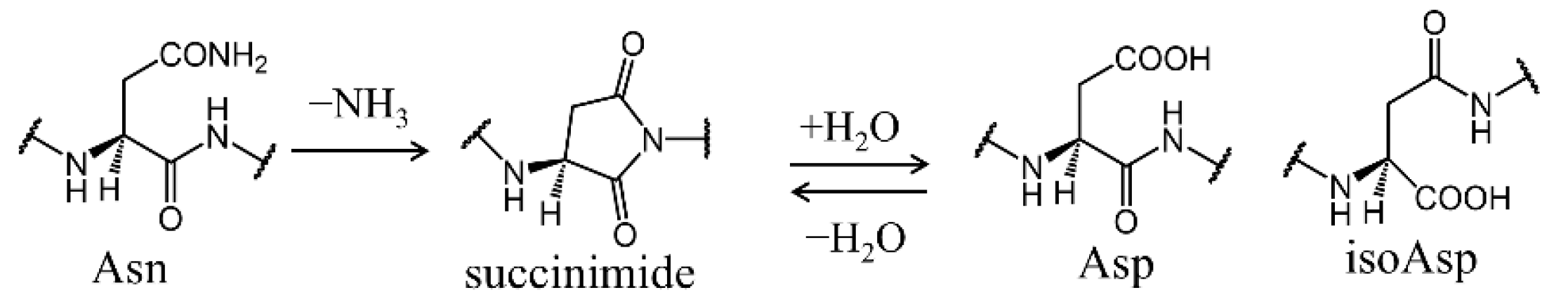
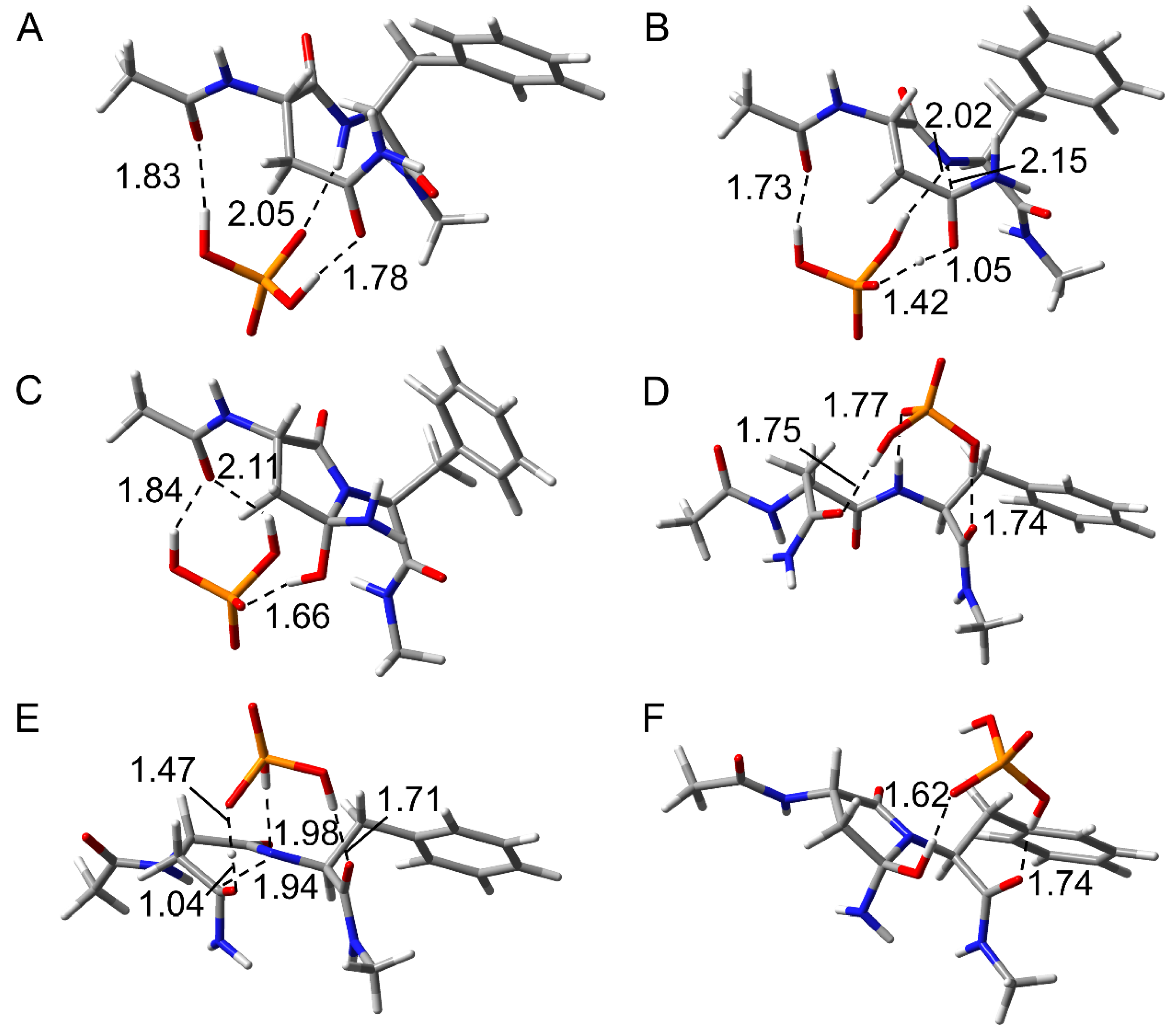
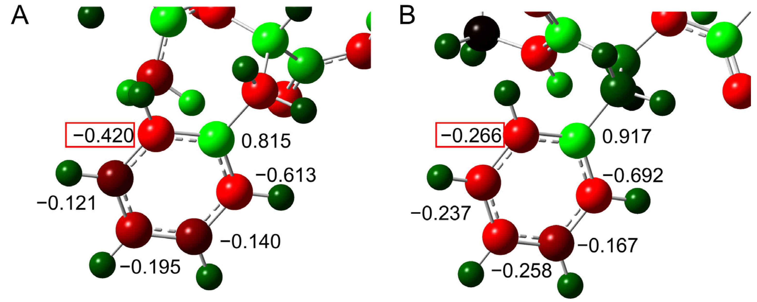
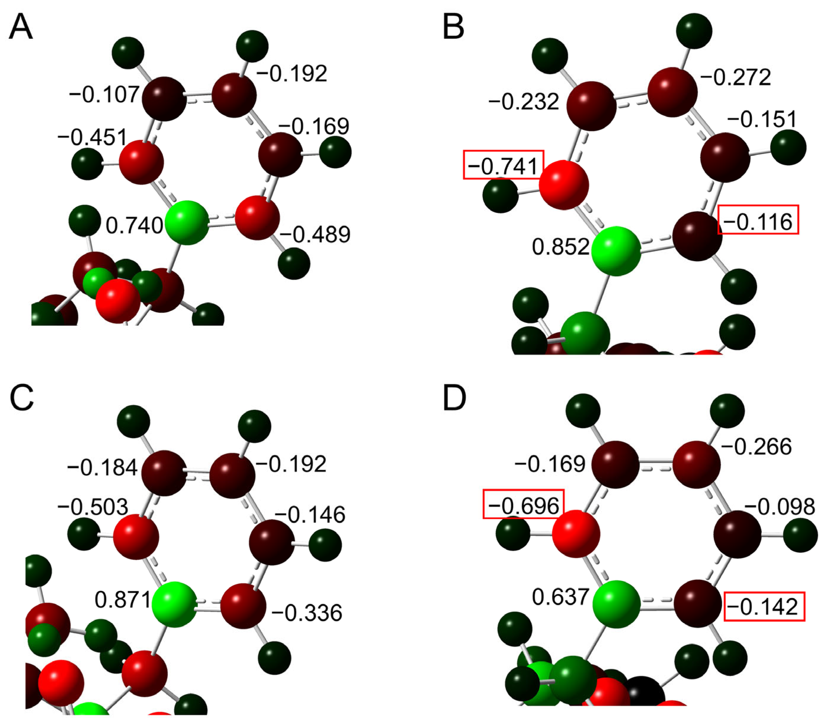
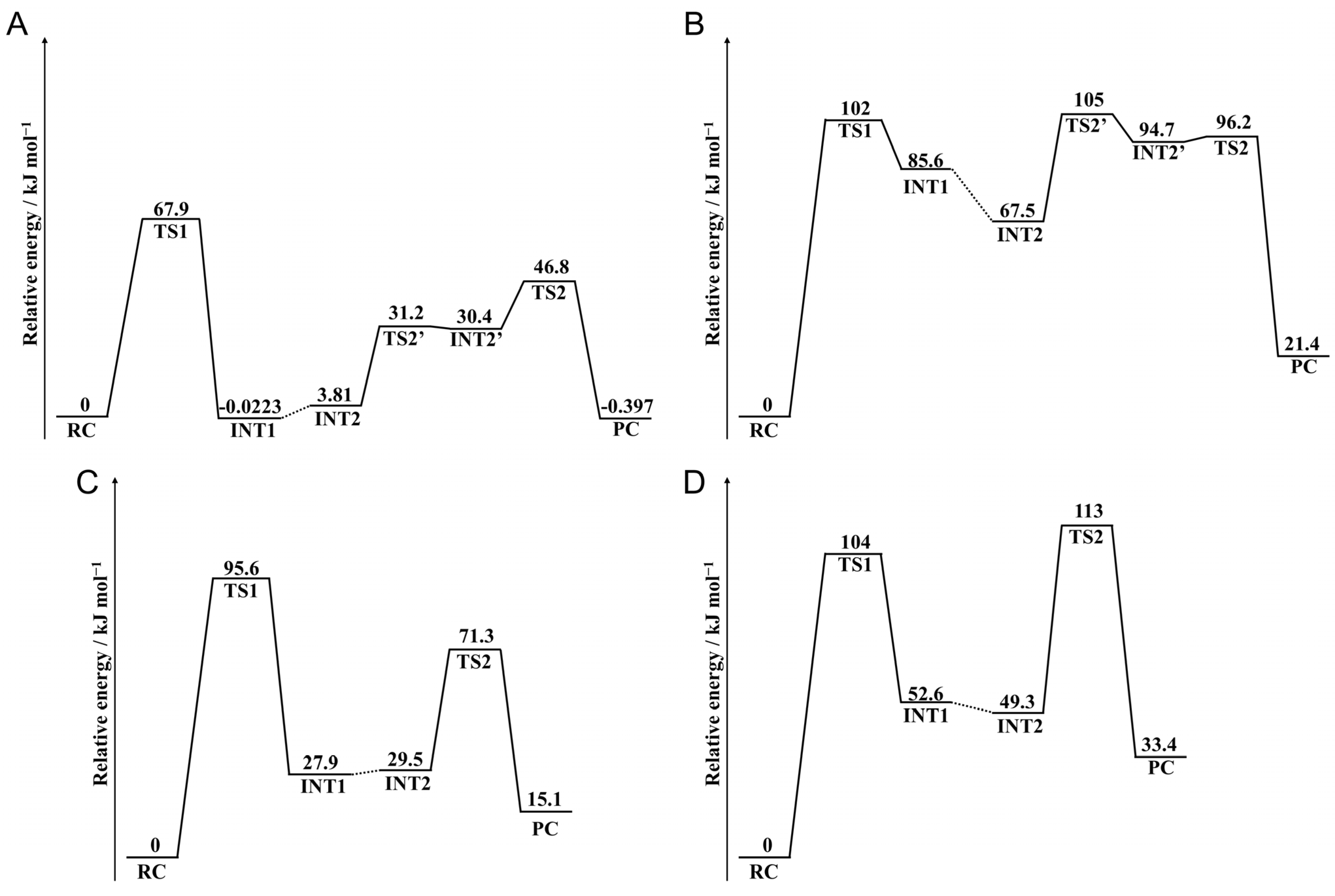
2.1. Cyclization Step

To investigate the effects of the (N + 1) Phe residue on the reaction mechanisms of Asn deamidation, the optimized geometries in the cyclization step are compared between pathways 1 and 2. The optimized geometries of reactant complex (RC), transition state 1 (TS1), and intermediate 1 (INT1) in the cyclization step for CH3CO-Asn-Phe-NHCH3 are shown in Figure 4. In the RC of both pathways, the H2PO4 ion bridged the side chain and main chain of the model compound through hydrogen bond formation (Figure 4A,D). The catalytic ion formed a hydrogen bond with the main chain oxygen on the N-terminal side in pathway 1, but formed a hydrogen bond with the main chain oxygen on the C-terminal side in pathway 2. This catalytic H2PO4 ion mediated the proton transfers involved in the cyclization step. In TS1, the distances between the main chain amide nitrogen and side chain amide carbon (C-N distance) were 2.15 and 1.94 Å in pathways 1 and 2, respectively (Figure 4B,E). This C-N distance in pathway 1 was similar to those of the previously reported phosphate-catalyzed reactions (2.20–2.23 Å) and carbonate-catalyzed reactions (2.01–2.24 Å) [30]. However, the C-N distance in pathway 2 was shorter. In the acetic acid-catalyzed reaction, the C-N distance (1.62–1.77 Å) was reported to be shorter than those of both pathways in this study [34]. The proton transfer and the nucleophilic attack by the main chain nitrogen were completed, and INT1 was formed. Three hydrogen bonds between the compound and the H2PO4 were observed in INT1 of pathway 1, while two were observed in INT1 of pathway 2 (Figure 4C,F). In pathway 1, the benzene ring of the Phe residue was maintained close to the amide nitrogen of the Asn side chain throughout the cyclization step. The distance between the closest carbon atom of the benzene ring and the hydrogen atom of the side chain amide was 2.88 Å in TS1 for pathway 1, and the distance between the centroid of the benzene ring and the hydrogen atom of the side chain amide was 3.15 Å. At this distance, the benzene ring was thought to interact with the amide moiety by N-H···π interaction in pathway 1 [35,36]. On the other hand, the benzene ring of the Phe residue remained far from the amide moiety in pathway 2.
The optimized geometries in the cyclization step for CH3CO-Asn-Ile-NHCH3 are shown in Figure S1. No differences were observed in the reaction mechanism between CH3CO-Asn-Phe-NHCH3 and CH3CO-Asn-Ile-NHCH3. In pathway 1 of CH3CO-Asn-Ile-NHCH3, the interatomic distances and number of hydrogen bonds were similar to those in CH3CO-Asn-Phe-NHCH3. There were no significant differences between each RC and TS1 in pathway 2 either. In INT1 of CH3CO-Asn-Ile-NHCH3 in pathway 2 of Ile, three hydrogen bonds were observed, which were similar to INT1 in pathway 1 of CH3CO-Asn-Phe-NHCH3 and CH3CO-Asn-Ile-NHCH3. The closest hydrogen atom of the methyl moiety and the nitrogen atom of the amide moiety were 3.01 Å in TS1 for pathway 1, which was a sufficiently interactive distance.
To elucidate the effects of the interaction of the benzene ring, the atomic charge distribution for the benzene ring was investigated (Figure 5). No significant difference was observed in the Mulliken charge for most of the carbon atoms between pathways 1 and 2. However, the charge on one carbon atom was −4.20 e in pathway 1 and −2.66 e in pathway 2. This carbon atom is closest to the side chain amide moiety, and the change in charge distribution is thought to be due to the interaction between the benzene ring and amide moiety.

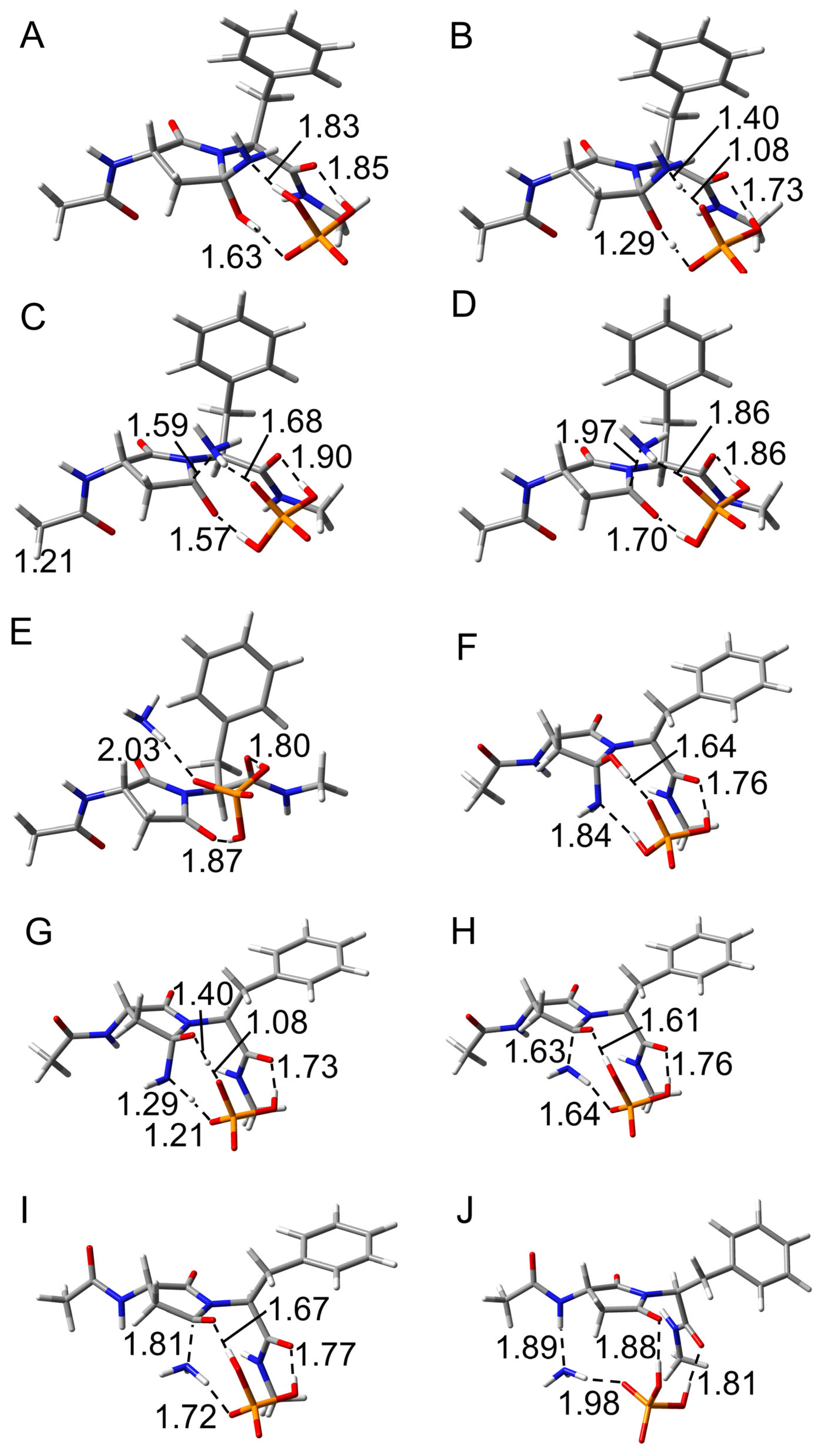
2.2. Deammoniation Step

The deammoniation step had two transition states and proceeded via the two stages in CH3CO-Asn-Phe-NHCH3. To compare the reaction mechanism in the deammoniation step between pathways 1 and 2, the optimized geometries of intermediate 2 (INT2), transition state 2 (TS2), intermediate 3 (INT3), transition state 3 (TS3), and product complex (PC) in the deammoniation step are shown in Figure 6. Although the position of the H2PO4 ion and the conformation of the gem-hydroxylamine moiety of INT2 were different from those of INT1, the changes were considered to be easily changed because these ions are abundant under physiological conditions, as H2PO4 ions are abundant in vivo. In INT2 of both pathways, the H2PO4 ion formed three hydrogen bonds with the gem-hydroxylamine moiety and the main chain oxygen of the Phe residue (Figure 6A,F). The migrating protons were located between the gem-hydroxylamine moiety and the H2PO4 ion, indicating that the proton transfer occurs in the first stage (Figure 6B,G). No differences were observed in the distance of each atom between pathways 1 and 2. The proton transfers were completed in INT3, whereas the distances between nitrogen and carbon of the gem-hydroxylamine moiety were 1.59 and 1.63 Å in pathways 1 and 2, respectively, suggesting that there is still an interaction between these atoms (Figure 6C,H). INT3 was directly connected to TS2 and TS3, and the ammonia is released in the second stage. TS3 had the longer distance between nitrogen and carbon of the gem-hydroxylamine moiety (1.97 Å and 1.81 Å in pathways 1 and 2, respectively) in comparison with INT3. In PC, the released ammonia molecule formed a hydrogen bond with the H2PO4 ion in both pathways, while the other hydrogen bond was formed with the main chain amide hydrogen of the Asn residue only in pathway 2. The H2PO4 ion formed two hydrogen bonds with the oxygen atoms of the succinimide and the main chain of the Phe residue in both pathways. In a previous study, the deammoniation step progressed in one stage, that is, simultaneous proton transfer and ammonia release [30]. The TS2 reported in the previous study was similar to TS3 of CH3CO-Asn-Phe-NHCH3 in the present study, and the proton transfers were completed in these TSs. A similar two-stage reaction was also observed in the Maillard reaction, where an amine is added to an aldehyde [37]. Since the same two-stage reaction proceeded in that reverse reaction and deammoniation step, the addition and elimination of amines are considered to likely proceed in two stages.
For CH3CO-Asn-Ile-NHCH3, the deammoniation step proceeds in one stage (Figure S2), the same as in the previous study [30]. The H2PO4 ion formed three hydrogen bonds with the gem-hydroxylamine moiety and the main chain oxygen of the Phe residue in INT2. On the other hand, the H2PO4 ion formed two hydrogen bonds with the gem-hydroxylamine moiety in INT2 of pathway 2, whereas no hydrogen bond was observed between the H2PO4 ion and the gem-hydroxylamine moiety. Proton transfers and an ammonia release occurred simultaneously, and succinimide was formed. In PC, the H2PO4 ion formed a hydrogen bond with the main chain oxygen of Phe in pathway 1. In contrast, the ammonia molecule formed a hydrogen bond with the main chain amide hydrogen of Asn in pathway 2. This hydrogen bond formation was similar to that observed for CH3CO-Asn-Phe-NHCH3. Therefore, there were no differences other than the simultaneous occurrence of proton transfer and deammoniation.
Differences in the atomic charge distribution of the benzene ring were investigated (Figure 7). In TS2, the charge of the δ-position carbon atom was evenly distributed on both sides in pathway 1, but a deviation was observed in pathway 2. Whereas the charge of that carbon atom in TS3 was deviated toward one carbon atom, the deviation was larger in pathway 2 than in pathway 1. In pathway 1, the distance between the centroid of the benzene ring and the closest hydrogen atom of the gem-hydroxylamine was 3.1 Å in TS2 and 3.3 Å in TS3, and the difference in the charge distribution was observed, suggesting that the benzene ring interacted with the amine.

2.3. Conformational Change Through the Deamidation

Dihedral angles for all geometries in CH3CO-Asn-Phe-NHCH3 were retrieved to investigate the conformational changes via the succinimide formation (Table 1 and Table 2). The dihedral angles φ and ψ characterize the main chain conformation, and χ characterizes the side chain conformation (Figure 2). In pathway 1, no significant conformational change of the main chain was observed through the succinimide formation. The largest change of φ was 5.0°, which occurred when INT1 was formed from TS1, and that of ψ was 8.0°, which occurred when TS1 was formed from RC. In contrast, a large conformational change of the main chain in pathway 2 was observed upon INT3 formation from TS2, and the change in φ was 51°. In addition, ψ was changed by 41° upon the TS1 formation from RC in pathway 2. Therefore, the deamidation in the Asn-Phe sequence in pathway 2 is considered to proceed with a large structural change of the protein, compared to the reaction in pathway 1. In a previous study, the conformational effect of the main chain on the Asn deamidation was computationally investigated, and the change of dihedral angles in syn conformation was smaller than that in anti-conformation [30]. The largest change in the φ and ψ values was observed in the TS1 formation for syn conformation, and those values were 13.3° and 31.2°, respectively. In CH3CO-Asn-Ile-NHCH3, structural changes in the main chain associated with deamidation were observed in both pathway 1 and pathway 2 (Tables S1 and S2). The largest change of ψ was 38°, which occurred when TS1 was formed from RC in pathway 1. In contrast, a large conformational change of the main chain in pathway 2 was observed upon INT2 formation from INT1, and the change in φ was 52°. The structural change in pathway 1 was larger for CH3CO-Asn-Ile-NHCH3 than for CH3CO-Asn-Phe-NHCH3. Therefore, the structural changes in the main chain for Asn deamidation in the Asn-Phe sequence are smaller than those in other sequences in pathway 1, while they are larger in pathway 2.

2.4. Barrier Heights

To evaluate the differences in activation energy barrier between pathways 1 and 2, the relative energies were calculated using the MP2/6-311+G(2d,2p)//B3LYP/6-31+G(d,p) level, and the energy profiles are shown in Figure 8. In pathway 1 of CH3CO-Asn-Phe-NHCH3, the barrier height of the cyclization step was higher than that of the deammoniation step (Figure 8A). Therefore, the cyclization step is the rate-determining step in pathway 1. In contrast, the barrier height of the cyclization and deammoniation steps was almost the same as in pathway 2 (Figure 8B). The barrier of the deammoniation step was slightly higher than that of the cyclization step, suggesting that the rate-determining step of pathway 2 was the deammoniation step. However, these barrier heights of pathway 2 were too high for the reaction progress under physiological conditions. A previous study has also reported that the barrier height of the deammoniation step in Gln deamidation with no catalyst or water as a catalyst is higher than or similar to that of the cyclization step, but this alters depending on the catalyst species [38]. The results of this study suggest that the rate-limiting step is also affected by conformation. In pathway 1, the barrier height of the succinimide formation was 67.9 kJ mol−1, which was lower than any of the previously reported values by computational studies. The calculated activation barriers of Asn deamidation were 82.7, 84.5, and 113 kJ mol−1 [23,30,36]. In Ref. [23], Asn deamidation was investigated in the Asn-His sequence. The adjacent His residue acted as a catalyst, whereas the activation barrier was higher than that in the Asn-Phe sequence in this study. In CH3CO-Asn-Ile-NHCH3, the rate-determining steps were cyclization and deammoniation steps in pathways 1 and 2, respectively. The barrier heights of both steps in pathway 2 were similar to those for CH3CO-Asn-Phe-NHCH3. However, the barrier height in pathway 1 was significantly higher than that for CH3CO-Asn-Phe-NHCH3. Therefore, the decrease in pathway 1 was observed in only CH3CO-Asn-Phe-NHCH3.
The single-point energies were also calculated using the M06-2X/6-31+G(d,p) level, and the obtained relative energies were similar to those with the MP2/6-311+G(2d,2p)//B3LYP/6-31+G(d,p) (Tables S33, S34, S37, and S38). In CH3CO-Asn-Phe-NHCH3, the barrier heights in the energies obtained using M06-2X/6-31+G(d,p) were 68.0 and 101 kJ mol−1 in pathways 1 and 2, respectively. In CH3CO-Asn-Phe-NHCH3, the barrier heights in the energies obtained using M06-2X/6-31+G(d,p) were 92.2 and 121 kJ mol−1 in pathways 1 and 2, respectively. The interaction of the benzene ring of the phenylalanine side chain was thought to reduce the barrier height of Asn deamination. The barrier height for Asn deamidation experimentally investigated using peptides is 80–100 kJ mol−1 [14,39]. The calculated values in this study were lower than these. This is thought to be because peptides do not form conformations like the model structure in pathway 1. The barrier height obtained in this study is likely to be applicable only when a similar conformation is formed in protein structures.

3. Materials and Methods

The initial structure of pathway 1 for both peptides was constructed based on the 3D structure of Asn14 and Asn53 of γS-crystallin registered in the protein data bank (PDB, PDB ID: 2M3T) [40], and that of pathway 2 was constructed based on the anti-conformation in a previous study [30]. All calculations were performed using Gaussian 16 software [41]. Optimization of energy minima and TS geometries was performed without any constraints by density functional theory (DFT) calculations using the B3LYP/6-31+G(d,p) level of theory [42]. Intrinsic reaction coordinate (IRC) calculations were performed from TSs, followed by full geometry optimizations, to confirm that each TS is connected to energy-minimum geometries. The relative energies reported include the zero-point energies calculated at B3LYP/6-31+G(d,p) level. Furthermore, the single-point energies of all optimized geometries were calculated by the MP2/6-311+G(2d,2p) and M06-2X/6-31+G(d,p) levels. All calculations included hydration effects by employing the polarizable continuum model (PCM). To evaluate the structural changes associated with the reaction, the dihedral angles φ (C–N–Cα–C) and ψ (N–Cα–C–N), which characterize the main chain conformation, and χ (N–Cα–Cβ–Cγ), which characterizes the side chain conformation, were retrieved.

4. Conclusions

We investigated the reaction mechanism and the activation barrier of the succinimide formation in the Asn-Phe and Asn-Ile sequences using quantum chemical calculations to elucidate the effects of the (N + 1) Phe residue on the Asn deamidation. Our calculation results indicated that the (N + 1) Phe residue affected the barrier height of the cyclization step and the reaction mechanism of the deammoniation step. In contrast, although the (N + 1) Ile residue was located near the amide moiety of TS1 in pathway 1, the barrier height of pathway 1 was not significantly lower than that of pathway 2. Our calculation results suggest that the (N + 1) Phe residue interacts with the amide moiety of the Asn side chain and reduces the barrier height of deamidation. The barrier height for CH3CO-Asn-Phe-NHCH3 in pathway 1 was lower than the previously reported values determined by experimental methods using peptides. However, the experimental data for peptides were inconsistent with the experimental reports for deamidation in proteins. The bulkiness of (N + 1) Phe residue is considered to impair Asn deamidation in peptides, and conformations such as the model structure in pathway 1 are hardly formed in peptides. In contrast, the 3D structure formation in proteins allows the Asn-Phe residues to form conformations such as the model structure in pathway 1, reducing the activation barrier of Asn deamidation. In an experimental structure (PDB ID: 2M3T), the aromatic ring of (N + 1) Phe and Tyr residues are oriented toward the Asn residues (Asn14, Asn54, or Asn143), suggesting that the conformation of these Asn and (N + 1) residues is similar to the model compound in pathway 1 (Figure 9). These conformations were the same for the other crystal structure (PDB ID: 6FD8) [43]. Therefore, the results of this study can explain the higher deamidation rate for Asn with Phe as the (N + 1) residue. In addition, the importance of analyzing deamidation while considering protein structure was demonstrated. In the future study, the effects of other (N + 1) residues on Asn deamidation should be analyzed in detail. Because the effects of D3 correction on the barrier heights are unclear [44], accumulation of the results obtained by applying D3 correction is important to advance computational research, including aromatic amino acids. Reaction calculations using HPO42− as a catalyst have been reported for the stereo-inversion of succinimide [45]. The differences in reaction mechanisms and activation barriers due to the catalysts should also be the focus of future research. In addition, the hydration effect using explicit water molecules should also be considered for future study.

Supplementary Materials

The following supporting information can be downloaded at: https://www.mdpi.com/article/10.3390/ijms26146819/s1.

Author Contributions

Conceptualization, K.K.; methodology, K.K.; software, K.K. and Y.I.; validation, T.N. and A.M.; formal analysis, K.K.; investigation, K.K. and H.A.; resources, K.K. and Y.I.; data curation, K.K. and H.A.; writing—original draft preparation, K.K.; writing—review and editing, H.A., T.N., A.O. and Y.I.; visualization, K.K. and H.A.; supervision, K.K.; project administration, K.K.; funding acquisition, K.K. and Y.I. All authors have read and agreed to the published version of the manuscript.

Funding

This work was supported by Grants-in-Aid for Scientific Research (21K15244) from the Japan Society for the Promotion of Science.

Institutional Review Board Statement

Not applicable.

Informed Consent Statement

Not applicable.

Data Availability Statement

Data is contained within the article or the Supplementary Materials (Figures S1 and S2, Tables S1–S38). The original contributions presented in this study are included in the article/Supplementary Materials. Further inquiries can be directed to the corresponding author.

Conflicts of Interest

The authors declare no conflicts of interest.

Abbreviations

The following abbreviations are used in this manuscript:
RCReactant complex
TSTransition state
INTIntermediate
PCProduct complex

References

  1. Lubsen, N.H.; Slingsby, C.; Tardieu, A. Ageing and Vision: Structure, Stability and Function of Lens Crystallins. Prog. Biophys. Mol. Biol. 2004, 86, 407–485. [Google Scholar] [CrossRef]
  2. Driessen, H.P.; Herbrink, P.; Bloemendal, H.; de Jong, W.W. Primary Structure of the Bovine β-crystallin Bp Chain. Internal Duplication and Homology with γ-Crystallin. Eur. J. Biochem. 1981, 121, 83–91. [Google Scholar] [CrossRef] [PubMed]
  3. Hogg, D.; Tsui, L.C.; Gorin, M.; Breitman, M.L. Characterization of the Human β-crystallin Gene HuβA3/A1 Reveals Ancestral Relationships among the βγ-crystallin Superfamily. J. Biol. Chem. 1986, 261, 12420–12427. [Google Scholar] [CrossRef] [PubMed]
  4. Garlick, R.L.; Mazer, J.S.; Chylack, L.T., Jr.; Tung, W.H.; Bunn, H.F. Nonenzymatic glycation of human lens crystallin. Effect of aging and diabetes mellitus. J. Clin. Investig. 1984, 74, 1742–1749. [Google Scholar] [CrossRef] [PubMed]
  5. Slingsby, C.; Clout, N.J. Structure of the Crystallins. Eye 1999, 13, 395–402. [Google Scholar] [CrossRef]
  6. Moreau, K.L.; King, J.A. Protein misfolding and aggregation in cataract disease and prospects for prevention. Trends Mol. Med. 2012, 18, 273–282. [Google Scholar] [CrossRef]
  7. Loh, D.; Reiter, R.J. Melatonin, ATP, and Cataracts: The Two Faces of Crystallin Phase Separation. Qeios 2024, D09YND. [Google Scholar] [CrossRef]
  8. Robinson, N.E.; Robinson, A.B. Molecular clocks. Proc. Natl. Acad. Sci. USA 2001, 98, 944–949. [Google Scholar] [CrossRef]
  9. Midelfort, C.F.; Mehler, A.H. Deamidation in vivo of an asparagine residue of rabbit muscle aldolase. Proc. Natl. Acad. Sci. USA 1972, 69, 1816–1819. [Google Scholar] [CrossRef]
  10. Solstad, T.; Flatmark, T. Microheterogeneity of recombinant human phenylalanine hydroxylase as a result of nonenzymatic deamidations of labile amide containing amino acids. Effects on catalytic and stability properties. Eur. J. Biochem. 2000, 267, 6302–6310. [Google Scholar] [CrossRef]
  11. Capasso, S.; Salvadori, S. Effect of the three-dimensional structure on the deamidation reaction of ribonuclease A. J. Pept. Res. 1999, 54, 377–382. [Google Scholar] [CrossRef] [PubMed]
  12. Deverman, B.E.; Cook, B.L.; Manson, S.R.; Niederhoff, R.A.; Langer, E.M.; Rosová, I.; Kulans, L.A.; Fu, X.; Weinberg, J.S.; Heinecke, J.W.; et al. Bcl-xL deamidation is a critical switch in the regulation of the response to DNA damage. Cell 2002, 111, 51–62. [Google Scholar] [CrossRef] [PubMed]
  13. Nowak, C.; Tiwari, A.; Liu, H. Asparagine deamidation in a complementarity determining region of a recombinant monoclonal antibody in complex with antigen. Anal. Chem. 2018, 90, 6998–7003. [Google Scholar] [CrossRef] [PubMed]
  14. Patel, K.; Borchardt, R.T. Chemical pathways of peptide degradation. II. Kinetics of deamidation of an asparaginyl residue in a model hexapeptide. Pharm. Res. 1990, 7, 703–711. [Google Scholar] [CrossRef]
  15. Kaji, Y.; Oshika, T.; Takazawa, Y.; Fukayama, M.; Fujii, N. Accumulation of D-beta-aspartic acid-containing proteins in age-related ocular diseases. Chem. Biodivers. 2010, 7, 1364–1370. [Google Scholar] [CrossRef]
  16. Palmer, W.G.; Papaconstantinou, J. Aging of alpha-crystallins during development of the lens. Proc. Natl. Acad. Sci. USA 1969, 64, 404–410. [Google Scholar] [CrossRef]
  17. Takemoto, L.; Boyle, D. Deamidation of specific glutamine residues from alpha-A crystallin during aging of the human lens. Biochemistry 1998, 37, 13681–13685. [Google Scholar] [CrossRef]
  18. Lampi, K.J.; Wilmarth, P.A.; Murray, M.R.; David, L.L. Lens β-crystallins: The role of deamidation and related modifications in aging and cataract. Prog. Biophys. Mol. Biol. 2014, 115, 21–31. [Google Scholar] [CrossRef]
  19. Nilsson, M.R.; Driscoll, M.; Raleigh, D.P. Low levels of asparagine deamidation can have a dramatic effect on aggregation of amyloidogenic peptides: Implications for the study of amyloid formation. Protein Sci. 2002, 11, 342–349. [Google Scholar] [CrossRef]
  20. Capasso, S. Estimation of the deamidation rate of asparagine side chains. J. Pept. Res. 2000, 55, 224–229. [Google Scholar] [CrossRef]
  21. Robinson, N.E.; Robinson, Z.W.; Robinson, B.R.; Robinson, A.L.; Robinson, J.A.; Robinson, M.L.; Robinson, A.B. Structure-dependent nonenzymatic deamidation of glutaminyl and asparaginyl pentapeptides. J. Pept. Res. 2004, 63, 426–436. [Google Scholar] [CrossRef] [PubMed]
  22. Phillips, J.J.; Buchanan, A.; Andrews, J.; Chodorge, M.; Sridharan, S.; Mitchell, L.; Burmeister, N.; Kippen, A.D.; Vaughan, T.J.; Higazi, D.R.; et al. Rate of Asparagine Deamidation in a Monoclonal Antibody Correlating with Hydrogen Exchange Rate at Adjacent Downstream Residues. Anal. Chem. 2017, 89, 2361–2368. [Google Scholar] [CrossRef] [PubMed]
  23. Takahashi, O.; Manabe, N.; Kirikoshi, R. A Computational Study of the Mechanism of Succinimide Formation in the Asn-His Sequence: Intramolecular Catalysis by the His Side Chain. Molecules 2016, 21, 327. [Google Scholar] [CrossRef] [PubMed]
  24. Takemoto, L. Deamidation of Asn-143 of gamma S crystallin from protein aggregates of the human lens. Curr. Eye Res. 2001, 22, 148–153. [Google Scholar] [CrossRef]
  25. Lapko, V.N.; Purkiss, A.G.; Smith, D.L.; Smith, J.B. Deamidation in human gamma S-crystallin from cataractous lenses is influenced by surface exposure. Biochemistry 2002, 41, 8638–8648. [Google Scholar] [CrossRef]
  26. Ray, N.J.; Hall, D.; Carver, J.A. Deamidation of N76 in human γS-crystallin promotes dimer formation. Biochim. Biophys. Acta 2016, 1860, 315–324. [Google Scholar] [CrossRef]
  27. Pande, A.; Mokhor, N.; Pande, J. Deamidation of Human γS-Crystallin Increases Attractive Protein Interactions: Implications for Cataract. Biochemistry 2015, 54, 4890–4899. [Google Scholar] [CrossRef]
  28. Hains, P.G.; Truscott, R.J. Age-dependent deamidation of lifelong proteins in the human lens. Investig. Ophthalmol. Vis. Sci. 2010, 51, 3107–3114. [Google Scholar] [CrossRef]
  29. Wilmarth, P.A.; Tanner, S.; Dasari, S.; Nagalla, S.R.; Riviere, M.A.; Bafna, V.; Pevzner, P.A.; David, L.L. Age-related changes in human crystallins determined from comparative analysis of post-translational modifications in young and aged lens: Does deamidation contribute to crystallin insolubility? J. Proteome Res. 2006, 5, 2554–2566. [Google Scholar] [CrossRef]
  30. Kato, K.; Nakayoshi, T.; Kurimoto, E.; Oda, A. Mechanisms of Deamidation of Asparagine Residues and Effects of Main-Chain Conformation on Activation Energy. Int. J. Mol. Sci. 2020, 21, 7035. [Google Scholar] [CrossRef]
  31. Kirikoshi, R.; Manabe, N.; Takahashi, O. Phosphate-Catalyzed Succinimide Formation from Asp Residues: A Computational Study of the Mechanism. Int. J. Mol. Sci. 2018, 19, 637. [Google Scholar] [CrossRef] [PubMed]
  32. Kato, K.; Nakayoshi, T.; Ishikawa, Y.; Kurimoto, E.; Oda, A. Computational Analysis of the Mechanism of Nonenzymatic Peptide Bond Cleavage at the C-Terminal Side of an Asparagine Residue. ACS Omega 2021, 6, 30078–30084. [Google Scholar] [CrossRef] [PubMed]
  33. Nakayoshi, T.; Kato, K.; Kurimoto, E.; Oda, A. Computational Studies on the Mechanisms of Nonenzymatic Intramolecular Cyclization of the Glutamine Residues Located at N-Termini Catalyzed by Inorganic Phosphate Species. ACS Omega 2020, 5, 9162–9170. [Google Scholar] [CrossRef] [PubMed]
  34. Nakayoshi, T.; Wanita, K.; Kato, K.; Kurimoto, E.; Oda, A. Computational analysis of nonenzymatic deamidation of asparagine residues catalysed by acetic acid. Mol. Phys. 2021, 119, e1827176. [Google Scholar] [CrossRef]
  35. Banerjee, A.; Saha, A.; Saha, B.K. Understanding the Behavior of π–π Interactions in Crystal Structures in Light of Geometry Corrected Statistical Analysis: Similarities and Differences with the Theoretical Models. Cryst. Growth Des. 2019, 19, 2245–2252. [Google Scholar] [CrossRef]
  36. Ciunik, Z.; Desiraju, G.R. Area correction of multi-atom–acceptor hydrogen bond frequency distributions. Chem. Commun. 2001, 703–704. [Google Scholar] [CrossRef]
  37. Kato, K.; Nakayoshi, T.; Shinohara, Y.; Kurimoto, E.; Oda, A.; Ishikawa, Y. Theoretical Studies on the Reaction Mechanism of Schiff Base Formation from Hexoses. J. Phys. Chem. B 2024, 128, 4952–4958. [Google Scholar] [CrossRef]
  38. Kato, K.; Nakayoshi, T.; Kurimoto, E.; Oda, A. Computational Studies on the Nonenzymatic Deamidation Mechanisms of Glutamine Residues. ACS Omega 2019, 4, 3508–3513. [Google Scholar] [CrossRef]
  39. Connolly, B.D.; Tran, B.; Moore, J.M.; Sharma, V.K.; Kosky, A. Specific catalysis of asparaginyl deamidation by carboxylic acids: Kinetic, thermodynamic, and quantitative structure-property relationship analyses. Mol. Pharm. 2014, 11, 1345–1358. [Google Scholar] [CrossRef]
  40. Kingsley, C.N.; Brubaker, W.D.; Markovic, S.; Diehl, A.; Brindley, A.J.; Oschkinat, H.; Martin, R.W. Preferential and specific binding of human αB-crystallin to a cataract-related variant of γS-crystallin. Structure 2013, 21, 2221–2227. [Google Scholar] [CrossRef]
  41. Frisch, M.J.; Trucks, G.W.; Schlegel, H.B.; Scuseria, G.E.; Robb, M.A.; Cheeseman, J.R.; Scalmani, G.; Barone, V.; Petersson, G.A.; Nakatsuji, H.; et al. Gaussian 16; Revision A.03; Gaussian, Inc.: Wallingford, CT, USA, 2016. [Google Scholar]
  42. Lee, C.; Yang, W.; Parr, R.G. Development of the Colle-Salvetti correlation-energy formula into a functional of the electron density. Phys. Rev. B 1988, 37, 785–789. [Google Scholar] [CrossRef]
  43. Thorn, D.C.; Grosas, A.B.; Mabbitt, P.D.; Thorn, D.C.; Grosas, A.B.; Mabbitt, P.D.; Ray, N.J.; Jackson, C.J.; Carver, J.A. The Structure and Stability of the Disulfide-Linked γS-Crystallin Dimer Provide Insight into Oxidation Products Associated with Lens Cataract Formation. J. Mol. Biol. 2019, 431, 483–497. [Google Scholar] [CrossRef]
  44. Grimme, S.; Antony, J.; Ehrlich, S.; Krieg, H. A consistent and accurate ab initio parametrization of density functional dispersion correction (DFT-D) for the 94 elements H-Pu. J. Chem. Phys. 2010, 132, 154104. [Google Scholar] [CrossRef]
  45. Takahashi, O. A Computational DFT Study of the Stereoinversion of Succinimide Residues Formed in Proteins and Peptides Catalyzed by a Hydrogen Phosphate Ion: An Unsymmetrical SE1 Mechanism. Symmetry 2024, 16, 1369. [Google Scholar] [CrossRef]
Figure 1. Deamidation pathway of an asparagine residue.
Ijms 26 06819 g001
Figure 2. The structures of the model compounds. (A) CH3CO-Asn-Phe-NHCH3 and (B) CH3CO-Asn-Ile-NHCH3. The dihedral angles φ (C–N–Cα–C) and ψ (N–Cα–C–N), which characterize the main chain conformation, and χ (N–Cα–Cβ–Cγ), which characterizes the side chain conformation.
Ijms 26 06819 g002
Figure 3. Reaction pathway of the succinimide formation through a gem-hydroxylamine intermediate.
Ijms 26 06819 g003
Figure 4. Optimized geometries in the cyclization step for CH3CO-Asn-Phe-NHCH3. (A) RC, (B) TS1, and (C) INT1 for pathway 1, and (D) RC, (E) TS1, and (F) INT1 for pathway 2 were shown. Carbon, oxygen, nitrogen, phosphorus, and hydrogen atoms were illustrated in gray, red, blue, orange, and white, respectively. Selected interatomic distances are in units of Å.
Ijms 26 06819 g004
Figure 5. The charges in Coulomb units of carbon atoms in the benzene ring of phenylalanine were calculated by Mulliken methods (/e) with the MP2/6-311+G(2d,2p)//B3LYP/6-31+G(d,p) level. The optimized geometries of TS1 of (A) pathway 1 and (B) pathway 2 are shown. Relatively negatively charged atoms are shown in red, and relatively positively charged atoms are green. The red boxes indicate the values of δ positions with large differences in the charges.
Ijms 26 06819 g005
Figure 6. Optimized geometries in the cyclization step for CH3CO-Asn-Phe-NHCH3. (A) INT2, (B) TS2, (C) INT3, (D) TS3, and (E) PC for pathway 1, and (F) INT2, (G) TS2, (H) INT3, (I) TS3, and (J) PC for pathway 2 were shown. Carbon, oxygen, nitrogen, phosphorus, and hydrogen atoms were illustrated in gray, red, blue, orange, and white, respectively. Selected interatomic distances are in units of Å.
Ijms 26 06819 g006
Figure 7. The charges in Coulomb units of carbon atoms in the benzene ring of phenylalanine are calculated by Mulliken methods (/e) with the MP2/6-311+G(2d,2p)//B3LYP/6-31+G(d,p) level. The optimized geometries of (A) TS2 in pathway 1, (B) TS2 in pathway 2, (C) TS3 in pathway 1, and (D) TS3 in pathway 2 are shown. Relatively negatively charged atoms are shown in red, and relatively positively charged atoms are green. The red boxes indicated the values of δ positions with large differences in the charges.
Ijms 26 06819 g007
Figure 8. Energy diagrams for the succinimide formation calculated using the MP2/6-311+G(2d,2p)//B3LYP/6-31+G(d,p) level. Entire energy profiles of (A) pathway1 and (B) pathway 2 in CH3CO-Asn-Phe-NHCH3 and those of (C) pathway1 and (D) pathway 2 in CH3CO-Asn-Ile-NHCH3 are shown.
Ijms 26 06819 g008
Figure 9. Conformation of the (N + 1) aromatic amino acid residues in γS-crystallin. The structures around (A) Asn14, (B) Asn54, and (C) Asn143 were retrieved from the experimentally determined structure (PDB ID: 2M3T). Carbon, nitrogen, oxygen, and hydrogen atoms are shown in cyan, blue, red, and white, respectively.
Ijms 26 06819 g009
Table 1. Dihedral angles of the optimized geometries in pathway 1.
Dihedral Angle
φψχ
RC61.8−139−166
TS157.6−147166
INT152.6−144147
INT255.8−146142
TS2′55.1−144140
INT355.6−144142
TS256.1−146144
PC56.3−142136
φ: C–N–Cα–C, ψ: N–Cα–C–N, and χ: N–Cα–Cβ–Cγ.
Table 2. Dihedral angles of the optimized geometries in pathway 2.
Dihedral Angle
φψχ
RC−155−17971.8
TS1−168−13898.0
INT1−166−132109
INT2−162−11298.0
TS2′−167−10894.7
INT3−116−127118
TS2−117−127118
PC−108−121117
φ: C–N–Cα–C, ψ: N–Cα–C–N, and χ: N–Cα–Cβ–Cγ.
Disclaimer/Publisher’s Note: The statements, opinions and data contained in all publications are solely those of the individual author(s) and contributor(s) and not of MDPI and/or the editor(s). MDPI and/or the editor(s) disclaim responsibility for any injury to people or property resulting from any ideas, methods, instructions or products referred to in the content.

