Article

Conserved Yet Divergent Smc5/6 Complex Degradation by Mammalian Hepatitis B Virus X Proteins

1
Department of Veterinary Science, Faculty of Agriculture, University of Miyazaki, Miyazaki, Miyazaki 889-2192, Japan
2
Graduate School of Medicine and Veterinary Medicine, University of Miyazaki, Miyazaki, Miyazaki 889-1692, Japan
3
Center for Animal Disease Control, University of Miyazaki, Miyazaki, Miyazaki 889-2192, Japan
*
Author to whom correspondence should be addressed.
These authors contributed equally to this work.
Int. J. Mol. Sci. 2025, 26(14), 6786; https://doi.org/10.3390/ijms26146786
Submission received: 16 May 2025 / Revised: 25 June 2025 / Accepted: 12 July 2025 / Published: 15 July 2025
(This article belongs to the Collection State-of-the-Art Macromolecules in Japan)

Abstract

Hepatitis B virus (HBV), belonging to the genus Orthohepadnavirus, can cause chronic hepatitis and hepatocarcinoma in humans. HBV ensures optimal replication by encoding X, a multifunctional protein responsible for degrading the structural maintenance of chromosomes (Smc) 5/6 complex, an anti-HBV factor in hepatocytes. Previous studies suggest that degradation of the Smc5/6 complex is conserved among viruses from the genus Orthohepadnavirus. Recently, a novel hepadnavirus in cats, domestic cat HBV (DCHBV), has been identified as genetically close to HBV. However, it remains unclear whether the DCHBV X protein possesses similar Smc5/6 complex-degrading properties. Here, we investigated the degradation of the Smc5/6 complex by X proteins from viruses of the genus Orthohepadnavirus, including DCHBV, in cells derived from primates and cats. We found that the DCHBV X protein degraded the Smc5/6 complex in the cells of several host species, and the degree of its anti-Smc5/6 complex activity differed depending on the host species. Furthermore, the DCHBV X protein degraded Smc6 independently of DNA-binding protein 1 (DDB1), which is a critical host factor for HBV X-mediated Smc6 degradation. Our findings highlight the conserved yet divergent degradation machinery for Smc6 of mammalian hepatitis B virus X proteins.

1. Introduction

Hepatitis B virus (HBV), of the family Hepadnaviridae and genus Orthohepadnavirus, is an enveloped virus with a partially double-stranded, circular DNA genome. Approximately 250 million individuals are chronically infected with HBV worldwide, and chronic HBV infections can cause cirrhosis and hepatocellular carcinoma [1]. Several therapeutic agents, including nucleoside analogs and interferon-α (IFN-α), have been approved to treat HBV infection; however, curing the infection, i.e., eliminating the virus from an individual, remains challenging. Therefore, there has been much research on developing antiviral therapies to achieve a cure. In addition, there is a need to identify new animal models for studying HBV infection and to study the interaction between host defense mechanisms and HBV. Therefore, it is important to elucidate the molecular interactions between hosts and viruses.
The viruses belonging to the genus Orthohepadnavirus encode four viral proteins: polymerase (P) protein, surface (S) protein, core (C) protein, and X protein [2]. While S and C proteins are structural proteins, the P protein is responsible for viral genome replication [3]. Meanwhile, the HBV X protein is a versatile regulator that influences host cellular processes and activates multiple transcriptional factors, including AP-1, AP-2, NF-κB, and the cAMP response element [4], playing a critical role in HBV replication and pathogenesis [5,6]. The HBV X protein comprises four key regions that are essential for transactivation, dimerization, p53 interaction, and binding to the 14-3-3 protein motif [7]. Notably, HBV X protein antagonizes the inhibitory effect of the structural maintenance of chromosomes (Smc) 5/6 complex on HBV in infected cells. The Smc5/6 complex, along with cohesin and condensin, constitutes one of the three major structural maintenance of chromosomes (Smc) complexes in eukaryotic cells. Similar to other Smc complexes, its core structure is composed of a heterodimer formed by two Smc proteins, Smc5 and Smc6, which interact with four additional proteins known as non-Smc elements (Nsmce1–4). These Smc complexes are critical for essential cellular processes, including chromosome replication, segregation, and DNA repair. To antagonize the inhibitory effect of the Smc5/6 complex, HBV X protein acts by degrading the Smc5/6 complex via interaction with DNA-binding protein 1 (DDB1)–Cul4 ubiquitin ligase machinery [8,9,10]. Moreover, the genus Orthohepadnavirus also includes HBV-like viruses that infect mammals, such as rodents, bats, and primates, and the function of X from viruses in the genus Orthohepadnavirus is conserved in primates, bats, and rodents [11].
However, it is unclear if the function of X is conserved in HBVs found in other animals. For example, an HBV-like virus, domestic cat hepatitis B virus (DCHBV), was identified in domestic cats in 2018. DCHBV infection displayed the signature of chronic hepatitis, suggesting an association between them [12,13]. DCHBV has been identified in several countries and regions, including Australia [12], Italy [14], the UK [15], the US, Malaysia [16], Thailand [17], Taiwan [18], Türkiye [19], Japan [20,21], Hong Kong [22], Chile [23], and Brazil [24]. Furthermore, we have recently demonstrated that DCHBV shares with HBV the sodium/bile acid cotransporter NTCP as an entry receptor [25]. This finding suggests that DCHBV infection in cats can be a promising animal model for studying HBV infection. However, whether the function of the DCHBV X protein is conserved in carnivores remains to be elucidated.
In this study, we analyzed the degradation of the Smc5/6 complex by mammalian hepatitis B virus X proteins in primate and feline cells. While western blotting is used to study Smc5/6 complex degradation by mammalian hepatitis B virus X proteins, this assay has a relatively low throughput. Thus, we improved the assay’s throughput by developing a new assay using a split-type red fluorescent protein to measure Smc6 degradation by mammalian hepatitis B virus X proteins quantitatively. We observed that while the human-derived HBV(A) X protein showed conserved Smc6 degradation activity in primate and feline cells, the DCHBV X protein displayed weaker Smc6 degradation activity in the cells of African green monkeys. Furthermore, we found that the DCHBV(KT116) X protein degraded Smc6 via a different mechanism than the HBV(A) X protein.
Our findings suggest that the X proteins in the viruses in the genus Orthohepadnavirus may have evolved to have conserved but differential Smc5/6 complex degradation activities, depending on the combination of viruses and host species. Collectively, our findings highlight the evolutionary traits of mammalian hepatitis B virus X proteins, which have arisen as a consequence of the arms race between hosts and viruses.

2. Results

2.1. Genetic Similarity Between Mammalian Hepatitis B Virus X Proteins

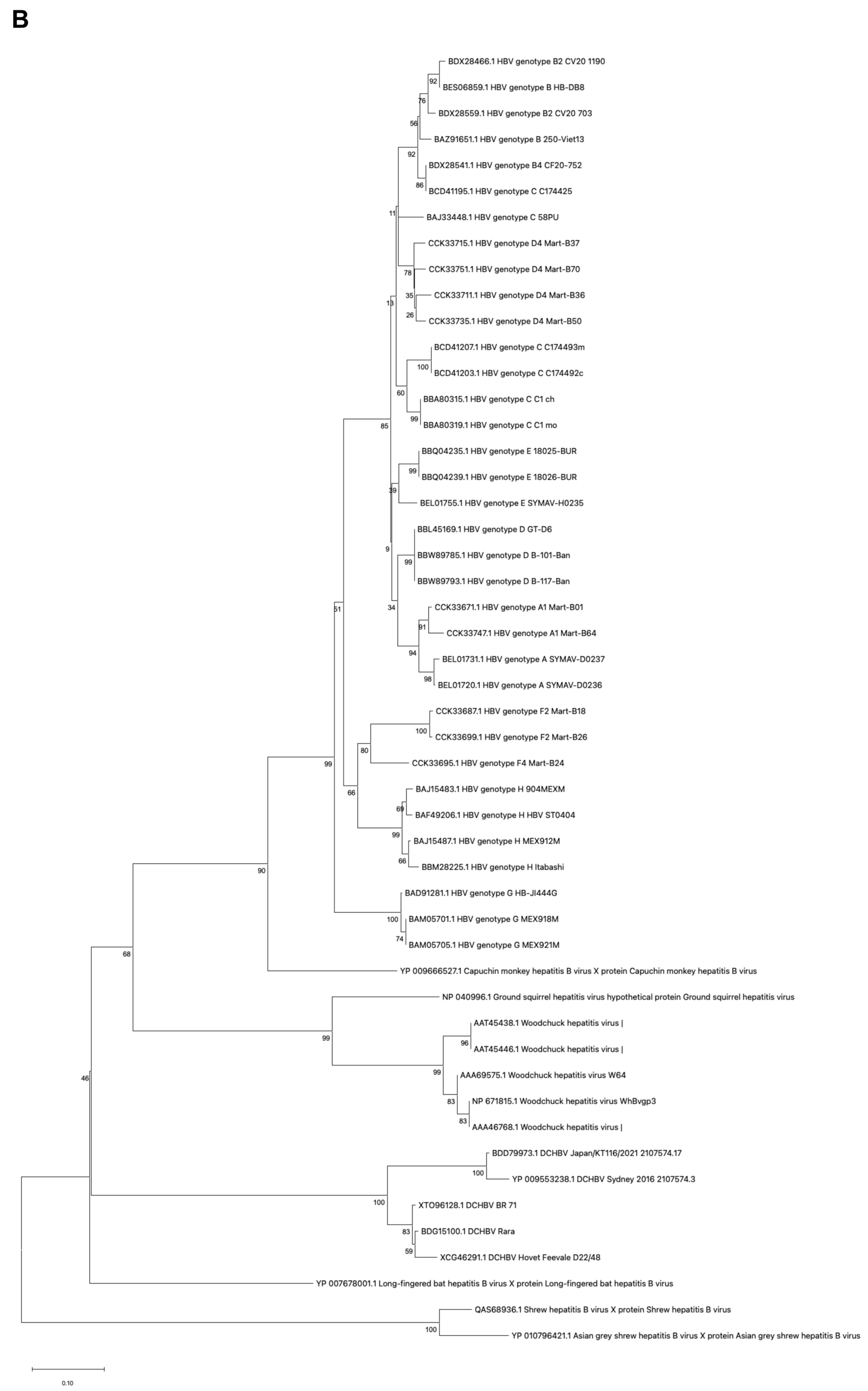
First, we analyzed the genetic diversity of mammalian hepatitis B virus X proteins by aligning the sequences of the X proteins of nine strains of human HBV, five strains of DCHBV, five species of bat hepadnavirus, one strain of woodchuck hepatitis virus, and 12 virus strains belonging to the genus Orthohepadnavirus (Figure 1A). Interestingly, DCHBV clustered independently but closely with domestic donkey HBV and bat HBV. Additionally, DCHBV exhibited distinct clustering patterns compared with primate HBV (Figure 1B). The phylogenetic tree revealed that, although several branches exhibited bootstrap values below 70%, the overall topology was consistent with host species-specific clustering.

2.2. Similarities Between Smc6 Proteins Among Mammals

Next, we analyzed the similarities between Smc6 proteins in humans and other mammals, including cats (Figure 2 and Figure S1). According to the phylogenetic tree, feline Smc6 was genetically close to primate Smc6 even though they were on different branches. The similarity levels of mammalian Smc6 to human Smc6 at the amino acid level were consistently high, with values of more than 90%, indicating relative conservation across species. Notably, platypus Smc6 showed lower sequence identity, at less than 70%, with human Smc6, suggesting greater evolutionary divergence in this species (Supplementary Table S1). Also, we found a 5-amino-acid indel in the N-terminal region of platypus Smc6 (Figure 2). Unlike hominoids and Old World monkeys (OWMs), New World monkeys and other mammals possessed a TXSFX motif between positions 36 and 48. Furthermore, feline Smc6 (isoform 1), which was derived from a cat fibroblast (Accession# XP_023107850.1), had an additional 5-amino-acid insertion at positions 216-KVRNT-222, which is absent in feline Smc6 (isoform 2) and other mammals (Figure 2).

2.3. Mammalian Hepatitis B Virus X Proteins Exhibit Differential Smc5/6 Degradation Activity Depending on the Host Species

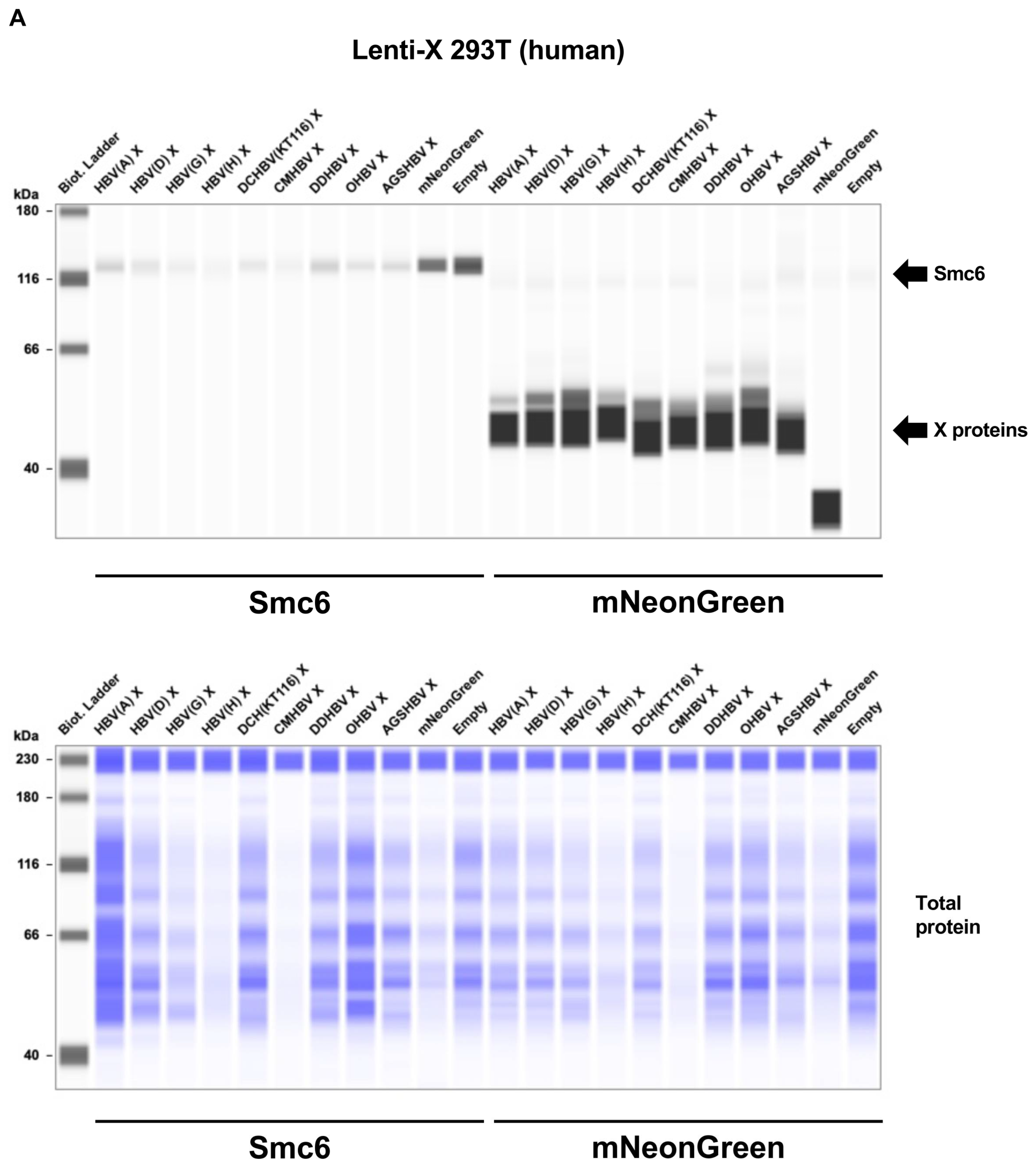
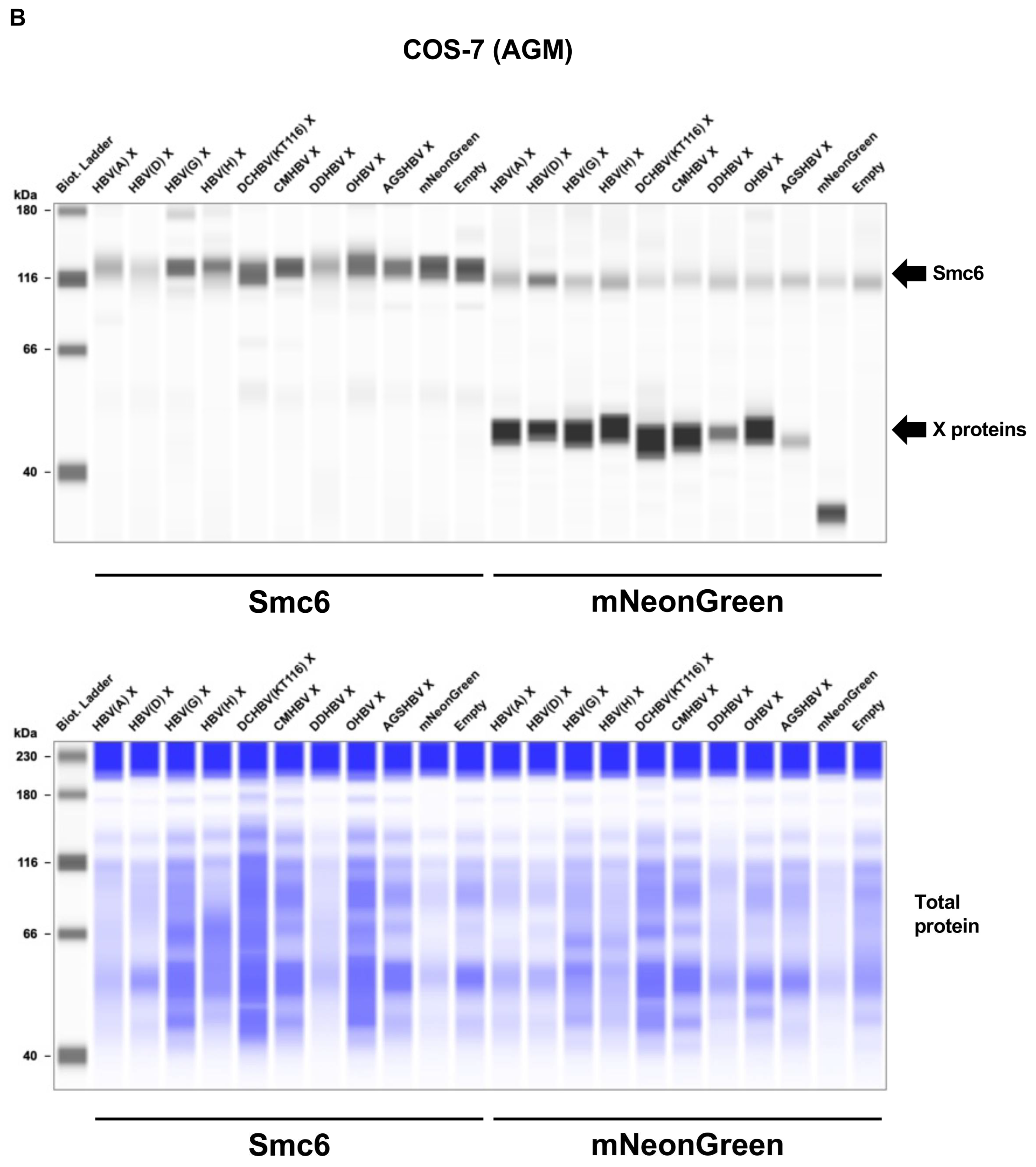
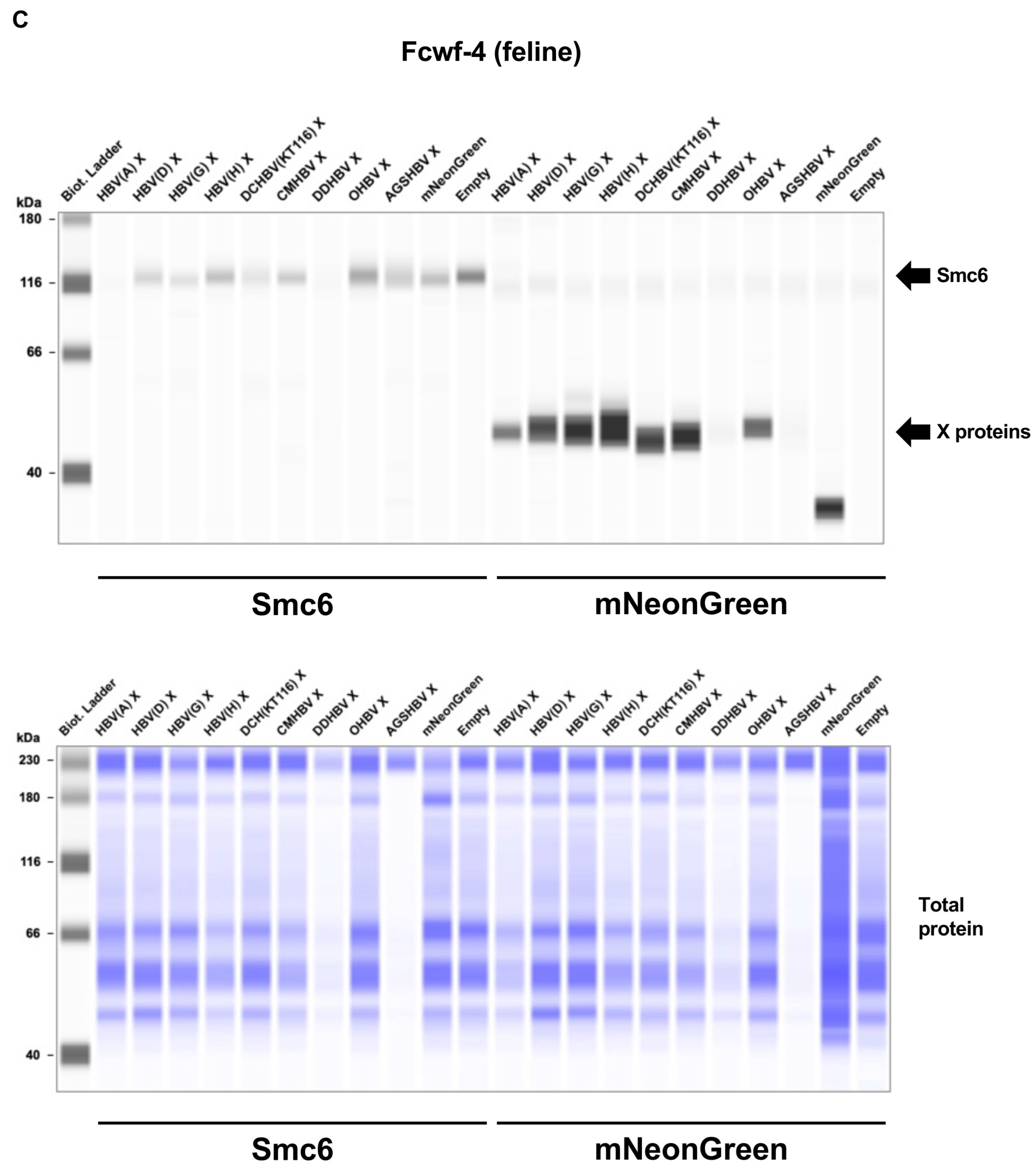
Four cell lines derived from the three mammalian species, i.e., Lenti-X 293T (human), COS-7 (African green monkey: AGM), Fcwf-4 (cat), and CRFK (cat), were transfected with expression vectors encoding mNeonGreen (26.6 kDa) or mammalian hepatitis B virus X proteins tagged with mNeonGreen (~50 kDa). We observed comparable levels of mNeonGreen-tagged X proteins in these cell lines (Figure 3, right side of each panel). While the expression of mNeonGreen alone did not affect the level of the Smc5/6 complex in each cell line, all mammalian hepatitis B virus X proteins showed significant degradation activity of the Smc5/6 complex (~130 kDa) in Lenti-X 293T and Fcwf-4 cells (Figure 3A,D). In contrast, we found differential Smc5/6 degradation activity of mammalian hepatitis B virus X proteins in COS-7 and CRFK cells (Figure 3B,D). Specifically, HBV(A) X had significant Smc5/6 degradation activity in COS-7 and CRFK cells. This result was consistent with a previous finding that HBV(A) X has conserved degradation activity for all mammalian Smc6, including in cell lines derived from OWMs (Vero cells and COS-7 cells) [11]. While DCHBV(KT116) X efficiently degraded Smc5/6 complexes in Lenti-X 293T, Fcwf-4, and CRFK cells (Figure 3A,C,D), it caused minimal Smc5/6 degradation in COS-7 cells (Figure 3B). In CRFK cells, HBV(H) X and orangutan HBV (OHBV) X failed to degrade Smc6 (Figure 3D). These results suggest that mammalian hepatitis B virus X proteins have differential Smc6 degradation activities depending on the host species and cell types.

2.4. DCHBV(KT116) X Is More Predominantly Localized in the Nucleus than HBV(A) X

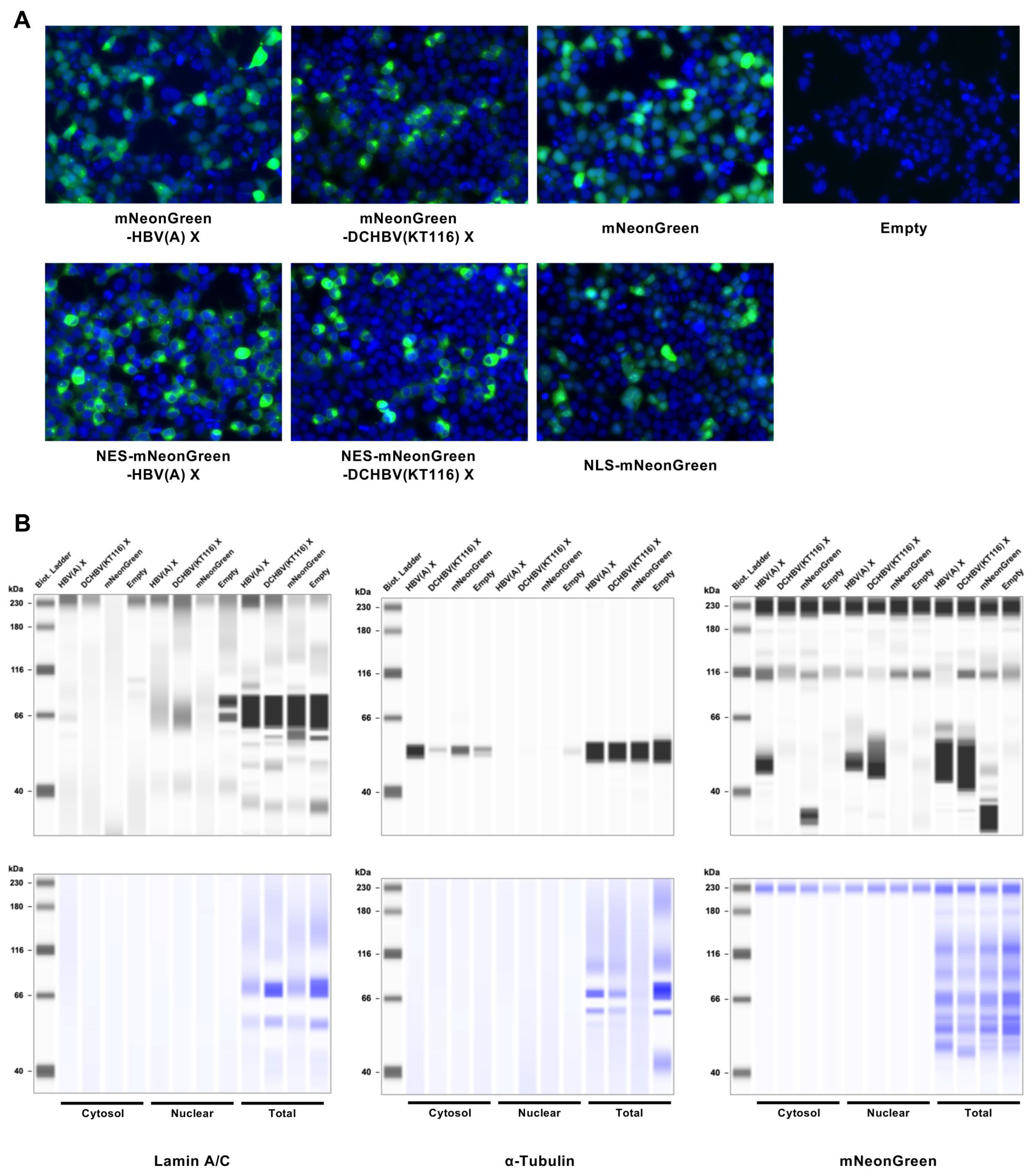
Although the subcellular localization of HBV X is both cytoplasmic and nuclear [26,27,28], about 80% of it is concentrated in the cytoplasm [26]. We examined the similarity of X subcellular localizations by comparing the localization of HBV(A) X and DCHBV(KT116) X in Lenti-X 293-T cells. While HBV X was observed both in the cytoplasm and nucleus, DCHBV(KT116) X was mainly localized in the nucleus (Figure 4A). Next, we investigated the subcellular localization of these proteins by adding either an SV40 large T-derived nuclear localization signals (NLS) signal (PKKKRKV) [29] or an HIV-1 Vpr-derived nuclear export signal (NES) signal (PLQLPPLERLTL) [30]. As expected, the addition of an NLS signal to mNeonGreen resulted in its localization to the nucleus. In contrast, the addition of an NES signal to the X protein abolished its nuclear localization.
In addition, we evaluated the distribution of X using a subcellular fractionation assay. First, we confirmed the efficient separation of the cytosolic and nuclear fractions (Figure 4B). Second, we observed that while HBV(A) X was distributed in both the cytoplasm and the nucleus, DCHBV(KT116) X was predominantly present in the nucleus (Figure 4B); these findings are consistent with observations made with fluorescence microscopy (Figure 4A).

2.5. DCHBV(KT116) X Degrades the Smc6 Complex Independently of DDB1

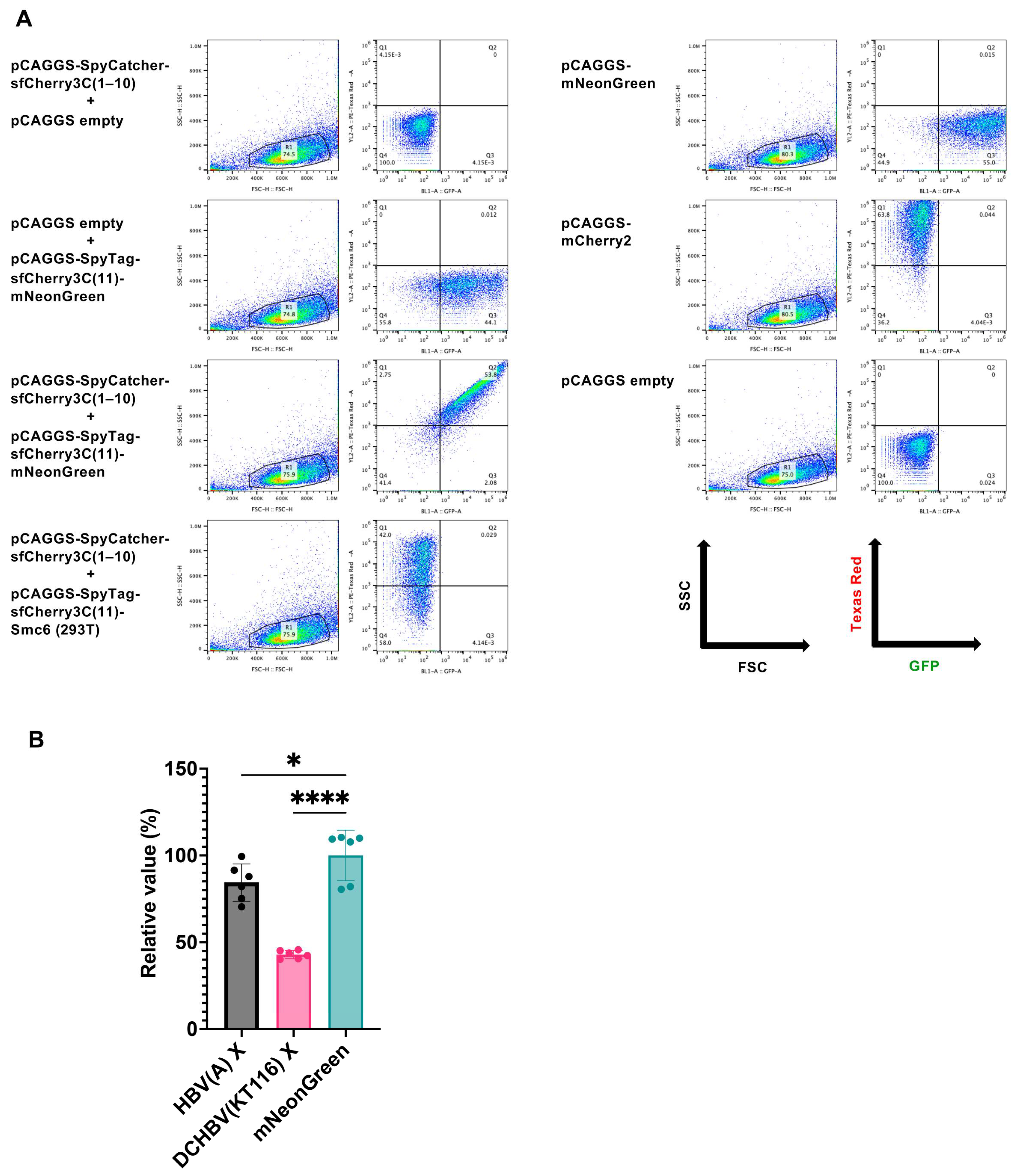
We improved the throughput of Smc5/6 degradation analysis by developing a new assay based on a split-type red fluorescent protein, sfCherry3C [31]. In this assay, Smc6 was tagged with SpyTag-sfCherry3C(11). The principle of the assay is that within the cell, the SpyTag-sfCherry3C(11)-labeled protein associates with SpyCatcher-sfCherry3C(1–10), reconstituting the full sfCherry3C structure and emitting fluorescence (Supplementary Figure S2). If Smc6 tagged with SpyTag-sfCherry3C(11) is degraded by X proteins, this association is disrupted, resulting in decreased fluorescence intensity. It should be noted that Smc5 was not expressed in this assay; therefore, the assay specifically evaluated the degradation of Smc6 alone. First, we tested the specificity of this system by transfecting Lenti-X 293T cells with either pCAGGS-SpyCatcher-sfCherry3C(1–10), pCAGGS-SpyTag-sfCherry3C(11)-mNeonGreen alone, or both. We found that pCAGGS-SpyCatcher-sfCherry3C(1–10) or pCAGGS-SpyTag-sfCherry3C(11)-mNeonGreen transfection did not produce any sfCherry3C signal. In contrast, the co-transfection of both plasmids produced double-positive signals of sfCherry3C and mNeonGreen (Figure 5A). We observed efficient levels of sfCherry3C after the co-transfection of pCAGGS-SpyCatcher-sfCherry3C(1–10) and pCAGGS-SpyTag-sfCherry3C(11)-Smc6 (Human) (Figure 5A); the finding highlighted the specificity of this system (Supplementary Figure S2).
Next, Lenti-X 293T cells were co-transfected with pCAGGS-SpyCatcher-sfCherry3C(1–10), pCAGGS-SpyTag-sfCherry3C(11)-Smc6 (Human, Cat, and Cat (+KVRNT)), and pCAGGS plasmids encoding mNeonGreen-tagged X proteins. The co-expression of the plasmids encoding HBV(A) X and DCHBV(KT116) X resulted in a lower level of sfCherry3C (Figure 5B), suggesting that this system could reproduce the Smc6 degradation visualized by western blotting (Figure 3).
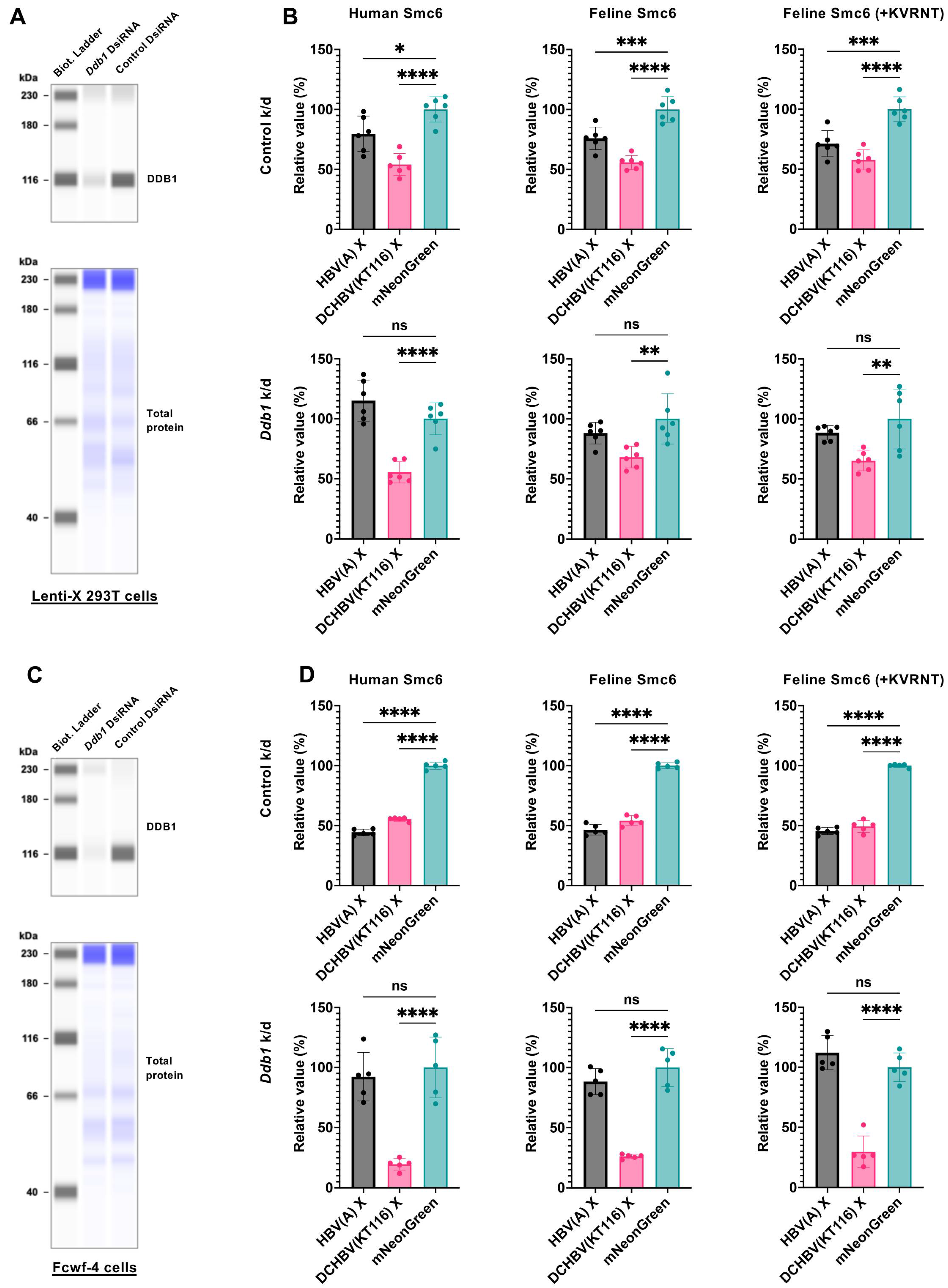
HBV X degrades the Smc5/6 complex by interacting with the host DDB1-containing E3 ubiquitin ligase complex for ubiquitin-mediated Smc5/6 degradation [8,9]. Thus, we determined whether HBV(A) X and DCHBV(KT116) X shared the Smc6 degradation mechanism by examining the effect of Ddb1 deletion on Smc6 degradation (Figure 6A). While knockdown of human Ddb1 in Lenti-X 293T cells completely inhibited HBV(A) X–mediated Smc6 degradation (Figure 6B), it did not affect DCHBV(KT116) X–induced Smc6 degradation (Figure 6B). This finding suggests that DCHBV(KT116) X degraded Smc6 by a different mechanism to HBV(A) X.
Further, we tested the role of feline DDB1 in X-mediated Smc6 degradation in feline-derived Fcwf-4 cells (Figure 6C). The knockout of Ddb1 negated the Smc6 degradation induced by HBV(A) X (Figure 6D), suggesting a similar role for human and feline DDB1 in HBV(A) X-mediated Smc6 degradation. In contrast, Smc6 degradation by DCHBV(KT116) X was more efficient in Ddb1-depleted cells. These results suggest that human DDB1 and feline DDB1 play different roles in DCHBV(KT116) X–mediated Smc6 degradation.

3. Discussion

In this study, we have demonstrated that the Smc5/6 degradation activities, mediated by mammalian hepatitis B virus X proteins, are divergent depending on the combination of the virus and the host species. Further, we have uncovered that HBV(A) X and DCHBV(KT116) X depend on DDB1 differently for Smc6 degradation.
The phylogenetic analysis of DCHBV X indicated its placement on a branch separate from that of HBV (Figure 1). In addition, Smc6 was found to be divergent in mammals (Figure 2 and Figure S1), suggesting different Smc5/6 degradation activities depending on the virus and host. Supporting this suggestion, the western blotting analysis demonstrated that HBV(A) X degraded Smc6 in human, monkey, and feline cells. In contrast, DCHBV(KT116) X minimally degraded Smc6 in monkey cells (Figure 3A–D), suggesting that it has a narrower range of Smc6 degradation than HBV(A) X. In addition, several X proteins, including HBV(H) X and orangutan HBV X, minimally degraded Smc6 in feline cells (Figure 3D), suggesting that the combination of the virus and host species significantly affects Smc6 degradation. Alternatively, feline cells may have the machinery to antagonize Smc6 degradation by several mammalian hepatitis B virus X proteins.
Using our novel Smc6 degradation assay (Figure 5 and Figure S2), we demonstrated that HBV(A) X-mediated Smc6 degradation was negated by Ddb1 depletion (Figure 6B). This finding is consistent with previous reports that HBV(A) X requires interaction with DDB1 for Smc6 degradation [8,9]. In marked contrast, DCHBV(KT116) X degraded Smc6 independently of DDB1 (Figure 6D).
Another difference between HBV(A) X and DCHBV(KT116) X is their cellular localization. The DCHBV(KT116) X protein was predominantly localized in the nucleus compared with HBV(A) X (Figure 4A,B). The cellular localization of HBV X affects the function of X [28,32,33,34]. Therefore, the predominantly nuclear localization of DCHBV X likely leads to different functions compared with HBV X. Although it remains unclear how different localizations are determined, HBV(A) X and DCHBV(KT116) X may interact with host factors differently.
DDB2 induces the nuclear localization of HBV X without the binding of DDB1 [35]. Because HBV X lacks a nuclear localization signal, its nuclear accumulation may rely on its interaction with other cellular proteins [36,37]. Therefore, it will be intriguing to test the molecular interaction of DCHBV X with DDB2 and other cellular proteins that also interact with HBV X.
In this study, we tested two major isoforms of the feline Smc6 gene. Isoform 1 has the KVRNT insertion, which is absent in other mammals, between amino acids 216 and 222 (Figure 2). Although our Smc6 degradation assay demonstrated that this insertion did not affect its sensitivity to mammalian hepatitis B virus X proteins (Figure 6B,D), the accuracy of this insertion and its impact on antiviral activity should be examined in future studies. Interestingly, we have identified similar insertions encoding one to three amino acids in the human Smc6 gene (rs1168792692, rs1669134078, and rs1669195959; https://www.ncbi.nlm.nih.gov/snp; accessed on 20 October 2024). The impact of these insertions, especially on a host’s susceptibility to HBV infection, can be investigated in future studies.
Mammalian hepatitis B virus X proteins have multiple functions in addition to Smc6 degradation. For example, HBV X modulates transcription, cell cycle control, cell growth, and apoptosis in hepatocytes [19,36]. Also, we have recently demonstrated that mammalian hepatitis B virus X proteins have conserved activities in preventing the TIR-domain-containing adaptor protein from inducing the IFN-β (TRIF)-mediated IFN-β signaling pathway through TRIF degradation [38]. We propose that mammalian hepatitis B virus X proteins exhibit different phenotypes depending on their functions and host species. It will be of interest to investigate which function is more and less conserved in mammalian hepatitis B virus X proteins. Furthermore, HBV X can be an attractive target for inhibitors [39].
There are several limitations in this study. First, we used an overexpression system to study Smc6 degradation by mammalian hepatitis B virus X proteins. It is possible that the use of overexpression may impact the physiological function of cells. It will also be valuable to examine the roles of mammalian hepatitis B virus X proteins in Smc6 degradation with physiological protein levels. Second, the effect of the mammalian hepatitis B virus X proteins should be tested using an infectious and X-deficient strain of HBV to directly evaluate the roles of the mammalian hepatitis B virus X proteins in viral replication [11]. Lastly, the domains or residues in HBV(A) X and DCHBV(KT116) X that determine different Smc6 degradation activities and DDB1 dependency can be investigated using chimeric proteins.
In summary, our findings demonstrate that X proteins of mammalian Orthohepadnavirus share a conserved ability to target the Smc6 protein, although the mechanisms and efficiency of degradation may differ among species. Further research into the interaction between DCHBV(KT116) X, the Smc5/Smc6 complex, and related molecules can provide valuable insights into the molecular mechanisms underlying DCHBV pathogenesis. Understanding how DCHBV X targets and modulates Smc6 may provide potential therapeutic insight into treating DCHBV-infected cats. Furthermore, it will contribute to the development of a novel HBV animal model using DCHBV.

4. Materials and Methods

4.1. Alignment of the Mammalian Hepatitis B Virus X Proteins and Phylogenetic Analysis

The complete amino acid sequences of the X protein from 32 virus strains of the genus Orthohepadnavirus were aligned using the MUSCLE algorithm in MEGA 11 (MEGA Software, Version 11.0.13) [40,41]. The alignment parameters were gap open at −2.90, gap extend at 0.00, and hydrophobicity multiplier at 1.20. Alignment visualization was conducted using CLC Genomics Workbench viewing mode Version 22.0.1
A phylogenetic tree was constructed using the alignment of X amino acid sequences retrieved from public databases, and evolutionary analysis was conducted using MEGA X. The evolutionary link was inferred using the maximum likelihood method and the Jones–Taylor–Thornton (JTT) matrix-based model. The initial trees for the heuristic search were obtained by applying the neighbor-joining method to a matrix of pairwise distances estimated using the JJT model. A discrete gamma distribution was used to model the site’s evolution. The tree is drawn to scale, with branch lengths measured in the number of substitutions per site.

4.2. Alignment of the Mammalian Smc6 Proteins and Phylogenetic Analysis

The regions around residues 36 to 48 and 216 to 222 of the Smc6 protein from 23 animal species and 3 human cells were aligned using the MUSCLE algorithm in MEGA 11 (MEGA Software). The alignment parameters were gap open at −2.90, gap extend at 0.00, and hydrophobicity multiplier at 1.20.
A phylogenetic tree was constructed using the alignment of Smc6 amino acid sequences retrieved from public databases. The evolutionary analysis was conducted in MEGA 11. The evolutionary link was inferred using the maximum likelihood method and the JTT matrix-based model [42]. The initial trees for the heuristic search were obtained by applying the neighbor-joining method to a matrix of pairwise distances estimated using the JJT model. Discrete gamma distribution was used to model the evolutionary rate differences among sites. The tree is drawn to scale, with branch lengths measured in the number of substitutions per site.

4.3. Calculation of the Identity of Smc6 Among Animal Species

The identity of Smc6 among the animal species was calculated using MEGA X with a pairwise distance matrix. Analyses were conducted using the JTT matrix-based model [42]. The analysis involved 26 amino acid sequences. All ambiguous positions were removed for each sequence pair (pairwise deletion option). There were a total of 1125 positions in the final dataset. Evolutionary analyses were conducted in MEGA11 [40,41].

4.4. Cell Culture

Lenti-X 293T cells (Homo sapiens or human; TaKaRa, Kusatsu, Japan, Cat# Z2180N), COS-7 (Cercopithecus aethiops or African green monkey [AGM]; Japanese Collection of Research Bioresources Cell Bank [JCRB], Ibaraki, Japan, Cat# JCRB9127), Fcwf-4 (Felis catus or cat; American Type Culture Collection, Manassas, VA, USA, Cat# CRL-2787), and CRFK (cat; JCRB, Cat# JCRB9035) cells were cultured in Dulbecco’s modified Eagle medium (DMEM; Nacalai Tesque, Kyoto, Japan, Cat# 08458-16) supplemented with 10% fetal bovine serum and 1 × penicillin–streptomycin (Nacalai Tesque, Cat# 09367-34).

4.5. Plasmids

The cDNA sequences of the X protein derived from 9 viruses of Orthohepadnavirus were previously synthesized with codon optimization to humans (Twist Bioscience, San Francisco, CA, USA) (Supplementary Table S2) [38]. cDNA fragments encoding X were cloned into a pCAGGS-mNeonGreen vector [43] predigested with AgeI-HF (New England Biolabs [NEB], Ipswich, MA, USA, Cat# R3552S) and NheI-HF (NEB, Cat# R3131M) using the In-Fusion Snap Assembly Master Mix (TaKaRa, Cat# Z8947N). The plasmids were amplified using NEB 5-alpha F′Iq Competent Escherichia coli (High Efficiency) (NEB, Cat# C2992H) and isolated using the PureYield Plasmid Miniprep System (Promega, Madison, WI, USA, Cat# A1222). The sequences of all the plasmids were verified using a SupreDye v3.1 Cycle Sequencing Kit (M&S TechnoSystems, Osaka, Japan, Cat# 063001) with a Spectrum Compact CE System (Promega).

4.6. Smc5/6 Complex Degradation Assay

Lenti-X 293T (1.25 × 10⁵ per well), COS-7 (4.17 × 10⁴ per well), CRFK (4.17 × 10⁴ per well), and Fcwf-4 cells (4.17 × 10⁴ per well) were plated in a 24-well plate (Thermo Fisher Scientific, Waltham, MA, USA, Cat# 142475). After overnight incubation, the cells were co-transfected with 500 ng of pCAGGS-mNeonGreen-X using the TransIT-LT1 Transfection Reagent (TaKaRa, Cat# V2300) or the TransIT-X2 Dynamic Delivery System (TaKaRa, Cat# V6100) in the Opti-MEM I Reduced Serum Medium (Thermo Fisher Scientific, Cat# 31985062). After 48 h, the cells were stained with the NucBlue Live ReadyProbes Reagent (Hoechst 33342) (Thermo Fisher Scientific, Cat# R37605), observed under the EVOS M7000 Imaging System (Thermo Fisher Scientific), and collected for western blotting analysis.

4.7. Western Blotting

Western blotting was used to examine Smc5/6 degradation by mammalian hepatitis B virus X proteins. First, pelleted cells were lysed in an M-PER Mammalian Protein Extraction Reagent (Thermo Fisher Scientific, Cat#78501) containing a Protease Inhibitor Cocktail Set I (×100) (Fujifilm, Osaka, Japan, Cat#165-26021). Protein concentrations were measured using the TaKaRa Bradford Protein Assay Kit (TaKaRa, Cat# T9310A). Then, 1 µg of the cellular extracts was mixed with 2 × Bolt LDS sample buffer (Thermo Fisher Scientific, Cat# B0008) containing 2% β-mercaptoethanol (Bio-Rad, Hercules, CA, USA, Cat# 1610710) and incubated at 70 °C for 10 min. The level of Smc6 was evaluated using a SimpleWestern Abby (ProteinSimple, San Jose, CA, USA) with an anti-SMC6L1 antibody (GeneTex, Irvine, CA, USA, Cat# GTX116832, ×50) and an Anti-Rabbit Detection Module (ProteinSimple, Cat# DM-001). The level of mNeonGreen-tagged X was measured with an anti-mNeonGreen antibody (Proteintech, Rosemont, IL, USA, Cat# 32F6-100, ×50) and an Anti-Mouse Detection Module (ProteinSimple, Cat# DM-002). The amount of input protein was measured using a Total Protein Detection Module (ProteinSimple, Cat# DM-TP01).
X protein localization was examined by first plating Lenti-X 293-T cells (1.25 × 10⁵ per well) in a 24-well plate. After overnight incubation, the cells were co-transfected with 500 ng of pCAGGS-mNeonGreen-X plasmids using the TransIT-X2 Dynamic Delivery System. After 48 h, the nuclear and cytosolic fractions were separated using a Minute Cytoplasmic and Nuclear Extraction Kit (Invent Biotechnologies, Inc., Cat# SC-003). The samples for western blotting were prepared as described above. The localization of X was evaluated using an anti-mNeonGreen antibody. The efficiency of subcellular fractionation was evaluated using an anti-Lamin A/C antibody (Cell Signaling Technology [CST], Danvers, MA, USA, Cat# 4777S, ×250) and anti-α-Tubulin antibody (CST, Cat# 2125S, ×250).

4.8. Smc6 Degradation Assay Using the SpyTag/SpyCatcher System

First, total RNA was extracted from Lenti-X 293T, COS-7 CRFK, and Fcwf-4 cells using an RNeasy Mini Kit (QIAGEN, Chuo-ku, Japan, Cat# 74104) and QIAshredder (QIAGEN, Cat# 79656). Then, Smc6 cDNA was amplified by RT-PCR using the PrimeScript II High Fidelity One Step RT-PCR Kit (TaKaRa, Cat#R026B) using specific primers (Supplementary Table S3). The PCR consisted of 1 cycle at 45 °C for 10 min; 1 cycle at 94 °C for 2 min; 40 cycles at 98 °C for 10 s, 60 °C for 15 s, and 68 °C for 20 s; and 1 cycle at 68 °C for 7 min. The amplified fragments were ligated into pCAGGS-SpyTag-sfCherry3C(11)-HA predigested with AgeI-HF and NheI-HF using an In-Fusion Snap Assembly Master Mix. The plasmids were verified by sequencing.
The system was validated by plating Lenti-X 293T cells at 2.5 × 10⁵ cells per well in a 12-well plate. Then the cells were transfected with pCAGGS-SpyCatcher-sfCherry3C(1–10) alone, pCAGGS-SpyTag-sfCherry3C(11)-mNeonGreen alone, or a combination of pCAGGS-SpyCatcher-sfCherry3C(1–10), pCAGGS-SpyTag-sfCherry3C(11)-mNeonGreen, and pCAGGS-SpyTag-sfCherry3C(11)-HA-Smc6 (Human). The signals of mNeonGreen and sfCherry3C were measured 2 days after transfection using an Attune NxT Flow Cytometer (Thermo Fisher Scientific). The mNeonGreen- and sfCherry3C- double-positive population was gated and analyzed using FlowJo v10.8.1 (Becton, Dickinson and Company, Franklin Lakes, NJ, USA).
To evaluate the role of DDB1 in Smc6 degradation, we depleted human DDB1 by transfecting Lenti-X 293T cells (1.9 × 10⁶ cells per well in a 6-well plate) with DsiRNA targeting human Ddb1 (Integrated DNA Technologies [IDT], Coralville, IA, USA, Reference# hs. Ri. DDB1.13.1, and hs. Ri. DDB1.13.3) or Negative Control DsiRNA (IDT, Cat# 109617253) with the TransIT-X2 Dynamic Delivery System. We depleted feline Ddb1 by transfecting Fcwf-4 cells (6.3 × 10⁵ cells per well in a 6-well plate) with DsiRNA targeting feline Ddb1 (IDT, Reference# CD.Ri.477260.13.1 and CD.Ri.477260.13.3) or Negative Control DsiRNA with the TransIT-X2 Dynamic Delivery System. After 2 days, the cells were re-plated in a new 96-well plate at 3 × 10⁴ Lenti-X 293T cells per well or 1 × 10⁴ Fcwf-4 cells per well. The cells were cultured overnight and co-transfected with pCAGGS-SpyCatcher-sfCherry3C(1–10) at 15 ng/well, pCAGGS-SpyTag-sfCherry3C(11)-HA-Smc6 at 15 ng/well, and pCAGGS-mNeonGreen-X at 70 ng/well. Smc6 degradation was measured with sfCherry3C-positivity 2 days after transfection as described above.

4.9. Statistical Analysis

The level of Smc6 degradation was evaluated by one-way ANOVA, followed by the Tukey test. A p-value of 0.05 or less was considered statistically significant. The analysis was performed using Prism 10 v10.2.3 (GraphPad Software).

Supplementary Materials

The following supporting information can be downloaded at https://www.mdpi.com/article/10.3390/ijms26146786/s1.

Author Contributions

M.S. and A.S. designed the experiments. M.S., Y.V.F., and A.S. performed the experiments. M.S., Y.V.F., and A.S. analyzed the results. M.S. and A.S. wrote the manuscript. All authors have read and agreed to the published version of the manuscript.

Funding

This work was supported by grants from the Japan Agency for Medical Research and Development (AMED) Research Program on HIV/AIDS JP24fk0410047, JP25fk0410075, JP25fk0410056, and JP25fk0410058 (to A.S.); the AMED Research Program on Emerging and Re-emerging Infectious Diseases JP23fk0108583, and JP24fk0108907 (to A.S.); the AMED Research Project for Practical Applications of Regenerative Medicine JP25bk0104177 (to A.S.); the JSPS KAKENHI Grant-in-Aid for Scientific Research (C) JP24K09227 (to A.S.); the JSPS KAKENHI Grant-in-Aid for Scientific Research (B) JP22H02500 (to A.S.) and JP21H02361 (to A.S.); the JSPS Bilateral Program JPJSBP120245706 (to A.S.); the JSPS Fund for the Promotion of Joint International Research (International Leading Research) JP23K20041 (to A.S.); the G-7 Grant (to A.S.); and the Ito Foundation Research Grant R6 KEN119 (to A.S.).

Institutional Review Board Statement

Not applicable.

Informed Consent Statement

Not applicable.

Data Availability Statement

Source data are available on request.

Acknowledgments

The authors thank Tomoko Nishiuchi, Yuki Shibatani, Miki Kawano, Natsumi Matsubara, and the staff of CADIC, University of Miyazaki, for their assistance. Supplementary Figure S1 was created with BioRender (https://biorender.com/, accessed on 13 April 2025).

Conflicts of Interest

The authors declare no conflicts of interest.

References

  1. Nguyen, M.H.; Wong, G.; Gane, E.; Kao, J.-H.; Dusheiko, G. Hepatitis B Virus: Advances in Prevention, Diagnosis, and Therapy. Clin. Microbiol. Rev. 2020, 33, e00046-19. [Google Scholar] [CrossRef] [PubMed]
  2. Magnius, L.; Mason, W.S.; Taylor, J.; Kann, M.; Glebe, D.; Dény, P.; Sureau, C.; Norder, H.; Ictv Report Consortium. ICTV Virus Taxonomy Profile: Hepadnaviridae. J. Gen. Virol. 2020, 101, 571–572. [Google Scholar] [CrossRef] [PubMed]
  3. Feng, H.; Chen, P.; Zhao, F.; Nassal, M.; Hu, K. Evidence for Multiple Distinct Interactions between Hepatitis B Virus P Protein and Its Cognate RNA Encapsidation Signal during Initiation of Reverse Transcription. PLoS ONE 2013, 8, e72798. [Google Scholar] [CrossRef]
  4. Murakami, S. Hepatitis B Virus X Protein: Structure, Function and Biology. Intervirology 1999, 42, 81–99. [Google Scholar] [CrossRef] [PubMed]
  5. Lucifora, J.; Arzberger, S.; Durantel, D.; Belloni, L.; Strubin, M.; Levrero, M.; Zoulim, F.; Hantz, O.; Protzer, U. Hepatitis B Virus X Protein Is Essential to Initiate and Maintain Virus Replication after Infection. J. Hepatol. 2011, 55, 996–1003. [Google Scholar] [CrossRef] [PubMed]
  6. Arbuthnot, P.; Capovilla, A.; Kew, M. Putative Role of Hepatitis B Virus X Protein in Hepatocarcinogenesis: Effects on Apoptosis, DNA Repair, Mitogen-Activated Protein Kinase and JAK/STAT Pathways. J. Gastroenterol. Hepatol. 2000, 15, 357–368. [Google Scholar] [CrossRef] [PubMed]
  7. Diao, J.; Garces, R.; Richardson, C.D. X Protein of Hepatitis B Virus Modulates Cytokine and Growth Factor Related Signal Transduction Pathways during the Course of Viral Infections and Hepatocarcinogenesis. Cytokine Growth Factor Rev. 2001, 12, 189–205. [Google Scholar] [CrossRef] [PubMed]
  8. Murphy, C.M.; Xu, Y.; Li, F.; Nio, K.; Reszka-Blanco, N.; Li, X.; Wu, Y.; Yu, Y.; Xiong, Y.; Su, L. Hepatitis B Virus X Protein Promotes Degradation of SMC5/6 to Enhance HBV Replication. Cell Rep. 2016, 16, 2846–2854. [Google Scholar] [CrossRef] [PubMed]
  9. Decorsière, A.; Mueller, H.; van Breugel, P.C.; Abdul, F.; Gerossier, L.; Beran, R.K.; Livingston, C.M.; Niu, C.; Fletcher, S.P.; Hantz, O.; et al. Hepatitis B Virus X Protein Identifies the Smc5/6 Complex as a Host Restriction Factor. Nature 2016, 531, 386–389. [Google Scholar] [CrossRef] [PubMed]
  10. Niu, C.; Livingston, C.M.; Li, L.; Beran, R.K.; Daffis, S.; Ramakrishnan, D.; Burdette, D.; Peiser, L.; Salas, E.; Ramos, H.; et al. The Smc5/6 Complex Restricts HBV When Localized to ND10 without Inducing an Innate Immune Response and Is Counteracted by the HBV X Protein Shortly after Infection. PLoS ONE 2017, 12, e0169648. [Google Scholar] [CrossRef] [PubMed]
  11. Abdul, F.; Filleton, F.; Gerossier, L.; Paturel, A.; Hall, J.; Strubin, M.; Etienne, L. Smc5/6 Antagonism by HBx Is an Evolutionarily Conserved Function of Hepatitis B Virus Infection in Mammals. J. Virol. 2018, 92, e00769-18. [Google Scholar] [CrossRef] [PubMed]
  12. Aghazadeh, M.; Shi, M.; Barrs, V.R.; McLuckie, A.J.; Lindsay, S.A.; Jameson, B.; Hampson, B.; Holmes, E.C.; Beatty, J.A. A Novel Hepadnavirus Identified in an Immunocompromised Domestic Cat in Australia. Viruses 2018, 10, 269. [Google Scholar] [CrossRef] [PubMed]
  13. Pesavento, P.A.; Jackson, K.; Scase, T.; Tse, T.; Hampson, B.; Munday, J.S.; Barrs, V.R.; Beatty, J.A. A Novel Hepadnavirus Is Associated with Chronic Hepatitis and Hepatocellular Carcinoma in Cats. Viruses 2019, 11, 969. [Google Scholar] [CrossRef] [PubMed]
  14. Lanave, G.; Capozza, P.; Diakoudi, G.; Catella, C.; Catucci, L.; Ghergo, P.; Stasi, F.; Barrs, V.; Beatty, J.; Decaro, N.; et al. Identification of Hepadnavirus in the Sera of Cats. Sci. Rep. 2019, 9, 10668. [Google Scholar] [CrossRef] [PubMed]
  15. Jeanes, E.C.; Wegg, M.L.; Mitchell, J.A.; Priestnall, S.L.; Fleming, L.; Dawson, C. Comparison of the Prevalence of Domestic Cat Hepadnavirus in a Population of Cats with Uveitis and in a Healthy Blood Donor Cat Population in the United Kingdom. Vet. Ophthalmol. 2022, 25, 165–172. [Google Scholar] [CrossRef] [PubMed]
  16. Anpuanandam, K.; Selvarajah, G.T.; Choy, M.M.K.; Ng, S.W.; Kumar, K.; Ali, R.M.; Rajendran, S.K.; Ho, K.L.; Tan, W.S. Molecular Detection and Characterisation of Domestic Cat Hepadnavirus (DCH) from Blood and Liver Tissues of Cats in Malaysia. BMC Vet. Res. 2021, 17, 9. [Google Scholar] [CrossRef] [PubMed]
  17. Piewbang, C.; Wardhani, S.W.; Chaiyasak, S.; Yostawonkul, J.; Chai-In, P.; Boonrungsiman, S.; Kasantikul, T.; Techangamsuwan, S. Insights into the Genetic Diversity, Recombination, and Systemic Infections with Evidence of Intracellular Maturation of Hepadnavirus in Cats. PLoS ONE 2020, 15, e0241212. [Google Scholar] [CrossRef] [PubMed]
  18. Silva, B.B.I.; Chen, J.-Y.; Villanueva, B.H.A.; Lu, Z.-Y.; Hsing, H.-Z.; Montecillo, A.D.; Shofa, M.; Minh, H.; Chuang, J.-P.; Huang, H.-Y.; et al. Genetic Diversity of Domestic Cat Hepadnavirus in Southern Taiwan. Viruses 2023, 15, 2128. [Google Scholar] [CrossRef] [PubMed]
  19. Korkulu, E.; Şenlik, E.İ.; Adıgüzel, E.; Artut, F.G.; Çetinaslan, H.D.; Erdem-Şahinkesen, E.; Oğuzoğlu, T.Ç. Status Quo of Feline Leukaemia Virus Infection in Turkish Cats and Their Antigenic Prevalence. Animals 2024, 14, 385. [Google Scholar] [CrossRef] [PubMed]
  20. Takahashi, K.; Kaneko, Y.; Shibanai, A.; Yamamoto, S.; Katagiri, A.; Osuga, T.; Inoue, Y.; Kuroda, K.; Tanabe, M.; Okabayashi, T.; et al. Identification of Domestic Cat Hepadnavirus from a Cat Blood Sample in Japan. J. Vet. Med. Sci. 2022, 84, 648–652. [Google Scholar] [CrossRef] [PubMed]
  21. Sakamoto, H.; Ito, G.; Goto-Koshino, Y.; Sakamoto, M.; Nishimura, R.; Momoi, Y. Detection of Domestic Cat Hepadnavirus by Next-Generation Sequencing and Epidemiological Survey in Japan. J. Vet. Med. Sci. 2023, 85, 642–646. [Google Scholar] [CrossRef] [PubMed]
  22. Beatty, J.A.; Choi, Y.R.; Nekouei, O.; Woodhouse, F.M.; Gray, J.J.; Hofmann-Lehmann, R.; Barrs, V.R. Epidemiology of Pathogenic Retroviruses and Domestic Cat Hepadnavirus in Community and Client-Owned Cats in Hong Kong. Viruses 2024, 16, 167. [Google Scholar] [CrossRef] [PubMed]
  23. Choi, Y.R.; Iturriaga, M.P.; Nekouei, O.; Tu, T.; Van Brussel, K.; Barrs, V.R.; Beatty, J.A. Domestic Cat Hepadnavirus and Pathogenic Retroviruses; A Sero-Molecular Survey of Cats in Santiago, Chile. Viruses 2023, 16, 46. [Google Scholar] [CrossRef] [PubMed]
  24. Tessmann, A.; Sumienski, J.; Sita, A.; Mallmann, L.; Birlem, G.E.; da Silva Nunes, N.J.; Lupion, C.G.; Eckert, J.S.; Demoliner, M.; Gularte, J.S.; et al. Domestic Cat Hepadnavirus Genotype B Is Present in Southern Brazil. Virus Genes 2024, 61, 81–86. [Google Scholar] [CrossRef] [PubMed]
  25. Shofa, M.; Ohkawa, A.; Kaneko, Y.; Saito, A. Conserved Use of the Sodium/Bile Acid Cotransporter (NTCP) as an Entry Receptor by Hepatitis B Virus and Domestic Cat Hepadnavirus. Antivir. Res. 2023, 217, 105695. [Google Scholar] [CrossRef] [PubMed]
  26. Hoare, J.; Henkler, F.; Dowling, J.J.; Errington, W.; Goldin, R.D.; Fish, D.; McGarvey, M.J. Subcellular Localisation of the X Protein in HBV Infected Hepatocytes. J. Med. Virol. 2001, 64, 419–426. [Google Scholar] [CrossRef] [PubMed]
  27. Cha, M.-Y.; Ryu, D.-K.; Jung, H.-S.; Chang, H.-E.; Ryu, W.-S. Stimulation of Hepatitis B Virus Genome Replication by HBx Is Linked to Both Nuclear and Cytoplasmic HBx Expression. J. Gen. Virol. 2009, 90, 978–986. [Google Scholar] [CrossRef] [PubMed]
  28. Henkler, F.; Hoare, J.; Waseem, N.; Goldin, R.D.; McGarvey, M.J.; Koshy, R.; King, I.A. Intracellular Localization of the Hepatitis B Virus HBx Protein. J. Gen. Virol. 2001, 82, 871–882. [Google Scholar] [CrossRef] [PubMed]
  29. Lu, J.; Wu, T.; Zhang, B.; Liu, S.; Song, W.; Qiao, J.; Ruan, H. Types of Nuclear Localization Signals and Mechanisms of Protein Import into the Nucleus. Cell Commun. Signal. 2021, 19, 60. [Google Scholar] [CrossRef] [PubMed]
  30. Xu, D.; Farmer, A.; Collett, G.; Grishin, N.V.; Chook, Y.M. Sequence and Structural Analyses of Nuclear Export Signals in the NESdb Database. Mol. Biol. Cell 2012, 23, 3677–3693. [Google Scholar] [CrossRef] [PubMed]
  31. Feng, S.; Varshney, A.; Coto Villa, D.; Modavi, C.; Kohler, J.; Farah, F.; Zhou, S.; Ali, N.; Müller, J.D.; Van Hoven, M.K.; et al. Bright Split Red Fluorescent Proteins for the Visualization of Endogenous Proteins and Synapses. Commun. Biol. 2019, 2, 344. [Google Scholar] [CrossRef] [PubMed]
  32. McClain, S.L.; Clippinger, A.J.; Lizzano, R.; Bouchard, M.J. Hepatitis B Virus Replication Is Associated with an HBx-Dependent Mitochondrion-Regulated Increase in Cytosolic Calcium Levels. J. Virol. 2007, 81, 12061–12065. [Google Scholar] [CrossRef] [PubMed]
  33. Clippinger, A.J.; Bouchard, M.J. Hepatitis B Virus HBx Protein Localizes to Mitochondria in Primary Rat Hepatocytes and Modulates Mitochondrial Membrane Potential. J. Virol. 2008, 82, 6798–6811. [Google Scholar] [CrossRef] [PubMed]
  34. Keasler, V.V.; Hodgson, A.J.; Madden, C.R.; Slagle, B.L. Hepatitis B Virus HBx Protein Localized to the Nucleus Restores HBx-Deficient Virus Replication in HepG2 Cells and in Vivo in Hydrodynamically-Injected Mice. Virology 2009, 390, 122–129. [Google Scholar] [CrossRef] [PubMed]
  35. Nag, A.; Datta, A.; Yoo, K.; Bhattacharyya, D.; Chakrabortty, A.; Wang, X.; Slagle, B.L.; Costa, R.H.; Raychaudhuri, P. DDB2 Induces Nuclear Accumulation of the Hepatitis B Virus X Protein Independently of Binding to DDB1. J. Virol. 2001, 75, 10383–10392. [Google Scholar] [CrossRef] [PubMed]
  36. Sitterlin, D.; Bergametti, F.; Transy, C. UVDDB P127-Binding Modulates Activities and Intracellular Distribution of Hepatitis B Virus X Protein. Oncogene 2000, 19, 4417–4426. [Google Scholar] [CrossRef] [PubMed]
  37. Weil, R.; Sirma, H.; Giannini, C.; Kremsdorf, D.; Bessia, C.; Dargemont, C.; Bréchot, C.; Israël, A. Direct Association and Nuclear Import of the Hepatitis B Virus X Protein with the NF-kappaB Inhibitor IkappaBalpha. Mol. Cell. Biol. 1999, 19, 6345–6354. [Google Scholar] [CrossRef] [PubMed]
  38. Choonnasard, A.; Shofa, M.; Okabayashi, T.; Saito, A. Conserved Functions of Orthohepadnavirus X Proteins to Inhibit Type-I Interferon Signaling. Int. J. Mol. Sci. 2024, 25, 3753. [Google Scholar] [CrossRef] [PubMed]
  39. Sekiba, K.; Otsuka, M.; Ohno, M.; Yamagami, M.; Kishikawa, T.; Suzuki, T.; Ishibashi, R.; Seimiya, T.; Tanaka, E.; Koike, K. Inhibition of HBV Transcription From cccDNA With Nitazoxanide by Targeting the HBx-DDB1 Interaction. Cell Mol. Gastroenterol. Hepatol. 2019, 7, 297–312. [Google Scholar] [CrossRef] [PubMed]
  40. Stecher, G.; Tamura, K.; Kumar, S. Molecular Evolutionary Genetics Analysis (MEGA) for macOS. Mol. Biol. Evol. 2020, 37, 1237–1239. [Google Scholar] [CrossRef] [PubMed]
  41. Kumar, S.; Stecher, G.; Li, M.; Knyaz, C.; Tamura, K. MEGA X: Molecular Evolutionary Genetics Analysis across Computing Platforms. Mol. Biol. Evol. 2018, 35, 1547–1549. [Google Scholar] [CrossRef] [PubMed]
  42. Jones, D.T.; Taylor, W.R.; Thornton, J.M. The Rapid Generation of Mutation Data Matrices from Protein Sequences. Comput. Appl. Biosci. 1992, 8, 275–282. [Google Scholar] [CrossRef] [PubMed]
  43. Niwa, H.; Yamamura, K.; Miyazaki, J. Efficient Selection for High-Expression Transfectants with a Novel Eukaryotic Vector. Gene 1991, 108, 193–199. [Google Scholar] [CrossRef] [PubMed]
Figure 1. Sequence alignment and phylogenetic analysis of the X proteins from viruses of the genus Orthohepadnavirus. (A) Amino acid sequence alignment of the X proteins of the genus Orthohepadnavirus. The residue numbering was based on the HBV genotype D (strain ayw). (B) The phylogenetic tree was constructed using the alignment of X protein sequences derived from viruses of the genus Orthohepadnavirus. The evolutionary link was inferred using the neighbor joining method. The percentage of replicate trees in which the associated taxa clustered together in the bootstrap test (1000 replicates) is shown next to the branches.
Ijms 26 06786 g001aIjms 26 06786 g001b
Figure 2. Sequence alignment of mammalian Smc6 and phylogenetic analysis of the entire mammalian Smc6 sequence. Alignment of the amino acid sequence of mammalian Smc6, specifically amino acids 36 to 48 and 216 to 222, was conducted. Subsequently, a phylogenetic tree for the complete mammalian Smc6 sequence was constructed. The numbering of residues was standardized in reference to the human Smc6 sequence.
Ijms 26 06786 g002
Figure 3. Mammalian hepatitis B virus X proteins exhibit differential Smc5/6 complex degradation activities depending on the host species. (A) Lenti-X 293T (human), (B) COS-7 (African green monkey), (C) Fcwf-4 (feline), and (D) CRFK (feline) cells were transfected with plasmids encoding X proteins tagged with mNeonGreen (~50 kDa), or mNeonGreen (26.6 kDa), or only pCAGGS empty plasmid. The levels of Smc6 (~130 kDa) and mNeonGreen at 2 days after transfection were analyzed by western blotting. The results are representative of at least 3 independent experiments.
Ijms 26 06786 g003aIjms 26 06786 g003bIjms 26 06786 g003cIjms 26 06786 g003d
Figure 4. Subcellular localization of HBV(A) X and DCHBV(KT116) X. (A) Lenti-X 293-T cells were transfected with pCAGGS plasmids encoding mNeonGreen-HBV(A) X, mNeonGreen-DCHBV(KT116) X, mNeonGreen, NES-mNeonGreen-HBV(A) X, NES-mNeonGreen-DCHBV(KT116) X, NLS-mNeonGreen or only pCAGGS. (B) The nuclear fraction, cytosolic fraction, and whole-cell lysates (total) were analyzed by western blotting using anti-lamin A/C (nuclear), anti-α-tubulin (cytosolic), and anti-mNeonGreen (X protein) antibodies. The results are representative of at least 3 independent experiments.
Ijms 26 06786 g004
Figure 5. A flow cytometry–based SMC6 degradation assay using sfCherry3C-Smc6. (A) Lenti-X 293T cells were transfected with pCAGGS plasmids encoding SpyCatcher-sfCherry3C(1–10) only, SpyTag-sfCherry3C(11)-mNeonGreen only, or a combination of SpyCatcher-sfCherry3C(1–10), SpyTag-sfCherry3C(11)-mNeonGreen, and SpyTag-sfCherry3C(11)-HA-Smc6 (human). The signals of mNeonGreen and sfCherry3C were measured 2 days after transfection. (B) Lenti-X 293T cells were co-transfected with pCAGGS plasmids encoding SpyCatcher-sfCherry3C(1–10) only, SpyTag-sfCherry3C(11)-HA-Smc6 (human) only, or in combination with mNeonGreen-HBV(A) X, mNeonGreen-DCHBV(KT116) X, or mNeonGreen. Smc6 degradation was evaluated by measuring sfCherry3C levels using flow cytometry. The results, presented as the mean and SD of sextuplicate measurements from 1 assay, are representative of at least 3 independent experiments. The differences in sfCherry3C positivity between the cells producing HBV(A) X, DCHBV(KT116) X, and mNeonGreen were evaluated using one-way ANOVA, followed by the Tukey test. * p < 0.05, **** p < 0.0001.
Ijms 26 06786 g005
Figure 6. DCHBV(KT116) X degrades Smc6 independently of DDB1. (A) Lenti-X 293-T cells were transfected with DsiRNA targeting human Ddb1 or non-targeting control DsiRNA. The level of DDB1 in Lenti-X 293-T cells was determined using western blotting. (B) After 2 days of DsiRNA transfection, the cells were co-transfected with pCAGGS plasmids encoding SpyCatcher-sfCherry3C(1–10), SpyTag-sfCherry3C(11)-HA-Smc6 (human, feline, or feline with KVRNT insertion), or in combination with mNeonGreen-HBV(A) X, mNeonGreen-DCHBV(KT116) X, or mNeonGreen. Smc6 degradation was evaluated by measuring sfCherry3C levels with flow cytometry. The results, presented as the mean and SD of sextuplicate measurements from 1 assay, are representative of at least 3 independent experiments. The differences in sfCherry3C positivity between the cells producing HBV(A) X, DCHBV(KT116) X, and mNeonGreen were evaluated using one-way ANOVA, followed by the Tukey test. * p < 0.05, ** p < 0.01, *** p < 0.001, **** p < 0.0001, ns (not significant). (C) Fcwf-4 cells were transfected with DsiRNA targeting cat Ddb1 or non-targeting control DsiRNA. The level of Ddb1 in Fcwf-4 cells was determined via western blotting. (D) After 2 days of DsiRNA transfection, the cells were co-transfected with pCAGGS plasmids encoding SpyCatcher-sfCherry3C(1–10), SpyTag-sfCherry3C(11)-HA-Smc6 (human, feline, or feline with +KVRNT insertion), in combination with mNeonGreen-HBV(A) X, mNeonGreen-DCHBV(KT116) X, or mNeonGreen. Smc6 degradation was evaluated by measuring sfCherry3C levels with flow cytometry. The results, presented as the mean and SD of sextuplicate measurements from 1 assay, are representative of at least 3 independent experiments. The differences in sfCherry3C positivity between cells producing HBV(A) X, DCHBV(KT116) X, and mNeonGreen were evaluated by one-way ANOVA, followed by the Tukey test. **** p < 0.0001, ns (not significant).
Ijms 26 06786 g006
Disclaimer/Publisher’s Note: The statements, opinions and data contained in all publications are solely those of the individual author(s) and contributor(s) and not of MDPI and/or the editor(s). MDPI and/or the editor(s) disclaim responsibility for any injury to people or property resulting from any ideas, methods, instructions or products referred to in the content.