Review

Microbiome-Based Products: Therapeutic Potential for Inflammatory Skin Diseases

by
Anamarija Rušanac
1,2,†,
Zara Škibola
1,2,†,
Mario Matijašić
1,2,*,
Hana Čipčić Paljetak
1,2 and
Mihaela Perić
1,2
1
Department of Intercellular Communication, Center for Translational and Clinical Research, School of Medicine, University of Zagreb, Šalata 2, 10000 Zagreb, Croatia
2
BIMIS—Biomedical Research Center Šalata, School of Medicine, University of Zagreb, 10000 Zagreb, Croatia
*
Author to whom correspondence should be addressed.
These authors contributed equally to this work.
Int. J. Mol. Sci. 2025, 26(14), 6745; https://doi.org/10.3390/ijms26146745
Submission received: 18 June 2025 / Revised: 9 July 2025 / Accepted: 10 July 2025 / Published: 14 July 2025
(This article belongs to the Section Molecular Pathology, Diagnostics, and Therapeutics)

Abstract

Maintaining a balanced skin microbiota is essential for skin health, whereas disruptions in skin microbiota composition, known as dysbiosis, can contribute to the onset and progression of various skin disorders. Microbiota dysbiosis has been associated with several inflammatory skin conditions, including atopic dermatitis, seborrheic dermatitis, acne, psoriasis, and rosacea. Recent advances in high-throughput sequencing and metagenomic analyses have provided a deeper understanding of the skin microbial communities in both health and disease. These discoveries are now being translated into novel therapeutic approaches aimed at restoring microbial balance and promoting skin health through microbiome-based interventions. Unlike conventional therapies that often disrupt the microbiota and lead to side effects or resistance, microbiome-based products offer a more targeted strategy for preventing and managing inflammatory skin diseases. These products, which include probiotics, prebiotics, postbiotics, and live biotherapeutic agents, are designed to modulate the skin ecosystem by enhancing beneficial microbial populations, suppressing pathogenic strains, and enhancing immune tolerance. As a result, they represent a promising class of products with the potential to prevent, manage, and even reverse inflammatory skin conditions. However, realizing the full therapeutic potential of microbiome-based strategies in dermatology will require continued research, robust clinical validation, and clear regulatory frameworks.

1. Introduction

The human body is inhabited by trillions of microorganisms, collectively known as the microbiota, residing in highly symbiotic and interdependent relationships both mutually and with their host. Microbiota composition varies across different regions of the body and is shaped by factors such as the environmental niche, the host’s immune status, and interactions between microbial species [1,2]. Over the past decade, there has been a significant expansion in microbiota research, leading to a more comprehensive understanding of its structural and functional dynamics. This progress recognized the importance of the microbiota in human health and opened opportunities for the development and application of microbiome-based products with therapeutic potential.
The skin serves as the body’s primary physical barrier against the external environment, protecting against pathogens, dehydration, UV light, and physical damage. It also hosts diverse microorganisms—bacteria, fungi, viruses, and archaea—that form communities supporting the immune system and defending against harmful microbes [3]. Maintaining a balanced skin microbiota is crucial for skin health while disruptions in skin microbiota composition, referred to as dysbiosis, can contribute to the development and progression of various diseases. Microbiota dysbiosis has been associated with several inflammatory skin conditions, including atopic dermatitis (AD), seborrheic dermatitis (SD), acne, psoriasis, and rosacea [2,4,5,6,7].
Members of the microbiota are being explored for diverse applications, including the treatment of dermatological and gastrointestinal conditions, as well as in improving skincare [2,8]. Although these approaches show significant promise, extensive human trials are necessary to evaluate potential risks, confirm their efficacy, and fully understand their direct and indirect effects. This review provides a summary of the key information about the human skin microbiota, its connection to inflammatory skin diseases, and its potential use as therapeutics for these conditions. To ensure clarity and consistency, the key terms used throughout this manuscript are defined in Table 1.

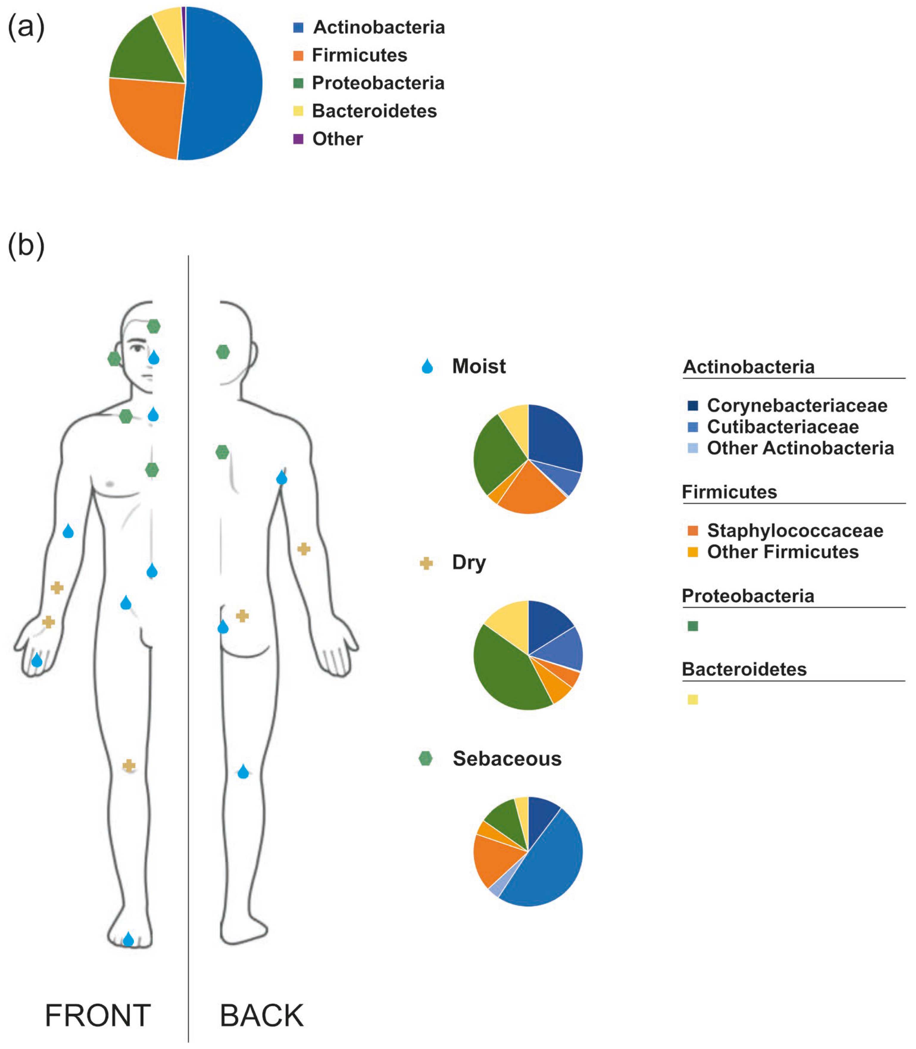
2. The Human Skin Microbiota

The skin, comprising approximately 15% of the adult body weight, functions as a complex barrier organ with physical, chemical, and immunological roles, while also supporting a site-specific microbiota through the provision of nutrients such as amino acids, fatty acids, and lactic acid [3,16]. It is well established that different microbial species, along with their structural components and metabolites play a crucial role in protecting against pathogenic or harmful microorganisms and maintaining skin homeostasis, modulating skin immunity, stimulating and activating various immune responses, metabolizing natural products, and producing antimicrobial peptides (AMPs) [2,17,18]. The initial skin microbiota colonization begins at birth and is strongly influenced by the mode of delivery and the maternal microbiota to which neonates are exposed during labour [6,16]. Throughout life, the microbiota composition is affected by various intrinsic factors, such as the skin site, age, ethnicity, gender, and intra- and interpersonal variability, as well as extrinsic factors, including the mode of delivery, lifestyle, living and working environment, diet, antibiotic exposure, hygiene practices, skin disinfection, cosmetic use, clothing textiles, climate, seasonality, and geographical location [2,4,19].
The normal human skin microbiota is predominantly composed of the bacterial phyla Actinobacteria (52%), Firmicutes (24%), Proteobacteria (17%), and Bacteroidetes (6%) but new taxa are being identified as methodological approaches advance [20]. The dominant genera include Corynebacterium, Cutibacterium (previously known as Propionibacterium), and Staphylococcus genera that comprise more than 60% of the bacterial skin population [16,21,22,23]. The skin also hosts various fungal species, such as Malassezia spp., Cryptococcus spp., Rhodotorula spp., Aspergillus spp., and Epicoccum spp., with Malassezia spp. being the most prevalent, comprising approximately 80% of the total fungal microbiota [4,5,6,16,24,25]. Several studies also indicate the presence of Candida species in the normal skin mycobiome [26,27]. However, fungi represent the least abundant microorganisms on the skin [28]. Viruses within the skin microbiota have been less explored than cellular microorganisms but a recent study identified a large set of viral metagenome-assembled genomes (vMAGs) on several body sites [20]. Among them, cutaneous human papillomaviruses are commonly found but in lower abundance compared to the bacteriophages which are linked to the modulation of bacterial populations and influence skin microbiota homeostasis [29,30]. Cutibacterium and Staphylococcus phages are the most abundant skin phages followed by Streptococcus and Corynebacterium phages [28]. Additionally, there are small mites from the Demodicidae family that naturally inhabit sebaceous areas of the human skin, such as the face and hair follicles. Specifically, two species have been isolated from human samples: Demodex folliculorum and Demodex brevis [29,31].
The composition of the human skin microbiota is relatively stable, though it varies according to the physiological traits of different body regions: sebaceous (e.g., face, chest, and back), moist (e.g., elbows, knees, and genitalia), and dry (e.g., palms) (Figure 1) [6,21,24,32]. Sebaceous (oily) areas show the lowest microbial diversity and are primarily inhabited by lipophilic microorganisms, such as Cutibacterium, Staphylococci, and Corynebacterium [21,24,33]. Moist skin areas, providing a thermally stable and warm environment, are dominated by Corynebacterium and Staphylococcus species, which are well-adapted to survive in these conditions [3,21]. Dry skin areas, by contrast, are among the most diverse sites on the human body in terms of microbial composition. Despite this diversity, dry skin is predominantly colonized by bacteria from the genera Corynebacterium, Flavobacteriales, and Proteobacteria [34].

3. Skin Microbiota Dysbiosis and Inflammatory Skin Diseases

Although the exact mechanisms underlying the maintenance and regulation of skin homeostasis remain unclear, it is well-established that the balance among the members of the skin microbiota plays a critical role in protecting against skin diseases [2]. Several chronic inflammatory skin diseases (ISDs), such as atopic dermatitis, seborrheic dermatitis, acne, psoriasis, and rosacea, are associated with skin microbiota dysbiosis [2,3,4,5,6,7,35]. These conditions are most commonly related to increased levels of Staphylococcus aureus and a decrease in Cutibacterium species. However, each disease has its own specific characteristics and microbial shifts that define its unique dysbiosis profile, which will be discussed in detail below. Table 2 summarizes the current knowledge of the main ISDs and the alterations in the human skin microbiota, presented as an increase or decrease in bacterial genera, species, or species ratios.
Beyond the skin microbiota dysbiosis paradigm, the skin-gut axis hypothesis proposes that the immune system, metabolic-hormonal pathways, and nervous system all play a role in linking gastrointestinal health and skin health [56]. The gut microbiota, as the primary modulator of the gut-skin axis, affects immunity, and a dysbiosis of the gut disrupts the immune system’s equilibrium [57]. According to the gut-skin axis hypothesis, inflammatory skin conditions are caused by a complex interaction between the immune system, lifestyle, and genetics that is constantly synchronized with the skin neuro–immuno–endocrine system [58,59]. The gut and cutaneous microbiota are important in these connections because the immune system and microbes are constantly interacting in the colon and on the skin [57]. Many skin disorders coexist with non-cutaneous conditions like gastrointestinal diseases, further supporting the interdependence of the skin and the gut.

3.1. Atopic Dermatitis

Atopic dermatitis, also known as eczema, is the most common cause of skin illness worldwide with a prevalence of 2.6%, affecting over 200 million people [60]. The hallmark of the disease is dry, red and itchy skin which typically first appears in childhood but can occasionally reappear in adulthood. The pathophysiology of AD remains an open debate. According to the external hypothesis, a basic barrier malfunction (such as a loss-of-function mutation in barrier genes like FLG, that codes for filaggrin) results in a defective epidermal barrier and increased allergen penetration, which in turn triggers the skin immune system and causes atopic inflammation [61]. In contrast, the inside-out hypothesis suggests that skin inflammation develops due to a primary immune defect, which in turn causes a secondary barrier malfunction, microbial dysbiosis and allergen penetration [62].
The skin of AD patients exhibits significant alterations in microbial communities compared to that of healthy individuals, marked by pronounced microbial imbalance and decreased diversity. This is manifested by the significant reduction in Cutibacterium, Streptococcus, Acinetobacter, Corynebacterium, and Prevotella, together with a marked overrepresentation of Staphylococcus, especially S. aureus [37]. Indeed, S. aureus is the primary bacterial species linked to AD, with its dominance especially pronounced during severe disease flares. In these instances, microbial diversity significantly decreases, often resulting in the dominance of a single S. aureus strain [6]. The key event in S. aureus skin colonization is its adhesion to corneocytes by binding to corneodesmosin, a glycoprotein adhesion molecule displayed on the surface of corneocytes. Due to the low levels of skin humectants and moisturizing factors in AD, corneocytes excessively display corneodesmosin and facilitate the binding of S. aureus [63]. S. aureus expresses numerous secreted and wall-anchored virulence factors, e.g., superantigens (toxins), enzymes, and other proteins, thereby compromising the skin barrier and allowing allergen presentation to epidermal dendritic cells, which intensifies AD symptoms [64]. The severity of the condition also correlates with a decrease in coagulase-negative species and a decrease in microbiota diversity [30,65]. Patients with AD tend to have reduced levels of protective commensal skin bacteria, specifically coagulase-negative Staphylococcus species like Staphylococcus epidermidis and Staphylococcus hominis, which shield healthy skin against harmful pathogens by secreting lantibiotics or other antimicrobial peptides (AMPs). Reintroducing these antimicrobial coagulase-negative strains to AD patients decreased S. aureus colonization, indicating that skin microbiota dysbiosis plays a role in the disease pathology [66].
However, it is still insufficiently clear how staphylococci contribute to the development of AD. To determine if elevated staphylococci levels precede clinical symptoms, one study monitored newborns by swabbing four distinct skin sites at three timepoints. The findings have shown that infants that developed AD by 12 months of age had significantly lower levels of commensal staphylococci present at 2 months compared to healthy controls, indicating that early exposure to commensal staphylococci may support the development of a healthy immune system and provide protection against the onset of disease later in life [67]. Similarly, another study found that infants who later developed AD had elevated levels of S. aureus on their skin at 3 months of age. Compared to unaffected, age-matched infants, S. aureus was more prevalent both at the time of AD onset and two months prior [68]. In contrast to S. aureus, S. epidermidis has been considered a skin commensal organism, essential in combating pathogens. Recent results, however, propose that S. epidermidis can contribute to the inflammatory reaction in AD, with its cysteine protease, EcpA, compromising the skin barrier. On the other hand, it was demonstrated that S. hominis, another commensal skin bacterium species, inhibits the S. epidermidis production of EcpA, highlighting the importance of interspecies interactions and the necessity of individualized and multitargeted therapeutic approaches in AD [69].
Compared to the extensive research on skin bacteria, studies examining the diversity of skin fungi in AD remain limited. Similarly to healthy individuals, Malassezia species, particularly Malassezia globosa and Malassezia restricta, are predominant in patients with AD [27]. In patients with mild to moderate AD, M. restricta tends to be more abundant than M. globosa, whereas in severe cases, both species are present in roughly equal proportions [27,70]. It has been demonstrated that Malassezia can penetrate the impaired epithelial barrier in AD, triggering immune cell activation and skin inflammation. Moreover, allergens produced by Malassezia can elicit a specific IgE-mediated immune response, further contributing to the pathogenesis of the disease [71].
A recent study by Wielscher et al. demonstrated that healthy and AD skin harbour distinct phageomes suggesting a causative relationship between shifts in viral and bacterial communities and the development of skin pathology. Despite notable inter-individual variability in phage composition, the study identified increased S.aureus-specific phages in patients with AD, indicating that these could provide a growth advantage for S. aureus. On the other hand, the abundance of certain phages decreased with increasing disease severity implying a possible skin protective role [30].

3.2. Seborrheic Dermatitis

Seborrheic dermatitis is characterized by itchy, red patches and greasy scales, typically affecting areas abundant in sebaceous glands—the scalp, face and upper chest. SD affects up to 5% of the general population, with a higher prevalence in immunocompromised individuals and those with neurological conditions [72]. Because of its multifactorial etiology it is very difficult to establish the primary origin of the disease, with its most prominent pathology factors including immune responses, skin microbiota dysbiosis and increased sebocyte activity [73].
Recent advances in microbiota research revealed an imbalance of fungal and bacterial communities on the skin of SD affected individuals [74]. It has been proposed that Malassezia spp. play an important role in the initiation of SD and are crucial contributors to the disease severity [74,75]. Although a part of the normal skin microbiota, predominantly residing on sebaceous areas, Malassezia can produce lipases and phospholipases that decompose sebum and release unsaturated fatty acids and lipid peroxides causing inflammation [76]. The activation of keratinocyte pattern recognition receptors triggers the NF-κB and MAPK pathways, leading to the release of cytokines from immune cells and the promotion of inflammation [77]. This leads to keratinocyte proliferation, stratum corneum thickening, and barrier disruption. A compromised barrier facilitates Malassezia invasion, resulting in transepidermal water loss, skin peeling, and other SD symptoms caused by ceramide depletion [78]. It has been demonstrated that Malassezia spp. host dsRNA mycoviruses, which can enhance fungal gene expression, and the presence of these mycoviruses is associated with a greater disease severity. Also, higher levels of Malassezia are linked to increased itching scores and greater disease severity, highlighting its role in disease manifestation [79].
In addition, Malassezia-derived unsaturated fatty acids increase skin pH, promoting S. aureus growth and its adhesion to keratinocytes. Indeed, S. aureus has been identified as the most prevalent bacterium on SD-affected skin, occurring significantly more often than in healthy controls [79,80]. Microbiota studies have shown that Cutibacterium is more abundant in non-lesional skin, whereas Staphylococcus and Streptococcus dominate lesional areas [81]. Similarly, Tanaka et al. found increased Cutibacterium at non-lesional sites and elevated Acinetobacter, Staphylococcus, and Streptococcus at lesional ones based on qPCR [41]. These sebum-degrading bacteria, together with Malassezia, may contribute to SD pathogenesis. Elevated Staphylococcus levels correlate with increased transepidermal water loss and higher skin pH, indicating barrier disruption, while Cutibacterium is associated with better hydration and barrier integrity [82]. Supporting this, a recent study reported scalp microbiota dysbiosis in SD, with a reduction in Corynebacterium and a predominance of Pseudomonas, Staphylococcus, and Micrococcus sp. [40].

3.3. Acne Vulgaris

One of the most prevalent dermatological disorders in the world, acne vulgaris affects the pilosebaceous unit, which includes the hair follicle, hair shaft, and sebaceous gland [83]. Cutibacterium acnes, the predominant organism in sebaceous skin areas, is linked to acne vulgaris [84]. Increased sebum production and changes in the fatty acid composition of sebum, hormonal microenvironment dysregulation, interaction with neuropeptides, follicular hyperkeratinization, inflammation induction, and innate and adaptive immune system dysfunction are some of the major mechanisms that contribute to the development of acne [85]. Despite similar relative abundances of C. acnes in the two cohorts (patients and healthy controls), metagenomic research revealed that population structures at the strain level differed dramatically. While certain strains were abundant in healthy skin, others were strongly linked to acne. Therefore, it appears that a loss of diversity and the predominance of phylotype IA1 and, to a lesser extent, phylotype IA2 (among six phylogenetic groups IA1, IA2, IB, IC, II, and III) contribute more to acne development than the relative abundance of C. acnes on the skin [43,85]. One study found that individuals with and without acne have different C. acnes gene expression profiles and proposed a mechanism of vitamin B12-mediated acne pathogenesis. Elevated levels of vitamin B12 in the host causes the inhibition of vitamin B12 production and elevated 2-oxoglutarate and L-glutamate levels in C. acnes. Due to the increased flux of L-glutamate to the porphyrin biosynthetic pathway C. acnes produces an excess of porphyrins. This triggers an inflammatory response in the host, contributing to the development of acne [86]. Dreno et al. showed that the severity of acne is correlated with an increase in Staphylococcus abundance [42]. However, commensal staphylococci have a crucial role in maintaining the equilibrium of C. acnes in healthy human skin. It has been demonstrated that microbial interference contributes to the homeostasis of healthy skin, and certain strains of Staphylococci may have anti-C. acnes properties [87]. S. epidermidis can prevent the growth of C. acnes [42] and reduce the skin irritation it causes [88]. S. epidermidis enhances the fermentation of naturally occurring skin glycerol by producing succinic acid, a byproduct of fatty acid fermentation, thus suppressing the C. acnes proliferation [89]. Toll-like receptor (TLR)-2 production is inhibited by lipoteichoic acid, which mediates the anti-inflammatory actions of S. epidermidis. In this way, S. epidermidis can inhibit the C. acnes-induced production of tumour necrosis factor (TNF)-alpha and IL-6 in keratinocytes [88].
Additionally, differences in C. acnes phage abundance have been observed between healthy individuals and acne patients [45] associating phage deficiency with acne development. Furthermore, a direct relationship between C. acnes phage abundance and age has been reported, suggesting that phage abundance may play a role in the reduced prevalence of acne vulgaris among ageing individuals.

3.4. Psoriasis

Psoriasis is triggered and sustained by Th17 cells and the production of their associated proinflammatory mediators. The hallmarks of the disease are flaky skin patches and scales due to the excessive keratinocyte proliferation and redness from dilated dermal blood vessels and immune cell infiltration [90]. While the precise etiology and pathology of the disease are still unknown, recent studies revealed an association between skin microbiota dysbiosis and psoriasis.
A significant reduction in the alpha- and beta-diversity of the skin microbiota was reported in patients with psoriasis, with lower abundance of Cutibacterium, Burkholderia, and Lactobacilli, along with increased levels of Corynebacterium kroppenstedii, Corynebacterium simulans, Neisseria spp., and Finegoldia spp. compared to healthy individuals [46,91]. The microbiota dysbiosis in psoriasis is characterized by elevated levels of the Staphylococcus genus increased in both lesional and non-lesional skin of patients compared to healthy controls [47,92]. S. aureus colonizes psoriatic skin lesions in almost 60% of patients, compared to 5% to 30% in healthy controls, with the generation of the staphylococcal enterotoxin highly correlated with the disease severity [93]. A study also reported that colonization by S. aureus sets off an inflammatory Th17 response sustaining keratinocyte proliferation, promoting the infiltration of immune cells and subsequently amplifying the inflammation process [47].
Streptococcus is the most common bacterial genus identified in psoriatic skin, with Streptococcus pyogenes infection of the pharynx recently recognized as a risk factor for psoriasis development [49]. S. pyogenes can produce streptococcal M protein and streptococcal peptidoglycan, functioning as superantigens that activate circulating cutaneous lymphocyte-associated antigen (CLA)-positive (+) memory T cells [94]. The activation of CLA+ T cells leads to the increased production of IL-17, the cytokine recognized as the key driver of psoriasis [95].
A variety of fungi in psoriatic skin have been found to trigger the disease by activating the innate immune system [96]. A meta-analysis by Pietrzak et al. reported higher levels of Candida spp. in psoriasis patients compared to controls [97]. In addition to C. albicans, members of the Malassezia genus were also found to aggravate psoriasis, even though no clear correlation was found between them and psoriatic lesions in general [48].
According to several studies, the dysbiosis linked to psoriasis may have its origins in the intestine. The findings that are most commonly reported are the increase in Firmicutes and Actinobacteria and the decrease in Bacteroides and Akkermansia spp. [75,98]. Bacterial translocation in the altered intestine causes inflammation and an abnormal microbiome, which in turn causes the inflammatory response to persist [99].
In a recent study, alterations in the phageome composition were correlated with psoriasis lesional skin [50] with the most abundant phage species showing decreased abundance in lesional areas compared to healthy skin from contralateral sites as well as family controls, indicating the state of dysbiosis of psoriatic skin and the potential effect of phages on bacterial diversity.

3.5. Rosacea

Rosacea is characterized by recurring facial erythema, pustules and papules, telangiectasia (visible blood vessels), and face flushing. Rosacea primarily affects the forehead, chin, nose and cheeks, with the disease alternating between flare-ups and remissions [100]. The current hypothesis suggests that the dysbiosis of the skin microbiota contributes to the disease pathophysiology by stimulating the innate immunity [101]. The dysbiosis of the skin microbiota in rosacea is primarily reflected in the increased load of Demodex mites, particularly Demodex folliculorum [51,54,102]. Demodex mites may have a role in the early inflammatory phase in rosacea by secreting bioactive compounds and influencing immunological responsiveness in sebocytes through the upregulation of TLR2 expression and elevated IL-8 [103]. Furthermore, the Demodex mite is an endosymbiont vector, and various bacterial species including S. epidermidis, Chlamydophila pneumoniae and several Bacillus species, including Bacillus oleronius, Bacillus cereus, Bacillus pumilus and Bacillus simplex, have been isolated from D. folliculorum [51].
The Gram-negative bacterium B. oleronius was initially identified in a D. folliculorum mite isolated from a rosacea patient’s face [104]. B. oleronius displayed antigens that considerably increased the peripheral blood mononuclear cell proliferation in rosacea patients compared to healthy controls, activating the production of proinflammatory cytokines and driving neutrophil recruitment [105]. Recently, a novel mechanism of rosacea pathogenesis was proposed, which includes microbial dysbiosis and a higher abundance of B. oleronius, inducing a proinflammatory response and triggering neoangiogenesis [106]. In addition, elevated temperature in the skin affected by rosacea can influence other bacterial communities and their metabolism. For instance, S. epidermidis, typically considered a commensal, can start behaving as a pathogen in altered skin conditions, generating virulence factors which are absent in individuals who are not rosacea-affected [107], suggesting that S. epidermidis can also play a role in the disease development [108].
According to a recent study, C. kroppenstedtii and D. folliculorum exist in a mutualistic relationship, and viable bacteria appear to be a requirement for the survival of Demodex mites [109]. Rainer et al. discovered that C. kroppenstedtii was one of the most prevalent bacteria and Roseomonas mucosa was decreased in rosacea patients compared to healthy controls [55].

4. Microbiome-Based Products and Skin Health—Treatment Approaches

There are many definitions and classifications of products containing microorganisms or their components, which can provide health benefits or be used as therapeutic agents [110]. Initially, the World Health Organization (WHO) defined probiotics as “live microorganisms that, when administered in adequate amounts, confer a health benefit on the host” [12]. However, this definition covers a wide range of applications, from food products and dietary supplements to medicines. To provide a more precise definition for products with medical applications, the Food and Drug Administration introduced the category “Live Biotherapeutic Products” (LBPs) [111]. This category specifically refers to the use of live microorganisms as therapeutics, but excludes inanimate microbes, their metabolites and components (collectively referred to as postbiotics) and bacteriophages. To comprehensively include all the products that are based on and derived from microorganisms with the potential to provide health benefits (Figure 2) and in light of the fact that the terminology is constantly evolving, the term “microbiome-based products (MBPs)” was used throughout this review.
As the general knowledge about the role of the microbiome in various skin conditions continues to expand, modulating the immune system by restoring microbial homeostasis has emerged as a promising area of research. One direct approach to achieving this balance involves the use of the MBPs, both orally and topically [2,112]. Over the past decade, there has been a significant increase in the use of MBPs in skincare products and dermatological therapies [2]. These treatments have shown potential in the prevention and management of inflammatory skin diseases, potentially offering an alternative to traditional topical or systemic steroids and antimicrobial agents. However, clinical trials and efficacy studies in this field are still relatively limited. Although the exact mechanisms by which MBPs improve skin health are not yet fully understood, numerous studies have demonstrated their beneficial effects [80,113,114,115].

4.1. Topical MBPs

Standard therapeutic approaches for inflammatory skin diseases typically involve the application of corticosteroids (AD and psoriasis) [113,116] and antibiotics (acne) [4,16]. While these treatments are effective in managing the disease and alleviating symptoms, their mechanisms of action and associated side effects limit their long-term use. As a result, they provide short-term symptom relief rather than offering a long-term solution.
In recent years, there has been a growing interest in the application of natural microbiota-based formulations as topical treatments to re-establish microbial homeostasis, restore the skin barrier function and maintain the physiological balance between the host and its microbiota [113]. These formulations are generally considered safe, with fewer adverse effects compared to standard therapies for skin diseases [117]. Topical MBPs may include live or inanimate microorganisms, fermented lysates and extracts, all designed to modulate the skin microbiota and promote skin health. Initially, most research focused on the impact of gut-targeted MBPs on skin conditions [56,118], while more recent studies have shifted the therapeutic strategy toward the direct topical application of MBPs [2,113,115,117,119,120]. Several topically applied MBPs have shown positive effects against inflammatory skin diseases, such as improving the skin barrier, producing antimicrobial peptides, and reducing inflammation. Lactic acid bacteria (LAB) are currently the most commonly used microorganisms in microbiome-based therapies due to their well-established safety profile and GRAS status. However, emerging research is increasingly exploring other bacterial species and bacteriophages as promising therapeutic options for inflammatory skin diseases.
The application of MBPs has been most extensively studied in the treatment of atopic dermatitis, with more limited research available on their use in seborrheic dermatitis. Di Marzio et al. found that the topical application of a cream containing sonicated Streptococcus thermophilus improved ceremide concentrations in the skin and resulted in the improvement of the symptoms in AD patients [121]. Two studies demonstrated that emollients containing Lactobacillus sakei extract [122] or heat-treated Lactobacillus johnsonii NCC 533 [123] inhibited the growth of S. aureus, also restoring the skin barrier and reducing symptoms in subjects with AD. A recent clinical study proved that a topical ointment containing live Lactobacillus reuteri DSM 17938 decreased disease symptoms in AD patients after four and eight weeks of continuous use, showing great potential as a novel topical product [124]. In addition to LAB, other bacterial species have also been studied for their beneficial effects on human skin. Several reports describe positive effects of Roseomonas mucosa (commensal skin bacteria) on alleviating AD symptoms. Myles et al. showed that topical treatment containing live R. mucosa was associated with a significant reduction in AD severity, topical steroid requirement and S. aureus colonization on the skin in children with AD [125]. Another study developed a living bacterial formulation that combines live R. mucosa with a polymeric matrix into a skin product, which demonstrated several positive effects on the skin, including a reduction in S. aureus proliferation and the alleviation of immune/inflammatory responses associated with AD in an animal model [126]. Silverberg et al. conducted a clinical study which assessed safety and efficacy of a topical spray including live ammonia-oxidizing bacterium Nitrosomonas eutropha, reporting reduced pruritus and improved eczema symptoms in patients with mild to moderate AD [127]. Several interesting clinical studies showed that topically applied extract and lysate of Vitreoscilla filiformis (bacterium isolated from sodium sulfurized thermal waters) significantly reduced the severity of AD and SD [128,129,130]. Another clinical trial investigated the efficacy of a topical treatment containing Lactobacillus extract in reducing dandruff, a condition often associated with SD. After two weeks of application, the treatment was shown to improve overall scalp health [131]. In a recent clinical study, Truglio et al. investigated the effects of a topical oily suspension containing Lactobacillus crispatus P17631 and Lacticaseibacillus paracasei I1688 (EUTOPLAC formulation) on the skin microbiome and its potential to reduce SD severity, reporting an improvement in symptoms, with significant reductions in the Malassezia genus in patients with SD [132].
Phage therapy shows promising, though still preliminary, effectiveness in treating atopic dermatitis, with encouraging results primarily from small-scale studies and animal models. Experimental evidence for its potential in treating AD was obtained in the sensitized and S. aureus challenged mouse skin model where S. epidermidis was topically administered with or without S. aureus phage SAGU1. The groups treated with the phage showed the greatest reductions in S. aureus CFUs and plasma IgE levels [133]. Similarly, but more thoroughly, Geng et al. showed that the topical application of the novel S. aureus phage SAP71 improved skin lesions, reduced bacterial loads in the skin, and prevented the development of AD-like skin pathological changes in an AD model [134].
In addition to AD and SD, several studies have also explored the use of topical MBPs in acne treatment. Lebeer et al. developed a topical product incorporating live lactobacilli (Lacticaseibacillus rhamnosus GG, Lactobacillus plantarum WCFS1, and Lactiplantibacillus pentosus KCA1) and demonstrated a positive impact on acne symptoms in a clinical trial involving patients with mild-to-moderate acne. This formulation showed potential in modulating the skin microbiota, including a reduction in the relative abundance of staphylococci and C. acnes [135]. In a recent study, Podrini et al. investigated the effects of topically applied serum containing live Lactobacillus plantarum in a 2D human primary sebocyte culture as a model for treatment of mild-to-moderate papulopustular acne. The study demonstrated a significant reduction in the production of inflammatory mediators and a decrease in the abundance of acne-associated pathogens, including the class-A C. acnes [136]. Additionally, two clinical studies have demonstrated the effectiveness of L. plantarum lysates in treating mild-to-moderate acne and reducing acne lesions [137,138]. Majeed et al. report a clinical study demonstrating that a cream containing cell-free supernatants (CFS) of Bacillus coagulans and inactivated cells of Bacillus longum was effective in treating acne lesions and other seborrheic conditions [139]. Interestingly, another study demonstrated that using a lotion with CFS of Enterococcus faecalis SL-5 (lactic acid bacteria isolated from human feces) significantly reduced acne compared to placebo. This may be due to enterocins, antibacterial molecules produced by E. faecalis [140].
Bacteriophage therapy to treat acne vulgaris has emerged as a potential therapeutic solution. In murine models with C. acnes, bacteriophage injections reduced inflammation, epidermal thickness, and microcomedone-like cysts [141], while topical phage treatment decreased inflammatory and apoptotic markers and was effective against multidrug-resistant C. acnes [142,143]. An MBP containing a three-phage cocktail topical gel formulation was not irritant to human skin or ocular tissues in the ex vivo models and did not permeate through human epidermis while in a cosmetic clinical study, phage gel was well tolerated and reduced the facial burden of C. acnes [144].
On the other hand, the application of topical MBPs for the treatment of psoriasis and rosacea still remains underexplored. To our knowledge, there is only one animal study that has confirmed the effect of probiotic Lactobacillus sakei proBio-65 extract on the severity of skin inflammation in a mouse model of psoriasis [145]. There have been some reports suggesting that bacteriophage therapy could hold promise for treating psoriasis by addressing cutaneous bacterial dysbiosis and modulating immune responses [50,146,147] but firm experimental evidence is still missing. Despite the growing market for skincare products, many of which claim effectiveness in managing these conditions, there is a significant lack of scientific research and clinical trials to validate the efficacy and safety of these topical treatments.
Due to their growing popularity, a number of skincare products based on the microbiota can be found on the market. These products may contain live bacteria, inactivated bacterial cells, or their components (e.g., extracts, lysates and cell-free supernatants). Examples of products available on the market, as well as their effect on skin, are listed in Table 3. The listed products were selected based on the availability of reliable sources, including official websites, that clearly disclosed the formulation composition, the specific microbial strains used, and the claimed dermatological effects.

4.2. Oral MBPs

The gut microbiota plays a crucial role in regulating the host’s immune system, and gut microbiota dysbiosis can trigger autoimmune and inflammatory conditions, not only within the intestines but also in other organs, including the skin [2,118,150]. Growing evidence indicates that gut microbiota dysbiosis is frequently associated with skin diseases such as atopic dermatitis, psoriasis, rosacea, and acne vulgaris, highlighting the potential of oral MBPs as a promising therapeutic strategy for managing these dermatological conditions [2,58,150]. Recent reports have highlighted the potential benefits of oral MBPs for skin health, with most studies focusing on treating atopic dermatitis and acne.
There is a number of animal studies using MBPs to improve AD symptoms in mouse models. Fecal microbiota transplantation modulated the gut microbiota homeostasis and affected the recovery from AD-related inflammation by affecting epidermal layer thicknesses and suppressing inflammatory cytokines [151]. Kim et al. investigated the beneficial effects of a combination therapy using Bifidobacterium longum and the prebiotic galactooligosaccharide (GOS), showing improvement in skin inflammation markers, TEWL and the deficiency of epidermal barrier proteins in the dinitrochlorobenzene (DNCB)-induced AD model [152]. Similar findings were reported for the oral administration of L. paracasei subsp. paracasei NTU 101 [153], tyndallizated L. rhamnosus IDCC 3201 [154], and Lactobacillus sakei WIKIM30 isolated from kimchi [155]. One study proved that the ingestion of yoghurt containing Lactococcus lactis 11/19-B1 significantly improved the severity of AD symptoms in a mouse model and in a clinical trial conducted on children suffering from AD [156]. There are several clinical trials showing that the combination of different lactic acid bacteria species with or without a prebiotic component can reduce the severity of AD symptoms. For instance, Navarro-López et al. investigated the effect of the oral intake of a capsule containing Bifidobacterium lactis CECT 8145, Bifidobacterium longum CECT 7347, L. casei CECT 9104 and maltodextrin (prebiotic) and reported efficacy in reducing the SCORAD index and reducing the use of topical steroids in children with moderate AD [157]. Another clinical study conducted on children with moderate-to-severe AD showed significant clinical improvement in AD severity after therapy containing a probiotic mixture of L. paracasei and Lactobacillus fermentum [158]. Although most studies are conducted on lactic acid bacteria due to their proven safety (GRAS status), recent research aimed to prove the positive effect of other species of human microbiota. Lee et al. used animal models to assess the beneficial effect of commensal gut bacteria Faecalibacterium prausnitzii and Akkermansia muciniphila in the pathogenesis of AD. The study resulted in improvements in AD-related markers and a reduction in TSLP levels, which suggest that these strains have therapeutic potential in AD [159].
In addition to AD, the use of oral MBPs has been most explored for acne treatment. Several in vitro studies have demonstrated the antibacterial activity and anti-inflammatory effects of oral MBPs, which may be beneficial in the treatment of acne [160,161,162,163,164]. Furthermore, Espinoza-Monje et al. proved the antimicrobial and immunomodulatory properties of lactic acid bacterium Weissella viridescens UCO-SMC3 and demonstrated its beneficial effect in the treatment of acne vulgaris in vitro and in a mouse model [165]. As for clinical trials, a recent study from Eguren et al. reported the oral application of a capsule containing L. rhamnosus (CECT30031) and the cyanobacterium Arthrospira platensis (BEA_IDA_0074B) as a safe and effective with clinical and statistically significant reduction in the severity of acne compared to placebo [166]. Another study tested the efficacy of a dietary supplement containing a mixture of Bifidobacterium breve BR03 DSM 16604, L. casei LC03 DSM 27537, Ligilactobacillus salivarius LS03 DSM 22776, and a botanical extract (containing lupeol from Solanum melongena L. and Echinacea extract) in patients with mild to moderate acne. The results from this study provide a promising alternative for the treatment of inflammatory acne as well as for the control of acne-prone skin [167]. In addition to the studies mentioned above, there are several clinical trials that have applied a combination of therapies including MBPs and standard treatments (e.g., antibiotics). Jung et al. conducted a study in acne patients using a probiotic mixture containing L. acidophilus, L. bulgaricus and B. bifidum, applied with or without minocycline. After 12 weeks of treatment, patients treated with the probiotic mixture plus minocycline showed significant improvement in their acne lesions [168]. Interestingly, another clinical study involving patients with intestinal-related dermatoses, including acne, explored the effects of strain E. coli Nissle 1917, and found that the patient group receiving the bacterium orally along with conventional topical therapy (tetracycline, steroids, and retinoids), showed significant improvement compared to patients in the conventional therapy-only group [169].
As previously noted, there is a lack of human studies examining the use of orally applied MBPs for other inflammatory skin conditions, such as SD, psoriasis, and rosacea. Chen et al. investigated the effect of orally administered Lactobacillus pentosus GMNL-77 in a psoriasis-like skin inflammation animal model and demonstrated that the therapy significantly reduced erythematous scaling lesions, a symptom characteristic for psoriasis [170]. A clinical study demonstrated the efficacy of orally administered L. paracasei NCC 2461 ST11 in managing dandruff (symptom typical for SD) and restoring a balanced scalp microbiota [171]. Research into the use of oral MBPs for the treatment of rosacea is increasingly gaining attention in clinical practice. For instance, Fortuna et al. conducted a case study on a rosacea patient who underwent a treatment of low-dose doxycycline alongside probiotic therapy, which included Bifidobacterium breve BR03 and L. salivarius LS01. Following the treatment, the patient displayed significant improvement in both cutaneous and ocular manifestations, leading the researchers to recommend discontinuing doxycycline and continuing with probiotic therapy [172].

4.3. Regulatory Landscape

The regulatory framework for microbiome-based products is developing quickly, driven by growing scientific insights and commercial interest. The regulatory classification depends heavily on the product’s intended use, whether for therapeutic, nutritional, or cosmetic purposes, which directly affects how it is reviewed and approved [15,173,174]. Although the rapid pace of scientific discoveries is well reflected in microbiome-based innovations and product development, regulatory alignment has yet to fully keep pace and account for the complexity and novelty of these products.
Consumer microbiome products, such as probiotics, dietary supplements, and personal care items, are usually regulated under food or cosmetic laws. These categories typically face limited pre-market oversight, leading to concerns about product quality, labelling accuracy, and consumer benefits and safety. In case therapeutic claims are made, they may be reclassified as drugs.
Microbiome-based therapies, particularly live biotherapeutic products (LBPs), are emerging as novel medical treatments designed to modulate the microbiome, improve health or treat disease. These products are generally regulated as drug or medicinal products by agencies such as the U.S. Food and Drug Administration (FDA) and the European Medicines Agency (EMA). Regulatory requirements for LBPs and similar therapies include extensive documentation on identity, purity, potency, and safety, as well as robust preclinical and clinical trial data. Microbiome-based therapies vary in complexity and donor dependence ranging from fully complex like fecal microbiota transplantation, whole microbial ecosystems, and rationally designed microbial consortia, to simpler ones like LBPs (single-strain or mixtures of multiple strains), non-living biotherapeutic products and bacteriophages [15]. The legal definition and regulatory pathway for authorizing MBPs remain key challenges for scientists, regulatory authorities, and product developers. For complex products, the origin of the microbial material is critical in determining the benefit–risk ratio, whereas for less complex products, thorough characterization and quality control play a more significant role.
As knowledge is increasing and new products advance, regulatory frameworks need to evolve to reflect the challenges of product design and manufacturing, delivery methods, and targeted indications. The U.S. has taken the lead with the approval of therapies for recurrent Clostridium difficile infection, while in the European Union there is a lack of harmonization regarding the regulatory frameworks applied to various MBPs but progress is expected through updated frameworks such as the proposed Substances of Human Origin (SoHO) legislation [15]. However, no topical microbiome therapeutic products for treating skin disease have been approved to date [113,117] and most topical formulations are still primarily used as personal skincare products [104,108].
Still, numerous scientific and regulatory hurdles remain, such as defining what constitutes a “healthy” microbiome, accounting for inter-individual variability, understanding complex host–microbiome interactions, evaluating long-term safety and validating/standardizing analytical methodology, strain isolation and banking as well as controlling manufacturing processes. Addressing these challenges will require continued collaboration among regulators, scientists, and industry stakeholders to ensure that safe, effective microbiome-based therapies reach patients and consumers worldwide.

5. Conclusions

MBPs represent a promising therapeutic approach for inflammatory skin diseases such as atopic dermatitis, seborrheic dermatitis, acne, psoriasis and rosacea. These formulations aim to restore microbial homeostasis and modulate immune responses, addressing the underlying pathophysiology of these chronic diseases. The advantages of MBPs are compelling, as they offer a multifaceted mechanism of action, including the modulation of the gut–skin axis, the enhancement of skin barrier function, and reduction in systemic inflammation. Several clinical studies have demonstrated that MBPs can reduce disease severity and inflammatory markers. In addition, MBPs generally exhibit favourable safety profiles, with minimal systemic absorption and a low risk of adverse effects.
However, several challenges must be addressed. The complexity of the skin microbiome and individual variability in microbiota composition complicate the development of standardized treatments. Furthermore, the long-term effects of microbiome modulation are not yet fully understood. Further research is needed to determine the mechanisms of action of MBPs, along with clinical trials to assess the safety and efficacy of topical formulations. Such advancements may pave the way for the development of the first topical MBPs as therapeutics for the treatment of inflammatory skin diseases.
Looking forward, the integration of personalized medicine, including microbiome profiling, holds promise for optimizing microbiome-based therapies. Additionally, interdisciplinary collaboration among dermatologists, microbiologists, and bioengineers will be essential to translate this promising science into clinically viable solutions. Further research is crucial to overcome existing limitations and fully harness the therapeutic potential of MBPs in managing inflammatory skin diseases.

Author Contributions

Conceptualization, A.R., Z.Š. and M.M.; writing—original draft preparation, A.R. and Z.Š.; writing—review and editing, M.M., H.Č.P. and M.P.; visualization, M.M.; supervision, M.P.; funding acquisition, M.M., H.Č.P. and M.P. All authors have read and agreed to the published version of the manuscript.

Funding

This work was supported by the projects “Synergistic innovative combination of microbiota components as a basis for the development of innovative topical products for the treatment and prevention of inflammatory conditions of human skin” (acronym PROBITECT) (KK.01.2.1.02.0137), “Novel pharmabiotics for inflammatory skin conditions” (acronym SKINBIOTIC) (NPOO.C3.2.R3-I1.04.0240), and “Novel topical preparations based on Adriatic microalgae for treatment of rosacea” (acronym ADRIROSA) (IP.1.1.03.0157), all funded by the EU through the ERDF.

Conflicts of Interest

The authors declare no conflicts of interest.

Abbreviations

The following abbreviations are used in this manuscript:
ADAtopic dermatitis
AMPAntimicrobial peptides
CFUColony forming units
FDAFood and Drug Administration
GRASGenerally Recognized as Safe
ISDsInflammatory skin diseases
LBPLive biotherapeutic product
MBPMicrobiome-based product
SCORADScoring atopic dermatitis
SDSeborrheic dermatitis

References

  1. Mousa, W.K.; Chehadeh, F.; Husband, S. Recent Advances in Understanding the Structure and Function of the Human Microbiome. Front. Microbiol. 2022, 13, 825338. [Google Scholar] [CrossRef]
  2. De Almeida, C.V.; Antiga, E.; Lulli, M. Oral and Topical Probiotics and Postbiotics in Skincare and Dermatological Therapy: A Concise Review. Microorganisms 2023, 11, 1420. [Google Scholar] [CrossRef] [PubMed]
  3. Hrestak, D.; Matijašić, M.; Paljetak, H.Č.; Drvar, D.L.; Hadžavdić, S.L.; Perić, M. Skin Microbiota in Atopic Dermatitis. Int. J. Mol. Sci. 2022, 23, 3503. [Google Scholar] [CrossRef] [PubMed]
  4. Carmona-Cruz, S.; Orozco-Covarrubias, L.; Sáez-de-Ocariz, M. The Human Skin Microbiome in Selected Cutaneous Diseases. Front. Cell Infect. Microbiol. 2022, 12, 834135. [Google Scholar] [CrossRef] [PubMed]
  5. Sanford, J.A.; Gallo, R.L. Functions of the Skin Microbiota in Health and Disease. Semin. Immunol. 2013, 25, 370–377. [Google Scholar] [CrossRef]
  6. Byrd, A.L.; Belkaid, Y.; Segre, J.A. The Human Skin Microbiome. Nat. Rev. Microbiol. 2018, 16, 143–155. [Google Scholar] [CrossRef]
  7. Lee, H.J.; Kim, M. Skin Barrier Function and the Microbiome. Int. J. Mol. Sci. 2022, 23, 13071. [Google Scholar] [CrossRef]
  8. Lee, E.S.; Song, E.J.; Nam, Y.-D.; Lee, S.Y. Probiotics in Human Health and Disease: From Nutribiotics to Pharmabiotics. J. Microbiol. 2018, 56, 773–782. [Google Scholar] [CrossRef]
  9. Berg, G.; Rybakova, D.; Fischer, D.; Cernava, T.; Vergès, M.C.C.; Charles, T.; Chen, X.; Cocolin, L.; Eversole, K.; Corral, G.H.; et al. Microbiome Definition Re-Visited: Old Concepts and New Challenges. Microbiome 2020, 8, 103. [Google Scholar] [CrossRef]
  10. Nguyen, U.T.; Kalan, L.R. Forgotten Fungi: The Importance of the Skin Mycobiome. Curr. Opin. Microbiol. 2022, 70, 102235. [Google Scholar] [CrossRef]
  11. Modi, S.R.; Lee, H.H.; Spina, C.S.; Collins, J.J. Antibiotic Treatment Expands the Resistance Reservoir and Ecological Network of the Phage Metagenome. Nature 2013, 499, 219. [Google Scholar] [CrossRef] [PubMed]
  12. Hill, C.; Guarner, F.; Reid, G.; Gibson, G.R.; Merenstein, D.J.; Pot, B.; Morelli, L.; Canani, R.B.; Flint, H.J.; Salminen, S.; et al. Expert Consensus Document: The International Scientific Association for Probiotics and Prebiotics Consensus Statement on the Scope and Appropriate Use of the Term Probiotic. Nat. Rev. Gastroenterol. Hepatol. 2014, 11, 506–514. [Google Scholar] [CrossRef] [PubMed]
  13. Gibson, G.R.; Scott, K.P.; Rastall, R.A.; Tuohy, K.M.; Hotchkiss, A.; Dubert-Ferrandon, A.; Gareau, M.; Murphy, E.F.; Saulnier, D.; Loh, G.; et al. Dietary Prebiotics: Current Status and New Definition. Food Sci. Technol. Bull. Funct. Foods 2010, 7, 1–19. [Google Scholar] [CrossRef]
  14. Salminen, S.; Collado, M.C.; Endo, A.; Hill, C.; Lebeer, S.; Quigley, E.M.M.; Sanders, M.E.; Shamir, R.; Swann, J.R.; Szajewska, H.; et al. The International Scientific Association of Probiotics and Prebiotics (ISAPP) Consensus Statement on the Definition and Scope of Postbiotics. Nat. Rev. Gastroenterol. Hepatol. 2021, 18, 649–667. [Google Scholar] [CrossRef] [PubMed]
  15. Rodriguez, J.; Cordaillat-Simmons, M.; Pot, B.; Druart, C. The Regulatory Framework for Microbiome-Based Therapies: Insights into European Regulatory Developments. NPJ Biofilms Microbiomes 2025, 11, 53. [Google Scholar] [CrossRef]
  16. McLoughlin, I.J.; Wright, E.M.; Tagg, J.R.; Jain, R.; Hale, J.D.F. Skin Microbiome—The Next Frontier for Probiotic Intervention. Probiotics Antimicrob. Proteins 2021, 14, 630–647. [Google Scholar] [CrossRef]
  17. Iwase, T.; Uehara, Y.; Shinji, H.; Tajima, A.; Seo, H.; Takada, K.; Agata, T.; Mizunoe, Y. Staphylococcus Epidermidis Esp Inhibits Staphylococcus Aureus Biofilm Formation and Nasal Colonization. Nature 2010, 465, 346–349. [Google Scholar] [CrossRef]
  18. Belkaid, Y.; Serge, J.A. Dialogue between Skin Microbiota and Immunity. Science 2014, 346, 954–959. [Google Scholar] [CrossRef]
  19. Skowron, K.; Bauza-Kaszewska, J.; Kraszewska, Z.; Wiktorczyk-Kapischke, N.; Grudlewska-Buda, K.; Kwiecińska-Piróg, J.; Wałecka-Zacharska, E.; Radtke, L.; Gospodarek-Komkowska, E. Human Skin Microbiome: Impact of Intrinsic and Extrinsic Factors on Skin Microbiota. Microorganisms 2021, 9, 543. [Google Scholar] [CrossRef]
  20. Saheb Kashaf, S.; Proctor, D.M.; Deming, C.; Saary, P.; Hölzer, M.; Mullikin, J.; Thomas, J.; Young, A.; Bouffard, G.; Barnabas, B.; et al. Integrating Cultivation and Metagenomics for a Multi-Kingdom View of Skin Microbiome Diversity and Functions. Nat. Microbiol. 2022, 7, 169–179. [Google Scholar] [CrossRef]
  21. Grice, E.A. The Intersection of Microbiome and Host at the Skin Interface: Genomic- and Metagenomic-Based Insights. Genome Res. 2015, 25, 1514–1520. [Google Scholar] [CrossRef] [PubMed]
  22. Tavaria, F.K. Topical Use of Probiotics: The Natural Balance. Porto Biomed. J. 2017, 2, 69–70. [Google Scholar] [CrossRef] [PubMed]
  23. Niemeyer-van der Kolk, T.; van der Wall, H.E.C.; Balmforth, C.; Van Doorn, M.B.A.; Rissmann, R. A Systematic Literature Review of the Human Skin Microbiome as Biomarker for Dermatological Drug Development. Br. J. Clin. Pharmacol. 2018, 84, 2178–2193. [Google Scholar] [CrossRef]
  24. Grice, E.A.; Segre, J.A. The Skin Microbiome. Nat. Rev. Microbiol. 2011, 9, 244–253. [Google Scholar] [CrossRef]
  25. Dréno, B.; Araviiskaia, E.; Berardesca, E.; Gontijo, G.; Sanchez Viera, M.; Xiang, L.F.; Martin, R.; Bieber, T. Microbiome in Healthy Skin, Update for Dermatologists. J. Eur. Acad. Dermatol. Venereol. 2016, 30, 2038–2047. [Google Scholar] [CrossRef]
  26. Rafat, Z.; Hashemi, S.J.; Ahamdikia, K.; Daie Ghazvini, R.; Bazvandi, F. Study of Skin and Nail Candida Species as a Normal Flora Based on Age Groups in Healthy Persons in Tehran-Iran. J. Mycol. Med. 2017, 27, 501–505. [Google Scholar] [CrossRef]
  27. Zhang, E.; Tanaka, T.; Tajima, M.; Tsuboi, R.; Nishikawa, A.; Sugita, T. Characterization of the Skin Fungal Microbiota in Patients with Atopic Dermatitis and in Healthy Subjects. Microbiol. Immunol. 2011, 55, 625–632. [Google Scholar] [CrossRef]
  28. Oh, J.; Byrd, A.L.; Park, M.; Kong, H.H.; Segre, J.A. Temporal Stability of the Human Skin Microbiome. Cell 2016, 165, 854–866. [Google Scholar] [CrossRef]
  29. Boxberger, M.; Cenizo, V.; Cassir, N.; La Scola, B. Challenges in Exploring and Manipulating the Human Skin Microbiome. Microbiome 2021, 9, 125. [Google Scholar] [CrossRef]
  30. Wielscher, M.; Pfisterer, K.; Samardzic, D.; Balsini, P.; Bangert, C.; Jäger, K.; Buchberger, M.; Selitsch, B.; Pjevac, P.; Willinger, B.; et al. The Phageome in Normal and Inflamed Human Skin. Sci. Adv. 2023, 9, eadg4015. [Google Scholar] [CrossRef]
  31. Forton, F.M.N.; De Maertelaer, V. Which Factors Influence Demodex Proliferation? A Retrospective Pilot Study Highlighting a Possible Role of Subtle Immune Variations and Sebaceous Gland Status. J. Dermatol. 2021, 48, 1210–1220. [Google Scholar] [CrossRef] [PubMed]
  32. Grice, E.A.; Kong, H.H.; Conlan, S.; Deming, C.B.; Davis, J.; Young, A.C.; Bouffard, G.G.; Blakesley, R.W.; Murray, P.R.; Green, E.D.; et al. Topographical and Temporal Diversity of the Human Skin Microbiome. Science 2009, 324, 1190–1192. [Google Scholar] [CrossRef] [PubMed]
  33. Timm, C.M.; Loomis, K.; Stone, W.; Mehoke, T.; Brensinger, B.; Pellicore, M.; Staniczenko, P.P.A.; Charles, C.; Nayak, S.; Karig, D.K. Isolation and Characterization of Diverse Microbial Representatives from the Human Skin Microbiome. Microbiome 2020, 8, 58. [Google Scholar] [CrossRef]
  34. Sanmiguel, A.; Grice, E.A. Interactions between Host Factors and the Skin Microbiome. Cell. Mol. Life Sci. 2015, 72, 1499–1515. [Google Scholar] [CrossRef]
  35. Zhang, X.E.; Zheng, P.; Ye, S.Z.; Ma, X.; Liu, E.; Pang, Y.-B.; He, Q.Y.; Zhang, Y.X.; Li, W.Q.; Zeng, J.H.; et al. Microbiome: Role in Inflammatory Skin Diseases. J. Inflamm. Res. 2024, 17, 1057–1082. [Google Scholar] [CrossRef] [PubMed]
  36. Nakatsuji, T.; Chen, T.H.; Two, A.M.; Chun, K.A.; Narala, S.; Geha, R.S.; Hata, T.R.; Gallo, R.L. Staphylococcus Aureus Exploits Epidermal Barrier Defects in Atopic Dermatitis to Trigger Cytokine Expression. J. Investig. Dermatol. 2016, 136, 2192–2200. [Google Scholar] [CrossRef]
  37. Kong, H.H.; Oh, J.; Deming, C.; Conlan, S.; Grice, E.A.; Beatson, M.A.; Nomicos, E.; Polley, E.C.; Komarow, H.D.; Mullikin, J.; et al. Temporal Shifts in the Skin Microbiome Associated with Disease Flares and Treatment in Children with Atopic Dermatitis. Genome Res. 2012, 22, 850–859. [Google Scholar] [CrossRef]
  38. Brodská, P.; Panzner, P.; Pizinger, K.; Schmid-Grendelmeier, P. IgE-Mediated Sensitization to Malassezia in Atopic Dermatitis: More Common in Male Patients and in Head and Neck Type. Dermatitis 2014, 25, 120–126. [Google Scholar] [CrossRef]
  39. Watanabe, S.; Kano, R.; Sato, H.; Nakamura, Y.; Hasegawa, A. The Effects of Malassezia Yeasts on Cytokine Production by Human Keratinocytes. J. Investig. Dermatol. 2001, 116, 769–773. [Google Scholar] [CrossRef]
  40. Dityen, K.; Soonthornchai, W.; Kueanjinda, P.; Kullapanich, C.; Tunsakul, N.; Somboonna, N.; Wongpiyabovorn, J. Analysis of Cutaneous Bacterial Microbiota of Thai Patients with Seborrheic Dermatitis. Exp. Dermatol. 2022, 31, 1949–1955. [Google Scholar] [CrossRef]
  41. Tanaka, A.; Cho, O.; Saito, C.; Saito, M.; Tsuboi, R.; Sugita, T. Comprehensive Pyrosequencing Analysis of the Bacterial Microbiota of the Skin of Patients with Seborrheic Dermatitis. Microbiol. Immunol. 2016, 60, 521–526. [Google Scholar] [CrossRef] [PubMed]
  42. Dreno, B.; Martin, R.; Moyal, D.; Henley, J.B.; Khammari, A.; Seité, S. Skin Microbiome and Acne Vulgaris: Staphylococcus, a New Actor in Acne. Exp. Dermatol. 2017, 26, 798–803. [Google Scholar] [CrossRef] [PubMed]
  43. Dagnelie, M.A.; Corvec, S.; Saint-Jean, M.; Nguyen, J.M.; Khammari, A.; Dréno, B. Cutibacterium Acnes Phylotypes Diversity Loss: A Trigger for Skin Inflammatory Process. J. Eur. Acad. Dermatol. Venereol. 2019, 33, 2340–2348. [Google Scholar] [CrossRef] [PubMed]
  44. Liu, J.; Yan, R.; Zhong, Q.; Ngo, S.; Bangayan, N.J.; Nguyen, L.; Lui, T.; Liu, M.; Erfe, M.C.; Craft, N.; et al. The Diversity and Host Interactions of Propionibacterium Acnes Bacteriophages on Human Skin. ISME J. 2015, 9, 2078–2093. [Google Scholar] [CrossRef]
  45. Barnard, E.; Shi, B.; Kang, D.; Craft, N.; Li, H. The Balance of Metagenomic Elements Shapes the Skin Microbiome in Acne and Health. Sci. Rep. 2016, 6, 39491. [Google Scholar] [CrossRef]
  46. Olejniczak-Staruch, I.; Ciążyńska, M.; Sobolewska-Sztychny, D.; Narbutt, J.; Skibińska, M.; Lesiak, A. Alterations of the Skin and Gut Microbiome in Psoriasis and Psoriatic Arthritis. Int. J. Mol. Sci. 2021, 22, 3998. [Google Scholar] [CrossRef]
  47. Chang, H.W.; Yan, D.; Singh, R.; Liu, J.; Lu, X.; Ucmak, D.; Lee, K.; Afifi, L.; Fadrosh, D.; Leech, J.; et al. Alteration of the Cutaneous Microbiome in Psoriasis and Potential Role in Th17 Polarization. Microbiome 2018, 6, 154. [Google Scholar] [CrossRef]
  48. Rudramurthy, S.M.; Honnavar, P.; Chakrabarti, A.; Dogra, S.; Singh, P.; Handa, S. Association of Malassezia Species with Psoriatic Lesions. Mycoses 2014, 57, 483–488. [Google Scholar] [CrossRef]
  49. Yousefi, A.; Karbalaei, M.; Keikha, M. Impact of Streptococcus Pyogenes Infection in Susceptibility to Psoriasis: A Systematic Review and Meta-Analysis. World J. Metaanal 2021, 9, 309–316. [Google Scholar] [CrossRef]
  50. Wang, H.; Chan, H.H.; Ni, M.Y.; Lam, W.W.; Chan, W.M.M.; Pang, H. Bacteriophage of the Skin Microbiome in Patients with Psoriasis and Healthy Family Controls. J. Investig. Dermatol. 2020, 140, 182–190.e5. [Google Scholar] [CrossRef]
  51. Holmes, A.D. Potential Role of Microorganisms in the Pathogenesis of Rosacea. J. Am. Acad. Dermatol. 2013, 69, 1025–1032. [Google Scholar] [CrossRef] [PubMed]
  52. Tatu, A.L.; Clatici, V.; Cristea, V. Isolation of Bacillus Simplex Strain from Demodex Folliculorum and Observations about Demodicosis Spinulosa. Clin. Exp. Dermatol. 2016, 41, 818–820. [Google Scholar] [CrossRef] [PubMed]
  53. Tutka, K.; Żychowska, M.; Reich, A. Diversity and Composition of the Skin, Blood and Gut Microbiome in Rosacea—A Systematic Review of the Literature. Microorganisms 2020, 8, 1756. [Google Scholar] [CrossRef]
  54. Zhu, W.; Hamblin, M.R.; Wen, X. Role of the Skin Microbiota and Intestinal Microbiome in Rosacea. Front. Microbiol. 2023, 14, 1108661. [Google Scholar] [CrossRef]
  55. Rainer, B.M.; Thompson, K.G.; Antonescu, C.; Florea, L.; Mongodin, E.F.; Bui, J.; Fischer, A.H.; Pasieka, H.B.; Garza, L.A.; Kang, S.; et al. Characterization and Analysis of the Skin Microbiota in Rosacea: A Case–Control Study. Am. J. Clin. Dermatol. 2020, 21, 139–147. [Google Scholar] [CrossRef]
  56. Mahmud, M.R.; Akter, S.; Tamanna, S.K.; Mazumder, L.; Esti, I.Z.; Banerjee, S.; Akter, S.; Hasan, M.R.; Acharjee, M.; Hossain, M.S.; et al. Impact of Gut Microbiome on Skin Health: Gut-Skin Axis Observed through the Lenses of Therapeutics and Skin Diseases. Gut Microbes 2022, 14, 2096995. [Google Scholar] [CrossRef]
  57. Salem, I.; Ramser, A.; Isham, N.; Ghannoum, M.A. The Gut Microbiome as a Major Regulator of the Gut-Skin Axis. Front. Microbiol. 2018, 9, 382698. [Google Scholar] [CrossRef]
  58. De Pessemier, B.; Grine, L.; Debaere, M.; Maes, A.; Paetzold, B.; Callewaert, C. Gut–Skin Axis: Current Knowledge of the In-terrelationship between Microbial Dysbiosis and Skin Conditions. Microorganisms 2021, 9, 353. [Google Scholar] [CrossRef]
  59. Slominski, R.M.; Raman, C.; Jetten, A.M.; Slominski, A.T. Neuro–Immuno–Endocrinology of the Skin: How Environment Regulates Body Homeostasis. Nat. Rev. Endocrinol. 2025, 21, 495–509. [Google Scholar] [CrossRef]
  60. Tian, J.; Zhang, D.; Yang, Y.; Huang, Y.; Wang, L.; Yao, X.; Lu, Q. Global Epidemiology of Atopic Dermatitis: A Comprehensive Systematic Analysis and Modelling Study. Br. J. Dermatol. 2023, 190, 55–61. [Google Scholar] [CrossRef]
  61. Stefanovic, N.; Irvine, A.D. Filaggrin and beyond: New Insights into the Skin Barrier in Atopic Dermatitis and Allergic Diseases, from Genetics to Therapeutic Perspectives. Ann. Allergy Asthma Immunol. 2024, 132, 187–195. [Google Scholar] [CrossRef] [PubMed]
  62. Schuler, C.F.; Tsoi, L.C.; Billi, A.C.; Harms, P.W.; Weidinger, S.; Gudjonsson, J.E. Genetic and Immunological Pathogenesis of Atopic Dermatitis. J. Investig. Dermatol. 2024, 144, 954–968. [Google Scholar] [CrossRef] [PubMed]
  63. Towell, A.M.; Feuillie, C.; Vitry, P.; da Costa, T.M.; Mathelié-Guinlet, M.; Kezic, S.; Fleury, O.M.; McAleer, M.A.; Dufrêne, Y.F.; Irvine, A.D.; et al. Staphylococcus Aureus Binds to the N-Terminal Region of Corneodesmosin to Adhere to the Stratum Corneum in Atopic Dermatitis. Proc. Natl. Acad. Sci. USA 2021, 118, e2014444118. [Google Scholar] [CrossRef] [PubMed]
  64. Geoghegan, J.A.; Irvine, A.D.; Foster, T.J. Staphylococcus Aureus and Atopic Dermatitis: A Complex and Evolving Relationship. Trends Microbiol. 2018, 26, 484–497. [Google Scholar] [CrossRef]
  65. Tauber, M.; Balica, S.; Hsu, C.Y.; Jean-Decoster, C.; Lauze, C.; Redoules, D.; Viodé, C.; Schmitt, A.M.; Serre, G.; Simon, M.; et al. Staphylococcus Aureus Density on Lesional and Nonlesional Skin Is Strongly Associated with Disease Severity in Atopic Dermatitis. J. Allergy Clin. Immunol. 2016, 137, 1272–1274.e3. [Google Scholar] [CrossRef]
  66. Nakatsuji, T.; Chen, T.H.; Narala, S.; Chun, K.A.; Two, A.M.; Yun, T.; Shafiq, F.; Kotol, P.F.; Bouslimani, A.; Melnik, A.V.; et al. Antimicrobials from Human Skin Commensal Bacteria Protect against Staphylococcus Aureus and Are Deficient in Atopic Dermatitis. Sci. Transl. Med. 2017, 9, eaah4680. [Google Scholar] [CrossRef]
  67. Kennedy, E.A.; Connolly, J.; Hourihane, J.O.B.; Fallon, P.G.; McLean, W.H.I.; Murray, D.; Jo, J.H.; Segre, J.A.; Kong, H.H.; Irvine, A.D. Skin Microbiome before Development of Atopic Dermatitis: Early Colonization with Commensal Staphylococci at 2 Months Is Associated with a Lower Risk of Atopic Dermatitis at 1 Year. J. Allergy Clin. Immunol. 2017, 139, 166–172. [Google Scholar] [CrossRef]
  68. Meylan, P.; Lang, C.; Mermoud, S.; Johannsen, A.; Norrenberg, S.; Hohl, D.; Vial, Y.; Prod’hom, G.; Greub, G.; Kypriotou, M.; et al. Skin Colonization by Staphylococcus Aureus Precedes the Clinical Diagnosis of Atopic Dermatitis in Infancy. J. Investig. Dermatol. 2017, 137, 2497–2504. [Google Scholar] [CrossRef]
  69. Cau, L.; Williams, M.R.; Butcher, A.M.; Nakatsuji, T.; Kavanaugh, J.S.; Cheng, J.Y.; Shafiq, F.; Higbee, K.; Hata, T.R.; Horswill, A.R.; et al. Staphylococcus Epidermidis Protease EcpA Can Be a Deleterious Component of the Skin Microbiome in Atopic Dermatitis. J. Allergy Clin. Immunol. 2021, 147, 955–966.e16. [Google Scholar] [CrossRef]
  70. Kim, J.E.; Kim, H.S. Microbiome of the Skin and Gut in Atopic Dermatitis (Ad): Understanding the Pathophysiology and Finding Novel Management Strategies. J. Clin. Med. 2019, 8, 444. [Google Scholar] [CrossRef]
  71. Nowicka, D.; Nawrot, U. Contribution of Malassezia Spp. to the Development of Atopic Dermatitis. Mycoses 2019, 62, 588–596. [Google Scholar] [CrossRef] [PubMed]
  72. Borda, L.J.; Wikramanayake, T.C. Seborrheic Dermatitis and Dandruff: A Comprehensive Review. J. Clin. Investig. Dermatol. 2015, 3, 10–13188. [Google Scholar] [CrossRef]
  73. Goldust, M.; Ranjkesh, M.R.; Amirinia, M.; Golforoushan, F.; Rezaee, E.; Rezazadeh Saatlou, M.A. Diagnosis and Treatment of Seborrheic Dermatitis. Am. Fam. Physician 2015, 91, 185–190. [Google Scholar] [CrossRef]
  74. Lin, Q.; Panchamukhi, A.; Li, P.; Shan, W.; Zhou, H.; Hou, L.; Chen, W. Malassezia and Staphylococcus Dominate Scalp Microbiome for Seborrheic Dermatitis. Bioprocess. Biosyst. Eng. 2020, 44, 965–975. [Google Scholar] [CrossRef]
  75. Chen, G.; Chen, Z.-m.; Fan, X.-y.; Jin, Y.-l.; Li, X.; Wu, S.-r.; Ge, W.-w.; Lv, C.-h.; Wang, Y.-k. Gut-Brain-Skin Axis in Psoriasis: A Review. Dermatol. Ther. 2021, 11, 25–38. [Google Scholar] [CrossRef]
  76. Adalsteinsson, J.A.; Kaushik, S.; Muzumdar, S.; Guttman, E.; Ungar, J. An Update on the Microbiology, Immunology and Genetics of Seborrheic Dermatitis. Exp. Dermatol. 2020, 29, 481–489. [Google Scholar] [CrossRef]
  77. Schwartz, J.R.; Mesenger, A.G.; Tosti, A.; Todd, G.; Hordinsky, M.; Hay, R.J.; Wang, X.; Zachariae, C.; Ker, K.M.; Henry, J.P.; et al. A Comprehensive Pathophysiology of Dandruff and Seborrheic Dermatitis–Towards a More Precise Definition of Scalp Health. Acta Derm. Venereol. 2013, 93, 131–137. [Google Scholar] [CrossRef]
  78. Wikramanayake, T.C.; Borda, L.J.; Miteva, M.; Paus, R. Seborrheic Dermatitis-Looking beyond Malassezia. Exp. Dermatol. 2019, 28, 991–1001. [Google Scholar] [CrossRef]
  79. Park, M.; Cho, Y.J.; Kim, D.; Yang, C.S.; Lee, S.M.; Dawson, T.L.; Nakamizo, S.; Kabashima, K.; Lee, Y.W.; Jung, W.H. A Novel Virus Alters Gene Expression and Vacuolar Morphology in Malassezia Cells and Induces a TLR3-Mediated Inflammatory Immune Response. mBio 2020, 11, e01521-20. [Google Scholar] [CrossRef]
  80. Tamer, F.; Kekilli, M. Exploring the Therapeutic Potential of Topical Probiotics in Dermatological Diseases: A Comprehensive Review of Clinical Studies. J. Ger. Soc. Dermatol. 2024, 22, 1195–1204. [Google Scholar] [CrossRef]
  81. Park, T.; Kim, H.J.; Myeong, N.R.; Lee, H.G.; Kwack, I.; Lee, J.; Kim, B.J.; Sul, W.J.; An, S. Collapse of Human Scalp Microbiome Network in Dandruff and Seborrhoeic Dermatitis. Exp. Dermatol. 2017, 26, 835–838. [Google Scholar] [CrossRef]
  82. Tao, R.; Li, R.; Wang, R. Skin Microbiome Alterations in Seborrheic Dermatitis and Dandruff: A Systematic Review. Exp. Dermatol. 2021, 30, 1546–1553. [Google Scholar] [CrossRef]
  83. Moradi Tuchayi, S.; Makrantonaki, E.; Ganceviciene, R.; Dessinioti, C.; Feldman, S.R.; Zouboulis, C.C. Acne Vulgaris. Nat. Rev. Dis. Prim. 2015, 1, 15029. [Google Scholar] [CrossRef] [PubMed]
  84. Fitz-Gibbon, S.; Tomida, S.; Chiu, B.H.; Nguyen, L.; Du, C.; Liu, M.; Elashoff, D.; Erfe, M.C.; Loncaric, A.; Kim, J.; et al. Propionibacterium Acnes Strain Populations in the Human Skin Microbiome Associated with Acne. J. Investig. Dermatol. 2013, 133, 2152–2160. [Google Scholar] [CrossRef] [PubMed]
  85. Dréno, B.; Dagnelie, M.A.; Khammari, A.; Corvec, S. The Skin Microbiome: A New Actor in Inflammatory Acne. Am. J. Clin. Dermatol. 2020, 21, 18–24. [Google Scholar] [CrossRef] [PubMed]
  86. Kang, D.; Shi, B.; Erfe, M.C.; Craft, N.; Li, H. Vitamin B12 Modulates the Transcriptome of the Skin Microbiota in Acne Pathogenesis. Sci. Transl. Med. 2015, 7, 293ra103. [Google Scholar] [CrossRef]
  87. Ahle, C.M.; Stødkilde, K.; Poehlein, A.; Bömeke, M.; Streit, W.R.; Wenck, H.; Reuter, J.H.; Hüpeden, J.; Brüggemann, H. Interference and Co-Existence of Staphylococci and Cutibacterium Acnes within the Healthy Human Skin Microbiome. Commun. Biol. 2022, 5, 293. [Google Scholar] [CrossRef]
  88. Skabytska, Y.; Biedermann, T. Staphylococcus Epidermidis Sets Things Right Again. J. Investig. Dermatol. 2016, 136, 559–560. [Google Scholar] [CrossRef]
  89. Wang, Y.; Kuo, S.; Shu, M.; Yu, J.; Huang, S.; Dai, A.; Two, A.; Gallo, R.L.; Huang, C.M. Staphylococcus Epidermidis in the Human Skin Microbiome Mediates Fermentation to Inhibit the Growth of Propionibacterium Acnes: Implications of Probiotics in Acne Vulgaris. Appl. Microbiol. Biotechnol. 2014, 98, 411–424. [Google Scholar] [CrossRef]
  90. Afonina, I.S.; Van Nuffel, E.; Beyaert, R. Immune Responses and Therapeutic Options in Psoriasis. Cell Mol. Life Sci. 2021, 78, 2709–2727. [Google Scholar] [CrossRef]
  91. Fyhrquist, N.; Muirhead, G.; Prast-Nielsen, S.; Jeanmougin, M.; Olah, P.; Skoog, T.; Jules-Clement, G.; Feld, M.; Barrientos-Somarribas, M.; Sinkko, H.; et al. Microbe-Host Interplay in Atopic Dermatitis and Psoriasis. Nat. Commun. 2019, 10, 4703. [Google Scholar] [CrossRef] [PubMed]
  92. Tett, A.; Pasolli, E.; Farina, S.; Truong, D.T.; Asnicar, F.; Zolfo, M.; Beghini, F.; Armanini, F.; Jousson, O.; De Sanctis, V.; et al. Unexplored Diversity and Strain-Level Structure of the Skin Microbiome Associated with Psoriasis. NPJ Biofilms Microbiomes 2017, 3, 14. [Google Scholar] [CrossRef] [PubMed]
  93. Tomi, N.S.; Kränke, B.; Aberer, E. Staphylococcal Toxins in Patients with Psoriasis, Atopic Dermatitis, and Erythroderma, and in Healthy Control Subjects. J. Am. Acad. Dermatol. 2005, 53, 67–72. [Google Scholar] [CrossRef]
  94. De Jesús-Gil, C.; Ruiz-Romeu, E.; Ferran, M.; Chiriac, A.; Deza, G.; Hóllo, P.; Celada, A.; Pujol, R.M.; Santamaria-Babí, L.F. CLA+ T Cell Response to Microbes in Psoriasis. Front. Immunol. 2018, 9, 1488. [Google Scholar] [CrossRef]
  95. Ruiz-Romeu, E.; Ferran, M.; Sagristà, M.; Gómez, J.; Giménez-Arnau, A.; Herszenyi, K.; Hóllo, P.; Celada, A.; Pujol, R.; Santamaria-Babí, L.F. Streptococcus Pyogenes-Induced Cutaneous Lymphocyte Antigen-Positive T Cell-Dependent Epidermal Cell Activation Triggers TH17 Responses in Patients with Guttate Psoriasis. J. Allergy Clin. Immunol. 2016, 138, 491–499.e6. [Google Scholar] [CrossRef]
  96. Fry, L.; Baker, B.S.; Powles, A.V.; Fahlen, A.; Engstrand, L. Is Chronic Plaque Psoriasis Triggered by Microbiota in the Skin? Br. J. Dermatol. 2013, 169, 47–52. [Google Scholar] [CrossRef]
  97. Pietrzak, A.; Grywalska, E.; Socha, M.; Roliński, J.; Franciszkiewicz-Pietrzak, K.; Rudnicka, L.; Rudzki, M.; Krasowska, D. Prevalence and Possible Role of Candida Species in Patients with Psoriasis: A Systematic Review and Meta-Analysis. Mediat. Inflamm. 2018, 2018, 9602362. [Google Scholar] [CrossRef]
  98. Navarro-López, V.; Martínez-Andrés, A.; Ramírez-Boscà, A.; Ruzafa-Costas, B.; Núñez-Delegido, E.; Carrión-Gutiérrez, M.A.; Prieto-Merino, D.; Codoñer-Cortés, F.; Ramón-Vidal, D.; Genovés-Martínez, S.; et al. Efficacy and Safety of Oral Administration of a Mixture of Probiotic Strains in Patients with Psoriasis: A Randomized Controlled Clinical Trial. Acta Derm. Venereol. 2019, 99, 1078–1084. [Google Scholar] [CrossRef]
  99. Ramírez-Boscá, A.; Navarro-López, V.; Martínez-Andrés, A.; Such, J.; Francés, R.; De La Parte, J.; Asín-Llorca, M. Identification of Bacterial DNA in the Peripheral Blood of Patients with Active Psoriasis. JAMA Dermatol. 2015, 151, 670–671. [Google Scholar] [CrossRef]
  100. Barakji, Y.A.; Rønnstad, A.T.M.; Christensen, M.O.; Zachariae, C.; Wienholtz, N.K.F.; Halling, A.S.; Maul, J.T.; Thomsen, S.F.; Egeberg, A.; Thyssen, J.P. Assessment of Frequency of Rosacea Subtypes in Patients with Rosacea: A Systematic Review and Meta-Analysis. JAMA Dermatol. 2022, 158, 617–625. [Google Scholar] [CrossRef]
  101. Two, A.M.; Wu, W.; Gallo, R.L.; Hata, T.R. Rosacea: Part I. Introduction, Categorization, Histology, Pathogenesis, and Risk Factors. J. Am. Acad. Dermatol. 2015, 72, 749–758. [Google Scholar] [CrossRef] [PubMed]
  102. Casas, C.; Paul, C.; Lahfa, M.; Livideanu, B.; Lie Lejeune, O.; Alvarez-Georges, S.; Saint-Martory, C.; Degouy, A.; Rie Mengeaud, V.; Ginisty, H.; et al. Quantification of Demodex Folliculorum by PCR in Rosacea and Its Relationship to Skin Innate Immune Activation. Exp. Dermatol. 2012, 21, 906–910. [Google Scholar] [CrossRef] [PubMed]
  103. Lacey, N.; Russell-Hallinan, A.; Zouboulis, C.C.; Powell, F.C. Demodex Mites Modulate Sebocyte Immune Reaction: Possible Role in the Pathogenesis of Rosacea. Br. J. Dermatol. 2018, 179, 420–430. [Google Scholar] [CrossRef] [PubMed]
  104. Lacey, N.; Delaney, S.; Kavanagh, K.; Powell, F.C. Mite-related Bacterial Antigens Stimulate Inflammatory Cells in Rosacea. Br. J. Dermatol. 2007, 157, 474–481. [Google Scholar] [CrossRef]
  105. McMahon, F.; Banville, N.; Bergin, D.A.; Smedman, C.; Paulie, S.; Reeves, E.; Kavanagh, K. Activation of Neutrophils via IP3 Pathway Following Exposure to Demodex-Associated Bacterial Proteins. Inflammation 2016, 39, 425–433. [Google Scholar] [CrossRef]
  106. Mylonas, A.; Hawerkamp, H.C.; Wang, Y.; Chen, J.; Messina, F.; Demaria, O.; Meller, S.; Homey, B.; Di Domizio, J.; Mazzolai, L.; et al. Type I IFNs Link Skin-Associated Dysbiotic Commensal Bacteria to Pathogenic Inflammation and Angiogenesis in Rosacea. JCI Insight 2023, 8, e151846. [Google Scholar] [CrossRef]
  107. Dahl, M.V.; Ross, A.J.; Schlievert, P.M. Temperature Regulates Bacterial Protein Production: Possible Role in Rosacea. J. Am. Acad. Dermatol. 2004, 50, 266–272. [Google Scholar] [CrossRef]
  108. Whitfeld, M.; Gunasingam, N.; Leow, L.J.; Shirato, K.; Preda, V. Staphylococcus Epidermidis: A Possible Role in the Pustules of Rosacea. J. Am. Acad. Dermatol. 2011, 64, 49–52. [Google Scholar] [CrossRef]
  109. Clanner-Engelshofen, B.M.; French, L.E.; Reinholz, M. Corynebacterium Kroppenstedtii Subsp. Demodicis Is the Endobacterium of Demodex Folliculorum. J. Eur. Acad. Dermatol. Venereol. 2020, 34, 1043–1049. [Google Scholar] [CrossRef]
  110. Cordaillat-Simmons, M.; Rouanet, A.; Pot, B. Live Biotherapeutic Products: The Importance of a Defined Regulatory Framework. Exp. Mol. Med. 2020, 52, 1397–1406. [Google Scholar] [CrossRef]
  111. FDA. Early Clinical Trials with Live Biotherapeutic Products: Chemistry, Manufacturing, and Control Information; Guidance for Industry; Food and Drug Administration: Muntinlupa, Philippines, 2016. [Google Scholar]
  112. Yu, Y.; Dunaway, S.; Champer, J.; Kim, J.; Alikhan, A. Changing Our Microbiome: Probiotics in Dermatology. Br. J. Dermatol. 2020, 182, 39–46. [Google Scholar] [CrossRef] [PubMed]
  113. Habeebuddin, M.; Karnati, R.K.; Shiroorkar, P.N.; Nagaraja, S.; Asdaq, S.M.B.; Anwer, M.K.; Fattepur, S. Topical Probiotics: More Than a Skin Deep. Pharmaceutics 2022, 14, 557. [Google Scholar] [CrossRef] [PubMed]
  114. Gowda, V.; Sarkar, R.; Verma, D.; Das, A. Probiotics in Dermatology: An Evidence-Based Approach. Indian Dermatol. Online J. 2024, 15, 571–583. [Google Scholar] [CrossRef] [PubMed]
  115. Alves, A.C.; Martins, S.M.d.S.B., Jr.; Belo, J.V.T.; Lemos, M.V.C.; Lima, C.E.d.M.C.; Silva, C.D.d.; Zagmignan, A.; Nascimento da Silva, L.C. Global Trends and Scientific Impact of Topical Probiotics in Dermatological Treatment and Skincare. Microorganisms 2024, 12, 2010. [Google Scholar] [CrossRef]
  116. Guttman-Yassky, E.; Renert-Yuval, Y.; Brunner, P.M. Atopic Dermatitis. Lancet 2025, 405, 583–596. [Google Scholar] [CrossRef]
  117. França, K.; Frost, P.; Ther, D. Topical Probiotics in Dermatological Therapy and Skincare: A Concise Review. Dermatol. Ther. 2021, 11, 71–77. [Google Scholar] [CrossRef]
  118. Gao, T.; Wang, X.; Li, Y.; Ren, F. The Role of Probiotics in Skin Health and Related Gut–Skin Axis: A Review. Nutrients 2023, 15, 3123. [Google Scholar] [CrossRef]
  119. Knackstedt, R.; Knackstedt, T.; Gatherwright, J. The Role of Topical Probiotics in Skin Conditions: A Systematic Review of Animal and Human Studies and Implications for Future Therapies. Exp. Dermatol. 2020, 29, 15–21. [Google Scholar] [CrossRef]
  120. Lee, G.R.; Maarouf, M.; Hendricks, A.J.; Lee, D.E.; Shi, V.Y. Topical Probiotics: The Unknowns behind Their Rising Popularity. Dermatol. Online J. 2019, 25, 5. [Google Scholar] [CrossRef]
  121. Di Marzio, L.; Centi, C.; Cinque, B.; Masci, S.; Giuliani, M.; Arcieri, A.; Zicari, L.; De Simone, C.; Cifone, M.G. Effect of the Lactic Acid Bacterium Streptococcus Thermophilus on Stratum Corneum Ceramide Levels and Signs and Symptoms of Atopic Dermatitis Patients. Exp. Dermatol. 2003, 12, 615–620. [Google Scholar] [CrossRef]
  122. Park, S.B.; Im, M.; Lee, Y.; Lee, J.H.; Lim, J.; Park, Y.H.; Seo, Y.J. Effect of Emollients Containing Vegetable-Derived Lactobacillus in the Treatment of Atopic Dermatitis Symptoms: Split-Body Clinical Trial. Ann. Dermatol. 2014, 26, 150–155. [Google Scholar] [CrossRef] [PubMed]
  123. Blanchet-Réthoré, S.; Bourdès, V.; Mercenier, A.; Haddar, C.H.; Verhoeven, P.O.; Andres, P. Effect of a Lotion Containing the Heat-Treated Probiotic Strain Lactobacillus Johnsonii NCC 533 on Staphylococcus Aureus Colonization in Atopic Dermatitis. Clin. Cosmet. Investig. Dermatol. 2017, 10, 249–257. [Google Scholar] [CrossRef] [PubMed]
  124. Butler, É.; Lundqvist, C.; Axelsson, J. Lactobacillus Reuteri DSM 17938 as a Novel Topical Cosmetic Ingredient: A Proof of Concept Clinical Study in Adults with Atopic Dermatitis. Microorganisms 2020, 8, 1026. [Google Scholar] [CrossRef]
  125. Myles, I.A.; Castillo, C.R.; Barbian, K.D.; Kanakabandi, K.; Virtaneva, K.; Fitzmeyer, E.; Paneru, M.; Otaizo-Carrasquero, F.; Myers, T.G.; Markowitz, T.E.; et al. Therapeutic Responses to Roseomonas Mucosa in Atopic Dermatitis May Involve Lipid-Mediated TNF-Related Epithelial Repair. Sci. Transl. Med. 2020, 12, eaaz8631. [Google Scholar] [CrossRef]
  126. Liu, X.; Qin, Y.; Dong, L.; Han, Z.; Liu, T.; Tang, Y.; Yu, Y.; Ye, J.; Tao, J.; Zeng, X.; et al. Living Symbiotic Bacteria-Involved Skin Dressing to Combat Indigenous Pathogens for Microbiome-Based Biotherapy toward Atopic Dermatitis. Bioact. Mater. 2023, 21, 253–266. [Google Scholar] [CrossRef]
  127. Silverberg, J.I.; Lio, P.A.; Simpson, E.L.; Li, C.; Brownell, D.R.; Gryllos, I.; Ng-Cashin, J.; Krueger, T.; Swaidan, V.R.; Bliss, R.L.; et al. Efficacy and Safety of Topically Applied Therapeutic Ammonia Oxidising Bacteria in Adults with Mild-to-Moderate Atopic Dermatitis and Moderate-to-Severe Pruritus: A Randomised, Double-Blind, Placebo-Controlled, Dose-Ranging, Phase 2b Trial. eClinicalMedicine 2023, 60, 102002. [Google Scholar] [CrossRef]
  128. Guéniche, A.; Cathelineau, A.; Bastien, P.; Esdaile, J.; Martin, R.; Queille Roussel, C.; Breton, L. Vitreoscilla Filiformis Biomass Improves Seborrheic Dermatitis. J. Eur. Acad. Dermatol. Venereol. 2008, 22, 1014–1015. [Google Scholar] [CrossRef]
  129. Gueniche, A.; Knaudt, B.; Schuck, E.; Volz, T.; Bastien, P.; Martin, R.; Röcken, M.; Breton, L.; Biedermann, T. Effects of Nonpathogenic Gram-Negative Bacterium Vitreoscilla Filiformis Lysate on Atopic Dermatitis: A Prospective, Randomized, Double-Blind, Placebo-Controlled Clinical Study. Br. J. Dermatol. 2008, 159, 1357–1363. [Google Scholar] [CrossRef]
  130. Gueniche, A.; Liboutet, M.; Cheilian, S.; Fagot, D.; Juchaux, F.; Breton, L. Vitreoscilla Filiformis Extract for Topical Skin Care: A Review. Front. Cell Infect. Microbiol. 2021, 11, 747663. [Google Scholar] [CrossRef]
  131. Fithian, E.; Thivalapill, N.; Kosner, J.; Necheles, J.; Bilaver, L. Natural Topical Treatment Contributes to a Reduction of Dry Scalp Symptoms in Children. Clin. Cosmet. Investig. Dermatol. 2023, 16, 2757–2762. [Google Scholar] [CrossRef]
  132. Truglio, M.; Sivori, F.; Cavallo, I.; Abril, E.; Licursi, V.; Fabrizio, G.; Cardinali, G.; Pignatti, M.; Toma, L.; Valensise, F.; et al. Modulating the Skin Mycobiome-Bacteriome and Treating Seborrheic Dermatitis with a Probiotic-Enriched Oily Suspension. Sci. Rep. 2024, 14, 2722. [Google Scholar] [CrossRef] [PubMed]
  133. Shimamori, Y.; Mitsunaka, S.; Yamashita, H.; Suzuki, T.; Kitao, T.; Kubori, T.; Nagai, H.; Takeda, S.; Ando, H. Staphylococcal Phage in Combination with Staphylococcus Epidermidis as a Potential Treatment for Staphylococcus Aureus-Associated Atopic Dermatitis and Suppressor of Phage-Resistant Mutants. Viruses 2021, 13, 7. [Google Scholar] [CrossRef] [PubMed]
  134. Geng, H.; Yang, X.; Zou, C.; Zhang, W.; Xiang, J.; Yang, K.; Shu, Y.; Luan, G.; Jia, X.; Lu, M. Isolation of the Novel Phage SAP71 and Its Potential Use against Staphylococcus Aureus in an Atopic Dermatitis Mouse Model. Virus Genes 2024, 60, 737–746. [Google Scholar] [CrossRef] [PubMed]
  135. Lebeer, S.; Oerlemans, E.F.M.; Claes, I.; Henkens, T.; Delanghe, L.; Wuyts, S.; Spacova, I.; van den Broek, M.F.L.; Tuyaerts, I.; Wittouck, S.; et al. Selective Targeting of Skin Pathobionts and Inflammation with Topically Applied Lactobacilli. Cell Rep. Med. 2022, 3, 100521. [Google Scholar] [CrossRef]
  136. Podrini, C.; Schramm, L.; Marianantoni, G.; Apolinarska, J.; McGuckin, C.; Forraz, N.; Milet, C.; Desroches, A.L.; Payen, P.; D’Aguanno, M.; et al. Topical Administration of Lactiplantibacillus Plantarum (SkinDuoTM) Serum Improves Anti-Acne Properties. Microorganisms 2023, 11, 417. [Google Scholar] [CrossRef]
  137. Cui, H.; Guo, C.; Wang, Q.; Feng, C.; Duan, Z. A Pilot Study on the Efficacy of Topical Lotion Containing Anti-Acne Postbiotic in Subjects with Mild -to -Moderate Acne. Front. Med. 2022, 9, 1064460. [Google Scholar] [CrossRef]
  138. Cui, H.; Feng, C.; Guo, C.; Duan, Z. Development of Novel Topical Anti-Acne Cream Containing Postbiotics for Mild-to-Moderate Acne. Indian. J. Dermatol. 2022, 67, 667–673. [Google Scholar] [CrossRef]
  139. Majeed, M.; Majeed, S.; Nagabhushanam, K.; Mundkur, L.; Rajalakshmi, H.R.; Shah, K.; Beede, K. Novel Topical Application of a Postbiotic, Lactosporin®, in Mild to Moderate Acne: A Randomized, Comparative Clinical Study to Evaluate Its Efficacy, Tolerability and Safety. Cosmetics 2020, 7, 70. [Google Scholar] [CrossRef]
  140. Kang, B.S.; Seo, J.-G.; Lee, G.-S.; Kim, J.-H.; Kim, S.Y.; Han, Y.W.; Kang, H.; Kim, H.O.; Rhee, J.H.; Chung, M.-J.; et al. Antimicrobial Activity of Enterocins from Enterococcus Faecalis SL-5 against Propionibacterium Acnes, the Causative Agent in Acne Vulgaris, and Its Therapeutic Effect. J. Microbiol. 2009, 47, 101–109. [Google Scholar] [CrossRef]
  141. Kim, M.J.; Eun, D.H.; Kim, S.M.; Kim, J.; Lee, W.J. Efficacy of Bacteriophages in Propionibacterium Acnes-Induced Inflammation in Mice. Ann. Dermatol. 2019, 31, 22–28. [Google Scholar] [CrossRef]
  142. Rimon, A.; Rakov, C.; Lerer, V.; Sheffer-Levi, S.; Oren, S.A.; Shlomov, T.; Shasha, L.; Lubin, R.; Zubeidat, K.; Jaber, N.; et al. Topical Phage Therapy in a Mouse Model of Cutibacterium Acnes-Induced Acne-like Lesions. Nat. Commun. 2023, 14, 1005. [Google Scholar] [CrossRef] [PubMed]
  143. Lam, H.Y.P.; Lai, M.J.; Chen, T.Y.; Wu, W.J.; Peng, S.Y.; Chang, K.C. Therapeutic Effect of a Newly Isolated Lytic Bacteriophage against Multi-Drug-Resistant Cutibacterium Acnes Infection in Mice. Int. J. Mol. Sci. 2021, 22, 7031. [Google Scholar] [CrossRef] [PubMed]
  144. Golembo, M.; Puttagunta, S.; Rappo, U.; Weinstock, E.; Engelstein, R.; Gahali-Sass, I.; Moses, A.; Kario, E.; Ben-Dor Cohen, E.; Nicenboim, J.; et al. Development of a Topical Bacteriophage Gel Targeting Cutibacterium Acnes for Acne Prone Skin and Results of a Phase 1 Cosmetic Randomized Clinical Trial. Ski. Health Dis. 2022, 2, e93. [Google Scholar] [CrossRef] [PubMed]
  145. Rather, I.A.; Bajpai, V.K.; Huh, Y.S.; Han, Y.K.; Bhat, E.A.; Lim, J.; Paek, W.K.; Park, Y.H. Probiotic Lactobacillus Sakei ProBio-65 Extract Ameliorates the Severity of Imiquimod Induced Psoriasis-like Skin Inflammation in a Mouse Model. Front. Microbiol. 2018, 9, 1021. [Google Scholar] [CrossRef]
  146. Zapi-Colín, L.A.; Gutiérrez-González, G.; Rodríguez-Martínez, S.; Cancino-Diaz, J.C.; Méndez-Tenorio, A.; Pérez-Tapia, S.M.; Gómez-Chávez, F.; Cedillo-Peláez, C.; Cancino-Diaz, M.E. A Peptide Derived from Phage-Display Limits Psoriasis-like Lesions in Mice. Heliyon 2020, 6, e04162. [Google Scholar] [CrossRef]
  147. Waghralkar, R.; Jhunjhunwala, B. Observational Case Studies of the Effect of Phage Laden Ganga Water on Psoriasis. IP Indian J. Clin. Exp. Dermatol. 2021, 7, 186–190. [Google Scholar] [CrossRef]
  148. Jain, R.; Voss, A.L.; Tagg, J.R.; Hale, J.D.F. Evaluation of the Preliminary Safety, Tolerability and Colonisation Efficacy of Topical Probiotic Formulations Containing Micrococcus Luteus Q24 in Healthy Human Adults. Cosmetics 2022, 9, 121. [Google Scholar] [CrossRef]
  149. Jain, R.; Voss, A.L.; Del Rosario, J.; Hale, J.D.F. Efficacy of a Topical Live Probiotic in Improving Skin Health. Int. J. Cosmet. Sci. 2025, 47, 488–496. [Google Scholar] [CrossRef]
  150. Szántó, M.; Dózsa, A.; Antal, D.; Szabó, K.; Kemény, L.; Bai, P. Targeting the Gut-Skin Axis—Probiotics as New Tools for Skin Disorder Management? Exp. Dermatol. 2019, 28, 1210–1218. [Google Scholar] [CrossRef]
  151. Jiang, X.; Liu, Z.; Ma, Y.; Miao, L.; Zhao, K.; Wang, D.; Wang, M.; Ruan, H.; Xu, F.; Zhou, Q.; et al. Fecal Microbiota Transplantation Affects the Recovery of AD-Skin Lesions and Enhances Gut Microbiota Homeostasis. Int. Immunopharmacol. 2023, 118, 110005. [Google Scholar] [CrossRef]
  152. Kim, S.; Han, S.Y.; Lee, J.; Kim, N.R.; Lee, B.R.; Kim, H.; Kwon, M.; Ahn, K.; Noh, Y.; Kim, S.J.; et al. Bifidobacterium Longum and Galactooligosaccharide Improve Skin Barrier Dysfunction and Atopic Dermatitis-like Skin. Allergy Asthma Immunol. Res. 2022, 14, 549–564. [Google Scholar] [CrossRef] [PubMed]
  153. Liu, C.F.; Shih, T.W.; Lee, C.L.; Pan, T.M. The Beneficial Role of Lactobacillus Paracasei Subsp. Paracasei NTU 101 in the Prevention of Atopic Dermatitis. Curr. Issues Mol. Biol. 2024, 46, 2236–2250. [Google Scholar] [CrossRef] [PubMed]
  154. Lee, S.H.; Yoon, J.M.; Kim, Y.H.; Jeong, D.G.; Park, S.; Kang, D.J. Therapeutic Effect of Tyndallized Lactobacillus Rhamnosus IDCC 3201 on Atopic Dermatitis Mediated by Down-Regulation of Immunoglobulin E in NC/Nga Mice. Microbiol. Immunol. 2016, 60, 468–476. [Google Scholar] [CrossRef] [PubMed]
  155. Kwon, M.S.; Lim, S.K.; Jang, J.Y.; Lee, J.; Park, H.K.; Kim, N.; Yun, M.; Shin, M.Y.; Jo, H.E.; Oh, Y.J.; et al. Lactobacillus Sakei WIKIM30 Ameliorates Atopic Dermatitis-like Skin Lesions by Inducing Regulatory T Cells and Altering Gut Microbiota Structure in Mice. Front. Immunol. 2018, 9, 1905. [Google Scholar] [CrossRef]
  156. Suzuki, T.; Nishiyama, K.; Kawata, K.; Sugimoto, K.; Isome, M.; Suzuki, S.; Nozawa, R.; Ichikawa, Y.; Watanabe, Y.; Suzutani, T. Effect of the Lactococcus Lactis 11/19-B1 Strain on Atopic Dermatitis in a Clinical Test and Mouse Model. Nutrients 2020, 12, 763. [Google Scholar] [CrossRef]
  157. Navarro-Lopez, V.; Ramirez-Bosca, A.; Ramon-Vidal, D.; Ruzafa-Costas, B.; Genoves-Martinez, S.; Chenoll-Cuadros, E.; Carrion-Gutierrez, M.; De La Parte, J.H.; Prieto-Merino, D.; Codoner-Cortes, F.M. Effect of Oral Administration of a Mixture of Probiotic Strains on SCORAD Index and Use of Topical Steroids in Young Patients with Moderate Atopic Dermatitis a Randomized Clinical Trial. JAMA Dermatol. 2018, 154, 37–43. [Google Scholar] [CrossRef]
  158. Wang, I.J.; Wang, J.Y. Children with Atopic Dermatitis Show Clinical Improvement after Lactobacillus Exposure. Clin. Exp. Allergy 2015, 45, 779–787. [Google Scholar] [CrossRef]
  159. Lee, Y.; Byeon, H.R.; Jang, S.Y.; Hong, M.G.; Kim, D.; Lee, D.; Shin, J.H.; Kim, Y.; Kang, S.G.; Seo, J.G. Oral Administration of Faecalibacterium Prausnitzii and Akkermansia Muciniphila Strains from Humans Improves Atopic Dermatitis Symptoms in DNCB Induced NC/Nga Mice. Sci. Rep. 2022, 12, 7324. [Google Scholar] [CrossRef]
  160. Lopes, E.G.; Moreira, D.A.; Gullón, P.; Gullón, B.; Cardelle-Cobas, A.; Tavaria, F.K. Topical Application of Probiotics in Skin: Adhesion, Antimicrobial and Antibiofilm in Vitro Assays. J. Appl. Microbiol. 2017, 122, 450–461. [Google Scholar] [CrossRef]
  161. Cosseau, C.; Devine, D.A.; Dullaghan, E.; Gardy, J.L.; Chikatamarla, A.; Gellatly, S.; Yu, L.L.; Pistolic, J.; Falsafi, R.; Tagg, J.; et al. The Commensal Streptococcus Salivarius K12 Downregulates the Innate Immune Responses of Human Epithelial Cells and Promotes Host-Microbe Homeostasis. Infect. Immun. 2008, 76, 4163–4175. [Google Scholar] [CrossRef]
  162. Lee, D.K.; Kim, M.J.; Ham, J.W.; An, H.M.; Cha, M.K.; Lee, S.W.; Park, C.I.; Shin, S.H.; Lee, K.O.; Kim, K.J.; et al. In Vitro Evaluation of Antibacterial Activities and Anti-Inflammatory Effects of Bifidobacterium Spp. Addressing Acne Vulgaris. Arch. Pharm. Res. 2012, 35, 1065–1071. [Google Scholar] [CrossRef]
  163. Al-Ghazzewi, F.H.; Tester, R.F. Effect of Konjac Glucomannan Hydrolysates and Probiotics on the Growth of the Skin Bacterium Propionibacterium Acnes in Vitro. Int. J. Cosmet. Sci. 2010, 32, 139–142. [Google Scholar] [CrossRef] [PubMed]
  164. Deidda, F.; Amoruso, A.; Nicola, S.; Graziano, T.; Pane, M.; Mogna, L. New Approach in Acne Therapy A Specific Bacteriocin Activity and a Targeted Anti IL-8 Property in Just 1 Probiotic Strain, the L. Salivarius LS03. J. Clin. Gastroenterol. 2018, 52, S78–S81. [Google Scholar] [CrossRef] [PubMed]
  165. Espinoza-Monje, M.; Campos, J.; Alvarez Villamil, E.; Jerez, A.; Dentice Maidana, S.; Elean, M.; Salva, S.; Kitazawa, H.; Villena, J.; García-Cancino, A. Characterization of Weissella Viridescens Uco-Smc3 as a Potential Probiotic for the Skin: Its Beneficial Role in the Pathogenesis of Acne Vulgaris. Microorganisms 2021, 9, 1486. [Google Scholar] [CrossRef]
  166. Eguren, C.; Navarro-Blasco, A.; Corral-Forteza, M.; Reolid-Pérez, A.; Setó-Torrent, N.; García-Navarro, A.; Prieto-Merino, D.; Núñez-Delegido, E.; Sánchez-Pellicer, P.; Navarro-López, V. A Randomized Clinical Trial to Evaluate the Efficacy of an Oral Probiotic in Acne Vulgaris. Acta Derm. Venereol. 2024, 104, adv33206. [Google Scholar] [CrossRef]
  167. Rinaldi, F.; Marotta, L.; Mascolo, A.; Amoruso, A.; Pane, M.; Giuliani, G.; Pinto, D. Facial Acne: A Randomized, Double-Blind, Placebo-Controlled Study on the Clinical Efficacy of a Symbiotic Dietary Supplement. Dermatol. Ther. 2022, 12, 577–589. [Google Scholar] [CrossRef]
  168. Jung, G.W.; Tse, J.E.; Guiha, I.; Rao, J. Prospective, Randomized, Open-Label Trial Comparing the Safety, Efficacy, and Tolerability of an Acne Treatment Regimen with and without a Probiotic Supplement and Minocycline in Subjects with Mild to Moderate Acne. J. Cutan. Med. Surg. 2013, 17, 114–122. [Google Scholar] [CrossRef]
  169. Manzhalii, E.; Hornuss, D.; Stremmel, W. Intestinal-Borne Dermatoses Significantly Improved by Oral Application of Escherichia Coli Nissle 1917. World J. Gastroenterol. 2016, 22, 5415–5421. [Google Scholar] [CrossRef]
  170. Chen, Y.H.; Wu, C.S.; Chao, Y.H.; Lin, C.C.; Tsai, H.Y.; Li, Y.R.; Chen, Y.Z.; Tsai, W.H.; Chen, Y.K. Lactobacillus Pentosus GMNL-77 Inhibits Skin Lesions in Imiquimod-Induced Psoriasis-like Mice. J. Food Drug Anal. 2016, 25, 559–566. [Google Scholar] [CrossRef]
  171. Reygagne, P.; Bastien, P.; Couavoux, M.P.; Philippe, D.; Renouf, M.; Castiel-Higounenc, I.; Gueniche, A. The Positive Benefit of Lactobacillus Paracasei NCC2461 ST11 in Healthy Volunteers with Moderate to Severe Dandruff. Benef. Microbes 2017, 8, 671–680. [Google Scholar] [CrossRef]
  172. Fortuna, M.C.; Garelli, V.; Pranteda, G.; Romaniello, F.; Cardone, M.; Carlesimo, M.; Rossi, A. A Case of Scalp Rosacea Treated with Low Dose Doxycycline and Probiotic Therapy and Literature Review on Therapeutic Options. Dermatol. Ther. 2016, 29, 249–251. [Google Scholar] [CrossRef] [PubMed]
  173. Vinderola, G.; Druart, C.; Gosálbez, L.; Salminen, S.; Vinot, N.; Lebeer, S. Postbiotics in the Medical Field under the Perspective of the ISAPP Definition: Scientific, Regulatory, and Marketing Considerations. Front. Pharmacol. 2023, 14, 1239745. [Google Scholar] [CrossRef] [PubMed]
  174. Regulation (EU) 2024/1938 of the European Parliament and of the Council of 13 June 2024 on Standards of Quality and Safety for Substances of Human Origin Intended for Human Application and Repealing Directives 2002/98/EC and 2004/23/EC (Text with EEA Relevance). Available online: https://eur-lex.europa.eu/legal-content/EN/TXT/?uri=OJ:L_202401938 (accessed on 13 June 2025).
Figure 1. (a) General overall composition of normal human skin microbiota; (b) Difference in composition of microbiota across different parts of human skin: moist, dry and sebaceous. Figure based on data from Grice et al. [32].
Ijms 26 06745 g001
Figure 2. Overview of microbiome-based products (MBPs) and their bioactive properties and applications. MBPs range from less characterized, donor-dependent approaches like microbiota transplantation and whole ecosystem-based products, to highly defined and controlled interventions such as rationally designed microbial consortia, live biotherapeutic products (LBPs), non-living biotherapeutics, and phage therapies.
Ijms 26 06745 g002
Table 1. Key terms used in this paper.
TermDefinitionRef.
MicrobiotaA community of microorganisms (bacteria, fungi, archaea and viruses) living within a specific environment (e.g., gut, oral, respiratory, and skin microbiota).[9]
MicrobiomeThe collection of microorganisms, their genomes, structural elements and metabolites, and the specific environment in which they live.[9]
MycobiomeTotal community of fungi and their genetic material present in a particular environment or host[10]
PhageomeTotal community of bacteriophages and their genetic material present in a particular environment or host[11]
ProbioticLive microorganisms that, when administered in adequate amounts, confer a health benefit on the host.[12]
PrebioticNon-digestible food ingredients which selectively stimulate the growth and/or activity of microorganisms with a positive influence on the health of the host.[13]
PostbioticBioactive compounds produced by microbiota, their components and/or inanimate microorganisms which confer a health benefit to the host.[14]
Microbiome-based product (MBP)A wide range of products, from food to medicinal products, including food supplements, foods for special medical purposes, cosmetics or medical devices, based on complex living systems.[15]
Table 2. Characteristics of human skin microbiota in inflammatory skin diseases.
DiseaseSkin Microbiota AlternationsRef.
IncreaseDecrease
Atopic dermatitisStaphylococcus spp.
Staphylococcus aureus
Malassezia restricta/
Malassezia globosa ratio
Staphilococcus aureus phages
Cutibacterium spp.
Streptococcus spp.
Acinetobacter spp.
Corynebacterium spp.
Prevotella spp.
[30,36,37,38,39]
Seborrheic dermatitisStaphylococcus spp.
Staphylococcus aureus
Pseudomonas spp.
Micrococcus spp.
Malassezia spp.
Acinetobacter spp.
Streptococcus spp.
Cutibacterium spp.
Corynebacterium spp.
[40,41]
AcneStaphylococcus spp.
Cutibacterium acnes IA1
Cutibacterium acnes phages [42,43,44,45]
PsoriasisStaphylococcus aureus
Streptococcus pyogenes
Corynebacterium kroppenstedtii
Corynebacterium simulans
Finegoldia spp.
Malassezia spp.
Cutibacterium spp.
Lactobacillus spp.
Bacteriophages
[46,47,48,49,50]
RosaceaDemodex folliculorum
Bacillus oleronius
Bacillus simplex
Staphylococcus epidermidis
Corynebacterium kroppenstedtii
Roseomonas mucosa[51,52,53,54,55]
Table 3. Selected commercial microbiota-based products for topical skin applications that contain viable microbial cells and/or their compounds (e.g., extract, ferment lysate, and supernatants).
No.Product/BrandMicrobial ComponentBenefits Claimed
Live bacteria
1Serum BLIS Q24
(Blis Technologies, New Zealand)
Micrococcus luteus Q24—strain isolated from skin of a healthy human adultBalances the skin microbiome; reduces oiliness, acne, redness/rosacea, rough skin, wrinkles [148,149].
2BAK Serum for acne-prone skin (BAK Skincare, Denmark)Lactobacillus plantarum LB356R and LB244R—isolated from fermented cabbage and beetsBalances the skin microbiome; restores skin barrier; reduces the formation of C. acnes biofilm.
3SkinDuo Probiotic Topical Serum (The BioArte, Malta)Lactobacillus plantarumBalances the dysbiosis in hair follicles environment; reduces sebum production and inflammation [136].
4Esse Probiotic Serum (Esse Skincare, South Africa)Lactobacillus spp. isolated from human gutComplements natural microbial diversity; strengthens the skin barrier.
5Squalane + Probiotic Gel Moisturizer (Biossance, USA)Lactobacillus, Lactococcus, Leuconostoc, Pediococcus, Saccharomyces, HansenulaSupports a healthy microbiome for balanced skin.
Suitable for oily, acne-prone, or sensitive skin.
6Youthful Serum (Gallinée, UK)Lactobacillus crispatus + Lactobacillus ferment lysateImproves skin hydration and balances skin microbiome.
Inanimate bacteria/extract/ferment lysate/cell-free supernatants
1LactoSporin (Sabinsa Cosmetics, USA)Cell-free supernatant of Bacillus coagulans and inactivated cells of Bacillus longumEffective against mild-to-moderate acne and other seborrheic conditions [139].
2Probiotic Cream (NiKEL, Croatia)Lactobacillus fermentRegenerates damaged skin, reduces inflammation, and accelerates skin recovery. It is suitable for conditions like redness, flaking, and seborrheic dermatitis, and can complement psoriasis treatments.
3The Probiotic Concentrate (Aurelia, UK)Bifida fermentStrengthens skin immune system and protect the skin barrier. Reduces skin irritation and inflammation.
Balances skin microbiome.
4Probiotic Concentrate (Columbia skincare, USA)Lactococcus ferment lysateEnhances the skin’s renewal process.
5Lipikar Eczema Cream (La Roche Posay, France)Vitreoscilla fermentVisibly reduces signs of eczema and provides long-lasting relief.
Helps relieve itching and irritation.
6Advanced Génifique serum (Lancome, Australia)Bifida ferment lysateStrengthens skin barrier function.
Balances skin microbiome.
Disclaimer/Publisher’s Note: The statements, opinions and data contained in all publications are solely those of the individual author(s) and contributor(s) and not of MDPI and/or the editor(s). MDPI and/or the editor(s) disclaim responsibility for any injury to people or property resulting from any ideas, methods, instructions or products referred to in the content.

