Article

Predicting the Damaging Potential of Uncharacterized KCNQ1 and KCNE1 Variants

by
Svetlana I. Tarnovskaya
1 and
Boris S. Zhorov
1,2,*
1
Sechenov Institute of Evolutionary Physiology & Biochemistry, Russian Academy of Sciences, St. Petersburg 194223, Russia
2
Department of Biochemistry and Biomedical Sciences, McMaster University, Hamilton, ON L8S 4K1, Canada
*
Author to whom correspondence should be addressed.
Int. J. Mol. Sci. 2025, 26(14), 6561; https://doi.org/10.3390/ijms26146561
Submission received: 31 May 2025 / Revised: 5 July 2025 / Accepted: 7 July 2025 / Published: 8 July 2025
(This article belongs to the Special Issue Genetic Variations in Human Diseases: 2nd Edition)

Abstract

Voltage-gated potassium channels Kv7.1, encoded by the gene KCNQ1, play critical roles in various physiological processes. In cardiomyocytes, the complex Kv7.1-KCNE1 mediates the slow component of the delayed rectifier potassium current that is essential for the action potential repolarization. Over 1000 KCNQ1 missense variants, many of which are associated with long QT syndrome, are reported in ClinVar and other databases. However, over 600 variants are of uncertain clinical significance (VUS), have conflicting interpretations of pathogenicity, or lack germline information. Computational prediction of the damaging potential of such variants is important for the diagnostics and treatment of cardiac disease. Here, we collected 1750 benign and pathogenic missense variants of Kv channels from databases ClinVar, Humsavar, and Ensembl Variation and tested 26 bioinformatics tools in their ability to identify known pathogenic or likely pathogenic (P/LP) variants. The best-performing tool, AlphaMissense, predicted the pathogenicity of 195 VUSs in Kv7.1. Among these, 79 variants of 66 wildtype residues (WTRs) are also reported as P/LP variants in sequentially matching positions of at least one hKv7.1 paralogue. In available cryoEM structures of Kv7.1 with activated and deactivated voltage-sensing domains, 52 WTRs form intersegmental contacts with WTRs of ClinVar-listed variants, including 21 WTRs with P/LP variants. ClinPred and paralogue annotation methods consistently predicted that 21 WTRs of KCNE1 have 34 VUSs with damaging potential. Among these, 8 WTRs are contacting 23 Kv7.1 WTRs with 13 ClinVar-listed variants in the AlphaFold3 model. Analysis of intersegmental contacts in CryoEM and AlphaFold3 structures suggests atomic mechanisms of dysfunction for some VUSs.

1. Introduction

Voltage-gated potassium channels Kv7.1, encoded by the gene KCNQ1, play important roles in the physiology and pathophysiology of the heart [1,2]. Homotetrameric Kv7.1 associated with different beta subunits, which are encoded by KCNE genes, are expressed in various organs including the colon, kidney, stomach, inner ear, and testis; for reviews, see [3,4]. In the heart, hKv7.1 channels mediate the slow-delayed rectifier potassium current IKs, which is essential for the normal cardiac rhythm [5]. Mutations in the KCNQ1 gene are associated with several heart diseases [3], including long QT syndrome [6,7], short QT syndrome [8,9], atrial fibrillation [10,11], and sudden cardiac death [12,13]. Recently, Kv7.1 was found to mediate sex-dependent cold sensation [14].
Each Kv7.1 subunit has six transmembrane (TM) segments. Segments S1 to S4 form the voltage-sensing domain (VSD). Segments S5 and S6, with a membrane-reentrant P-loop between them, contribute a quarter to the pore domain. The C-terminal domain, which is critical for the channel tetramerization, binds calmodulin, which regulates gating [15]. KCNE1 is the primary accessory subunit for the cardiac channel Kv7.1 [16]. Kv7.1-mediated slow potassium current IKs plays a relatively minor role under normal heart conditions, but becomes crucial under strong adrenergic stimulation, such as during stress or exercise [17]. Other voltage-gated potassium channels involved in repolarization include Kv1.4, Kv1.5, Kv1.11, Kv4.2, and Kv4.3; see ref. [18] for a review. Thousands of disease-associated mutations in potassium channels are reported in ClinVar [19] and other databases. Here, we focus on missense disease mutations in KCNQ1 and KCNE1.
As of 6 March 2025, the ClinVar database lists about 1000 disease-associated missense variants of KCNQ1. Among these, 519 variants are of unknown clinical significance (VUSs), 131 variants are reported with conflicting interpretations of pathogenicity (CIP), and germline classification is not provided (NP) for 70 variants. The most common diseases associated with Kv7.1 variants are long QT syndrome [20] and short QT interval syndrome [9]. The KCNE1 β subunit has only 103 residues, but as many as 624 missense variants are reported in ClinVar. Among these, 100 variants are VUSs, 21 variants are CIPs, and 24 are NP variants. Artificial neuronal networks have been used to predict some biophysical characteristics of KCNE1 variants [21]. Functional studies, including high-throughput electrophysiology, were employed to reclassify many Kv7.1 VUSs as P/LP variants [22] and reveal molecular mechanisms underlying pathogenicity [23]. The large number of yet uncharacterized VUSs motivates the application of computational methods to predict their damaging potential.
The American College of Medical Genetics and Genomics and the Association for Molecular Pathology recommend using in silico predictive algorithms for the interpretation of variants [24]. Many computational tools based on different principles have been developed to predict the likelihood that a mutation would damage the protein function [25,26]. The performance of in silico tools ranges between 60 and 80% and may depend on the disease phenotype [27] and the protein type [28]. For instance, MetaLR, MetaSVM, and MCap have shown top performance in predicting pathogenicity for variants associated with cardiovascular abnormalities [25]. However, some methods yielded many false-positive and false-negative predictions of pathogenicity in individual protein families [25,29]. Thus, MetaSVM predicted a pathogenic effect for 75% of benign variants of the cardiac sodium channel Nav1.5 [30]. Selecting a tool with a high success rate of correct predictions for specific protein families and adjusting the pathogenicity threshold can improve predictions.
Previously, we used various bioinformatics tools combined with the paralogue annotation method [31] to predict the damaging potential for numerous VUSs of the cardiac sodium channel Nav1.5 [30], calcium channel Cav1.2 [32], and the TRPM4 channel [33]. The paralogue annotation method employs multiple sequence alignment of functionally and structurally related proteins, focusing on residues in sequentially matching positions where a disease mutation is known for at least one family member. A VUS in the matching position of the channel under investigation is then predicted as a likely damaging (LD) variant.
In this study, we collected 1750 benign and pathogenic missense variants of Kv channels from several databases and tested 26 bioinformatics tools in identifying damaging variants. The best-performing tool, AlphaMissense, in combination with the paralogues annotation method, predicted damaging potential for 79 VUSs. We further used another tool, ClinPred, in combination with paralogue annotations to predict the damaging potential for 34 VUSs of 21 WTRs in KCNE1. In available cryoEM structures of Kv7.1 with activated and deactivated VSDs and in the AlphaFold model of KCNE1-bound Kv7.1, 52 WTRs of likely damaging VUSs form intersegmental contacts with ClinVar-listed WTRs, including 21 WTRs with P/LP variants. Analysis of state-dependent contacts revealed in 3D-aligned structures suggests an atomic mechanism of dysfunction for some VUSs.

2. Results and Discussions

2.1. Universal Residue Labels of Kv7.1 and Its Paralogues

In this study, we show UniProt residue numbers of Kv7.1 and its paralogues, as well as PLIC labels, which are universal for P-loop ion channels [34]. A PLIC label refers to the segment, the channel subunit (when necessary), and residue position relative to the reference residue in the segment, which is most conserved in the multiple sequence alignment of P-loop channels. The reference residues have numbers 550 in TM helices or 850 in P-loops. Subunits are designated “A”, “B”, “C”, and “D”. When viewed from the extracellular side, subunits A to D are arranged clockwise [35]. PLIC labels facilitate recognition of residue locations in different P-loop channels. Several 3D-aligned structures of Kv7.1 and Kv7.2 channels with PLIC labels are available in the database https://plic3da.com. Supplemental file PLIC_Uniprot_Kv.xlsx provides relations between PLIC labels and UniProt numbers for potassium channels mentioned in this study.

2.2. Composing a Broad Dataset of Missense Variants for Channel hKv7.1 and Its Paralogues

According to the Ensemble database, there are 31 paralogues of KCNQ1 (ENSG00000053918), but only 14 of them have P/LP variants that are used in the paralogue annotation method. For KCNQ1 and its 14 paralogues (Table 1), we collected a total of 5632 missense variants from the gnomAD, ClinVar, Ensembl, and Humsavar databases (Table S1). These include 1059 P/LP variants, 691 common neutral variants (with AF > 0.00001), and 3882 uncharacterized variants or VUSs. We refer to this dataset as the “broad dataset.” The largest number of P/LP variants (394) was identified in KCNQ2. For the hKv7.1 channel, we found 299 P/LP variants, 43 benign variants (with AF > 0.00001), and 519 VUSs (Table 1 and Table S1).

2.3. Distribution of Missense Variants in Topological Regions of hKv7.1

Many pathogenic variants are localized in the C-terminal region and P-loop of the channel. Most of the P/LP variants are associated with the long QT syndrome 1 (LQT1).

2.4. Comparing Performance of Bioinformatics Tools

We have chosen only those tools that predicted pathogenicity for >70% variants in our dataset. Thus, we excluded from our analysis algorithms EVE, MutPred, and MutationTaster. To compare the performance of 26 prediction tools (Table 2), we compiled a test set with 691 true-positive (TP) and 1059 true-negative (TN) observations obtained from our broad dataset (Table S1). Pre-computed algorithm scores were retrieved from the database dbNSFP v4.5. For each tool, we determined the optimal pathogenicity threshold and calculated sensitivity, specificity, and accuracy (Table 2). We used the area under the Receiver Operating Characteristic (ROC) curve as the performance measure (Figure 1A).
AlphaMissense and ClinPred demonstrated the best performance (AUC = 0.96) in the broad dataset (Figure 1), followed by VARITYR (AUC = 0.95), MetaRNN (AUC = 0.95), and DEOGEN2 (AUC = 0.95). AlphaMissense correctly classified 96% of the P/LP variants as pathogenic variants and 85% of the common neutral variants as tolerated variants (Table 2).
DEOGEN2, MetaRNN, and VARITYR also have high predictive parameters with AUC = 0.95. The lowest accuracy across all methods was found for ESM1b (AUC = 0.52), FATHMM (0.64), and GenoCanyon (0.68). The results indicate that AlphaMissense and ClinPred are the best-performing pathogenicity predictors for variants in the Kv family.

2.5. Paralogue Annotation of Kv7.1 Variants

Using multiple sequence alignments of hKv7.1 and its paralogues, we mapped each residue with a known P/LP variant from a paralogue protein onto the corresponding amino acid position of hKv7.1. A total of 555 known P/LP variants in paralogues were mapped to 195 amino acid positions in the hKv7.1 channel (Table S2). In these positions, we found 410 variants of hKv7.1, including 174 VUSs, 226 P/LP variants, and 10 common neutral variants. In some cases, multiple variants were mapped to the same sequence position. Forty-three P/LP variants of paralogues were mapped to the P-loop region of hKv7.1, forty-seven P/LP paralogue variants to the C-terminal region, and twenty-four P/LP variants to the S4 segment (Table S2).

2.6. AlphaMissense and Paralogue Annotations Consensually Predicted Damaging Potential for 79 VUSs

As many as 519 variants of Kv7.1 in our dataset are currently classified as VUSs (Table 1). We used the best-performing tool, AlphaMissense, to predict the damaging potential of these VUSs. AlphaMissense identified 195 VUSs with a pathogenicity threshold > 0.5 as P/LP variants. Among these, we further selected those variants that are annotated as P/LP in at least one of the fourteen paralogues of hKv7.1 (Table 1) with a conservation score across paralogues Cs > 0.3. Both methods consensually predicted damaging potential for 79 VUSs (Table 3).
Alfred George and co-authors [22] functionally assessed 78 Kv7.1 variants using a repurposed automated electrophysiology platform originally designed for drug discovery. The authors reclassified over 65% of the examined VUSs and found strong concordance with conventional electrophysiology results. Among the 79 likely damaging VUSs predicted in our study (Table 3), 6 were functionally characterized [22]; 5 of these exhibited a strong reduction in current density, and 1 showed a moderate decrease (Table 3). These experimental data support the predictive power of our approach. In the next section, we describe intersegmental contacts of WTRs in likely damaging VUSs, which may help guide their selection for functional studies.

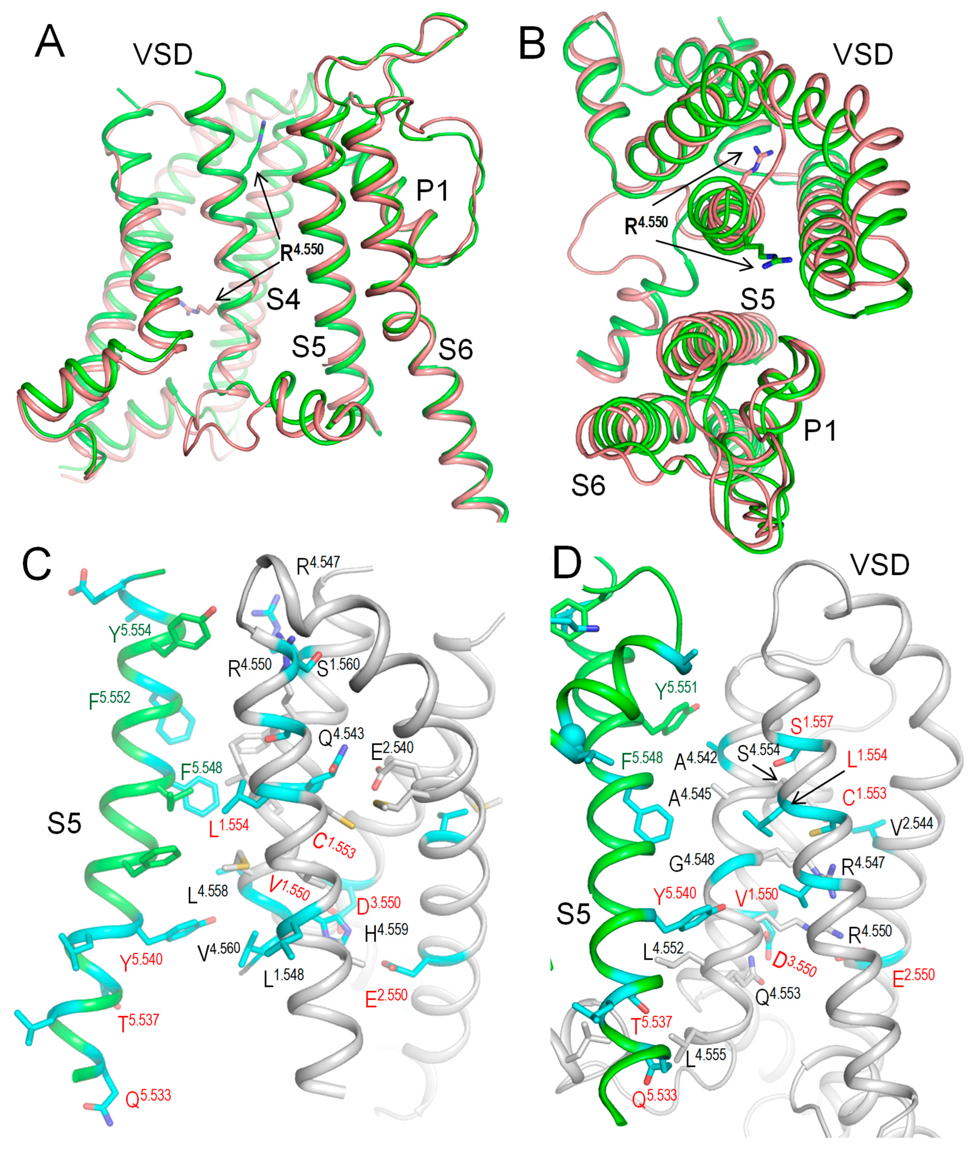
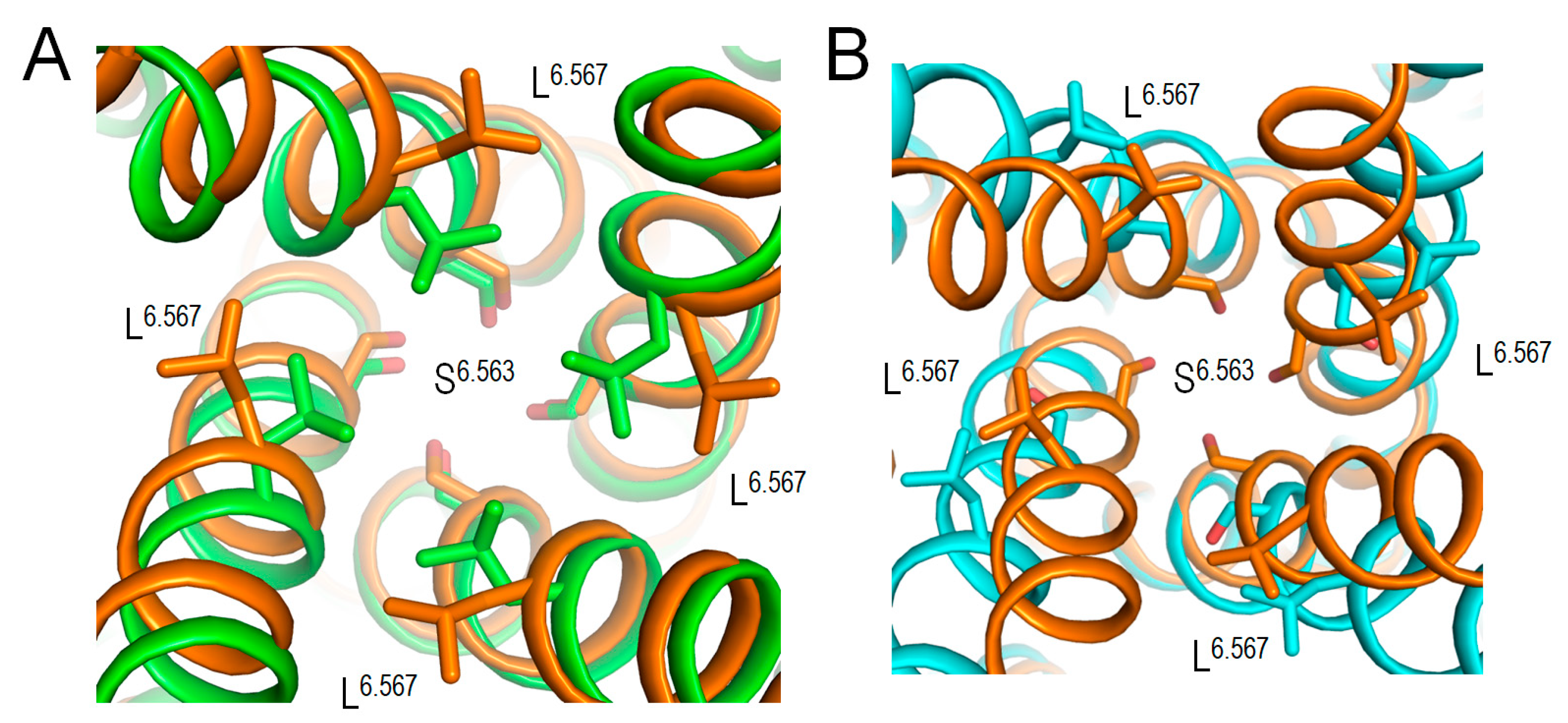
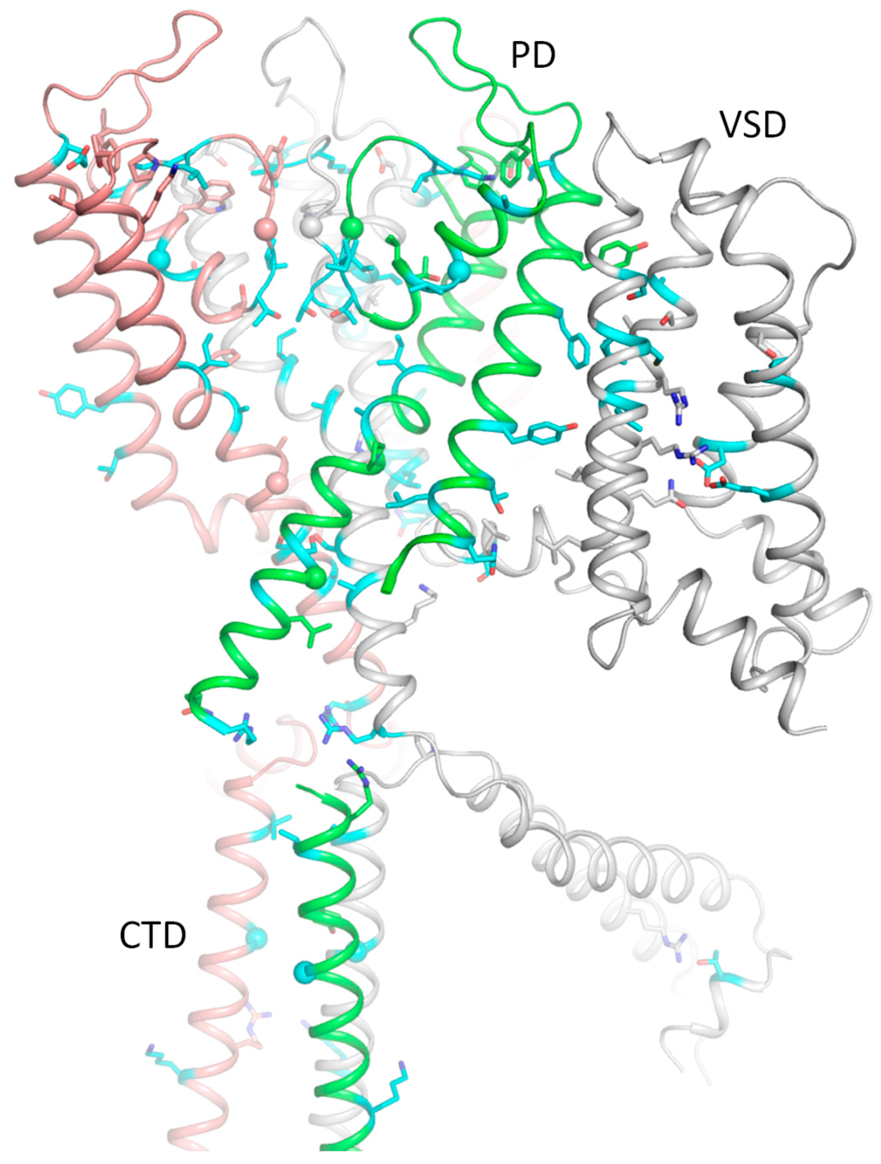
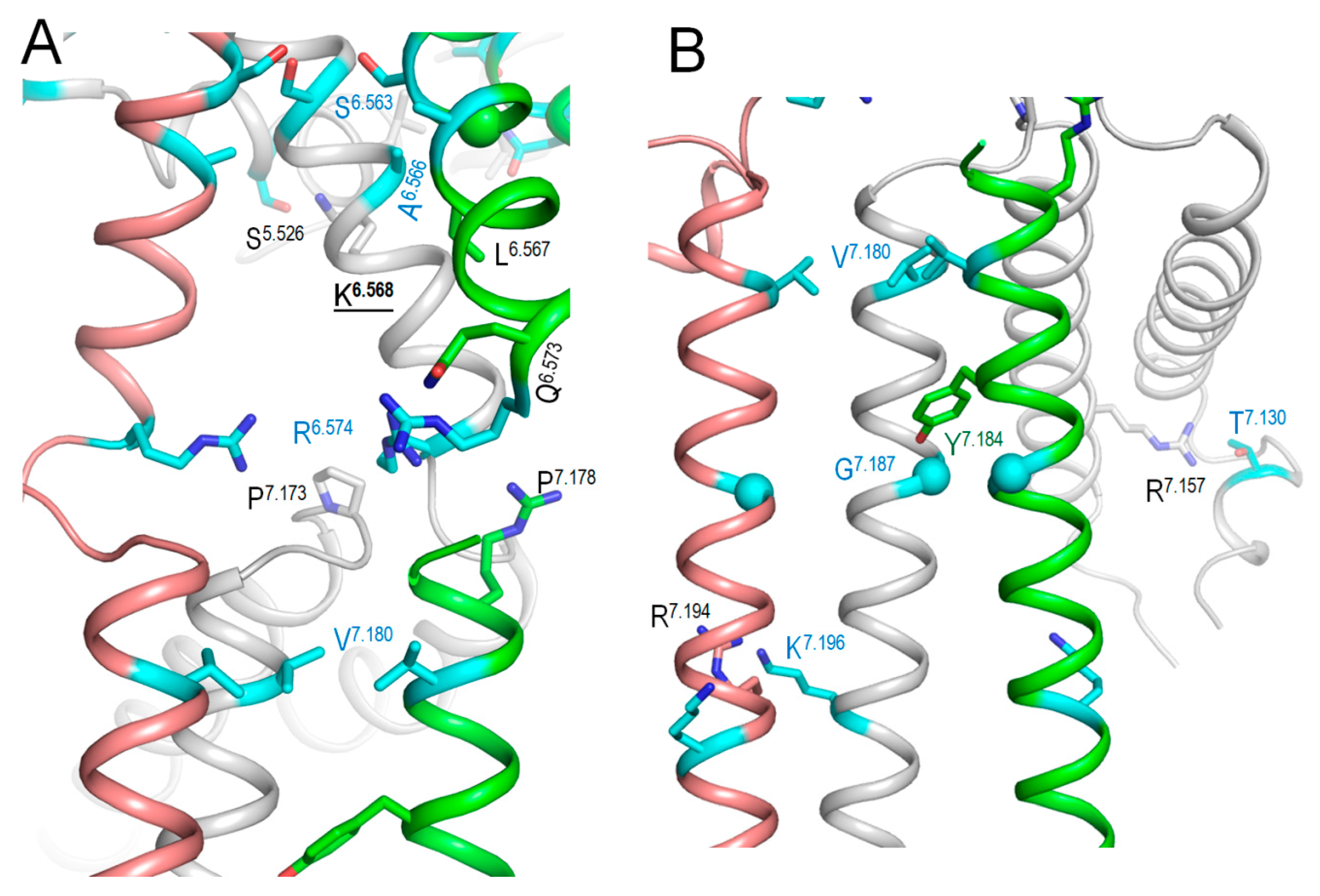
2.7. Intersegmental Contacts Involving WTRs with Likely Damaging VUSs

The above bioinformatics approaches strongly suggest the damaging potential of 79 VUSs in Kv7.1, but the molecular mechanisms of the variants’ dysfunction are unclear. Analysis of intersegmental contacts and their state dependency may suggest mechanisms of stabilization or destabilization of specific channel states that underlie the molecular mechanisms. We this goal in mind, we 3D-aligned cryoEM structures of Kv7.1 with VSDs in the activated (8sik) and deactivated (8sin) states [36], as described in the Methods. Upon a VSD deactivation, helix S4 underwent a substantial downshift (Figure 2A) and rotation (Figure 2B), while rearrangements of helices S5 and S6 were rather small. In both 8sik and 8sin structures, the pore at the level of serines S6.563 is narrow (Figure 3A), implying a closed channel [36]. Surprisingly, leucines L6.567 in the 8sik structure form a much tighter constriction than in 8sin (Figure 3A). However, CA atoms of leucines L6.567 in both structures are close to each other, and respective CA-CB bonds are collinear (Figure 3A), indicating that the tighter constriction in 8sik is due to the sidechain rotations rather than rearrangement of the S6 bundle.
In epithelial cells, KCNE3 associates with KCNQ1 to form apparently voltage-independent channels. By using voltage clamp fluorimetry, Larsson and co-authors [37] found that KCNE3 shifts the voltage dependence of S4 movement to extreme hyperpolarized potentials, making the channel constitutively open. These results suggest that KCNE3 primarily affects the voltage-sensing domain and only indirectly affects the gate. In a later study, Kasuya and Nakajo used the KCNQ1–KCNE3–calmodulin complex structure to examine amino acid residues in KCNE3 and the S1 segment of the KCNQ1 [38]. By changing the side-chain bulkiness of these interacting residues, the authors found that the distance between the S1 segment and KCNE3 is optimized to achieve constitutive activity, suggesting that the tight binding of the S1 segment and KCNE3 is crucial for controlling the VSDs.
While VSDs in the cryoEM structure (6v01) of KCNE3-bound Kv7.1 are deactivated [6], the S6 bundle is much wider than that in 8sin (Figure 3B), indicating that KCNE3 binding is sufficient to widen the pore even when VSDs are deactivated. Thus, although cryoEM structures of Kv7.1 with activated VSDs and open pore domain are lacking, available cryoEM structures of Kv7.1 allow analyzing intersegmental contacts of WTRs of likely damaging VUSs and, to some extent, their state-dependency.
We visualized intersegmental contacts in VSDs involving WTRs of likely damaging VUSs and WTRs of other ClinVar-reported variants in structures with deactivated (Figure 2C) and activated (Figure 2D) VSDs. Since the pore domain and especially its extracellular half are more 3D-conserved than VSDs, we analyzed intersegmental contacts in the pore domain in the structure with deactivated VSDs (Figure 4, Figure 5 and Figure 6), and then in the model with the KCNE1-bound channel. These contacts are listed in Table 4, Table 5, Table 6 and Table 7.
The vast majority of Kv7.1 P/LP variants, which are reported in ClinVar, as well as likely damaging VUSs (Table 3), are associated with long QT syndrome, indicating that respective mutations destabilize the open channel, decrease the IKs current, and thus prolong the action potential. In the case of the cardiac channel Nav1.5, for many intersegmental state-dependent contacts involving WTRs with ClinVar-reported variants, substitution of either contact partner causes the channel dysfunction associated with the same syndrome [39]. By analogy, we propose that if two WTRs of Kv7.1 form an intersegmental contact with one partner having a ClinVar-reported P/LP variant and another partner is a likely damaging VUS, it strongly increases the reliability of our prediction on the likely damaging VUS.
Table 4. Likely damaging (LD) VUSs whose WTRs contact WTRs of ClinVar-reported P/LP variants a.
LD VUS P/LP State bLD VUS P/LP State
V133/1.550A R231/4.550L L262/5.535V P343/6.557A
Q234/4.553P o P343/6.557A/S
C136/1.553F S225/4.544L/W T264/5.537SL233/4.552M
L156/2.536P o L251/5.524Q/P
Q234/4.553P G269/5.542R/S/V/D
L137/1.554PS225/4.544L/W Y267/5.540FG229/4.548D
G229/4.548D L233/4.552M
Q234/4.553P E284/5.557G T322/5.859P/A/R
I235/4.554N P320/5.857S
I274/5.547D G325/6.539W
R231/4.550S/C/L/H oG306/5.843E L273/5.546I/V/P/R/F
Q234/4.553P oV307/5.844M/L/E S330/\6.544Y
I235/4.554L/N oT312/5.849S T312/5.849I/S
S140/1.557R S225/4.544L/W I313/5.850FG314/5.851R/D/S
L156/2.536P T312/5.849S/I
R231/4.550S/C/L/H o T309/5.846I
Q234/4.553P oV319/5.856M Y315/5.852S
L156/2.536P o W304/5.841R/L/S
S143/1.560FR231/4.550S/C/L/H o W304/5.841G
V164/2.544AS209/3.557P o Y315/5.852D
E170/2.550GR231/4.550S/H/C/L I337/6.551MF340/6.554L
D202/3.550VQ234/4.553P F339/6.553VL251/5.524Q
R231/4.550S/C/L/H L251/5.524P
R234/4.562S/C/L/H/P A341/6.555T A344/6.558E/V
R243/4.562S/C/L/H/P oS349/6.563A/L G345/6.559R/V/E
Q260/A5.533H L251/D5.524Q/P R360/6.574K/TR539/7.178W/Q
L262/5.535VP343/6.557A/S oK557/7.196R R555/7.194S/C
a ClinVar of 4 March 2025; b ↓, contacts in the hKv7.1 cryoEM structure with “down” VSDs (PDB ID: 8sin) [36]; ↑, contacts in the hKv7.1 cryoEM structure with “up” VSDs (PDB ID: 8sik) [36]; o, Contacts in hKv7.1-KCNE3-PIP3 cryoEM structure with open PD and “down” VSDs (PD ID: 6v01) [40].
Table 5. Likely damaging (LD) VUSs whose WTRs form intersegmental contacts with WTRs of other ClinVar-reported VUSs a.
LD VUS Contact b LD VUS Contact b
V133/A1.550I/A R228/A4.547WVUS → LDK318/A5.855ND301/A5.838YVUS
G229/A4.548S/VVUSS349/A6.563A/L S349/B,D6.563A/LVUS → LD
Y267/B5.540FVUS → LD A352/B,D6.566P/DVUS → LD
C136/A1.553F R228/A4.547WVUS→ LDA352/A6.566P/D L353/B6.567PVUS
L137/A1.554PG229/A4.548S/VVUS S349/B6.563A/LVUS → LD
R228/A4.547WVUS → LD G350/B6.564RVUS
D202/A3.550VQ234/A4.553L/RVUS → LDR360/A6.574K/TP535/A7.174TVUS
S253/A5.526AK354/A6.568RVUS → LDT391/A7.030PR518/A7.157Q/PVUS
Y267/A5.540FG229/D4.548S/VVUSV541/A7.180I V541/B,D7.180IVUS → LD
E284/A5.557G V324/6.538L/I/FVUS Y545/B7.184FVUS
A300/A5.837S/GK326/B6.540EVUSG548/A7.187D Y545/B7.184FVUS
T312/A5.849S I313/5.850FVUS → LD
I337/A6.551MVUS → LD
a ClinVar Data on 4 March 2025; b In the cryoEM structure of hKv7.1 (PDB ID: 8sin) with voltage sensor in the down state [36].
Table 6. Intersegmental contacts a between WTRs of likely damaging (LD) VUSs and WTRs of CIP or NP variants b.
LD VUSContactLD VUS Contact
V164/A2.544A S209/A3.557PNPI313/A5.850F G314/B5.851C/ANP
A223/A4.542T Y278/B5.551HNP T309/B5.846S/RNP
Q260/A5.533H L236/D4.555R/PCIP V308/B5.845DNP
T264/A5.537SL236/D4.555R/PCIPK318/A5.855ND301/A5.838VCIP
L233/D4.552PCIPI337/A6.551MT311/D5.848ANP
Y267/A5.540FL233/4.552PCIP T311/D5.848ICIP
F275/A5.548L A226/D4.545VCIPA341/A6.555T A344/D6.558T/GCIP
E284/A5.557G T322/5.859KCIPS349/A6.563A/L S349/B,D6.563PCIP
F296/5.611SCIPA352/A6.566P/D S349/B6.563PCIP
T312/A5.849S I337/A6.551FCIP G350/B6.564VCIP
T391/A7.030PR518/A7.157GCIP
a In the cryoEM structure of hKv7.1 (PDB ID: 8sin) with the voltage sensor in the down state [36]; b ClinVar Data of 4 March 2025.
Table 7. CryoEM structure a with activated VSDs: intersegmental contacts of WTRs in VSD and S5 b.
LD VUS Contact
C136/A1.553F M159/2539LVUS
S140/A1.557RQ260/A5.533HLD VUS
V164/A2.544AM210/3558T/IVUS
E170/A2.550GH240/4.559QLD VUS
V129/1546I/G/AVUS
D202/A3.550VH240/4.559QLD VUS
L239/4.558VY267/A5.540FLD VUS
F275/A5.548S/LLD VUS
H240/4.559Q/RE170/A2.550GLD VUS
D202/A3.550HLD VUS
V241/4.560IY267/5.540FVUS
Y267/A5.540FV241/4.560ILD VUS
L239/4.558VLD VUS
F275/A5.548LL239/4.558VLD VUS
a In the cryoEM structure of hKv7.1 (PDB ID: 8sik) with the voltage sensor in the up state [36]; b ClinVar Data of 4 March 2025.
Such contacts with known P/LP residues involve 31 likely damaging VUSs of 21 WTRs, including eight variants in VSD, seven variants in S5, eight variants in P-loop, and eight variants in S6 (Table 4). Contacts within a VSD and between VSD and S5 are clearly state-dependent (cf. Figure 2C,D). In particular, H-bonds R4.550---S1.560, Q4.553---E2.540, salt bridges H4.559: E2.550 and H4.559: D3.550, and hydrophobic interactions L4.558: F5.548 stabilize the “up” state of S4 and thus activated state of VSD (Figure 2D). Mutations R4.550L, D3.550H, Q4.553P, E2.550G (Table 4), L4.558V, and F275/A5.548S/L (Table 7) would weaken or eliminate these contacts and destabilize the activated VSD. This would decrease the probability of the channel open state, reduce IKs, and prolong the action potential in cardiomyocytes, explaining why respective variants are associated with the gain-of-function long QT syndrome. However, some of the above mutations (e.g., R4.550L and E2.550G) would also destabilize deactivated VSD (Figure 2D). Therefore, the long QT syndrome associated with these variants indicates that respective mutations destabilize the activated state of VSD more strongly than its deactivated state.
Table 4 also shows seven likely damaging VUSs in the cytoplasmic part of S6 (positions 6.553–6.574) and six likely damaging VUSs in the cytoplasmic part of S5 (positions 5.533–5.540). These residues undergo substantial movements upon activation gating, implying that respective mutations destabilize the channel open state. Importantly, WTRs of the respective variants contact WTRs with ClinVar-reported P/LP variants (Table 4). These residues are marked with red labels in Figure 4.
The second category of contacts includes 16 WTRs of 26 likely damaging VUSs and 25 partner WTRs of 35 ClinVar-reported VUSs (Table 5). Among the contact partners, 13 WTRs have 18 VUSs that we also predicted as likely damaging variants (Table 4). The fact that VUSs of both partners in such contacts were independently selected by AlphaMissense and paralogue annotation methods further justifies our predictions that respective VUSs are likely damaging, making them promising objects for future functional analyses.
The third category of contacts includes 15 WTRs of 20 likely damaging VUSs and 16 contact-partner WTRs for which 26 variants are reported in ClinVar as either CIP variants or their germline classification is not provided (Table 6). We suggest a damaging potential of the CIP variants because at least one submitter reported likely pathogenicity of the variant, and the respective WTR is contacting a WTR with a likely damaging VUS.
We found four WTRs of ClinVar-reported benign/likely benign (B/LB) variants that form intersegmental contacts with WTRs of likely damaging VUSs (Supplementary Figure S1). Among these contacts, only B/LB mutation S1.560F would affect the polar contact with T2.553 and thus the stability of the VSD state.

2.8. Predicting Damaging Potential for KCNE1 VUSs with High ClinPred Score and ClinVar-Reported Variants in Sequentially Matching Positions of Paralogues

KCNE1 association with Kv7.1 is important for normal heart rhythm; for reviews, see [5,16]. As of March 2025, over 600 variants of KCNE1 are reported in ClinVar, but only three missense variants (T7I, M1L, and G52R) are classified as pathogenic. Among four known KCNE1 paralogs (KCNE2, KCNE3, KCNE4, and KCNE5), KCNE2 has five P/LP variants, while for the other paralogues, only VUSs are reported. Therefore, the paralogues annotation method for KCNE subunits is less reliable than for Kv channels. Since AlphaMissense correctly predicted the damaging effect of fewer than 60% of KCNE variants, we used the second top-performing tool (Section 2.4), ClinPred, with the standard cutoff score (deleterious threshold) of 0.5. ClinPred and the paralogue annotations consensually predicted the damaging potential of 34 VUSs for 21 WTRs in KCNE1 (Table 8). Among the 34 VUSs, 20 variants were explored in a recent high-throughput functional study of 68 KCNE1 variants [41], and for most of these, the pick current decrease in the KCNQ1-KCNE1 assembly is reported (Table 8). These experimental data confirm the reliability of our approach to predict the damaging potential of KNCE1 variants.

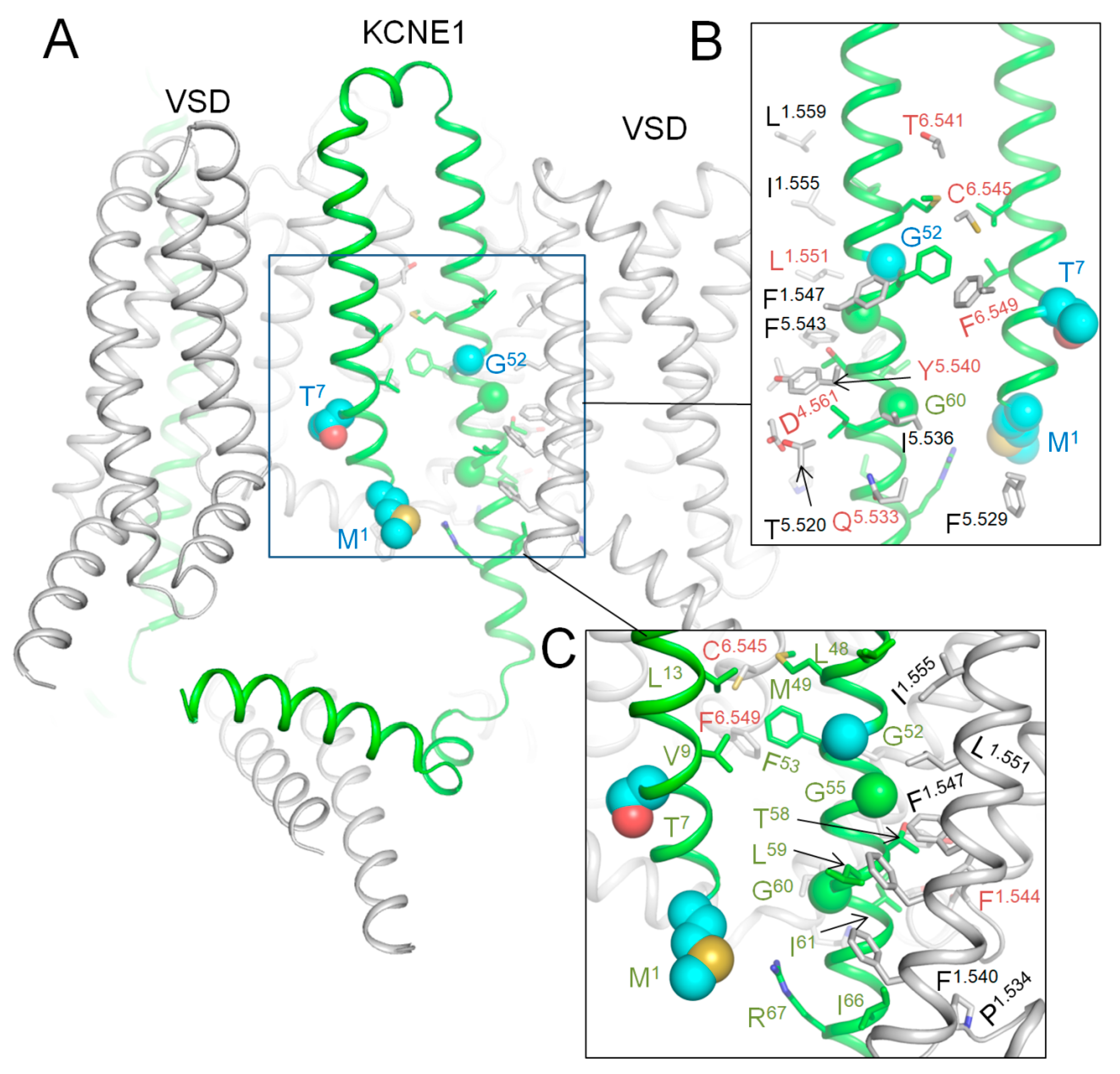
2.9. AlphaFold 3 (AF3) Model of Kv7.1 with KCNE1

In the lack of cryoEM structures of Kv7.1-KCNE1, we created an AF3 multimer model [42,43] of complex Kv7.1-KCNE1 and Monte Carlo (MC) minimized its energy with rigid backbones (Figure 7). In this model, the large N-terminal portion of each KCNE1 folded up to fill the space between two VSDs, while the smaller C-terminal part extended to the cytoplasm. WTRs of pathogenic variants M1L, T7I, and G52R are shown as blue spheres. Likely damaging VUSs of KCNE1 (Table 8) are shown as sticks in close-up views of Figure 7. Among multiple Kv7.1 WTRs that form contacts with KCNE1, six have known P/LP variants, and several others have likely damaging VUSs (Table 3). Since these data alone are insufficient to prove our AF3 model of Kv7.1-KCNE1, we compared the model with the cryoEM structure of KCNE3-bound Kv7.1 as described in the next section.

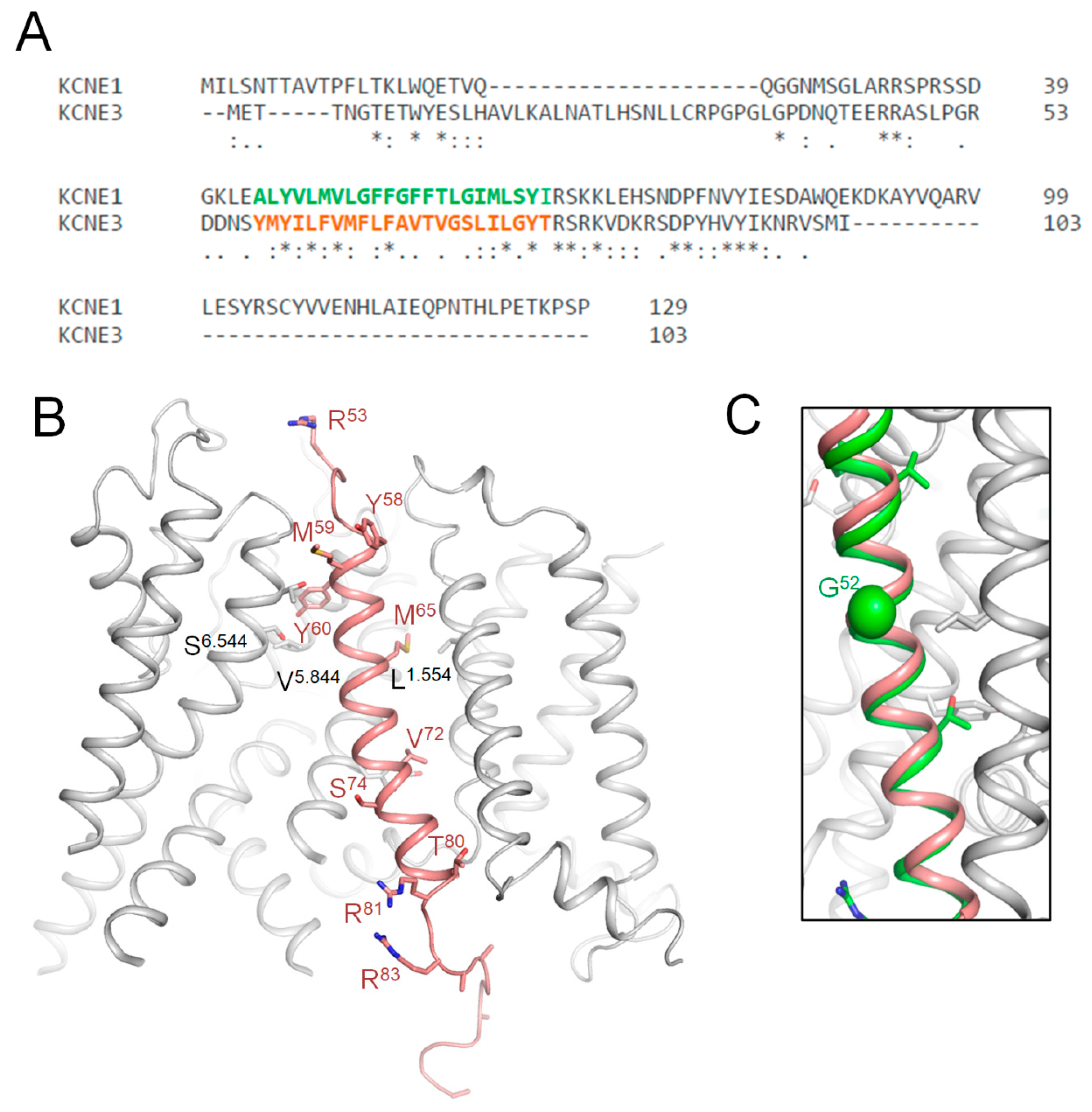
2.10. CryoEM Structure of KCNE3-Bound Kv7.1

Kv7.1 co-assembles with KCNE3 in non-excitable cells [40,44], but there is evidence that such complexes are also expressed in the human heart, especially in diseased hearts [45]. KCNE1 and KCNE3 sequences are very different (Figure 8A), but there is sequence similarity in the middle part where KCNE3, which is resolved in the cryoEM structure (6v01) of KCNE3-bound Kv7.1 [40], where the TM helix is bound between S1 of VSD and the extracellular parts of helices S5 and S6 (Figure 8B). In the 3D-aligned cryoEM structure of Kv7.1-KCNE3 and the AF3 model of Kv7.1-KCNE1, the TM part of the KCNE3 helix (M60 to T80) overlaps with the KCNE1 helix from L45 to I66 (Figure 8C). It should be noted that the full-fledged cryoEM structure and the AF3 model are 3D-aligned by minimizing RMS deviations of sequentially matching CA atoms in P-loop helices P1 (see Methods) rather than sequentially matching CA atoms in the KCNE TM helices (Figure 8A). The perfect 3D match of the TM helices validates the position of the respective KCNE1 TM helix in our AF3 model (Figure 7).

3. Methods and Materials

3.1. Sequence Data of Human Channels and Collection of Variants

The sequence of hKv7.1 was obtained from the UniProt database [46] (accession number P51787). Paralogues of the hKv7.1 channel were identified using Ensembl [47]. Missense mutations for Kv7.1 and its paralogues were collected from three databases: Humsavar, Ensembl Variation [48], and ClinVar [49]. Only P or LP variants were extracted from Ensembl Variation, ClinVar, and Humsavar. VUSs were also obtained from ClinVar. Common benign (neutral) variants, along with their minor allele frequencies (AF), were sourced from the population database gnomAD [50]. Variants with AF > 0.00001 that are not present in ClinVar were considered benign [51,52]. The total number of collected P/LP, VUS, and common neutral variants is given in Table 1. All variants were compiled into a comprehensive dataset (Table S1).

3.2. Topology of the Kv7.1 Channel

The hKv7.1 regions were defined in accordance with the UniProt entry P51787. The pore-forming α1 subunit of Kv7.1 assembles from four subunits. A large cytoplasmic domain plays critical roles in subunit assembly, interactions with regulatory proteins, and modulation of channel function [53].

3.3. Multiple Sequence Alignment and Paralogue Annotation

The paralogue annotation method identifies P/LP missense variants by transferring annotations across families of related proteins [31]. Previously, we applied a modified version of this method to predict likely damaging variants for channels hNav1.5 [30], hCav1.2 [32], and TRPM4 [33]. Here, we used the same approach to predict likely damaging variants for VUSs of hKv7.1.
For each paralogue channel, P/LP variants were collected. The amino acid sequences of hKv7.1 and its paralogue channels were aligned using the multiple sequence alignment program Tcoffee [54]. Proteins lacking P/LP variants were excluded from the alignment [26]. Since disease-causing mutations tend to occur at evolutionarily conserved positions, we computed position-specific conservation scores (Cs), which range from 0 (no conservation) to 1 (identical), with Cs = 0.8 indicating high conservation. These scores reflect the conservation of physicochemical properties (small, polar, hydrophobic, tiny, charged, negative, positive, aromatic, aliphatic, and proline) in the alignment [55]. Cs values were calculated using the Zvelebil method [56], as implemented in the Amino Acid Conservation Calculation Service [57]. Variants occurring at positions with Cs > 0.3 were classified as P/LP variants.

3.4. Sequence-Based Prediction of Likely Damaging Variants

Missense variants were annotated with scores from 29 algorithms (REVEL, VEST4, MVP, CADD, LIST.S2, VARITY_R, VARITY_ER, AlphaMissense, EVE, MPC, MVP, DANN, CenoCanyon, PrimateAI, DEOGEN2, M-CAP, MetaLR, MetaSVM, MetaRNN, FATHMM, PROVEAN, MutationAssessor, MutPred, PolyPhen2-HVAR, PolyPhen2-HDIV, SIFT, SIFT4G, LRT, MutationTaster), which were obtained from the dbNSFPv4.5 database [58]. To generate binary predictions (Damaging/Tolerated), we used thresholds determined as the optimal pathogenicity threshold from the AUC-ROC curve (Table 2).
The ‘probably damaging’ and ‘possibly damaging’ classes predicted by Polyphen were merged into a single ‘damaging’ class. For results from the MutationAssessor server, which subdivides mutants into four categories, we treated categories high (‘H’) or medium (‘M’) as ‘Damaging’, and categories low (‘L’) or neutral (‘N’) as ‘Tolerated’.
The overall prediction performance of the 29 methods was assessed by calculating sensitivity, specificity, Matthews Correlation Coefficient (MCC), and accuracy (ACC) as follows:
S e n s i t i v i t y = T P T P + F N ;
S p e c i f i c i t y = T N T N + F P ;
M C C = T P × T N F P × F N ( T P + F P ) ( T P + F N ) ( T N + F P ) ( T N + F N ) ;
A C C = T P + T N T P + F P + T N + F N .
The following abbreviations are used in these equations:
  • TP (true-positive) is the number of disease-causing variants correctly predicted to be pathogenic;
  • FN (false-negative) is the number of disease-causing variants incorrectly predicted as tolerated;
  • TN (true-negative) is the number of neutral variants correctly predicted as tolerated;
  • FP (false-positive) is the number of neutral variants incorrectly predicted as pathogenic;
  • MCC is a correlation coefficient between the observed and predicted binary classification, ranging from −1 (total disagreement between prediction and observation) to 1 (perfect prediction).
For the test dataset, we selected common neutral and P/LP variants from our comprehensive dataset (Supplementary Table S1). We also calculated the area under the ROC (Receiver Operating Characteristic) curve (AUC) using the pROC library in the R programming language. ROC curves were generated by plotting sensitivity against (1—specificity) at each threshold for each algorithm. The AUC can range from 0 (completely random) to 1 (perfectly correct prediction). The absence of a variant annotation negatively impacts prediction accuracy. Therefore, we included only those algorithms that predicted the pathogenicity of over 30% of variants in our dataset (Table S1).

3.5. Molecular Modeling

All computations were performed with the freely available ZMM program (www.zmmsift.ca). Energy was calculated using the AMBER force field [59] with environment- and distance-dependent dielectric function [60]. Energy was optimized with Monte Carlo (MC) minimizations [61] in the space of generalized coordinates [62]. We used the AlphaFold3 (AF3) server (https://alphafold.ebi.ac.uk) to predict multimeric complexes of hKv7.1 with four full-fledged KCNE1 subunits. CryoEM structures of Kv7.1 with the activated and deactivated VSDs, as well as AF3 models, were imported by the ZMM program, and side chain conformations were MC-minimized with rigid backbones. MC-minimizing trajectories were terminated when the last 100th energy minimization did not improve the protein energy. All structures were 3D-aligned by minimizing the root mean square deviations of Cα atoms in P1 helices against a reference crystal structure of the chimeric potassium channels Kv1.2-Kv2.1 channel (PDB ID: 2R9R), the first eukaryotic P-loop potassium channels whose crystal structure was obtained with a resolution below 2.5 Å [63].
Intersegmental contacts are defined as those where two sidechains are within 5Å from each other. Such contacts were automatically identified by ZMM in MC-minimized structures. We used PyMol v099 (Schrödinger, New York, NY, USA) to visualize cryoEM structures and AF3 models of the Kv7.1 channel and its complex with KCNE1. Other details of computations may be found elsewhere [64].

4. Conclusions

In this study, we compiled a comprehensive dataset encompassing known pathogenic/likely pathogenic (P/LP) variants of the hKv7.1 channel and its 14 paralogues. Our analysis identified AlphaMissense as the top-performing bioinformatics tool for predicting (P/LP) variants in Kv channels. AlphaMissense and the paralogue annotations method consensually predicted 79 VUSs of Kv7.1 as likely damaging (LD) variants. We further predicted that 34 KCNE1 VUSs are LD variants. Many wildtype residues (WTRs) of LD variants make state-dependent intersegmental contacts with WTRs of known P/LP variants or LD variants in cryo-EM structures or AlphaFold3 models, suggesting atomic mechanisms of the variants’ dysfunction. Respective variants of Kv7.1 and KCNE1 are promising objects for future functional analyses.

Study Limitations

Our computations predicted the damaging potential of many reported disease-associated mutations in KCNQ1 and KCNE1, but functional studies are necessary to reclassify respective VUSs as P/LP variants. Since the cryoEM structures of Kv7.1 considered here do not provide relations between VSD activation and the pore opening, mutations with likely damaging potential within the VSD do not necessarily affect the IKs current; functional studies are necessary to explore these variants. We analyzed contacts of WTRs but not respective VUSs and suggested that mutations would modify these contacts. Despite these limitations, we hope that our study, which incorporates paralogue annotations and analysis of experimental and AF3-predicted 3D structures, provides more reliable predictions of the damaging potential of VUSs than more traditional bioinformatics approaches.

Supplementary Materials

The following supporting information can be downloaded at https://www.mdpi.com/article/10.3390/ijms26146561/s1.

Author Contributions

Methodology, S.I.T. and B.S.Z.; Software, B.S.Z.; Validation, B.S.Z.; Investigation, S.I.T. and B.S.Z.; Data curation, S.I.T.; Writing—original draft, S.I.T.; Writing—review and editing, B.S.Z.; Visualization, B.S.Z.; Supervision, B.S.Z.; Project administration, B.S.Z.; Funding acquisition, B.S.Z. All authors have read and agreed to the published version of the manuscript.

Funding

This research was funded by the Natural Sciences and Engineering Research Council of Canada (RGPIN-2020-07100) and the ongoing funding of the Sechenov Institute, RAS (N.A.).

Institutional Review Board Statement

Not applicable.

Informed Consent Statement

Not applicable.

Data Availability Statement

The original contributions presented in this study are included in the article/Supplementary Materials. Further inquiries can be directed to the corresponding author.

Acknowledgments

We thank Vyacheslav Korkosh for the administration of the database www.plic3da.

Conflicts of Interest

The authors declare no conflicts of interest.

Abbreviations

AF3AlphaFold3
AUCArea under the ROC curve
CIPConflicting Interpretation of Pathogenicity
LDLikely damaging variant
LQTSLong QT Syndrome
MCMonte Carlo
NPGermline classification of pathogenicity is Not Provided
P/LPPathogenic/Likely Pathogenic variant
PLICP-loop ion channel
TMTransmembrane
ROCReceiver Operating Characteristic
VSDVoltage-sensing domain
VUSVariant of unknown clinical significance
WTRWildtype residue

References

  1. Wu, X.; Larsson, H.P. Insights into Cardiac IKs (KCNQ1/KCNE1) Channels Regulation. Int. J. Mol. Sci. 2020, 21, 9440. [Google Scholar] [CrossRef] [PubMed]
  2. Kekenes-Huskey, P.M.; Burgess, D.E.; Sun, B.; Bartos, D.C.; Rozmus, E.R.; Anderson, C.L.; January, C.T.; Eckhardt, L.L.; Delisle, B.P. Mutation-Specific Differences in Kv7.1 (KCNQ1) and Kv11.1 (KCNH2) Channel Dysfunction and Long QT Syndrome Phenotypes. Int. J. Mol. Sci. 2022, 23, 7389. [Google Scholar] [CrossRef] [PubMed]
  3. Abbott, J.W. Biology of the KCNQ1 Potassium Channel. New J. Sci. 2014, 2014, 1–26. [Google Scholar] [CrossRef]
  4. Jespersen, T.; Grunnet, M.; Olesen, S.P. The KCNQ1 potassium channel: From gene to physiological function. Physiology 2005, 20, 408–416. [Google Scholar] [CrossRef]
  5. Sanguinetti, M.C.; Seebohm, G. Physiological Functions, Biophysical Properties, and Regulation of KCNQ1 (K(V)7.1) Potassium Channels. Adv. Exp. Med. Biol. 2021, 1349, 335–353. [Google Scholar] [CrossRef]
  6. Sun, J.; MacKinnon, R. Cryo-EM Structure of a KCNQ1/CaM Complex Reveals Insights into Congenital Long QT Syndrome. Cell 2017, 169, 1042–1050.e9. [Google Scholar] [CrossRef] [PubMed]
  7. Bains, S.; Giammarino, L.; Nimani, S.; Alerni, N.; Tester, D.J.; Kim, C.S.J.; Christoforou, N.; Louradour, J.; Horvath, A.; Beslac, O.; et al. KCNQ1 suppression-replacement gene therapy in transgenic rabbits with type 1 long QT syndrome. Eur. Heart J. 2024, 45, 3751–3763. [Google Scholar] [CrossRef]
  8. Rothenberg, I.; Piccini, I.; Wrobel, E.; Stallmeyer, B.; Muller, J.; Greber, B.; Strutz-Seebohm, N.; Schulze-Bahr, E.; Schmitt, N.; Seebohm, G. Structural interplay of K(V)7.1 and KCNE1 is essential for normal repolarization and is compromised in short QT syndrome 2 (K(V)7.1-A287T). Hear. Case Rep. 2016, 2, 521–529. [Google Scholar] [CrossRef]
  9. Bellocq, C.; van Ginneken, A.C.; Bezzina, C.R.; Alders, M.; Escande, D.; Mannens, M.M.; Baro, I.; Wilde, A.A. Mutation in the KCNQ1 gene leading to the short QT-interval syndrome. Circulation 2004, 109, 2394–2397. [Google Scholar] [CrossRef]
  10. Chen, Y.H.; Xu, S.J.; Bendahhou, S.; Wang, X.L.; Wang, Y.; Xu, W.Y.; Jin, H.W.; Sun, H.; Su, X.Y.; Zhuang, Q.N.; et al. KCNQ1 gain-of-function mutation in familial atrial fibrillation. Science 2003, 299, 251–254. [Google Scholar] [CrossRef]
  11. Hateley, S.; Lopez-Izquierdo, A.; Jou, C.J.; Cho, S.; Schraiber, J.G.; Song, S.; Maguire, C.T.; Torres, N.; Riedel, M.; Bowles, N.E.; et al. The history and geographic distribution of a KCNQ1 atrial fibrillation risk allele. Nat. Commun. 2021, 12, 6442. [Google Scholar] [CrossRef] [PubMed]
  12. Campuzano, O.; Sarquella-Brugada, G.; Brugada, R.; Brugada, J. Genetics of channelopathies associated with sudden cardiac death. Glob. Cardiol. Sci. Pract. 2015, 2015, 39. [Google Scholar] [CrossRef] [PubMed]
  13. Albert, C.M.; MacRae, C.A.; Chasman, D.I.; VanDenburgh, M.; Buring, J.E.; Manson, J.E.; Cook, N.R.; Newton-Cheh, C. Common variants in cardiac ion channel genes are associated with sudden cardiac death. Circ. Arrhythmia Electrophysiol. 2010, 3, 222–229. [Google Scholar] [CrossRef]
  14. Kiper, A.K.; Wegner, S.; Kadala, A.; Rinne, S.; Schutte, S.; Winter, Z.; Bertoune, M.A.R.; Touska, F.; Matschke, V.; Wrobel, E.; et al. KCNQ1 is an essential mediator of the sex-dependent perception of moderate cold temperatures. Proc. Natl. Acad. Sci. USA 2024, 121, e2322475121. [Google Scholar] [CrossRef]
  15. Sachyani, D.; Dvir, M.; Strulovich, R.; Tria, G.; Tobelaim, W.; Peretz, A.; Pongs, O.; Svergun, D.; Attali, B.; Hirsch, J.A. Structural basis of a Kv7.1 potassium channel gating module: Studies of the intracellular c-terminal domain in complex with calmodulin. Structure 2014, 22, 1582–1594. [Google Scholar] [CrossRef]
  16. Wrobel, E.; Tapken, D.; Seebohm, G. The KCNE Tango—How KCNE1 Interacts with Kv7.1. Front. Pharmacol. 2012, 3, 142. [Google Scholar] [CrossRef] [PubMed]
  17. Banyasz, T.; Jian, Z.; Horvath, B.; Khabbaz, S.; Izu, L.T.; Chen-Izu, Y. Beta-adrenergic stimulation reverses the I Kr-I Ks dominant pattern during cardiac action potential. Pflug. Arch. 2014, 466, 2067–2076. [Google Scholar] [CrossRef]
  18. Burg, S.; Attali, B. Targeting of Potassium Channels in Cardiac Arrhythmias. Trends Pharmacol. Sci. 2021, 42, 491–506. [Google Scholar] [CrossRef]
  19. Landrum, M.J.; Lee, J.M.; Benson, M.; Brown, G.R.; Chao, C.; Chitipiralla, S.; Gu, B.; Hart, J.; Hoffman, D.; Jang, W.; et al. ClinVar: Improving access to variant interpretations and supporting evidence. Nucleic Acids. Res. 2018, 46, D1062–D1067. [Google Scholar] [CrossRef]
  20. Huang, H.; Kuenze, G.; Smith, J.A.; Taylor, K.C.; Duran, A.M.; Hadziselimovic, A.; Meiler, J.; Vanoye, C.G.; George, A.L., Jr.; Sanders, C.R. Mechanisms of KCNQ1 channel dysfunction in long QT syndrome involving voltage sensor domain mutations. Sci. Adv. 2018, 4, eaar2631. [Google Scholar] [CrossRef]
  21. Phul, S.; Kuenze, G.; Vanoye, C.G.; Sanders, C.R.; George, A.L., Jr.; Meiler, J. Predicting the functional impact of KCNQ1 variants with artificial neural networks. PLoS Comput. Biol. 2022, 18, e1010038. [Google Scholar] [CrossRef] [PubMed]
  22. Vanoye, C.G.; Desai, R.R.; Fabre, K.L.; Gallagher, S.L.; Potet, F.; DeKeyser, J.M.; Macaya, D.; Meiler, J.; Sanders, C.R.; George, A.L., Jr. High-Throughput Functional Evaluation of KCNQ1 Decrypts Variants of Unknown Significance. Circ. Genom. Precis. Med. 2018, 11, e002345. [Google Scholar] [CrossRef] [PubMed]
  23. Brewer, K.R.; Vanoye, C.G.; Huang, H.; Clowes Moster, K.R.; Desai, R.R.; Hayes, J.B.; Burnette, D.T.; George, A.L.; Sanders, C.R. Integrative analysis of KCNQ1 variants reveals molecular mechanisms of type 1 long QT syndrome pathogenesis. Proc. Natl. Acad. Sci. USA 2025, 122, e2412971122. [Google Scholar] [CrossRef]
  24. Richards, S.; Aziz, N.; Bale, S.; Bick, D.; Das, S.; Gastier-Foster, J.; Grody, W.W.; Hegde, M.; Lyon, E.; Spector, E.; et al. Standards and guidelines for the interpretation of sequence variants: A joint consensus recommendation of the American College of Medical Genetics and Genomics and the Association for Molecular Pathology. Genet. Med. 2015, 17, 405–424. [Google Scholar] [CrossRef]
  25. Dong, C.; Wei, P.; Jian, X.; Gibbs, R.; Boerwinkle, E.; Wang, K.; Liu, X. Comparison and integration of deleteriousness prediction methods for nonsynonymous SNVs in whole exome sequencing studies. Hum. Mol. Genet. 2015, 24, 2125–2137. [Google Scholar] [CrossRef]
  26. Niroula, A.; Vihinen, M. Variation Interpretation Predictors: Principles, Types, Performance, and Choice. Hum. Mutat. 2016, 37, 579–597. [Google Scholar] [CrossRef]
  27. Ghosh, R.; Oak, N.; Plon, S.E. Evaluation of in silico algorithms for use with ACMG/AMP clinical variant interpretation guidelines. Genome Biol. 2017, 18, 225. [Google Scholar] [CrossRef] [PubMed]
  28. Tordai, H.; Torres, O.; Csepi, M.; Padanyi, R.; Lukacs, G.L.; Hegedus, T. Analysis of AlphaMissense data in different protein groups and structural context. Sci. Data 2024, 11, 495. [Google Scholar] [CrossRef]
  29. Anderson, D.; Lassmann, T. A phenotype centric benchmark of variant prioritisation tools. NPJ Genom. Med. 2018, 3, 5. [Google Scholar] [CrossRef]
  30. Tarnovskaya, S.I.; Korkosh, V.S.; Zhorov, B.S.; Frishman, D. Predicting novel disease mutations in the cardiac sodium channel. Biochem. Biophys. Res. Commun. 2020, 521, 603–611. [Google Scholar] [CrossRef]
  31. Walsh, R.; Peters, N.S.; Cook, S.A.; Ware, J.S. Paralogue annotation identifies novel pathogenic variants in patients with Brugada syndrome and catecholaminergic polymorphic ventricular tachycardia. J. Med. Genet. 2014, 51, 35–44. [Google Scholar] [CrossRef] [PubMed]
  32. Tarnovskaya, S.I.; Kostareva, A.A.; Zhorov, B.S. L-Type Calcium Channel: Predicting Pathogenic/Likely Pathogenic Status for Variants of Uncertain Clinical Significance. Membranes 2021, 11, 599. [Google Scholar] [CrossRef]
  33. Tarnovskaya, S.I.; Kostareva, A.A.; Zhorov, B.S. In silico analysis of TRPM4 variants of unknown clinical significance. PLoS ONE 2023, 18, e0295974. [Google Scholar] [CrossRef]
  34. Tikhonov, D.B.; Korkosh, V.S.; Zhorov, B.S. 3D-aligned tetrameric ion channels with universal residue labels for comparative structural analysis. Biophys. J. 2025, 124, 458–470. [Google Scholar] [CrossRef]
  35. Gigolaev, A.M.; Iurevaa, D.A.; Lagosha, S.V.; Brazhe, A.R.; Zhorov, B.S.; Vassilevski, A.A. Golden Gate cloning enables efficient concatemer construction for biophysical analysis of heterozygous potassium channel variants from patients with epilepsy. Int. J. Biol. Macromol. 2025; in press. [Google Scholar] [CrossRef] [PubMed]
  36. Mandala, V.S.; MacKinnon, R. The membrane electric field regulates the PIP(2)-binding site to gate the KCNQ1 channel. Proc. Natl. Acad. Sci. USA 2023, 120, e2301985120. [Google Scholar] [CrossRef] [PubMed]
  37. Barro-Soria, R.; Perez, M.E.; Larsson, H.P. KCNE3 acts by promoting voltage sensor activation in KCNQ1. Proc. Natl. Acad. Sci. USA 2015, 112, E7286–E7292. [Google Scholar] [CrossRef]
  38. Kasuya, G.; Nakajo, K. Optimized tight binding between the S1 segment and KCNE3 is required for the constitutively open nature of the KCNQ1-KCNE3 channel complex. eLife 2022, 11, e81683. [Google Scholar] [CrossRef]
  39. Korkosh, V.S.; Zaytseva, A.K.; Kostareva, A.A.; Zhorov, B.S. Intersegment Contacts of Potentially Damaging Variants of Cardiac Sodium Channel. Front. Pharmacol. 2021, 12, 756415. [Google Scholar] [CrossRef]
  40. Sun, J.; MacKinnon, R. Structural Basis of Human KCNQ1 Modulation and Gating. Cell 2020, 180, 340–347.E9. [Google Scholar] [CrossRef]
  41. Vanoye, C.G.; Desai, R.R.; John, J.D.; Hoffman, S.C.; Fink, N.; Zhang, Y.; Venkatesh, O.G.; Roe, J.; Adusumilli, S.; Jairam, N.P.; et al. Functional profiling of KCNE1 variants informs population carrier frequency of Jervell and Lange-Nielsen syndrome type 2. bioRxiv 2025. [Google Scholar] [CrossRef]
  42. Jumper, J.; Evans, R.; Pritzel, A.; Green, T.; Figurnov, M.; Ronneberger, O.; Tunyasuvunakool, K.; Bates, R.; Zidek, A.; Potapenko, A.; et al. Highly accurate protein structure prediction with AlphaFold. Nature 2021, 596, 583–589. [Google Scholar] [CrossRef]
  43. Evans, R.; O’Neill, M.; Pritzel, A.; Antropova, N.; Senior, A.; Green, T.; Green, T.; Bates, R.; Blackwell, S.; Yim, J.; et al. Protein complex prediction with AlphaFold-Multimer. bioRxiv 2021. [Google Scholar] [CrossRef]
  44. Abbott, G.W. KCNE1 and KCNE3: The yin and yang of voltage-gated K(+) channel regulation. Gene 2016, 576, 1–13. [Google Scholar] [CrossRef] [PubMed]
  45. Lundquist, A.L.; Manderfield, L.J.; Vanoye, C.G.; Rogers, C.S.; Donahue, B.S.; Chang, P.A.; Drinkwater, D.C.; Murray, K.T.; George, A.L., Jr. Expression of multiple KCNE genes in human heart may enable variable modulation of I(Ks). J. Mol. Cell. Cardiol. 2005, 38, 277–287. [Google Scholar] [CrossRef] [PubMed]
  46. UniProt, C. UniProt: A hub for protein information. Nucleic Acids. Res. 2015, 43, D204–D212. [Google Scholar] [CrossRef]
  47. Dyer, S.C.; Austine-Orimoloye, O.; Azov, A.G.; Barba, M.; Barnes, I.; Barrera-Enriquez, V.P.; Becker, A.; Bennett, R.; Beracochea, M.; Berry, A.; et al. Ensembl 2025. Nucleic Acids. Res. 2025, 53, D948–D957. [Google Scholar] [CrossRef]
  48. Boutet, E.; Lieberherr, D.; Tognolli, M.; Schneider, M.; Bansal, P.; Bridge, A.J.; Poux, S.; Bougueleret, L.; Xenarios, I. UniProtKB/Swiss-Prot, the Manually Annotated Section of the UniProt KnowledgeBase: How to Use the Entry View. Methods Mol. Biol. 2016, 1374, 23–54. [Google Scholar] [CrossRef]
  49. Landrum, M.J.; Lee, J.M.; Benson, M.; Brown, G.; Chao, C.; Chitipiralla, S.; Gu, B.; Hart, J.; Hoffman, D.; Hoover, J.; et al. ClinVar: Public archive of interpretations of clinically relevant variants. Nucleic Acids. Res. 2016, 44, D862–D868. [Google Scholar] [CrossRef]
  50. Karczewski, K.J.; Francioli, L.C.; Tiao, G.; Cummings, B.B.; Alföldi, J.; Wang, Q.; Collins, R.L.; Laricchia, K.M.; Ganna, A.; Birnbaum, D.P.; et al. The mutational constraint spectrum quantified from variation in 141,456 humans. Nature 2020, 581, 434–443. [Google Scholar] [CrossRef]
  51. Kaltman, J.R.; Evans, F.; Fu, Y.-P. Re-evaluating pathogenicity of variants associated with the long QT syndrome. J. Cardiovasc. Electrophysiol. 2018, 29, 98–104. [Google Scholar] [CrossRef] [PubMed]
  52. Walsh, R.; Thomson, K.L.; Ware, J.S.; Funke, B.H.; Woodley, J.; McGuire, K.J.; Mazzarotto, F.; Blair, E.; Seller, A.; Taylor, J.C.; et al. Reassessment of Mendelian gene pathogenicity using 7,855 cardiomyopathy cases and 60,706 reference samples. Genet. Med. 2017, 19, 192–203. [Google Scholar] [CrossRef] [PubMed]
  53. Haitin, Y.; Wiener, R.; Shaham, D.; Peretz, A.; Cohen, E.B.; Shamgar, L.; Pongs, O.; Hirsch, J.A.; Attali, B. Intracellular domains interactions and gated motions of I(KS) potassium channel subunits. EMBO J. 2009, 28, 1994–2005. [Google Scholar] [CrossRef] [PubMed]
  54. Sievers, F.; Wilm, A.; Dineen, D.; Gibson, T.J.; Karplus, K.; Li, W.; Lopez, R.; McWilliam, H.; Remmert, M.; Söding, J.; et al. Fast, scalable generation of high-quality protein multiple sequence alignments using Clustal Omega. Mol. Syst. Biol. 2011, 7, 539. [Google Scholar] [CrossRef]
  55. Livingstone, C.D.; Barton, G.J. Protein sequence alignments: A strategy for the hierarchical analysis of residue conservation. Comput. Appl. Biosci. CABIOS 1993, 9, 745–756. [Google Scholar] [CrossRef]
  56. Zvelebil, M.J.; Barton, G.J.; Taylor, W.R.; Sternberg, M.J. Prediction of protein secondary structure and active sites using the alignment of homologous sequences. J. Mol. Biol. 1987, 195, 957–961. [Google Scholar] [CrossRef]
  57. Golicz, A.; Troshin, P.V.; Madeira, F.; Martin, D.M.A.; Procter, J.B.; Barton, G.J. AACon: A Fast Amino Acid Conservation Calculation Service. 2018; submitted. [Google Scholar]
  58. Liu, X.; Li, C.; Mou, C.; Dong, Y.; Tu, Y. dbNSFP v4: A comprehensive database of transcript-specific functional predictions and annotations for human nonsynonymous and splice-site SNVs. Genome Med. 2020, 12, 103. [Google Scholar] [CrossRef]
  59. Weiner, S.J.; Kollman, P.A.; Nguyen, D.T.; Case, D.A. An all atom force field for simulations of proteins and nucleic acids. J. Comput. Chem. 1986, 7, 230–252. [Google Scholar] [CrossRef]
  60. Garden, D.P.; Zhorov, B.S. Docking flexible ligands in proteins with a solvent exposure- and distance-dependent dielectric function. J. Comput. Aided Mol. Des. 2010, 24, 91–105. [Google Scholar] [CrossRef]
  61. Li, Z.; Scheraga, H.A. Monte Carlo-minimization approach to the multiple-minima problem in protein folding. Proc. Natl. Acad. Sci. USA 1987, 84, 6611–6615. [Google Scholar] [CrossRef]
  62. Zhorov, B.S. Vector method for calculating derivatives of energy of atom-atom interactions of complex molecules according to generalized coordiantes. J. Struct. Chem. 1981, 22, 4–8. [Google Scholar] [CrossRef]
  63. Long, S.B.; Tao, X.; Campbell, E.B.; MacKinnon, R. Atomic structure of a voltage-dependent K+ channel in a lipid membrane-like environment. Nature 2007, 450, 376–382. [Google Scholar] [CrossRef] [PubMed]
  64. Zhorov, B.S. Possible Mechanism of Ion Selectivity in Eukaryotic Voltage-Gated Sodium Channels. J. Phys. Chem. B 2021, 125, 2074–2088. [Google Scholar] [CrossRef] [PubMed]
Figure 1. (A) ROC curves for prediction algorithms on the broad dataset illustrate the performance of quantitative predictions. The larger the area under the ROC curve (AUC), the better the algorithm’s performance. (B) AUC values arranged to decrease from the best-performing algorithm (AlphaMissense) to the worst-performing algorithm (ESMB1b).
Ijms 26 06561 g001
Figure 2. CryoEM structures of hKv7.1. (A,B) Side (A) and extracellular (B) views of the interface between a VSD and a quarter of PD in cryoEM structures with activated (green, 8sik) and deactivated (brown, 8sin) VSDs. Note a significant downshift of R4.550 (A) and its anticlockwise rotation (B) upon VSD deactivation. (C,D) Intersegmental contacts of WTRs with likely damaging VUSs (cyan carbons) in the cryoEM structures with activated (C) and deactivated (D) VSDs. Specific contacts are listed in Table 4, Table 5, Table 6 and Table 7. Red labels indicate residues that contact WTRs with known P/LP variants (Table 4).
Ijms 26 06561 g002
Figure 3. Cytoplasmic views of the Kv7.1 pore lumen in 3D-aligned cryoEM structures. (A) In structures with activated (8sik, green) and deactivated (8sin, brown) VSDs, the bundle of S6 helices is rather conserved, and the pore-facing serines S6.563 apparently block the lumen, suggesting the closed channel [36]. The pore constriction at the level of leucines L6.576 in 8sik is narrower than in 8sin, but this difference is mainly due to rotations of the leucine side chains. (B) In the structure with KCNE3-bound Kv7.1 and down VSDs (6v01, cyan), the bundle of S6 helices is much wider than in the Kv7.1 structure with deactivated VSD and without KCNE (8sin, brown). In the former structure, neither S6.563 nor L6.567 apparently blocks the pore, in agreement with the notion that KCNE3 binding creates constitutively open Kv7.1.
Ijms 26 06561 g003
Figure 4. CryoEM structure of hKv7.1 with VSDs in the down state (PDB ID: 8sin). Subunit C and some other parts of the channel are removed for clarity. Subunits A, B, and D are gray, green, and brown, respectively. WTRs involved in intersegmental contacts (Table 4, Table 5, Table 6 and Table 7) are shown as sticks (CA atoms of glycines as spheres). WTRs of likely damaging VUSs (Table 3) are shown with cyan carbons, and their contact WTRs with ClinVar-listed variants are shown with carbons colored as respective ribbons. Close-up views of specific contacts are given in Figure 5 and Figure 6.
Ijms 26 06561 g004
Figure 5. Close-up views of contacts in the extracellular part of Kv7.1 from Figure 4. WTRs involved in intersegmental contacts (Table 4) are shown as sticks, and CA atoms of glycines as spheres. WTRs of likely damaging VUSs (Table 3) are shown with cyan carbons. Their WTR contacts with ClinVar-listed variants are shown with carbons colored as respective ribbons. (A) View from the pore at the selectivity filter region. (B) Extracellular view at contacts involving P-loops. (C) View from the pore on contacts involving the P-loop.
Ijms 26 06561 g005
Figure 6. Close-up views of contacts in the cytoplasmic part of Kv7.1 from Figure 4. (A) View at the border between transmembrane and cytoplasmic domains. (B) View at the lower part of the cytoplasmic domain. WTRs with likely damaging VUSs are shown with cyan carbons.
Ijms 26 06561 g006
Figure 7. AF3 model of KCNE1-bound hNav7.1. (A) Membrane view. WTRs of ClinVar-reported KCNE1 pathogenic missense variants are shown as spheres with cyan carbons. (B,C) Close-up views of KCNE1 contacts with Kv7.1. Backbones of Kv7.1 are not shown for clarity in panel (B). KCNE1 WTRs with likely damaging variants (Table 8) are shown with green carbons. Kv7.1 residues that contact KCNE1 are shown with gray carbons. Residues listed in ClinVar are marked with red labels (Table 9).
Ijms 26 06561 g007
Figure 8. KCNE3-bound Kv7.1 (6v01) vs. AF3 model of KCNE1-bound Kv7.1. (A) Sequence alignment of KCNE1 and KCNE3. TM helix of KCNE3 in the cryoEM structure (Y58 to T80, orange) and the TM helix in the AF3 model (A44 to I66, green) have 6 identical and 12 similar residues. (B) CryoEM structure of Kv7.1 (gray) with resolved part of KCNE3 (brown). Shown are KCNE3 WTRs with ClinVar-reported P/LP variants (Table 8) and WTRs of their Kv7.1 contacts with likely damaging VUSs (Table 3). (C) TM helix of KCNE1 in the AF3 model (green) matches the KCNE3 helix (brown) in the cryoEM structure (6v01).
Ijms 26 06561 g008
Table 1. Known missense variants of hKv7.1 and its paralogues.
Gene aUniProt ID bP/LP cVUS dCommon
Neutral e
KCNA1Q094703824514
KCNA2P163894420710
KCNA5P22460427656
KCNB1Q147218721262
KCNC1P485471114911
KCNC2Q96PR1124437
KCNC3Q140031017382
KCND2Q9NZV8717622
KCND3Q9UK172120717
KCNQ1P5178729951943
KCNQ2O4352639447457
KCNQ3O435253945849
KCNQ4P566962815363
KCNQ5Q9NR822022865
KCNV2Q8TDN245361103
a Genes of voltage-gated potassium ion channels, which have P/LP variants in public databases; b Accession number of a protein in the UniprotKB database; c Number of P/LP variants; d Number of VUSs; e Number of variants from the gnomAD database, which occur in a population with allele frequency > 0.00001 and are absent in the ClinVar database.
Table 2. Performance of variant interpretation tools.
ToolDeleterious
Threshold a
Sensitivity bSpecificity cMCC dACC eAUC f
AlphaMissense>0.50.950.860.810.90.96
ClinPred>0.80.970.790.770.870.95
DEOGEN2>0.50.960.670.650.810.95
MetaRNN>0.60.970.820.790.890.95
VARITYR>0.50.930.860.780.890.95
REVEL>0.450.990.560.590.760.94
VARITYER>0.50.90.850.750.870.93
VEST4>0.50.970.730.720.850.93
LISTS2>0.850.970.540.550.730.91
PROVEAN<−1.50.960.620.610.780.91
SIFT4G<0.050.90.750.650.820.9
CADD>30.930.680.630.80.89
MetaLR>0.40.990.150.250.540.88
MPC>20.570.920.510.740.88
MutationAssessor >1.70.890.680.580.780.88
MVP>0.750.990.430.490.680.88
PPH_HVAR>0.450.910.690.610.790.88
SIFT<0.00450.820.80.620.810.88
MetaSVM>00.990.280.370.610.86
PPH_HDIV>0.450.930.560.520.730.86
MCap>0.0510.190.310.570.84
PrimateAI>0.60.980.460.50.70.84
DANN>0.510.040.140.520.76
GenoCanyon>0.70.890.350.290.620.68
FATHMM<−10.990.080.180.50.64
ESM1b<−30.690.350.050.510.52
a Deleterious threshold is the custom pathogenicity threshold that divides variants into two categories: pathogenic or benign. The larger or smaller the score is than the threshold, the more likely the variant is damaging. b Sensitivity characterizes the number of P/LP variants, which were predicted as P/LP by the tool. c Specificity characterizes the number of benign variants, which were predicted as benign by the tool. d MCC, Matthews Correlation Coefficient. e ACC, Accuracy indicates the predictive accuracy of the tool. f AUC, Area under the ROC curve.
Table 3. Likely damaging VUSs of hKv7.1 a.
PLIC
Label
hKv7.1
Variant
ParalogueClinPredα MissenseCurrent
Change b
1.532E115DKCNQ2-E86K0.8780.997
1.545A128PKCNQ2-Y98X0.1850.861
1.548L131PKCNQ2-L101H0.9950.958↓↓
1.550V133AKCNA1-I177N, KCNB1-I199F,
KCNQ2-V103D
0.9840.964
1.553C136FKCNQ2-C106G10.954↓↓
1.554L137PKCNQ2-L107F0.9860.999
1.557S140RKCNA1-F184C0.9960.999↓↓
1.560S143FKCNB1-N209K0.9970.953
2.544V164AKCNQ2-I134N0.9980.797
2.550E170GKCNQ2-E140A10.987
2.551Y171HKCNV2-Y317X0.9980.998
2.556W176SKCNQ2-W146X10.926
2.609G189AKCNQ2-G159E/R/V0.9980.978↓↓
3.549I201VKCNA2-I258N0.9440.507
3.550D202VKCNQ2-D172G10.998
3.560V212AKCNQ2-V182M0.9940.958
3.560V212FKCNQ2-V182M0.9970.917
3.600G219EKCNA1-E283K, KCNQ2-G189D0.9890.817
4.542A223TKCNQ2-A193D/V0.9940.772
4.547R228WKCNA2-R294H, KCNQ2-R198W10.966
4.553Q234LKCNA2-R300S, KCNB1-R303Q,
KCNC3-R420H, KCNQ2-Q204H
0.9980.965
4.553Q234RKCNA2-R300S, KCNB1-R303Q,
KCNC3-R420H, KCNQ2-Q204H
0.970.992
4.558L239VKCNQ2-I209S/T0.8290.86
4.559H240RKCNQ2-R210C/H/P0.3310.949
4.559H240QKCNQ2-R210C/H/P0.9930.991
5.518G245RKCND3-S304F0.9980.998↓↓
5.523L250PKCNA1-I314T, KCNB1-S319F/Y0.9951
5.525G252RKCNB1-G321S10.996
5.525G252SKCNB1-G321S0.9980.885
5.526S253AKCNQ2-S223F/P0.9720.792
5.530I257SKCNQ2-A227V0.9930.947
5.533Q260HKCNQ2-K230M0.9910.99
5.535L262VKCNC3-F448L0.9960.952
5.536I263KKCNB1-G332V0.9970.997
5.537T264SKCNQ2-T234A/P0.9920.895
5.540Y267FKCNQ2-Y237C0.9960.644
5.541I268VKCNC2-F388S0.8780.674
5.548F275LKCNQ2-L245P0.9960.986
5.552F279CKCND3-V338E, KCNQ2-L249P0.9990.788
5.556A283TKCNQ2-A253S/T0.9960.504
5.557E284GKCNQ2-E254D10.972
5.613S298RKCNQ2-T263A/I0.9970.977
5.837A300GKCNQ2-A265P/T/V0.9860.747
5.843G306EKCNQ2-G271D/R/S/V,
KCNQ3-G310D/V
0.9990.999
5.844V307MKCNB1-T372N/I0.9910.79
5.844V307LKCNB1-T372N/I0.990.895
5.844V307EKCNB1-T372M/I0.9930.989
5.849T312SKCNA2-T374A, KCNC2-T437A,
KCNQ2-T277N/P/S
0.9950.966
5.850I313FKCNQ2-I278F/M/T,
KCNQ3-I317M/T
0.9930.991
5.855K318NKCNQ2-K283E0.9580.93
5.856V319MKCNQ2-Y284C/D0.9950.616
6.543A329VKCND3-G384S, KCNQ2-A294G/S0.9990.979
6.543A329TKCND3-G384S, KCNQ2-A294G/S0.9870.935
6.544S330YKCND3-S385P0.9980.993
6.545C331YKCNQ2-T296P0.9980.976
6.549F335CKCNA1-A395S0.9970.856
6.551I337MKCNA2-V399M0.9620.775
6.553F339VKCNQ2-F304C, KCNQ2-F304S0.9990.972
6.555A341TKCNA1-A401V, KCNB1-A406V,
KCNQ2-A306P/T/V/E
0.9960.976
6.563S349AKCNB1-N414D0.9950.92
6.563S349LKCNB1-N414D0.9990.995
6.566A352PKCNB1-S417P, KCNQ2-A317T,
KCNQ3-A356T
0.9990.997
6.566A352DKCNB1-S417P, KCNQ2-A317T,
KCNQ3-A356T
0.9981
6.569K354RKCNQ2-K319E0.9920.768
6.571Q357RKCNA1-R417X, KCNB1-E422A0.9960.984
6.571Q357EKCNA1-R417X, KCNB1-E422A0.9390.566
6.574R360TKCNQ2-R325G, KCNQ3-R364C/H0.9990.999
6.574R360KKCNQ2-R325G, KCNQ3-R364C/H0.9920.976
7.013L374VKCNQ2-L339Q, KCNQ2-L339R0.9950.85
7.030T391PKCNQ2-T359K0.9980.898
7.165K526EKCNQ2-K552N0.9320.967
7.165K526QKCNQ2-K552N0.8380.717
7.165K526NKCNQ2-K552N0.9840.995
7.180V541IKCNQ2-V567D0.9030.814
7.187G548DKCNQ2-G574D/S0.9990.999
7.194R555LKCNQ2-R581G0.9980.994
7.196K557RKCNQ2-K583N0.9820.63
7.222R583SKCNQ2-K606X0.8850.902
7.222R583GKCNQ2-K606X0.790.606
7.230R591PKCNQ2-R622P0.9970.997
a Shown are PLIC labels that are universal for P-loop channels [34] and UniProt residue numbers of specific channels. b Current density decreases strongly (↓↓) or moderately (↓) according to [22].
Table 8. KCNE1 VUSs reclassified as LP variants.
VUSCsFunctional Study aClinPredParalogue
S28L0.6Smaller current0.818KCNE5-VUS:D44H
R32C0.8faster activation0.622KCNE3-VUS:R47G,KCNE3-VUS:R47Q,
KCNE3-VUS:R47W
P35S0.4faster activation0.957KCNE2-VUS:V41A
L48I0.9~ current0.963KCNE2-Disease:M54T,
KCNE2-VUS:M54V
L48F0.9faster activation0.808KCNE2-Disease:M54T,
KCNE2-VUS:M54V
L48P0.9 0.999KCNE2-Disease:M54T,
KCNE2-VUS:M54V
M49T0.7 0.958KCNE5-VUS:L65F
F53L0.8~ current0.756KCNE2-VUS:M59I, KCNE5-VUS:F69V
F53C0.8Small current0.996KCNE2-VUS:M59I,KCNE5-VUS:F69V
G55R0.8 0.994KCNE2-VUS:S61P
T58P0.6Slower activation0.982NA-Disease:T58P,KCNE3-VUS:V72G
T58A0.6 0.987NA-Disease:T58P,KCNE3-VUS:V72G
L59P0.7LoF0.978KCNE2-VUS:V65L,KCNE2-VUS:V65M,
KCNE5-VUS:G75R
G60D0.8Small current0.994KCNE2-VUS:A66V,KCNE3-VUS:S74R,
KCNE5-VUS:G76D
G60V0.8 0.995KCNE2-VUS:A66V,KCNE3-VUS:S74R,
KCNE5-VUS:G76D
I61F1 0.983KCNE2-VUS:I67M
I66L0.7 0.905KCNE3-VUS:T80I
R67G0.9Small current 0.996KCNE3-VUS:R81C
R67S0.9Small current 0.987KCNE3-VUS:R81C
R67L0.9Small current 0.99KCNE3-VUS:R81C
R67H0.9Small current 0.873KCNE3-VUS:R81C
K69E0.9 0.953KCNE3-VUS:R83C,KCNE3-VUS:R83P,
KCNE5-VUS:R85H
K70Q0.9 0.977KCNE3-VUS:K84E,KCNE5-VUS:K86E
K70M0.9Small current0.994KCNE3-VUS:K84E,KCNE5-VUS:K86E
K70E0.9 0.983KCNE3-VUS:K84E,KCNE5-VUS:K86E
L71V0.4 0.956KCNE2-Disease:R77W,
KCNE3-VUS:V85A
E72K0.4 0.984KCNE5-VUS:V88D,KCNE5-VUS:V88I
H73R0.6 0.931KCNE2-VUS:H79R,KCNE5-VUS:E89K
S74W0.4Small current0.998KCNE2-VUS:S80P,KCNE3-VUS:R88C,
KCNE3-VUS:R88H
S74P0.4Smaller current0.629KCNE2-VUS:S80P,KCNE3-VUS:R88C,
KCNE3-VUS:R88H
Y81C0.7Smaller current0.998KCNE2-VUS:Y87C
I82M0.4Smaller current0.885KCNE3-VUS:I96S
I82V0.4Smaller current0.978KCNE3-VUS:I96S
I82F0.4 0.993KCNE3-VUS:I96S
a ref. [41]; ~ current means “approximately the same current as in the wildtype channel”.
Table 9. AF3 model: KCNE1 variants with WTRs, which contact WTRs of ClinVar-reported Kv7.1 variants.
KCNE1
Variant
Classifi-cationCurrentContact with
Change aKCNE1Kv7.1
M1L/I/R/T/K/VP/VUS R67G/S/L/HVUSF256/5.529
L48I/F/PLD VUS~ I138/1.555
L142/1.559
M49T/ILD VUS L13R/P/V/MNPC331/6.545Y
T327/6.541D
LD VUS
VUS
G52R/V/E/AP/NPR ↓ L134/1.551PCIP
F53L/CLD VUSC ↓
L ~
L13R/P/V/M
V95I
NP
VUS
F335/6.549C
F270/5.543
LD VUS
G55RLD VUS L134/1.551P
F130/1.547
CIP
T58P/ALD VUSP ↓ F130/1.547
Y267/5.540F
V241/4.560I
LD VUS
LD VUS
L59PLD VUS F127/1.544L
F123/1.540
NP
G60D/VLD VUSD ↓ I263/5.536VVUS
I61FLD VUS Y267/5.540F
D242/4.561Y/N/E
Q260/5.533H
I263/5.536V
T247/5.520
LD VUS
P/LP
LD VUS
VUS
I66LLD VUS F123/1.540
P117/1.534T/S/L
LP
R67G/S/L/HLD VUSAll ↓M1L/I/R/T/K/VP/NP
a Measured for the indicated variant [41]; "~" means a similar current as in the wildtype channel; "↓" means the decreased current.
Disclaimer/Publisher’s Note: The statements, opinions and data contained in all publications are solely those of the individual author(s) and contributor(s) and not of MDPI and/or the editor(s). MDPI and/or the editor(s) disclaim responsibility for any injury to people or property resulting from any ideas, methods, instructions or products referred to in the content.

