Review

Dysfunction of Microcirculation in Atherosclerosis: Implications of Nitric Oxide, Oxidative Stress, and Inflammation

1
Laboratory of Preclinical Research and Environmental Agents, Mossakowski Medical Research Institute, Polish Academy of Sciences, 5 A. Pawińskiego Street, 02-106 Warsaw, Poland
2
Laboratory of Centre for Preclinical Research, Department of Experimental Physiology and Pathophysiology, Medical University of Warsaw, 3C Pawińskiego Street, 02-106 Warsaw, Poland
3
Department of Molecular Biotechnology and Genetics, Faculty of Biology and Environmental Protection, University of Lodz, 12/16 Banacha Street, 90-237 Lodz, Poland
4
Department of Renal and Body Fluid Physiology, Mossakowski Medical Research Institute, Polish Academy of Sciences, 5 A. Pawińskiego Street, 02-106 Warsaw, Poland
*
Author to whom correspondence should be addressed.
Int. J. Mol. Sci. 2025, 26(13), 6467; https://doi.org/10.3390/ijms26136467
Submission received: 29 May 2025 / Revised: 27 June 2025 / Accepted: 30 June 2025 / Published: 4 July 2025

Abstract

Cardiovascular diseases (CVDs) are the leading causes of death worldwide, and most of them are connected with atherosclerosis (AS). Hypertension (HT), hyperlipidemia (HPL), and hyperglycaemia (HG) are the main risk factors responsible for CVD and have become a significant public health issue. AS might be a prime causative factor in CVD, and it originates from endothelial cell dysfunction. On the other hand, the factors mentioned above might cause endothelial cell damage as a consequence of endothelial dysfunction (ED) or might be regarded as a consequence of ED. Thus, endothelial cells are critical for maintaining vascular health and homeostasis, and their function is a key contributor to the initiation and progression of AS. The autoregulation of microcirculation, which is functionally present in the brain and kidneys, and from the physiological and pathophysiological point of view, is of high importance to preserve the proper function of the endothelium of blood vessels. The key factor responsible for cardiovascular system regulation and proper action is nitric oxide (NO). Disturbances in NO synthesis and/or bioavailability, caused by oxidative stress and/or inflammation, accompany or even precede diseases such as HT, angiogenesis-associated disorders, HPL, and HG, which are on the pathway of AS development. In the present review, we attempted to synthesize recent advances in understanding the pathophysiology of multifactorial-related AS.

Graphical Abstract

1. Introduction

According to data presented by the World Health Organisation (WHO), hypertension (HT), hyperlipidemia (HPL), and hyperglycaemia (HG) are the main risk factors responsible for cardiovascular diseases and are leading causes of death worldwide, and all of them are connected with atherosclerosis (AS). All of them might be considered cardiovascular diseases (CVDs), which prevail as leading causes of death worldwide and have become a significant public health issue [1]. It is worth indicating that AS might be a prime causative factor in cardiovascular disease, and it originates from endothelial cell dysfunction. In addition, the above factors might cause endothelial cell damage as a consequence of endothelial dysfunction (ED) or might be regarded as a consequence of ED. Endothelial cells (ECs) are critical for maintaining vascular health and homeostasis, and their function is a key contributor to the initiation and progression of AS [1].
ED is regarded as one of the factors responsible for the formation of atherosclerotic plaque and, as a consequence, AS development [2]. The brain and kidneys are regarded as organs of high importance, due to the fact that both possess a very specific blood flow regulation system, and the autoregulation of microcirculation is functionally present in these organs; here we will focus mostly on the brain vasculature.
The key factor responsible for cardiovascular system regulation and proper action is NO. Abnormalities in NO synthesis and/or bioavailability accompany or even precede diseases such as HT, angiogenesis-associated disorders, AS, and diabetes [3].
After the last 20 years, since Ignarro’s Nobel Prize, NO is still one of the most studied molecule. Briefly, after Hadi and Suwaidi (2007), endogenous NO is synthesized by the conversion of L-arginine to L-citrulline, by the group of enzymes called NO synthases (NOSs), which produce picomolar amounts of NO able to act on a very short distance (paracrine, local effect) [4]. Three distinct genes encode NOS isozymes, which catalyse the production of NO from L-arginine: neuronal NOS (nNOS or NOS-1), cytokine-inducible NOS (iNOS or NOS-2), and endothelial NOS (eNOS or NOS-3) [3]. The eNOS-dependent NO is produced within the endothelium cells after diffusion to the vascular smooth muscle (VSM) activates the enzyme guanylyl cyclase, and via cGMP, induces vasodilation, as a consequence of VSM relaxation.
From the physiological and pathophysiological point of view, it is of high importance to preserve the proper physiological level of NO, by preventing its fast over-synthesis (e.g., caused by early diabetes) and rapid metabolism and reactive oxygen species (ROS) production or even overproduction. ROS caused strong negative effects, leading, among others, to the mentioned endothelial dysfunction and, in consequence, AS. AS is primarily regarded as an inflammatory reaction of the cardiovascular system caused by endothelial damage. Microcirculation, comprising arterioles, capillaries, and venules with a diameter smaller than 20 µm, plays a crucial metabolic role, participating in oxygen delivery, nutrient exchange, and maintaining tissue homeostasis. Microcirculation is most characteristic of the retina, heart, brain, and kidneys, and those organs are the most susceptible to injuries, called microangiopathy. Microangiopathy observed within vessels in the specific vascular beds might lead to life-threatening diseases of these two crucial organs: the brain and kidneys.
The scope of the AS basis is as follows:
  • oxidative stress;
  • endothelial dysfunction;
  • inflammation.
All of these are also responsible for other cardiovascular diseases, which are on the pathway to AS development.
HT induces primary structural changes in the systemic microcirculation: rarefaction, which means a reduction in vessel density, and remodelling, i.e., structural modifications of resistance small vessels, arteries, and arterioles, i.e., microcirculation [5]. In essential HT, remodelling often involves a narrowing of the internal lumen and an increase in the thickness of the tunica media or total/whole vessel wall [5]. HT exerts a profound impact on the microcirculation, causing both structural and functional alterations that contribute to systemic and organ-specific vascular damage. As it was mentioned by Hadi and Suwaidi (2007), endothelial dysfunction has been demonstrated in patients with HT, which is one of the features of the insulin resistance syndrome [4].
Diabetes is a heterogeneous group of diseases characterized by hyperglycaemia. Despite advances in care, patients with diabetes have a two- to fourfold increased risk for developing cardiovascular disease compared with individuals who do not have diabetes and are in a high-risk group.
As described by Derosa and Maffioli (2016), several epidemiological studies showed an evident linkage between diabetes and cardiovascular events [6]. As mentioned, hyperglycaemia enhances the secretion of vasoconstricting factor, endothelin-1 (ET-1), and decreases NO production in the aorta of diabetic rats and coronary microvessels in humans [6]. Of note, according to Hadi and Suwaidi (2007), acute experimental hyperglycaemia is responsible for significantly increased plasma NO levels, more in subjects with diabetic glucose tolerance than in subjects with normal and impaired glucose tolerance at baseline [4].
In the context of hyperglycaemia, the suppression of endothelial NO (synthesized via eNOS) production leads to microcirculation AS, heightened inflammation, and abnormal intimal growth. Hyperglycaemia causes peripheral vascular changes that result in ED and decreased vasodilator secretion, leading to ischemia [7]. Endothelial dysfunction has been demonstrated in insulin-resistant states in both animals and humans and might be considered as a first step to AS development [4].
AS is a complex pathologic process involving various cellular and molecular events, and ED has been regarded as a critical and initiating factor in the pathogenesis of AS [1,8]. Also, there is an increasing amount of evidence that suggests hyperinsulinemia is linked with the development of AS in patients with diabetes.
As mentioned by Shao and coworkers (2024), AS is caused by an inflammatory response resulting from damage to the cardiovascular endothelium, leading to the progressive thickening and hardening of the vascular wall [2]. Within this process is observed a developing and progressing in the presence of risk factors, including hyperlipidaemia, hypercholesterolemia, and chronic inflammation, among others [9]. The pro-atherogenic stimuli causing endothelial dysfunction, next to the mentioned inflammation, include hypercholesterolemia, oxidative stress (OS), HT, metabolic disorder, sex hormone dysregulation, aging, and haemodynamic forces [1,10].
Mounting evidence indicates that ED serves as an initial trigger and pivotal step in the development of AS [10,11]. As mentioned, ED is considered a hallmark of AS and might be treated as a target for AS prevention and management [1].
ECs form a precise barrier between the blood vessel wall and blood, performing numerous essential functions [11]. ECs play a crucial role in maintaining vascular homeostasis, optimizing redox balance, and regulating inflammatory responses. ECD has a critical role in the pathophysiology of AS (in generating the atherosclerotic plaque/lesion), among others, by promoting the upregulation of adhesion molecules, enhanced LDL (low-density lipoprotein) oxidation, platelet activation, and VSM cell (VSMC) proliferation and migration [12]. A healthy endothelium inhibits platelet and leukocyte adhesion to the vascular surface and maintains a balance of profibrinolytic and prothrombotic activity [4]. As was stated by Hadi and Suwaidi (2007), the endothelium is a complex organ, with paracrine and autocrine function, which is in fact a “first line” physiological defence against AS [4].
Sies and coworkers (2017), for the first time, described the imbalance between oxidant production and antioxidant defence as an “OS” [13]. ECs produce mediators that might induce vasoconstriction (e.g., endothelin, prostaglandins, and angiotensin II) and vasodilators like NO, which is the most famous one. Endothelium-dependent vasodilation might be assessed in both coronary and peripheral circulation, mostly microcirculation.
To make the OS easier to understand, it is worth knowing that mammalian bodies are permanently producing a large number of free radicals, on a daily basis. If free radicals are not neutralized by any antioxidant, this might lead to the situation of the mentioned OS.
In the context of hyperglycaemia, the suppression of endothelial NO production by NOS inhibition leads to an elevated level of ROS, notably superoxide radicals. A key consequence of OS is the reduction in the availability of NO, a critical molecule for maintaining vascular homeostasis through vasodilation [5].
OS negatively affects multiple biochemical pathways. Free radicals are generated as by-products of normal metabolic processes, primarily through mitochondrial respiration and energy production, leading to the formation of ROS such as superoxide anions and hydrogen peroxide. It was estimated that the maintenance of vascular function by endothelial NO is essential under physiological conditions. Although, in blood vessel walls, NO is mainly produced from L-arginine by eNOS, other mechanisms of vascular NO production exist. L-arginine was the first discovered and best-characterized source of NO as the substrate for NOS [3].

2. Factors Involved in Endothelial Dysfunction in Atherosclerosis

A key factor in the development of AS is ED. This dysfunction not only promotes plaque formation in larger arteries but also impairs the ability of the microcirculation to dilate and constrict properly, leading to further impairments in blood flow regulation. ED is predominantly characterized by the secretion of vasoconstrictors such as ET-1 and thromboxane A2 rather than vasodilators like NO and prostacyclin (PGI2) [10,14]. The main factor of endothelial dysfunction is OS, which is linked to elevated ROS concentration [15]. According to Cassuto et al. (2014), one of these, superoxide anion (O2), combines with NO to form the aggressive nitrogen free radical peroxynitrite [16]. This, in turn, damages the endothelium caveolae, resulting in the uncoupling of endothelial NO synthase (eNOS) and a decrease in NO bioavailability. In addition to OS, inflammation is also an important factor in the development of ED [17]. In the microvessels, the cytokines such as IL-1β and IL-6 might promote the development of inducible NOS (iNOS) [18]. In healthy vascular tissue, inducible NOS is typically undetectable, but when it is produced, it generates toxically high levels of NO, which causes eNOS uncoupling [19]. TNF-α is another strong inducer of iNOS activation in endothelial cells [20]. In microvessels, TNF-α has been shown to be a strong inhibitor of NO-mediated endothelium-dependent relaxation [21]. Additionally, eNOS mRNA levels in endothelial cells are directly decreased by high TNF-α concentrations [22,23].
Elevated glucose levels, dyslipidemia, and other metabolic alterations are involved in the pathogenesis of AS. In this section, we discuss their contribution, in addition to OS and inflammation, to endothelial dysfunction in AS. Together, these elements contribute to a cycle of vascular injury that underpins the development of atherosclerotic disease (Figure 1).

2.1. Oxidative Stress

The cause of OS is an imbalance between the generation of oxidants and their scavengers [15]. One of the main indicators of OS and a major contributor to the development of AS is the production of ROS. Increased ROS levels, especially from oxidized LDL (ox-LDL), are a crucial early event in AS and contribute to ED. Some experimental studies have demonstrated an increase in the production of superoxide anion in blood vessels in AS [24,25]. Furthermore, superoxide anion binds NO in a reaction that produces an aggressive nitrogen free radical, peroxynitrite (ONOO). The disruption of endothelial caveolae by peroxynitrite results in the uncoupling of eNOS and a decrease in NO bioavailability [16]. As expected, vascular nitrosative stress was reported in animal models of AS [26]. Simultaneously, high-fat diets reduce the expression of genes involved in free radical scavenging [27], which additionally contributes to the imbalance between the oxidants and their scavengers. For example, Zhang and coworkers (2014) showed decreased activity of superoxide dismutase (SOD), an antioxidant enzyme, in cerebral microvessels in atherosclerotic groups of animals [28].
Decreased NO levels cause vasoconstriction [29]. Enhanced production of vasoconstricting ET-1 [30,31] and decreased production of vasodilating NO [32,33] have often been described in AS. For example, Fan with colleagues (2000) showed that ET-1 and ET receptors are upregulated in both human and experimental animal atherosclerotic lesions. Moreover, plasma ET-1 levels were significantly elevated in hypercholesterolemic subjects and cholesterol-fed animals [31]. Deficiency in NO leads to impaired endothelium-dependent relaxation in vessels of atherosclerotic patients and hypercholesterolemic animal models [26].
Deficiency in the NO pathway due to vascular OS also contributes to the progression of AS by facilitating smooth muscle cell proliferation and inflammatory processes [34,35]. ED and plaque instability result from OS impact on the expression of several genes linked to inflammatory responses, such as adhesion molecules and chemokines [36,37]. Inflammation-induced OS results, in turn, in an increased accumulation of ROS, mainly derived from mitochondria [37]. High OS has also been shown to be associated with increased lipid build-up and macrophage infiltration in artery walls, which accelerates atherogenesis [38,39]. Conditions such as hypercholesterolemia, where lipid metabolism abnormalities dramatically increase OS levels, additionally aggravate this process [40,41].
Notably, therapies aimed at reducing OS have shown promise in mitigating the effects of AS. Restoring the equilibrium between ROS and the body’s antioxidant defences can be greatly aided by antioxidants. Antioxidant treatments may prevent AS by lowering ROS levels, enhancing endothelial function, and lowering inflammatory reactions [29]. For instance, compounds like curcumin and fisetin have been studied for their ability to lower OS, suggesting they could be helpful additional treatments for AS [36,38].
Statins, aside from lowering cholesterol, present some supplementary non-lipid effects, i.e., pleiotropic effects. They also boost eNOS activity, leading to increased NO synthesis, but not always its bioavailibilty; which might be reduced by increased ROS synthesis. Recently, it was reported in studies using animal models that the beneficial effect of statins could be based on the improvement of e-NOS expression and NO production in the vasculature [42,43].
They can also attenuate downstream deleterious effects of factors like protein Rac1 or nicotinamide adenine dinucleotide phosphate (NADPH) oxidase, involved in the production of ROS, which can contribute to ED, inflammation, and oxidation of LDL particles, which all contribute to the development of AS [44,45]. This may partially explain how statins mediate oxidative stress and inhibit AS.
On the other hand, Krueppel-like factors (KLFs), essential transcription factors in eukaryotes, were reported to be induced by statins (mevastatin, simvastatin, and lovastatin). They were found to induce KLF2 expression, while reduced KLF2 expression attenuates the statin-mediated accumulation of eNOS and platelet-derived growth factor levels [46].
As reported by Sikora et al., the next effect of statins is the reduction in platelet aggregation and the exertion of antithrombotic action [47]. Reduction in oxidative stress and anti-inflammatory properties are two other important pleiotropic traits of statins. They are responsible for the suppression of the formation of ROS and the emission of proinflammatory cytokines [48].
Going further with the pleiotropic properties of statins, they stabilize the plaque and amend the composition of atheroma by decreasing non-calcified plaque and increasing the fraction of the density of calcified plaque. Several clinical trials using the intravenous ultrasound technique confirmed their plaque stabilization action [48].
Next to statins, natural flavonoids (plant-derived phenolic compounds) are in the spotlight of attention as potential drugs with endothelium-protective properties. They act mostly through regulating the endothelial function, targeting inflammation, ROS, impaired NO synthesis, and apoptosis. More details can be found in the recent review of Zhong and coworkers (2025) [1].

2.2. Inflammation

Inflammation is a complex biological process influenced by multiple factors and involving various cell types, such as endothelial cells, VSMCs, and immune cells like neutrophils, monocytes/macrophages, and lymphocytes, along with mediators such as cytokines, chemokines, and ROS [49,50]. In general, this process aims to remove or destroy the cause of cellular damage and eliminate any cells or necrotic debris formed due to the damage [49]. From a clinical point of view, the inflammation can be acute or chronic. Acute inflammation represents an immediate systemic response to recruit leukocytes to the injury site to eliminate the causative agent. It is characterized by a short duration, the presence of an exudate, and a predominant neutrophilic infiltrate [49]. On the other hand, chronic inflammation is associated with many cells, including monocytes, monocyte-derived macrophages, dendritic cells, and T cells [49,51,52]. Macrophages are the most important listed cell types characterizing chronic inflammation because they secrete many biologically active substances [52].
There is strong evidence that chronic inflammation increases the risk of cardiovascular disease [53]. Regardless of the severity of the inflammation, if it lasts for a long time, it increases the risk of AS, regardless of the primary disease. This is what happens in periodontitis, which increases the risk of myocardial infarction and stroke [54]. This is most likely due to the presence of bacteria in the blood that stimulate the immune system to produce inflammatory cytokines, thus causing vascular damage. A similar situation can be observed in transplanted organs, where inflammation and vascular damage are the main factors responsible for acute and chronic rejection [55]. On the other hand, there is no evidence suggesting that acute inflammation, typically associated with infections, increases the risk of atherosclerotic disease. The formation of ROS is one of the key components of the inflammatory response. In the vascular system, they are produced by cells such as VSMCs and endothelial cells [56]. Unfortunately, free radicals also react with LDL, leading to the production of ox-LDL, directly involved in atherogenesis. In addition, VSMCs react to higher levels of ox-LDL. These cells start to produce proinflammatory cytokines. This creates a cycle in which the problem gets worse over time [57].
AS is a long-term inflammatory condition marked by ED and changes in blood vessel structure [58]. It starts with the activation of ECs prompted by proinflammatory factors, including ox-LDL and irregular blood flow patterns. This activation increases adhesion molecules like vascular cell adhesion molecule-1 (VCAM-1) and intracellular adhesion molecule-1 (ICAM-1) on the endothelial surface [58,59,60]. These molecules facilitate the adhesion and migration of monocytes into the intima, where they transform into macrophages and consume modified lipids, forming foam cells [50]. These factors interact via multiple receptors, for example, toll-like receptors (TLRs), and NOD-like receptors on immune and vascular cells, that activate proinflammatory signalling pathways such as the nuclear factor kappa-light-chain-enhancer of activated B cell (NF-κB) transcriptional cascade [61].
NF-κB increases the likelihood of atherosclerotic plaque rupture by increasing metalloproteinase (MMP) synthesis [14,61]. Additionally, proteins in this pathway directly affect the release of proinflammatory cytokines, such as IL-6 and IL-1β. Studies on mice have shown that IL-6 is involved in remodelling the extracellular matrix and atherosclerotic plaque. Elevated blood levels of IL-6 are associated with poor cardiovascular prognosis and overall mortality in humans, regardless of race or ethnic group [62]. Despite the evident role of inflammation in AS, commonly used anti-inflammatory drugs (e.g., NSAIDs and steroids) do not protect the cardiovascular system. This is most likely due to the inhibition of prostacyclin production and the mineralocorticoid activity of glucocorticosteroids, which leads to increased blood pressure [63,64]. A meta-analysis of 14 observational studies showed that tocilizumab (an IL-6 receptor inhibitor) reduces the risk of major adverse cardiovascular events in patients with severe rheumatoid arthritis [65]. Similar results were obtained for canakinumab, a drug that blocks the IL-1β receptor. Unfortunately, the usefulness of these immunosuppressive drugs is limited because they increase the risk of opportunistic infections and cancer.
However, it is worth mentioning that, while statins are not typically anti-inflammatory drugs, they also exhibit an important anti-inflammatory component in their pleiotropic action [66]. Older drugs such as colchicine, which has an anti-inflammatory effect by inhibiting the activation of the NLRP3 inflammasome, have an FDA-approved role in the secondary prevention of coronary artery disease [67]. Additionally, new strategies using nanoparticle-mediated drug delivery systems are attracting a lot of attention, as they can deliver anti-inflammatory agents directly to inflamed plaques, potentially reducing the risk of adverse effects [68].

2.3. NO and Shear Stress

Vascular endothelial cells are continuously exposed to the unique environment created by pulsatile blood flow, where shear stress, the tangential force exerted by flowing blood on the vessel’s inner wall, plays a significant role. This shear stress increases proportionally with the volume of blood flow and is inversely proportional to the diameter of the blood vessel. Changes in vessel diameter thus directly affect the magnitude of shear stress experienced by the endothelium [69]. There are a few kinds of shear stress that differ in their impact on the vessels. Disturbed shear stress (low, turbulent, or oscillatory, occurring in vascular areas like branches, curvatures, or bifurcations) increases the expression and activity of proinflammatory, proapoptotic, vasoconstrictor, and oxidant factors in endothelial cells, leading to atherosclerotic plaque formation [70]. Disturbed shear stress upregulates the expression of adhesion molecules such as vascular cell adhesion molecule 1 (VCAM-1) and intercellular adhesion molecule 1 (ICAM-1), facilitating leukocyte adhesion and transmigration into the vessel wall [71,72]. This proinflammatory environment is further amplified by increased secretion of chemokines like monocyte chemoattractant protein-1 (MCP-1) and activation of transcription factors including NF-κB and activator protein 1 (AP-1), which drive the production of cytokines such as tumour necrosis factor α (TNF-α) and IL-6 [71,73]. Concurrently, disturbed shear stress promotes OS through enhanced activity of nicotinamide adenine dinucleotide phosphate (NADPH) oxidases (Nox4, gp91phox), leading to elevated ROS generation and endothelial damage. The suppression of protective factors like KLF2 (Krüppel-like factor 2) and Nrf2 (nuclear factor erythroid 2-related factor 2) under these conditions impairs antioxidant defences, exacerbating oxidative injury and lipid oxidation [70]. In contrast, laminar and high shear stress induce atheroprotective signalling, thereby maintaining endothelial homeostasis and suppressing inflammation [74].
NO, synthesized by NO synthase (NOS), is a key signalling molecule that exerts complex, often dichotomous, effects on inflammation. The inducible form (iNOS), upregulated by proinflammatory cytokines and microbial components, produces sustained high levels of NO, which can promote vascular permeability, leukocyte adhesion, and cytokine release [75]. Elevated NO levels enhance the production of TNF-α, IL-6, and IL-1β by immune and endothelial cells, contributing to the amplification of inflammatory responses [76,77]. In ED, NO facilitates neutrophil transmigration and promotes OS through peroxynitrite formation and mitochondrial impairment, particularly under pathological conditions such as septic shock and AS [77]. However, NO also exhibits anti-inflammatory properties by inhibiting NF-κB activation, suppressing adhesion molecule expression, and promoting regulatory T cell (Treg) differentiation [75]. In cutaneous and neuroinflammatory models, NO donors reduced inflammatory infiltration, cytokine release, and disease severity, partly via modulation of inflammasome activity and T cell apoptosis [76,78]. These effects are context-dependent: low physiological NO concentrations maintain vascular homeostasis, while excessive or prolonged NO production can be cytotoxic and immunosuppressive [79]. Thus, the dual role of NO in inflammation reflects a delicate balance of concentration, NOS isoform activity, and local cellular context, positioning NO as both a driver and a regulator of inflammatory pathways.

2.4. Hypercholesterolemia

Hypercholesterolemia is a lipid disorder in which the level of LDL is increased in the blood [80,81]. The relationship between LDL and AS is supported by numerous cellular and biochemical mechanisms that lead to plaque formation [82,83]. The main mechanism by which LDL is involved in the development of AS is its oxidation because ox-LDL is particularly atherogenic, as it is readily taken up by macrophages via scavenger receptors such as CD36. It leads, in turn, to the formation of foam cells [84]. Additionally, ox-LDL contributes to a vicious cycle of inflammation by causing endothelial cell dysfunction, which facilitates leukocytes and other immune cells to infiltrate the artery wall [84,85].
Hypercholesterolemia is a main risk factor for coronary and peripheral vascular diseases [80,86]. However, there is increasing evidence that hypercholesterolemia also leads to microvascular dysfunction long before the appearance of atherosclerotic lesions in large vessels [81]. Hypercholesterolemia-induced microvascular dysfunction manifests itself in arterioles as impaired endothelium-dependent vasodilation, as well as leukocyte and platelet migration [81]. Endothelial activation and increased levels of proinflammatory cytokines contribute to microvascular changes in hypercholesterolemia. These events may ultimately lead to thrombosis [87]. Indeed, Kraft et al. (2017) demonstrated erythrocyte stasis and thrombotic occlusions of microvessels in hypercholesterolemic animals [88]. Additionally, hypercholesterolemia causes increased expression of endothelial proteins, including P-selectin and adhesion molecules [86,89], increased blood viscosity [90], and autoregulatory changes [91], which consequently cause an inflammatory/thrombotic state in the microvessels. Long-term increases in LDL induce plaque formation in cerebral microvasculature [92]. Hypercholesterolemia also leads to OS in cerebral arterioles, which in turn, causes ED in these vessels [93].
Despite the adverse effects of hypercholesterolemia on cerebral microvessels, the relationship between high cholesterol and ischemic stroke is not obvious. Some studies indicate a close relationship between cholesterol levels and ischemic stroke [94,95,96]. On the other hand, other studies have shown no correlation between them [97,98,99]. Clinical studies have demonstrated that, for example, type 2 diabetes or HT poses a greater risk of cerebral small vessel disease compared to hypercholesterolemia [92]. This discrepancy could be associated with stroke subtypes, sex, and the age of patients [98,100]. Furthermore, cerebral vessels differ from other vascular beds with respect to endothelial cell function and resistance to injuries relative to coronary vessels or other peripheral vessels [101].
Moreover, no positive relationship between cholesterol and stroke mortality, especially at older ages (70–89 years vs. 40–59 years of age) or higher blood pressures, has been explained, and the topic needs further research [98].
Interestingly, despite the inconsistent association between cholesterol and stroke, statins, which lower cholesterol, reduced the risk of ischemic stroke [102,103]. This could be related to the beneficial effects of statins on endothelial function, as well as the anti-inflammatory and antithrombotic effects of the drugs [93,102].

2.5. Hyperglycaemia

Hyperglycaemia is defined as a state in which blood glucose levels exceed physiological ranges. The most common cause of hyperglycaemia is diabetes mellitus, in which there is impaired insulin production (type 1 diabetes) or resistance to the peripheral actions of insulin (type 2 diabetes). Type 1 and type 2 diabetes are significant risk factors for coronary artery disease and stroke [104]. Diabetes is a multifactorial condition, and in addition to hyperglycaemia, it is linked to abnormal glucose fluctuations, lipid changes, hormone changes, and a proinflammatory state [105].
Hyperglycaemia causes undesirable changes in vascular tissue that, in turn, accelerate the atherosclerotic process [93]. Its effect on blood vessels is mainly related to inflammatory processes and OS, which together contribute to the pathogenesis of AS. Hyperglycemia is associated with increased inflammation in cerebral microvessels [106]. Nishizawa et al. (2014) showed that high glucose conditions cause greater macrophage accumulation in the vessel wall [107]. Furthermore, Nagareddy et al. (2013) found that hyperglycaemia promotes myelopoiesis, leading to enhanced numbers of proinflammatory monocytes [108]. Elevated glucose leads to increased release of cytokines and chemokines from macrophages [105,106]. Moreover, Rom et al. (2019) showed an increase in gene expression of adhesion molecules in cerebral microvessels [106].
In addition to inflammation, high glucose levels also increase OS. An increased superoxide level was reported in cerebral parenchymal arterioles in hyperglycaemia [109]. Zheng et al. (2010) demonstrated that intermittent high glucose exposure can evoke apoptosis of ECs due to mitochondrial superoxide overproduction [110]. Hyperglycaemia can also reduce levels of natural antioxidants [111,112]. The increased expression of genes responsible for the release of free radicals is closely related to the interaction of advanced glycosylation end products (AGEs) and their receptor REGE [104,113]. AGEs are the products of the reaction of monosaccharides, including glucose, with proteins, lipids, and nucleic acids. The interaction of AGEs with their receptors has been shown to evoke inflammatory and thrombotic reactions [114]. The presence of RAGE receptors has been reported in all cells relevant to the atherosclerotic process, including monocyte-derived macrophages and endothelial cells [115,116]. Receptor-mediated mechanisms activate the mechanisms leading to dysfunction of the endothelium through increased expression of adhesion molecules [115], increased intracellular stress [113], and increased procoagulant activity [117].
Dysfunction of the endothelium was reported in cerebral parenchymal arterioles in hyperglycaemia [118]. High concentrations of glucose impair the endothelium-dependent relaxation of parenchymal arterioles [118]. Among other things, the response of parenchymal arterioles to the vasodilators acetylcholine and ADP was impaired [118]. On the other hand, some data showed that hyperglycaemia had no effect on cerebral blood flow and basal tone of parenchymal arterioles [119]. Moreover, a beneficial effect of hyperglycaemia on lacunar stroke was reported [120,121]. This can be explained by the fact that, under conditions in which NO release is impaired, another vasodilator, endothelium-dependent hyperpolarizing factor (EDHF), can compensate for the loss of NO [119]. Interestingly, it was reported that the poor outcome in acute ischemic stroke (AIS) could not be attributable to the diabetic status. During a review of medical records of patients with AIS, no difference was noted in the outcome between diabetic and nondiabetic patients [122].
However, a meta-analysis of 68 prospective studies (including around 1 million adults) indicated that diabetic patients were inclined to increased vascular mortality, particularly from occlusive causes, including ischaemic stroke, or other atherosclerotic deaths. Moreover, the sex differences analysis indicated that a higher relative risk exists among women than among men. However, what exactly made these differences still remained unexplained. It seemed that they could not be related solely to established major vascular risk factors, such as blood pressure, total cholesterol, body mass index (BMI), and smoking status [123].
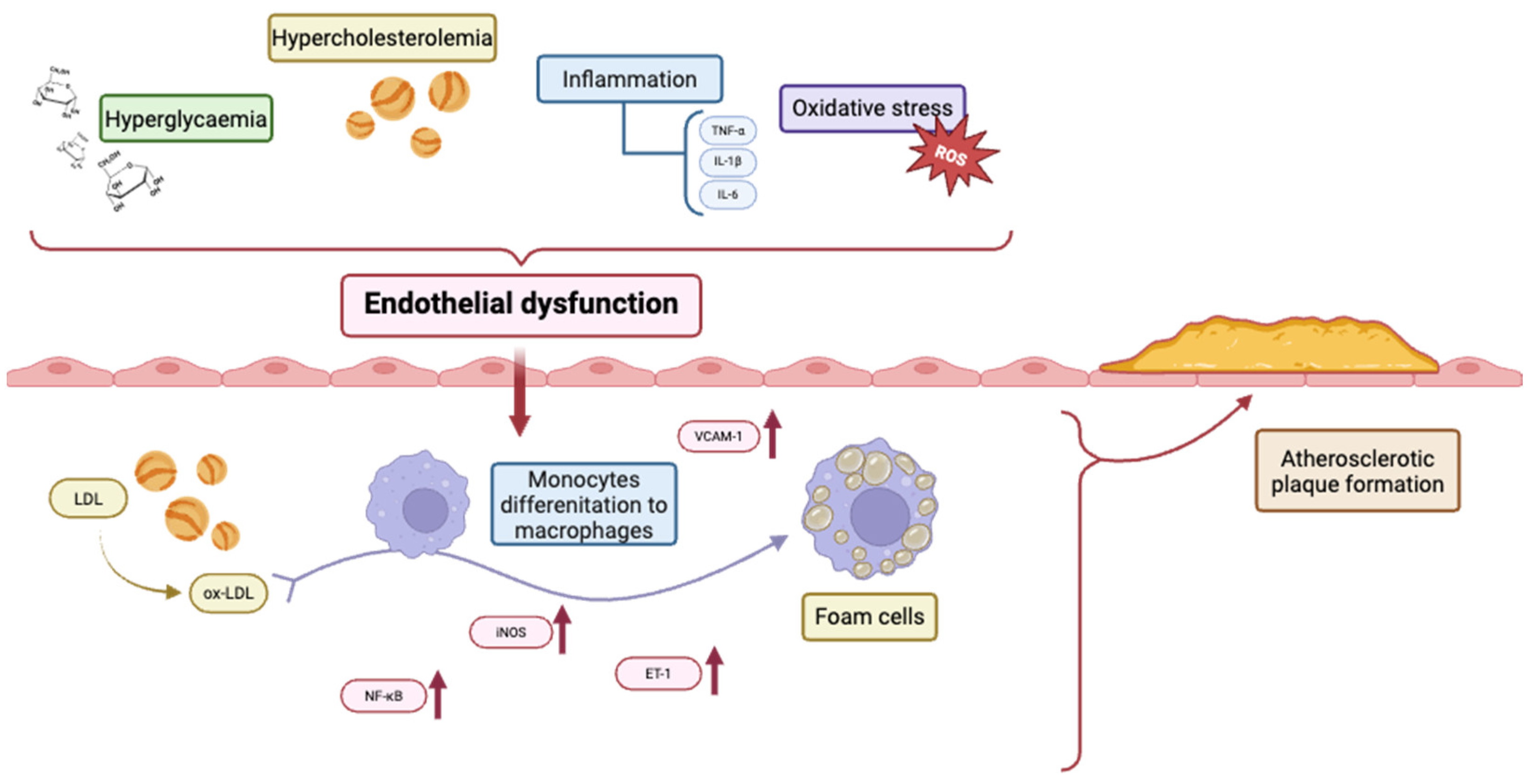
The figure below (Figure 2) illustrates the interaction between hyperglycaemia and hypercholesterolemia in inducing endothelial dysfunction through the release of inflammatory cytokines (e.g., TNF-α, IL-1β, and IL-6) and the generation of oxidative stress (reactive oxygen species, ROS). These processes increase vascular permeability and the expression of adhesion molecules such as VCAM-1, aiding the recruitment of monocytes. Low-density lipoproteins (LDLs) undergo oxidative modification (ox-LDL) in the subendothelial space and are absorbed by differentiated macrophages, which turn into foam cells. Activation of NF-κB, inducible nitric oxide synthase (iNOS), and endothelin-1 (ET-1) further heightens inflammation. The build-up of foam cells and unresolved inflammation contributes to the formation and progression of atherosclerotic plaques.

3. Cerebral Microcirculation in Atherosclerosis

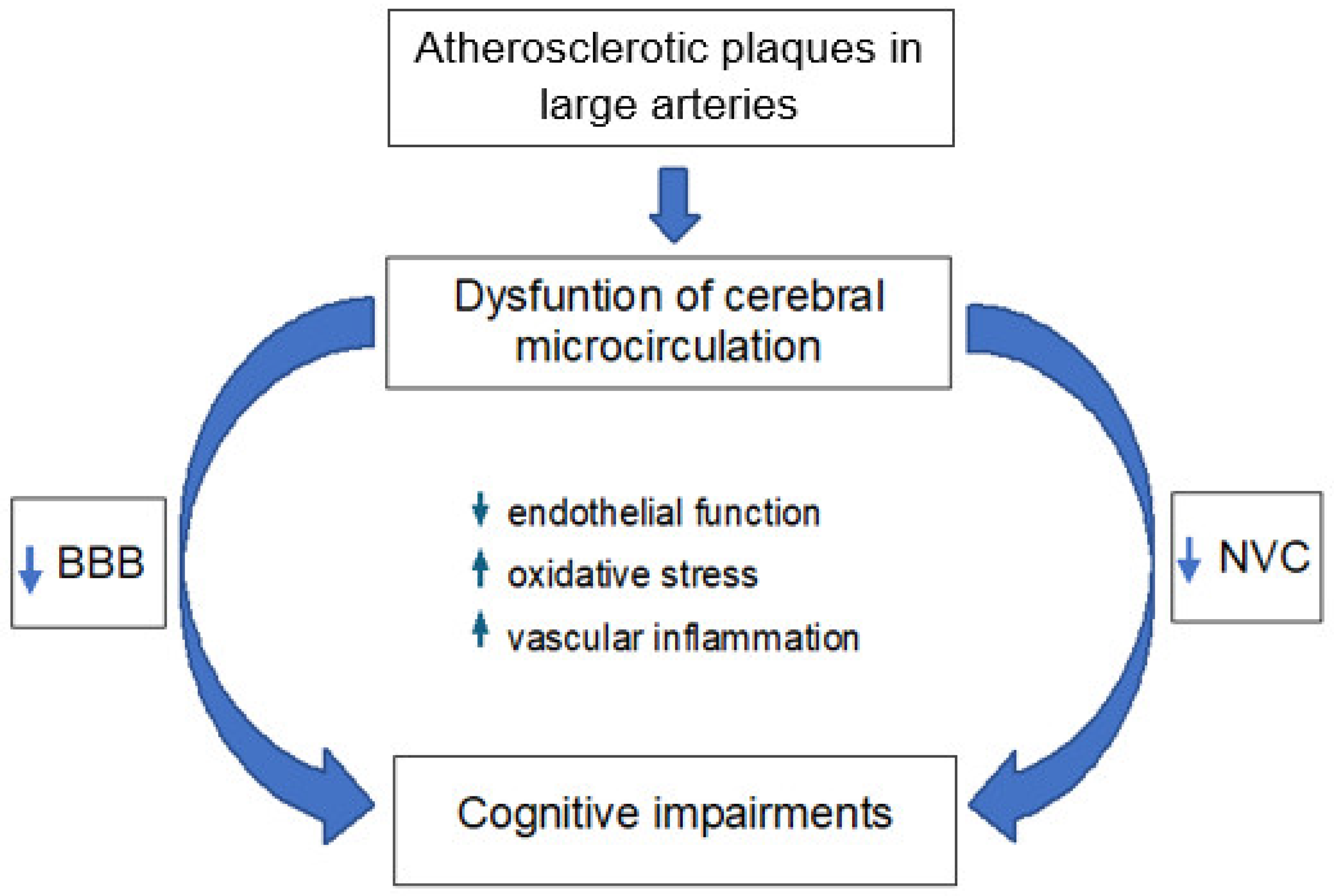
Although atherosclerotic plaques grow within the large arteries, such as the carotid arteries and large cerebral arteries, they also reduce the overall cerebral blood flow (CBF). This leads to improper perfusion in the downstream microcirculation [124,125], thereby contributing to the development of cerebral small vessel disease (CSVD). Moreover, increased arterial stiffness of large arteries leads to increased pulse-wave velocity and pulse pressure [126,127], which in turn, may influence cerebral small vessels. Microcirculation, defined as the network of parenchymal arterioles, capillaries, and venules, is crucial for maintaining proper cerebral perfusion. Microvessels directly determine CBF through changes in their diameter. Dysfunction of microvessels may result in the impairment of functional hyperemia, also named neurovascular coupling (NVC), and disruption of the blood–brain barrier (BBB). Impairments within both NVC and the BBB may lead to cognitive impairments [128,129] (Figure 3). In the section below, we discuss the influence of AS on NVC, the BBB, and consequently on the occurrence of cognitive disorders.

3.1. Neurovascular Coupling

In a normal brain vascular system, vasodilation adjusts the cerebral microflow in response to increased demand for oxygen and nutrients during increased neuronal activity. This NVC is reduced or absent in many forms of cerebrovascular pathology [130]. NVC requires the structural and functional integrity of the neurovascular unit. The neurovascular unit [NVU] is composed of microvascular endothelial cells, astrocytes, neurons, and pericytes, which are interconnected and function in concert. Neuronal activity is communicated to the cerebral vasculature at the level of the neurovascular unit and precapillary arterioles to adjust the local perfusion of brain tissue to neuronal metabolism. An increase in neuronal activity is accompanied by vasodilation and by an increase in local cerebral blood flow. This neurovascular coupling is essential for the homeostasis and proper functioning of the brain, and endothelial cells are an indispensable component of the neurovascular unit, contributing to the regulation of the microflow [131].
Dysfunction of cerebral microcirculation can critically affect neurovascular coupling. Alterations within microvessels, particularly those induced by aging and AS, could lead to neurodegenerative conditions and cognitive dysfunction [132]. It is known that AS affects cerebral microcirculation, contributing to cognitive decline and increased risk of ischemic events [133,134,135]. Some studies demonstrated that disruptions in neurovascular coupling, possibly due to microvascular changes from systemic atherosclerotic factors, significantly impair local CBF regulation. Eyre et al. (2025) demonstrated a disturbed response to whisker stimulation in mice with AS [135]. Lu et al. (2019) found similar results, demonstrating that the somatosensory cortex of aged atherosclerotic (ATX) mice had a reduced hemodynamic response to whisker stimulation [133]. Compared to juvenile atherosclerotic mice, ATX animals also showed a lower and more variable partial pressure of oxygen (pO2) in cerebral tissue. Additionally, hypoxic micro-pockets in cortical tissue were found in old, but not young, ATX mice. Another study confirmed that old atherosclerotic mice exhibit a higher spatial heterogeneity of tissue pO2, suggesting a less efficient oxygen extraction [134].
The worsened microvascular perfusion contributes to ischemic damage in the cerebral tissues. AS causes unnoticed strokes that occur in areas of the brain with compromised blood flow. Over time, these silent strokes can accumulate, leading to subcortical ischemic changes [136]. Some studies confirmed that atherosclerotic disease plays a causative role in the onset of ischemic cerebrovascular diseases [137,138]. Clinical studies showed that, in patients with AS, lacunar infarcts are observed [139,140]. Interestingly, smaller lacunes (<3 mm) were associated with diabetes, and larger lacunes were associated with high LDL cholesterol [141].

3.2. Blood–Brain Barrier

Endothelial cells in the cerebral microvessels form the BBB, which limits the transport of undesirable substances from the bloodstream to the brain. Cerebral endothelial cells are connected by tight junctions (TJs), which are composed of integral membrane proteins such as occludin and claudin-5. Both membrane proteins are linked to the actin cytoskeleton by TJ-associated proteins such as zonula occludens-1 (ZO-1) [142].
The majority of research on the relationship between AS and the BBB structure shows that carotid artery stenosis and AS in large arteries cause disruption of the BBB [143,144]. More precisely, increased BBB permeability and tight junction dysregulation have been reported [124,145,146,147]. Furthermore, increased BBB permeability is linked to AS risk factors like HT, HPL, and HG [148,149]. Changes in the expression and structure of endothelial TJs have been documented in these comorbidities [150,151,152]. Additionally, it has been demonstrated that proinflammatory cytokines impair the endothelial barrier [153,154]. Elevated OS and inflammatory mediators interact to create a vicious cycle that damages the endothelium and increases the BBB’s permeability (Figure 3). Consequently, it facilitates potentially harmful substances to enter the central nervous system, which can lead to neurological conditions and cognitive decline [155,156]. On the other hand, some studies indicated circumscribed BBB breakdown in the white matter in AS [88], which could, however, be an early predictor of white matter damage commonly found in cerebral small vessel diseases [157].
OS and inflammation cause BBB dysfunction, leading to impaired brain function in patients with AS. Understanding the impact of AS on BBB permeability is crucial to developing treatment strategies that will preserve the integrity of the BBB and reduce the incidence of neurodegenerative diseases associated with this systemic disorder [156,158].

3.3. Cognitive Impairments

Normal cognitive functions require an adequate, well-regulated delivery of blood to the neurons [159]. The oxygen and glucose are delivered to neurons by microvessels; therefore, microvascular dysfunction may be associated with worse cognitive performance. Accumulating evidence supports the role of disturbance of neurovascular coupling and BBB functions in the pathophysiology of cognitive decline [128,129,160]. Despite disturbed BBB and NVC functions in AS [124,133,134,146,147], conclusions regarding the effect of AS on cognitive function appear inconsistent. The vast majority of studies indicate that AS leads to reduced cognition [140,161,162,163,164]. Cognitive disorders occur even in subclinical AS [165]. On the other hand, a few studies showed no association between markers of AS and cognitive dysfunction [166,167,168]. For example, Auperin et al. (1996) did not identify any statistically significant correlations between AS and cognitive function in women [166]. The discrepancy between studies indicating the existence or absence of a correlation between AS and cognitive disorders may be related to study sample variability, cognitive testing techniques, and AS severity. Additionally, it should be noted that factors that often accompany AS, e.g., older age, can themselves cause the development of cognitive disorders.
In conclusion, the connection between AS and cerebral microcirculation is essential to comprehending the pathogenesis of vascular cognitive impairment (Figure 3). The detrimental effects of AS on cerebral microcirculation may be mitigated by early management and systemic microvascular health monitoring, preserving cognitive function and reducing the risk of dementia.

4. Final Remarks

Our review article described the multifactorial pathophysiology of AS, emphasising the key roles of microcirculatory dysfunction, OS, inflammation, and NO bioavailability. Atherosclerosis remains the leading cause of CVDs, which are the leading cause of death worldwide. The endothelium is crucial for vascular health, with its dysfunction serving as both a marker and a mediator of AS progression. This article highlights how hypertension, hyperlipidaemia, and hyperglycaemia impair NO synthesis, increase reactive oxygen species (ROS), and promote chronic inflammation. These mechanisms promote endothelial injury, plaque development, and vascular remodelling. Special attention is given to cerebral microcirculation, where AS impairs neurovascular coupling and BBB function, which may lead to cognitive decline. Hypercholesterolemia and hyperglycaemia further impair endothelial dysfunction through ROS overproduction and advanced glycation end products. This article also discusses disturbed shear stress and proinflammatory cytokines such as TNF-α and IL-6, which disrupt eNOS regulation and intensify inflammatory cascades. Experimental and clinical evidence support the notion that maintaining NO levels and reducing oxidative and inflammatory triggers may help lessen atherosclerosis. Future research should focus on identifying specific molecular markers and NO-modulating pathways to improve targeted therapies for atherosclerosis. Advanced diagnostics and antioxidant-based interventions could allow earlier prevention of AS-related microvascular and cognitive complications.

Funding

The authors declare that no funds, grants, or other support were received during the preparation of this manuscript.

Conflicts of Interest

The authors declare no conflicts of interest.

References

  1. Zhong, C.; Deng, K.; Lang, X.; Shan, D.; Xie, Y.; Pan, W.; Yu, J. Therapeutic potential of natural flavonoids in atherosclerosis through endothelium-protective mechanisms: An update. Pharmacol. Ther. 2025, 271, 108864. [Google Scholar] [CrossRef] [PubMed]
  2. Shao, R.; Chen, R.; Zheng, Q.; Yao, M.; Li, K.; Cao, Y.; Jiang, L. Oxidative stress disrupts vascular microenvironmental homeostasis affecting the development of atherosclerosis. Cell Biol. Int. 2024, 48, 1781–1801. [Google Scholar] [CrossRef] [PubMed]
  3. Zhao, Y.; Vanhoutte, P.M.; Leung, S.W. Vascular nitric oxide: Beyond eNOS. J. Pharmacol. Sci. 2015, 129, 83–94. [Google Scholar] [CrossRef]
  4. Hadi, H.A.; Suwaidi, J.A. Endothelial dysfunction in diabetes mellitus. Vasc. Health Risk Manag. 2007, 3, 853–876. [Google Scholar] [PubMed]
  5. Durante, A.; Mazzapicchi, A.; Baiardo Redaelli, M. Systemic and Cardiac Microvascular Dysfunction in Hypertension. Int. J. Mol. Sci. 2024, 25, 13294. [Google Scholar] [CrossRef]
  6. Derosa, G.; Maffioli, P. A review about biomarkers for the investigation of vascular function and impairment in diabetes mellitus. Vasc. Health Risk Manag. 2016, 12, 415–419. [Google Scholar] [CrossRef] [PubMed]
  7. Kim, J. The pathophysiology of diabetic foot: A narrative review. J. Yeungnam Med. Sci. 2023, 40, 328–334. [Google Scholar] [CrossRef]
  8. Libby, P. The changing landscape of atherosclerosis. Nature 2021, 592, 524–533. [Google Scholar] [CrossRef]
  9. Rai, V. Current and future role of biomarkers in the monitoring and prognosis of coronary artery disease. Future Cardiol. 2025, 21, 331–333. [Google Scholar] [CrossRef]
  10. Gimbrone, M.A., Jr.; García-Cardeña, G. Endothelial Cell Dysfunction and the Pathobiology of Atherosclerosis. Circ. Res. 2016, 118, 620–636. [Google Scholar] [CrossRef]
  11. Yang, L.; Li, X.; Ni, L.; Lin, Y. Treatment of endothelial cell dysfunction in atherosclerosis: A new perspective integrating traditional and modern approaches. Front. Physiol. 2025, 16, 1555118. [Google Scholar] [CrossRef] [PubMed]
  12. Singh, R.B.; Mengi, S.A.; Xu, Y.J.; Arneja, A.S.; Dhalla, N.S. Pathogenesis of atherosclerosis: A multifactorial process. Exp. Clin. Cardiol. 2002, 7, 40–53. [Google Scholar] [PubMed]
  13. Sies, H.; Berndt, C.; Jones, D.P. Oxidative Stress. Annu. Rev. Biochem. 2017, 86, 715–748. [Google Scholar] [CrossRef] [PubMed]
  14. Hadi, H.A.; Carr, C.S.; Al Suwaidi, J. Endothelial dysfunction: Cardiovascular risk factors, therapy, and outcome. Vasc. Health Risk Manag. 2005, 1, 183–198. [Google Scholar] [PubMed]
  15. Faraci, F.M. Reactive oxygen species: Influence on cerebral vascular tone. J. Appl. Physiol. 2006, 100, 739–743. [Google Scholar] [CrossRef]
  16. Cassuto, J.; Dou, H.; Czikora, I.; Szabo, A.; Patel, V.S.; Kamath, V.; Belin de Chantemele, E.; Feher, A.; Romero, M.J.; Bagi, Z. Peroxynitrite disrupts endothelial caveolae leading to eNOS uncoupling and diminished flow-mediated dilation in coronary arterioles of diabetic patients. Diabetes 2014, 63, 1381–1393. [Google Scholar] [CrossRef]
  17. Poggesi, A.; Pasi, M.; Pescini, F.; Pantoni, L.; Inzitari, D.J. Circulating biologic markers of endothelial dysfunction in cerebral small vessel disease: A review. Cereb. Blood Flow. Metab. 2016, 36, 72–94. [Google Scholar] [CrossRef]
  18. Park, S.J.; Shin, J.I.; Korean, J. Inflammation and hyponatremia: An underrecognized condition? Korean J. Pediatr. 2013, 56, 519–522. [Google Scholar] [CrossRef]
  19. Vila, E.; Salaices, M. Cytokines and vascular reactivity in resistance arteries. Am. J. Physiol. Heart Circ. Physiol. 2005, 288, H1016–H1021. [Google Scholar] [CrossRef]
  20. Kofler, S.; Nickel, T.; Weis, M. Role of cytokines in cardiovascular diseases: A focus on endothelial responses to inflammation. Clin. Sci. 2005, 108, 205–213. [Google Scholar] [CrossRef]
  21. De Palma, C.; Meacci, E.; Perrotta, C.; Bruni, P.; Clementi, E. Endothelial nitric oxide synthase activation by tumor necrosis factor alpha through neutral sphingomyelinase 2, sphingosine kinase 1, and sphingosine 1 phosphate receptors: A novel pathway relevant to the pathophysiology of endothelium. Arterioscler. Thromb. Vasc. Biol. 2006, 26, 99–105. [Google Scholar] [CrossRef] [PubMed]
  22. Yoshizumi, M.; Perrella, M.A.; Burnett, J.C., Jr.; Lee, M.E. Tumor necrosis factor downregulates an endothelial nitric oxide synthase mRNA by shortening its half-life. Circ. Res. 1993, 73, 205–209. [Google Scholar] [CrossRef]
  23. Sprague, A.H.; Khalil, R.A. Inflammatory cytokines in vascular dysfunction and vascular disease. Biochem. Pharmacol. 2009, 78, 539–552. [Google Scholar] [CrossRef]
  24. Miller, F.J., Jr.; Gutterman, D.D.; Rios, C.D.; Heistad, D.D.; Davidson, B.L. Superoxide production in vascular smooth muscle contributes to oxidative stress and impaired relaxation in atherosclerosis. Circ. Res. 1998, 82, 1298–1305. [Google Scholar] [CrossRef]
  25. Collin, B.; Busseuil, D.; Zeller, M.; Perrin, C.; Barthez, O.; Duvillard, L.; Vergely, C.; Bardou, M.; Dumas, M.; Cottin, Y.; et al. Increased superoxide anion production is associated with early atherosclerosis and cardiovascular dysfunctions in a rabbit model. Mol. Cell. Biochem. 2007, 294, 225–235. [Google Scholar] [CrossRef]
  26. White, C.R.; Brock, T.A.; Chang, L.Y.; Crapo, J.; Briscoe, P.; Ku, D.; Bradley, W.A.; Gianturco, S.H.; Gore, J.; Freeman, B.A.; et al. Superoxide and peroxynitrite in atherosclerosis. Proc. Natl. Acad. Sci. USA 1994, 91, 1044–1048. [Google Scholar] [CrossRef] [PubMed]
  27. Sreekumar, R.; Unnikrishnan, J.; Fu, A.; Nygren, J.; Short, K.R.; Schimke, J.; Barazzoni, R.; Nair, K.S. Impact of high-fat diet and antioxidant supplement on mitochondrial functions and gene transcripts in rat muscle. Am. J. Physiol. Endocrinol. Metab. 2002, 282, E1055–E1061. [Google Scholar] [CrossRef] [PubMed]
  28. Zhang, L.; Ya, B.; Yang, P.; Sun, F.; Zhang, L.; Li, Y.; Li, L. Impact of carotid atherosclerosis combined with hypercholesterolemia on cerebral microvessels and brain parenchyma in a new complex rat model. Neurochem. Res. 2014, 39, 653–660. [Google Scholar] [CrossRef]
  29. Ito, F.; Sono, Y.; Ito, T. Measurement and Clinical Significance of Lipid Peroxidation as a Biomarker of Oxidative Stress: Oxidative Stress in Diabetes, Atherosclerosis, and Chronic Inflammation. Antioxidants 2019, 8, 72. [Google Scholar] [CrossRef]
  30. Lerman, A.; Edwards, B.S.; Hallett, J.W.; Heublein, D.M.; Sandberg, S.M.; Burnett, J.C., Jr. Circulating and tissue endothelin immunoreactivity in advanced atherosclerosis. N. Engl. J. Med. 1991, 325, 997–1001. [Google Scholar] [CrossRef]
  31. Fan, J.; Unoki, H.; Iwasa, S.; Watanabe, T. Role of endothelin-1 in atherosclerosis. Ann. N. Y Acad. Sci. 2000, 902, 84–93; discussion 93–94. [Google Scholar] [CrossRef]
  32. Kawashima, S.; Yokoyama, M. Dysfunction of endothelial nitric oxide synthase and atherosclerosis. Arterioscler. Thromb. Vasc. Biol. 2004, 24, 998–1005. [Google Scholar] [CrossRef] [PubMed]
  33. Freiman, P.C.; Mitchell, G.G.; Heistad, D.D.; Armstrong, M.L.; Harrison, D.G. Atherosclerosis impairs endothelium-dependent vascular relaxation to acetylcholine and thrombin in primates. Circ. Res. 1986, 58, 783–789. [Google Scholar] [CrossRef] [PubMed]
  34. Ozaki, M.; Kawashima, S.; Yamashita, T.; Hirase, T.; Namiki, M.; Inoue, N.; Hirata, K.; Yasui, H.; Sakurai, H.; Yoshida, Y.; et al. Overexpression of endothelial nitric oxide synthase accelerates atherosclerotic lesion formation in apoE-deficient mice. J. Clin. Investig. 2002, 110, 331–340. [Google Scholar] [CrossRef]
  35. Ignarro, L.J.; Buga, G.M.; Wei, L.H.; Bauer, P.M.; Wu, G.; del Soldato, P. Role of the arginine-nitric oxide pathway in the regulation of vascular smooth muscle cell proliferation. Proc. Natl. Acad. Sci. USA 2001, 98, 4202–4208. [Google Scholar] [CrossRef] [PubMed]
  36. Song, L.; Zhang, J.; Lai, R.; Li, Q.; Ju, J.; Xu, H. Chinese Herbal Medicines and Active Metabolites: Potential Antioxidant Treatments for Atherosclerosis. Front. Pharmacol. 2021, 12, 675999. [Google Scholar] [CrossRef]
  37. Scioli, M.G.; Storti, G.; D’Amico, F.; Rodríguez Guzmán, R.; Centofanti, F.; Doldo, E.; Céspedes Miranda, E.M.; Orlandi, A. Oxidative Stress and New Pathogenetic Mechanisms in Endothelial Dysfunction: Potential Diagnostic Biomarkers and Therapeutic Targets. J. Clin. Med. 2020, 9, 1995. [Google Scholar] [CrossRef]
  38. Jiang, H.; Wang, J.; Sheng, L.; Xu, X.; Zhou, B.; Shen, L.; Wu, M. Tongnao Decoction (TND) Alleviated Atherosclerosis by Playing Lowering Lipid, Anti-Inflammatory, and Antioxidant Roles. Oxid. Med. Cell. Longev. 2022, 2022, 6061197. [Google Scholar] [CrossRef]
  39. Higashi, Y. Roles of Oxidative Stress and Inflammation in Vascular Endothelial Dysfunction-Related Disease. Antioxidants 2022, 11, 1958. [Google Scholar] [CrossRef]
  40. Li, X.; Guo, D.; Hu, Y.; Chen, Y. Oxidative Stress and Inflammation Are Associated with Coexistent Severe Multivessel Coronary Artery Stenosis and Right Carotid Artery Severe Stenosis in Elderly Patients. Oxid. Med. Cell. Longev. 2021, 2021, 2976447. [Google Scholar] [CrossRef]
  41. Tabaei, S.; Tabaee, S.S. DNA methylation abnormalities in atherosclerosis. Artif. Cells Nanomed. Biotechnol. 2019, 47, 2031–2041. [Google Scholar] [CrossRef] [PubMed]
  42. Laufs, U.; Gertz, K.; Dirnagl, U.; Boehm, M.; Nickenig, G.; Endres, M. Rosuvastatin, a new HMG-CoA reductase inhibitor, upregulates endothelial nitric oxide synthase and protects from ischemic stroke in mice. Brain Res. 2002, 942, 23–30. [Google Scholar] [CrossRef]
  43. Thomas, R.G.; Kim, J.H.; Kim, J.H.; Yoon, J.; Choi, K.H.; Jeong, Y.Y. Treatment of ischemic stroke by atorvastatin-loaded PEGylated liposome. Transl. Stroke Res. 2023, 15, 388–398. [Google Scholar] [CrossRef] [PubMed]
  44. Miller, Y.I.; Choi, S.-H.; Fang, L.; Tsimikas, S. Lipoprotein modification and macrophage uptake: Role of pathologic cholesterol transport in atherogenesis. In Cholesterol Binding and Cholesterol Transport Proteins: Structure and Function in Health and Disease; Harris, J.R., Ed.; Springer: Dordrecht, The Netherlands, 2010; pp. 229–251. [Google Scholar]
  45. Nowak, W.N.; Deng, J.; Ruan, X.Z.; Xu, Q. Reactive oxygen species generation and atherosclerosis. Arterioscler. Thromb. Vasc. Biol. 2017, 37, e41–e52. [Google Scholar] [CrossRef]
  46. Sen-Banerjee, S.; Mir, S.; Lin, Z.; Hamik, A.; Atkins, G.B.; Das, H.; Banerjee, P.; Kumar, A.; Jain, M.K. Kruppel-like factor 2 as a novel mediator of statin effects in endothelial cells. Circulation 2005, 112, 720–726. [Google Scholar] [CrossRef] [PubMed]
  47. Sikora, J.; Kostka, B.; Marczyk, I.; Krajewska, U.; Chałubiński, M.; Broncel, M. Effect of statins on platelet function in patients with hyperlipidemia. Arch. Med. Sci. 2013, 9, 622–628. [Google Scholar] [CrossRef]
  48. Choudhary, A.; Rawat, U.; Kumar, P.; Mittal, P. Pleotropic effects of statins: The dilemma of wider utilization of statin. Egypt. Heart J. 2023, 75, 1. [Google Scholar] [CrossRef]
  49. Mussbacher, M.; Salzmann, M.; Haigl, B.; Basílio, J.; Hochreiter, B.; Gleitsmann, V.; Moser, B.; Hoesel, B.; Suur, B.E.; Puhm, F.; et al. Ikk2-mediated inflammatory activation of arterial endothelial cells promotes the development and progression of atherosclerosis. Atherosclerosis 2020, 307, 21–31. [Google Scholar] [CrossRef]
  50. Ebert, R.; Cumbana, R.; Lehmann, C.; Kutzner, L.; Toewe, A.; Ferreirós, N.; Parnham, M.J.; Schebb, N.H.; Steinhilber, D.; Kahnt, A.S. Long-term stimulation of toll-like receptor-2 and -4 upregulates 5-LO and 15-LO-2 expression thereby inducing a lipid mediator shift in human monocyte-derived macrophages. Biochim. Biophys. Acta (BBA)-Mol. Cell Biol. Lipids 2020, 1865, 158702. [Google Scholar] [CrossRef]
  51. Shi, J.; Zhao, Y.; Wang, K.; Shi, X.; Wang, Y.; Huang, H.; Zhuang, Y.; Cai, T.; Wang, F.; Shao, F. Cleavage of GSDMD by inflammatory caspases determines pyroptotic cell death. Nature 2015, 526, 660–665. [Google Scholar] [CrossRef]
  52. Liu, D.; Zeng, X.; Li, X.; Mehta, J.L.; Wang, X. Role of NLRP3 inflammasome in the pathogenesis of cardiovascular diseases. Basic Res. Cardiol. 2018, 113, 5. [Google Scholar] [CrossRef]
  53. Henein, M.Y.; Vancheri, S.; Longo, G.; Vancheri, F. The Role of Inflammation in Cardiovascular Disease. Int. J. Mol. Sci. 2022, 23, 12906. [Google Scholar] [CrossRef] [PubMed]
  54. Rydén, L.; Buhlin, K.; Ekstrand, E.; de Faire, U.; Gustafsson, A.; Holmer, J.; Kjellström, B.; Lindahl, B.; Norhammar, A.; Klinge, B. Periodontitis Increases the Risk of a First Myocardial Infarction: A Report From the PAROKRANK Study. Circulation 2016, 133, 576–583. [Google Scholar] [CrossRef]
  55. von Rossum, A.; Laher, I.; Choy, J.C. Immune-mediated vascular injury and dysfunction in transplant arteriosclerosis. Front. Immunol. 2015, 5, 684. [Google Scholar] [CrossRef]
  56. Nguyen Dinh Cat, A.; Montezano, A.C.; Burger, D.; Touyz, R.M. Angiotensin II, NADPH oxidase, and redox signaling in the vasculature. Antioxid. Redox Signal. 2013, 19, 1110–1120. [Google Scholar] [CrossRef]
  57. Lim, S.; Park, S. Role of vascular smooth muscle cell in the inflammation of atherosclerosis. BMB Rep. 2014, 47, 1–7. [Google Scholar] [CrossRef] [PubMed]
  58. Peng, W.; Li, T.; Pi, S.; Huang, L.; Liu, Y. Suppression of circular RNA circDHCR24 alleviates aortic smooth muscle cell proliferation and migration by targeting miR-149-5p/MMP9 axis. Biochem. Biophys. Res. Commun. 2020, 529, 753–759. [Google Scholar] [CrossRef]
  59. Förstermann, U.; Sessa, W.C. Nitric oxide synthases: Regulation and function. Eur. Heart J. 2021, 42, 3230–3242. [Google Scholar] [CrossRef]
  60. Rui, W.; Guan, L.; Zhang, F.; Zhang, W.; Ding, W. PM2.5-induced oxidative stress increases adhesion molecules expression in human endothelial cells through the ERK/AKT/NF-κB-dependent pathway. J. Appl. Toxicol. 2016, 36, 48–59. [Google Scholar] [CrossRef]
  61. Monaco, C.; Andreakos, E.; Kiriakidis, S.; Mauri, C.; Bicknell, C.; Foxwell, B.; Cheshire, N.; Paleolog, E.; Feldmann, M. Canonical pathway of nuclear factor kappa B activation selectively regulates proinflammatory and prothrombotic responses in human atherosclerosis. Proc. Natl. Acad. Sci. USA 2004, 101, 5634–5639. [Google Scholar] [CrossRef]
  62. Khan, M.S.; Talha, K.M.; Maqsood, M.H.; Rymer, J.A.; Borlaug, B.A.; Docherty, K.F.; Pandey, A.; Kahles, F.; Cikes, M.; Lam, C.S.P.; et al. Interleukin-6 and Cardiovascular Events in Healthy Adults: MESA. JACC Adv. 2024, 3, 101063. [Google Scholar] [CrossRef] [PubMed]
  63. Varga, Z.; Sabzwari, S.R.A.; Vargova, V. Cardiovascular Risk of Nonsteroidal Anti-Inflammatory Drugs: An Under-Recognized Public Health Issue. Cureus 2017, 9, e1144. [Google Scholar] [CrossRef]
  64. Pujades-Rodriguez, M.; Morgan, A.W.; Cubbon, R.M.; Wu, J. Dose-dependent oral glucocorticoid cardiovascular risks in people with immune-mediated inflammatory diseases: A population-based cohort study. PLoS Med. 2020, 17, e1003432. [Google Scholar] [CrossRef]
  65. Singh, S.; Fumery, M.; Singh, A.G.; Singh, N.; Prokop, L.J.; Dulai, P.S.; Sandborn, W.J.; Curtis, J.R. Comparative Risk of Cardiovascular Events With Biologic and Synthetic Disease-Modifying Antirheumatic Drugs in Patients With Rheumatoid Arthritis: A Systematic Review and Meta-Analysis. Arthritis Care Res. 2020, 72, 561–576. [Google Scholar] [CrossRef]
  66. Diamantis, E.; Kyriakos, G.; Quiles-Sanchez, L.V.; Farmaki, P.; Troupis, T. The Anti-Inflammatory Effects of Statins on Coronary Artery Disease: An Updated Review of the Literature. Curr. Cardiol. Rev. 2017, 13, 209–216. [Google Scholar] [CrossRef]
  67. Bonaventura, A.; Abbate, A. Colchicine for cardiovascular prevention: The dawn of a new era has finally come. Eur. Heart J. 2023, 44, 3303–3304. [Google Scholar] [CrossRef] [PubMed]
  68. Perera, B.; Wu, Y.; Nguyen, N.T.; Ta, H.T. Advances in drug delivery to atherosclerosis: Investigating the efficiency of different nanomaterials employed for different type of drugs. Mater. Today Bio 2023, 22, 100767. [Google Scholar] [CrossRef] [PubMed]
  69. Ryu, J.Y.; Kim, Y.H.; Lee, J.S.; Lee, J.W.; Oh, E.J.; Kim, H.M.; Lee, S.J.; Lee, J.; Lee, S.Y.; Huh, S.; et al. Oscillatory shear stress promotes angiogenic effects in arteriovenous malformations endothelial cells. Mol. Med. 2021, 27, 31. [Google Scholar] [CrossRef]
  70. Marchio, P.; Guerra-Ojeda, S.; Vila, J.M.; Aldasoro, M.; Victor, V.M.; Mauricio, M.D. Targeting Early Atherosclerosis: A Focus on Oxidative Stress and Inflammation. Oxid. Med. Cell. Longev. 2019, 2019, 8563845. [Google Scholar] [CrossRef]
  71. Zhou, H.L.; Jiang, X.Z.; Ventikos, Y. Role of blood flow in endothelial functionality: A review. Front. Cell Dev. Biol. 2023, 11, 1259280. [Google Scholar] [CrossRef]
  72. Cook-Mills, J.M.; Marchese, M.E.; Abdala-Valencia, H. Vascular cell adhesion molecule-1 expression and signaling during disease: Regulation by reactive oxygen species and antioxidants. Antioxid. Redox Signal 2011, 15, 1607–1638. [Google Scholar] [CrossRef] [PubMed]
  73. Traub, O.; Berk, B.C. Laminar shear stress: Mechanisms by which endothelial cells transduce an atheroprotective force. Arterioscler. Thromb. Vasc. Biol. 1998, 18, 677–685. [Google Scholar] [CrossRef] [PubMed]
  74. Pan, S. Molecular mechanisms responsible for the atheroprotective effects of laminar shear stress. Antioxid. Redox Signal 2009, 11, 1669–1682. [Google Scholar] [CrossRef]
  75. Man, M.Q.; Wakefield, J.S.; Mauro, T.M.; Elias, P.M. Regulatory Role of Nitric Oxide in Cutaneous Inflammation. Inflammation 2022, 45, 949–964. [Google Scholar] [CrossRef]
  76. Liy, P.M.; Puzi, N.N.A.; Jose, S.; Vidyadaran, S. Nitric oxide modulation in neuroinflammation and the role of mesenchymal stem cells. Exp. Biol. Med. 2021, 246, 2399–2406. [Google Scholar] [CrossRef]
  77. Boveris, A.; Alvarez, S.; Navarro, A. The role of mitochondrial nitric oxide synthase in inflammation and septic shock. Free Radic. Biol. Med. 2002, 33, 1186–1193. [Google Scholar] [CrossRef] [PubMed]
  78. Grant, R.W.; Dixit, V.D. Mechanisms of disease: Inflammasome activation and the development of type 2 diabetes. Front. Immunol. 2013, 4, 50. [Google Scholar] [CrossRef]
  79. Shrishrimal, S.; Chatterjee, A.; Kosmacek, E.A.; Davis, P.J.; McDonald, J.T.; Oberley-Deegan, R.E. Manganese porphyrin, MnTE-2-PyP, treatment protects the prostate from radiation-induced fibrosis (RIF) by activating the NRF2 signaling pathway and enhancing SOD2 and sirtuin activity. Free Radic. Biol. Med. 2020, 152, 255–270. [Google Scholar] [CrossRef]
  80. Watts, G.F.; Catapano, A.L.; Masana, L.; Zambon, A.; Pirillo, A.; Tokgözoğlu, L. Hypercholesterolemia and cardiovascular disease: Focus on high cardiovascular risk patients. Atheroscler. Suppl. 2020, 42, e30–e34. [Google Scholar] [CrossRef]
  81. Stokes, K.Y.; Cooper, D.; Tailor, A.; Granger, D.N. Hypercholesterolemia promotes inflammation and microvascular dysfunction: Role of nitric oxide and superoxide. Free Radic. Biol. Med. 2002, 33, 1026–1036. [Google Scholar] [CrossRef]
  82. Stoll, G.; Bendszus, M. Inflammation and atherosclerosis: Novel insights into plaque formation and destabilization. Stroke 2006, 37, 1923–1932. [Google Scholar] [CrossRef] [PubMed]
  83. Napoli, C. Oxidation of LDL, atherogenesis, and apoptosis. Ann. N. Y. Acad. Sci. 2003, 1010, 698–709. [Google Scholar] [CrossRef] [PubMed]
  84. Jiang, H.; Zhou, Y.; Nabavi, S.M.; Sahebkar, A.; Little, P.J.; Xu, S.; Weng, J.; Ge, J. Mechanisms of Oxidized LDL-Mediated Endothelial Dysfunction and Its Consequences for the Development of Atherosclerosis. Front. Cardiovasc. Med. 2022, 9, 925923. [Google Scholar] [CrossRef]
  85. Frostegård, J. Immunity, atherosclerosis and cardiovascular disease. BMC Med. 2013, 11, 117. [Google Scholar] [CrossRef]
  86. Prasad, K.; Mishra, M. Mechanism of Hypercholesterolemia-Induced Atherosclerosis. Rev. Cardiovasc. Med. 2022, 23, 212. [Google Scholar] [CrossRef]
  87. Esmon, C.T. Inflammation and thrombosis. J. Thromb. Haemost. 2003, 1, 1343–1348. [Google Scholar] [CrossRef] [PubMed]
  88. Kraft, P.; Schuhmann, M.K.; Garz, C.; Jandke, S.; Urlaub, D.; Mencl, S.; Zernecke, A.; Heinze, H.J.; Carare, R.O.; Kleinschnitz, C.; et al. Hypercholesterolemia induced cerebral small vessel disease. PLoS ONE 2017, 12, e0182822. [Google Scholar] [CrossRef]
  89. Ishikawa, M.; Stokes, K.Y.; Zhang, J.H.; Nanda, A.; Granger, D.N. Cerebral microvascular responses to hypercholesterolemia: Roles of NADPH oxidase and P-selectin. Circ. Res. 2004, 94, 239–244. [Google Scholar] [CrossRef]
  90. Song, S.H.; Kim, J.H.; Lee, J.H.; Yun, Y.M.; Choi, D.H.; Kim, H.Y. Elevated blood viscosity is associated with cerebral small vessel disease in patients with acute ischemic stroke. BMC Neurol. 2017, 17, 20. [Google Scholar] [CrossRef]
  91. Lopes, F.G.; Bottino, D.A.; Oliveira, F.J.; Mecenas, A.S.; Clapauch, R.; Bouskela, E. In elderly women moderate hypercholesterolemia is associated to endothelial and microcirculatory impairments. Microvasc. Res. 2013, 85, 99–103. [Google Scholar] [CrossRef]
  92. Todate, Y.; Ishigaki, Y. Effect of Hypercholesterolemia on the Characteristics of Cerebral Microvasculature. Diabetes 2018, 67 (Suppl. S1), 463-P. [Google Scholar] [CrossRef]
  93. Kitayama, J.; Faraci, F.M.; Lentz, S.R.; Heistad, D.D. Cerebral vascular dysfunction during hypercholesterolemia. Stroke 2007, 38, 2136–2141. [Google Scholar] [CrossRef] [PubMed]
  94. Wannamethee, S.G.; Shaper, A.G.; Ebrahim, S. HDL-Cholesterol, total cholesterol, and the risk of stroke in middle-aged British men. Stroke 2000, 31, 1882–1888. [Google Scholar] [CrossRef]
  95. Shi, Y.; Guo, L.; Chen, Y.; Xie, Q.; Yan, Z.; Liu, Y.; Kang, J.; Li, S. Risk factors for ischemic stroke: Differences between cerebral small vessel and large artery atherosclerosis aetiologies. Folia Neuropathol. 2021, 59, 378–385. [Google Scholar] [CrossRef]
  96. Demchuk, A.M.; Hess, D.C.; Brass, L.M.; Yatsu, F.M. Is cholesterol a risk factor for stroke?: Yes. Arch. Neurol. 1999, 56, 1518–1520, discussion 1524. [Google Scholar] [CrossRef]
  97. Kannel, W.B.; Dawber, T.R.; McNamara, P.M. Vascular Disease of the Brain—Epidemiologic Aspects: The Framingham Study. Am. J. Public Health Nations Health 1965, 55, 1355–1366. [Google Scholar] [CrossRef] [PubMed]
  98. Lewington, S.; Whitlock, G.; Clarke, R.; Sherliker, P.; Emberson, J.; Halsey, J.; Qizilbash, N.; Peto, R.; Collins, R. Blood cholesterol and vascular mortality by age, sex, and blood pressure: A meta-analysis of individual data from 61 prospective studies with 55,000 vascular deaths. Lancet 2007, 370, 1829–1839, Erratum in Lancet 2008, 372, 292. [Google Scholar] [CrossRef]
  99. Beheshti, S.; Madsen, C.M.; Varbo, A.; Benn, M.; Nordestgaard, B.G. Relationship of Familial Hypercholesterolemia and High Low-Density Lipoprotein Cholesterol to Ischemic Stroke: Copenhagen General Population Study. Circulation 2018, 138, 578–589. [Google Scholar] [CrossRef]
  100. Amarenco, P.; Labreuche, J. Lipid management in the prevention of stroke: Review and updated meta-analysis of statins for stroke prevention. Lancet Neurol. 2009, 8, 453–463. [Google Scholar] [CrossRef]
  101. Joubert, J.; Lemmer, L.B.; Fourie, P.A.; van Gelder, A.L.; Darazs, B. Are clinical differences between black and white stroke patients caused by variations in the atherosclerotic involvement of the arterial tree? S. Afr. Med. J. 1990, 77, 248–251. [Google Scholar]
  102. Vaughan, C.J.; Delanty, N.; Basson, C.T. Statin therapy and stroke prevention. Curr. Opin. Cardiol. 2001, 16, 219–224. [Google Scholar] [CrossRef] [PubMed]
  103. Ference, B.A.; Ginsberg, H.N.; Graham, I.; Ray, K.K.; Packard, C.J.; Bruckert, E.; Hegele, R.A.; Krauss, R.M.; Raal, F.J.; Schunkert, H.; et al. Low-density lipoproteins cause atherosclerotic cardiovascular disease. 1. Evidence from genetic, epidemiologic, and clinical studies. A consensus statement from the European Atherosclerosis Society Consensus Panel. Eur. Heart J. 2017, 38, 2459–2472. [Google Scholar] [CrossRef] [PubMed]
  104. Aronson, D.; Rayfield, E.J. How hyperglycemia promotes atherosclerosis: Molecular mechanisms. Cardiovasc. Diabetol. 2002, 1, 1. [Google Scholar] [CrossRef]
  105. Bornfeldt, K.E. Does Elevated Glucose Promote Atherosclerosis? Pros and Cons. Circ. Res. 2016, 119, 190–193. [Google Scholar] [CrossRef]
  106. Rom, S.; Zuluaga-Ramirez, V.; Gajghate, S.; Seliga, A.; Winfield, M.; Heldt, N.A.; Kolpakov, M.A.; Bashkirova, Y.V.; Sabri, A.K.; Persidsky, Y. Hyperglycemia-Driven Neuroinflammation Compromises BBB Leading to Memory Loss in Both Diabetes Mellitus (DM) Type 1 and Type 2 Mouse Models. Mol. Neurobiol. 2019, 56, 1883–1896. [Google Scholar] [CrossRef] [PubMed]
  107. Nishizawa, T.; Kanter, J.E.; Kramer, F.; Barnhart, S.; Shen, X.; Vivekanandan-Giri, A.; Wall, V.Z.; Kowitz, J.; Devaraj, S.; O’Brien, K.D.; et al. Testing the role of myeloid cell glucose flux in inflammation and atherosclerosis. Cell Rep. 2014, 7, 356–365. [Google Scholar] [CrossRef]
  108. Nagareddy, P.R.; Murphy, A.J.; Stirzaker, R.A.; Hu, Y.; Yu, S.; Miller, R.G.; Ramkhelawon, B.; Distel, E.; Westerterp, M.; Huang, L.S.; et al. Hyperglycemia promotes myelopoiesis and impairs the resolution of atherosclerosis. Cell Metab. 2013, 17, 695–708. [Google Scholar] [CrossRef]
  109. Nakahata, K.; Kinoshita, H.; Azma, T.; Matsuda, N.; Hama-Tomioka, K.; Haba, M.; Hatano, Y. Propofol restores brain microvascular function impaired by high glucose via the decrease in oxidative stress. Anesthesiology 2008, 108, 269–275. [Google Scholar] [CrossRef] [PubMed]
  110. Zheng, F.; Lu, W.; Jia, C.; Li, H.; Wang, Z.; Jia, W. Relationships between glucose excursion and the activation of oxidative stress in patients with newly diagnosed type 2 diabetes or impaired glucose regulation. Endocrine 2010, 37, 201–208. [Google Scholar] [CrossRef]
  111. Karpen, C.W.; Cataland, S.; O’Dorisio, T.M.; Panganamala, R.V. Production of 12-hydroxyeicosatetraenoic acid and vitamin E status in platelets from type I human diabetic subjects. Diabetes 1985, 34, 526–531. [Google Scholar] [CrossRef]
  112. Chen, M.S.; Hutchinson, M.L.; Pecoraro, R.E.; Lee, W.Y.; Labbé, R.F. Hyperglycemia-induced intracellular depletion of ascorbic acid in human mononuclear leukocytes. Diabetes 1983, 32, 1078–1081. [Google Scholar] [CrossRef] [PubMed]
  113. Yan, S.D.; Chen, X.; Schmidt, A.M.; Brett, J.; Godman, G.; Zou, Y.S.; Scott, C.W.; Caputo, C.; Frappier, T.; Smith, M.A.; et al. Glycated tau protein in Alzheimer disease: A mechanism for induction of oxidant stress. Proc. Natl. Acad. Sci. USA 1994, 91, 7787–7791. [Google Scholar] [CrossRef]
  114. Yamagishi, S.I.; Matsui, T. Role of Ligands of Receptor for Advanced Glycation End Products (RAGE) in Peripheral Artery Disease. Rejuvenation Res. 2018, 21, 456–463. [Google Scholar] [CrossRef]
  115. Schmidt, A.M.; Hori, O.; Brett, J.; Yan, S.D.; Wautier, J.L.; Stern, D. Cellular receptors for advanced glycation end products. Implications for induction of oxidant stress and cellular dysfunction in the pathogenesis of vascular lesions. Arterioscler. Thromb. A J. Vasc. Biol. 1994, 14, 1521–1528. [Google Scholar] [CrossRef]
  116. Brett, J.; Schmidt, A.M.; Yan, S.D.; Zou, Y.S.; Weidman, E.; Pinsky, D.; Nowygrod, R.; Neeper, M.; Przysiecki, C.; Shaw, A.; et al. Survey of the distribution of a newly characterized receptor for advanced glycation end products in tissues. Am. J. Pathol. 1993, 143, 1699–1712. [Google Scholar] [PubMed]
  117. Esposito, C.; Gerlach, H.; Brett, J.; Stern, D.; Vlassara, H. Endothelial receptor-mediated binding of glucose-modified albumin is associated with increased monolayer permeability and modulation of cell surface coagulant properties. J. Exp. Med. 1989, 170, 1387–1407. [Google Scholar] [CrossRef] [PubMed]
  118. Mayhan, W.G.; Simmons, L.K.; Sharpe, G.M. Mechanism of impaired responses of cerebral arterioles during diabetes mellitus. Am. J. Physiol. 1991, 260 Pt 2, H319–H326. [Google Scholar] [CrossRef] [PubMed]
  119. Cipolla, M.J.; Godfrey, J.A. Effect of hyperglycemia on brain penetrating arterioles and cerebral blood flow before and after ischemia/reperfusion. Transl. Stroke Res. 2010, 1, 127–134. [Google Scholar] [CrossRef]
  120. Ginsberg, M.D.; Prado, R.; Dietrich, W.D.; Busto, R.; Watson, B.D. Hyperglycemia reduces the extent of cerebral infarction in rats. Stroke 1987, 18, 570–574. [Google Scholar] [CrossRef]
  121. Uyttenboogaart, M.; Koch, M.W.; Stewart, R.E.; Vroomen, P.C.; Luijckx, G.J.; De Keyser, J. Moderate hyperglycaemia is associated with favourable outcome in acute lacunar stroke. Brain 2007, 130 Pt 6, 1626–1630. [Google Scholar] [CrossRef]
  122. Nair, S.S.; Sylaja, P.N.; Sreedharan, S.E.; Sarma, S. Maintenance of normoglycemia may improve outcome in acute ischemic stroke. Ann. Indian. Acad. Neurol. 2017, 20, 122–126. [Google Scholar] [PubMed]
  123. Prospective Studies Collaboration and Asia Pacific Cohort Studies Collaboration. Sex-specific relevance of diabetes to occlusive vascular and other mortality: A collaborative meta-analysis of individual data from 980,793 adults from 68 prospective studies. Lancet Diabetes Endocrinol. 2018, 6, 538–546. [Google Scholar] [CrossRef]
  124. Nyúl-Tóth, Á.; Patai, R.; Csiszar, A.; Ungvari, A.; Gulej, R.; Mukli, P.; Yabluchanskiy, A.; Benyo, Z.; Sotonyi, P.; Prodan, C.I.; et al. Linking peripheral atherosclerosis to blood-brain barrier disruption: Elucidating its role as a manifestation of cerebral small vessel disease in vascular cognitive impairment. Geroscience 2024, 46, 6511–6536. [Google Scholar] [CrossRef] [PubMed]
  125. Csiszar, A.; Ungvari, A.; Patai, R.; Gulej, R.; Yabluchanskiy, A.; Benyo, Z.; Kovacs, I.; Sotonyi, P.; Kirkpartrick, A.C.; Prodan, C.I.; et al. Atherosclerotic burden and cerebral small vessel disease: Exploring the link through microvascular aging and cerebral microhemorrhages. Geroscience 2024, 46, 5103–5132. [Google Scholar] [CrossRef]
  126. O’Rourke, M.F.; Safar, M.E. Relationship between aortic stiffening and microvascular disease in brain and kidney: Cause and logic of therapy. Hypertension 2005, 46, 200–2004. [Google Scholar] [CrossRef] [PubMed]
  127. van Popele, N.M.; Grobbee, D.E.; Bots, M.L.; Asmar, R.; Topouchian, J.; Reneman, R.S.; Hoeks, A.P.; van der Kuip, D.A.; Hofman, A.; Witteman, J.C. Association between arterial stiffness and atherosclerosis: The Rotterdam Study. Stroke 2001, 32, 454–460. [Google Scholar] [CrossRef]
  128. Che, J.; Sun, Y.; Deng, Y.; Zhang, J. Blood-brain barrier disruption: A culprit of cognitive decline? Fluids Barriers CNS 2024, 21, 63. [Google Scholar] [CrossRef]
  129. Tarantini, S.; Tran, C.H.T.; Gordon, G.R.; Ungvari, Z.; Csiszar, A. Impaired neurovascular coupling in aging and Alzheimer’s disease: Contribution of astrocyte dysfunction and endothelial impairment to cognitive decline. Exp. Gerontol. 2017, 94, 52–58. [Google Scholar] [CrossRef]
  130. Bøthun, M.L.; Haaland, Ø.A.; Moen, G.; Logallo, N.; Svendsen, F.; Thomassen, L.; Helland, C.A. Impaired cerebrovascular reactivity may predict delayed cerebral ischemia after aneurysmal subarachnoid hemorrhage. J. Neurol. Sci. 2019, 407, 116539. [Google Scholar] [CrossRef]
  131. Izawa, Y.; Gu, Y.H.; Osada, T.; Kanazawa, M.; Hawkins, B.T.; Koziol, J.A.; Papayannopoulou, T.; Spatz, M.; Del Zoppo, G.J. β1-integrin-matrix interactions modulate cerebral microvessel endothelial cell tight junction expression and permeability. J. Cereb. Blood Flow. Metab. 2018, 38, 641–658. [Google Scholar] [CrossRef]
  132. Toth, P.; Tarantini, S.; Ashpole, N.M.; Tucsek, Z.; Milne, G.L.; Valcarcel-Ares, N.M.; Menyhart, A.; Farkas, E.; Sonntag, W.E.; Csiszar, A.; et al. IGF-1 deficiency impairs neurovascular coupling in mice: Implications for cerebromicrovascular aging. Aging Cell 2015, 14, 1034–1044. [Google Scholar] [CrossRef] [PubMed]
  133. Lu, Y.; Zhang, C.; Lu, X.; Moeini, M.; Thorin, E.; Lesage, F. Impact of atherosclerotic disease on cerebral microvasculature and tissue oxygenation in awake LDLR-/-hApoB+/+ transgenic mice. Neurophotonics 2019, 6, 045003. [Google Scholar] [CrossRef]
  134. Li, B.; Lu, X.; Moeini, M.; Sakadžić, S.; Thorin, E.; Lesage, F. Atherosclerosis is associated with a decrease in cerebral microvascular blood flow and tissue oxygenation. PLoS ONE 2019, 14, e0221547. [Google Scholar] [CrossRef]
  135. Eyre, B.; Shaw, K.; Drew, D.; Rayson, A.; Shabir, O.; Lee, L.; Francis, S.; Berwick, J.; Howarth, C. Characterizing vascular function in mouse models of Alzheimer’s disease, atherosclerosis, and mixed Alzheimer’s and atherosclerosis. Neurophotonics 2025, 12 (Suppl. S1), S14610. [Google Scholar] [CrossRef]
  136. Vilar-Bergua, A.; Riba-Llena, I.; Nafría, C.; Bustamante, A.; Llombart, V.; Delgado, P.; Montaner, J. Blood and CSF biomarkers in brain subcortical ischemic vascular disease: Involved pathways and clinical applicability. J. Cereb. Blood Flow. Metab. 2016, 36, 55–71. [Google Scholar] [CrossRef] [PubMed]
  137. Banerjee, C.; Chimowitz, M.I. Stroke Caused by Atherosclerosis of the Major Intracranial Arteries. Circ. Res. 2017, 120, 502–513. [Google Scholar] [CrossRef]
  138. Ellulu, M.S.; Patimah, I.; Khaza’ai, H.; Rahmat, A.; Abed, Y.; Ali, F. Atherosclerotic cardiovascular disease: A review of initiators and protective factors. Inflammopharmacology 2016, 24, 1–10. [Google Scholar] [CrossRef]
  139. Komura, S.; Nomura, T.; Imaizumi, T.; Inamura, S.; Kanno, A.; Honda, O.; Hashimoto, Y.; Mikami, T.; Nonaka, T. Asymptomatic cerebral findings on 3-Tesla MRI in patients with severe carotid artery stenoses. J. Clin. Neurosci. 2022, 101, 106–111. [Google Scholar] [CrossRef] [PubMed]
  140. Baradaran, H.; Dahlstrom, K.A.; Culleton, S.; Sarrami, A.H.; McFarland, M.M.; Romero, J.R. Association between Extracranial Carotid Artery Plaque and Cognitive Dysfunction: A Systematic Review and Meta-Analysis. Dement. Geriatr. Cogn. Disord. 2022, 51, 377–385. [Google Scholar] [CrossRef]
  141. Bezerra, D.C.; Sharrett, A.R.; Matsushita, K.; Gottesman, R.F.; Shibata, D.; Mosley, T.H., Jr.; Coresh, J.; Szklo, M.; Carvalho, M.S.; Selvin, E. Risk factors for lacune subtypes in the Atherosclerosis Risk in Communities (ARIC) Study. Neurology 2012, 78, 102–108. [Google Scholar] [CrossRef]
  142. Krueger, M.; Härtig, W.; Reichenbach, A.; Bechmann, I.; Michalski, D. Blood-brain barrier breakdown after embolic stroke in rats occurs without ultrastructural evidence for disrupting tight junctions. PLoS ONE 2013, 8, e56419. [Google Scholar] [CrossRef] [PubMed]
  143. Van Skike, C.E.; Jahrling, J.B.; Olson, A.B.; Sayre, N.L.; Hussong, S.A.; Ungvari, Z.; Lechleiter, J.D.; Galvan, V. Inhibition of mTOR protects the blood-brain barrier in models of Alzheimer’s disease and vascular cognitive impairment. Am. J. Physiol. Heart Circ. Physiol. 2018, 314, H693–H703. [Google Scholar] [CrossRef]
  144. Roberts, J.M.; Maniskas, M.E.; Bix, G.J. Bilateral carotid artery stenosis causes unexpected early changes in brain extracellular matrix and blood-brain barrier integrity in mice. PLoS ONE 2018, 13, e0195765. [Google Scholar] [CrossRef]
  145. Chen, L.; Chen, Z.; Ge, M.; Tang, O.; Cheng, Y.; Zhou, H.; Shen, Y.; Qin, F. Monocytic cell junction proteins serve important roles in atherosclerosis via the endoglin pathway. Mol. Med. Rep. 2017, 16, 6750–6756. [Google Scholar] [CrossRef] [PubMed]
  146. Cheng, X.; Wang, X.; Wan, Y.; Zhou, Q.; Zhu, H.; Wang, Y. Myosin light chain kinase inhibitor ML7 improves vascular endothelial dysfunction via tight junction regulation in a rabbit model of atherosclerosis. Mol. Med. Rep. 2015, 12, 4109–4116. [Google Scholar] [CrossRef]
  147. Kisucka, J.; Chauhan, A.K.; Zhao, B.Q.; Patten, I.S.; Yesilaltay, A.; Krieger, M.; Wagner, D.D. Elevated levels of soluble P-selectin in mice alter blood-brain barrier function, exacerbate stroke, and promote atherosclerosis. Blood 2009, 113, 6015–6022. [Google Scholar] [CrossRef] [PubMed]
  148. Gyanwali, B.; Tan, C.S.; Petr, J.; Escobosa, L.L.T.; Vrooman, H.; Chen, C.; Mutsaerts, H.J.; Hilal, S. Arterial Spin-Labeling Parameters and Their Associations with Risk Factors, Cerebral Small-Vessel Disease, and Etiologic Subtypes of Cognitive Impairment and Dementia. AJNR Am. J. Neuroradiol. 2022, 43, 1418–1423. [Google Scholar] [CrossRef]
  149. Lin, Z.; Sur, S.; Liu, P.; Li, Y.; Jiang, D.; Hou, X.; Darrow, J.; Pillai, J.J.; Yasar, S.; Rosenberg, P.; et al. Blood-Brain Barrier Breakdown in Relationship to Alzheimer and Vascular Disease. Ann. Neurol. 2021, 90, 227–238. [Google Scholar] [CrossRef]
  150. Kalayci, R.; Kaya, M.; Uzun, H.; Bilgic, B.; Ahishali, B.; Arican, N.; Elmas, I.; Küçük, M. Influence of hypercholesterolemia and hypertension on the integrity of the blood-brain barrier in rats. Int. J. Neurosci. 2009, 119, 1881–1904. [Google Scholar] [CrossRef]
  151. Cong, X.; Kong, W. Endothelial tight junctions and their regulatory signaling pathways in vascular homeostasis and disease. Cell. Signal. 2020, 66, 109485. [Google Scholar] [CrossRef]
  152. El Ali, A.; Doeppner, T.R.; Zechariah, A.; Hermann, D.M. Increased blood-brain barrier permeability and brain edema after focal cerebral ischemia induced by hyperlipidemia: Role of lipid peroxidation and calpain-1/2, matrix metalloproteinase-2/9, and RhoA overactivation. Stroke 2011, 42, 3238–3244. [Google Scholar] [CrossRef] [PubMed]
  153. Erdő, F.; Denes, L.; de Lange, E. Age-associated physiological and pathological changes at the blood-brain barrier: A review. J. Cereb. Blood Flow. Metab. 2017, 37, 4–24. [Google Scholar] [CrossRef]
  154. Enciu, A.M.; Gherghiceanu, M.; Popescu, B.O. Triggers and effectors of oxidative stress at blood-brain barrier level: Relevance for brain ageing and neurodegeneration. Oxid. Med. Cell. Longev. 2013, 2013, 297512. [Google Scholar] [CrossRef]
  155. Milej, D.; Abdalmalak, A.; Desjardins, L.; Ahmed, H.; Lee, T.Y.; Diop, M.; Lawrence, K.S. Quantification of blood-brain barrier permeability by dynamic contrast-enhanced NIRS. Sci. Rep. 2017, 7, 1702. [Google Scholar] [CrossRef] [PubMed]
  156. Kurz, C.; Walker, L.; Rauchmann, B.S.; Perneczky, R. Dysfunction of the blood-brain barrier in Alzheimer’s disease: Evidence from human studies. Neuropathol. Appl. Neurobiol. 2022, 48, e12782. [Google Scholar] [CrossRef]
  157. Ihara, M.; Yamamoto, Y. Emerging Evidence for Pathogenesis of Sporadic Cerebral Small Vessel Disease. Stroke 2016, 47, 554–560. [Google Scholar] [CrossRef]
  158. Montagne, A.; Zhao, Z.; Zlokovic, B.V. Alzheimer’s disease: A matter of blood-brain barrier dysfunction? J. Exp. Med. 2017, 214, 3151–3169. [Google Scholar] [CrossRef] [PubMed]
  159. Ogoh, S.; Tarumi, T. Cerebral blood flow regulation and cognitive function: A role of arterial baroreflex function. J. Physiol. Sci. 2019, 69, 813–823. [Google Scholar] [CrossRef]
  160. Taheri, S.; Gasparovic, C.; Huisa, B.N.; Adair, J.C.; Edmonds, E.; Prestopnik, J.; Grossetete, M.; Shah, N.J.; Wills, J.; Qualls, C.; et al. Blood-brain barrier permeability abnormalities in vascular cognitive impairment. Stroke 2011, 42, 2158–2163. [Google Scholar] [CrossRef]
  161. Suri, M.F.K.; Zhou, J.; Qiao, Y.; Chu, H.; Qureshi, A.I.; Mosley, T.; Gottesman, R.F.; Wruck, L.; Sharrett, A.R.; Alonso, A.; et al. Cognitive impairment and intracranial atherosclerotic stenosis in general population. Neurology 2018, 90, e1240–e1247. [Google Scholar] [CrossRef]
  162. Rossetti, H.C.; Weiner, M.; Hynan, L.S.; Cullum, C.M.; Khera, A.; Lacritz, L.H. Subclinical atherosclerosis and subsequent cognitive function. Atherosclerosis 2015, 241, 36–41. [Google Scholar] [CrossRef]
  163. Sabayan, B.; Goudarzi, R.; Ji, Y.; Borhani-Haghighi, A.; Olson-Bullis, B.A.; Murray, A.M.; Sedaghat, S. Intracranial Atherosclerosis Disease Associated With Cognitive Impairment and Dementia: Systematic Review and Meta-Analysis. J. Am. Heart Assoc. 2023, 12, e032506. [Google Scholar] [CrossRef] [PubMed]
  164. Liang, J.; Pan, Y.; Zhang, W.; Gao, D.; Ma, J.; Zhang, Y.; Ji, M.; Dai, Y.; Liu, Y.; Wang, Y.; et al. Associations Between Atherosclerosis and Subsequent Cognitive Decline: A Prospective Cohort Study. J. Am. Heart Assoc. 2024, 13, e036696. [Google Scholar] [CrossRef] [PubMed]
  165. Ihle-Hansen, H.; Ihle-Hansen, H.; Sandset, E.C.; Hagberg, G. Subclinical Carotid Artery Atherosclerosis and Cognitive Function: A Mini-Review. Front. Neurol. 2021, 12, 705043. [Google Scholar] [CrossRef]
  166. Auperin, A.; Berr, C.; Bonithon-Kopp, C.; Touboul, P.J.; Ruelland, I.; Ducimetiere, P.; Alperovitch, A. Ultrasonographic assessment of carotid wall characteristics and cognitive functions in a community sample of 59- to 71-year-olds. The EVA Study Group. Stroke 1996, 27, 1290–1295. [Google Scholar] [CrossRef] [PubMed]
  167. Anbar, R.; Sultan, S.R.; Al Saikhan, L.; Alkharaiji, M.; Chaturvedi, N.; Hardy, R.; Richards, M.; Hughes, A. Is carotid artery atherosclerosis associated with poor cognitive function assessed using the Mini-Mental State Examination? A systematic review and meta-analysis. BMJ Open 2022, 12, e055131. [Google Scholar] [CrossRef]
  168. Gardener, H.; Caunca, M.R.; Dong, C.; Cheung, Y.K.; Elkind, M.S.V.; Sacco, R.L.; Rundek, T.; Wright, C.B. Ultrasound Markers of Carotid Atherosclerosis and Cognition: The Northern Manhattan Study. Stroke 2017, 48, 1855–1861. [Google Scholar] [CrossRef]
Figure 1. Schematic pathway of the relationship between the factors affecting atherosclerosis, endothelial dysfunction, and hypertension. NO—nitric oxide, ROS—reactive oxygen species.
Ijms 26 06467 g001
Figure 2. Pathophysiological mechanisms contributing to atherosclerotic plaque formation. “Created in BioRender. Konop, M. (2025) https://BioRender.com/md0y6li” (accessed on 23 June 2025).
Ijms 26 06467 g002
Figure 3. Correlation between dysfunction of microvessels and cognitive impairments. Atherosclerotic plaques in large arteries lead to dysfunction of cerebral microcirculation, which in turn, translates into dysfunction of the blood–brain barrier (BBB) and neurovascular coupling (NVC). Both of these are involved in the development of cognitive impairments and ischemic events. Dysfunction of cerebral microcirculation in atherosclerosis is associated with disordered functions of the endothelium, due to vascular inflammation and oxidative stress.
Ijms 26 06467 g003
Disclaimer/Publisher’s Note: The statements, opinions and data contained in all publications are solely those of the individual author(s) and contributor(s) and not of MDPI and/or the editor(s). MDPI and/or the editor(s) disclaim responsibility for any injury to people or property resulting from any ideas, methods, instructions or products referred to in the content.