Article

Metagenomic Investigation of Intestinal Microbiota of Insectivorous Synanthropic Bats: Densoviruses, Antibiotic Resistance Genes, and Functional Profiling of Gut Microbial Communities

by
Ilia V. Popov
1,
Andrey D. Manakhov
2,3,
Vladislav E. Gorobets
1,
Kristina B. Diakova
1,
Ekaterina A. Lukbanova
1,
Aleksey V. Malinovkin
1,
Koen Venema
4,
Alexey M. Ermakov
1 and
Igor V. Popov
1,5,*
1
Faculty “Bioengineering and Veterinary Medicine”, Don State Technical University, 344000 Rostov-on-Don, Russia
2
Division of Genetics, Center for Genetics and Life Science, Sirius University of Science and Technology, 354340 Sirius, Russia
3
Vavilov Institute of General Genetics, Russian Academy of Sciences, 119991 Moscow, Russia
4
Beneficial Microbes® Consultancy, 6709 TN Wageningen, The Netherlands
5
Division of Immunobiology and Biomedicine, Center of Genetics and Life Sciences, Sirius University of Science and Technology, 354340 Sirius, Russia
*
Author to whom correspondence should be addressed.
Int. J. Mol. Sci. 2025, 26(13), 5941; https://doi.org/10.3390/ijms26135941
Submission received: 11 May 2025 / Revised: 18 June 2025 / Accepted: 18 June 2025 / Published: 20 June 2025

Abstract

Bats serve as key ecological reservoirs of diverse microbial communities, including emerging viruses and antibiotic resistance genes. This study investigates the intestinal microbiota of two insectivorous bat species, Nyctalus noctula and Vespertilio murinus, at the Rostov Bat Rehabilitation Center in Southern Russia using whole metagenome shotgun sequencing. We analyzed taxonomic composition, functional pathways, antibiotic resistance genes, and virulence factors. Densoviruses, especially those closely related to Parus major densovirus, were the most dominant viral sequences identified. Metagenome-assembled densovirus genomes showed high sequence similarity with structural variations and clustered phylogenomically with viruses from mealworms and birds, reflecting both dietary origins and the potential for vertebrate infection. Functional profiling revealed microbial pathways associated with cell wall biosynthesis, energy metabolism, and biofilm formation. A total of 510 antibiotic resistance genes, representing 142 unique types, mainly efflux pumps and β-lactamases, were identified. Additionally, 870 virulence factor genes were detected, with a conserved set of iron acquisition systems and stress response regulators across all samples. These findings highlight the ecological complexity of bat-associated microbiota and viromes and suggest that synanthropic bats may contribute to the circulation of insect-associated viruses and antimicrobial resistance in urban settings.

Graphical Abstract

1. Introduction

Chiroptera, the second most diverse order of mammals after rodents, includes more than 1400 species and represents approximately 20% of the world’s mammal population [1]. These animals play a crucial role in the global microbial ecology and One Health, serving as natural reservoirs for an extensive diversity of microorganisms [2,3,4]. Unique physiological features contribute to their exceptional ecological significance, including their ability to fly, efficient immune system, and metabolic adaptations that allow them to asymptomatically coexist with numerous pathogens [5,6]. Their ability to fly, combined with a lifespan significantly longer than that of other mammals of similar size, enables bats to facilitate the widespread dissemination of microorganisms across diverse environments over both spatial and temporal scales [7]. These features also contribute to the spread of antibiotic resistance genes, posing a significant challenge to both public health and animal welfare [8]. As bats interact with diverse ecosystems, their movements can unexpectedly facilitate the transfer of these genes between microbial communities, potentially impacting the production performance of farm animals and the effectiveness of antibiotic therapy [9]. Consequently, bats can be viewed as “living bioreactors” for microbial and antibiotic resistance gene spillovers [7].
The most infamous examples of the role of bats in microbial dynamics include their involvement in emerging pathogens, such as coronaviruses (e.g., SARS-CoV, MERS-CoV, and SARS-CoV-2), Ebola virus, Nipah virus, and Marburg virus [10]. Although there are no definitive conclusions on whether bats were the original hosts for some of the progenitors of these viruses, the detection of homologous viruses closely related to those responsible for epidemics and pandemics in bat populations underscores their involvement in zoonotic transmission chains [11,12].
The ecological role of bats extends far beyond their interactions with various microbial communities. Based on their dietary preferences, bats serve as pollinators (nectarivorous species), seed dispersers (frugivorous species), and regulators of insect populations (insectivorous species) [13,14,15]. These functions are vital for maintaining ecological balance and supporting the balance of agricultural and urban environments, ultimately contributing to the economy and One Health [16,17]. The influence of bats on the economy, agriculture, and healthcare is evident in the connection between bat population dynamics, agricultural productivity, and infant mortality rates. While the relationship between bats and agriculture is obvious, the impact of bat populations on infant mortality rates has only recently been established. Eyal G. Frank demonstrated that the loss of bat biodiversity caused by white-nose syndrome, one of the deadliest infections in bats, leads to a significant increase in the use of insecticides to compensate for the decline in natural pest control provided by bats, which in turn affects the quality of food and subsequently human infant health [18]. The causative agent of the white-nose syndrome is Pseudogymnoascus destructans, which infects the skin of hibernating bats, which are insectivorous and contribute to pest control [19]. Fungi belonging to the Pseudogymnoascus genus are usually isolated from soil or plants, which suggests that the progenitor of P. destructans originated from the environment [20]. Also, P. destructans can persist and grow in environmental substrates like soil, wood, and guano, which supports the hypothesis that it originated from environment-associated fungi [21]. Thus, these findings demonstrate that bats and their associated microbial communities are deeply integrated into both ecological and One Health frameworks, underscoring the need for comprehensive research and thoughtful interpretation of the results.
In this study, we aimed to assess the intestinal microbial communities of common noctules (Nyctalus noctula) and parti-colored bats (Vespertilio murinus) from the Rostov Bat Rehabilitation Center, Rostov-on-Don, Russia. These bats are insectivorous and abundant in the Rostov region, especially N. noctula, which, according to the bat rehabilitation data, is the most dominant species in urban areas. The gut microbiota composition of these bat species was previously studied based on V3-V4 16S rRNA metaprofiling data, which has limitations in terms of selective analysis of composition and diversity of bacteria (and archaea), while completely excluding the investigation of other representatives of microbial communities, including viruses and fungi, and their genomic features [22]. This study is the continuation of microbial ecology studies of synanthropic bats, and is focused on a deeper investigation of metagenomic signatures of their intestinal microbes.

2. Results

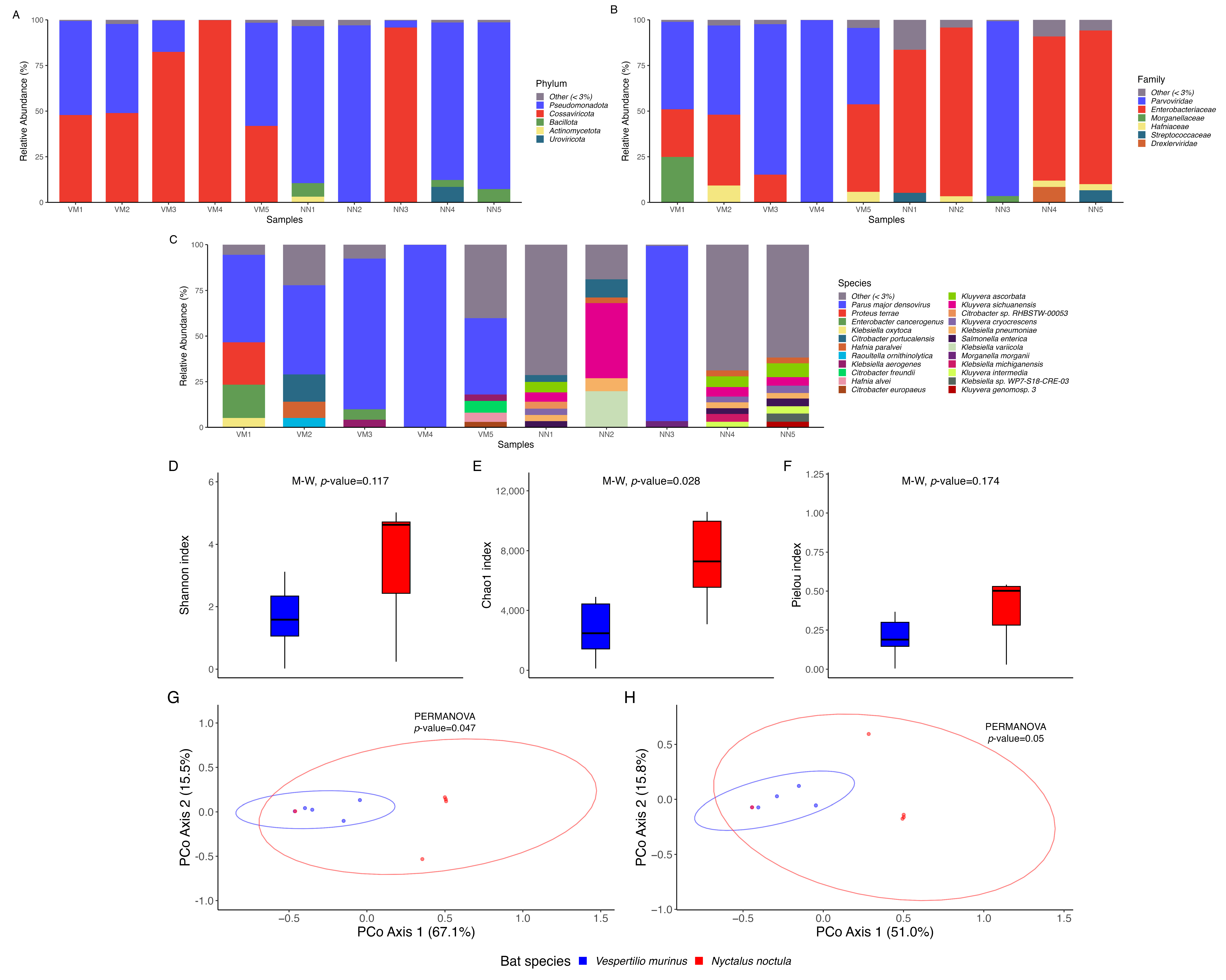
2.1. Microbial Community Composition and Diversity Analysis

Taxonomic profiling revealed the relative abundance of microorganisms in each sample (Figure 1A–C). Notably, Parus major densovirus dominated the microbial composition in two samples (NN3 and VM4), accounting for over 90% of the total sequences (Figure 1C), and was also highly abundant (>40%) in four additional samples (VM1–VM3, VM5). On the phylum level (Figure 1A), despite the marked dominance of the Cossaviricota, to which Parus major densovirus belongs, the microbial communities were largely composed of Pseudomonadota across most samples. Bacillota, Actinomycetota, and Uroviricota were also present in some samples in lower relative abundances. Family-level analysis (Figure 1B) of the gut microbial communities revealed a notable dominance of Parvoviridae and Enterobacteriaceae alongside lower abundances of Morganellaceae, Hafniaceae, Streptococcaceae, and Drexlerviridae. On the species level (Figure 1C), the intestinal microbial communities of the studied bat species were composed of multiple taxa, such as Proteus terrae, Enterobacter cancerogenus, Klebsiella oxytoca, Citrobacter portucalensis, Hafnia paralvei, Raoultella ornithinolytica, Klebsiella aerogenes, Citrobacter freundii, Hafnia alvei, Citrobacter europaeus, Kluyvera ascorbata, Kluyvera sichuanensis, Citrobacter sp. RHBSTW-00053, Kluyvera cryocrescens, Klebsiella pneumoniae, Salmonella enterica, Klebsiella variicola, Morganella morganii, Klebsiella michiganensis, Kluyvera intermedia, Klebsiella sp. WP7-S18-CRE-03, Kluyvera genomosp. 3, and others. However, as mentioned earlier, the overall species composition in the studied samples was overwhelmingly dominated by Parus major densovirus, which skewed the abundance of other representatives of the studied microbial communities.
Alpha diversity analysis did not reveal relevant differences between the two bat species (V. murinus and N. noctula). The Shannon (p = 0.117) (Figure 1D) and Pielou (p = 0.174) (Figure 1E) indices indicated no substantial variation in overall diversity and evenness, while Chao1 (p = 0.028) (Figure 1D) suggested a difference in species richness. Beta diversity analysis showed a weak but statistically significant effect. PERMANOVA results for Bray–Curtis (p = 0.047) (Figure 1E) and Jaccard (p = 0.05) (Figure 1F) distances suggested some level of community structure differentiation.
Differential abundance analysis did not detect statistically significant taxonomic shifts between the two groups at any of the tested taxonomic levels (Figures S1–S6).

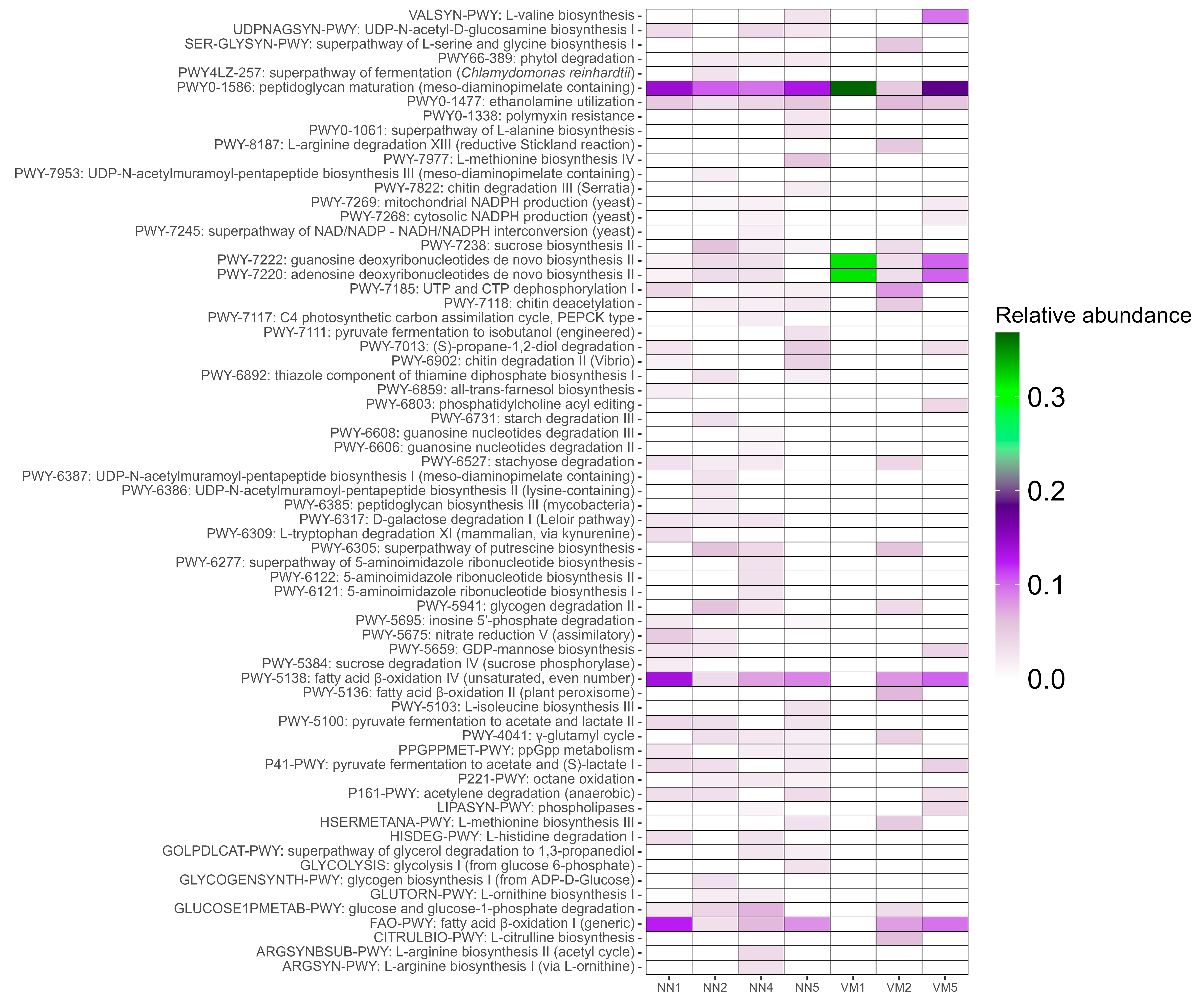
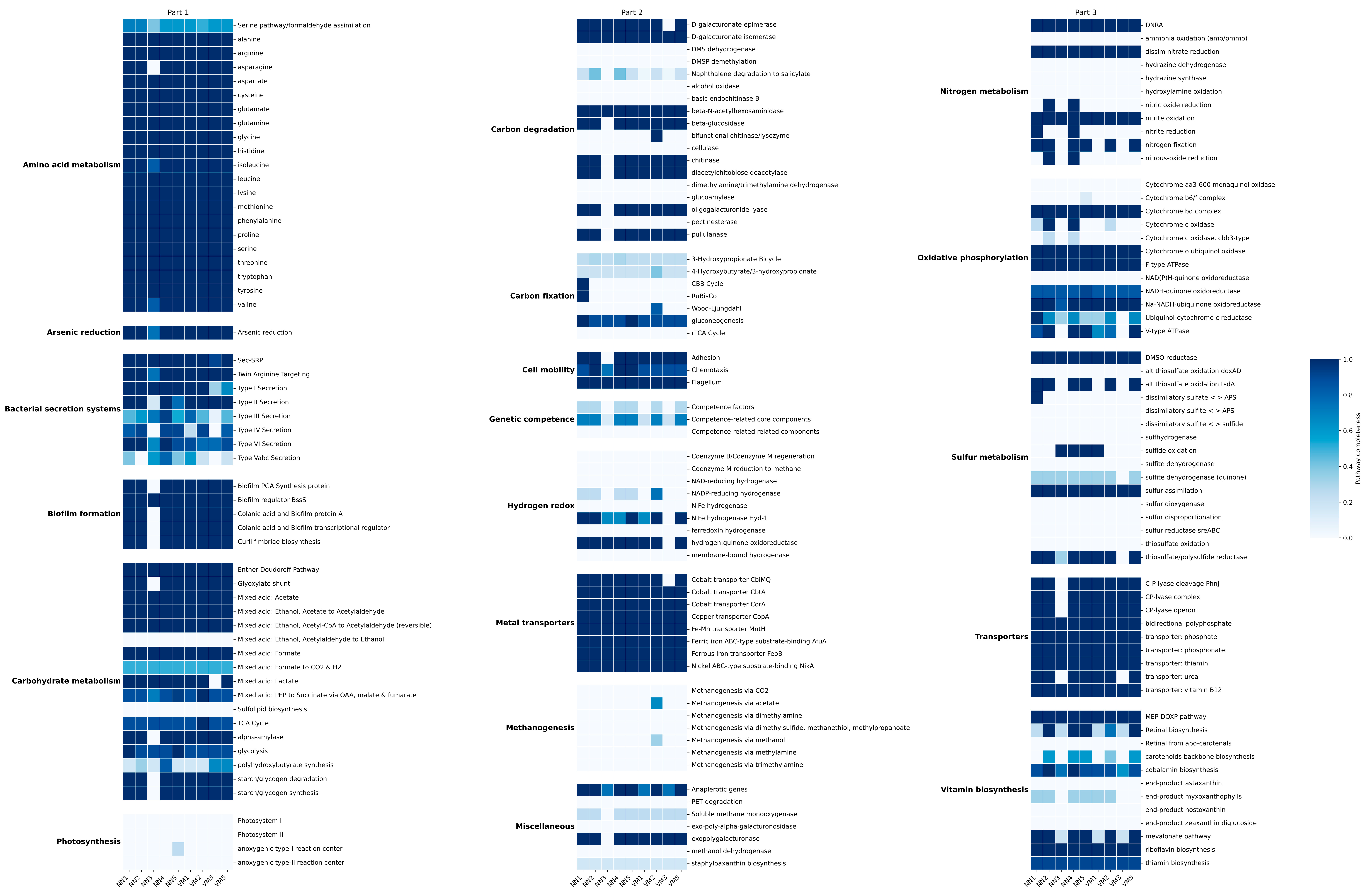
2.2. Functional and Metabolic Profiling

Annotation of metabolic pathways using HUMAnN3 identified 67 pathways across seven samples, while no pathways were annotated in three samples (NN3, VM3, and VM4). The heatmap (Figure 2) of relative pathway abundance highlighted peptidoglycan maturation as the most prevalent across all annotated samples, suggesting a significant presence of actively growing bacteria with cell walls and their role in microbial interactions. The VM1 sample exhibited the lowest functional diversity, with only three pathways detected: peptidoglycan maturation, guanosine deoxyribonucleotides de novo biosynthesis II, and adenosine deoxyribonucleotides de novo biosynthesis II. This result aligns with taxonomic profiling and biodiversity assessment, which revealed low species diversity in the VM1 sample, consisting of only four taxa: Parus major densovirus, Proteus terrae, Enterobacter cancerogenus, and Klebsiella oxytoca. Differential pathway abundance analysis did not yield statistically significant results (Figure S7).
Further annotation of KEGG metabolic pathways using eggNOG-mapper revealed six major functional categories consistently represented across most samples (Figure 3): (i) amino acid metabolism, (ii) carbon degradation, (iii) nitrogen and sulfur metabolism, (iv) oxidative phosphorylation, (v) bacterial secretion systems, and (vi) biofilm formation. These results indicate active microbial participation in amino acid turnover, efficient organic matter processing, and involvement in biogeochemical nitrogen and sulfur cycles, alongside robust energy production via aerobic respiration. The presence of bacterial secretion systems and biofilm formation pathways aligns with the HUMAnN3 annotation, reinforcing the validity of both analyses. The NN3 sample exhibited the lowest functional annotation completeness in the KEGG pathway analysis, while VM4 had no annotated pathways. The distinctiveness of NN3 was further supported by correlation network analysis of sample relationships (Figure 4).

2.3. Antimicrobial Resistance and Virulence Genes

Screening for ARGs across ten samples resulted in a total of 510 detections using the CARD and ResFinder databases (Figure 5A–C). In total, 142 unique ARGs were identified across the dataset: 121 from CARD and 53 from ResFinder, with 32 genes detected by both databases (Figure 5D). Virulence factor identification using the VFDB database revealed a total of 870 gene detections across the ten samples (Figure 5A). These correspond to 314 unique virulence factors identified across the entire dataset (Figure 5E). The NN3 sample exhibited the lowest ARG count, with only eight detected (five from CARD and three from ResFinder), whereas VM4 lacked any annotated resistance genes.
Among the ARGs (Figure 5B), CRP was the only one present in all nine annotated samples. This global regulator represses the MdtEF multidrug efflux pump and is associated with resistance to penicillins, fluoroquinolones, and macrolides. Additionally, 17 genes were consistently detected in eight samples, primarily encoding efflux pumps from the resistance-nodulation-cell division (RND) family (mdtC, mdtB, acrD, acrB), ATP-binding cassette transporters (msbA), and porins with reduced permeability (marA, OmpA, MdtQ). These genes mediate resistance to a broad spectrum of antibiotics, including aminoglycosides, beta-lactams, tetracyclines, fluoroquinolones, and carbapenems. The major facilitator superfamily efflux system was also well represented, with emrR, emrB, and Klebsiella pneumoniae KpnH/KpnG contributing to resistance against macrolides, cephalosporins, and peptide antibiotics.
A smaller subset of ARGs was found in seven out of nine samples, including ramA (a regulator affecting multiple resistance mechanisms), oqxA/oqxB (RND efflux pumps conferring resistance to tetracyclines and fluoroquinolones), and eptB/ArnT (enzymes modifying bacterial membranes to counteract antimicrobial peptides). Notably, Klebsiella pneumoniae OmpK37, a porin linked to reduced beta-lactam permeability, was also widespread.
Unlike the CARD database, which includes all known ARGs, ResFinder specifically identifies acquired resistance determinants. Screening against this database (Figure 5C) did not reveal any distinct trends, as genes were distributed unevenly across the samples without a single dominant resistance determinant present in most or all cases. However, instead of isolated genes with high prevalence, this dataset featured well-represented gene groups sharing common prefixes.
The largest category included 22 bla genes, encoding β-lactamases that hydrolyze the β-lactam ring, conferring resistance to penicillins and cephalosporins. Tetracycline resistance was attributed to five tet genes—tet(S), tet(M), tet(H), tet(D), and tet(57)—all encoding ribosomal protection proteins that prevent antibiotic binding. A similar pattern was observed for fosfomycin resistance, with five fosA variants, each encoding a glutathione transferase responsible for drug inactivation.
Additional resistance mechanisms included three qnr genes, which protect DNA gyrase and topoisomerase IV from quinolone inhibition, and three cat genes, encoding chloramphenicol acetyltransferases that enzymatically modify chloramphenicol to prevent ribosomal binding. Aminoglycoside resistance was linked to three aph(3′) genes, coding for phosphotransferases that inactivate aminoglycosides through phosphorylation. Trimethoprim resistance, in turn, was associated with two dfr genes encoding dihydrofolate reductase variants, which bypass the drug’s inhibitory effect on folate metabolism. Notably, two oqx genes, previously identified in the CARD-based screening, were also detected in this dataset, reinforcing their broad distribution across samples.
Analysis of virulence factors using the VFDB database (Figure 5D) revealed notable variation across samples. NN2 and NN4 exhibited the highest number of annotated genes, while the remaining samples contained at least twice as few. Despite this overall disparity, most genes were sporadically distributed, appearing in some samples but absent in others. However, a subset of 17 genes was consistently present across all datasets, suggesting a conserved core of virulence-associated functions.
Among these, multiple genes were linked to iron acquisition, including components of the ferric enterobactin transport system (fepA, fepC, fepD, and fepG), the ferric aerobactin receptor iutA, and regulatory elements such as the ferric uptake regulator fur. Enterobactin biosynthesis and export were similarly well represented, with entA, entB, entE, entF, and entS forming a complete pathway for siderophore-mediated iron scavenging. Additionally, transcriptional regulation was supported by the presence of rpoS and rcsB, key players in bacterial stress response and virulence control. Other conserved factors included ompA, encoding an outer membrane protein involved in adhesion and immune evasion, acrA, a multidrug efflux pump subunit, and fimA, which encodes a major fimbrial subunit associated with host colonization. Notably, galF, encoding UTP-glucose-1-phosphate uridylyltransferase, was also uniformly detected. This enzyme plays a key role in polysaccharide biosynthesis, potentially influencing capsule formation and immune evasion strategies.

2.4. Genomic Characterization and Phylogenetic Placement of Identified Densoviruses

Genomic viral sequences extracted from assembled metagenomes were named Densovirinae sp. Isolate RBHC_NN_1–5 and Densovirinae sp. Isolate RBHC_VN_1–5 to reflect their origin from the Rostov Bat Rehabilitation Center (RBHC) and their respective bat hosts—Nyctalus noctula (NN) and Vespertilio murinus (VM), with the numbering indicating individual sequence variants or isolates within each group. Validation of the viral nature of these genomic sequences using VirSorter2 confirmed their 100% viral origin.
Comparison of complete viral genomic sequences based on ANI (Figure 6A) revealed a high level of similarity, with a minimum identity of 99.2%. The most divergent virus was derived from sample NN4. Genome annotation using Prokka identified a genome structure consisting of two structural (SPs) and two non-structural proteins (NSPs), with one exception: the virus from sample NN5 contained two SPs and three NSPs. Gene nomenclature in our dataset may differ from that in publicly available Densovirinae genomes due to inconsistencies in NCBI RefSeq annotations. For instance, the same gene might be labeled NSP-1 in one genome and NSP-2 in another. While the numbering of genes may vary, our annotations consistently preserve their structural or non-structural classification. Comparative genomic analysis further confirmed the high degree of similarity in gene sequences across the dataset. The least similar pair, Parus major densovirus and Tenebrio molitor densovirus, still exhibited approximately 97% identity. Full-genome inversion was observed in viruses from VM1, VM2, VM4, VM5, NN1, NN3, NN4, and NN5. Additionally, the virus from NN5 displayed a duplication of the NSP-2 gene, whose length appeared to fluctuate among the studied viruses (Figure 6B). Table S1 summarizes genomic features and pairwise genomic identity of detected densoviruses.
A complete genome inversion was observed in two viral isolates (VM3 and NN1), which were mirror images of the other samples except for NN5.
Orthology analysis with Proteinortho identified 45 viruses in which NSP-1 was a single-copy ortholog (10 from this study, 34 from RefSeq, and 1 from GenBank), 22 viruses with a single-copy SP-1 ortholog (10 from this study, 11 from RefSeq, and 1 from GenBank), and 12 viruses with a single-copy SP-2 ortholog (10 from this study, 1 from RefSeq, and 1 from GenBank). Separate phylogenetic trees were constructed for NSP-1 (Figure 7C) and SP-1 (Figure 7B), alongside a phylogenomic tree incorporating both genes (Figure 7A). The resulting phylogenomic tree displayed strong bootstrap support, with only three branches falling below 70%.
Across all three trees, the identified densoviruses from bat fecal metagenomes did not cluster with other bat-associated densoviruses. Instead, they consistently formed a distinct branch alongside Parus major densovirus. This virus was isolated from a great tit (Parus major) in China in 2014 [23]. Notably, Tenebrio molitor densovirus, which caused an outbreak with high mortality in a commercial mealworm farm [24], appeared in the same clade. Furthermore, the close relationship between Parus major densovirus and Tenebrio molitor densovirus was corroborated by an additional single-copy orthologous gene—SP-2. This gene was shared by 12 viruses, including all 10 from this study, Parus major densovirus, and Tenebrio molitor densovirus. Multiple sequence alignment of this protein (Figure 7D,E) revealed a conserved length of 213 amino acids, with only 12 amino acid variations among all 12 viruses.

3. Discussion

In this study, we present the first comprehensive metagenomic characterization of gut microbial communities in two synanthropic insectivorous bats, N. noctula and V. murinus. According to the taxonomical profiling of metagenomic data, the most remarkable signature of their gut microbiota was the dominant abundance of the reads belonging to the Densovirus genera. Currently, densoviruses are known to infect insects from six orders—Blattodea, Diptera, Hemiptera, Hymenoptera, Lepidoptera, and Orthoptera—as well as decapod crustaceans, such as shrimp and crayfish, and echinoderms, including sea stars [25,26]. Densoviruses cause significant damage to commercially produced invertebrate populations, including crickets, silkworms, and shrimp [27,28,29]. Many densoviruses are polytropic, exhibiting a variable host range, while others display high specificity to hosts and even organs [26]. Due to their high pathogenicity toward pests, particularly mosquitoes, densoviruses have the potential to be used as biological control agents [30]. However, the long-term impact of viral spillover on non-target native insect populations remains unclear [31]. Also, densoviruses are frequently observed in gut metagenomes of insectivorous species, particularly birds and bats. For example, Ge et al., in their metagenomic study of bat fecal samples in China, identified densoviruses among the most frequent reads and assembled contigs related to eukaryotic viruses. The authors suggested that these viruses were present in the bats’ gut virome due to the insectivorous diet of the studied bats [32]. In addition, densoviruses were detected in metagenomic sequencing of nucleic acids extracted from bat guano, feces, and gastrointestinal samples in studies in South Africa, the USA, and Croatia [33,34,35]. In all these studies, researchers also suggested the dietary origins of these viruses in the gut microbiome of bats. Given that the bats included in our study are also insectivorous, the presence of densoviruses in their gut microbiota likely originates from the consumption of infected insects. However, some findings of our bioinformatical analysis suggest that the occurrence of densoviruses in the gut microbiome of studied bats could be associated with factors beyond dietary transmission.
We assembled complete genomes of densoviruses from all studied bat-associated samples. Densovirus genomes analyzed in previous studies were composed of single-stranded linear DNA ranging from 4 to 6 kb in length and contained several major ORFs for non-structural and structural proteins [25,36]. In our research, the genome size of densoviruses was around 5 kb; nine of the densoviruses contained four ORFs, and one of them had five ORFs. According to the phylogenomic analysis targeting several genome regions of detected densoviruses, all of them appeared in one clade with Parus major densovirus. This virus was isolated by Yang et al. from the lung tissue of a great tit (Parus major) in the Jilin province of China and demonstrated to be infectious in vertebrate cell lines. The authors stated that it remains unclear whether the bird was directly infected by the densovirus or if the virus originated from insects consumed by the bird without infecting avian cells, suggesting further laboratory experiments to investigate pathogenic mechanisms in avian species [23]. Nevertheless, this virus was shown to infect vertebrate cell lines, and the high genetic homology between Parus major densovirus and bat-associated densoviruses from our study suggests that these viruses may also have the capacity to infect bats, potentially identifying bats as hosts for them. This hypothesis is supported by the results of the comparative genomic structure analysis, where gene inversions of identified densoviruses were observed. Some studies show that spontaneous gene inversions in the genomes of microbes increase their mutation rates, potentiating their capacity to adapt to new environments, including new hosts, and evolve virulence [37,38,39]. Although it is considered that DNA viruses have stable genomes in comparison to RNA viruses, there is an exception to this rule for DNA viruses with shorter genomes, as they often exhibit higher mutation rates due to their reduced replication fidelity and less efficient repair mechanisms [40,41]. In previous studies, densoviruses showed a high mutation rate, allowing them to adapt to new hosts, primarily invertebrates [42,43]. The majority of the studies in which the ability of densoviruses to infect vertebrate cells was investigated showed that most densoviruses exhibit limited or no active replication within vertebrate cells, pointing to the fact that their host range is limited to invertebrates [44,45]. Thus, the results of Yang et al., where Parus major densovirus showed the capacity to infect vertebrate cell lines, challenge the traditional notion that densoviruses are confined exclusively to invertebrate hosts [23]. Given the high genetic similarity between the Parus major densovirus and the densoviruses detected in bats and their different genome structures due to gene inversions, it is conceivable that these bat-associated strains may possess or be evolving the capacity to infect vertebrates, including bats themselves. Given these phylogenomic and comparative genomic analysis results, we included genomic records of densovirus isolated from commercial mealworms (Tenebrio molitor) from Armién et al. [24] to track the homology and genomic differences between densoviruses detected in the studied bat gut microbiota and densovirus from mealworms, which were included in the diet of bats from the Rostov Bat Rehabilitation Center. As a result, a mealworm-associated densovirus appeared in one clade with Parus major densovirus and the discovered bat-associated densoviruses. The mealworm-associated densovirus included in our bioinformatic analyses exhibited a high sequence identity (97–98%) with densoviruses associated with bats and birds, whereas its identity with insect-associated densoviruses was significantly lower, at approximately 50% [24], which generally corresponds to the results of our study, where this virus was phylogenetically distant from other insect-associated viruses. Thus, this genetic variant of this Tenebrio molitor densovirus could have evolved to increase virulence, which could have given it the ability to infect a broader range of hosts, including vertebrates such as bats and birds. However, it is important to recognize that this remains speculative and requires confirmation through comprehensive ecological monitoring and long-term surveillance of densovirus prevalence in both wild and farmed insect populations, as well as in bat populations. Such studies would help track evolutionary trends and transmission pathways. Additionally, experiments using multiple cell lines are essential to verify the ability of densoviruses to infect bats, birds, and other vertebrates, providing critical insights into their host range and pathogenic potential.
While the high homology of detected bat-associated densoviruses with Parus major densovirus points to the debatable ability of these viruses to infect bats due to the capability of the latter virus to infect vertebrate cells, the presence of these densoviruses in the gut microbiota of bats and their genomic variations points to the more realistic ecological impact of this finding. Bats are considered one of the most unpredictable carriers of emerging pathogens due to their ability to fly long distances and long lifespan relative to mammals of the same size, which allows them to maintain and disseminate infectious agents over broad geographic areas and extended time frames [7]. Their mobility means that pathogens can spread across different ecosystems, bypassing natural barriers that would typically limit microbe transmission. This spatial and temporal flexibility makes disease outbreaks linked to bats particularly difficult to predict and control [46]. Previously, all these features were mainly considered for emerging viruses that target humans and animals. However, the detection of densoviruses with potentially expanded host ranges in bats suggests that similar dynamics may apply to insect-associated viruses as well. If these densoviruses are capable of infecting vertebrate cells, bats could act not only as passive carriers but also as active amplifiers or reservoirs, facilitating viral adaptation and cross-species transmission. Even if bats are not the true hosts of densoviruses, their role as carriers through the gut microbiome may still contribute significantly to the environmental spread of these viruses. Through defecation, bats may introduce viable viral particles into new habitats, including agricultural fields, urban green spaces, and insect farming facilities, thereby creating opportunities for densovirus transmission to susceptible invertebrate populations. This role as ecological vectors underscores the broader influence of insectivorous bats in modulating viral circulation at the interface of natural and human-modified environments. Although our study was cross-sectional, the consistent presence and high relative abundance of densoviruses in multiple samples, combined with their genomic instability and mutation potential, suggest that the bat gut environment may support the maintenance and possibly the diversification of these viruses. Given that genomes of densoviruses detected in our metagenomic study exhibited considerable similarity to vertebrate-infecting strains and presented signs of genomic plasticity, such as gene inversions, it is reasonable to consider that the bat gut environment itself may contribute to the ongoing evolution of these viruses. Notably, this environment may act not just as a passive conduit but as an evolutionary incubator, where microbial interactions and selective pressures foster the emergence of new viral variants. This is particularly relevant for insect-targeting viruses such as densoviruses, which may undergo genetic reshuffling or host-range expansions within the bat gastrointestinal tract. Recent findings suggest that the gut microbiota of bats, particularly species such as N. noctula, exhibits pronounced pro-mutagenic activity, likely influencing both microbial and viral genetic variability. According to one of our previous studies, the majority of lactic acid bacteria and bacilli isolated from bat feces demonstrated increased RecA expression in lux-biosensor assays, indicative of DNA damage and activation of the SOS response system—a process known to accelerate mutation rates in both bacteria and associated viral communities [47]. This suggests that the bat gut environment, shaped by its unique microbiota, may serve as a hotspot for viral genetic diversification. As such, it could play a more active role in shaping the evolutionary trajectory of densoviruses than previously recognized.
We suggest that the dominance of densoviruses in the gut microbiome of the studied bat species is associated with their specific diet at the Rostov Bat Rehabilitation Center, which mostly consists of superworms and mealworms. This assumption is supported by the results of our phylogenomic analysis, which showed that the mealworm-infecting Tenebrio molitor densovirus identified by Armién et al. [24] clustered in the same clade as the densoviruses detected in the bat gut microbiome. However, it is important to note that while Armién et al. reported high mortality caused by Tenebrio molitor densovirus at a commercial mealworm farm, no such mortality was observed in the mealworm colony at the Rostov Bat Rehabilitation Center. This discrepancy may reflect differences in viral strain virulence, farm conditions, or host resistance. In any case, dominance in the abundance of densoviruses in the metagenomic data of the gut microbiome of the studied bat species skewed the estimates of microbial diversity. According to the alpha diversity analysis, there were no significant differences in the Shannon and Pielou indices. However, the Chao1 index indicated higher alpha diversity in the gut microbiome of N. noctula compared to V. murinus. This difference is likely due to the higher occurrence of dominant densoviruses in V. murinus samples in comparison to those obtained from N. noctula (5 vs. 1), which reduced the relative abundance of rarer taxa. Since the Chao1 index estimates species richness by giving more weight to low-abundance or rare taxa [48,49], the dominance of densoviruses in V. murinus likely suppressed the detection of less abundant microbes, resulting in a lower estimated richness. This conclusion is further supported by the beta diversity analysis: the PCoA plot showed that samples with densovirus dominance clustered tightly together in ordination space, suggesting that the variation between samples was largely influenced by the overwhelming abundance of a single viral species, rather than by overall differences in microbial community structure. However, it is important to note that the sample size in this study (n = 10) is relatively small, which limits the statistical power to detect subtle species-specific differences in microbiota composition. Therefore, conclusions regarding species-level diversity comparisons should be interpreted with caution. Nevertheless, in our previous study implementing 16S rRNA gut microbiota metaprofiling, N. noctula and V. murinus showed no significant differences in alpha diversity across Shannon, Chao1, and Pielou indices, aligning with the current shotgun results except for Chao1. Similarly, beta diversity analyses in both studies revealed overlapping microbial communities between the two species, with no significant separation by host identity once the diet was controlled for [22]. Together, these findings suggest that the gut microbiota of N. noctula and V. murinus are highly similar in both diversity and structure when not confounded by viral overrepresentation.
Our findings highlight a critical methodological consideration for future metagenomic studies of bat gut microbiota: the composition of the diet, especially in rehabilitation settings, can substantially bias microbial community profiles. In this study, bats were predominantly fed mealworms, which likely served as the source of the highly abundant densoviruses detected. This dietary skew not only resulted in an overrepresentation of densoviral reads but also masked the broader microbial diversity, as shown by alpha and beta diversity estimates. Providing a more diverse diet could minimize the dominance of single dietary-origin taxa, reduce community skew, and allow a more accurate and representative assessment of gut microbial ecosystems, reflective of wild animals. Therefore, feeding insectivorous bats with a variety of insect species under laboratory and rehabilitation center conditions is essential for obtaining robust and ecologically meaningful metagenomic profiles of their gut microbiota. While numerous studies have demonstrated that bat gut microbiome composition is strongly influenced by diet [22,50,51,52,53], the overwhelming dominance of densoviral reads observed in our metagenomic data was unexpected, even in light of these prior findings. Another option could be sampling from wild bats, as gut microbial communities of wild and captive animals differ significantly [54,55]; however, this is more challenging than conducting studies on bats in captive conditions.
Excluding densoviruses, the microbial component of the bat gut microbiota was largely dominated by members of the phylum Pseudomonadota, with notable contributions from Bacillota and Actinomycetota in several samples, which generally corresponds to previous observations [22]. At the family level, Enterobacteriaceae was predominant, accompanied by Morganellaceae, Hafniaceae, and to a lesser extent Streptococcaceae and Drexlerviridae. These families are commonly found in the guts of mammals and are associated with nutrient processing, host interaction, and in some cases, opportunistic pathogenicity [56,57,58,59,60,61,62]. Additionally, the presence of Drexlerviridae, a family of tailed bacteriophages, is noteworthy, as many of its members specifically infect Enterobacteriaceae [63,64,65]. These bacteriophages may play a regulatory role in shaping bacterial population dynamics within the gut, influencing microbial composition, horizontal gene transfer, and potentially contributing to the control of opportunistic pathogens [66,67,68].
Species-level analysis revealed a diverse set of enteric and environmental bacteria, including Proteus terrae, Enterobacter cancerogenus, Klebsiella oxytoca, Citrobacter portucalensis, and Hafnia paralvei. These taxa are consistent with prior findings in N. noctula from the same rehabilitation setting, where Klebsiella, Citrobacter, and Hafnia species were also frequently isolated, particularly from active bats prior to hibernation [69]. Their prevalence in both cultivable and metagenomic datasets supports the idea that these bacteria represent stable components of the gut community in synanthropic noctules, likely shaped by exposure to shared environmental sources such as insects, water, and human-influenced habitats. Notably, several of these taxa are recognized as opportunistic pathogens in humans and animals [70,71,72,73], suggesting a potential public health and domestic animal well-being relevance of bat-associated microbiomes. Furthermore, the detection of metabolically versatile species belonging to the Enterobacter and Proteus genera, capable of persisting in both host and non-host environments [71,74], reinforces the notion that the gut microbiota of insectivorous bats is highly adaptable and shaped by both intrinsic physiological factors and extrinsic ecological pressures.
Functional annotation of the metagenomic data revealed a relatively conserved repertoire of microbial pathways across most samples, despite the dominance of densoviruses in several of them. The most consistently detected function, peptidoglycan maturation, indicates an active community of cell wall-synthesizing bacteria, which are essential components of gut ecosystems due to their roles in colonization resistance and immune modulation [75,76,77,78]. In addition, pathways related to nucleotide biosynthesis (e.g., guanosine and adenosine deoxyribonucleotides) were recovered, though at lower prevalence, suggesting that microbial replication capacity may be constrained in low-diversity samples such as VM1 [79,80,81]. This observation is consistent with the reduced taxonomic richness and metabolic complexity in samples where densovirus dominance suppressed the representation of other taxa.
Further insights from KEGG pathway analysis revealed microbial engagement in core metabolic functions, including amino acid metabolism, carbon degradation, and energy generation via oxidative phosphorylation. Pathways associated with nitrogen and sulfur turnover, secretion systems, and biofilm formation suggest ecological versatility and adaptation to variable gut conditions [82,83,84,85]. These functions are typical of facultatively anaerobic bacteria and align with known traits of Enterobacteriaceae and related gut-associated taxa detected in the samples [58,86,87]. Importantly, the absence or incompleteness of functional annotations in samples like VM4 and NN3 likely reflects reduced microbial diversity due to densoviral overrepresentation, again highlighting the confounding impact of densovirus abundance.
The analysis of antibiotic resistance genes revealed a diverse resistome in the gut microbiota of both N. noctula and V. murinus, with over 500 ARGs detected across samples, and with 142 ARGs in the whole analyzed dataset. The majority of these genes encoded efflux pumps, membrane porins, and regulatory proteins associated with multidrug resistance phenotypes. The presence of acrB, acrD, mdtB/C, msbA, and marA, well-characterized components of the RND and major facilitator superfamily (MFS) transporters [88,89], suggests that intrinsic resistance mechanisms are prevalent within the microbial communities of these bats. These efflux systems confer resistance to a broad spectrum of antibiotics, including β-lactams, macrolides, aminoglycosides, and fluoroquinolones [90,91,92], indicating that the bat gut serves as a reservoir for multidrug resistance determinants.
Interestingly, the core resistome was conserved across samples, with crp identified in all metagenomes containing ARG annotations. However, samples such as NN3 and VM4, which exhibited strong densovirus dominance, showed minimal ARG representation, once again underscoring how viral overabundance can mask functional attributes of the broader microbial community. When screened against the ResFinder database, the resistome included a substantial number of bla genes (β-lactamases), tet genes (tetracycline resistance), and fosA variants (fosfomycin resistance), consistent with exposure to environmental or foodborne microbial communities where these ARGs are common [93,94,95,96].
The detection of virulence factors followed a similar pattern. While overall gene richness varied among samples, a core set of virulence-associated functions, particularly those related to iron acquisition (e.g., fep, ent, iutA), stress response (rpoS, rcsB), and adhesion (fimA, ompA), was consistently detected. These traits are hallmarks of gut-associated Enterobacteriaceae and suggest that members of this family not only persist in the bat gut but are potentially well-adapted to colonize and interact with host tissues [97,98,99].
In line with these observations, culture-based and molecular surveys of bat populations in different continents have repeatedly documented the same convergence of resistance and virulence traits. Multidrug-resistant E. coli isolated from insectivorous and frugivorous bats in southeast Nigeria, for example, carried blaCTX-M-15, blaTEM, tet(A), and int1 genes together with the intimin adhesin gene eae, confirming that clinically important extended-spectrum beta-lactamase (ESBLs) and colonization factors can co-mobilize in bat-associated Enterobacteriaceae [100]. Costa et al. found that free-tailed bats (Tadarida brasiliensis) from Southern Brazil harbor enterococci carrying ermC, tetM, and vanC1/2/3 together with virulence genes gelE and ace (with occasional agg, cylA, esp), highlighting the tight linkage of resistance and pathogenic traits in bat-associated Gram-positive commensals [101]. European free-tailed bats (Tadarida teniotis) in Portugal hosted ESBL-E. coli in about 10% of fecal samples; isolates carried blaCTX-M-1/-3, often with blaSHV, blaTEM, or blaOXA, as well as tet(A)/tet(B), and invariably the fimA adhesin, again coupling mobilizable β-lactamases with colonization factors in bat-borne Enterobacteriaceae [102]. Lesser horseshoe bats (Rhinolophus monoceros) from India, likewise, yielded gut isolates resistant to β-lactams such as penicillin G and cefoxitin, suggesting underlying β-lactamase ARGs, and Bacillus and Pseudomonas species from the same samples produced hemolysin and protease, coupling antimicrobial resistance with clear virulence functions [103]. Similarly, a metagenomic survey by Huang et al. revealed over a thousand ARGs across wild bats with diverse diets in China, with multidrug and polymyxin resistance genes being particularly prevalent. High-risk ARGs were genetically linked to mobile elements on zoonotic pathogens, and their abundance and diversity were diet-dependent, most pronounced in carnivorous and sanguivorous bats, suggesting that feeding ecology shapes both the resistome and associated risks. This study further emphasizes bats as potent reservoirs and vectors of clinically relevant resistance traits with mobilization potential [104].
Collectively, these findings support the growing recognition of synanthropic bats as environmental reservoirs of both antimicrobial resistance and virulence traits. While direct transmission risks to humans or animals remain speculative, the co-occurrence of ARGs and virulence genes within mobile or pathogenic bacterial taxa poses a potential One Health concern, especially in urbanized or agricultural interfaces where bats and humans frequently interact. Additionally, the detection of clinically relevant resistance genes in wildlife microbiomes further supports calls for expanded surveillance of resistomes beyond traditional agricultural and hospital environments.

4. Materials and Methods

4.1. Sampling

Sampling was conducted in October 2022 at the Rostov Bat Rehabilitation Center (Don State Technical University, Rostov-on-Don, Russia) for metagenomic investigation of gut microbiota signatures. For these studies, fecal samples were collected from common noctules (Nyctalus noctula [n = 5, NN1–5]) and parti-colored bats (Vespertilio murinus [n = 5, VM1–5]). Bats included in this research were in the active state, seized from households of Rostov-on-Don at the owners’ request, and then arrived at the bat rehabilitation facility, where sampling was performed after their arrival and feeding with superworms and mealworms. Handling of animals at the bat rehabilitation center was approved by the Don State Technical University local ethics committee (Protocol No. 5, 2022). A minimum of 0.5 g of fecal samples were collected from bats into sterile 1.5 mL tubes, snap-frozen, and then stored at −80 °C.

4.2. DNA Extraction and High-Throughput Sequencing

Extraction of DNA was performed with a DNA-Sorb B kit (AmpliSens, Moscow, Russia) according to the manufacturer’s protocol. Libraries were prepared using the NEBNext Ultra II DNA Library Prep Kit (New England Biolabs, Beverly, MA, USA) and sequenced using the Illumina NovaSeq 6000 sequencing system according to the manufacturer’s instructions (Illumina, San Diego, CA, USA) with the generation of 2 × 150 bp reads.

4.3. Quality Control of Raw Reads and Taxonomic Classification

The quality of raw sequencing reads was assessed using FastQC v.0.12.1 (https://www.bioinformatics.babraham.ac.uk/projects/fastqc/, accessed on 10 May 2025), followed by report aggregation for each paired-end sample using MultiQC v.1.23 [105]. Taxonomic classification of microbial communities in metagenomic DNA samples was performed using Kraken2 v.2.1.3 [106] with the PlusPF database (Standard plus RefSeq protozoa & fungi; 28 December 2024). Taxon abundance at the genus and species levels was re-estimated using Bracken v.3.1 to improve classification accuracy [107]. The resulting taxonomic profiles were formatted into structured tables using KrakenParser v.0.1.51 (https://github.com/PopovIILab/KrakenParser, accessed on 10 May 2025).

4.4. Diversity and Differential Abundance Analysis

Taxonomic composition profiles were visualized as bar charts representing the relative abundance of microorganisms across samples at the phylum, family, and species levels. To assess biological diversity, both alpha and beta diversity metrics were calculated. The visualizations were generated in R using the tidyverse, ggtext, patchwork, paletteer, data.table, tibble, dplyr, tidyr, ggplot2, and scales packages.
Alpha diversity was quantified using the Shannon [108], Pielou [109], and Chao1 [110] indices. Differences in alpha diversity between the two bat species were evaluated using the Mann–Whitney U test [111], with results visualized as box plots. Beta diversity was assessed using the Bray–Curtis [112] and Jaccard [113] indices, with statistical comparisons performed using permutational multivariate analysis of variance (PERMANOVA) [114]. Principal coordinates analysis (PCoA) plots were used to visualize beta diversity patterns. Alpha and beta diversity metrics were visualized in R with tidyverse, ggtext, patchwork, vegan, glue, and scales.
To identify taxa with significantly different abundance levels between N. noctula and V. murinus, differential abundance analysis was conducted using MaAsLin2 [115]. The analysis employed a linear model with total sum scaling (TSS) normalization and Benjamini–Hochberg correction for False Discovery Rate control [116], using the following parameters: min_prevalence = 0.01, min_abundance = 50,000, analysis_method = LM, max_significance = 0.05. Results were visualized as volcano plots generated in R using the tidyverse, ggtext, and ggrepel packages.

4.5. Metagenome Assembly and Functional Profiling

Metagenome assembly was performed using metaSPAdes v.4.0.0 [117], and assembly quality was assessed with metaQUAST v.5.2.0 [118]. To minimize noise and improve downstream analyses, contigs shorter than 3500 nucleotides were filtered out after quality evaluation.
Functional annotation of metabolic pathways was conducted using HUMAnN3 v.3.9 [119], a tool designed for precise quantitative assessment of functional potential. Annotation relied on the ChocoPhlAn and UniRef databases [120]. HUMAnN3 results were merged and normalized via total sum scaling, yielding relative pathway abundances across metagenomes. All unmapped, unintegrated, and unclassified values were removed to ensure clarity. Pathway abundances were visualized as heatmaps using tidyverse, ggplot2, ggtext, patchwork, and vegan. Differential pathway abundance was further assessed with MaAsLin2 v.1.7.3 [115] using identical model parameters but omitting normalization, as it had already been applied. Volcano plots were generated to illustrate significant differences in pathway abundance between groups.
In addition, microbial community functional annotation was performed using eggNOG-mapper v.2.1.12 [121]. The analysis was optimized for metagenomic inputs, employing Prodigal v.2.6.3 [122] for gene recognition. To assess the completeness of KEGG orthologous group (KO)-based metabolic pathways, annotation results were processed with KEGGaNOG v.0.7.42 (https://github.com/iliapopov17/KEGGaNOG, accessed on 10 May 2025), a custom utility designed to decode KEGG_ko values via KEGG Decoder [123]. KEGGaNOG generated heatmaps representing pathway completeness across metagenomes. To compare KEGG pathway completeness between samples, a correlation matrix was constructed, retaining only correlations above 0.7 for visualization clarity. The correlation network was rendered to highlight key differences between samples.
The dual approach to functional annotation—HUMAnN3 for pathway-level integration and quantification, and eggNOG-mapper for broader gene annotation—ensured a comprehensive functional characterization of the metagenomes.
Finally, assembled metagenomes were screened for antibiotic resistance genes (ARGs) and virulence factors using ABRicate v.1.0.1 (https://github.com/tseemann/abricate, accessed on 10 May 2025), leveraging the CARD [124], ResFinder [125], and VFDB [126] databases. The distribution of identified genes across samples was visualized using bar plots, while presence/absence patterns were represented as heatmaps. The identification of the numbers of overlapping and non-overlapping genes by different databases was visualized using ggVennDiagram [127]. All visualizations were implemented in R using tidyverse, ggtext, patchwork, paletteer, data.table, tibble, dplyr, tidyr, ggplot2, and scales.

4.6. Genomic Characterization and Phylogenetic Analysis of Densoviruses

Given the high relative abundance of Parus major densovirus in most analyzed samples, as identified by Kraken2, densoviruses were subjected to an in-depth investigation.
Complete genomes of all known densoviruses were retrieved from the NCBI RefSeq database [128] using Entrez-direct [129], with the following query: Densovirus AND “complete genome” AND srcdb_refseq[PROP]. A custom BLAST v.2.16.0 database [130] was then constructed using 44 densovirus genomes from RefSeq. All metagenome assemblies were screened against this local database to extract viral sequences. To validate BLAST hits and confirm viral origin, VirSorter2 v.2.2.4 [131] was employed, ultimately identifying 10 near-complete densovirus genomes from the metagenomic data.
To assess their genomic similarity, pairwise Average Nucleotide Identity (ANI) was calculated using FastANI v.1.34 [132], and the results were visualized as a heatmap using pandas, matplotlib, and seaborn Python libraries. Genome annotation was performed with Prokka v.1.14.6 [133], incorporating protein sequences of densoviruses from the NCBI Protein database (https://www.ncbi.nlm.nih.gov/protein/, accessed on 10 May 2025). The annotation results served as the foundation for further phylogenomic analysis.
The phylogenomic framework included proteomes of the 10 newly identified densoviruses, the 44 RefSeq genomes, and an additional densovirus described by Armién et al. [24]. Orthologous gene identification was conducted using Proteinortho v.6.3.2 [134], retaining only single-copy orthologs for downstream analysis. Two orthologous genes were selected for phylogenetic inference, while an additional gene, detected in a limited number of species, was used solely for sequence alignment visualization.
Multiple sequence alignment was performed using MAFFT v.7.526 [135], with substitution models determined via ModelFinder [136]. Maximum likelihood phylogenies were reconstructed with IQ-TREE2 v.2.3.6 [137], incorporating 1000 ultrafast bootstrap replicates [138]. Separate trees were generated for the two genes, along with a combined dataset tree integrating both. Phylogenetic trees were visualized and annotated using ggtree [139], incorporating metadata on host species, geographic origin, and year of isolation. Host taxa were represented by silhouettes sourced from PhyloPic (https://www.phylopic.org, accessed on 10 May 2025). Annotation data were retrieved from NCBI metadata using Phyloki v.0.5.51 (https://github.com/iliapopov17/phyloki, accessed on 10 May 2025). Sequence alignment was visualized with ggmsa [140].
Finally, genome architectures of the densoviruses were compared using pyGenomeViz v.1.5.0 (https://github.com/moshi4/pyGenomeViz, accessed on 10 May 2025). Prokka annotation results were integrated with Proteinortho data to generate a consensus gene map. Viral genomes were aligned vertically, displaying all identified genes. Genomic synteny was assessed using MUMmer v.3.23 [141], with collinear regions depicted as linking bands to highlight homologous segments across genomes.

4.7. Data Analysis and Visualization

Throughout the data analysis process, Snakemake pipelines [142] were extensively employed to automate and parallelize tasks. Data transformation and visualization steps were carried out using custom scripts written in Python (version 3.12), R (version 4.4.2), and bash.

5. Conclusions

This study provides novel metagenomic insights into the gut microbiota of two synanthropic insectivorous bat species, Nyctalus noctula and Vespertilio murinus, emphasizing their ecological roles as reservoirs and potential disseminators of diverse microbial and viral taxa. While the bacterial communities were dominated by facultative anaerobes such as Enterobacteriaceae, which are important for public health and domestic animal welfare, the most striking finding was the consistently high abundance of densoviruses, particularly those closely related to Parus major densovirus, known for its infectivity in vertebrate cell lines.
This observation raises the possibility that bats, long recognized as carriers of zoonotic viruses affecting mammals, may also serve as previously overlooked hosts for insect-targeting viruses, including highly pathogenic strains. The detection of gene inversions and high genomic similarity to densoviruses implicated in lethal outbreaks in farmed insects suggests that the bat gut environment may not only preserve but also promote the evolution and potential host-range expansion of these viruses. This is further supported by the previously investigated pro-mutagenic potential of bat gut microbiota, which may act as a hotspot for viral diversification and adaptation.
In addition to their viral ecology, bats in this study harbored a broad and diverse repertoire of antimicrobial resistance genes, including multidrug efflux pumps, β-lactamases, and tetracycline and fosfomycin resistance determinants. These findings align with the growing recognition of wildlife as a reservoir of clinically relevant ARGs, with synanthropic bats potentially contributing to the environmental spread of resistance through fecal shedding in urban and agricultural ecosystems. The co-occurrence of ARGs and virulence factors within gut-associated bacterial communities further underscores the One Health implications of bat microbiomes.
Taken together, our findings call for a broader ecological reassessment of bats, not only as key players in the emergence of mammalian pathogens but also as active participants in both the circulation and emergence of insect-targeting viruses and antimicrobial resistance genes. Future research should integrate longitudinal sampling, functional validation, and virome-focused approaches to elucidate the full spectrum of microbial and viral dynamics within bat populations, particularly in the context of rapidly changing anthropogenic environments.

Supplementary Materials

The following supporting information can be downloaded at https://www.mdpi.com/article/10.3390/ijms26135941/s1.

Author Contributions

Conceptualization, I.V.P. (Igor V. Popov); methodology, A.V.M., I.V.P. (Igor V. Popov) and A.D.M.; software, I.V.P. (Ilia V. Popov); validation, A.D.M., K.V. and I.V.P. (Igor V. Popov); formal analysis, I.V.P. (Ilia V. Popov); investigation, A.D.M., V.E.G., K.B.D. and A.V.M.; resources, V.E.G., K.B.D. and E.A.L.; data curation, V.E.G. and E.A.L.; writing—original draft preparation, I.V.P. (Ilia V. Popov) and I.V.P. (Igor V. Popov); writing—review and editing, K.V.; visualization, I.V.P. (Ilia V. Popov); supervision, K.V. and I.V.P. (Igor V. Popov); project administration, A.M.E.; funding acquisition, A.M.E. and I.V.P. (Igor V. Popov). All authors have read and agreed to the published version of the manuscript.

Funding

Sampling, DNA extraction, and bioinformatic analysis were funded by the Russian Science Foundation (project 23-14-00316). Library preparation and sequencing were funded by the Ministry of Science and Higher Education of the Russian Federation (Agreement 075-10-2025-017). The funders had no role in study design, data collection and analysis, decision to publish, or preparation of the manuscript.

Institutional Review Board Statement

The reported study does not involve the results of any in vivo intervention experiments. Handling of animals at the bat rehabilitation center was approved by the Don State Technical University local ethics committee (Protocol No. 5, 17 January 2022).

Informed Consent Statement

Not applicable.

Data Availability Statement

The raw sequencing data used for this study are available in the National Center for Biotechnological Information’s Short Read Archive under BioProject PRJNA1253095. The pipeline used for the in silico and bioinformatic data analysis has been deposited in GitHub: https://github.com/PopovIILab/BatShotMetaFlow (accessed on 10 May 2025).

Acknowledgments

The authors acknowledge the support of the Genomic Research Resource Center of Sirius University of Science and Technology for high-throughput sequencing and the Department of Informatization of the Don State Technical University for the support and configuration of the server for bioinformatic calculations. Igor V. Popov and Ilia V. Popov acknowledge the support of the Ministry of Science and Higher Education of the Russian Federation (Orders No. 486 of 15 June 2021 and No. 825 of 29 August 2022) for the internships in metagenomic bioinformatics under the supervision of Koen Venema.

Conflicts of Interest

The authors declare no conflicts of interest.

Abbreviations

The following abbreviations are used in this manuscript:
ARGsAntibiotic Resistance Genes
VFDBVirulence Factors of Bacterial Database
CARDComprehensive Antibiotic Resistance Database
PCoAPrincipal Coordinates Analysis
ANIAverage Nucleotide Identity
ORFsOpen Reading Frames
NSPNon-Structural Protein
SPStructural Protein
RNDResistance-Nodulation-Cell Division
MFSMajor Facilitator Superfamily
KEGGKyoto Encyclopedia of Genes and Genomes
KOKEGG Orthologous
PERMANOVAPermutational Multivariate Analysis of Variance
DNADeoxyribonucleic Acid
RNARibonucleic Acid

References

  1. Pena, S.A.; Alencastre-Santos, A.B.; da Silva, J.B.; Correia, L.L.; Urbieta, G.L.; Graciolli, G.; Palheta, L.R.; Vieira, T.B. Bats (Mammalia, Chiroptera) and Bat Flies (Diptera, Streblidae) from the Cazumbá-Iracema and Chico Mendes Reserve, Western Brazilian Amazon. Parasitol. Res. 2023, 122, 451–459. [Google Scholar] [CrossRef] [PubMed]
  2. Bazzoni, E.; Cacciotto, C.; Zobba, R.; Pittau, M.; Martella, V.; Alberti, A. Bat Ecology and Microbiome of the Gut: A Narrative Review of Associated Potentials in Emerging and Zoonotic Diseases. Animals 2024, 14, 3043. [Google Scholar] [CrossRef]
  3. Ohlopkova, O.V.; Stolbunova, K.A.; Popov, I.V.; Popov, I.V.; Kabwe, E.; Davidyuk, Y.N.; Stepanyuk, M.A.; Moshkin, A.D.; Kononova, Y.V.; Lukbanova, E.A.; et al. Detection of Brno Loanvirus (Loanvirus brunaense) in Common Noctule Bats (Nyctalus Noctula) in Southern Russia. Braz. J. Microbiol. 2025, 56, 675–682. [Google Scholar] [CrossRef]
  4. Popov, I.V.; Ohlopkova, O.V.; Donnik, I.M.; Zolotukhin, P.V.; Umanets, A.; Golovin, S.N.; Malinovkin, A.V.; Belanova, A.A.; Lipilkin, P.V.; Lipilkina, T.A.; et al. Detection of Coronaviruses in Insectivorous Bats of Fore-Caucasus, 2021. Sci. Rep. 2023, 13, 2306. [Google Scholar] [CrossRef] [PubMed]
  5. Banerjee, A.; Baker, M.L.; Kulcsar, K.; Misra, V.; Plowright, R.; Mossman, K. Novel Insights Into Immune Systems of Bats. Front. Immunol. 2020, 11, 26. [Google Scholar] [CrossRef] [PubMed]
  6. Demian, W.L.; Cormier, O.; Mossman, K. Immunological Features of Bats: Resistance and Tolerance to Emerging Viruses. Trends Immunol. 2024, 45, 198–210. [Google Scholar] [CrossRef]
  7. Lagunas-Rangel, F.A. Why Do Bats Live so Long?-Possible Molecular Mechanisms. Biogerontology 2020, 21, 1–11. [Google Scholar] [CrossRef]
  8. Carrillo Gaeta, N.; Cavalcante Brito, J.E.; Nunes Batista, J.M.; Gagete Veríssimo de Mello, B.; Dias, R.A.; Heinemann, M.B. Bats Are Carriers of Antimicrobial-Resistant Staphylococcaceae in Their Skin. Antibiotics 2023, 12, 331. [Google Scholar] [CrossRef]
  9. Devnath, P.; Karah, N.; Graham, J.P.; Rose, E.S.; Asaduzzaman, M. Evidence of Antimicrobial Resistance in Bats and Its Planetary Health Impact for Surveillance of Zoonotic Spillover Events: A Scoping Review. Int. J. Environ. Res. Public Health 2022, 20, 243. [Google Scholar] [CrossRef]
  10. Letko, M.; Seifert, S.N.; Olival, K.J.; Plowright, R.K.; Munster, V.J. Bat-Borne Virus Diversity, Spillover and Emergence. Nat. Rev. Microbiol. 2020, 18, 461–471. [Google Scholar] [CrossRef]
  11. Crits-Christoph, A.; Levy, J.I.; Pekar, J.E.; Goldstein, S.A.; Singh, R.; Hensel, Z.; Gangavarapu, K.; Rogers, M.B.; Moshiri, N.; Garry, R.F.; et al. Genetic Tracing of Market Wildlife and Viruses at the Epicenter of the COVID-19 Pandemic. Cell 2024, 187, 5468–5482.e11. [Google Scholar] [CrossRef] [PubMed]
  12. Tian, J.; Sun, J.; Li, D.; Wang, N.; Wang, L.; Zhang, C.; Meng, X.; Ji, X.; Suchard, M.A.; Zhang, X.; et al. Emerging Viruses: Cross-Species Transmission of Coronaviruses, Filoviruses, Henipaviruses, and Rotaviruses from Bats. Cell Rep. 2022, 39, 110969. [Google Scholar] [CrossRef] [PubMed]
  13. Aguiar, L.M.S.; Bueno-Rocha, I.D.; Oliveira, G.; Pires, E.S.; Vasconcelos, S.; Nunes, G.L.; Frizzas, M.R.; Togni, P.H.B. Going out for Dinner—The Consumption of Agriculture Pests by Bats in Urban Areas. PLoS ONE 2021, 16, e0258066. [Google Scholar] [CrossRef] [PubMed]
  14. Shilton, L.A.; Altringham, J.D.; Compton, S.G.; Whittaker, R.J. Old World Fruit Bats Can Be Long–Distance Seed Dispersers through Extended Retention of Viable Seeds in the Gut. Proc. R. Soc. Lond. B 1999, 266, 219–223. [Google Scholar] [CrossRef]
  15. Stewart, A.B.; Srilopan, S.; Wayo, K.; Hassa, P.; Dudash, M.R.; Bumrungsri, S. Bat Pollinators: A Decade of Monitoring Reveals Declining Visitation Rates for Some Species in Thailand. Zoological Lett. 2024, 10, 5. [Google Scholar] [CrossRef]
  16. Boyles, J.G.; Cryan, P.M.; McCracken, G.F.; Kunz, T.H. Economic Importance of Bats in Agriculture. Science 2011, 332, 41–42. [Google Scholar] [CrossRef]
  17. Martinez, S.; Sullivan, A.; Hagan, E.; Goley, J.; Epstein, J.H.; Olival, K.J.; Saylors, K.; Euren, J.; Bangura, J.; Zikankuba, S.; et al. Living Safely With Bats: Lessons in Developing and Sharing a Global One Health Educational Resource. Glob. Health Sci. Pract. 2022, 10, e2200106. [Google Scholar] [CrossRef]
  18. Frank, E.G. The Economic Impacts of Ecosystem Disruptions: Costs from Substituting Biological Pest Control. Science 2024, 385, eadg0344. [Google Scholar] [CrossRef]
  19. Hoyt, J.R.; Langwig, K.E.; Sun, K.; Parise, K.L.; Li, A.; Wang, Y.; Huang, X.; Worledge, L.; Miller, H.; White, J.P.; et al. Environmental Reservoir Dynamics Predict Global Infection Patterns and Population Impacts for the Fungal Disease White-Nose Syndrome. Proc. Natl. Acad. Sci. USA 2020, 117, 7255–7262. [Google Scholar] [CrossRef]
  20. Minnis, A.M.; Lindner, D.L. Phylogenetic Evaluation of Geomyces and Allies Reveals No Close Relatives of Pseudogymnoascus Destructans, Comb. Nov., in Bat Hibernacula of Eastern North America. Fungal Biol. 2013, 117, 638–649. [Google Scholar] [CrossRef]
  21. Urbina, J.; Chestnut, T.; Allen, J.M.; Levi, T. Pseudogymnoascus Destructans Growth in Wood, Soil and Guano Substrates. Sci. Rep. 2021, 11, 763. [Google Scholar] [CrossRef]
  22. Popov, I.V.; Popov, I.V.; Krikunova, A.A.; Lipilkina, T.A.; Derezina, T.N.; Chikindas, M.L.; Venema, K.; Ermakov, A.M. Gut Microbiota Composition of Insectivorous Synanthropic and Fructivorous Zoo Bats: A Direct Metagenomic Comparison. Int. J. Mol. Sci. 2023, 24, 17301. [Google Scholar] [CrossRef] [PubMed]
  23. Yang, W.-T.; Shi, S.-H.; Jiang, Y.-L.; Zhao, L.; Chen, H.-L.; Huang, K.-Y.; Yang, G.-L.; Wang, C.-F. Genetic Characterization of a Densovirus Isolated from Great Tit (Parus Major) in China. Infect. Genet. Evol. 2016, 41, 107–112. [Google Scholar] [CrossRef] [PubMed]
  24. Armién, A.G.; Polon, R.; Rejmanek, D.; Moeller, R.B.; Crossley, B.M. Outbreak of Densovirus with High Mortality in a Commercial Mealworm (Tenebrio Molitor) Farm: A Molecular, Bright-Field, and Electron Microscopic Characterization. Vet. Pathol. 2023, 60, 689–703. [Google Scholar] [CrossRef] [PubMed]
  25. Cotmore, S.F.; Agbandje-McKenna, M.; Canuti, M.; Chiorini, J.A.; Eis-Hubinger, A.-M.; Hughes, J.; Mietzsch, M.; Modha, S.; Ogliastro, M.; Pénzes, J.J.; et al. ICTV Virus Taxonomy Profile: Parvoviridae. J. Gen. Virol. 2019, 100, 367–368. [Google Scholar] [CrossRef]
  26. Tijssen, P.; Pénzes, J.J.; Yu, Q.; Pham, H.T.; Bergoin, M. Diversity of Small, Single-Stranded DNA Viruses of Invertebrates and Their Chaotic Evolutionary Past. J. Invertebr. Pathol. 2016, 140, 83–96. [Google Scholar] [CrossRef]
  27. Dhar, A.K.; Cruz-Flores, R.; Caro, L.F.A.; Siewiora, H.M.; Jory, D. Diversity of Single-Stranded DNA Containing Viruses in Shrimp. Virusdisease 2019, 30, 43–57. [Google Scholar] [CrossRef]
  28. Li, Y.; Zádori, Z.; Bando, H.; Dubuc, R.; Fédière, G.; Szelei, J.; Tijssen, P. Genome Organization of the Densovirus from Bombyx Mori (BmDNV-1) and Enzyme Activity of Its Capsid. J. Gen. Virol. 2001, 82, 2821–2825. [Google Scholar] [CrossRef]
  29. Liu, K.; Li, Y.; Jousset, F.-X.; Zadori, Z.; Szelei, J.; Yu, Q.; Pham, H.T.; Lépine, F.; Bergoin, M.; Tijssen, P. The Acheta Domesticus Densovirus, Isolated from the European House Cricket, Has Evolved an Expression Strategy Unique among Parvoviruses. J. Virol. 2011, 85, 10069–10078. [Google Scholar] [CrossRef]
  30. Li, J.; Dong, Y.; Sun, Y.; Lai, Z.; Zhao, Y.; Liu, P.; Gao, Y.; Chen, X.; Gu, J. A Novel Densovirus Isolated From the Asian Tiger Mosquito Displays Varied Pathogenicity Depending on Its Host Species. Front. Microbiol. 2019, 10, 1549. [Google Scholar] [CrossRef]
  31. Impact of Biological Invasions on Ecosystem Services; Vilà, M., Hulme, P.E., Eds.; Springer International Publishing: Cham, Switzerland, 2017; ISBN 978-3-319-45119-0. [Google Scholar]
  32. Ge, X.; Li, Y.; Yang, X.; Zhang, H.; Zhou, P.; Zhang, Y.; Shi, Z. Metagenomic Analysis of Viruses from Bat Fecal Samples Reveals Many Novel Viruses in Insectivorous Bats in China. J. Virol. 2012, 86, 4620–4630. [Google Scholar] [CrossRef] [PubMed]
  33. Geldenhuys, M.; Mortlock, M.; Weyer, J.; Bezuidt, O.; Seamark, E.C.J.; Kearney, T.; Gleasner, C.; Erkkila, T.H.; Cui, H.; Markotter, W. A Metagenomic Viral Discovery Approach Identifies Potential Zoonotic and Novel Mammalian Viruses in Neoromicia Bats within South Africa. PLoS ONE 2018, 13, e0194527. [Google Scholar] [CrossRef] [PubMed]
  34. Li, L.; Victoria, J.G.; Wang, C.; Jones, M.; Fellers, G.M.; Kunz, T.H.; Delwart, E. Bat Guano Virome: Predominance of Dietary Viruses from Insects and Plants plus Novel Mammalian Viruses. J. Virol. 2010, 84, 6955–6965. [Google Scholar] [CrossRef]
  35. Šimić, I.; Zorec, T.M.; Lojkić, I.; Krešić, N.; Poljak, M.; Cliquet, F.; Picard-Meyer, E.; Wasniewski, M.; Zrnčić, V.; Ćukušić, A.; et al. Viral Metagenomic Profiling of Croatian Bat Population Reveals Sample and Habitat Dependent Diversity. Viruses 2020, 12, 891. [Google Scholar] [CrossRef]
  36. Tang, S.; Song, X.; Xue, L.; Wang, X.; Wang, X.; Xu, P.; Ren, G. Characterization and Distribution Analysis of a Densovirus Infecting Myzus Persicae Nicotianae (Hemiptera: Aphididae). J. Econ. Entomol. 2016, 109, 580–587. [Google Scholar] [CrossRef]
  37. Merrikh, C.N.; Merrikh, H. Gene Inversion Potentiates Bacterial Evolvability and Virulence. Nat. Commun. 2018, 9, 4662. [Google Scholar] [CrossRef]
  38. Pepin, K.M.; Domsic, J.; McKenna, R. Genomic Evolution in a Virus under Specific Selection for Host Recognition. Infect. Genet. Evol. 2008, 8, 825–834. [Google Scholar] [CrossRef]
  39. Sanjuán, R.; Pereira-Gómez, M.; Risso, J. Genome Instability in DNA Viruses. In Genome Stability; Elsevier: Amsterdam, The Netherlands, 2016; pp. 37–47. ISBN 978-0-12-803309-8. [Google Scholar]
  40. Bradwell, K.; Combe, M.; Domingo-Calap, P.; Sanjuán, R. Correlation between Mutation Rate and Genome Size in Riboviruses: Mutation Rate of Bacteriophage Qβ. Genetics 2013, 195, 243–251. [Google Scholar] [CrossRef] [PubMed]
  41. Sanjuán, R.; Domingo-Calap, P. Mechanisms of Viral Mutation. Cell Mol. Life Sci. 2016, 73, 4433–4448. [Google Scholar] [CrossRef]
  42. Bruemmer, A.; Scholari, F.; Lopez-Ferber, M.; Conway, J.F.; Hewat, E.A. Structure of an Insect Parvovirus (Junonia Coenia Densovirus) Determined by Cryo-Electron Microscopy. J. Mol. Biol. 2005, 347, 791–801. [Google Scholar] [CrossRef]
  43. Jaroenram, W.; Chaivisuthangkura, P.; Owens, L. One Base Pair Deletion and High Rate of Evolution: Keys to Viral Accommodation of Australian Penaeus Stylirostris Densovirus. Aquaculture 2015, 443, 40–48. [Google Scholar] [CrossRef]
  44. El-Far, M.; Li, Y.; Fédière, G.; Abol-Ela, S.; Bergoin, M.; Tijssen, P. Abortive Infection of the Insect Parvovirus, Mythimna Loreyi Densovirus (MIDNV) in Mammalian Cells. IOBC Wprs Bulletin 2003, 26, 229–232. [Google Scholar]
  45. Li, T.; Li, H.; Wu, Y.; Li, S.; Yuan, G.; Xu, P. Identification of a Novel Densovirus in Aphid, and Uncovering the Possible Antiviral Process During Its Infection. Front. Immunol. 2022, 13, 905628. [Google Scholar] [CrossRef]
  46. Donnik, I.M.; Popov, I.V.; Sereda, S.V.; Popov, I.V.; Chikindas, M.L.; Ermakov, A.M. Coronavirus Infections of Animals: Future Risks to Humans. Biol. Bull. Russ. Acad. Sci. 2021, 48, 26–37. [Google Scholar] [CrossRef] [PubMed]
  47. Popov, I.V.; Mazanko, M.S.; Kulaeva, E.D.; Golovin, S.N.; Malinovkin, A.V.; Aleshukina, I.S.; Aleshukina, A.V.; Prazdnova, E.V.; Tverdokhlebova, T.I.; Chikindas, M.L.; et al. Gut Microbiota of Bats: Pro-Mutagenic Properties and Possible Frontiers in Preventing Emerging Disease. Sci. Rep. 2021, 11, 21075. [Google Scholar] [CrossRef]
  48. Chao, A.; Colwell, R.K. Thirty Years of Progeny from Chao’s Inequality: Estimating and Comparing Richness with Incidence Data and Incomplete Sampling. SORT 2017, 41, 3–54. [Google Scholar]
  49. Hill, T.C.J.; Walsh, K.A.; Harris, J.A.; Moffett, B.F. Using Ecological Diversity Measures with Bacterial Communities. FEMS Microbiol. Ecol. 2003, 43, 1–11. [Google Scholar] [CrossRef]
  50. Aizpurua, O.; Nyholm, L.; Morris, E.; Chaverri, G.; Herrera Montalvo, L.G.; Flores-Martinez, J.J.; Lin, A.; Razgour, O.; Gilbert, M.T.P.; Alberdi, A. The Role of the Gut Microbiota in the Dietary Niche Expansion of Fishing Bats. Anim. Microbiome 2021, 3, 76. [Google Scholar] [CrossRef]
  51. Dai, W.; Leng, H.; Li, J.; Li, A.; Li, Z.; Zhu, Y.; Li, X.; Jin, L.; Sun, K.; Feng, J. The Role of Host Traits and Geography in Shaping the Gut Microbiome of Insectivorous Bats. mSphere 2024, 9, e0008724. [Google Scholar] [CrossRef]
  52. Li, J.; Chu, Y.; Yao, W.; Wu, H.; Feng, J. Differences in Diet and Gut Microbiota Between Lactating and Non-Lactating Asian Particolored Bats (Vespertilio Sinensis): Implication for a Connection Between Diet and Gut Microbiota. Front. Microbiol. 2021, 12, 735122. [Google Scholar] [CrossRef]
  53. Mena Canata, D.A.; Benfato, M.S.; Pereira, F.D.; Ramos Pereira, M.J.; Hackenhaar, F.S.; Mann, M.B.; Frazzon, A.P.G.; Rampelotto, P.H. Comparative Analysis of the Gut Microbiota of Bat Species with Different Feeding Habits. Biology 2024, 13, 363. [Google Scholar] [CrossRef] [PubMed]
  54. Popov, I.V.; Popov, I.V.; Chebotareva, I.P.; Tikhmeneva, I.A.; Peshkova, D.A.; Krikunova, A.A.; Tkacheva, E.V.; Algburi, A.R.; Abdulhameed, A.M.; Jargalsaikhan, A.; et al. Differences in Gut Microbiota Composition, Diversity, and Predicted Functional Activity between Wild and Captive Zoo Carollia Perspicillata in a One Health Perspective. Braz. J. Microbiol. 2025, 56, 1291–1302. [Google Scholar] [CrossRef] [PubMed]
  55. Xiao, Y.; Xiao, G.; Liu, H.; Zhao, X.; Sun, C.; Tan, X.; Sun, K.; Liu, S.; Feng, J. Captivity Causes Taxonomic and Functional Convergence of Gut Microbial Communities in Bats. PeerJ 2019, 7, e6844. [Google Scholar] [CrossRef]
  56. Adams, S.; Knapp, J.P.; Neujahr, A.; Burkey, T.; Miller, P.S.; Fernando, S.C. 276 Investigating the Colonization History of Early-Life Microbiome of Piglets. J. Anim. Sci. 2023, 101, 165–166. [Google Scholar] [CrossRef]
  57. Adeolu, M.; Alnajar, S.; Naushad, S.; S Gupta, R. Genome-Based Phylogeny and Taxonomy of the “Enterobacteriales”: Proposal for Enterobacterales Ord. Nov. Divided into the Families Enterobacteriaceae, Erwiniaceae Fam. Nov., Pectobacteriaceae Fam. Nov., Yersiniaceae Fam. Nov., Hafniaceae Fam. Nov., Morganellaceae Fam. Nov., and Budviciaceae Fam. Nov. Int. J. Syst. Evol. Microbiol. 2016, 66, 5575–5599. [Google Scholar] [CrossRef] [PubMed]
  58. Khorsand, B.; Asadzadeh Aghdaei, H.; Nazemalhosseini-Mojarad, E.; Nadalian, B.; Nadalian, B.; Houri, H. Overrepresentation of Enterobacteriaceae and Escherichia Coli Is the Major Gut Microbiome Signature in Crohn’s Disease and Ulcerative Colitis; a Comprehensive Metagenomic Analysis of IBDMDB Datasets. Front. Cell Infect. Microbiol. 2022, 12, 1015890. [Google Scholar] [CrossRef]
  59. Liu, H.; Zhu, J.; Hu, Q.; Rao, X. Morganella Morganii, a Non-Negligent Opportunistic Pathogen. Int. J. Infect. Dis. 2016, 50, 10–17. [Google Scholar] [CrossRef]
  60. Ma, J.; Song, X.; Li, M.; Yu, Z.; Cheng, W.; Yu, Z.; Zhang, W.; Zhang, Y.; Shen, A.; Sun, H.; et al. Global Spread of Carbapenem-Resistant Enterobacteriaceae: Epidemiological Features, Resistance Mechanisms, Detection and Therapy. Microbiol. Res. 2023, 266, 127249. [Google Scholar] [CrossRef]
  61. Moreira de Gouveia, M.I.; Bernalier-Donadille, A.; Jubelin, G. Enterobacteriaceae in the Human Gut: Dynamics and Ecological Roles in Health and Disease. Biology 2024, 13, 142. [Google Scholar] [CrossRef]
  62. Shah, T.; Wang, Y.; Wang, Y.; Li, Q.; Zhou, J.; Hou, Y.; Wang, B.; Xia, X. A Comparative Analysis of the Stomach, Gut, and Lung Microbiomes in Rattus Norvegicus. Microorganisms 2023, 11, 2359. [Google Scholar] [CrossRef]
  63. German, G.J.; DeGiulio, J.V.; Ramsey, J.; Kropinski, A.M.; Misra, R. The TolC and Lipopolysaccharide-Specific Escherichia Coli Bacteriophage TLS-the Tlsvirus Archetype Virus. Phage 2024, 5, 173–183. [Google Scholar] [CrossRef]
  64. Kumar, P.; Meghvansi, M.K.; Kamboj, D.V. Phenotypic Characterization and Whole-Genome Analysis of a Novel Bacteriophage HCF1 Infecting Citrobacter Amalonaticus and C. Freundii. Front. Microbiol. 2021, 12, 644013. [Google Scholar] [CrossRef] [PubMed]
  65. Pan, L.; Li, D.; Lin, W.; Liu, W.; Qin, W.; Xu, L.; Tong, Y. Genomic Analysis of a Novel Active Prophage of Hafnia Paralvei. Arch. Virol. 2022, 167, 2027–2034. [Google Scholar] [CrossRef]
  66. Muniesa, M.; Jofre, J. Identifying and Analyzing Bacteriophages in Human Fecal Samples: What Could We Discover? Future Microbiol. 2014, 9, 879–886. [Google Scholar] [CrossRef]
  67. Pilati, G.V.T.; Cadamuro, R.D.; Filho, V.B.; Dahmer, M.; Elois, M.A.; Savi, B.P.; Salles, G.B.C.; Muniz, E.C.; Fongaro, G. Bacteriophage-Associated Antimicrobial Resistance Genes in Avian Pathogenic Escherichia Coli Isolated from Brazilian Poultry. Viruses 2023, 15, 1485. [Google Scholar] [CrossRef]
  68. Song, S.J.; Sanders, J.G.; Delsuc, F.; Metcalf, J.; Amato, K.; Taylor, M.W.; Mazel, F.; Lutz, H.L.; Winker, K.; Graves, G.R.; et al. Comparative Analyses of Vertebrate Gut Microbiomes Reveal Convergence between Birds and Bats. mBio 2020, 11, e02901-19. [Google Scholar] [CrossRef]
  69. Popov, I.V.; Berezinskaia, I.S.; Popov, I.V.; Martiusheva, I.B.; Tkacheva, E.V.; Gorobets, V.E.; Tikhmeneva, I.A.; Aleshukina, A.V.; Tverdokhlebova, T.I.; Chikindas, M.L.; et al. Cultivable Gut Microbiota in Synanthropic Bats: Shifts of Its Composition and Diversity Associated with Hibernation. Animals 2023, 13, 3658. [Google Scholar] [CrossRef] [PubMed]
  70. Mavziutov, A.R.; Bondarenko, V.M.; Zherebtsova, N.I.; Valishin, D.A. [Pathogenicity factors of opportunistic enterobacteria and its role in development of diarrhea]. Zh Mikrobiol. Epidemiol. Immunobiol. 2007, 1, 89–97. [Google Scholar]
  71. Osei Sekyere, J.; Reta, M.A. Global Evolutionary Epidemiology and Resistome Dynamics of Citrobacter Species, Enterobacter Hormaechei, Klebsiella Variicola, and Proteeae Clones. Environ. Microbiol. 2021, 23, 7412–7431. [Google Scholar] [CrossRef]
  72. Palusiak, A. Proteus Mirabilis and Klebsiella Pneumoniae as Pathogens Capable of Causing Co-Infections and Exhibiting Similarities in Their Virulence Factors. Front. Cell Infect. Microbiol. 2022, 12, 991657. [Google Scholar] [CrossRef]
  73. Sanches, M.S.; Rodrigues da Silva, C.; Silva, L.C.; Montini, V.H.; Lopes Barboza, M.G.; Migliorini Guidone, G.H.; Dias de Oliva, B.H.; Nishio, E.K.; Faccin Galhardi, L.C.; Vespero, E.C.; et al. Proteus Mirabilis from Community-Acquired Urinary Tract Infections (UTI-CA) Shares Genetic Similarity and Virulence Factors with Isolates from Chicken, Beef and Pork Meat. Microb. Pathog. 2021, 158, 105098. [Google Scholar] [CrossRef]
  74. Dobrindt, U.; Hentschel, U.; Kaper, J.B.; Hacker, J. Genome Plasticity in Pathogenic and Nonpathogenic Enterobacteria. Curr. Top. Microbiol. Immunol. 2002, 264, 157–175. [Google Scholar] [PubMed]
  75. Clarke, T.B.; Davis, K.M.; Lysenko, E.S.; Zhou, A.Y.; Yu, Y.; Weiser, J.N. Recognition of Peptidoglycan from the Microbiota by Nod1 Enhances Systemic Innate Immunity. Nat. Med. 2010, 16, 228–231. [Google Scholar] [CrossRef]
  76. Gupta, D.; Jing, X.; Dziarski, R. Peptidoglycan Recognition Protein 3-Regulated Microbiome Maintains Healthy Levels of Treg Cells and Protects Mice from Colitis (INC1P.346). J. Immunol. 2015, 194, 54.3. [Google Scholar] [CrossRef]
  77. Tosoni, G.; Conti, M.; Diaz Heijtz, R. Bacterial Peptidoglycans as Novel Signaling Molecules from Microbiota to Brain. Curr. Opin. Pharmacol. 2019, 48, 107–113. [Google Scholar] [CrossRef] [PubMed]
  78. Wolf, A.J. Peptidoglycan-Induced Modulation of Metabolic and Inflammatory Responses. Immunometabolism 2023, 5, e00024. [Google Scholar] [CrossRef]
  79. Baud, G.L.C.; Prasad, A.; Ellegaard, K.M.; Engel, P. Turnover of Strain-Level Diversity Modulates Functional Traits in the Honeybee Gut Microbiome between Nurses and Foragers. Genome Biol. 2023, 24, 283. [Google Scholar] [CrossRef]
  80. Murga-Garrido, S.M.; Ulloa-Pérez, E.J.; Díaz-Benítez, C.E.; Orbe-Orihuela, Y.C.; Cornejo-Granados, F.; Ochoa-Leyva, A.; Sanchez-Flores, A.; Cruz, M.; Castañeda-Márquez, A.C.; Plett-Torres, T.; et al. Virulence Factors of the Gut Microbiome Are Associated with BMI and Metabolic Blood Parameters in Children with Obesity. Microbiol. Spectr. 2023, 11, e0338222. [Google Scholar] [CrossRef]
  81. Watson, A.R.; Füssel, J.; Veseli, I.; DeLongchamp, J.Z.; Silva, M.; Trigodet, F.; Lolans, K.; Shaiber, A.; Fogarty, E.; Runde, J.M.; et al. Metabolic Independence Drives Gut Microbial Colonization and Resilience in Health and Disease. Genome Biol. 2023, 24, 78. [Google Scholar] [CrossRef]
  82. Kanehisa, M.; Furumichi, M.; Sato, Y.; Matsuura, Y.; Ishiguro-Watanabe, M. KEGG: Biological Systems Database as a Model of the Real World. Nucleic Acids Res. 2025, 53, D672–D677. [Google Scholar] [CrossRef]
  83. Kanehisa, M.; Furumichi, M.; Sato, Y.; Kawashima, M.; Ishiguro-Watanabe, M. KEGG for Taxonomy-Based Analysis of Pathways and Genomes. Nucleic Acids Res. 2023, 51, D587–D592. [Google Scholar] [CrossRef] [PubMed]
  84. Xu, Y.; Xu, J.; Zhu, Y.; Mao, H.; Li, J.; Kong, X.; Zhu, X.; Zhang, J. Investigating Gut Microbiota-Blood and Urine Metabolite Correlations in Early Sepsis-Induced Acute Kidney Injury: Insights from Targeted KEGG Analyses. Front. Cell Infect. Microbiol. 2024, 14, 1375874. [Google Scholar] [CrossRef] [PubMed]
  85. Zhang, Q.; Meng, N.; Li, R.; He, Q.; Liang, X.; Zhou, F.; Zheng, W.; Ma, J.; Yu, X.; Tan, K.; et al. Gut Microbe-Metabolite Profiles Are Associated with Microbial Pathways of Longevity in Women: A Cross-Sectional Study Conducted in China. Gerontology 2024, 70, 76–89. [Google Scholar] [CrossRef]
  86. Warinner, J.; ElSaadani, M.; Rosenau, K.; Kim, J.H.; Hassan, S.A.; Bhogoju, S.; Wempe, L.; Goretsky, T.; Alam, M.A.; Awuah, S.; et al. AUPHOS, A NOVEL DRUG THAT IMPROVES COLITIS BY MICROBIOME INDUCED METABOLIC CHANGES. Inflamm. Bowel Dis. 2023, 29, S49. [Google Scholar] [CrossRef]
  87. Xu, L.; Wu, Y.; Yang, X.; Pang, X.; Wu, Y.; Li, X.; Liu, X.; Zhao, Y.; Yu, L.; Wang, P.; et al. The Fe-S Cluster Biosynthesis in Enterococcus Faecium Is Essential for Anaerobic Growth and Gastrointestinal Colonization. Gut Microbes 2024, 16, 2359665. [Google Scholar] [CrossRef]
  88. El-Demerdash, A.S.; Kamel, S.A.; Elariny, E.Y.T.; Henidi, H.; Mahran, Y.; Alahdal, H.; Saleh, A.M.; Ibrahim, R.A. Natural Inhibitors of Salmonella MDR Efflux Pumps AcrAB and AcrD: An Integrated In Silico, Molecular, and In Vitro Investigation. Int. J. Mol. Sci. 2024, 25, 12949. [Google Scholar] [CrossRef]
  89. Yap, P.S.X.; Ahmad Kamar, A.; Chong, C.W.; Yap, I.K.S.; Teh, C.S.J. Whole Genome Analysis of Multidrug-Resistant Citrobacter Freundii B9-C2 Isolated from Preterm Neonate’s Stool in the First Week. J. Glob. Antimicrob. Resist. 2020, 21, 246–251. [Google Scholar] [CrossRef]
  90. Albarri, O.; AlMatar, M.; Var, I.; Köksal, F. Antimicrobial Resistance of Clinical Klebsiella Pneumoniae Isolates: Involvement of AcrAB and OqxAB Efflux Pumps. Curr. Mol. Pharmacol. 2024, 17, e310323215266. [Google Scholar] [CrossRef]
  91. Kang, H.-W.; Woo, G.-J. Increase of Multidrug Efflux Pump Expression in Fluoroquinolone-Resistant Salmonella Mutants Induced by Ciprofloxacin Selective Pressure. Res. Vet. Sci. 2014, 97, 182–186. [Google Scholar] [CrossRef]
  92. Schuster, S.; Vavra, M.; Schweigger, T.M.; Rossen, J.W.A.; Matsumura, Y.; Kern, W.V. Contribution of AcrAB-TolC to Multidrug Resistance in an Escherichia Coli Sequence Type 131 Isolate. Int. J. Antimicrob. Agents 2017, 50, 477–481. [Google Scholar] [CrossRef]
  93. Baron, S.; Le Devendec, L.; Lucas, P.; Larvor, E.; Jové, T.; Kempf, I. Characterisation of Plasmids Harbouring Extended-Spectrum Cephalosporin Resistance Genes in Escherichia Coli from French Rivers. Vet. Microbiol. 2020, 243, 108619. [Google Scholar] [CrossRef] [PubMed]
  94. Bonanno Ferraro, G.; Bonomo, C.; Brandtner, D.; Mancini, P.; Veneri, C.; Briancesco, R.; Coccia, A.M.; Lucentini, L.; Suffredini, E.; Bongiorno, D.; et al. Characterisation of Microbial Communities and Quantification of Antibiotic Resistance Genes in Italian Wastewater Treatment Plants Using 16S rRNA Sequencing and Digital PCR. Sci. Total Environ. 2024, 933, 173217. [Google Scholar] [CrossRef] [PubMed]
  95. Glenn, L.M.; Englen, M.D.; Lindsey, R.L.; Frank, J.F.; Turpin, J.E.; Berrang, M.E.; Meinersmann, R.J.; Fedorka-Cray, P.J.; Frye, J.G. Analysis of Antimicrobial Resistance Genes Detected in Multiple-Drug-Resistant Escherichia Coli Isolates from Broiler Chicken Carcasses. Microb. Drug Resist. 2012, 18, 453–463. [Google Scholar] [CrossRef]
  96. Rajaei, M.; Moosavy, M.-H.; Gharajalar, S.N.; Khatibi, S.A. Antibiotic Resistance in the Pathogenic Foodborne Bacteria Isolated from Raw Kebab and Hamburger: Phenotypic and Genotypic Study. BMC Microbiol. 2021, 21, 272. [Google Scholar] [CrossRef]
  97. Singh, N.; Goel, G.; Raghav, M. Insights into Virulence Factors Determining the Pathogenicity of Cronobacter Sakazakii. Virulence 2015, 6, 433–440. [Google Scholar] [CrossRef]
  98. Wibberg, D.; Szczepanowski, R.; Eikmeyer, F.; Pühler, A.; Schlüter, A. The IncF Plasmid pRSB225 Isolated from a Municipal Wastewater Treatment Plant’s on-Site Preflooder Combining Antibiotic Resistance and Putative Virulence Functions Is Highly Related to Virulence Plasmids Identified in Pathogenic E. Coli Isolates. Plasmid 2013, 69, 127–137. [Google Scholar] [CrossRef]
  99. Yuan, L.; Li, X.; Du, L.; Su, K.; Zhang, J.; Liu, P.; He, Q.; Zhang, Z.; Peng, D.; Shen, L.; et al. RcsAB and Fur Coregulate the Iron-Acquisition System via entC in Klebsiella Pneumoniae NTUH-K2044 in Response to Iron Availability. Front. Cell Infect. Microbiol. 2020, 10, 282. [Google Scholar] [CrossRef] [PubMed]
  100. Obodoechi, L.O.; Carvalho, I.; Chenouf, N.S.; Martínez-Álvarez, S.; Sadi, M.; Nwanta, J.A.; Chah, K.F.; Torres, C. Antimicrobial Resistance in Escherichia Coli Isolates from Frugivorous (Eidolon Helvum) and Insectivorous (Nycteris Hispida) Bats in Southeast Nigeria, with Detection of CTX-M-15 Producing Isolates. Comp. Immunol. Microbiol. Infect. Dis. 2021, 75, 101613. [Google Scholar] [CrossRef]
  101. Costa, L.D.F.X.; Grassotti, T.T.; Canani, C.R.; de Lira, A.D.; de Moura, T.M.; Campos, A.A.S.; Frazzon, J.; Frazzon, A.P.G. Diversidade, Perfis de Resistência e Virulência de Enterococcus Spp. Em Fezes de Morcegos Urbanos Tadarida Brasiliensis (Brazilian Free-Tailed Bats). Rev. Bras. De Biociências 2019, 17, 43–52. [Google Scholar]
  102. Garcês, A.; Correia, S.; Amorim, F.; Pereira, J.E.; Igrejas, G.; Poeta, P. First report on extended-spectrum beta-lactamase (ESBL) producing Escherichia coli from European free-tailed bats (Tadarida teniotis) in Portugal: A one-health approach of a hidden contamination problem. J. Hazard. Mater. 2019, 370, 219–224. [Google Scholar] [CrossRef]
  103. Selvin, J.; Lanong, S.; Syiem, D.; De Mandal, S.; Kayang, H.; Kumar, N.S.; Kiran, G.S. Culture-Dependent and Metagenomic Analysis of Lesser Horseshoe Bats’ Gut Microbiome Revealing Unique Bacterial Diversity and Signatures of Potential Human Pathogens. Microb. Pathog. 2019, 137, 103675. [Google Scholar] [CrossRef]
  104. Huang, L.; Dai, W.; Sun, X.; Pu, Y.; Feng, J.; Jin, L.; Sun, K. Diet-Driven Diversity of Antibiotic Resistance Genes in Wild Bats: Implications for Public Health. Microbiol. Res. 2025, 293, 128086. [Google Scholar] [CrossRef] [PubMed]
  105. Ewels, P.; Magnusson, M.; Lundin, S.; Käller, M. MultiQC: Summarize Analysis Results for Multiple Tools and Samples in a Single Report. Bioinformatics 2016, 32, 3047–3048. [Google Scholar] [CrossRef] [PubMed]
  106. Wood, D.E.; Lu, J.; Langmead, B. Improved Metagenomic Analysis with Kraken 2. Genome Biol. 2019, 20, 257. [Google Scholar] [CrossRef]
  107. Lu, J.; Rincon, N.; Wood, D.E.; Breitwieser, F.P.; Pockrandt, C.; Langmead, B.; Salzberg, S.L.; Steinegger, M. Metagenome Analysis Using the Kraken Software Suite. Nat. Protoc. 2022, 17, 2815–2839. [Google Scholar] [CrossRef] [PubMed]
  108. Shannon, C.E. A Mathematical Theory of Communication. Bell Syst. Tech. J. 1948, 27, 379–423. [Google Scholar] [CrossRef]
  109. Pielou, E.C. The Measurement of Diversity in Different Types of Biological Collections. J. Theor. Biol. 1966, 13, 131–144. [Google Scholar] [CrossRef]
  110. Chao, A.; Bunge, J. Estimating the Number of Species in a Stochastic Abundance Model. Biometrics 2002, 58, 531–539. [Google Scholar] [CrossRef]
  111. Mann, H.B.; Whitney, D.R. On a Test of Whether One of Two Random Variables Is Stochastically Larger than the Other. Ann. Math. Statist. 1947, 18, 50–60. [Google Scholar] [CrossRef]
  112. Bray, J.R.; Curtis, J.T. An Ordination of the Upland Forest Communities of Southern Wisconsin. Ecol. Monogr. 1957, 27, 325–349. [Google Scholar] [CrossRef]
  113. Jaccard, P. Étude Comparative de La Distribution Florale Dans Une Portion Des Alpes et Du Jura; Imprimerie Corbaz & Comp.: Lexington, MA, USA, 1901. [Google Scholar] [CrossRef]
  114. Anderson, M.J. Permutational Multivariate Analysis of Variance (PERMANOVA). In Wiley StatsRef: Statistics Reference Online; Kenett, R.S., Longford, N.T., Piegorsch, W.W., Ruggeri, F., Eds.; Wiley: Hoboken, NJ, USA, 2017; pp. 1–15. ISBN 978-1-118-44511-2. [Google Scholar]
  115. Mallick, H.; Rahnavard, A.; McIver, L.J.; Ma, S.; Zhang, Y.; Nguyen, L.H.; Tickle, T.L.; Weingart, G.; Ren, B.; Schwager, E.H.; et al. Multivariable Association Discovery in Population-Scale Meta-Omics Studies. PLoS Comput. Biol. 2021, 17, e1009442. [Google Scholar] [CrossRef] [PubMed]
  116. Benjamini, Y.; Hochberg, Y. Controlling the False Discovery Rate: A Practical and Powerful Approach to Multiple Testing. J. R. Stat. Soc. Ser. B Stat. Methodol. 1995, 57, 289–300. [Google Scholar] [CrossRef]
  117. Nurk, S.; Meleshko, D.; Korobeynikov, A.; Pevzner, P.A. metaSPAdes: A New Versatile Metagenomic Assembler. Genome Res. 2017, 27, 824–834. [Google Scholar] [CrossRef]
  118. Mikheenko, A.; Saveliev, V.; Gurevich, A. MetaQUAST: Evaluation of Metagenome Assemblies. Bioinformatics 2016, 32, 1088–1090. [Google Scholar] [CrossRef] [PubMed]
  119. Beghini, F.; McIver, L.J.; Blanco-Míguez, A.; Dubois, L.; Asnicar, F.; Maharjan, S.; Mailyan, A.; Manghi, P.; Scholz, M.; Thomas, A.M.; et al. Integrating Taxonomic, Functional, and Strain-Level Profiling of Diverse Microbial Communities with bioBakery 3. Elife 2021, 10, e65088. [Google Scholar] [CrossRef]
  120. Suzek, B.E.; Huang, H.; McGarvey, P.; Mazumder, R.; Wu, C.H. UniRef: Comprehensive and Non-Redundant UniProt Reference Clusters. Bioinformatics 2007, 23, 1282–1288. [Google Scholar] [CrossRef] [PubMed]
  121. Cantalapiedra, C.P.; Hernández-Plaza, A.; Letunic, I.; Bork, P.; Huerta-Cepas, J. eggNOG-Mapper v2: Functional Annotation, Orthology Assignments, and Domain Prediction at the Metagenomic Scale. Mol. Biol. Evol. 2021, 38, 5825–5829. [Google Scholar] [CrossRef]
  122. Hyatt, D.; Chen, G.-L.; Locascio, P.F.; Land, M.L.; Larimer, F.W.; Hauser, L.J. Prodigal: Prokaryotic Gene Recognition and Translation Initiation Site Identification. BMC Bioinform. 2010, 11, 119. [Google Scholar] [CrossRef]
  123. Graham, E.D.; Heidelberg, J.F.; Tully, B.J. Potential for Primary Productivity in a Globally-Distributed Bacterial Phototroph. ISME J. 2018, 12, 1861–1866. [Google Scholar] [CrossRef]
  124. Jia, B.; Raphenya, A.R.; Alcock, B.; Waglechner, N.; Guo, P.; Tsang, K.K.; Lago, B.A.; Dave, B.M.; Pereira, S.; Sharma, A.N.; et al. CARD 2017: Expansion and Model-Centric Curation of the Comprehensive Antibiotic Resistance Database. Nucleic Acids Res. 2017, 45, D566–D573. [Google Scholar] [CrossRef]
  125. Zankari, E.; Hasman, H.; Cosentino, S.; Vestergaard, M.; Rasmussen, S.; Lund, O.; Aarestrup, F.M.; Larsen, M.V. Identification of Acquired Antimicrobial Resistance Genes. J. Antimicrob. Chemother. 2012, 67, 2640–2644. [Google Scholar] [CrossRef] [PubMed]
  126. Chen, L.; Zheng, D.; Liu, B.; Yang, J.; Jin, Q. VFDB 2016: Hierarchical and Refined Dataset for Big Data Analysis--10 Years On. Nucleic Acids Res. 2016, 44, D694–D697. [Google Scholar] [CrossRef] [PubMed]
  127. Gao, C.; Chen, C.; Akyol, T.; Dusa, A.; Yu, G.; Cao, B.; Cai, P. ggVennDiagram: Intuitive Venn Diagram Software Extended. iMeta 2024, 3, e177. [Google Scholar] [CrossRef]
  128. O’Leary, N.A.; Wright, M.W.; Brister, J.R.; Ciufo, S.; Haddad, D.; McVeigh, R.; Rajput, B.; Robbertse, B.; Smith-White, B.; Ako-Adjei, D.; et al. Reference Sequence (RefSeq) Database at NCBI: Current Status, Taxonomic Expansion, and Functional Annotation. Nucleic Acids Res. 2016, 44, D733–D745. [Google Scholar] [CrossRef]
  129. Kans, J. Entrez Direct: E-Utilities on the UNIX Command Line. In Entrez Programming Utilities Help [Internet]; National Center for Biotechnology Information (US): Bethesda, MD, USA, 2024. [Google Scholar]
  130. Camacho, C.; Coulouris, G.; Avagyan, V.; Ma, N.; Papadopoulos, J.; Bealer, K.; Madden, T.L. BLAST+: Architecture and Applications. BMC Bioinform. 2009, 10, 421. [Google Scholar] [CrossRef]
  131. Guo, J.; Bolduc, B.; Zayed, A.A.; Varsani, A.; Dominguez-Huerta, G.; Delmont, T.O.; Pratama, A.A.; Gazitúa, M.C.; Vik, D.; Sullivan, M.B.; et al. VirSorter2: A Multi-Classifier, Expert-Guided Approach to Detect Diverse DNA and RNA Viruses. Microbiome 2021, 9, 37. [Google Scholar] [CrossRef]
  132. Jain, C.; Rodriguez-R, L.M.; Phillippy, A.M.; Konstantinidis, K.T.; Aluru, S. High Throughput ANI Analysis of 90K Prokaryotic Genomes Reveals Clear Species Boundaries. Nat. Commun. 2018, 9, 5114. [Google Scholar] [CrossRef] [PubMed]
  133. Seemann, T. Prokka: Rapid Prokaryotic Genome Annotation. Bioinformatics 2014, 30, 2068–2069. [Google Scholar] [CrossRef]
  134. Lechner, M.; Findeiss, S.; Steiner, L.; Marz, M.; Stadler, P.F.; Prohaska, S.J. Proteinortho: Detection of (Co-)Orthologs in Large-Scale Analysis. BMC Bioinform. 2011, 12, 124. [Google Scholar] [CrossRef]
  135. Katoh, K.; Standley, D.M. MAFFT Multiple Sequence Alignment Software Version 7: Improvements in Performance and Usability. Mol. Biol. Evol. 2013, 30, 772–780. [Google Scholar] [CrossRef]
  136. Kalyaanamoorthy, S.; Minh, B.Q.; Wong, T.K.F.; von Haeseler, A.; Jermiin, L.S. ModelFinder: Fast Model Selection for Accurate Phylogenetic Estimates. Nat. Methods 2017, 14, 587–589. [Google Scholar] [CrossRef] [PubMed]
  137. Minh, B.Q.; Schmidt, H.A.; Chernomor, O.; Schrempf, D.; Woodhams, M.D.; von Haeseler, A.; Lanfear, R. IQ-TREE 2: New Models and Efficient Methods for Phylogenetic Inference in the Genomic Era. Mol. Biol. Evol. 2020, 37, 1530–1534. [Google Scholar] [CrossRef] [PubMed]
  138. Hoang, D.T.; Chernomor, O.; von Haeseler, A.; Minh, B.Q.; Vinh, L.S. UFBoot2: Improving the Ultrafast Bootstrap Approximation. Mol. Biol. Evol. 2018, 35, 518–522. [Google Scholar] [CrossRef]
  139. Xu, S.; Li, L.; Luo, X.; Chen, M.; Tang, W.; Zhan, L.; Dai, Z.; Lam, T.T.; Guan, Y.; Yu, G. Ggtree: A Serialized Data Object for Visualization of a Phylogenetic Tree and Annotation Data. Imeta 2022, 1, e56. [Google Scholar] [CrossRef] [PubMed]
  140. Zhou, L.; Feng, T.; Xu, S.; Gao, F.; Lam, T.T.; Wang, Q.; Wu, T.; Huang, H.; Zhan, L.; Li, L.; et al. Ggmsa: A Visual Exploration Tool for Multiple Sequence Alignment and Associated Data. Brief. Bioinform. 2022, 23, bbac222. [Google Scholar] [CrossRef]
  141. Marçais, G.; Delcher, A.L.; Phillippy, A.M.; Coston, R.; Salzberg, S.L.; Zimin, A. MUMmer4: A Fast and Versatile Genome Alignment System. PLoS Comput. Biol. 2018, 14, e1005944. [Google Scholar] [CrossRef]
  142. Mölder, F.; Jablonski, K.P.; Letcher, B.; Hall, M.B.; Tomkins-Tinch, C.H.; Sochat, V.; Forster, J.; Lee, S.; Twardziok, S.O.; Kanitz, A.; et al. Sustainable Data Analysis with Snakemake. F1000Res 2021, 10, 33. [Google Scholar] [CrossRef]
Figure 1. Taxonomic composition and diversity metrics of bat gut microbiota. Relative abundance of microbial taxa at the phylum level (A), family level (B), and species level (C) across 10 metagenomic samples. Samples VM1–VM5 correspond to Vespertilio murinus, while NN1–NN5 represent Nyctalus noctula. (D)—Shannon index, (E)—Chao1 richness index, (F)—Pielou’s evenness index (alpha diversity). (G)—Bray–Curtis dissimilarity, (H)—Jaccard index (beta diversity).
Ijms 26 05941 g001
Figure 2. Functional potential of bat gut microbiota based on metabolic pathway abundance. Heatmap of relative abundances of metabolic pathways predicted by HUMAnN3. Rows are individual pathways predicted by HUMAnN3; columns are samples (NN1–NN5 = Nyctalus noctula, VM1–VM5 = Vespertilio murinus). Color intensity represents normalized relative abundance (each column sums to 1).
Ijms 26 05941 g002
Figure 3. Completeness of KEGG metabolic pathways in bat gut microbiota. Heatmap displaying the completeness of over 170 individual KEGG pathways, divided into three nearly equal sections for clarity. Each cell corresponds to a specific metabolic function, labeled on the right. Functions are grouped by category, with group names indicated on the left. Gaps separate different functional groups. Columns represent samples (NN1–NN5 = Nyctalus noctula, VM1–VM5 = Vespertilio murinus).
Ijms 26 05941 g003
Figure 4. KEGG metabolic profile correlation network. Network illustrating correlations greater than 0.7, with weak correlations shown in blue and strong correlations in red. The thickness of the lines increases with the strength of the correlation.
Ijms 26 05941 g004
Figure 5. Identification and presence of antibiotic resistance genes and virulence factors across metagenomic samples. (A)—Bar plot of gene counts from CARD, ResFinder, and VFDB databases identified in each sample. Blue bars represent CARD genes, orange bars represent ResFinder genes, and green bars represent VFDB genes. A black dashed line separates the samples. (B,C)—Presence tables for CARD and ResFinder genes, respectively, across the metagenomic samples. (D)—Vienn diagram showing overlapping and non-overlapping identifications of antibiotic resistance genes by CARD (blue) and ResFinder (orange) databases. (E)—Presence tables for VFDB genes across the metagenomic samples. X-axis labels on bar plot (A) and columns in heatmaps (B,C,E) represent samples (NN1–NN5 = Nyctalus noctula, VM1–VM5 = Vespertilio murinus).
Ijms 26 05941 g005
Figure 6. Comparative genomic analysis of densoviruses. (A)—Heatmap of pairwise ANI comparison of viral genomic sequences. ANI values represent the nucleotide identity between each pair of sequences, with higher values indicating greater similarity. (B)—Comparative genomic analysis of Parus major densovirus, Tenebrio molitor densovirus, and the densoviruses studied in this research. Genes included in the phylogenetic analysis are outlined in green, while those not included are outlined in red. Green connecting lines represent direct relationships between genes, while red lines indicate inverse relationships. Line color saturation reflects the degree of similarity. Gene names are displayed at the top of the graph without numbering.
Ijms 26 05941 g006
Figure 7. Phylogenomic analysis and protein sequence alignment. (AC)—Maximum likelihood phylogenetic trees constructed using NSP-1 and SP-1 sequences. (A)—Tree based on NSP-1 and SP-1 using the Q.pfam+I+G4 substitution model. (B)—Tree based on SP-1 using the VT+I+G4 substitution model. (C)—Tree based on NSP-1 using the Q.pfam+F+I+G4 substitution model. Clades with bootstrap support below 70 are shown in gray, while those with higher support are shown in black. Trees are annotated with virus host silhouettes from the PhyloPic database, flags indicating the countries of virus isolation, and a heatmap displaying the years of isolation. (D,E)—Visualization of multiple sequence alignment of SP-2. (D)—Sequence region from amino acid 1 to 100. (E)—Sequence region from amino acid 101 to 213. Consensus sequences are shown at the top, with bar charts below each sequence representing the divergence from the consensus for each amino acid.
Ijms 26 05941 g007
Disclaimer/Publisher’s Note: The statements, opinions and data contained in all publications are solely those of the individual author(s) and contributor(s) and not of MDPI and/or the editor(s). MDPI and/or the editor(s) disclaim responsibility for any injury to people or property resulting from any ideas, methods, instructions or products referred to in the content.

