Review

Antimicrobial Efficacy of Nd:YAG Laser in Polymicrobial Root Canal Infections: A Systematic Review of In Vitro Studies

Department of Periodontal Diseases and Oral Mucosa Diseases, Faculty of Medical Sciences in Zabrze, Medical University of Silesia, 40-055 Katowice, Poland
*
Authors to whom correspondence should be addressed.
Int. J. Mol. Sci. 2025, 26(12), 5631; https://doi.org/10.3390/ijms26125631
Submission received: 17 May 2025 / Revised: 7 June 2025 / Accepted: 10 June 2025 / Published: 12 June 2025
(This article belongs to the Section Molecular Pathology, Diagnostics, and Therapeutics)

Abstract

Endodontic infections are characterized by complex polymicrobial communities residing within the intricate root canal system. Traditional chemomechanical methods frequently fail to achieve complete microbial eradication, especially in cases involving biofilm-forming and resistant species. This systematic review synthesizes current evidence on the molecular basis and antimicrobial efficacy of the neodymium-doped yttrium aluminum garnet (Nd:YAG) laser in root canal disinfection, particularly against polymicrobial infections. A comprehensive literature search was conducted in the PubMed, Embase, Scopus, and Cochrane databases in accordance with PRISMA 2020 guidelines. Experimental and preclinical studies evaluating the bactericidal properties of Nd:YAG laser therapy were included. The Nd:YAG laser demonstrated significant reductions in total microbial load through photothermal effects, including denaturation of proteins, disruption of cell membranes, and degradation of mixed-species biofilms. Although complete sterilization was not consistently achieved, its ability to penetrate dentinal tubules and target microbial consortia offers substantial adjunctive value. Standardization of laser parameters and further clinical studies are needed to validate these findings and establish Nd:YAG laser use in routine endodontic disinfection protocols.

1. Introduction

Successful endodontic therapy fundamentally depends on the thorough and effective elimination of microbial pathogens from the intricate and often inaccessible areas of the root canal system [1,2,3,4]. Despite significant progress in mechanical instrumentation and the development of sophisticated chemical irrigation techniques, achieving complete and sustained disinfection of the complex root canal anatomy continues to pose a formidable clinical challenge for practitioners [5,6]. This difficulty is primarily due to the presence of anatomical complexities such as lateral canals, isthmuses, and apical deltas that harbor resilient microbial populations beyond the reach of traditional treatment modalities [7]. Among these microbial species, Enterococcus faecalis is particularly notorious for its resistance to eradication [8]. This facultative anaerobe exhibits an extraordinary ability to survive under nutrient-deprived conditions and to deeply infiltrate the dentinal tubules, forming persistent biofilms that are impervious to conventional chemomechanical protocols [9,10]. Its survival in treated canals has been closely associated with endodontic treatment failures, thus highlighting the need for innovative disinfection strategies [11,12]. In response to these challenges, there has been a growing interest in the development and clinical integration of adjunctive technologies that can augment the antimicrobial efficacy of existing methods [13]. One such promising approach involves the application of laser-based systems, with particular attention being given to the neodymium-doped yttrium aluminum garnet (Nd:YAG) laser [14]. This laser, which operates at a wavelength of 1064 nanometers, exerts its primary antimicrobial effects through photothermal interactions, leading to localized heat generation capable of penetrating deeply into the dentinal tissues [15,16]. Unlike conventional irrigants, which often struggle to reach the depths of the dentinal tubules, the Nd:YAG laser offers superior tissue penetration, potentially enhancing its ability to eliminate deeply embedded microorganisms [17]. Furthermore, the Nd:YAG laser has demonstrated the capacity to disrupt bacterial cell walls and degrade biofilm matrices, making it an attractive candidate for endodontic disinfection, particularly in recalcitrant cases where standard treatment approaches prove insufficient [18,19,20]. A growing body of in vitro and ex vivo research has investigated the bactericidal potential of the Nd:YAG laser, frequently comparing its performance with that of other laser systems such as the erbium-doped yttrium aluminum garnet (Er:YAG) and erbium, chromium-doped yttrium, scandium, gallium, and garnet (Er,Cr:YSGG) lasers, as well as with commonly used chemical irrigants like sodium hypochlorite and chlorhexidine [21,22,23,24,25]. In addition, some studies have explored its utility alongside emerging methods such as antimicrobial photodynamic therapy [26]. However, despite these encouraging findings, results across individual studies have been inconsistent and sometimes contradictory [27,28]. Variability in experimental methodologies, including differences in laser energy settings, pulse durations, microbial strains targeted, and methods of microbial quantification, have contributed to a lack of consensus in the literature [29,30,31,32]. This inconsistency is further compounded by the predominance of laboratory-based investigations and the relative scarcity of well-designed clinical trials, which limits the generalizability of findings to real-world clinical settings [33]. Moreover, the absence of standardized treatment protocols impedes the reproducibility and comparability of outcomes across studies. Given these limitations and the need for evidence-based clinical guidance, there is a critical imperative to systematically evaluate and synthesize the available evidence regarding the antimicrobial efficacy of the Nd:YAG laser as an adjunctive root canal disinfectant.
The primary objective of this systematic review is to collate and critically appraise microbiological outcomes reported in the existing body of literature, with a particular focus on measures such as bacterial eradication rates and reductions in microbial load. By undertaking a rigorous analysis of recent experimental and preclinical studies, this review seeks to clarify the potential role of Nd:YAG laser therapy in enhancing endodontic disinfection. Ultimately, the findings aim to inform future research directions and support the development of standardized clinical protocols for the effective integration of laser technology into routine endodontic practice.

2. Methods

2.1. Focused Question

A systematic review was conducted using the PICO framework [34], structured as follows: infected root canal system models (Population), does treatment with the Nd:YAG laser (Intervention), compared to conventional antimicrobial therapies, alternative laser modalities, or no laser treatment (Comparison), result in improved bacterial eradication or reduction of microbial load (Outcome)?

2.2. Search Strategy

This systematic review, registered with PROSPERO (ID: CRD420251038941), was conducted in accordance with the PRISMA 2020 guidelines to ensure transparent and structured reporting [35]. This review followed PRISMA 2020 guidelines and included a clearly defined focused question (PICO), search strategy, selection criteria, risk of bias assessment, and data extraction protocols to ensure methodological transparency and reproducibility. A comprehensive literature search was performed across major electronic databases, including PubMed/Medline, Embase, Scopus, and the Cochrane Library, to identify studies evaluating the antibacterial and bactericidal efficacy of the Nd:YAG laser in root canal disinfection. The complete search strategy is illustrated in Section 3.1. Three independent reviewers executed the database queries using a predefined set of search terms focused on microbiological outcomes associated with Nd:YAG laser use. Language restrictions were applied to include only studies published in English without date restrictions. The study selection process began with title and abstract screening based on established inclusion criteria (outlined in Table 1), followed by independent full-text assessments conducted by two reviewers. A snowballing technique was also employed, reviewing the reference lists of included articles to identify additional eligible studies. The primary aim of this review was to synthesize available microbiological evidence on the effectiveness of Nd:YAG laser therapy in reducing or eliminating bacterial pathogens within the root canal system, whether used alone or as an adjunct to conventional disinfection protocols. Final study inclusion was based on strict adherence to predefined eligibility criteria. The literature search was conducted from database inception to February 2025.

2.3. Study Selection Process

To ensure methodological rigor and minimize potential bias, all identified records underwent a structured and independent screening process carried out by multiple reviewers. Titles and abstracts were systematically evaluated for consistency with predefined eligibility criteria. In cases of disagreement, consensus was reached through discussion among reviewers to maintain uniform decision-making standards. The selection criteria were specifically designed to identify high-quality experimental studies assessing the antimicrobial efficacy of the Nd:YAG laser in root canal disinfection (Table 2).
This evaluation, conducted in alignment with PRISMA 2020 guidelines, was designed to ensure transparency, reproducibility, and methodological integrity. Only studies demonstrating clear relevance and scientific rigor were selected for inclusion. This stringent selection process was implemented to generate a credible and evidence-based synthesis of current microbiological data on the antibacterial and bactericidal efficacy of the Nd:YAG laser in the context of root canal disinfection.

2.4. Risk of Bias in Individual Studies

To promote impartiality and reduce the likelihood of selection bias, the initial screening of titles and abstracts retrieved from the search strategy was conducted independently by three reviewers. To quantify the consistency of their evaluations, the Cohen’s kappa coefficient was calculated, offering an objective measure of interviewer agreement [36]. When disagreements arose concerning the eligibility of specific studies, they were addressed through deliberate, structured dialogue until unanimous agreement was achieved. This coordinated and transparent process was designed to preserve the methodological integrity of the review and ensure a reliable selection of relevant studies.

2.5. Quality Assessment

The methodological quality of the included studies was independently assessed by three reviewers, with a focus on key elements related to the design, execution, and reporting of Nd:YAG laser interventions. A structured scoring system was used to evaluate risk of bias, assigning 1 point for each criterion met (“yes”) and 0 points for unmet items (“no”), across nine predefined domains: (1) clear reporting of Nd:YAG laser operating parameters (e.g., power settings, frequency, pulse duration); (2) identification of the laser device or manufacturer; (3) comprehensive description of the irradiation protocol, including exposure duration and treatment area; (4) provision of full technical specifications such as wavelength, spot size, energy fluence, and repetition rate; (5) use of dosimetric validation tools (e.g., power meter); (6) inclusion of an appropriate control group (e.g., untreated, placebo, or comparative antimicrobial treatment); (7) application of valid statistical analysis for microbiological outcomes; (8) transparent outcome reporting without selective or missing data; and (9) disclosure of conflicts of interest and funding sources. Each study received a total score out of 9, and risk of bias was categorized as high (0–3 points), moderate (4–6 points), or low (7–9 points). Final quality ratings were made in accordance with the Cochrane Handbook for Systematic Reviews of Interventions [37] (Table 3).

2.6. Data Extraction

Once a consensus was reached on the final list of studies for inclusion, two reviewers independently extracted data using a predefined and standardized protocol to promote uniformity and minimize errors. The extracted information encompassed key study characteristics, including bibliographic details (first author, year of publication), study design, microbial species or strains evaluated, descriptions of experimental and control groups, follow-up duration where applicable, and the primary and secondary microbiological outcomes assessed. Particular attention was given to comprehensive technical details related to the Nd:YAG laser intervention, such as wavelength, power output, pulse frequency, energy fluence, and fiber type, as well as any adjunctive agents or methods used in conjunction with the laser. Procedural variables, including treatment duration, exposure technique, and application mode, were also systematically recorded to facilitate cross-study comparison and synthesis.

3. Results

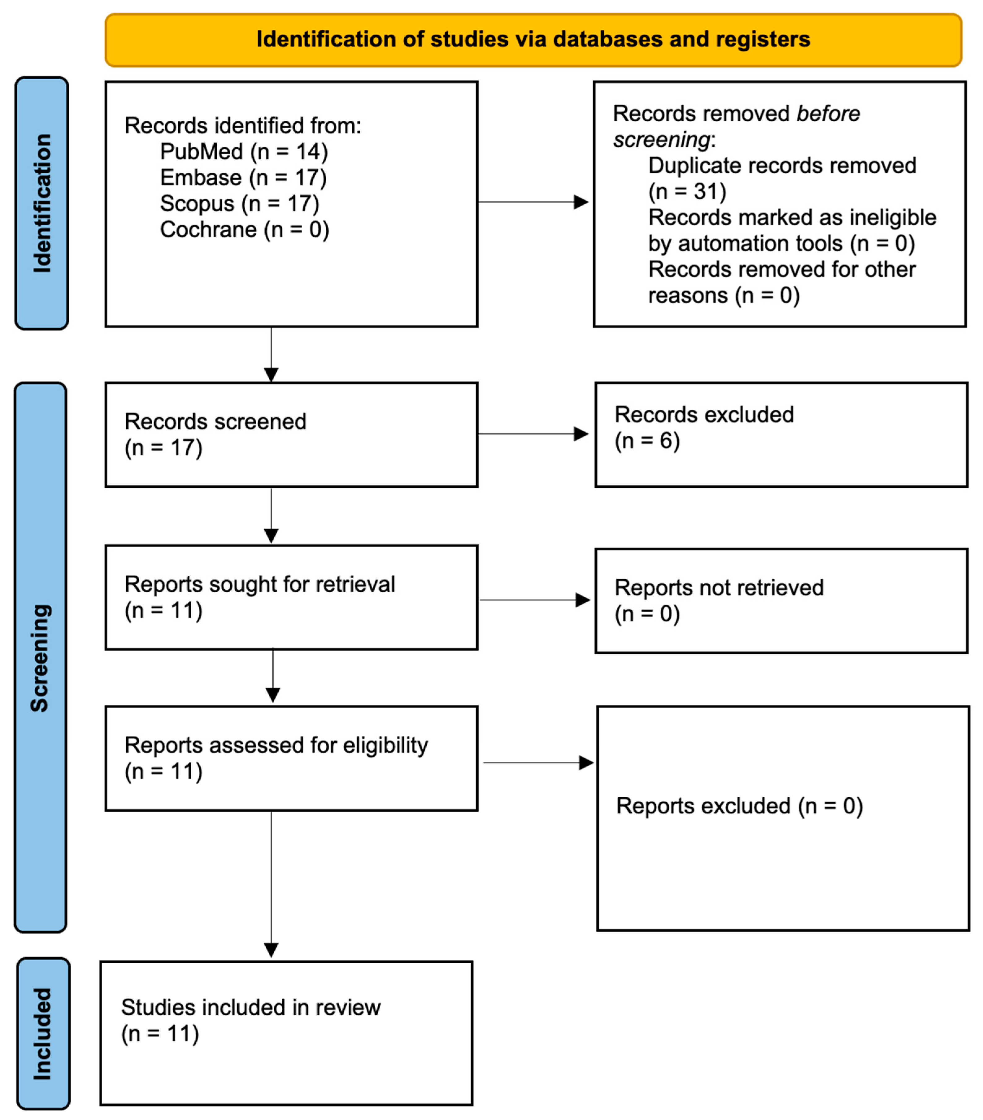
3.1. Study Selection

In alignment with the PRISMA 2020 guidelines, the study selection process is summarized in Figure 1. The initial database search retrieved 48 records, which were reduced to 17 distinct entries following the removal of duplicates. A preliminary screening of titles and abstracts confirmed the relevance of 11 studies, which were subsequently subjected to full-text review. No studies were excluded at this stage, resulting in the final inclusion of 11 publications for analysis. These studies were selected based on their direct investigation of the antimicrobial and bactericidal properties of the Nd:YAG laser in endodontic disinfection.

3.2. Data Presentation

Table 4, Table 5, Table 6 and Table 7 present a detailed and organized synthesis of the results from the ten included studies, highlighting essential outcomes, methodological characteristics, and microbiological data pertaining to the antibacterial and bactericidal properties of the Nd:YAG laser.

3.3. Overview of Study Characteristics

Table 4 outlines the geographical distribution of the studies included in this review.
Table 4. The geographical origins of the included research.
StudyCountry
Bergmans et al., 2006 [38]Belgium
Cheng et al., 2012 [39]China
Jurić et al., 2016 [40]Croatia
Kasić et al., 2017 [41]Croatia and France
Katalinić et al., 2014 [42]Croatia and Austria
Meire et al., 2012 [43]Belgium
Moshonov et al., 1995 [44]USA
Pirnat et al., 2011 [45]Slovenia
Schoop et al., 2004 [46]Austria
Vatkar et al., 2016 [47]India
Wang et al., 2018 [48]China

3.4. Main Study Outcomes

The outcomes of the included studies are summarised in Table 5.
Table 5. Summary of principal results and study details.
Reference NumberAuthor and YearMicroorganisms TestedStudy GroupsOutcomes
[38]Bergmans et al., 2006Enterococcus faecalis (LMG 7937), Streptococcus anginosus, and Actinomyces naeslundiiThe experimental design included three main models. In the microbiological analysis, six root canals were inoculated with E. faecalis and divided into laser-treated and control groups, using a cross-over approach. For CSEM, dentine discs were inoculated with S. anginosus and A. naeslundii, then treated with one, two, or three cycles of laser irradiation through 1 mm thick dentine. ESEM was employed to assess the effect of direct laser irradiation on biofilms formed by E. faecalis or micro-colonies of S. anginosus, with specimens undergoing either two or three irradiation cycles and being analyzed in situ.
-
Nd:YAG laser reduced E. faecalis load in root canals by 99.7%.
-
CSEM showed increasing bacterial damage with each laser cycle, with A. naeslundii more susceptible than S. anginosus.
-
No intact cells remained after three cycles.
-
ESEM revealed that while S. anginosus micro-colonies were mostly eradicated after 2–3 cycles, E. faecalis biofilms remained partially intact, with viable bacteria persisting in deeper layers, underscoring the difficulty of eliminating biofilm-associated infections.
[39]Cheng et al., 2012Enterococcus faecalis (ATCC 4083)The study comprised seven groups: five experimental groups—(1) Nd:YAG laser, (2) Er:YAG/NaClO/NS/DW, (3) Er:YAG/NS/DW, (4) Er,Cr:YSGG laser, and (5) aPDT—and two control groups: 5.25% NaClO as the positive control and 0.9% normal saline as the negative control. Each group included 20 specimens for bacteriological analysis and 10 for SEM imaging. Laser settings and procedural specifics followed manufacturer guidelines.
-
All laser protocols significantly reduced bacterial loads, with the Er:YAG/NaClO/NS/DW group achieving complete elimination on canal walls and up to 200 μm into dentinal tubules.
-
While 5.25% NaClO achieved 99.99% surface reduction, it was less effective in deeper layers.
-
Nd:YAG, Er,Cr:YSGG, and aPDT showed strong but lower efficacy. SEM confirmed no intact bacteria in Er:YAG/NaClO/NS/DW samples at all depths, unlike other groups.
-
The study concluded this combination was the most effective for endodontic disinfection.
[40]Jurić et al., 2016Enterococcus faecalis (ATCC 29212)A total of 65 dentine samples were randomly assigned to five groups: aPDT using phenothiazinium chloride and 660 nm laser; Nd:YAG laser irradiation at 2 W, 15 Hz; QMiX solution (a mix of CHX, EDTA, and detergent); 5.25% NaOCl as the negative control (gold standard disinfectant); and a positive control group with no disinfection. Each group included 15 samples for quantitative and microscopic analysis.
-
All methods significantly reduced E. faecalis CFUs versus controls.
-
The most effective was 5.25% NaOCl (99.99% reduction; complete eradication in some cases), followed by QMiX (99.3%) and aPDT (98.8%) with similar efficacy. Nd:YAG laser was least effective (96% reduction), with FISH confirming residual viable biofilm.
-
Overall, QMiX and aPDT were effective NaOCl alternatives, while laser alone showed limited efficacy.
[41]Kasić et al., 2017Enterococcus faecalis and Candida albicansThirty extracted single-rooted human teeth were divided into three groups (n = 10), each disinfected with a different laser system plus saline irrigation. Group 1 received Er:YAG laser using the PIPS technique, Group 2 underwent Nd:YAG laser treatment with spiral fiber motion, and Group 3 was treated with Er,Cr:YSGG laser using a radially firing tip. CFUs were measured before and after treatment to evaluate antimicrobial effectiveness.
-
The Er,Cr:YSGG laser most effectively reduced E. faecalis and C. albicans, significantly outperforming Er:YAG and Nd:YAG (p < 0.05). Er:YAG also reduced CFUs significantly, but less so. Nd:YAG showed no statistically significant effect.
-
These results highlight erbium lasers, especially Er,Cr:YSGG, as better suited for root canal disinfection due to superior irrigant activation and deeper penetration in biofilm-rich environments.
[42]Katalinić et al., 2014*Sixty extracted single-rooted anterior teeth were divided into four groups (n = 15) based on the final disinfection method: (1) 2.5% NaOCl, (2) 0.2% CHX, (3) gaseous ozone, and (4) Nd:YAG laser. After disinfection, canals were filled, post spaces prepared, and fiber-reinforced composite posts cemented with a self-etch adhesive. Push-out bond strength was measured using a universal testing machine.
-
Nd:YAG laser treatment produced the highest bond strength values, significantly outperforming the NaOCl group (p = 0.004). Sodium hypochlorite irrigation resulted in the lowest bond strength, potentially due to its oxidizing effect on dentine. No significant differences were found between the CHX, ozone, and Nd:YAG groups, nor between CHX, ozone, and NaOCl. The findings suggest that laser treatment enhances bonding effectiveness, while NaOCl may compromise it.
[43]Meire et al., 2012Enterococcus faecalis (ATCC 10541), Candida albicans (ATCC 10231), and Propionibacterium acnes (LMG 16711)Two laser systems were tested in vitro: Er:YAG (2940 nm) in single-pulse mode with energies from 40–400 mJ and pulse durations of 100–1000 μs, and Nd:YAG (1064 nm) in pulse train mode at 15 W, 100 Hz, applied for 5–120 s. Microbe-inoculated agar plates were irradiated through a 5 mm spot; for Nd:YAG, spot size was varied to assess irradiance effects. Antimicrobial efficacy was evaluated by measuring inhibition or clear zone diameters post-irradiation.
-
Er:YAG laser effectively inhibited all tested microorganisms on agar at low energy thresholds (TITs ≤ 220 mJ), with efficacy varying by pulse length and species, P. acnes being most sensitive. Shorter pulses worked better for bacteria.
-
In contrast, Nd:YAG required much higher energy densities (e.g., 5300 J/cm2 for C. albicans, 7100 J/cm2 for P. acnes) and was ineffective against surface E. faecalis, though embedded cells showed moderate susceptibility.
-
Overall, Er:YAG offered superior surface antimicrobial action, while Nd:YAG showed deeper but less efficient photothermal effects.
[44]Moshonov et al., 1995Enterococcus faecalisSeventy-five teeth were assigned to six groups: (1) non-infected control, (2) infected control, (3) infected + Nd:YAG laser with nigrosin dye, (4) infected + dye only, (5) infected + dye and air-water spray (no laser), and (6) infected + 1% NaOCl (positive control). In Group 3, laser irradiation was applied using a 400 µm fiber at 4.5 W, alternating between apical and coronal directions. Post-treatment, all teeth were sampled for bacterial growth, and selected specimens underwent SEM analysis.
-
Nd:YAG laser treatment reduced cultivable E. faecalis by over 99%, indicating strong but incomplete disinfection.
-
NaOCl achieved complete elimination. SEM confirmed partial canal wall cleaning and smear layer reduction with some residual bacteria.
-
Dye and air-water spray controls showed minimal effect, highlighting the laser’s specific antibacterial action.
-
Nd:YAG demonstrated notable potential, though less effective than NaOCl in achieving full sterility.
[45]Pirnat et al., 2011Enterococcus faecalis (ATCC 29212)The experiment tested two Nd:YAG laser heating protocols: (1) single 25 ms pulses (60–100 J/cm2) and (2) pulse trains of ten 1 ms pulses with 30 ms intervals (total fluence 100–260 J/cm2). Bacterial viability was assessed via culture plates and flow cytometry. Thermal behavior and disinfection efficacy were also compared between healthy and carious dentin using modeled heat pulses with fluences from 30 to 300 J/cm2.
-
Viability of E. faecalis decreased significantly with increasing peak temperature and laser fluence.
-
A ≥90% reduction (1-log10) in viability was achieved at around 102 °C with an 8 ms effective exposure time.
-
Sub-millisecond pulse trains produced cumulative thermal effects comparable to longer pulses, and the effectiveness was modeled accurately with the Arrhenius-based TDSC.
-
Disinfection was more efficient in carious than in healthy dentin due to higher absorption.
-
The study concluded that sub-millisecond pulsed Nd:YAG lasers are more effective and energy-efficient for root canal disinfection than continuous-wave lasers, minimizing unwanted thermal damage.
[46]Schoop et al., 2004Escherichia coli (ATCC 25922), Enterococcus faecalis (ATCC 29212)Four dental lasers were tested: Nd:YAG (1064 nm), diode (810 nm), Er:YAG (2940 nm), and Er,Cr:YSGG (2780 nm), each at 1 W and 1.5 W, forming eight test groups plus controls. Each group included 20 dentin slices. Laser irradiation was applied indirectly through the non-inoculated side to simulate clinical deep-layer disinfection. Control groups received no irradiation. Temperature measurements were also recorded to evaluate thermal effects.
-
All four lasers showed bactericidal effects, strongest at 1.5 W, with E. coli more easily eradicated than E. faecalis.
-
Er:YAG was most effective, fully eliminating E. coli and reducing E. faecalis by up to two log steps.
-
Diode and Er,Cr:YSGG were less effective, especially against E. faecalis. Nd:YAG and Er:YAG outperformed others in both reduction and penetration.
-
Temperature rises were moderate (max ~8.7 °C) and unrelated to efficacy.
-
Overall, all lasers were effective for deep dentin disinfection, with Er:YAG showing the highest efficiency.
[47]Vatkar et al., 2016Enterococcus faecalis (ATCC 29212)Group I (control) received no disinfection, allowing assessment of natural bacterial colonization. Group II was irrigated with 0.9% saline, Group III with 5.25% NaOCl, and Group IV with 2% CHX. Groups V and VI underwent laser disinfection using Nd:YAG and diode lasers, respectively, with specified power settings and fiber-optic delivery.
-
The control group showed deep E. faecalis colonization (965–1176 µm).
-
Normal saline was ineffective, with no zone of dead bacteria (ZDB). NaOCl and CHX produced limited ZDBs (88–110 µm and 110–194 µm, respectively).
-
In contrast, Nd:YAG and diode lasers achieved complete bacterial eradication, with ZDBs spanning nearly the full dentin depth (761–1145 µm).
-
Laser methods were markedly more effective than traditional irrigants in disinfecting dentinal tubules.
[48]Wang et al., 2018Enterococcus faecalis (ATCC 29212)Specimens were randomly divided into six groups, each tested at 1 and 3 min intervals: (A) 5.25% NaOCl, (B) Nd:YAG laser, (C) diode laser, (D) Nd:YAP laser, (E) Er,Cr:YSGG + NaOCl, and (F) Er:YAG + NaOCl. Bactericidal effects were evaluated using CLSM with LIVE/DEAD staining, comparing outcomes across laser types and exposure times.
-
All treatments significantly reduced E. faecalis biofilms, with greater efficacy at 3 min.
-
Er:YAG + NaOCl and Er,Cr:YSGG + NaOCl showed the highest bactericidal activity (76–89% and 73–85% cell death), outperforming all other groups.
-
Nd:YAP was next most effective, while Nd:YAG and diode lasers were less effective but still better than NaOCl alone.
-
The study emphasized the synergistic effect of erbium lasers with NaOCl, enhancing disinfection via photoacoustic and cavitation mechanisms for deeper irrigant penetration.
CSEM—conventional scanning electron microscopy, ESEM—environmental scanning electron microscopy, Nd:YAG—neodymium-doped yttrium aluminum garnet, Er:YAG—erbium-doped yttrium aluminum garnet, NaClO—sodium hypochlorite, NS—normal saline, DW—distilled water, Er,Cr:YSGG—erbium, chromium-doped yttrium, scandium, gallium, and garnet, aPDT—antimicrobial photodynamic therapy, CHX—chlorhexidine, NaOCl—sodium hypochlorite, CFUs—Colony-Forming Units, PIPS—Photon-Induced Photoacoustic Streaming, P. acnesPropionibacterium acnes, CLSM—confocal laser scanning microscopy, LIVE/DEAD—LIVE/DEAD viability staining, Nd:YAP—neodymium-doped yttrium aluminum perovskite, ZDB—zone of dead bacteria, TDSC—thermal damage survival curve. * Organisms not stated. SEM used as outcome measure.

3.5. Characteristics of Light Sources Used in PDT

Table 6 summarizes the main molecular properties of the Nd:YAG laser in this context. Table 7 presents the key physical properties of the light sources utilized in the studies meeting the inclusion criteria.
Table 6. Summary of the properties of Nd:YAG.
PropertyDescription/Significance
Wavelength1064 nm (near-infrared).
Chromophore AbsorptionHighly absorbed by bacterial pigments (melanin, dark pigments); poor absorption in water and hydroxyapatite.
Mode of ActionPrimarily photothermal; energy absorbed by chromophores within bacteria leads to localized heating and bacterial destruction.
Penetration DepthEffective bactericidal penetration up to 1 mm into dentinal tubules, deeper than many other lasers.
Antibacterial MechanismThermal denaturation of proteins, disruption of bacterial cell membranes, and direct bactericidal effects due to intracellular heating.
Effect on Smear LayerCauses evaporation, melting, contraction, and recrystallization of smear layer; at higher energies, complete removal and structural changes occur.
Morphological EffectsMelting, glazing, recrystallization, and partial or complete occlusion of dentinal tubules observed.
Thermal EffectsRisk of thermal damage and carbonization at higher energy levels (>3 W); optimal bactericidal effects achieved at controlled parameters (15 Hz, 100 mJ, 1.5 W).
Clinical Usage RecommendationUsed as adjunct to conventional chemical irrigation (NaOCl); does not fully replace chemical disinfection methods.
Source: [38,39,40,41,42,43,44,45,46,47,48].
Table 7. Characteristics of Nd:YAG laser used.
Author and YearLight SourceOperating ModePower Output (mW)Irradiation Time (s)
Bergmans et al., 2006 [38]Nd:YAG laser (Smarty A10; DEKA, Firenze, Italy)15 Hz, short-pulsed mode15004 × 5
Cheng et al., 2012 [39]Nd:YAG laser (Fontona Lasers, Stegne-7-1210, Ljubljana, Slovenia)15 Hz, pulsed15004 × 4
Jurić et al., 2016 [40]Nd:YAG (Fotona, Ljubljana, Slovenia)15 Hz, pulsed20004 × 5
Kasić et al., 2017 [41]Nd:YAG (LightWalker, Fotona, Ljubljana, Slovenia)15 Hz, pulsed1500Not specified
Katalinić et al., 2014 [42]Nd:YAG (Fotona, Ljubljana, Slovenia)10 Hz, very short-pulsed mode400010
Meire et al., 2012 [43]Nd:YAG (AT Fidelis, Fotona)100 Hz, pulsed15,00010–120
Moshonov et al., 1995 [44]Nd:YAGNot reported45004 × 15
Pirnat et al., 2011 [45]Nd:YAG (XP-2, Fotona d.d., Slovenia)PulsedNot reportedSub-second, ms pulses
Schoop et al., 2004 [46]Nd:YAG (American Dental Technologies, Texas, USA)10–200 Hz, pulsed200–50000.1
Vatkar et al., 2016 [47]Nd:YAG (Fotona Fidelis III, Slovenia, Europe)Continuous mode15005
Wang et al., 2018 [48]Nd:YAG (Fotona, Ljubljana, Slovenia)15 Hz, pulsed150015 × 4

4. Discussion

4.1. Results in the Context of Other Evidence

The neodymium-doped yttrium aluminum garnet (Nd:YAG) laser, operating at a wavelength of 1064 nm, exhibits unique molecular and photothermal properties that make it a valuable tool in root canal disinfection [38,49]. Its high penetration capacity into dentinal tubules allows it to target deeply embedded microbial colonies, especially pigmented bacteria, via rapid thermal effects that denature proteins, disrupt cell membranes, and induce coagulative necrosis [50,51]. This selective absorption by bacterial chromophores enhances its bactericidal potential while sparing surrounding tissue [38,39,40]. Numerous in vitro studies have demonstrated the Nd:YAG laser’s efficacy against key endodontic pathogens. For example, Moritz et al. reported a 99.16% reduction in Enterococcus faecalis and Escherichia coli populations [52], while Gutknecht et al. achieved a 99.92% reduction in E. faecalis using settings of 15 Hz and 100 mJ [53]. Similarly, another study demonstrated up to 99.7% bacterial reduction in root canals, although complete sterilization was rarely attained [38,52,53]. These outcomes highlight the laser’s potential to manage polymicrobial infections, especially when compared to traditional chemical irrigants [47,52,53]. Despite these promising results, limitations remain. The Nd:YAG laser’s bactericidal efficacy is primarily thermal, relying on localized heating to disrupt cell walls and denature intracellular proteins [47,52]. However, its poor absorption in water reduces its effectiveness against non-pigmented bacteria and dense biofilms, often requiring elevated energy densities to achieve meaningful reductions [47,52,54]. Although it can reach bactericidal depths of up to 1000 μm—surpassing the 1100 μm bacterial invasion depth reported in some cases—its effectiveness may be attenuated at these depths, especially in clinical scenarios with persistent biofilms [47,54,55,56,57,58]. Comparative studies suggest that erbium lasers, such as Er:YAG and Er,Cr:YSGG, offer superior bacterial eradication and deeper dentinal tubule penetration than Nd:YAG lasers [39,41,49]. These systems also show greater efficacy in smear layer removal and irrigant activation due to their photoacoustic or photomechanical mechanisms [39,41,59]. In contrast, Nd:YAG relies solely on photothermal interactions, which may be less effective in complex anatomical regions or against mature biofilms. Adjunctive use of Nd:YAG with sodium hypochlorite has shown to improve bacterial clearance and reduce inflammatory markers compared to irrigation alone, offering enhanced clinical outcomes [43]. The laser’s ability to increase push-out bond strength of fiber posts and alter dentin surface properties further underscores its utility [42]. Photothermal effects contribute to smear layer modification and the removal of intracanal medicaments and debris, although these outcomes are often secondary observations and inconsistently quantified [42,44,50]. Sub-millisecond pulsed Nd:YAG modes have emerged as more energy-efficient and bactericidal compared to longer-pulse or continuous-wave settings [46]. Importantly, scanning electron microscopy (SEM) and confocal laser scanning microscopy (CLSM) have confirmed that Nd:YAG can significantly disrupt biofilms, although viable bacteria often persist in deeper canal layers [38,45]. Moreover, temperature rises during laser use have been moderate and not directly correlated with disinfection outcomes, reaffirming the importance of optimizing fluence and pulse parameters rather than relying on heat generation alone [47]. In terms of structural effects, Nd:YAG irradiation induces noticeable dentin alterations such as melting and recrystallization, potentially occluding or exposing dentinal tubules [47,60,61,62,63,64,65,66]. These morphological changes differ substantially from those caused by chemical irrigants like sodium hypochlorite (NaOCl), EDTA, H2O2, or chlorhexidine (CHX) and may influence the mechanical properties and mineral content of dentin, including the calcium-to-phosphorus ratio—factors relevant to post-treatment restoration [47,50,58]. Clinically, Nd:YAG lasers have shown promising results, especially in cases with significant periapical pathology. Long-term follow-ups have documented favorable outcomes following laser-enhanced disinfection, particularly when optimal settings (e.g., 15 Hz, 100 mJ, 1.5 W) and careful fiber optic tip movement are employed [47,53,56,59,67,68]. Nevertheless, studies agree that the laser should serve as a complementary tool rather than a standalone solution, as it has not demonstrated superiority over conventional irrigants such as NaOCl [29,38,60]. This conclusion is further reinforced by recent reviews. Dawasaz et al. (2022) found that diode lasers could significantly reduce microbial counts in vivo, although protocol heterogeneity and limited long-term data impeded strong clinical recommendations [66]. Fransson et al. (2013) reported improved microbial reductions with laser-assisted chemomechanical preparation but could not confirm consistent benefits in periapical healing due to methodological variability [59]. Similarly, Hazrati et al. (2024) suggested that laser-assisted therapy may enhance periapical healing, yet high-quality randomized trials remain scarce [67]. Despite its strengths, the Nd:YAG laser’s reduced efficacy against non-pigmented bacteria and mature biofilms, along with its inability to achieve complete sterilization, remain critical limitations [52,55,62,63]. Therefore, future research should focus on refining laser parameters, exploring hybrid modalities such as photodynamic therapy (PDT) and laser-activated irrigation (LAI), and advancing the understanding of laser–biofilm interactions at the molecular level [51,52]. These innovations may further enhance the clinical efficacy and integration of Nd:YAG lasers in endodontic disinfection protocols [51,52,68,69,70,71].

4.2. Limitations of the Evidence

The current body of evidence on the efficacy of Nd:YAG lasers in root canal disinfection is constrained by several key limitations. Most notably, the overwhelming reliance on in vitro studies limits the generalizability of findings to clinical practice, as these controlled laboratory settings do not accurately replicate the anatomical, biological, and microbial complexities of the human oral environment. Additionally, there is considerable variability in experimental protocols, including differences in the laser settings, exposure times, fiber types, and microbial strains used. This heterogeneity hampers direct comparison between studies and undermines the ability to draw consistent conclusions. Moreover, many studies lack standardized outcome measures, and few assess long-term antimicrobial effects or potential microbial recolonization, which are critical for evaluating clinical relevance. The limited number of high-quality in vivo investigations and randomized controlled trials further weakens the strength of the evidence. These gaps highlight the urgent need for more rigorous, standardized, and clinically oriented research to fully elucidate the therapeutic value of Nd:YAG lasers in endodontic disinfection.

4.3. Limitations of the Review Process

Despite providing a comprehensive synthesis of the existing literature, this systematic review has several noteworthy limitations. Firstly, all included studies were either in vitro or ex vivo, limiting the direct applicability of findings to clinical practice due to the absence of complex biological interactions present in in vivo settings. Secondly, significant heterogeneity in study protocols—such as variations in Nd:YAG laser parameters, microbial species tested, sample preparation, and disinfection outcome measures—makes it difficult to compare results across studies or conduct meaningful meta-analyses. Thirdly, while the included studies were assessed as having low risk of bias, their overall methodological diversity and lack of long-term follow-up reduce confidence in their durability and reproducibility. Additionally, the exclusion of non-English publications and gray literature introduces potential language and publication bias, possibly omitting relevant findings. Finally, the limited number of clinical trials in the evidence base highlights the need for more robust, standardized, and clinically focused research to better define the efficacy and practical integration of Nd:YAG laser therapy in endodontic disinfection.
A quantitative meta-analysis was not performed in this review due to substantial heterogeneity among the included studies. The sources of heterogeneity included the following: (1) variations in experimental design; (2) diverse microbial targets, with some studies evaluating mono-species infections and others assessing mixed-species or polymicrobial biofilms; (3) inconsistent Nd:YAG laser parameters such as wavelength calibration, power output, irradiation duration, and pulse frequency; (4) differing control groups and disinfection comparators across studies; and (5) lack of uniform outcome measures for microbial viability or reduction (e.g., CFU counts, SEM analysis, CLSM, ZDB). Due to these methodological discrepancies, data pooling would not yield valid or interpretable summary estimates, and meta-analytic synthesis was deemed inappropriate. Instead, a qualitative synthesis was conducted to preserve the integrity of the individual study contexts and findings.

4.4. Implications for Practice, Policy, and Future Research

The findings from this systematic review on the efficacy of Nd:YAG lasers in root canal disinfection have significant implications for clinical practice, policy, and future research. Clinically, the Nd:YAG laser shows considerable promise as an adjunctive treatment for root canal disinfection, especially in cases involving persistent biofilms and challenging anatomical structures like lateral canals and dentinal tubules. Dental practitioners are encouraged to integrate Nd:YAG laser treatment alongside traditional chemical irrigants, such as sodium hypochlorite and chlorhexidine, to improve disinfection outcomes and reduce treatment failure rates associated with resilient pathogens, particularly Enterococcus faecalis. From a policy perspective, the current absence of standardized clinical protocols highlights the urgent necessity for developing consensus guidelines that detail precise laser operation parameters, such as optimal pulse durations, power settings, and irradiation techniques. This standardization is essential to ensure consistent, safe, and effective clinical implementation. Regarding future research, it is imperative to conduct robust, randomized controlled clinical trials to validate the laboratory findings, investigate the long-term sustainability of antimicrobial effects, assess potential complications or adverse events, and compare the efficacy of Nd:YAG lasers with alternative laser systems and conventional endodontic treatments. Additionally, further investigation into the molecular mechanisms underlying the laser’s bactericidal effects could provide valuable insights, contributing to more effective and targeted therapeutic strategies.
While the Nd:YAG laser demonstrates significant antimicrobial effects in vitro, a major limitation is the absence of clinical evidence confirming its impact on long-term endodontic treatment outcomes. Specifically, no high-quality studies have shown that laser-assisted disinfection improves healing rates, reduces treatment failure, or enhances patient-reported outcomes compared to conventional protocols. The current evidence base is predominantly composed of laboratory-based research with limited translational value. As such, the therapeutic benefit of Nd:YAG lasers in routine endodontic practice remains hypothetical until validated by rigorously designed clinical trials.

5. Conclusions

The Nd:YAG laser shows promising antimicrobial efficacy in root canal disinfection, particularly due to its deep penetration and photothermal effects. While in vitro studies report significant bacterial load reductions, complete biofilm eradication remains inconsistent, and Er:YAG or Er,Cr:YSGG lasers often demonstrate superior outcomes. However, the current evidence is limited by methodological heterogeneity, lack of standardized protocols, and absence of clinical trials confirming improved treatment outcomes. As such, the Nd:YAG laser should be considered an adjunct to, not a replacement for, conventional irrigation. Future research should focus on well-designed clinical trials, standardized laser parameters, and molecular studies to clarify the laser’s clinical utility and optimize its application in endodontic practice.

Author Contributions

Conceptualization, J.F.-R. and R.W.; methodology, J.F.-R., D.S. and R.W.; software, J.F.-R.; formal analysis, J.F.-R., D.S. and R.W.; investigation, J.F.-R., D.S. and R.W.; writing—original draft preparation, J.F.-R., D.S. and R.W.; writing—review and editing, J.F.-R., D.S. and R.W.; supervision, D.S. and R.W.; funding acquisition, D.S. and R.W. All authors have read and agreed to the published version of the manuscript.

Funding

This research received no external funding.

Data Availability Statement

No new data were created or analyzed in this study.

Conflicts of Interest

The authors declare no conflicts of interest.

References

  1. Gomes, B.P.F.A.; Aveiro, E.; Kishen, A. Irrigants and irrigation activation systems in Endodontics. Braz. Dent. J. 2023, 34, 1–33. [Google Scholar] [CrossRef] [PubMed] [PubMed Central]
  2. Iqbal, A. Antimicrobial irrigants in the endodontic therapy. Int. J. Health Sci. 2012, 6, 186–192. [Google Scholar] [CrossRef] [PubMed] [PubMed Central]
  3. American Association of Endodontists. Treatment Standards; American Association of Endodontists: Chicago, IL, USA, 2018; Available online: https://www.aae.org/specialty/wp-content/uploads/sites/2/2018/04/TreatmentStandards_Whitepaper.pdf (accessed on 21 April 2025).
  4. Al Ajmi, N.F.; Alshenaifi, M.K.; Binsalem, M.M.; Alahmary, K.A.; Al Shahrani, N.A.; Alasim, A.M.; Alotaibi, F.R.; Ali, A.S.; Attar, S.M.; Al Shahrani, N.M. Microbial challenges and solutions in root canal therapy. Int. J. Community Med. Public Health 2024, 11, 3672–3676. [Google Scholar] [CrossRef]
  5. Saklar, F.; Öncü, A.; Sevgi, S.; Çelikten, B. Endodontic Treatment of Premolar Teeth with Different Root Canal Anatomy: Two Case Reports and Literature Review. Cyprus J. Med. Sci. 2023, 8, 453–456. [Google Scholar] [CrossRef]
  6. Muteeb, G.; Rehman, M.T.; Shahwan, M.; Aatif, M. Origin of Antibiotics and Antibiotic Resistance, and Their Impacts on Drug Development: A Narrative Review. Pharmaceuticals 2023, 16, 1615. [Google Scholar] [CrossRef]
  7. Mamat, R.; Nik Abdul Ghani, N.R. The Complexity of the Root Canal Anatomy and Its Influence on Root Canal Debridement in the Apical Region: A Review. Cureus 2023, 15, e49024. [Google Scholar] [CrossRef] [PubMed] [PubMed Central]
  8. Kristich, C.J.; Rice, L.B.; Arias, C.A. Enterococcal Infection—Treatment and Antibiotic Resistance. In Enterococci: From Commensals to Leading Causes of Drug Resistant Infection; Gilmore, M.S., Clewell, D.B., Ike, Y., Shanker, N., Eds.; Massachusetts Eye and Ear Infirmary: Boston, MA, USA, 2014. [Google Scholar]
  9. Xiao, S.; Sun, G.; Huang, S.; Lin, C.; Li, Y. Nanoarchitectonics-Based Materials as a Promising Strategy in the Treatment of Endodontic Infections. Pharmaceutics 2024, 16, 759. [Google Scholar] [CrossRef] [PubMed] [PubMed Central]
  10. Saleh, I.M.; Ruyter, I.E.; Haapasalo, M.; Ørstavik, D. Survival of Enterococcus faecalis in infected dentinal tubules after root canal filling with different root canal sealers in vitro. Int. Endod. J. 2004, 37, 193–198. [Google Scholar] [CrossRef] [PubMed]
  11. Mehta, D.; Coleman, A.; Lessani, M. Success and failure of endodontic treatment: Predictability, complications, challenges and maintenance. Br. Dent. J. 2025, 238, 527–535. [Google Scholar] [CrossRef] [PubMed] [PubMed Central]
  12. Prada, I.; Micó-Muñoz, P.; Giner-Lluesma, T.; Micó-Martínez, P.; Collado-Castellano, N.; Manzano-Saiz, A. Influence of microbiology on endodontic failure. Literature review. Med. Oral Patol. Oral Cir. Bucal 2019, 24, e364–e372. [Google Scholar] [CrossRef] [PubMed] [PubMed Central]
  13. Boyd, N.K.; Teng, C.; Frei, C.R. Brief Overview of Approaches and Challenges in New Antibiotic Development: A Focus on Drug Repurposing. Front. Cell Infect. Microbiol. 2021, 11, 684515. [Google Scholar] [CrossRef] [PubMed] [PubMed Central]
  14. Borkenstein, A.F.; Borkenstein, E.M. Neodymium-doped yttrium aluminum garnet (Nd: YAG) laser treatment in ophthalmology: A review of the most common procedures Capsulotomy and Iridotomy. Lasers Med. Sci. 2024, 39, 167. [Google Scholar] [CrossRef] [PubMed]
  15. Penberthy, W.T.; Vorwaller, C.E. Utilization of the 1064 nm Wavelength in Photobiomodulation: A Systematic Review and Meta-Analysis. J. Lasers Med. Sci. 2021, 12, e86. [Google Scholar] [CrossRef] [PubMed] [PubMed Central]
  16. Grönqvist, A.; Wiström, J.; Axner, O.; Monsen, T.J. Bactericidal effect of pulsed 1,064 nm Nd:YAG laser light on Staphylococcus epidermidis is of photothermal origin: An in vitro study. Lasers Surg. Med. 2000, 27, 336–340. [Google Scholar] [CrossRef] [PubMed]
  17. Ghorbanzadeh, A.; Aminsobhani, M.; Sohrabi, K.; Chiniforush, N.; Ghafari, S.; Shamshiri, A.R.; Noroozi, N. Penetration Depth of Sodium Hypochlorite in Dentinal Tubules after Conventional Irrigation, Passive Ultrasonic Agitation and Nd:YAG Laser Activated Irrigation. J. Lasers Med. Sci. 2016, 7, 105–111. [Google Scholar] [CrossRef] [PubMed] [PubMed Central]
  18. Dembicka-Mączka, D.; Gryka-Deszczyńska, M.; Sitkiewicz, J.; Makara, A.; Fiegler-Rudol, J.; Wiench, R. Evaluation of the Disinfection Efficacy of Er-YAG Laser Light on Single-Species Candida Biofilms: Systematic Review. Microorganisms 2025, 13, 942. [Google Scholar] [CrossRef]
  19. Granevik Lindström, M.; Wolf, E.; Fransson, H. The Antibacterial Effect of Nd:YAG Laser Treatment of Teeth with Apical Periodontitis: A Randomized Controlled Trial. J. Endod. 2017, 43, 857–863. [Google Scholar] [CrossRef] [PubMed]
  20. Fiegler-Rudol, J.; Grzech-Leśniak, Z.; Tkaczyk, M.; Grzech-Leśniak, K.; Zawilska, A.; Wiench, R. Enhancing Root Canal Disinfection with Er:YAG Laser: A Systematic Review. Dent. J. 2025, 13, 101. [Google Scholar] [CrossRef]
  21. Kurt, S.; Kırtıloğlu, T.; Yılmaz, N.A.; Ertaş, E.; Oruçoğlu, H. Evaluation of the effects of Er:YAG laser, Nd:YAG laser, and two different desensitizers on dentin permeability: In Vitro study. Lasers Med. Sci. 2018, 33, 1883–1890. [Google Scholar] [CrossRef] [PubMed]
  22. Huang, Q.; Li, Z.; Lyu, P.; Zhou, X.; Fan, Y. Current Applications and Future Directions of Lasers in Endodontics: A Narrative Review. Bioengineering 2023, 10, 296. [Google Scholar] [CrossRef] [PubMed] [PubMed Central]
  23. Scarano, A.; Lorusso, F.; Inchingolo, F.; Postiglione, F.; Petrini, M. The Effects of Erbium-Doped Yttrium Aluminum Garnet Laser (Er: YAG) Irradiation on Sandblasted and Acid-Etched (SLA) Titanium, an In Vitro Study. Materials. 2020, 13, 4174. [Google Scholar] [CrossRef] [PubMed]
  24. Meng, Y.; Huang, F.; Wang, S.; Li, M.; Lu, Y.; Pei, D.; Li, A. Bonding Performance of Universal Adhesives Applied to Nano-Hydroxyapatite Desensitized Dentin Using Etch-and-Rinse or Self-Etch Mode. Materials 2021, 14, 4746. [Google Scholar] [CrossRef] [PubMed] [PubMed Central]
  25. Ateş, M.O.; Uğur Aydın, Z. Evaluation of the effectiveness of different treatment approaches in preventing coronal discoloration caused by regenerative endodontic treatment. Clin. Oral Investig. 2023, 27, 4595–4603. [Google Scholar] [CrossRef] [PubMed]
  26. Hamblin, M.R.; Hasan, T. Photodynamic therapy: A new antimicrobial approach to infectious disease? Photochem. Photobiol. Sci. 2004, 3, 436–450. [Google Scholar] [CrossRef] [PubMed] [PubMed Central]
  27. Deaton, A.; Cartwright, N. Understanding and misunderstanding randomized controlled trials. Soc. Sci. Med. 2018, 210, 2–21. [Google Scholar] [CrossRef] [PubMed] [PubMed Central]
  28. Linden, A.H.; Hönekopp, J. Heterogeneity of Research Results: A New Perspective from Which to Assess and Promote Progress in Psychological Science. Perspect. Psychol. Sci. 2021, 16, 358–376. [Google Scholar] [CrossRef] [PubMed] [PubMed Central]
  29. Clark, A.E.; Kaleta, E.J.; Arora, A.; Wolk, D.M. Matrix-assisted laser desorption ionization-time of flight mass spectrometry: A fundamental shift in the routine practice of clinical microbiology. Clin. Microbiol. Rev. 2013, 26, 547–603. [Google Scholar] [CrossRef] [PubMed] [PubMed Central]
  30. Franco-Duarte, R.; Černáková, L.; Kadam, S.; Kaushik, K.S.; Salehi, B.; Bevilacqua, A.; Corbo, M.R.; Antolak, H.; Dybka-Stępień, K.; Leszczewicz, M.; et al. Advances in Chemical and Biological Methods to Identify Microorganisms—From Past to Present. Microorganisms 2019, 7, 130. [Google Scholar] [CrossRef] [PubMed] [PubMed Central]
  31. Wassenberg, M.W.M.; Kluytmans, J.A.J.W.; Box, A.T.A.; Bosboom, R.W.; Buiting, A.G.M.; Van Elzakker, E.P.M.; Melchers, W.J.G.; Van Rijen, M.M.L.; Thijsen, S.F.T.; Troelstra, A.; et al. Rapid screening of methicillin-resistant Staphylococcus aureus using PCR and chromogenic agar: A prospective study to evaluate costs and effects. Clin. Microbiol. Infect. 2010, 16, 1754–1761. [Google Scholar] [CrossRef]
  32. van Belkum, A.; Durand, G.; Peyret, M.; Chatellier, S.; Zambardi, G.; Schrenzel, J.; Shortridge, D.; Engelhardt, A.; Dunne, W.M., Jr. Rapid clinical bacteriology and its future impact. Ann. Lab. Med. 2010, 33, 14–27. [Google Scholar] [CrossRef]
  33. He, Z.; Tang, X.; Yang, X.; Guo, Y.; George, T.J.; Charness, N.; Quan Hem, K.B.; Hogan, W.; Bian, J. Clinical Trial Generalizability Assessment in the Big Data Era: A Review. Clin. Transl. Sci. 2020, 13, 675–684. [Google Scholar] [CrossRef] [PubMed] [PubMed Central]
  34. Schardt, C.; Adams, M.B.; Owens, T.; Keitz, S.; Fontelo, P. Utilization of the PICO framework to improve searching PubMed for clinical questions. BMC Med. Inform. Decis. Mak. 2007, 7, 16. [Google Scholar] [CrossRef] [PubMed]
  35. Page, M.J.; McKenzie, J.E.; Bossuyt, P.M.; Boutron, I.; Hoffmann, T.C.; Mulrow, C.D.; Shamseer, L.; Tetzlaff, J.M.; Akl, E.A.; Brennan, S.E.; et al. The PRISMA 2020 statement: An updated guideline for reporting systematic reviews. BMJ 2021, 372, n71. [Google Scholar] [CrossRef] [PubMed]
  36. Watson, P.F.; Petrie, A. Method agreement analysis: A review of correct methodology. Theriogenology 2010, 73, 1167–1179. [Google Scholar] [CrossRef]
  37. Higgins, J.P.T.; Thomas, J.; Chandler, J.; Cumpston, M.; Li, T.; Page, M.J.; Welch, V.A. (Eds.) Cochrane Handbook for Systematic Reviews of Interventions Version 6.5 (Updated August 2024); Cochrane: London, UK, 2024. [Google Scholar]
  38. Bergmans, L.; Moisiadis, P.; Teughels, W.; Van Meerbeek, B.; Quirynen, M.; Lambrechts, P. Bactericidal effect of Nd:YAG laser irradiation on some endodontic pathogens ex vivo. Int. Endod. J. 2006, 39, 547–557. [Google Scholar] [CrossRef]
  39. Cheng, X.; Guan, S.; Lu, H.; Zhao, C.; Chen, X.; Li, N.; Bai, Q.; Tian, Y.; Yu, Q. Evaluation of the bactericidal effect of Nd:YAG, Er:YAG, Er,Cr:YSGG laser radiation, and antimicrobial photodynamic therapy (aPDT) in experimentally infected root canals. Lasers Surg. Med. 2012, 44, 824–831. [Google Scholar] [CrossRef]
  40. Juric, I.B.; Plecko, V.; Anic, I.; Plesko, S.; Jakovljevic, S.; Rocca, J.P.; Medioni, E. Antimicrobial efficacy of photodynamic therapy, Nd:YAG laser and QMiX solution against Enterococcus faecalis biofilm. Photodiagnosis Photodyn. Ther. 2016, 13, 238–243. [Google Scholar] [CrossRef]
  41. Kasic, S.; Knezovic, M.; Beader, N.; Gabric, D.; Ivanisevic Malcic, A.; Baraba, A. Efficacy of three different lasers on eradication of Enterococcus faecalis and Candida albicans biofilms in root canal system. Photomed. Laser Surg. 2017, 35, 372–377. [Google Scholar] [CrossRef]
  42. Katalinić, I.; Glockner, K.; Anić, I. Influence of several root canal disinfection methods on pushout bond strength of self-etch post and core systems. Int. Endod. J. 2014, 47, 140–146. [Google Scholar] [CrossRef]
  43. Meire, M.A.; Coenye, T.; Nelis, H.J.; De Moor, R.J.G. In vitro inactivation of endodontic pathogens with Nd:YAG and Er:YAG lasers. Lasers Med. Sci. 2012, 27, 695–701. [Google Scholar] [CrossRef]
  44. Moshonov, J.; Ørstavik, D.; Yamauchi, S.; Pettiette, M.; Trope, M. Nd:YAG laser irradiation in root canal disinfection. Endod. Dent. Traumatol. 1995, 11, 220–224. [Google Scholar] [CrossRef] [PubMed]
  45. Pirnat, S.; Lukac, M.; Ihan, A. Thermal tolerance of E. faecalis to pulsed heating in the millisecond range. Lasers Med. Sci. 2011, 26, 229–237. [Google Scholar] [CrossRef]
  46. Schoop, U.; Kluger, W.; Moritz, A.; Nedjelik, N.; Georgopoulos, A.; Sperr, W. Bactericidal effect of different laser systems in the deep layers of dentin. Lasers Surg. Med. 2004, 35, 111–116. [Google Scholar] [CrossRef]
  47. Vatkar, N.A.; Hegde, V.; Sathe, S. Vitality of Enterococcus faecalis inside dentinal tubules after five root canal disinfection methods. J. Conserv. Dent. 2016, 19, 445–449. [Google Scholar] [CrossRef] [PubMed] [PubMed Central]
  48. Wang, X.; Cheng, X.; Liu, X.; Wang, Z.; Wang, J.; Guo, C.; Zhang, Y.; He, W. Bactericidal effect of various laser irradiation systems on Enterococcus faecalis biofilms in dentinal tubules: A confocal laser scanning microscopy study. Photomed. Laser Surg. 2018, 36, 514–522. [Google Scholar] [CrossRef] [PubMed]
  49. Dembicka-Mączka, D.; Kępa, M.; Fiegler-Rudol, J.; Grzech-Leśniak, Z.; Matys, J.; Grzech-Leśniak, K.; Wiench, R. Evaluation of the Disinfection Efficacy of Er: YAG Laser Light on Single-Species Candida Biofilms—An In Vitro Study. Dent. J. 2025, 13, 88. [Google Scholar] [CrossRef]
  50. Golob Deeb, J.; Reddy, N.; Kitten, T.; Carrico, C.K.; Grzech-Leśniak, K. Viability of bacteria associated with root caries after Nd:YAG laser application in combination with various antimicrobial agents: An in vitro study. Dent. Med. Probl. 2023, 60, 649–655. [Google Scholar] [CrossRef]
  51. Basavegowda, N.; Baek, K.H. Combination Strategies of Different Antimicrobials: An Efficient and Alternative Tool for Pathogen Inactivation. Biomedicines 2022, 10, 2219. [Google Scholar] [CrossRef] [PubMed] [PubMed Central]
  52. Moritz, A.; Schoop, U.; Goharkhay, K.; Schauer, P.; Doertbudak, O.; Sperr, W. The bactericidal effect of Nd:YAG laser irradiation within root canal dentin. J. Clin. Laser Med. Surg. 1997, 15, 29–31. [Google Scholar]
  53. Gutknecht, N.; Moritz, A.; Conrads, G.; Sievert, T.; Lampert, F. Bactericidal effect of the Nd:YAG laser in in vitro root canals. J. Clin. Laser Med. Surg. 1996, 14, 77–80. [Google Scholar] [CrossRef]
  54. Klinke, T.; Klimm, W.; Gutknecht, N. Antibacterial effects of Nd:YAG laser irradiation within root canal dentine. J. Clin. Laser Med. Surg. 1997, 15, 29–31. [Google Scholar] [CrossRef] [PubMed]
  55. Ozses Ozkaya, B.; Gulsahi, K.; Ungor, M.; Gocmen, J.S. A Comparison of Er:YAG Laser with Photon-Initiated Photoacoustic Streaming, Nd:YAG Laser, and Conventional Irrigation on the Eradication of Root Dentinal Tubule Infection by Enterococcus faecalis Biofilms: A Scanning Electron Microscopy Study. Scanning 2017, 2017, 6215482. [Google Scholar] [CrossRef] [PubMed] [PubMed Central]
  56. Gutknecht, N. Lasers in Endodontics. Laser Health Acad. Rev. 2008, 4, 1–10. [Google Scholar]
  57. Tsuka, Y.; Kunimatsu, R.; Gunji, H.; Abe, T.; Medina, C.C.; Nakajima, K.; Kimura, A.; Hiraki, T.; Nakatani, A.; Tanimoto, K. Examination of the Effect of the Combined Use of Nd: YAG Laser Irradiation and Mechanical Force Loading on Bone Metabolism Using Cultured Human Osteoblasts. J. Lasers Med. Sci. 2020, 11, 138–143. [Google Scholar] [CrossRef] [PubMed] [PubMed Central]
  58. Dilber, E.; Malkoc, M.A.; Ozturk, A.N.; Ozturk, F. Effect of various laser irradiations on the mineral content of dentin. Eur. J. Dent. 2013, 7, 74–80. [Google Scholar] [PubMed] [PubMed Central]
  59. Hazrati, P.; Azadi, A.; Tizno, A.; Asnaashari, M. The Effect of Lasers on the Healing of Periapical Lesion: A Systematic Review. J. Lasers Med. Sci. 2024, 15, e6. [Google Scholar] [CrossRef] [PubMed] [PubMed Central]
  60. Rahimi, S.; Shahi, S.; Gholizadeh, S.; Shakouie, S.; Rikhtegaran, S.; Soroush Barhaghi, M.H.; Ghojazadeh, M.; Froughreyhani, M.; Abdolrahimi, M. Bactericidal effects of Nd:YAG laser irradiation and sodium hypochlorite solution on Enterococcus faecalis biofilm. Photomed. Laser Surg. 2012, 30, 637–641. [Google Scholar] [CrossRef] [PubMed]
  61. White, J.M.; Goodis, H.E.; Rose, C.M. Nd:YAG pulsed infrared laser for treatment of root surface. J. Calif. Dent. Assoc. 1991, 19, 55–58. [Google Scholar] [PubMed]
  62. de Souza, E.B.; Cai, S.; Simionato, M.R.; Lage-Marques, J.L. High-power diode laser in the disinfection in depth of the root canal dentin. Oral. Surg. Oral. Med. Oral. Pathol. Oral. Radiol. Endod. 2008, 106, e68–e72. [Google Scholar] [CrossRef]
  63. Warakomska, A.; Fiegler-Rudol, J.; Kubizna, M.; Skaba, D.; Wiench, R. The Role of Photodynamic Therapy Mediated by Natural Photosensitisers in the Management of Peri-Implantitis: A Systematic Review. Pharmaceutics 2025, 17, 443. [Google Scholar] [CrossRef] [PubMed] [PubMed Central]
  64. Asnaashari, M.; Safavi, N. Disinfection of contaminated canals by different lasers: An in vitro study. J. Lasers Med. Sci. 2013, 4, 8–16. [Google Scholar] [PubMed]
  65. Dawasaz, A.A. In Vivo Efficacy of Diode Laser as a Monotherapy in Root Canal Disinfection: A Systematic Review and Meta-Analysis. Photobiomodul. Photomed. Laser Surg. 2022, 40, 59–70. [Google Scholar] [CrossRef] [PubMed]
  66. Fransson, H.; Larsson, K.M.; Wolf, E. Efficacy of lasers as an adjunct to chemo-mechanical disinfection of infected root canals: A systematic review. Int. Endod. J. 2013, 46, 296–307. [Google Scholar] [CrossRef] [PubMed]
  67. Kruczek-Kazibudzka, A.; Lipka, B.; Fiegler-Rudol, J.; Tkaczyk, M.; Skaba, D.; Wiench, R. Toluidine Blue and Chlorin-e6 Mediated Photodynamic Therapy in the Treatment of Oral Potentially Malignant Disorders: A Systematic Review. Int. J. Mol. Sci. 2025, 26, 2528. [Google Scholar] [CrossRef]
  68. Asnaashari, M.; Sadeghian, A.; Hazrati, P. The Effect of High-Power Lasers on Root Canal Disinfection: A Systematic Review. J. Lasers Med. Sci. 2022, 13, e66. [Google Scholar] [CrossRef] [PubMed] [PubMed Central]
  69. Fiegler-Rudol, J.; Kapłon, K.; Kotucha, K.; Moś, M.; Skaba, D.; Kawczyk-Krupka, A.; Wiench, R. Hypocrellin-Mediated PDT: A Systematic Review of Its Efficacy, Applications, and Outcomes. Int. J. Mol. Sci. 2025, 26, 4038. [Google Scholar] [CrossRef] [PubMed] [PubMed Central]
  70. Del Fabbro, M.; Corbella, S.; Sequeira-Byron, P.; Tsesis, I.; Rosen, E.; Lolato, A.; Taschieri, S. Endodontic procedures for retreatment of periapical lesions. Cochrane Database Syst. Rev. 2016, 10, CD005511. [Google Scholar] [CrossRef] [PubMed] [PubMed Central]
  71. Noferesti, M.; Darmiani, S.; Rastegar, H. A 980 nm Diode Laser as an Adjunctive Therapy on the Healing of Apical Periodontitis Following Endodontic Retreatment: A Randomized Controlled Clinical Trial Study. J. Lasers Med. Sci. 2024, 15, e36. [Google Scholar] [CrossRef] [PubMed] [PubMed Central]
Figure 1. PRISMA 2020 flow diagram.
Ijms 26 05631 g001
Table 1. Search syntax used in this study.
SourceSearch TermNumber of Results
PubMed(“Nd:YAG” [Title/Abstract] OR “neodymium:YAG” [Title/Abstract] OR “Nd:YAG laser” [Title/Abstract]) AND (“root canal disinfection” [Title/Abstract] OR “endodontic disinfection” [Title/Abstract] OR “root canal therapy” [Title/Abstract]) AND (disinfection [Title/Abstract] OR sterilization [Title/Abstract] OR “microbial reduction” [Title/Abstract])14
Embase(‘nd:yag’:ab,ti OR ‘neodymium:yag’:ab,ti OR ‘nd:yag laser’:ab,ti) AND (‘root canal disinfection’:ab,ti OR ‘endodontic disinfection’:ab,ti OR ‘root canal therapy’:ab,ti) AND (disinfection:ab,ti OR sterilization:ab,ti OR ‘microbial reduction’:ab,ti)17
Scopus(TITLE-ABS(“Nd:YAG”) OR TITLE-ABS(“neodymium:YAG”) OR TITLE-ABS(“Nd:YAG laser”)) AND (TITLE-ABS(“root canal disinfection”) OR TITLE-ABS(“endodontic disinfection”) OR TITLE-ABS(“root canal therapy”)) AND (TITLE-ABS(disinfection) OR TITLE-ABS(sterilization) OR TITLE-ABS(“microbial reduction”))17
Cochrane(“Er:YAG laser”:ti,ab OR “erbium:YAG laser”:ti,ab) AND (disinfection:ti,ab OR antibacterial:ti,ab OR bactericidal:ti,ab) AND (efficacy:ti,ab OR effectiveness:ti,ab) AND (bacteria:ti,ab OR microbial:ti,ab OR microbiological:ti,ab)0
Table 2. Selection criteria used in this study.
Inclusion CriteriaExclusion Criteria
  • In vitro studies investigating the antimicrobial or bactericidal effects of the Nd:YAG laser.
  • Research evaluating microbial susceptibility to Nd:YAG laser treatment across various bacterial or fungal species.
  • Studies in which the Nd:YAG laser serves as the primary intervention within the disinfection protocol.
  • Investigations assessing potential synergistic effects of combining Nd:YAG laser therapy with conventional antimicrobial agents.
  • Studies employing controlled experimental designs, including comparisons with untreated samples, placebo groups, or other antimicrobial technologies.
  • Studies comparing the efficacy of Nd:YAG laser disinfection with standard antimicrobial methods in terms of microbial load reduction or eradication.
  • Non-peer-reviewed literature, including conference abstracts, case reports, editorials, opinion pieces, book chapters, and unpublished theses.
  • Studies not meeting minimum standards of scientific rigor or lacking detailed methodological reporting.
  • Articles published in languages other than English.
  • Duplicate publications or multiple articles derived from the same dataset without additional or novel findings.
  • Research focused on non-microbial applications of the Nd:YAG laser.
  • Studies lacking a control or comparison group to contextualize antimicrobial outcomes.
  • Studies examining other laser types.
  • Pathogens not relevant to endodontic infections.
  • In vitro studies using highly artificial models with limited clinical translatability or real-world relevance.
Table 3. The results of the quality assessment and risk of bias across the studies.
Study12345678910TotalRisk
Bergmans et al., 2006 [38]11111111109Low
Cheng et al., 2012 [39]111111111110Low
Jurić et al., 2016 [40]111111111110Low
Kasić et al., 2017 [41]11111111109Low
Katalinić et al., 2014 [42]111111111110Low
Meire et al., 2012 [43]111111111110Low
Moshonov et al., 1995 [44]11111111019Low
Pirnat et al., 2011 [45]111111111110Low
Schoop et al., 2004 [46]11111111018Low
Vatkar et al., 2016 [47]11110111118Low
Wang et al., 2018 [48]11110111017Low
Disclaimer/Publisher’s Note: The statements, opinions and data contained in all publications are solely those of the individual author(s) and contributor(s) and not of MDPI and/or the editor(s). MDPI and/or the editor(s) disclaim responsibility for any injury to people or property resulting from any ideas, methods, instructions or products referred to in the content.

