Review

Cell-Based Therapies for Solid Tumors: Challenges and Advances

1
Center of Cellular Immunotherapies, Warsaw University of Life Sciences, 02-787 Warsaw, Poland
2
Faculty of Biology and Biotechnology, Warsaw University of Life Sciences, 02-787 Warsaw, Poland
*
Author to whom correspondence should be addressed.
Int. J. Mol. Sci. 2025, 26(12), 5524; https://doi.org/10.3390/ijms26125524
Submission received: 13 May 2025 / Revised: 5 June 2025 / Accepted: 7 June 2025 / Published: 9 June 2025
(This article belongs to the Special Issue Macrophages in Human Diseases and Their Treatment)

Abstract

Solid tumors pose significant therapeutic challenges due to their resistance to conventional treatments and the complexity of the tumor microenvironment. Cell-based immunotherapies offer a promising approach, enabling precise, personalized treatment through immune system modulation. This review explores several emerging cellular therapies for solid tumors, including tumor-infiltrating lymphocytes, T cell receptor-engineered T cells, CAR T cells, CAR natural killer cells, and macrophages. Tumor-infiltrating lymphocytes and their modified versions, T cell receptor-engineered T cells and CAR T cells, provide personalized immune responses, although their effectiveness can be limited by factors like variation in tumor antigens and the suppressive nature of the tumor environment. Natural killer cells engineered with chimeric receptors offer safer, non-major histocompatibility complex-restricted targeting, while modified macrophages exploit their natural ability to enter tumors and reshape the immune landscape. CAR-modified macrophages and macrophages conjugated with drugs are also considered as therapy for solid tumors. The review also examines the implications of autologous versus allogeneic cell sources. Autologous therapies ensure immunologic compatibility but are limited by scalability and manufacturing constraints. Allogeneic approaches offer “off-the-shelf” potential but require gene editing to avoid immune rejection. Integrating synthetic biology, gene editing, and combinatorial strategies will be essential to enhance efficacy and expand the clinical utility of cellular immunotherapies for solid tumors.

1. Introduction

Cancer remains one of the most rapidly progressing global health challenges. According to the World Health Organization (WHO), approximately 20 million new cancer cases and 9.7 million cancer-related deaths were reported in 2022 [1]. It is projected that by the year 2050, the global incidence of cancer will exceed 35 million new cases, representing a 77% increase compared to the estimated 20 million cases in 2022 [1]. These statistics underscore the critical need for continuously advancing novel, highly effective therapeutic strategies and the rigorous implementation of ongoing clinical trials. The sustained development of scientific research is essential for enhancing cancer treatment efficacy and transitioning from conventional therapies to more personalized approaches.
According to the National Cancer Institute’s Dictionary of Cancer Terms, solid tumors represent abnormal tissue proliferation, typically lacking cystic structures or fluid-filled areas. The management of solid tumors poses considerable challenges, particularly in terms of surgical resection, as well as resistance to conventional radiotherapy and chemotherapy, often leading to recurrence [2,3]. This has been linked to the presence of cancer stem-like cells (CSCs), a subpopulation of tumor cells with self-renewal capacity and the ability to differentiate into non-stem cancer cells (NSCCs), which constitute the bulk of the tumor’s mass [4,5,6]. A comprehensive understanding of CSC biology is crucial for developing therapeutic strategies targeting these resilient cancer cell populations. A deeper understanding and advancement of cell-based therapies can potentially enhance solid tumor treatment by specifically targeting both CSCs and NSCCs, thereby improving tumor eradication and reducing the risk of recurrence.
A key advantage of cell-based therapies is their ability to provide personalized treatment modalities tailored to individual patients [7]. Unlike conventional first-line cancer treatments, which often exhibit variable efficacy across different patients, cell therapies are designed to enhance patient-specific immune responses [8]. The primary objective of cell therapy for solid tumors is the precise identification and targeting of malignant cells, coupled with the activation, stimulation, and support of the immune system to eliminate tumor cells effectively [9].
Cell-based therapies encompass a diverse range of strategies, including both stem and non-stem cell approaches and single-cell and multicellular formulations with distinct immunophenotypic characteristics and mechanisms of action [10]. The immune system plays a pivotal role in recognizing and eradicating malignancies; however, cancer cells have evolved sophisticated mechanisms to evade immune surveillance [11]. Moreover, malignant cells can exploit immune system components to promote tumor progression and immune escape [12]. Cell therapies aim to augment or reprogram immune system components to counteract tumor-induced immunosuppression and enhance anti-tumor efficacy [13,14,15]. Significant progress has been made in elucidating mechanisms of cancer immune evasion and tumor–host interactions [16,17,18,19,20]. Still, further advancements are required to develop more effective strategies to counteract the ability of cancer cells to evade immune surveillance and resist immune-mediated elimination.
According to a report by Precedence Research, the global cell therapy market was valued at approximately USD 6.04 billion in 2024 and is projected to expand at a compound annual growth rate (CAGR) of 22.96% through 2034, reaching USD 47.72 billion. The oncology segment accounted for approximately 29.8% of the global cell therapy market share in 2023. This suggests that cell-based cancer therapies were valued at approximately USD 1.8 billion that year. Despite the promising potential of cell therapies, the majority remain in early-phase clinical trials (phases I and II), primarily due to safety concerns and adverse effects that hinder their integration into standard oncology treatment regimens [10,21]. Additionally, the highly individualized nature of these therapies contributes to their substantial costs, posing further challenges to widespread clinical adoption [22].
This review will explore cellular therapies with potential applications in solid tumor treatment, including tumor-infiltrating lymphocytes (TILs) and T cell receptor-engineered T cells. Furthermore, innovative therapies utilizing chimeric antigen receptor (CAR) T cells, CAR natural killer cells (CAR NK), and macrophages will be discussed. Their mechanisms of action are shown in the figure (Figure 1). The discussion will also encompass the implications of autologous versus allogeneic therapeutic approaches in the context of cellular immunotherapy for solid tumors.

2. Cell-Based Therapies in Solid Tumor Treatment

Innovative approaches in cancer treatment have led to the development of advanced therapeutic strategies, with immunotherapy standing out as a promising modality. Immunotherapy often involves T lymphocytes, which are crucial immune cells responsible for coordinating immune responses and eliminating pathogen-infected and cancerous cells [23,24]. Cell-based therapies for solid tumors increasingly incorporate other immune effector cells, such as NK cells and macrophages. These alternative cell types offer complementary mechanisms of action, such as innate cytotoxicity, phagocytosis, and tumor microenvironment remodeling, which may overcome resistance pathways and physical barriers that limit T cell efficacy [25,26]. The most important milestones in the development of the described cell-based therapeutic modalities—TILs, TCR T, CAR T, CAR NK, and modified macrophages—are summarized in Figure 2. This timeline highlights key translational breakthroughs, from first-in-human applications to pivotal regulatory approvals and ongoing clinical advances.

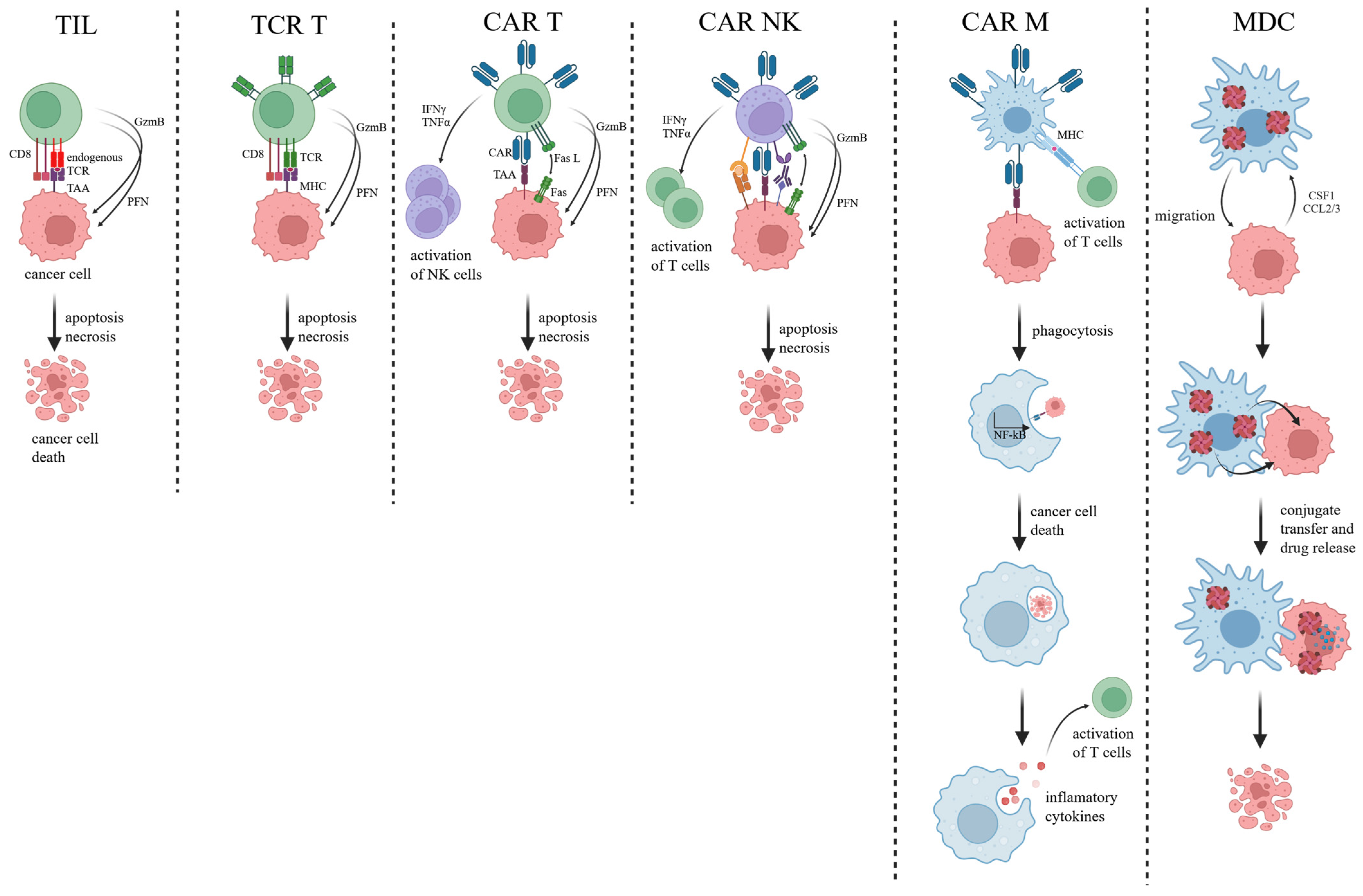
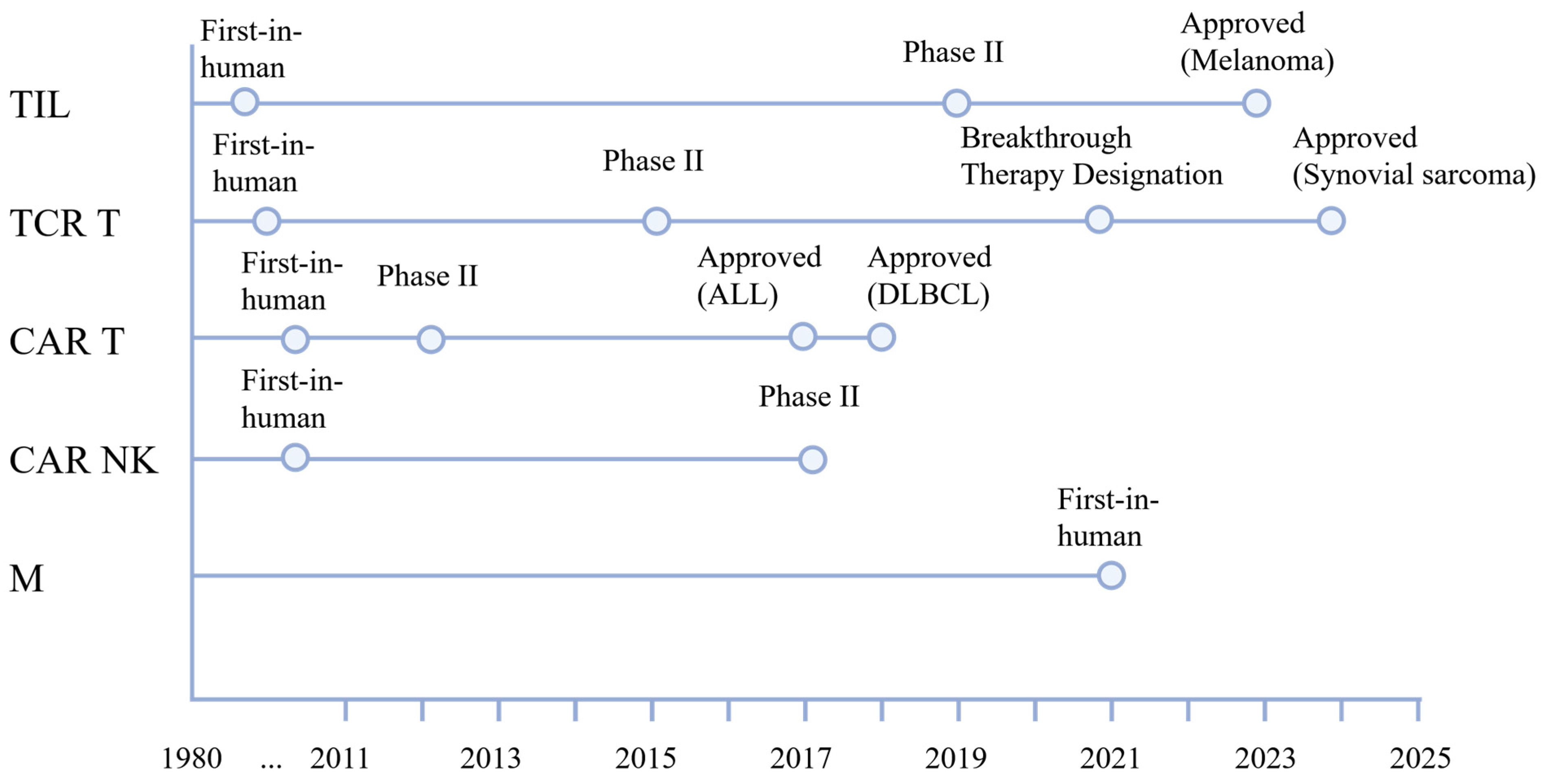
2.1. Tumor-Infiltrating Lymphocytes

Among the most promising strategies of novel therapeutic approaches in solid tumor treatment is using TILs—a component of the adaptive immune system—as a form of personalized immunotherapy [32]. TILs can be isolated directly from a patient’s tumor, expanded ex vivo to clinically significant numbers, and subsequently reinfused into the patient, typically following lymphodepleting chemotherapy [33]. This approach aims to potentiate the endogenous anti-tumor immune response and overcome immunosuppressive barriers, including dense stromal architecture, hypoxia, inhibitory cytokines (e.g., TGF-β, IL-10), and immune checkpoint expression (e.g., PD-L1, TIM-3, LAG-3) within the tumor microenvironment (TME).

2.1.1. Mechanism

The standard approach to TIL-based therapy begins with surgical resection of the tumor. TILs are isolated and cultured ex vivo in the presence of high concentrations of interleukin-2 (IL-2) to promote their activation and proliferation. After sufficient expansion, these TILs are infused back into the patient, typically following a lymphodepleting regimen to eliminate endogenous immune cells to enhance TIL engraftment, persistence, and functionality [33].
TILs primarily comprise T lymphocytes, especially CD8+ cytotoxic T lymphocytes (CTLs) and CD4+ helper T cells, although other immune subsets, including B cells and natural killer (NK) cells, may also be present [34,35]. CTLs play a central role in recognizing and destroying malignant cells through interactions with tumor-specific antigens presented by major histocompatibility complex (MHC) molecules. TILs are particularly valuable due to their intrinsic specificity for tumor-associated antigens, including neoantigens derived from tumor-specific mutations, enabling precise and potent immune targeting of cancer cells [36,37].
The TME, comprising malignant cells, stromal elements, immune infiltrates, and extracellular matrix components, presents numerous challenges to effective TIL function. The immunosuppressive nature of the TME is mediated by elements like regulatory T cells (Tregs), myeloid-derived suppressor cells (MDSCs), and immunomodulatory cytokines, like transforming growth factor-beta (TGF-β) and interleukin-10 (IL-10). It can significantly impair the effector functions of TILs [38,39,40]. Despite these barriers, the presence of TILs within tumors has been positively correlated with improved clinical outcomes in various malignancies, including melanoma, colorectal cancer, and ovarian cancer [41,42,43].

2.1.2. Clinical Data

Clinical trials, particularly in metastatic melanoma, have demonstrated that TIL therapy can induce durable clinical responses, with some patients achieving complete remission [44,45]. TILs can overcome the TME’s immunosuppressive barriers by reintroducing a large pool of tumor-reactive T cells with restored effector function and enhanced cytotoxicity. Upon reinfusion, TILs possess the capacity to penetrate immunologically “cold” tumors, resist local T cell dysfunction, and exert targeted cytolytic activity even in the presence of immunosuppressive mediators. Furthermore, the polyclonality of TIL populations allows for recognition of multiple tumor-associated neoantigens, reducing the likelihood of immune escape and broadening anti-tumor coverage. Clinical studies have shown that TIL therapy can effectively remodel the TME, shifting it toward a pro-inflammatory and immune-permissive state, thereby facilitating durable responses in checkpoint-refractory solid tumors [46,47,48]. An example of these abilities to overcome barriers is the FDA-approved product Lifileucel (AmtagviTM) for PD-1-refractory metastatic melanoma, which restores anti-tumor immunity through ex vivo-expanded autologous lymphocytes capable of mediating durable responses despite prior checkpoint inhibitor failure [49].
These promising outcomes have stimulated efforts to adapt TIL therapy to other solid tumor types. However, several challenges persist, including patient selection criteria, variability in TIL expansion success, and potential treatment-related toxicities [50,51].

2.1.3. Limitations

Despite their clinical promise, TILs face several limitations that constrain their broad applicability. First, TIL therapy requires extensive ex vivo expansion from surgically resected tumor tissue, a process that is both labor-intensive and not always feasible for patients with inaccessible or low-yield tumors [51]. Additionally, the composition and functionality of TILs can vary widely among patients, often influenced by the immunosuppressive TME, which may render them exhausted or antigen-non-specific [52,53]. Moreover, TILs are largely HLA-restricted, limiting their effectiveness across genetically diverse patient populations, and their persistence after infusion is often limited without adjunctive IL-2 support, which can itself cause severe toxicities [54].
To address these limitations, researchers are investigating next-generation strategies, such as genetic modification of TILs to enhance persistence and cytotoxicity, integration of an immune checkpoint blockade to mitigate TME-induced inhibition, and combinatorial regimens incorporating TILs with targeted therapies, vaccines, or cytokine support. These innovations aim to improve the efficacy and broaden the applicability of TIL-based immunotherapies [55,56].
TIL therapy represents a compelling and evolving approach to treating solid tumors. With their natural ability to recognize and eliminate cancer cells and ongoing technological and therapeutic advancements, TILs offer significant promise, particularly for patients with treatment-refractory malignancies.

2.2. T Cell Receptor-Engineered T Cells (TCR T)

T cell receptor-engineered T (TCR T) cells are genetically modified autologous or allogeneic T lymphocytes that express tumor-specific T cell receptors capable of recognizing intracellular antigens presented by MHC molecules. Their anti-tumor function is mediated through the specific recognition of tumor-associated antigens (TAAs) presented by MHC-I molecules on the surface of malignant cells, followed by targeted cytolysis [57]. This enables their application in tumors like melanoma, synovial sarcoma, and non-small cell lung cancer (NSCLC), where shared or patient-specific HLA-restricted tumor epitopes (e.g., NY-ESO-1, MAGE-A4, KRAS-G12D) are present [58,59,60].
Adaptive T cell therapy employing ex vivo expanded TRCs represents one of the earliest clinical applications of cell-based immunotherapy. Initial successes were observed in virus-associated malignancies, including Epstein–Barr virus (EBV)-positive lymphomas and nasopharyngeal carcinoma, where the immunodominance of viral antigens facilitates robust T cell targeting [61]. In these protocols, autologous CTLs are typically generated through in vitro stimulation of peripheral blood mononuclear cells (PBMCs) with antigen-loaded dendritic cells or irradiated tumor cells, followed by cytokine-supported expansion and subsequent reinfusion into the patient [61].

2.2.1. Mechanism

TCR T cells are generated by isolating peripheral blood T lymphocytes from a patient or donor and genetically modifying them to express a tumor-specific TCR with high affinity for antigens presented by MHC molecules on tumor cells. The TCR transgene is typically introduced using viral vectors, such as lentivirus or retrovirus. After transduction, the modified T cells are expanded ex vivo and infused back into the patient following a lymphodepleting conditioning regimen to enhance their persistence and function [62]. The engineering of T cells to express TCRs has enabled precise targeting and elimination of malignant cells [63].

2.2.2. Clinical Data

This approach has led to durable clinical responses in hematological malignancies and is increasingly being adapted for the treatment of solid tumors [63]. TCR T cells have shown promise in clinical trials, particularly in solid tumors where CAR T cell therapy has had limited success. Trials using NY-ESO-1-specific TCR T cells in synovial sarcoma and melanoma have demonstrated durable responses in some patients [64,65]. However, their effectiveness is influenced by factors like HLA restriction, tumor antigen heterogeneity, and immune evasion via MHC downregulation.

2.2.3. Limitations

Despite these advances, the therapeutic efficacy of TRCs, like other CTLs, in solid tumors remains constrained by multiple immunologic and tumor-intrinsic barriers, including antigenic heterogeneity, defective antigen presentation machinery, and the immunosuppressive TME, which collectively impair TCR T cells’ infiltration, survival, and function [66]. To circumvent these obstacles, recent strategies have focused on the genetic engineering of TRCs to augment their persistence and resistance to immunosuppressive signals, such as through dominant-negative TGF-β receptor expression or the incorporation of costimulatory signaling domains to enhance effector function and proliferation. CAR T cells are an example of another approach for T cell application in cancer treatment [67,68,69].

2.3. Chimeric Antigen Receptor T Cells (CAR T)

The effectiveness of chimeric antigen receptor CAR T cell therapy lies in the genetic modification of T cells to express CARs, enabling them to recognize and target specific antigens present on the surface of tumor cells, thereby activating T cells independently of the MHC [70,71]. This enhancement of T cell function presents new treatment avenues for patients with certain cancers.

2.3.1. Mechanism

CARs typically consist of three key components: an extracellular antigen-binding domain, a transmembrane domain, and an intracellular signaling domain [72,73]. The extracellular domain, responsible for antigen recognition, imparts specificity to CAR T cells, typically derived from single-chain variable fragments (scFvs) of antibodies that target TAAs or tumor-specific antigens (TSAs), such as HER2, mesothelin, EGFRvIII, GD2, and Claudin 18.2. The transmembrane domain anchors the CAR to the T cell membrane, ensuring its proper stability and signaling functionality [74]. The intracellular signaling domain transmits activation signals following antigen recognition and typically includes the CD3ζ domain, which can be combined with domains like CD28, 4-1BB (CD137), or OX40 (CD134) [75,76]. Including these co-stimulatory domains improves CAR T cell persistence, proliferation, and anti-tumor efficacy.
CAR T cells have evolved through several generations, with first-generation CARs incorporating only the CD3ζ domain, which has shown reduced in vivo efficacy [77]. Second-generation CARs incorporate a single co-stimulatory domain [78], while third-generation CARs contain two or more co-stimulatory domains [79]. Fourth-generation CARs, also known as TRUCKs (T cells Redirected for Universal Cytokine-mediated Killing), are designed to release cytokines upon antigen recognition, thus enhancing their tumor-killing potential [80,81].

2.3.2. Clinical Data

CAR T cell therapy has shown remarkable efficacy in treating hematologic malignancies, such as leukemia and lymphoma, where cancer cells circulate in the blood or the lymphatic system [80]. However, the treatment landscape becomes more complex when addressing solid tumors. Unlike hematologic cancers, solid tumors present unique challenges hindering CAR T cell therapy. These include immune evasion mechanisms, which allow tumors to avoid detection by the immune system, and a hostile TME that can inhibit CAR T cell function [14,68]. The TME includes dense extracellular matrices (ECM), non-cancerous stromal cells, and immune suppressive factors that obstruct CAR T cell infiltration and survival. Additionally, the abnormal vasculature often found in solid tumors limits the effective delivery of CAR T cells [14].
Although CAR T cell therapy has shown remarkable success in hematologic malignancies, several early-phase clinical trials have also demonstrated encouraging efficacy in solid tumors. For example, GD2-targeted CAR T cells have led to objective responses in patients with neuroblastoma and diffuse midline gliomas, including a case of radiographic remission in a child with pontine glioma [82]. Claudin 18.2 CAR T cells have shown promise in gastric and pancreatic cancers, with high response rates reported in a phase I study [83]. Furthermore, mesothelin-directed CAR T cells have induced disease stabilization in mesothelioma and pancreatic cancer when combined with regional or intrapleural delivery approaches [84]. These studies illustrate the feasibility and potential of CAR T therapy in solid tumors when combined with tumor-specific targeting, optimized delivery, and resistance mitigation strategies.

2.3.3. Limitations

One of the most significant hurdles in CAR T cell therapy for solid tumors is the identification of suitable target antigens that are both highly expressed on tumor cells and absent or minimally present in normal tissues [85,86,87]. Prominent TAAs under investigation include HER2, mesothelin, EGFRvIII, GD2, and Claudin 18.2, each associated with specific tumor types, such as breast, ovarian, glioblastoma, neuroblastoma, and gastric cancers, respectively [84,88,89,90,91]. Many tumor-associated proteins are also found in healthy tissues, raising the risk of off-target effects, including those tragically observed in early trials of HER2-CAR T cell related fatality due to cytokine-storm-like syndrome, accompanied by rapid respiratory failure and multi-organ dysfunction. Post-mortem analyses suggested that the CAR T cells had reacted not only with tumor cells but also with low-level HER2 expression in the pulmonary epithelium, leading to massive immune activation and widespread tissue damage [85]. Furthermore, solid tumors exhibit antigen heterogeneity, with varying levels or types of antigens across different tumor cells, making it challenging for CAR T cells to target and eliminate all malignant cells [92].
To address these challenges, various strategies have been explored. These include engineering CAR T cells to recognize and target multiple antigens [93], incorporating enzymes into CAR T cells that can degrade the ECM and facilitate better tumor infiltration [94], and identifying novel, highly specific antigens unique to cancer cells to improve targeting precision and reduce damage to healthy tissues [95]. Developing personalized CAR T cell therapies based on the unique antigen profile of a patient’s tumor is another promising strategy for overcoming antigen heterogeneity.
An emerging strategy to enhance the efficacy of CAR T cells in solid tumors involves co-targeting not only malignant cells but also the tumor stroma, particularly cancer-associated fibroblasts (CAFs), which play a central role in immune exclusion and therapeutic resistance. Huang et al. (2025) reported the development of a universal CAR T cell platform capable of redirecting cytotoxic activity against both tumor cells and CAFs through the use of bispecific adapter molecules [96]. Their design enabled the simultaneous engagement of tumor-associated antigens and fibroblast activation protein, a surface marker enriched on CAFs, resulting in synergistic tumor killing and stromal disruption in preclinical models of solid cancer. Importantly, the universal targeting system allowed for dynamic modulation of specificity and intensity via adapter dosing, offering a tunable approach to reduce off-tumor effects. This approach aligns with broader developments in modular CAR architectures, such as those described in the WO2024191887A2 patent, which detail adapter-mediated systems engineered to engage multiple components of the TME concurrently, thereby addressing spatial heterogeneity and immune evasion mechanisms inherent to solid tumors.
Notably, bispecific constructs targeting disialoganglioside (GD2) and prostate-specific membrane antigen (PSMA) have gained attention for their application in neuroectodermal and prostate malignancies, respectively. Preclinical models and early-phase clinical trials have shown that co-targeting GD2 and PSMA enhances T cell infiltration and cytotoxicity while potentially limiting immune escape mechanisms associated with monovalent CAR therapies. Ongoing trials, such as NCT05437315, continue to evaluate the safety, persistence, and anti-tumor activity of GD2/PSMA bispecific CAR T cells in solid tumors. These findings highlight the clinical feasibility of dual-targeting approaches and support the broader development of bispecific CAR platforms to overcome tumor antigen heterogeneity.
Ongoing investigations into CAR T cell therapy for solid tumors are being conducted across various cancers, including glioblastoma (NCT05577091, NCT04077866, NCT05353530), renal (NCT05420519, NCT04969354), prostate (NCT03873805), ovarian (NCT06305299, NCT05211557), and lung cancers (NCT06903117). Although early clinical trials have not yielded the same level of success as seen in hematologic malignancies [97,98], advances in CAR engineering and a deeper understanding of the challenges posed by the TME will drive progress in this field.

2.4. CAR Natural Killer Cells

CAR NK cells represent an emerging and innovative modality in the field of adoptive cell therapy designed to augment the innate cytotoxic potential of NK cells through tumor-specific redirection. In contrast to CAR T cells, which have demonstrated robust efficacy in hematologic malignancies, CAR NK cells offer several distinct immunological and safety advantages, including MHC-independent tumor recognition, a negligible risk of graft-versus-host disease (GvHD), and a lower incidence of cytokine release syndrome (CRS) and neurotoxicity [31,99].

2.4.1. Mechanism

CAR NK cells can be generated from various sources, including peripheral blood, umbilical cord blood, induced pluripotent stem cells (iPSCs), and established NK cell lines, such as NK-92. Each source offers distinct advantages regarding cytotoxic efficacy, expandability, and suitability for genetic modification [100]. For example, NK-92 cells are readily expandable and amenable to CAR transduction but require irradiation prior to infusion to prevent in vivo proliferation due to their malignant origin [101].
NK cells exert innate anti-tumor activity via a balance of activating and inhibitory receptor signaling, culminating in the direct lysis of transformed cells through perforin- and granzyme-mediated apoptosis. Additionally, NK cells mediate antibody-dependent cellular cytotoxicity (ADCC) through the expression of the FcγRIIIa receptor (CD16), further expanding their cytotoxic repertoire [102]. Incorporating CAR constructs into NK cells significantly enhances their tumor specificity and cytolytic potency, allowing antigen-directed killing via synthetic antibody-like scFv domains while preserving their endogenous effector mechanisms [31].

2.4.2. Clinical Data

CAR-NK cells’ clinical feasibility and therapeutic potential were first demonstrated in a landmark phase I/II clinical trial involving patients with relapsed or refractory CD19-positive lymphoid malignancies. In that study, umbilical-cord-blood-derived CAR NK cells, co-expressing interleukin-15 (IL-15) and an inducible caspase-9 safety switch, yielded objective responses in 8 out of 11 patients (73%), with no incidence of CRS, neurotoxicity, or GvHD, highlighting the favorable safety profile of this therapeutic platform [31].
Still, the application of CAR NK cells in solid tumors has been met with challenges analogous to those encountered in CAR T cell therapy. These include antigenic heterogeneity, physical and stromal barriers to infiltration, and immunosuppressive elements within the TME. Nonetheless, preclinical models have demonstrated encouraging anti-tumor activity of CAR NK cells targeting HER2, mesothelin, and epidermal growth factor receptor (EGFR) in malignancies like glioblastoma, breast cancer, and ovarian cancer [103,104,105,106].

2.4.3. Limitations

To enhance efficacy in the context of solid tumors, current efforts are focused on the development of “armored” CAR NK cells equipped with transgenes encoding pro-survival cytokines (e.g., IL-15), dominant-negative TGF-β receptors, and chemokine receptors to improve trafficking and persistence in the TME [107]. Moreover, generating “off-the-shelf” CAR-NK cell products from allogeneic sources, such as cord blood or iPSCs, presents a scalable and potentially more accessible therapeutic alternative to autologous CAR T cell therapy [108].

2.5. Macrophages

In response to challenges in applying CAR T cells in solid tumors [109,110,111], macrophages have emerged as a promising alternative effector cell type for CAR engineering owing to their intrinsic tumor-homing capacity and versatile immunomodulatory functions [112,113].

2.5.1. Mechanism

Recent advances have enabled the genetic modification of human macrophages to express CARs, thereby redirecting their phagocytic activity toward tumor cells [114]. Notably, using a chimeric adenoviral vector has facilitated efficient gene delivery by circumventing innate immune barriers typically encountered in macrophage transduction. This approach has successfully reprogrammed macrophages to adopt a sustained pro-inflammatory (M1-like) phenotype, enhancing their anti-tumor potential [114].

2.5.2. Clinical Data

In vitro studies have demonstrated that CAR macrophages (CAR M) exhibit antigen-specific phagocytosis and cytotoxicity against tumor cells [26]. Moreover, CAR M therapy promotes a shift in the macrophage phenotype from immunosuppressive M2 to pro-inflammatory M1, elevates pro-inflammatory cytokine and chemokine production, enhances antigen presentation, and recruits T cells into the tumor microenvironment. These effects collectively contribute to remodeling of the tumor milieu and potentiate adaptive anti-tumor immunity. In in vivo studies using humanized mouse models, CAR Ms have been shown to induce robust anti-tumor responses and promote T-lymphocyte infiltration and activity, further supporting their therapeutic potential in solid tumors [115].
Currently, several biotechnology companies are advancing CAR M platforms with distinct technological strategies:
SIRPαnt Immunotherapeutics has developed SIRPant-MTM (SI-101), an autologous, non-genetically engineered macrophage therapy utilizing the PhagoActTM platform. This method activates and educates patient-derived macrophages ex vivo to recognize and eliminate cancer cells through intrinsic immune mechanisms. SIRPant-MTM engages both cellular and humoral immune responses and promotes long-term anti-tumor immunity. The therapy is presently in preclinical development in relapsed or refractory non-Hodgkin lymphoma (ClinicalTrials.gov ID NCT05967416). SIRPant Immunotherapeutics announced in late 2023 that it had received FDA clearance to begin a phase 1 clinical trial of SIRPant-MTM for the treatment of various solid tumors, such as head and neck cancer, non-melanoma skin cancers, bladder and kidney cancers, low-grade prostate cancer, triple-negative breast cancer, and certain sarcomas. Under the newly cleared Investigational New Drug, the company plans to initiate clinical investigation of SIRPant-M as a monotherapy and in combination with other immuno-stimulatory modalities, such as radiotherapy and immune checkpoint inhibitors, for the treatment of select solid tumor indications.
Carisma Therapeutics is developing CT-0508, a genetically engineered CAR M product targeting HER2-positive solid tumors (ClinicalTrials.gov ID NCT04660929). Preclinical data suggest that CT-0508 can effectively infiltrate tumors, kill malignant cells, reprogram the tumor microenvironment, and facilitate the recruitment of adaptive immune cells. The study demonstrated a favorable safety profile with no dose-limiting toxicities. Approximately 44% of patients with HER2 3+ tumors achieved stable disease as the best overall response. However, the development of this platform has paused as the company shifts its focus to other technologies.
Cellis has introduced a novel platform called Macrophage Drug Conjugates (MDCs). This approach leverages macrophages loaded with ferritin-based drug complexes for targeted delivery into tumor cells [116]. Central to this platform is the TRAIN (Targeted Reprogramming of Antigen-presenting Immune Networks) mechanism, which enables precise cytoplasmic delivery of therapeutic payloads, inducing tumor cell death and immunogenic modulation of the microenvironment. MDCs have demonstrated enhanced survival, reduced tumor burden, and decreased metastases in preclinical models of solid tumors. The company is preparing to initiate first-in-human phase 1 clinical trials for its lead candidate, MDC-735, in the second quarter of 2025. These trials are planned to take place in Switzerland, Germany, and Poland, targeting multiple solid tumors, including glioblastoma, ovarian, bladder, lung, and head and neck cancers. The technology is on track to enter clinical trials in Q2 2025.
Collectively, these innovations underscore the potential of CAR macrophage therapies as next-generation cell-based immunotherapies, particularly in the context of solid tumors, where conventional CAR T strategies have faced limitations.

3. Current Clinical Trials of Cell-Based Therapies Against Solid Tumors

Clinical trials investigating cell-based therapies in solid tumors are essential for advancing the field of cancer immunotherapy beyond hematologic malignancies. These trials provide critical insights into the safety, feasibility, and preliminary efficacy of novel therapeutic modalities, such as TILs, TCR T, CAR T cells, CAR NK cells, and engineered macrophages, in the context of the immunosuppressive TME. As solid tumors account for the vast majority of cancer-related morbidity and mortality, the successful translation of cell-based approaches could significantly improve clinical outcomes. Clinical trials using cell-based therapies in solid tumor therapy conducted to date are summarized in Table 1.

4. Advances and Challenges in Cell-Based Therapy for Solid Tumors

Cell-based immunotherapies represent a promising approach for the treatment of various malignancies. Among these, CAR T therapy has shown potential efficacy against multiple cancer types, including pancreatic, colorectal, liver, lung, and gastric cancers. Despite this progress, significant challenges remain in the application of CAR T therapies to solid tumors. One major obstacle is the limited trafficking and infiltration of immune cells into solid tumor tissues. The TME exhibits strong immunosuppressive characteristics, severely impairing T cells’ anti-tumor activity [109].
Aberrant tumor vasculature further impedes T cell trafficking. Dysregulated angiogenesis, characterized by an imbalance between pro- and anti-angiogenic factors, forms large, unbranched vessels that fail to adequately perfuse the tumor tissue [117]. This poor perfusion results in hypoxia, increased levels of reactive oxygen species, and the activation of hypoxia-inducible factors (HIF1A and HIF2A), which promote the expression of immunosuppressive cytokines and growth factors [118].
Recent strategies to enhance T cell infiltration include tumor necrosis factor superfamily member 14 (LIGHT) expression on T cells. LIGHT expression induces the formation of tertiary lymphoid structures (TLSs) within tumors, significantly boosting local anti-tumor immune responses [119]. TLSs, which represent organized lymphoid aggregates within non-lymphoid tissues, facilitate enhanced immune surveillance and activation [120].
Moreover, when combined with vascular targeting peptides (VTPs), LIGHT expression contributes to vascular normalization within tumors. Studies in murine Lewis lung carcinoma and melanoma models have demonstrated that LIGHT-VTP treatment reduced hypoxia and improved tumor perfusion [121]. Similar findings have been reported in models of primary brain cancer [122], highlighting the potential of this approach to improve immune cell infiltration into solid tumors.
Another major obstacle is the role of immune checkpoints in the TME, which protect healthy tissues from immune-mediated damage but simultaneously limit anti-tumor responses [123]. Specifically, programmed cell death protein 1 (PD-1) expressed on activated T cells binds to its ligand PD-L1, which is often overexpressed by tumor-associated macrophages (TAMs) and other cells in the TME [124].
To counteract PD-1/PD-L1-mediated immunosuppression, small-molecule inhibitors, such as styryl carbamates, have been explored. These compounds, characterized by chemical stability and cellular permeability, can disrupt PD-1/PD-L1 interactions. In vitro co-cultures of cancer cells and T cells treated with styryl carbamates exhibited decreased cancer cell viability without significantly affecting immune cell survival. Moreover, styryl carbamates reduced PD-L1 expression and the formation of the PD-1/PD-L1 complex [125].
Additionally, CAR T cells engineered to secrete PD-1-blocking scFvs have demonstrated enhanced anti-tumor efficacy. In murine models of human lymphoma, treatment with such engineered CAR T cells resulted in increased tumor cell lysis and a reduction in PD-1 expression on T cell surfaces compared to control groups [126].
IL-15 co-expression in CAR-engineered immune cells has emerged as a strategy to enhance in vivo persistence, metabolic fitness, and anti-tumor function, particularly in NK and T cell platforms. Several preclinical and early-phase clinical studies have demonstrated that IL-15 promotes homeostatic proliferation and supports long-term survival of CAR T and CAR NK cells, translating into improved tumor control in solid and hematologic malignancies [31,127]. However, the inclusion of IL-15 is not without controversy. Elevated systemic levels of IL-15—especially when produced in an unregulated or constitutive manner—have been associated with increased risks of cytokine-mediated toxicities, including lymphoproliferation, neuroinflammation, and symptoms resembling CRS [128]. Furthermore, the effects of IL-15 appear to be highly context dependent, varying with the immune cell type, tumor microenvironment, route of administration, and vector design. Some studies report enhanced persistence without toxicity when IL-15 is membrane-bound or regulated by inducible systems, while others note adverse inflammatory consequences with secreted IL-15 constructs [129]. Thus, while IL-15 co-expression holds considerable therapeutic promise, its incorporation into CAR platforms requires precise control mechanisms and rigorous safety evaluation to balance efficacy with tolerability.
Together, these strategies provide promising avenues to overcome the hostile TME and enhance the therapeutic efficacy of CAR T cell therapies in the treatment of solid tumors.

5. The Implications of Autologous Versus Allogeneic Therapeutic Approaches in the Context of Cellular Immunotherapy for Solid Tumors

The selection of autologous versus allogeneic cellular sources constitutes a pivotal consideration in the development of adoptive immunotherapies for solid tumors. These two paradigms exhibit distinct immunological, logistical, and translational characteristics, each profoundly influencing therapeutic efficacy, manufacturing scalability, clinical feasibility, and patient accessibility.
Autologous cellular immunotherapy involves the procurement and ex vivo expansion or genetic modification of immune effector cells derived from the patient, followed by reinfusion of the modified product into the same individual. This approach underpins several advanced therapeutic strategies, including tumor-infiltrating lymphocyte (TIL) therapy and autologous CAR T cell therapy. A primary advantage of autologous approaches is their inherent immunological compatibility, thereby mitigating the risk of GvHD and recipient immune rejection. Moreover, the tumor specificity afforded by TILs—selected directly from the tumor microenvironment—permits the targeting of patient-specific neoantigens, offering a high degree of personalization particularly relevant for genetically complex solid tumors [130]. Nevertheless, autologous cell therapies are encumbered by substantial manufacturing constraints, including individualized processing workflows, prolonged production timelines, and variable product quality, which may be adversely affected by the patient’s prior treatments or immunocompromised status. These limitations pose significant barriers to scalability and timely clinical implementation.
Conversely, allogeneic cell therapies utilize immune cells sourced from healthy donors, which are expanded and genetically engineered for therapeutic administration to unrelated recipients [131,132]. This approach offers several practical advantages, chief among them the feasibility of developing standardized, “off-the-shelf” cell products that can be cryopreserved, stored, and deployed rapidly upon clinical need [133]. Allogeneic therapies significantly reduce per-patient manufacturing costs and facilitate batch processing, thus enabling broader dissemination and accessibility. In addition, donor-derived cells can be pre-selected for optimal functionality, cytotoxic potential, and fitness. However, the immunogenicity of allogeneic products presents a major obstacle. Unmodified donor T cells can induce GvHD, while host immune rejection may limit the persistence and efficacy of the therapeutic cells. To mitigate these risks, advanced gene-editing strategies, such as T cell receptor disruption, β2-microglobulin knockout, and HLA class I/II silencing, have been employed to reduce immunological incompatibility and extend in vivo durability [134]. Recent advancements in allogeneic NK cell therapy have been significantly driven by the development of robust ex vivo expansion platforms, particularly those employing genetically engineered feeder cell systems. The use of irradiated K562 cells modified to express membrane-bound interleukin-15 (mbIL-15) and 4-1BB ligand has markedly enhanced NK cell proliferation, survival, and cytotoxicity against tumor targets [135]. These feeder-based protocols enable the generation of clinical-scale NK cell populations with high purity, potent activation markers, and reduced exhaustion phenotypes, forming the backbone of several current good manufacturing practice-compliant protocols [31]. More recently, feeder cells have been further optimized to express mbIL-21, which supports greater NK cell expansion and maintains a less differentiated phenotype compared to IL-15-driven systems [136,137]. These innovations have laid the groundwork for scalable, off-the-shelf NK cell products that are being tested in early-phase clinical trials for both hematological and solid malignancies.
The application of autologous and allogeneic modalities to solid tumors is further complicated by tumor-intrinsic features impairing immune cell function and trafficking. These include pronounced antigenic heterogeneity, an immunosuppressive TME characterized by regulatory immune subsets and inhibitory cytokines, and a dense stromal architecture constituting a formidable physical barrier to cell infiltration [66]. While autologous therapies may offer superior tumor specificity and functional adaptability due to their endogenous origin, allogeneic approaches benefit from logistical flexibility and the potential for large-scale manufacturing. Notably, the development of allogeneic CAR NK cells and iPSC-derived immune effectors offers a promising avenue to bridge the gap between efficacy and accessibility by combining low immunogenicity with uniformity in therapeutic product composition. Allogeneic CAR NK cells have shown encouraging results in first-in-human trials, including high response rates (e.g., 73%) and minimal toxicity in hematologic malignancies [31]. Similarly, iPSC-derived NK cells have demonstrated consistent phenotype, potent cytotoxicity, and enhanced scalability in preclinical and early-phase clinical studies [138].
In conclusion, both autologous and allogeneic cell-based therapies possess unique and complementary strengths, and the optimal platform may vary depending on clinical context, tumor biology, therapeutic urgency, and infrastructure capabilities. The integration of next-generation technologies, including CRISPR-mediated gene editing, synthetic biology platforms, and combination immunotherapeutic regimens, will be essential to surmount existing barriers and maximize the therapeutic potential of cellular immunotherapy in solid tumors [139,140].

6. Conclusions

Cell-based immunotherapies have emerged as a rapidly advancing class of therapeutic strategies that could redefine the treatment paradigm for solid tumors. These approaches—including tumor-infiltrating lymphocytes, T cell receptor-engineered T cells, chimeric antigen receptor-engineered T cells, natural killer cells, and modified macrophages—offer the capacity for antigen-specific tumor targeting, immunological memory, and durable clinical responses. However, the translation of these therapies from hematologic malignancies to solid tumors remains impeded by a multitude of tumor-intrinsic and extrinsic barriers.
Key limitations include the heterogeneity of tumor antigen expression, the suppressive architecture of the tumor microenvironment, limited trafficking and persistence of therapeutic cells, and the potential for off-tumor toxicity. Furthermore, patient-derived autologous therapies’ logistical and manufacturing complexities constrain their scalability and timely clinical deployment. In contrast, allogeneic strategies offer the potential for standardized, “off-the-shelf” products with broader applicability but require sophisticated genetic engineering to mitigate immunogenicity and preserve therapeutic efficacy.
Advancing the clinical utility of cellular immunotherapy for solid tumors will depend on the continued integration of synthetic biology, gene-editing technologies, and rationally designed combinatorial regimens. These innovations, coupled with rigorous preclinical modeling and robust clinical trial design, will be essential to overcoming current limitations and establishing the next generation of effective, scalable, and safe immunotherapies for solid malignancies.

Author Contributions

Conceptualization, A.S., Z.K. and M.N.; methodology, J.S., J.T. and M.W.; investigation, R.O. and A.S.; writing—original draft preparation, Z.K., M.N., J.S., J.T., M.W., R.O. and A.S.; writing—review and editing, A.S.; supervision, A.S.; funding acquisition, A.S. All authors have read and agreed to the published version of the manuscript.

Funding

The publication was financed by the Science Development Fund of the Warsaw University of Life Sciences—SGGW.

Acknowledgments

The illustrations in this publication were created with https://BioRender.com, accessed on 10 April 2025.

Conflicts of Interest

The authors declare no conflict of interest.

Abbreviations

The following abbreviations are used in this manuscript:
ACTAdoptive Cell Therapy
ADCAntibody-Dependent Cellular Cytotoxicity
ALLAcute Lymphoblastic Leukemia
CAFsCancer-Associated Fibroblasts
CARChimeric Antigen Receptor
CAR MCAR Macrophages
CAR NKCAR NK Cells
CAR TCAR T Cells
CEACarcinoembryonic Antigen
CRSCytokine Release Syndrome
CTLsCytotoxic T Lymphocytes
CSCCancer Stem-Like Cells
DLBCLDiffuse Large B Cell Lymphoma
ECMExtracellular Matrix
EGFREpidermal Growth Factor Receptor
GD2Disialoganglioside
GvHDGraft-Versus-Host Disease
HER2Human Epidermal Growth Factor Receptor 2
HLAHuman Leukocyte Antigen
HPV16 E7Human Papillomavirus 16 Protein E7
iPSCInduced Pluripotent Stem Cell
IL2Interleukin-2
IL10Interleukin-10
IL13Rα2Interleukin-13 Receptor Alpha 2
IL15Interleukin-15
MAGE-A4Melanoma Antigen Gene A4
MAGE-C2Melanoma-Associated Gene C2
MART-1Melanoma Antigen Recognized by T Cells 1
mbIL-15Membrane-Bound Interleukin-15
MDCMacrophage–Drug Conjugate
MDSCMyeloid-Derived Suppressor Cell
MHCMajor Histocompatibility Complex
MUC1Mucin 1
NKNatural Killer
NSCCNon-Stem Cancer Cells
NY-ESO-1New York Esophageal Squamous Cell Carcinoma 1
PBMCPeripheral Blood Mononuclear Cell
PSMAProstate-Specific Membrane Antigen
ROR1Receptor Tyrosine Kinase Like Orphan Receptor 1
scFvSingle-Chain Variable Fragment
TAATumor-Associated Antigen
TCRT Cell Receptor
TCR TT Cell Receptor-Engineered T Cells
TGFβTransforming Growth Factor Beta
TILsTumor Infiltrating Lymphocytes
TMETumor Microenvironment
TregRegulatory T Cell
TROP2Trophoblast Cell Surface Antigen 2
TRUCKsT Cells Redirected For Universal Cytokine-Mediated Killing
WHOWorld Health Organization

References

  1. Bray, F.; Laversanne, M.; Sung, H.; Ferlay, J.; Siegel, R.L.; Soerjomataram, I.; Jemal, A. Global Cancer Statistics 2022: GLOBOCAN Estimates of Incidence and Mortality Worldwide for 36 Cancers in 185 Countries. CA Cancer J. Clin. 2024, 74, 229–263. [Google Scholar] [CrossRef]
  2. Wu, Y.; Song, Y.; Wang, R.; Wang, T. Molecular Mechanisms of Tumor Resistance to Radiotherapy. Mol. Cancer 2023, 22, 96. [Google Scholar] [CrossRef]
  3. Rebucci, M.; Michiels, C. Molecular Aspects of Cancer Cell Resistance to Chemotherapy. Biochem. Pharmacol. 2013, 85, 1219–1226. [Google Scholar] [CrossRef]
  4. Mai, Y.; Su, J.; Yang, C.; Xia, C.; Fu, L. The Strategies to Cure Cancer Patients by Eradicating Cancer Stem-like Cells. Mol. Cancer 2023, 22, 171. [Google Scholar] [CrossRef] [PubMed]
  5. Shen, S.; Xu, X.; Lin, S.; Zhang, Y.; Liu, H.; Zhang, C.; Mo, R. A Nanotherapeutic Strategy to Overcome Chemotherapeutic Resistance of Cancer Stem-like Cells. Nat. Nanotechnol. 2021, 16, 104–113. [Google Scholar] [CrossRef] [PubMed]
  6. Saha, T.; Lukong, K.E. Breast Cancer Stem-Like Cells in Drug Resistance: A Review of Mechanisms and Novel Therapeutic Strategies to Overcome Drug Resistance. Front. Oncol. 2022, 12, 856974. [Google Scholar] [CrossRef] [PubMed]
  7. Comoli, P.; Chabannon, C.; Koehl, U.; Lanza, F.; Urbano-Ispizua, A.; Hudecek, M.; Ruggeri, A.; Secondino, S.; Bonini, C.; Pedrazzoli, P. Development of Adaptive Immune Effector Therapies in Solid Tumors. Ann. Oncol. 2019, 30, 1740–1750. [Google Scholar] [CrossRef]
  8. Bastien, J.P.; Minguy, A.; Dave, V.; Roy, D.C. Cellular Therapy Approaches Harnessing the Power of the Immune System for Personalized Cancer Treatment. Semin. Immunol. 2019, 42, 101306. [Google Scholar] [CrossRef]
  9. Grimes, J.M.; Carvajal, R.D.; Muranski, P. Cellular Therapy for the Treatment of Solid Tumors. Transfus. Apher. Sci. 2021, 60, 103056. [Google Scholar] [CrossRef]
  10. El-Kadiry, A.E.H.; Rafei, M.; Shammaa, R. Cell Therapy: Types, Regulation, and Clinical Benefits. Front. Med. 2021, 8, 756029. [Google Scholar] [CrossRef]
  11. Mundhara, N.; Sadhukhan, P. Cracking the Codes behind Cancer Cells’ Immune Evasion. Int. J. Mol. Sci. 2024, 25, 8899. [Google Scholar] [CrossRef] [PubMed]
  12. Gonzalez, H.; Hagerling, C.; Werb, Z. Roles of the Immune System in Cancer: From Tumor Initiation to Metastatic Progression. Genes Dev. 2018, 32, 1267–1284. [Google Scholar] [CrossRef] [PubMed]
  13. Ricci, J.E. Tumor-Induced Metabolic Immunosuppression: Mechanisms and Therapeutic Targets. Cell Rep. 2025, 44, 115206. [Google Scholar] [CrossRef]
  14. Xia, X.; Yang, Z.; Lu, Q.; Liu, Z.; Wang, L.; Du, J.; Li, Y.; Yang, D.H.; Wu, S. Reshaping the Tumor Immune Microenvironment to Improve CAR-T Cell-Based Cancer Immunotherapy. Mol. Cancer 2024, 23, 175. [Google Scholar] [CrossRef]
  15. Tie, Y.; Tang, F.; Wei, Y.-Q.; Wei, X.-W. Immunosuppressive Cells in Cancer: Mechanisms and Potential Therapeutic Targets. J. Hematol. Oncol. 2022, 15, 61. [Google Scholar] [CrossRef] [PubMed]
  16. Pogge von Strandmann, E.; Reinartz, S.; Wager, U.; Müller, R. Tumor–Host Cell Interactions in Ovarian Cancer: Pathways to Therapy Failure. Trends Cancer 2017, 3, 137–148. [Google Scholar] [CrossRef]
  17. Mandalà, M.; De Logu, F.; Merelli, B.; Nassini, R.; Massi, D. Immunomodulating Property of MAPK Inhibitors: From Translational Knowledge to Clinical Implementation. Lab. Investig. 2017, 97, 166–175. [Google Scholar] [CrossRef]
  18. Badoual, C.; Sandoval, F.; Pere, H.; Hans, S.; Gey, A.; Merillon, N.; Van Ryswick, C.; Quintin-Colonna, F.; Bruneval, P.; Brasnu, D.; et al. Better Understanding Tumor–Host Interaction in Head and Neck Cancer to Improve the Design and Development of Immunotherapeutic Strategies. Head Neck. 2010, 32, 946–958. [Google Scholar] [CrossRef] [PubMed]
  19. Mellman, I.; Chen, D.S.; Powles, T.; Turley, S.J. The Cancer-Immunity Cycle: Indication, Genotype, and Immunotype. Immunity 2023, 56, 2188–2205. [Google Scholar] [CrossRef]
  20. Binnewies, M.; Roberts, E.W.; Kersten, K.; Chan, V.; Fearon, F.; Merad, M.; Coussens, L.M.; Gabrilovich, D.I.; Ostrand-Rosenberg, S.; Hedrick, C.C.; et al. Understanding the Tumor Immune Microenvironment (TIME) for Effective Therapy. Nat. Med. 2018, 24, 541–550. [Google Scholar] [CrossRef]
  21. Mount, N.M.; Ward, S.J.; Kefalas, P.; Hyllner, J. Cell-Based Therapy Technology Classifications and Translational Challenges. Philos. Trans. R. Soc. B Biol. Sci. 2015, 370, 20150017. [Google Scholar] [CrossRef] [PubMed]
  22. Dodson, B.P.; Levine, A.D. Challenges in the Translation and Commercialization of Cell Therapies. BMC Biotechnol. 2015, 15, 70. [Google Scholar] [CrossRef]
  23. Sun, L.; Su, Y.; Jiao, A.; Wang, X.; Zhang, B. T Cells in Health and Disease. Signal Transduct. Target. Ther. 2023, 8, 235. [Google Scholar] [CrossRef]
  24. Deo, A.S.; Shrijana; Sruthika, S.U.; Karun, S.; Bisaria, K.; Sarkar, K. Participation of T Cells in Generating Immune Protection against Cancers. Pathol. Res. Pract. 2024, 262, 155534. [Google Scholar] [CrossRef]
  25. Daher, M.; Melo Garcia, L.; Li, Y.; Rezvani, K. CAR-NK Cells: The next Wave of Cellular Therapy for Cancer. Clin. Transl. Immunol. 2021, 10, e1274. [Google Scholar] [CrossRef] [PubMed]
  26. Liu, M.; Liu, J.; Liang, Z.; Dai, K.; Gan, J.; Wang, Q.; Xu, Y.; Chen, Y.H.; Wan, X. CAR-Macrophages and CAR-T Cells Synergistically Kill Tumor Cells In Vitro. Cells 2022, 11, 3692. [Google Scholar] [CrossRef] [PubMed]
  27. Rosenberg, S.A.; Packard, B.S.; Aebersold, P.M.; Solomon, D.; Topalian, S.; Toy, S.; Simon, P.; Lotze, M.T.; Yang, J.C.; Seipp, C.; et al. Use of Tumor-Infiltrating Lumphocytes and Interleukin-2 in the Immunotherapy of Patients with Metastatic Melanoma. N. Engl. J. Med. 1988, 319, 1676–1680. [Google Scholar] [CrossRef]
  28. Morgan, R.A.; Dudley, M.E.; Wunderlich, J.R.; Hughes, M.S.; Yang, J.C.; Sherry, R.M.; Royal, R.E.; Topalían, S.L.; Kammula, U.S.; Restifo, N.P.; et al. Cancer Regression in Patients after Transfer of Genetically Engineered Lymphocytes. Science 2006, 314, 126–129. [Google Scholar] [CrossRef]
  29. Porter, D.L.; Levine, B.L.; Kalos, M.; Bagg, A.; June, C.H. Chimeric Antigen Receptor–Modified T Cells in Chronic Lymphoid Leukemia. N. Engl. J. Med. 2011, 365, 725–733. [Google Scholar] [CrossRef]
  30. Maude, S.L.; Frey, N.; Shaw, P.; Aplenc, R.; Barrett, D.M.; Bunin, N.J.; Chew, A.; Gonzalez, V.E.; Zheng, Z.; Lacey, S.F.; et al. Chimeric Antigen Receptor T Cells for Sustained Remissions in Leukemia. N. Engl. J. Med. 2014, 371, 1507–1517. [Google Scholar] [CrossRef]
  31. Liu, E.; Marin, D.; Banerjee, P.; Macapinlac, H.A.; Thompson, P.; Basar, R.; Nassif Kerbauy, L.; Overman, B.; Thall, P.; Kaplan, M.; et al. Use of CAR-Transduced Natural Killer Cells in CD19-Positive Lymphoid Tumors. N. Engl. J. Med. 2020, 382, 545–553. [Google Scholar] [CrossRef] [PubMed]
  32. Paijens, S.T.; Vledder, A.; de Bruyn, M.; Nijman, H.W. Tumor-Infiltrating Lymphocytes in the Immunotherapy Era. Cell Mol. Immunol. 2021, 18, 842–859. [Google Scholar] [CrossRef] [PubMed]
  33. Nguyen, L.T.; Saibil, S.D.; Sotov, V.; Le, M.X.; Khoja, L.; Ghazarian, D.; Bonilla, L.; Majeed, H.; Hogg, D.; Joshua, A.M.; et al. Phase II Clinical Trial of Adoptive Cell Therapy for Patients with Metastatic Melanoma with Autologous Tumor-Infiltrating Lymphocytes and Low-Dose Interleukin-2. Cancer Immunol. Immunother. 2019, 68, 773–785. [Google Scholar] [CrossRef]
  34. Han, E.; Choi, H.Y.; Kwon, H.J.; Chung, Y.R.; Shin, H.C.; Kim, E.K.; Suh, K.J.; Kim, S.H.; Kim, J.H.; Park, S.Y. Characterization of Tumor-Infiltrating Lymphocytes and Their Spatial Distribution in Triple-Negative Breast Cancer. Breast Cancer Res. 2024, 26, 180. [Google Scholar] [CrossRef]
  35. Liu, D.; Heij, L.R.; Czigany, Z.; Dahl, E.; Lang, S.A.; Ulmer, T.F.; Luedde, T.; Neumann, U.P.; Bednarsch, J. The Role of Tumor-Infiltrating Lymphocytes in Cholangiocarcinoma. J. Exp. Clin. Cancer Res. 2022, 41, 127. [Google Scholar] [CrossRef]
  36. Zhao, X.; Pan, X.; Wang, Y.; Zhang, Y. Targeting Neoantigens for Cancer Immunotherapy. Biomark. Res. 2021, 9, 61. [Google Scholar] [CrossRef]
  37. Leko, V.; Rosenberg, S.A. Identifying and Targeting Human Tumor Antigens for T Cell-Based Immunotherapy of Solid Tumors. Cancer Cell 2020, 38, 454–472. [Google Scholar] [CrossRef] [PubMed]
  38. Krishnamoorthy, M.; Gerhardt, L.; Vareki, S.M. Immunosuppressive Effects of Myeloid-derived Suppressor Cells in Cancer and Immunotherapy. Cells 2021, 10, 1170. [Google Scholar] [CrossRef]
  39. Qin, D.; Zhang, Y.; Shu, P.; Lei, Y.; Li, X.; Wang, Y. Targeting Tumor-Infiltrating Tregs for Improved Antitumor Responses. Front. Immunol. 2024, 15, 1325946. [Google Scholar] [CrossRef]
  40. Hofer, F.; Sario, G.D.; Musiu, C.; Sartoris, S.; Sanctis, F.D.; Ugel, S. A Complex Metabolic Network Confers Immunosuppressive Functions to Myeloid-Derived Suppressor Cells (MDSCs) within the Tumour Microenvironment. Cells 2021, 10, 2700. [Google Scholar] [CrossRef]
  41. Amaria, R.; Knisely, A.; Vining, D.; Kopetz, S.; Overman, M.J.; Javle, M.; Antonoff, M.B.; Tzeng, C.W.D.; Wolff, R.A.; Pant, S.; et al. Efficacy and Safety of Autologous Tumor-Infiltrating Lymphocytes in Recurrent or Refractory Ovarian Cancer, Colorectal Cancer, and Pancreatic Ductal Adenocarcinoma. J. Immunother. Cancer 2024, 12, e006822. [Google Scholar] [CrossRef] [PubMed]
  42. Tarabay, J.; Ren, D.; Ifegu, I.; Zada, S.; Edwards, R.; Chan, J.; Kuan, E.C.; Wang, B. Prognostic Significance of Tumor-Infiltrating Lymphocytes and Anti-Programmed Death-Ligand 1 Therapy in Sinonasal Mucosal Melanoma: A 10-Year Experience at a Single Institution. J. Clin. Transl. Pathol. 2023, 3, 115–119. [Google Scholar] [CrossRef]
  43. Brummel, K.; Eerkens, A.L.; de Bruyn, M.; Nijman, H.W. Tumour-Infiltrating Lymphocytes: From Prognosis to Treatment Selection. Br. J. Cancer 2023, 128, 451–458. [Google Scholar] [CrossRef] [PubMed]
  44. Rosenberg, S.A.; Yang, J.C.; Sherry, R.M.; Kammula, U.S.; Hughes, M.S.; Phan, G.Q.; Citrin, D.E.; Restifo, N.P.; Robbins, P.F.; Wunderlich, J.R.; et al. Durable Complete Responses in Heavily Pretreated Patients with Metastatic Melanoma Using T-Cell Transfer Immunotherapy. Clin. Cancer Res. 2011, 17, 4550–4557. [Google Scholar] [CrossRef]
  45. Haanen, J.; Los, C.; Phan, G.Q.; Betof Warner, A. Adoptive Cell Therapy for Solid Tumors: Current Status in Melanoma and Next-Generation Therapies. Am. Soc. Clin. Oncol. Educ. B 2024, 44, e431608. [Google Scholar] [CrossRef]
  46. Rosenberg, S.A.; Restifo, N.P. Adoptive Cell Transfer as Personalized Immunotherapy for Human Cancer. Science 2015, 348, 62–68. [Google Scholar] [CrossRef]
  47. Tran, E.; Turcotte, S.; Gros, A.; Robbins, P.F.; Lu, Y.; Dudley, M.E.; Parkhurst, M.R.; Yang, J.C.; Rosenberg, S. A Cancer Immunotherapy Based on Mutation-Specific CD4+ T Cells in a Patient with Epithelial Cancer. Science 2014, 9, 641–645. [Google Scholar] [CrossRef]
  48. Duhen, T.; Duhen, R.; Montler, R.; Moses, J.; Moudgil, T.; De Miranda, N.F.; Goodall, C.P.; Blair, T.C.; Fox, B.A.; McDermott, J.E.; et al. Co-Expression of CD39 and CD103 Identifies Tumor-Reactive CD8 T Cells in Human Solid Tumors. Nat. Commun. 2018, 9, 2724. [Google Scholar] [CrossRef]
  49. Sarnaik, A.A.; Hamid, O.; Khushalani, N.I.; Lewis, K.D.; Medina, T.; Kluger, H.M.; Thomas, S.S.; Domingo-Musibay, E.; Pavlick, A.C.; Whitman, E.D.; et al. Lifileucel, a Tumor-Infiltrating Lymphocyte Therapy, in Metastatic Melanoma. J. Clin. Oncol. 2021, 39, 2656–2666. [Google Scholar] [CrossRef]
  50. Qiu, X.; Li, S.; Fan, T.; Zhang, Y.; Wang, B.; Zhang, B.; Zhang, M.; Zhang, L. Advances and Prospects in Tumor Infiltrating Lymphocyte Therapy. Discov. Oncol. 2024, 15, 630. [Google Scholar] [CrossRef]
  51. Matsueda, S.; Chen, L.; Li, H.; Yao, H.; Yu, F. Recent Clinical Researches and Technological Development in TIL Therapy. Cancer Immunol. Immunother. 2024, 73, 232. [Google Scholar] [CrossRef]
  52. Thommen, D.S.; Schumacher, T.N. Perspective T Cell Dysfunction in Cancer. Cancer Cell 2018, 33, 547–562. [Google Scholar] [CrossRef] [PubMed]
  53. Zhang, W.; Liu, Y.; Li, D.; Liu, S. Research Progress on Tumor- in Fi Ltrating Lymphocyte Therapy for Cervical Cancer. Front. Immunol. 2025, 16, 1524842. [Google Scholar] [CrossRef]
  54. Tas, L.; Jedema, I.; Haanen, J.B.A.G. Novel Strategies to Improve Efficacy of Treatment with Tumor-Infiltrating Lymphocytes (TILs) for Patients with Solid Cancers. Curr. Opin. Oncol. 2023, 35, 107–113. [Google Scholar] [CrossRef] [PubMed]
  55. Vera, J.F.; Hoyos, V.; Savoldo, B.; Quintarelli, C.; Giordano Attianese, G.M.P.; Leen, A.M.; Liu, H.; Foster, A.E.; Heslop, H.E.; Rooney, C.M.; et al. Genetic Manipulation of Tumor-Specific Cytotoxic T Lymphocytes to Restore Responsiveness to IL-7. Mol. Ther. 2009, 17, 880–888. [Google Scholar] [CrossRef]
  56. Lee, K.L.; Schlom, J.; Hamilton, D.H. Combination Therapies Utilizing Neoepitope-Targeted Vaccines. Cancer Immunol. Immunother. 2021, 70, 875–885. [Google Scholar] [CrossRef]
  57. Neefjes, J.; Jongsma, M.L.M.; Paul, P.; Bakke, O. Towards a Systems Understanding of MHC Class I and MHC Class II Antigen Presentation. Nat. Rev. Immunol. 2011, 11, 823–836. [Google Scholar] [CrossRef]
  58. Schumacher, T.N.; Schreiber, R.D. Neoantigens in Cancer Immunotherapy. Science 2015, 348, 69–74. [Google Scholar] [CrossRef]
  59. Robbins, P.F.; Morgan, R.A.; Feldman, S.A.; Yang, J.C.; Sherry, R.M.; Dudley, M.E.; Wunderlich, J.R.; Nahvi, A.V.; Helman, L.J.; Mackall, C.L.; et al. Tumor Regression in Patients with Metastatic Synovial Cell Sarcoma and Melanoma Using Genetically Engineered Lymphocytes Reactive with NY-ESO-1. J. Clin. Neurosci. 2011, 29, 917–924. [Google Scholar] [CrossRef]
  60. Miao, R.; Yu, J.; Kim, R.D. Targeting the KRAS Oncogene for Patients with Metastatic Colorectal Cancer. Cancers 2025, 17, 1336–1347. [Google Scholar] [CrossRef]
  61. Bollard, C.M.; Aguilar, L.; Straathof, K.C.; Gahn, B.; Huls, M.H.; Rousseau, A.; Sixbey, J.; Gresik, M.V.; Carrum, G.; Hudson, M.; et al. Cytotoxic T Lymphocyte Therapy for Epstein-Barr Virus+ Hodgkin’s Disease. J. Exp. Med. 2004, 200, 1623–1633. [Google Scholar] [CrossRef] [PubMed]
  62. Shafer, P.; Kelly, L.M.; Hoyos, V. Cancer Therapy with TCR-Engineered T Cells: Current Strategies, Challenges, and Prospects. Front. Immunol. 2022, 13, 835762. [Google Scholar] [CrossRef] [PubMed]
  63. Fesnak, A.D.; June, C.H.; Levine, B.L. Engineered T Cells: The Promise and Challenges of Cancer Immunotherapy. Nat. Rev. Cancer 2016, 16, 566–581. [Google Scholar] [CrossRef]
  64. D’Angelo, S.P.; Melchiori, L.; Merchant, M.S.; Bernstein, D.; Glod, J.; Kaplan, R.; Grupp, S.; Tap, W.D.; Chagin, K.; Binder, G.K.; et al. Antitumor Activity Associated with Prolonged Persistence of Adoptively Transferred NY-ESO-1 C259 T Cells in Synovial Sarcoma. Cancer Discov. 2018, 8, 944–957. [Google Scholar] [CrossRef]
  65. Morgan, R.A.; Chinnasamy, N.; Abate-Daga, D.; Gros, A.; Robbins, P.F.; Zheng, Z.; Dudley, M.E.; Feldman, S.A.; Yang, J.C.; Sherry, R.M.; et al. Cancer Regression and Neurological Toxicity Following Anti-MAGE-A3 TCR Gene Therapy. J. Immunother. 2013, 36, 133–151. [Google Scholar] [CrossRef] [PubMed]
  66. Mortezaee, K. Immune Escape: A Critical Hallmark in Solid Tumors. Life Sci. 2020, 258, 118110. [Google Scholar] [CrossRef]
  67. Zhang, Z.Z.; Wang, T.; Wang, X.F.; Zhang, Y.Q.; Song, S.X.; Ma, C. qing Improving the Ability of CAR-T Cells to Hit Solid Tumors: Challenges and Strategies. Pharmacol. Res. 2022, 175, 106036. [Google Scholar] [CrossRef]
  68. Kankeu Fonkoua, L.A.; Sirpilla, O.; Sakemura, R.; Siegler, E.L.; Kenderian, S.S. CAR T Cell Therapy and the Tumor Microenvironment: Current Challenges and Opportunities. Mol. Ther. Oncolytics 2022, 25, 69–77. [Google Scholar] [CrossRef]
  69. Hong, M.; Clubb, J.D.; Chen, Y.Y. Engineering CAR-T Cells for Next-Generation Cancer Therapy. Cancer Cell 2020, 38, 473–488. [Google Scholar] [CrossRef]
  70. Eshhar, Z.; Waks, T.; Gkoss, G.; Schindler, D.G. Specific Activation and Targeting of Cytotoxic Lymphocytes through Chimeric Single Chains Consisting of Antibody-Binding Domains and the γ or ζ Subunits of the Immunoglobulin and T-Cell Receptors. Proc. Natl. Acad. Sci. USA 1993, 90, 720–724. [Google Scholar] [CrossRef]
  71. Ayala Ceja, M.; Khericha, M.; Harris, C.M.; Puig-Saus, C.; Chen, Y.Y. CAR-T Cell Manufacturing: Major Process Parameters and next-Generation Strategies. J. Exp. Med. 2024, 221, e20230903. [Google Scholar] [CrossRef] [PubMed]
  72. Zhang, C.; Liu, J.; Zhong, J.F.; Zhang, X. Engineering CAR-T Cells. Biomark. Res. 2017, 5, 3–8. [Google Scholar] [CrossRef] [PubMed]
  73. Ramos, C.A.; Dotti, G. Chimeric Antigen Receptor (CAR)-Engineered Lymphocytes for Cancer Therapy. Expert Opin. Biol. Ther. 2011, 11, 855–873. [Google Scholar] [CrossRef]
  74. Fujiwara, K.; Tsunei, A.; Kusabuka, H.; Ogaki, E.; Tachibana, M.; Okada, N. Hinge and Transmembrane Domains of Chimeric Antigen Receptor Regulate Receptor Expression and Signaling Threshold. Cells 2020, 9, 1182. [Google Scholar] [CrossRef]
  75. Mazinani, M.; Rahbarizadeh, F. CAR-T Cell Potency: From Structural Elements to Vector Backbone Components. Biomark. Res. 2022, 10, 70. [Google Scholar] [CrossRef]
  76. Jayaraman, J.; Mellody, M.P.; Hou, A.J.; Desai, R.P.; Fung, A.W.; Pham, A.H.T.; Chen, Y.Y.; Zhao, W. CAR-T Design: Elements and Their Synergistic Function. EBioMedicine 2020, 58, 102931. [Google Scholar] [CrossRef] [PubMed]
  77. Kershaw, M.H.; Westwood, J.A.; Parker, L.L.; Wang, G.; Eshhar, Z.; Mavroukakis, S.A.; White, D.E.; Wunderlich, J.R.; Canevari, S.; Rogers-Freezer, L.; et al. A Phase I Study on Adoptive Immunotherapy Using Gene-Modified T Cells for Ovarian Cancer. Clin. Cancer Res. 2006, 12, 6106–6115. [Google Scholar] [CrossRef]
  78. Savoldo, B.; Ramos, C.A.; Liu, E.; Mims, M.P.; Keating, M.J.; Carrum, G.; Kamble, R.T.; Bollard, C.M.; Gee, A.P.; Mei, Z.; et al. CD28 Costimulation Improves Expansion and Persistence of Chimeric Antigen Receptor–Modified T Cells in Lymphoma Patients. J. Clin. Investig. 2011, 121, 1822–1826. [Google Scholar] [CrossRef]
  79. Zhong, X.S.; Matsushita, M.; Plotkin, J.; Riviere, I.; Sadelain, M. Chimeric Antigen Receptors Combining 4-1BB and CD28 Signaling Domains Augment PI 3 Kinase/AKT/Bcl-X L Activation and CD8 T Cell-Mediated Tumor Eradication. Mol. Ther. 2010, 18, 413–420. [Google Scholar] [CrossRef]
  80. Khan, S.H.; Choi, Y.; Veena, M.; Lee, J.K.; Shin, D.S. Advances in CAR T Cell Therapy: Antigen Selection, Modifications, and Current Trials for Solid Tumors. Front. Immunol. 2024, 15, 1489827. [Google Scholar] [CrossRef]
  81. Labanieh, L.; Majzner, R.G.; Klysz, D.; Sotillo, E.; Fisher, C.J.; Vilches-Moure, J.G.; Pacheco, K.Z.B.; Malipatlolla, M.; Xu, P.; Hui, J.H.; et al. Enhanced Safety and Efficacy of Protease-Regulated CAR-T Cell Receptors. Cell 2022, 185, 1745–1763.e22. [Google Scholar] [CrossRef] [PubMed]
  82. Majzner, R.G.; Ramakrishna, S.; Yeom, K.W.; Patel, S.; Chinnasamy, H.; Schultz, L.M.; Richards, R.M.; Jiang, L.; Barsan, V.; Mancusi, R.; et al. GD2-CAR T Cell Therapy for H3K27M-Mutated Diffuse Midline Gliomas. Nature 2022, 603, 934–941. [Google Scholar] [CrossRef] [PubMed]
  83. Qi, C.; Gong, J.; Li, J.; Liu, D.; Qin, Y.; Ge, S.; Zhang, M.; Peng, Z.; Zhou, J.; Cao, Y.; et al. Claudin18.2-Specific CAR T Cells in Gastrointestinal Cancers: Phase 1 Trial Interim Results. Nat. Med. 2022, 28, 1189–1198. [Google Scholar] [CrossRef]
  84. Haas, A.R.; Tanyi, J.L.; Hara, M.H.O.; Gladney, W.L.; Lacey, S.F.; Torigian, D.A.; Soulen, M.C.; Tian, L.; Mcgarvey, M.; Nelson, A.M.; et al. Phase I Study of Lentiviral-Transduced Chimeric Antigen Receptor-Modified T Cells Recognizing Mesothelin in Advanced Solid Cancers. Mol. Ther. 2019, 27, 1919–1929. [Google Scholar] [CrossRef] [PubMed]
  85. Morgan, R.A.; Yang, J.C.; Kitano, M.; Dudley, M.E.; Laurencot, C.M.; Rosenberg, S.A. Case Report of a Serious Adverse Event Following the Administration of t Cells Transduced with a Chimeric Antigen Receptor Recognizing ERBB2. Mol. Ther. 2010, 18, 843–851. [Google Scholar] [CrossRef]
  86. Majzner, R.G.; Mackall, C.L. Tumor Antigen Escape from Car T-Cell Therapy. Cancer Discov. 2018, 8, 1219–1226. [Google Scholar] [CrossRef]
  87. Gardner, R.; Wu, D.; Cherian, S.; Fang, M.; Hanafi, L.A.; Finney, O.; Smithers, H.; Jensen, M.C.; Riddell, S.R.; Maloney, D.G.; et al. Acquisition of a CD19-Negative Myeloid Phenotype Allows Immune Escape of MLL-Rearranged B-ALL from CD19 CAR-T-Cell Therapy. Blood 2016, 127, 2406–2410. [Google Scholar] [CrossRef]
  88. Priceman, S.; Aguilar, B.; Starr, R.; Tilakawardane, D.; Park, A.; Gerdts, E.; Chang, W.; Wright, S.; Forman, S.J.; Brown, C.E. Development of HER2-Specific Chimeric Antigen Receptor T Cells for the Treatment of Breast-to-Brain Metastasis. J. Immunother. Cancer 2015, 3, P121. [Google Scholar] [CrossRef]
  89. O’Rourke, D.M.; Nasrallah, M.P.; Desai, A.; Melenhorst, J.J.; Mansfield, K.; Morrissette, J.J.D.; Martinez-Lage, M.; Brem, S.; Maloney, E.; Shen, A.; et al. A Single Dose of Peripherally Infused EGFRvIII-Directed CAR T Cells Mediates Antigen Loss and Induces Adaptive Resistance in Patients with Recurrent Glioblastoma. Sci. Transl. Med. 2017, 9, eaaa0984. [Google Scholar] [CrossRef]
  90. Pule, M.A.; Savoldo, B.; Myers, G.D.; Rossig, C.; Russell, H.V.; Dotti, G.; Huls, M.H.; Liu, E.; Gee, A.P.; Mei, Z.; et al. Virus-Specific T Cells Engineered to Coexpress Tumor-Specific Receptors: Persistence and Antitumor Activity in Individuals with Neuroblastoma. Nat. Med. 2008, 14, 1264–1270. [Google Scholar] [CrossRef]
  91. Jiang, H.; Shi, Z.; Wang, P.; Wang, C.; Yang, L.; Du, G.; Zhang, H.; Shi, B.; Jia, J.; Li, Q.; et al. Claudin18. 2-Specific Chimeric Antigen Receptor Engineered T Cells for the Treatment of Gastric Cancer. J. Natl. Cancer Inst. 2019, 111, 409–418. [Google Scholar] [CrossRef] [PubMed]
  92. Swanton, C. Intratumor Heterogeneity: Evolution through Space and Time. Cancer Res. 2012, 72, 4875–4882. [Google Scholar] [CrossRef] [PubMed]
  93. Hegde, M.; Corder, A.; Chow, K.K.; Mukherjee, M.; Ashoori, A.; Kew, Y.; Zhang, Y.J.; Baskin, D.S.; Merchant, F.A.; Brawley, V.S.; et al. Combinational Targeting Offsets Antigen Escape and Enhances Effector Functions of Adoptively Transferred T Cells in Glioblastoma. Mol. Ther. 2013, 21, 2087–2101. [Google Scholar] [CrossRef]
  94. Caruana, I.; Savoldo, B.; Hoyos, V.; Weber, G.; Liu, H.; Kim, E.S.; Ittmann, M.M.; Marchetti, D.; Dotti, G. Heparanase Promotes Tumor Infiltration and Antitumor Activity of CAR-Redirected T Lymphocytes. Nat. Med. 2015, 21, 524–529. [Google Scholar] [CrossRef]
  95. Roybal, K.T.; Rupp, L.J.; Morsut, L.; Walker, W.J.; McNally, K.A.; Park, J.S.; Lim, W.A. Precision Tumor Recognition by T Cells with Combinatorial Antigen-Sensing Circuits. Cell 2016, 164, 770–779. [Google Scholar] [CrossRef]
  96. Huang, B.; Zheng, S.; Sudarshan, K.; Mukkamala, R.; Srinivasarao, M.; Sardesai, T.; Yang, X.; Chu, H.; Low, P.S. Use of a Universal Targeting CAR T Cell to Simultaneously Kill Cancer Cells and Cancer-Associated Fibroblasts. Front. Immunol. 2025, 16, 1539265. [Google Scholar] [CrossRef]
  97. Yang, X.; Wang, G.X.; Zhou, J.F. CAR T Cell Therapy for Hematological Malignancies. Curr. Med. Sci. 2019, 39, 874–882. [Google Scholar] [CrossRef] [PubMed]
  98. Pasqui, D.M.; Latorraca, C.d.O.C.; Pacheco, R.L.; Riera, R. CAR-T Cell Therapy for Patients with Hematological Malignancies. A Systematic Review. Eur. J. Haematol. 2022, 109, 601–618. [Google Scholar] [CrossRef]
  99. Olson, J.A.; Leveson-Gower, D.B.; Gill, S.; Baker, J.; Beilhack, A.; Negrin, R.S. NK Cells Mediate Reduction of GVHD by Inhibiting Activated, Alloreactive T Cells While Retaining GVT Effects. Blood 2010, 115, 4293–4301. [Google Scholar] [CrossRef]
  100. Klingemann, H.; Boissel, L.; Toneguzzo, F. Natural Killer Cells for Immunotherapy—Advantages of the NK-92 Cell Line over Blood NK Cells. Front. Immunol. 2016, 7, 91. [Google Scholar] [CrossRef]
  101. Tam, Y.K.; Miyagawa, B.; Ho, V.C.; Klingemann, H.G. Immunotherapy of Malignant Melanoma in a SCID Mouse Model Using the Highly Cytotoxic Natural Killer Cell Line NK-92. J. Hematother. 1999, 8, 281–290. [Google Scholar] [CrossRef]
  102. Vivier, E.; Tomasello, E.; Baratin, M.; Walzer, T.; Ugolini, S. Functions of Natural Killer Cells. Nat. Immunol. 2008, 9, 503–510. [Google Scholar] [CrossRef] [PubMed]
  103. Nowak, J.; Bentele, M.; Kutle, I.; Zimmermann, K.; Lühmann, J.L.; Steinemann, D.; Kloess, S.; Koehl, U.; Roßberg, W.; Ahmed, A.; et al. CAR-NK Cells Targeting HER1 (EGFR) Show Efficient Anti-Tumor Activity against Head and Neck Squamous Cell Carcinoma (HNSCC). Cancers 2023, 15, 3169. [Google Scholar] [CrossRef] [PubMed]
  104. Xia, W.; Chen, J.; Hou, W.; Chen, J.; Xiong, Y.; Li, H.; Qi, X.; Xu, H.; Xie, Z.; Li, M.; et al. Engineering a HER2-CAR-NK Cell Secreting Soluble Programmed Cell Death Protein with Superior Antitumor Efficacy. Int. J. Mol. Sci. 2023, 24, 6843. [Google Scholar] [CrossRef] [PubMed]
  105. Kutle, I.; Polten, R.; Stalp, J.L.; Hachenberg, J.; Todzey, F.; Hass, R.; Zimmermann, K.; von der Ohe, J.; von Kaisenberg, C.; Neubert, L.; et al. Anti-Mesothelin CAR-NK Cells as a Novel Targeted Therapy against Cervical Cancer. Front. Immunol. 2024, 15, 1485461. [Google Scholar] [CrossRef]
  106. Liu, Y.; Zhang, M.; Shen, X.; Xia, C.; Hu, F.; Huang, D.; Weng, Q.; Zhang, Q.; Liu, L.; Zhu, Y.; et al. Mesothelin CAR-Engineered NK Cells Derived from Human Embryonic Stem Cells Suppress the Progression of Human Ovarian Cancer in Animals. Cell Prolif. 2024, 57, e13727. [Google Scholar] [CrossRef]
  107. Ren, Y.; Xue, M.; Hui, X.; Liu, X.; Farooq, M.A.; Chen, Y.; Ji, Y.; Duan, Y.; Ajmal, I.; Yao, J.; et al. Chimeric Cytokine Receptor TGF-β RII/IL-21R Improves CAR-NK Cell Function by Reversing the Immunosuppressive Tumor Microenvironment of Gastric Cancer. Pharmacol. Res. 2025, 212, 107637. [Google Scholar] [CrossRef]
  108. Lin, X.; Sun, Y.; Dong, X.; Liu, Z.; Sugimura, R.; Xie, G. IPSC-Derived CAR-NK Cells for Cancer Immunotherapy. Biomed. Pharmacother. 2023, 165, 115123. [Google Scholar] [CrossRef]
  109. Marofi, F.; Motavalli, R.; Safonov, V.A.; Thangavelu, L.; Yumashev, A.V.; Alexander, M.; Shomali, N.; Chartrand, M.S.; Pathak, Y.; Jarahian, M.; et al. CAR T Cells in Solid Tumors: Challenges and Opportunities. Stem Cell Res. Ther. 2021, 12, 81. [Google Scholar] [CrossRef]
  110. Rojas-Quintero, J.; Díaz, M.P.; Palmar, J.; Galan-Freyle, N.J.; Morillo, V.; Escalona, D.; González-Torres, H.J.; Torres, W.; Navarro-Quiroz, E.; Rivera-Porras, D.; et al. Car T Cells in Solid Tumors: Overcoming Obstacles. Int. J. Mol. Sci. 2024, 25, 4170. [Google Scholar] [CrossRef]
  111. Majzner, R.G.; Mackall, C.L. Clinical Lessons Learned from the First Leg of the CAR T Cell Journey. Nat. Med. 2019, 25, 1341–1355. [Google Scholar] [CrossRef] [PubMed]
  112. Lu, J.; Ma, Y.; Li, Q.; Xu, Y.; Xue, Y.; Xu, S. CAR Macrophages: A Promising Novel Immunotherapy for Solid Tumors and Beyond. Biomark. Res. 2024, 12, 86. [Google Scholar] [CrossRef] [PubMed]
  113. Franken, L.; Schiwon, M.; Kurts, C. Macrophages: Sentinels and Regulators of the Immune System. Cell. Microbiol. 2016, 18, 475–487. [Google Scholar] [CrossRef] [PubMed]
  114. Klichinsky, M.; Ruella, M.; Shestova, O.; Lu, X.M.; Best, A.; Zeeman, M.; Schmierer, M.; Gabrusiewicz, K.; Anderson, N.R.; Petty, N.E.; et al. Human Chimeric Antigen Receptor Macrophages for Cancer Immunotherapy. Nat. Biotechnol. 2020, 38, 947–953. [Google Scholar] [CrossRef]
  115. Pierini, S.; Gabbasov, R.; Oliveira-Nunes, M.C.; Qureshi, R.; Worth, A.; Huang, S.; Nagar, K.; Griffin, C.; Lian, L.; Yashiro-Ohtani, Y.; et al. Chimeric Antigen Receptor Macrophages (CAR-M) Sensitize HER2+ Solid Tumors to PD1 Blockade in Pre-Clinical Models. Nat. Commun. 2025, 16, 706. [Google Scholar] [CrossRef]
  116. Bialasek, M.; Sun, M.; Taciak, B.; Gorczak, M.; Marszalek, I.; Osadchuk, O.; Kurpiel, D.; Lipinski, W.; Sas, Z.; Weller, M.; et al. Immunotherapy: Macrophage-Drug Conjugate As a Cell-Based Therapy for Glioblastoma. Cytotherapy 2023, 25, S215. [Google Scholar] [CrossRef]
  117. Lugano, R.; Ramachandran, M.; Dimberg, A. Tumor Angiogenesis: Causes, Consequences, Challenges and Opportunities. Cell. Mol. Life Sci. 2020, 77, 1745–1770. [Google Scholar] [CrossRef]
  118. Wu, Q.; You, L.; Nepovimova, E.; Heger, Z.; Wu, W.; Kuca, K.; Adam, V. Hypoxia-Inducible Factors: Master Regulators of Hypoxic Tumor Immune Escape. J. Hematol. Oncol. 2022, 15, 77. [Google Scholar] [CrossRef]
  119. Zhang, N.; Liu, X.; Qin, J.; Sun, Y.; Xiong, H.; Lin, B.; Liu, K.; Tan, B.; Zhang, C.; Huang, C.; et al. LIGHT/TNFSF14 Promotes CAR-T Cell Trafficking and Cytotoxicity through Reversing Immunosuppressive Tumor Microenvironment. Mol. Ther. 2023, 31, 2575–2590. [Google Scholar] [CrossRef]
  120. Ribatti, D. Tertiary Lymphoid Structures, a Historical Reappraisal. Tissue Cell 2024, 86, 102288. [Google Scholar] [CrossRef]
  121. He, B.; Johansson-Percival, A.; Backhouse, J.; Li, J.; Lee, G.Y.F.; Hamzah, J.; Ganss, R. Remodeling of Metastatic Vasculature Reduces Lung Colonization and Sensitizes Overt Metastases to Immunotherapy. Cell Rep. 2020, 30, 714–724.e5. [Google Scholar] [CrossRef] [PubMed]
  122. He, B.; Jabouille, A.; Steri, V.; Johansson-Percival, A.; Michael, I.P.; Kotamraju, V.R.; Junckerstorff, R.; Nowak, A.K.; Hamzah, J.; Lee, G.; et al. Vascular Targeting of LIGHT Normalizes Blood Vessels in Primary Brain Cancer and Induces Intratumoural High Endothelial Venules. J. Pathol. 2018, 245, 209–221. [Google Scholar] [CrossRef] [PubMed]
  123. Arner, E.N.; Rathmell, J.C. Metabolic Programming and Immune Suppression in the Tumor Microenvironment. Cancer Cell 2023, 41, 421–433. [Google Scholar] [CrossRef]
  124. Boutilier, A.J.; Elsawa, S.F. Macrophage Polarization States in the Tumor Microenvironment. Int. J. Mol. Sci. 2021, 22, 6995. [Google Scholar] [CrossRef] [PubMed]
  125. Bou-Puerto, A.; Carda, M.; Falomir, E. Styryl Carbamate Backbones for the Discovery of TME-Disrupting Agents. Results Chem. 2024, 7, 101372. [Google Scholar] [CrossRef]
  126. Rafiq, S.; Yeku, O.O.; Jackson, H.J.; Purdon, T.J.; van Leeuwen, D.G.; Drakes, D.J.; Song, M.; Miele, M.M.; Li, Z.; Wang, P.; et al. Targeted Delivery of a PD-1-Blocking ScFV by CAR-T Cells Enhances Anti-Tumor Efficacy in Vivo. Nat. Biotechnol. 2018, 36, 847–858. [Google Scholar] [CrossRef]
  127. Kagoya, Y.; Tanaka, S.; Guo, T.; Anczurowski, M.; Wang, C.H.; Saso, K.; Butler, M.O.; Minden, M.D.; Hirano, N. A Novel Chimeric Antigen Receptor Containing a JAK-STAT Signaling Domain Mediates Superior Antitumor Effects. Nat. Med. 2018, 24, 352–359. [Google Scholar] [CrossRef]
  128. Zhang, X.; Guo, Y.; Ji, Y.; Gao, Y.; Zhang, M.; Liu, Y.; Zhu, W.; Lu, P. Cytokine Release Syndrome After Modified CAR-NK Therapy in an Advanced Non-Small Cell Lung Cancer Patient: A Case Report. Cell Transplant. 2022, 31, 9636897221094244. [Google Scholar] [CrossRef]
  129. Zhang, Y.; Zhuang, Q.; Wang, F.; Zhang, C.; Xu, C.; Gu, A.; Zhong, W.H.; Hu, Y.; Zhong, X. Co-Expression IL-15 Receptor Alpha with IL-15 Reduces Toxicity via Limiting IL-15 Systemic Exposure during CAR-T Immunotherapy. J. Transl. Med. 2022, 20, 432. [Google Scholar] [CrossRef]
  130. Borgers, J.S.W.; Lenkala, D.; Kohler, V.; Jackson, E.K.; Linssen, M.D.; Hymson, S.; McCarthy, B.; O’Reilly Cosgrove, E.; Balogh, K.N.; Esaulova, E.; et al. Personalized, Autologous Neoantigen-Specific T Cell Therapy in Metastatic Melanoma: A Phase 1 Trial. Nat. Med. 2025, 31, 881–893. [Google Scholar] [CrossRef]
  131. DiNofia, A.M.; Grupp, S.A. Will Allogeneic CAR T Cells for CD19+ Malignancies Take Autologous CAR T Cells ‘off the Shelf’? Nat. Rev. Clin. Oncol. 2021, 18, 195–196. [Google Scholar] [CrossRef] [PubMed]
  132. Benjamin, R.; Graham, C.; Yallop, D.; Jozwik, A.; Mirci-Danicar, O.C.; Lucchini, G.; Pinner, D.; Jain, N.; Kantarjian, H.; Boissel, N.; et al. Genome-Edited, Donor-Derived Allogeneic Anti-CD19 Chimeric Antigen Receptor T Cells in Paediatric and Adult B-Cell Acute Lymphoblastic Leukaemia: Results of Two Phase 1 Studies. Lancet 2020, 396, 1885–1894. [Google Scholar] [CrossRef]
  133. Depil, S.; Duchateau, P.; Grupp, S.A.; Mufti, G.; Poirot, L. ‘Off-the-Shelf’ Allogeneic CAR T Cells: Development and Challenges. Nat. Rev. Drug Discov. 2020, 19, 185–199. [Google Scholar] [CrossRef] [PubMed]
  134. Eyquem, J.; Mansilla-Soto, J.; Giavridis, T.; Van Der Stegen, S.J.C.; Hamieh, M.; Cunanan, K.M.; Odak, A.; Gönen, M.; Sadelain, M. Targeting a CAR to the TRAC Locus with CRISPR/Cas9 Enhances Tumour Rejection. Nature 2017, 543, 113–117. [Google Scholar] [CrossRef] [PubMed]
  135. Imai, C.; Iwamoto, S.; Campana, D. Genetic Modification of Primary Natural Killer Cells Overcomes Inhibitory Signals and Induces Specific Killing of Leukemic Cells. Blood 2005, 106, 376–383. [Google Scholar] [CrossRef]
  136. Kweon, S.; Phan, M.T.T.; Chun, S.; Yu, H.B.; Kim, J.; Kim, S.; Lee, J.; Ali, A.K.; Lee, S.H.; Kim, S.K.; et al. Expansion of Human NK Cells Using K562 Cells Expressing OX40 Ligand and Short Exposure to IL-21. Front. Immunol. 2019, 10, 879. [Google Scholar] [CrossRef]
  137. Denman, C.J.; Senyukov, V.V.; Somanchi, S.S.; Phatarpekar, P.V.; Kopp, L.M.; Johnson, J.L.; Singh, H.; Hurton, L.; Maiti, S.N.; Huls, M.H.; et al. Membrane-Bound IL-21 Promotes Sustained Ex Vivo Proliferation of Human Natural Killer Cells. PLoS ONE 2012, 7, e30264. [Google Scholar] [CrossRef]
  138. Goldenson, B.H.; Hor, P.; Kaufman, D.S. IPSC-Derived Natural Killer Cell Therapies—Expansion and Targeting. Front. Immunol. 2022, 13, 841107. [Google Scholar] [CrossRef]
  139. Lei, T.; Wang, Y.; Zhang, Y.; Yang, Y.; Cao, J.; Huang, J.; Chen, J.; Chen, H.; Zhang, J.; Wang, L.; et al. Leveraging CRISPR Gene Editing Technology to Optimize the Efficacy, Safety and Accessibility of CAR T-Cell Therapy. Leukemia 2024, 38, 2517–2543. [Google Scholar] [CrossRef]
  140. Feng, X.; Li, Z.; Liu, Y.; Chen, D.; Zhou, Z. CRISPR/Cas9 Technology for Advancements in Cancer Immunotherapy: From Uncovering Regulatory Mechanisms to Therapeutic Applications. Exp. Hematol. Oncol. 2024, 13, 102. [Google Scholar] [CrossRef]
Figure 1. Mechanisms of cell-based therapies against solid tumors described in the review. Tumor-infiltrating lymphocytes (TIL), T cell receptor-engineered T cells (TCR T), chimeric antigen receptor T cells (CAR T), CAR natural killer cells (CAR NK), CAR macrophages (CAR M), and macrophage–drug conjugate (MDC). TILs are naturally tumor-reactive T cells extracted and expanded from a patient’s own tumor, offering polyclonal responses but requiring surgical access. TCR T cells are engineered T cells expressing defined tumor-specific TCRs, enabling precise targeting of intracellular neoantigens but requiring matched HLA and careful validation to avoid off-target toxicity. CAR T cells eliminate cancer cells by recognizing tumor-associated surface antigens (TAA) through their engineered chimeric antigen receptors (CAR), triggering activation, cytokine release, and targeted cytotoxic killing via perforin (PFN) and granzyme (GzmB) pathways. CAR NK cells recognize tumor-specific surface antigens through chimeric antigen receptors, initiating rapid cytotoxic responses via perforin- and granzyme-mediated killing, along with antibody-dependent cellular cytotoxicity (ADCC) and cytokine secretion, contributing to direct tumor clearance and immunomodulation. CAR Ms recognize tumor-associated surface antigens via engineered chimeric antigen receptors, leading to their activation, the phagocytosis of cancer cells, antigen presentation, and pro-inflammatory cytokine release that reshapes the TME and promotes anti-tumor immunity. Macrophage–drug conjugates target and bind to tumor-associated antigens, enabling the macrophage to deliver cytotoxic payloads directly to cancer cells while simultaneously modulating the tumor microenvironment through innate immune activation.
Ijms 26 05524 g001
Figure 2. The clinical development milestones for cell-based therapeutic modalities. Tumor-infiltrating lymphocytes (TIL) [27], T cell receptor-engineered T cells (TCR T) [28], chimeric antigen receptor T cells (CAR T) [29,30], CAR natural killer cells (CAR NK) [31], modified macrophages (M). ALL—acute lymphoblastic leukemia, DLBCL—diffuse large B-cell lymphoma.
Ijms 26 05524 g002
Table 1. Clinical trials of cell-based therapies in solid tumors (clinicaltrials.gov).
Cell TypeTrial Name/IDIDCancer Type(s)Phase
TILNCT05727904-Advanced MelanomaPhase 3
NCT05361174-Melanoma, NSCLCPhase 1/2
NCT06481592-Endometrial CancerPhase 2
NCT06060613-Melanoma, NSCLC, Lung CancerPhase 1/2
NCT05470283-MelanomaPhase 1
TCR TNCT04044768MAGE-A4Synovial SarcomaPhase 2
NCT04526509NY-ESO-1/LAGE-1aVarious Solid TumorsPhase 1
NCT04729543MAGE-C2Melanoma, HNSCCPhase 1/2
NCT03912831HPV16 E7HPV-Associated CancersPhase 1
NCT02650986NY-ESO-1Melanoma, Synovial Sarcoma, Ovarian Carcinoma, Peritoneal CarcinomaPhase 1/2
NCT00670748NY-ESO-1Various Solid TumorsPhase 2
CAR TNCT00910650MART-1MelanomaPhase 2
NCT04581473Claudin18.2Gastric, Pancreatic CancersPhase 1/2
NCT02208362IL13Rα2GlioblastomaPhase 1
NCT04196413GD2Diffuse Midline GliomasPhase 1
NCT02706392ROR1Triple-Negative Breast Cancer, NSCLCPhase 1
NCT01869166EGFRAdvanced EGFR-positive Solid TumorsPhase 1/2
NCT02349724CEAColorectal, Lung, Gastric, Breast, Pancreatic CancersPhase 1
NCT02159716MesothelinPancreatic, Ovarian, MesotheliomaPhase 1
NCT05239143MUC1Breast, Ovarian, Pancreatic, Colorectal, Gastric Cancers, NSCLC, HNSCCPhase 1
NCT02541370CD133Liver, Pancreatic, Brain, Breast, Ovarian, Colorectal Cancers, Hepatocellular CarcinomaPhase 1/2
NCT04897321B7-H3Pediatric Solid TumorsPhase 1
CAR NKNCT06066424TROP2Advanced Solid TumorsPhase 1
NCT05410717Claudin6, GPC3, Mesothelin, AXLOvarian, Endometrial, Urologic CancersPhase 1
NCT06572956Various (CAR-T/CAR-NK)Pancreatic, Prostate, Breast, GliomaEarly Phase 1
CAR M NCT04660929HER2HER2-overexpressing Solid TumorsPhase 1
Disclaimer/Publisher’s Note: The statements, opinions and data contained in all publications are solely those of the individual author(s) and contributor(s) and not of MDPI and/or the editor(s). MDPI and/or the editor(s) disclaim responsibility for any injury to people or property resulting from any ideas, methods, instructions or products referred to in the content.

